Embed Size (px)
Citation preview
1
Genome sequencing of Mycobacterium abscessus isolates from patients in the United States 1
and comparisons to globally diverse clinical strains 2
3
Rebecca M. Davidsona, Nabeeh Hasana, b, Paul R. Reynoldsc, Sarah Tottend, Benjamin Garciaa, b, 4
Adrah Levine, Preveen Ramamoorthyf, Leonid Heifetsd, Charles L. Daleye, Michael Stronga, b # 5
6
aIntegrated Center for Genes, Environment and Health, National Jewish Health, Denver, 7
Colorado, USA; bComputational Bioscience Program, University of Colorado Denver, School of 8
Medicine, Aurora, Colorado, USA; cDepartment of Pediatrics, National Jewish Health, Denver, 9
Colorado, USA; dMycobacteriology and Pharmacokinetics, National Jewish Health, Denver, 10
Colorado, USA; eDivision of Mycobacterial and Respiratory Infections, National Jewish Health, 11
Denver, Colorado, USA; fDepartment of Academic Affairs, National Jewish Health, Denver, 12
Colorado, USA 13
14
15
Running Title: Mycobacterium abscessus genomics 16
17
#Address correspondence to Michael Strong, Ph.D. [email protected]. 18
19
20
21
22
23
JCM Accepts, published online ahead of print on 23 July 2014J. Clin. Microbiol. doi:10.1128/JCM.01144-14Copyright © 2014, American Society for Microbiology. All Rights Reserved.
2
Abstract 24
Nontuberculous mycobacterial (NTM) infections caused by Mycobacterium abscessus are 25
responsible for a range of disease manifestations from pulmonary to skin infections and are 26
notoriously difficult to treat due to innate resistance to many antibiotics. Previous population 27
studies of clinical M. abscessus utilized multi locus sequence typing or pulsed field gel 28
electrophoresis, but high resolution examinations of the genetic diversity at the whole genome 29
level has not been well characterized, particularly among clinical isolates derived in the United 30
States (US). We performed whole genome sequencing of eleven clinical M. abscessus isolates 31
derived from eight US patients with pulmonary NTM infections, compared them to 30 globally 32
diverse clinical isolates and investigated intra-patient genomic diversity and evolution. 33
Phylogenomic analyses revealed a cluster of closely related US and Western European-derived 34
M. abscessus ssp. abscessus isolates that are genetically distinct from other European and all 35
Asian isolates. Large-scale variation analyses suggested genome content differences of 0.3 – 36
8.3% relative to the reference strain, ATCC 19977T. Longitudinally sampled isolates showed 37
very few single nucleotide polymorphisms and correlated genomic deletion patterns suggesting 38
homogenous infection populations. Our study explores the genomic diversity of clinical M. 39
abscessus strains from multiple continents, and provides insight into the genome plasticity of an 40
opportunistic pathogen.41
3
Introduction 42
Nontuberculous mycobacteria (NTM) consist of a diverse group of environmental and 43
pathogenic bacteria that are increasing in clinical prevalence in the United States (1) and other 44
countries (2, 3). NTM are thought to be primarily acquired from environmental exposure (4) as 45
they reside in water, biofilms, and soil environments (5, 6), though a few recent studies provide 46
evidence of possible person-to person transmission in individuals with cystic fibrosis (7, 8). 47
Mycobacterium abscessus is the second most clinically prevalent NTM species in pulmonary 48
NTM infections (1, 9, 10), with Mycobacterium avium complex (MAC) the most prevalent. M. 49
abscessus infections are challenging to treat because they are innately resistant to many 50
antimicrobials including some that are effective against Mycobacterium tuberculosis and other 51
mycobacteria (11-13). Acquired antibiotic resistance has been observed in some M. abscessus 52
strains, with mechanisms of resistance ranging from mutational resistance to aminoglycosides 53
(14) to mutational (15) and inducible (16) resistance to macrolides. 54
The current taxonomy of M. abscessus recognizes two subspecies, M. abscessus ssp. 55
abscessus and M. abscessus ssp. bolletii (17), though recent population and comparative 56
genomic studies support the three previously recognized subspecies, M. abscessus ssp. abscessus, 57
M. abscessus ssp. massiliense and M. abscessus ssp. bolletii (18-21). This delineation is 58
important because clinical studies suggest differing treatment outcomes for patients infected with 59
M. abscessus ssp. abscessus versus M. abscessus ssp. massiliense (22, 23). The increased 60
recognition of M. abscessus as an emerging pathogen has reinforced the need to better 61
understand the population structure of clinical M. abscessus at the subspecies and genomic levels. 62
Previous population studies of clinical M. abscessus have primarily used multi-locus sequencing 63
typing (MLST) or pulsed field gel electrophoresis (PFGE), but these methods have limited 64
4
resolution for within-subspecies strain typing. We, therefore, hypothesized that a high-resolution 65
analysis of globally diverse clinical M. abscessus isolates could reveal distinct pathogen lineages 66
and may uncover genetic components relevant to mycobacterial disease. 67
Whole genome sequencing (WGS) and phylogenomics are important methods for 68
studying population structure and genome evolution of bacterial pathogens as they enable high 69
resolution analysis of genetic variants ranging from single nucleotide polymorphisms (SNPs) to 70
large scale deletions. Analogous to MLST, core genome analyses compare genomic positions 71
that are shared among all isolates in order to create phylogenies of isolate populations. For 72
example, a recent retrospective study of M. abscessus isolates derived from cystic fibrosis (CF) 73
patients in the United Kingdom (UK) utilized WGS and core genome analyses to compare 74
patient isolates and provide the first suggestive evidence of person to person transmission of M. 75
abscessus subsp. massiliense (8). Our subsequent studies using WGS and core genome analysis 76
revealed high genetic relatedness between a subgroup of UK CF clinic isolates and M. abscessus 77
subsp. massiliense strains from an epidemic of skin infections in Brazil (24) and an M. abscessus 78
outbreak in Seattle, Washington (25). 79
In addition to phylogenomic comparisons of the core genome, WGS also enables the 80
identification of large-scale insertions and deletions, often referred to as the accessory genome. 81
These genomic regions can include phage genes and plasmid elements and may represent 82
genomic islands acquired through horizontal gene transfer (26). Their loss or gain can be 83
attributed to environmental adaptations that can influence antibiotic resistance, virulence or host 84
range restriction (26, 27). A recent study of M. abscessus subsp. massiliense isolates, for 85
example, found that the absence of a cluster of glycopeptidolipid genes within a 24.8Kb genomic 86
deletion conferred a rough versus smooth colony morphology (28). Genome content variation in 87
5
lung associated, opportunistic pathogens ranges from 22% in Staphlococcus aureus (29) to 10% 88
in Pseudomonas aeruginosa (30), but is currently uncharacterized for M. abscessus clinical 89
isolates. Knowledge of genome content variation among M. abscessus isolates could provide 90
clues to genetic mechanisms contributing to virulence, antibiotic resistance and transmission, and 91
may suggest biomarkers of diagnostic utility. 92
Here we report the genome sequences and genomic features of eleven Mycobacterium 93
abscessus isolates derived from eight patients with pulmonary NTM disease in the United States 94
(US). The sequenced strains were evaluated for in vitro susceptibility to 19 drugs. Using the 95
complete genome of the M. abscessus subsp. abscessus type strain ATCC 19977T as a reference, 96
we identified core genome SNPs in the US-derived isolates as well as isolates from the UK, 97
France, Brazil, Malaysia, and China, to evaluate the global genetic population structure of 98
clinical M. abscessus. In our analysis, we detected large-scale genomic polymorphisms, 99
reflecting gene content variation and genome plasticity, among closely and distantly related 100
isolates. Lastly, we performed longitudinal genomic comparisons of isolates derived from 101
individual patients to examine infection homogeneity and genetic mutations that may arise 102
during M. abscessus lung infections. 103
104
Methods 105
Bacterial DNA Isolation and PCR Genotyping 106
M. abscessus isolates were grown in Middlebrook 7H9 liquid media for 5 days, and genomic 107
DNA was extracted using standard protocols (31). Gene specific primers were used to amplify a 108
partial segment of the RNA polymerase beta subunit (rpoB) gene (32) and the hsp65 gene (19) 109
for DNA sequencing using an ABI 3730xL Genetic Analyzer. The erythromycin ribosomal 110
6
methylase gene, erm(41) target was amplified using specific primers (16), and gel 111
electrophoresis was carried out using the Lonza Flash Gel System to determine the absence or 112
presence of a 273 base pair deletion within the erm(41) gene. 113
114
In vitro Drug Susceptibility Testing 115
National Jewish Health (NJH) isolates were evaluated for drug susceptibility to a panel of 19 116
drugs using the microdilution method (33), and results are reported as minimum inhibitory 117
concentrations (Table 3). NJH2 & NJH3 were not included in the testing because they did not 118
grow out in the testing media. 119
120
Genome Re-sequencing 121
Approximately 1ug of total genomic DNA was used for library preparation for SOLiD fragment 122
sequencing according to the manufacturer’s protocol. Fragments were size selected between 100-123
250bp, and genome sequencing was performed using the SOLiD 5500 (Life Technologies 124
Corporation, Carlsbad, CA) platform. 75bp single-end reads were produced, and sequence reads 125
were filtered using the default purity filter threshold in the Lifescope Genome Analysis Software 126
(Life Technologies Corporation, Carlsbad, CA). Only purity filtered reads were used for 127
downstream analyses. 128
129
Single Nucleotide Polymorphism Detection and Annotation 130
For strains with next generation re-sequencing data, including all NJH isolates and UK isolates 131
from Bryant et. al (8) (Table 2), sequence reads were mapped to the M. abscessus reference 132
genome sequence (34), that includes a 5,067,172bp chromosome and 23,319bp plasmid, using 133
7
the Lifescope Genome Analysis Software (Life Technologies Corporation, Carlsbad, CA) whole 134
genome re-sequencing pipeline for SOLiD data or the Bowtie mapping algorithm for Illumina 135
data (35). SNP and reference base calls were identified with the pileup program in SAMtools 136
version 0.1.7 (36). SNPs were filtered using a custom Perl script and the following parameters: 137
SNP quality score > 20, minimum of 10x read depth, >50% of base calls support the variant base, 138
and less than 25% of variant calls at the beginning or end of fragment reads. 139
For M. abscessus strains with publically available draft genomes (including 6G0728, 140
6G0125, 3A0119R, 3A0122R, V06705, M152, M94, M93, 9808, BD, M24, 5S0304, 4S0116S, 141
4S0116R, 47J26, CRM0020, M154, CCUG49998T; Table 2), we performed multi-genome 142
alignments of each draft genome to the M. abscessus reference genome sequence (34). Whole 143
genome alignments and single nucleotide polymorphism (SNP) identification were performed 144
with the progressiveMAUVE algorithm in Mauve 2.3.1 (38). 145
Genotype matrices for core genome comparisons were created for chromosomal positions 146
for which high quality variant and/or reference calls were available for all isolates. Genomic 147
positions with ambiguous bases and/or missing data were excluded from the analyses. High 148
confidence SNP sites were annotated as genic or intergenic with a SQLite database and custom 149
DBI-Perl scripts using the gene annotation provided for the reference genome (34). Genic SNPs 150
were further annotated for amino acid level changes using the ANNOVAR software (37). 151
152
Phylogenomic analysis 153
To elucidate the phylogeny of all 41 isolates, high confidence genotype data at 2,479 core 154
genomic positions were concatenated into a FASTA sequence for each strain using a custom Perl 155
script. A Neighbor-Joining (NJ) phylogeny was estimated using the observed differences 156
8
between each of the concatenated sequences with 1000 bootstrap replicates in SeaView 4.4.2 157
(39). To further resolve the clade of 18 US/European isolates, a high-resolution genotype dataset 158
of 128,074 core genomic positions was generated and a NJ phylogeny was estimated as 159
described above. 160
161
Large-Scale Polymorphism Analyses 162
For 11 NJH isolates and 9 UK isolates with next generation sequence data, sequence reads were 163
mapped to the complete genome of the type strain ATCC 19977T. Read counts were normalized 164
for GC content bias using a modified version of a previous method (40). Sequence coverage 165
values were estimated with a custom Perl script that counts all uniquely mapped reads in sliding, 166
non-overlapping 1 Kb windows across the entire ATCC 19977T chromosome (5,068 total 167
windows). Read counts for all 18 strains were converted to z-scores, and the genome-wide, 168
normalized read counts were clustered by hierarchical clustering using a Pearson correlation 169
distance metric and average linkage with the Multiple Experiment Viewer (MeV) Java package 170
(41). Pearson correlations of genome-wide deletion patterns of intra-patient isolates were 171
performed with the R statistical package (42). 172
Putative large-scale deletions were identified as contiguous regions with z-scores less 173
than -2.0, that are greater than 30Kb. To determine the sequence homology of six identified 174
deletions regions, genomic sequences were extracted from the reference genome with a custom 175
Perl script and were queried against the NCBI non-redundant nucleotide (nt) database using the 176
blastn algorithm. Blast results were filtered and sequence homology was defined with the 177
following criteria; E-value of 0.0, >=70% sequence identity, and >=30% query coverage. 178
179
9
Nucleotide Sequence Accession Numbers. 180
SRX641283, SRX641284, SRX641291, SRX641292, SRX641293, SRX641294, SRX641295, 181
SRX339602, SRX339603 182
183
Results and Discussion 184
Clinical and Microbiological Attributes of Patients and Isolates 185
M. abscessus isolates examined in this study were obtained from sputum or bronchoalveolar 186
lavage (BAL) samples from eight patients referred to National Jewish Health (NJH), in Denver, 187
Colorado for management of chronic M. abscessus-related pulmonary infections between 2009 188
and 2011 (Table 1). All but one patient had a ≥ 2-year history of M. abscessus pulmonary 189
infection and all had received prolonged treatment with multiple anti-mycobacterial drugs 190
including macrolides and aminoglycosides. Isolates NJH1 to NJH4 were sampled longitudinally 191
from the same patient at three time points during a 6-month period; isolates NJH2 and NJH3 192
were individual colonies collected at the second time point. All patients’ primary residences were 193
in the northern or eastern United States, with the exception of one individual from Puerto Rico 194
(isolate NJH7). Patients were primarily female (6/8), greater than 60 years old (7/8), and the 195
majority of patients were smear negative (6/7) at the time of sputum collection. Four of the 196
patients had adult cystic fibrosis (NJH1, 5, 8, 9) and two were carriers of a CFTR polymorphic 197
allele (NJH7, 11). 198
All isolates were initially identified to the sub-species level by Sanger sequencing of the 199
rpoB and hsp65 genes. Based on sequence homology to type strains, isolates NJH1 to NJH10 200
were identified as M. abscessus subsp. abscessus, and isolate NJH11 was identified as M. 201
abscessus subsp. massiliense. All isolates were also evaluated for the erm(41) deletion [16]. In 202
10
agreement with the rpoB and hsp65 results, isolates NJH1 – NJH10 exhibited a full length 203
erm(41) amplicon consistent with M. abscessus ssp. abscessus, while NJH11 showed a smaller, 204
deletion-containing amplicon consistent with M. abscessus ssp. massiliense. 205
206
Genomic sequencing and Core Genome Relationships Among Diverse Clinical Isolates 207
Genomic sequence reads from the pulmonary-derived NJH clinical isolates were mapped to the 208
reference genome of the M. abscessus type strain ATCC 19977T (34) that was initially isolated 209
from a knee infection with subcutaneous, abscess-like lesions (43). The NJH isolates were deep 210
sequenced at levels of 97 to 214x coverage (Table S1). Sequence reads from only one isolate, 211
NJH9, showed substantial read mapping to a mercury resistance plasmid present in the reference 212
strain, indicating that this plasmid is not widespread in the isolates studied. 213
To investigate the phylogenomic relationships of the NJH M. abscessus isolates, 214
compared to globally diverse clinical isolates, we assembled 30 publically available M. 215
abscessus genomes from the US, UK, France, China, Malaysia and Brazil for our analysis (Table 216
2), including 11 representative M. abscessus isolates from a recent retrospective study of CF 217
patients in the UK (8). First, we performed a Neighbor-Joining (NJ) phylogenetic reconstruction 218
for 2,479 core genome positions (2,320 genic: 159 intergenic) that are shared across all 41 219
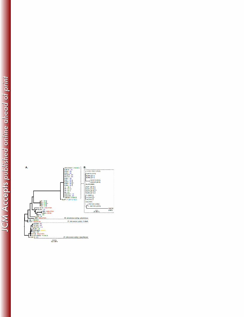
isolates (Fig. 1A). The resulting phylogeny supports the three previously recognized M. 220
abscessus subspecies as monophyletic, consistent with population studies utilizing MLST 221
genotyping (19, 21), and the NJH isolates group into expected clades consistent with subspecies 222
identifications based on rpoB, hsp65 and erm(41). Our phylogeny is also consistent with 223
previously reported, independent phylogenomic comparisons (8, 20, 24, 25, 44). 224
11
Within the M. abscessus ssp. abscessus subspecies included in our analysis, we observed 225
two major subclades (Fig. 1A). The basal subclade includes all Asian isolates, one NJH isolate 226
(NJH8) and six isolates from three UK-CF patients. This clade is genetically distinct from a 227
cluster of 18 isolates exclusively from the US or Europe, which includes the ATCC 19977T type 228
strain (43), nearly all of the NJH isolates, four additional US isolates, one French isolate and four 229
isolates from the UK. In this analysis, the 18 clustered isolates are distinguished by only 9 of 230
2,479 core genome SNP sites (0.36%). An additional 198 SNP sites discriminate the adjacent 231
Puerto Rican isolate (NJH7). These results suggest that among the M. abscessus ssp. abscessus 232
strains analyzed, the Asian isolates are divergent from most US isolates, while the UK isolates 233
are divided between the two genetic subgroups. Further sampling and future studies are needed 234
to validate these regional subgroups. 235
The lack of SNP variation in the US/Europe M. abscessus ssp. abscessus cluster is 236
intriguing given the temporal and geographical disparities among isolates. To further resolve the 237
18 clustered isolates and test for temporal or geographic groupings, a second, high-resolution 238
phylogeny was constructed using genotype information at 128,074 core genome nucleotide 239
positions (Fig. 1B). A total of 320 SNPs (320/128,074 = 0.25%) were observed in this dataset, 240
similar to the proportion of variation observed in the initial phylogeny. The majority of SNP 241
variation occurred in the French isolate, V06705, which is the most divergent in the cluster. 242
Overall, the branching patterns do not distinguish temporal or geographic groups, suggesting 243
independent acquisitions of the isolates. The high genetic relatedness may, therefore, represent a 244
dominant clinical M. abscessus ssp. abscessus subgroup, or may reflect similarities in the isolates’ 245
geographic or environmental niches. 246
247
12
In vitro Drug Susceptibility Results and Variants Associated with Resistance 248
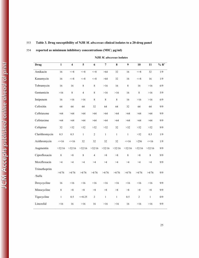
Nine of eleven NJH clinical isolates were evaluated for in vitro drug susceptibility to 19 anti-249
microbial drugs. These drugs span several drug classes, and the results are reported as minimum 250
inhibitory concentrations (MIC) (Table 3). Most isolates exhibited resistance to imipenem, 251
augmentin, two of four aminoglycosides (tobramycin and gentamicin), four generations of 252
cephalosporins, two of three tetracyclines (doxycycline and minocyclin), and two 253
flouroquinolones (ciprofloxacin and moxifloxacin), confirming the innate resistance of M. 254
abscessus to several drug classes (11, 13). Drugs that showed in vitro activity against the M. 255
abscessus strains include the aminoglycosides, amikacin and kanamycin, and the macrolides, 256
clarithromycin and azithromycin, which are commonly used to treat pulmonary M. abscessus 257
infections (12) as well as the less commonly prescribed, tigecycline. 258
Among the NJH isolates tested, only two exhibited different susceptibility phenotypes. 259
NJH7 was the only isolate with observed in vitro resistance to amikacin and kanamycin. Based 260
on our analysis, this resistance can be explained by the presence of a 16S rRNA mutation, 261
previously associated with aminoglycoside resistance in M. abscessus and M. chelonae isolates, 262
and corresponding to the A1408G mutation in Escherichia coli (14). NJH10 was the only isolate 263
with observed resistance to clarithromycin and azithromycin. This isolate does not have a 264
resistance-conferring mutation in the 23S rRNA (15) and may, therefore, contain a novel 265
mechanism of resistance. 266
267
Large-scale Polymorphic Genomic Regions in M. abscessus Clinical Isolates 268
Given the high genetic relatedness in the core genomes of clustered US/European isolates, we 269
also investigated large-scale genomic variations among a subset of clustered and non-clustered 270
13
isolates. We analyzed sequence coverage in 1kb windows relative to the ATCC 19977T reference 271
genome and identified 1kb windows with substantially low coverage (<2x), indicative of 272
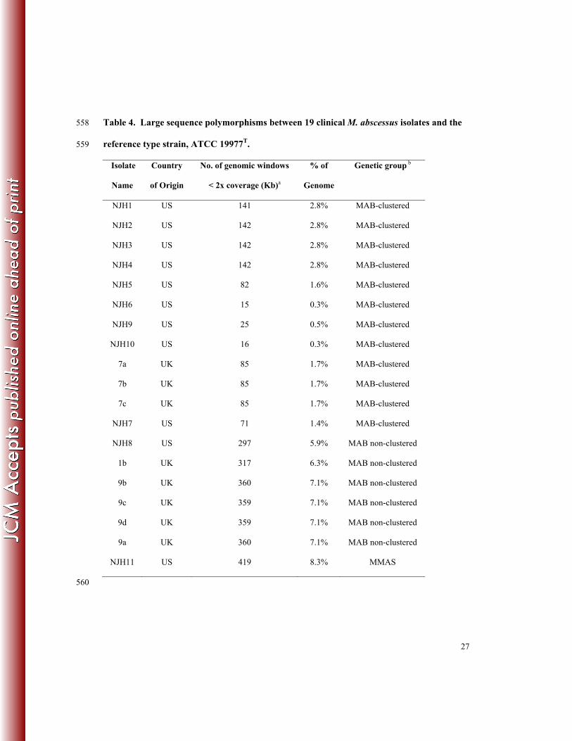
deletions or highly divergent regions of the genome. Across the 19 isolates analyzed, we 273
observed a range of 0.3 - 8.3% of the reference genome that is not represented in the clinical 274
isolates (Table 4). The least divergence was observed among clustered isolates (0.3 – 2.8%) and 275
higher divergence was observed among non-clustered isolates (5.9 – 7.1%). As expected, the 276
most divergence was observed in the M. abscessus ssp. massiliense isolate (NJH11; 8.3%). This 277
diversity is similar to genome content differences observed among clinical M. tuberculosis 278
isolates (45) and illustrates the genome plasticity and diversity among US and European M. 279
abscessus isolates. 280
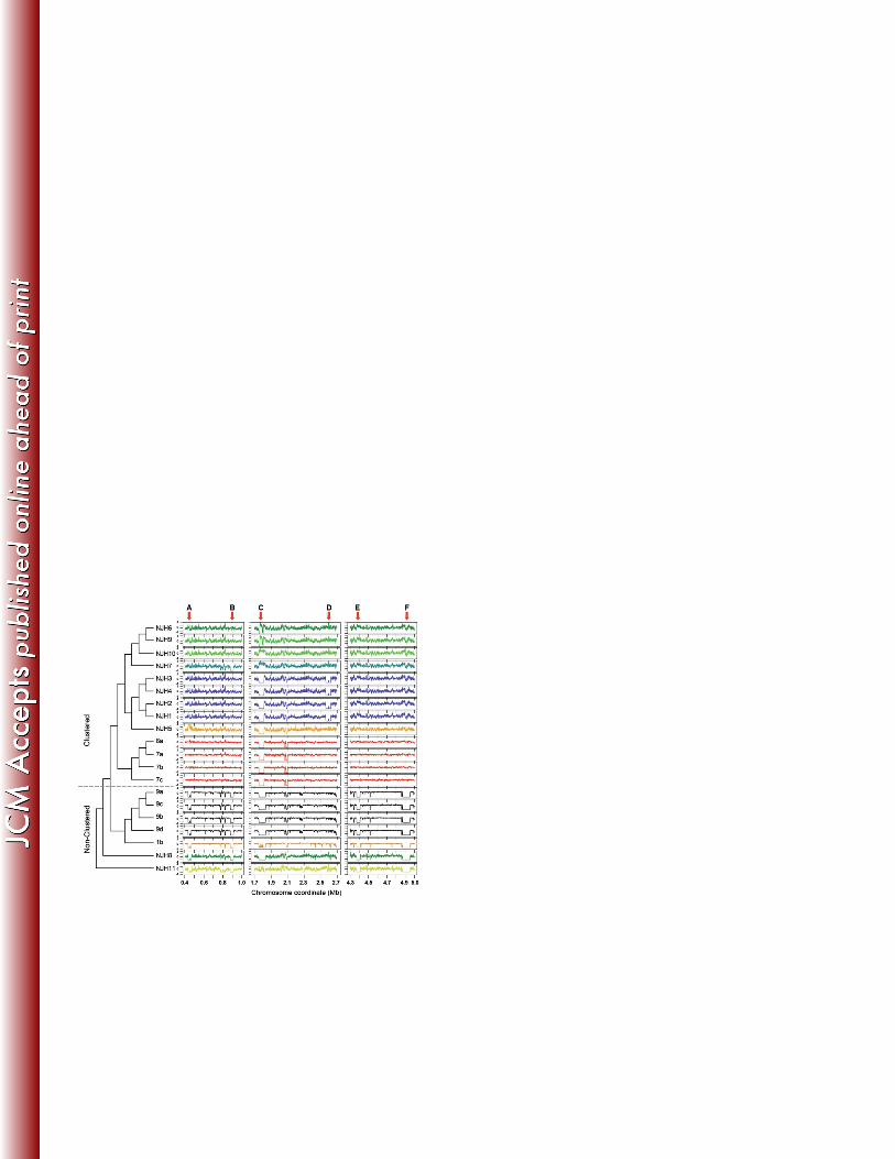
To test whether the clustered isolates share the same deletions, we performed a cluster 281
analysis of genome wide coverage patterns (Fig. 2). The clustering results are similar to the 282
initial core genome phylogeny (Fig. 1A), as the US/European clustered isolates group into a 283
major clade that is distinct from non-clustered isolates. One exception is the Puerto Rican isolate 284
(NJH7) that clusters with the US isolates based on deletion patterns, but is somewhat divergent 285
from the US and UK isolates in the core genome phylogeny (Fig. 1A). In contrast to the high-286
resolution phylogeny (Fig. 1B), the US/European clustered isolates group together largely by 287
geographic region. This, along with the grouping of NJH7 with other North American isolates, 288
suggests that evolution of the accessory genome may be driven by events within local 289
microenvironments. 290
Further analyses of six large-scale deletions (>30Kb) (Fig. 2, A – F), relative to the 291
reference M. abscessus genome, reveal potential mechanisms of accessory genome evolution 292
among clinical M. abscessus strains (Table S2). Regions A and F, which are deleted in all non-293
14
clustered isolates, include operons for copper ion and phosphate ion transport, respectively. Both 294
genomic regions contain sequences that are homologous to plasmids and transposons, providing 295
evidence for recombination or horizontal gene transfer (26). Region C, which is absent in 14 of 296
19 isolates, shows sequence homology only to mycobacteriophage, consistent with previous 297
annotation as a prophage (34). Two US isolates (NJH7 and NJH10) have excessive sequence 298
coverage in this region suggesting multiple genomic copies of prophage sequences. Regions B, 299
D and E show no evidence of recombination or homology to phage DNA. Region E, which is 300
deleted in non-clustered isolates and M. abscessus subsp. massiliense (NJH11), includes a 301
cassette of biphenyl and aromatic hydrocarbon metabolic enzymes, that functionally enable 302
degradation of environmental chemicals (46) in some cases. Overall, the results suggest that gene 303
content variation in clinical M. abscessus is due, in part, to recombination and/or metabolic 304
adaptations. 305
306
Genomic Comparisons of Longitudinally Sampled Isolates from Individual Patients 307
To evaluate intra-patient strain variation, we compared genomic information derived from three 308
sets of isolates from individual patients. From the UK-CF M. abscessus study (8), we studied 309
isolates from patients 7 and 9. Isolates 7a, 7b and 7c were collected at a single time point. 310
Isolates 9a and 9b were collected at an initial time point during treatment, and 9c and 9d were 311
collected one month later. From NJH, four isolates (NJH1–4) were longitudinally isolated from a 312
single patient at three time points during a six-month period, including two colonies (NJH2 and 313
NJH3) that were collected at the second time point. 314
In the previous core genome phylogenies (Fig. 1) with limited numbers of genomic 315
positions (2,479bp and 128,074bp), all three sets of isolates showed identical genotypes among 316
15
themselves within each set. We therefore performed in depth core genome analyses for each 317
patient-isolate group to explore infection homogeneity and the potential for genomic mutations 318
during infection and treatment. Using all high quality base calls within each group of patient 319
isolates, we found 4 SNPs among the three patient 7 isolates across 4,971,231 genomic positions 320
(0.00008%); 12 SNPs among the four patient 9 isolates across 4,503,047 positions (0.0002%); 321
and 10 SNPs among the four NJH patient 1 isolates across 4,587,198 positions (0.0002%). In 322
contrast to SNPs in the overall phylogeny (Fig. 1A) in which the minority of coding SNPs are 323
non-synonymous (20.9%) versus synonymous (79.5%), the majority of SNPs in the longitudinal 324
groups are non-synonymous changes (55.6% - 87.5%) suggesting positive selective pressures as 325
a possible driving force of genetic mutation. Examples of genes with nonsynonymous SNPs 326
observed among NJH1 patient isolates include an amino acid permease family protein 327
(MAB_0950c), a probable deoxyribonuclease TatD (MAB_1129), a putative membrane protein 328
MmpL (MAB_2303), sulfate adenylate transferase CysD (MAB_4181) and an arsenic-transport 329
integral membrane protein ArsC (MAB_4863). 330
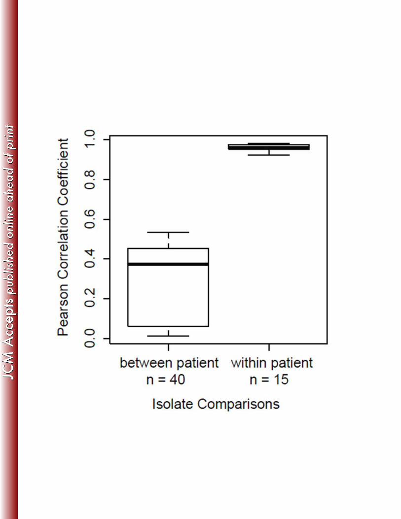
To test for large-scale variation between intra-patient isolates, correlations of genome 331
wide sequence coverage patterns were performed for all pair wise combinations of isolates 332
within and between each patient group (Fig. 3). Results demonstrate that all within-patient 333
isolate pairs are highly correlated compared to between-patient isolates, and we saw no evidence 334
of isolate-specific deletions within patients. These data, along with minimal SNP variation, 335
suggest homogenous populations in the patients studied, consistent with previous longitudinal 336
studies of M. abscessus (8, 47). The high-resolution comparisons enabled by WGS provide 337
evidence for genetic mutation and possible adaptation within the host. 338
339
16
Conclusions 340
Our study examines the genome sequences of eleven clinically relevant M. abscessus isolates 341
from eight US patients with persistent pulmonary infections that exhibit in vitro resistance to 342
several classes of antimicrobials. Moreover, it provides a comprehensive phylogenomic 343
assessment of US M. abscessus isolates compared to globally diverse clinical isolates. Our core 344
genome comparisons, across 41 M. abscessus isolates, reveal three genetic subgroups consistent 345
with previous subspecies classifications. We observed that most of the NJH isolates (9/11) 346
examined belong to a cluster of M. abscessus subsp. abscessus isolates with low core genome 347
diversity, that are primarily from US or Western European origin. This cluster is genetically 348
distinct from most UK and all Asian isolates examined in the study. Future studies with 349
expanded populations of both clinical and environmental isolates could reveal whether this 350
cluster represents a clinical subtype of M. abscessus or an ecological subgroup related through a 351
shared environmental niche. 352
Despite the core genome similarities observed among clustered US/European isolates, we 353
found substantial accessory genome variation in the form of multiple large-scale deletions. This 354
illustrates the resolving power of WGS in detecting not only SNPs, but also the presence or 355
absence of genomic islands, which can confer clinically important traits associated with 356
virulence and antibiotic resistance. Overall, we observed genome content variation of 0.3 to 357
7.1% within subspecies (M. abscessus subsp. abscessus) and 8.5% between subspecies (subsp. 358
abscessus versus subsp. massiliense) including one large deletion that is shared across subspecies 359
(Fig. 2; F). Functional annotation analyses of the deletion regions suggest that genome content 360
variation in M. abscessus can be attributed to recombination and/or metabolic adaptations. 361
17
Analyses of longitudinally collected samples from individual patients show low levels of 362
SNP variation and no evidence of large-scale variation, suggesting homogeneous populations in 363
the hosts studied. SNPs in the longitudinal studies were enriched for non-synonymous mutations 364
compared to the overall background population suggesting that pathogen genome evolution in 365
the host may be under positive selection from prolonged exposure to the host immune system, 366
antibiotic treatment, or other selective pressures. Further WGS studies will be needed to explore 367
long-term in vivo mutation rates in the host and potential implications for antibiotic targets. 368
369
Acknowledgements 370
This work was supported by the National Jewish Health NTM Center of Excellence, the 371
Colorado Bioscience Discovery Program, the Eppley Foundation, the Potts Memorial Foundation, 372
the Boettcher Foundation Webb-Waring Biomedical Research Program to MS, and a NIH 373
Biomedical Informatics training grant 2T15LM009451-06 to NH and BG. 374
375
18
References 376
1. Prevots DR, Shaw PA, Strickland D, Jackson LA, Raebel MA, Blosky M, Oca RM, Shea YR, 377 Seitz AE, Holland SM, Olivier KN. 2010. Nontuberculous Mycobacterial Lung Disease 378 Prevalence at Four Integrated Health Care Delivery Systems. Am J Respir Crit Care Med 379 182:970–976. 380
2. Moore JE, Kruijshaar ME, Ormerod LP, Drobniewski F, Abubakar I. 2010. Increasing 381 reports of non-tuberculous mycobacteria in England, Wales and Northern Ireland, 1995-2006. 382 BMC Public Health 10:612. 383
3. Marras TK, Chedore P, Ying AM, Jamieson F. 2007. Isolation prevalence of pulmonary non-384 tuberculous mycobacteria in Ontario, 1997 2003. Thorax 62:661-666. 385
4. Primm TP, Lucero CA, Falkinham JO, 3rd. 2004. Health impacts of environmental 386 mycobacteria. Clin Microbiol Rev 17:98-106. 387
5. Falkinham JO, 3rd. 2011. Nontuberculous mycobacteria from household plumbing of patients 388 with nontuberculous mycobacteria disease. Emerg Infect Dis 17:419-424. 389
6. Feazel LM, Baumgartner LK, Peterson KL, Frank DN, Harris JK, Pace NR. 2009. 390 Opportunistic pathogens enriched in showerhead biofilms. Proc Natl Acad Sci U S A 106:16393-391 16399. 392
7. Aitken ML, Limaye A, Pottinger P, Whimbey E, Goss CH, Tonelli MR, Cangelosi GA, 393 Dirac MA, Olivier KN, Brown-Elliott BA, McNulty S, Wallace RJ, Jr. 2012. Respiratory 394 outbreak of Mycobacterium abscessus subspecies massiliense in a lung transplant and cystic 395 fibrosis center. Am J Respir Crit Care Med 185:231-232. 396
8. Bryant JM, Grogono DM, Greaves D, Foweraker J, Roddick I, Inns T, Reacher M, 397 Haworth CS, Curran MD, Harris SR, Peacock SJ, Parkhill J, Floto RA. 2013. Whole-398 genome sequencing to identify transmission of Mycobacterium abscessus between patients with 399 cystic fibrosis: a retrospective cohort study. Lancet 381:1551-1560. 400
9. Winthrop KL, McNelley E, Kendall B, Marshall-Olson A, Morris C, Cassidy M, Saulson A, 401 Hedberg K. 2010. Pulmonary nontuberculous mycobacterial disease prevalence and clinical 402 features: an emerging public health disease. Am J Respir Crit Care Med 182:977-982. 403
10. Jonsson BE, Gilljam M, Lindblad A, Ridell M, Wold AE, Welinder-Olsson C. 2007. 404 Molecular epidemiology of Mycobacterium abscessus, with focus on cystic fibrosis. J Clin 405 Microbiol 45:1497-1504. 406
11. Griffith DE. 2011. The talking Mycobacterium abscessus blues. Clin Infect Dis 52:572-574. 407 12. Jarand J, Levin A, Zhang L, Huitt G, Mitchell JD, Daley CL. 2011. Clinical and 408
microbiologic outcomes in patients receiving treatment for Mycobacterium abscessus pulmonary 409 disease. Clin Infect Dis 52:565-571. 410
13. Nessar R, Cambau E, Reyrat JM, Murray A, Gicquel B. 2012. Mycobacterium abscessus: a 411 new antibiotic nightmare. J Antimicrob Chemother 67:810-818. 412
14. Prammananan T, Sander P, Brown BA, Frischkorn K, Onyi GO, Zhang Y, Bottger EC, 413 Wallace RJ, Jr. 1998. A single 16S ribosomal RNA substitution is responsible for resistance to 414 amikacin and other 2-deoxystreptamine aminoglycosides in Mycobacterium abscessus and 415 Mycobacterium chelonae. J Infect Dis 177:1573-1581. 416
15. Wallace RJ, Jr., Meier A, Brown BA, Zhang Y, Sander P, Onyi GO, Bottger EC. 1996. 417 Genetic basis for clarithromycin resistance among isolates of Mycobacterium chelonae and 418 Mycobacterium abscessus. Antimicrob Agents Chemother 40:1676-1681. 419
16. Nash KA, Brown-Elliott BA, Wallace RJ, Jr. 2009. A novel gene, erm(41), confers inducible 420 macrolide resistance to clinical isolates of Mycobacterium abscessus but is absent from 421 Mycobacterium chelonae. Antimicrob Agents Chemother 53:1367-1376. 422
17. Leao SC, Tortoli E, Euzeby JP, Garcia MJ. 2011. Proposal that Mycobacterium massiliense 423 and Mycobacterium bolletii be united and reclassified as Mycobacterium abscessus subsp. bolletii 424
19
comb. nov., designation of Mycobacterium abscessus subsp. abscessus subsp. nov. and emended 425 description of Mycobacterium abscessus. Int J Syst Evol Microbiol 61:2311-2313. 426
18. Macheras E, Roux AL, Bastian S, Leao SC, Palaci M, Sivadon-Tardy V, Gutierrez C, 427 Richter E, Rusch-Gerdes S, Pfyffer G, Bodmer T, Cambau E, Gaillard JL, Heym B. 2011. 428 Multilocus sequence analysis and rpoB sequencing of Mycobacterium abscessus (sensu lato) 429 strains. J Clin Microbiol 49:491-499. 430
19. Zelazny AM, Root JM, Shea YR, Colombo RE, Shamputa IC, Stock F, Conlan S, McNulty 431 S, Brown-Elliott BA, Wallace RJ, Jr., Olivier KN, Holland SM, Sampaio EP. 2009. Cohort 432 study of molecular identification and typing of Mycobacterium abscessus, Mycobacterium 433 massiliense, and Mycobacterium bolletii. J Clin Microbiol 47:1985-1995. 434
20. Heydari H, Wee WY, Lokanathan N, Hari R, Mohamed Yusoff A, Beh CY, Yazdi AH, 435 Wong GJ, Ngeow YF, Choo SW. 2013. MabsBase: A Mycobacterium abscessus Genome and 436 Annotation Database. PLoS One 8:e62443. 437
21. Cho YJ, Yi H, Chun J, Cho SN, Daley CL, Koh WJ, Jae Shin S. 2013. The Genome Sequence 438 of 'Mycobacterium massiliense' Strain CIP 108297 Suggests the Independent Taxonomic Status 439 of the Mycobacterium abscessus Complex at the Subspecies Level. PLoS One 8:e81560. 440
22. Koh WJ, Jeon K, Lee NY, Kim BJ, Kook YH, Lee SH, Park YK, Kim CK, Shin SJ, Huitt 441 GA, Daley CL, Kwon OJ. 2011. Clinical significance of differentiation of Mycobacterium 442 massiliense from Mycobacterium abscessus. Am J Respir Crit Care Med 183:405-410. 443
23. Harada T, Akiyama Y, Kurashima A, Nagai H, Tsuyuguchi K, Fujii T, Yano S, Shigeto E, 444 Kuraoka T, Kajiki A, Kobashi Y, Kokubu F, Sato A, Yoshida S, Iwamoto T, Saito H. 2012. 445 Clinical and Microbiological Differences between Mycobacterium abscessus and Mycobacterium 446 massiliense Lung Diseases. J Clin Microbiol 50:3556-3561. 447
24. Davidson RM, Hasan NA, de Moura VC, Duarte RS, Jackson M, Strong M. 2013. 448 Phylogenomics of Brazilian epidemic isolates of Mycobacterium abscessus subsp. bolletii reveals 449 relationships of global outbreak strains. Infect Genet Evol 20:292-297. 450
25. Tettelin H, Davidson RM, Agrawal S, Aitken ML, Shallom S, Hasan NA, Strong M, de 451 Moura VC, De Groote MA, Duarte RS, Hine E, Parankush S, Su Q, Daugherty SC, Fraser 452 CM, Brown-Elliott BA, Wallace RJ, Jr., Holland SM, Sampaio EP, Olivier KN, Jackson M, 453 Zelazny AM. 2014. High-level relatedness among Mycobacterium abscessus subsp. massiliense 454 strains from widely separated outbreaks. Emerg Infect Dis 20:364-371. 455
26. Juhas M, van der Meer JR, Gaillard M, Harding RM, Hood DW, Crook DW. 2009. 456 Genomic islands: tools of bacterial horizontal gene transfer and evolution. FEMS Microbiol Rev 457 33:376-393. 458
27. Jackson RW, Vinatzer B, Arnold DL, Dorus S, Murillo J. 2011. The influence of the 459 accessory genome on bacterial pathogen evolution. Mob Genet Elements 1:55-65. 460
28. Kim BJ, Kim BR, Lee SY, Kook YH. 2013. Rough colony morphology of Mycobacterium 461 massiliense Type II genotype is due to the deletion of glycopeptidolipid locus within its genome. 462 BMC Genomics 14:890. 463
29. Feng Y, Chen CJ, Su LH, Hu S, Yu J, Chiu CH. 2008. Evolution and pathogenesis of 464 Staphylococcus aureus: lessons learned from genotyping and comparative genomics. FEMS 465 Microbiol Rev 32:23-37. 466
30. Mathee K, Narasimhan G, Valdes C, Qiu X, Matewish JM, Koehrsen M, Rokas A, Yandava 467 CN, Engels R, Zeng E, Olavarietta R, Doud M, Smith RS, Montgomery P, White JR, 468 Godfrey PA, Kodira C, Birren B, Galagan JE, Lory S. 2008. Dynamics of Pseudomonas 469 aeruginosa genome evolution. Proc Natl Acad Sci U S A 105:3100-3105. 470
31. van Soolingen D, de Haas PE, Kremer K. 2001. Restriction fragment length polymorphism 471 typing of mycobacteria. Methods Mol Med 54:165-203. 472
32. Adekambi T, Colson P, Drancourt M. 2003. rpoB-based identification of nonpigmented and 473 late-pigmenting rapidly growing mycobacteria. J Clin Microbiol 41:5699-5708. 474
20
33. CLSI. 2011. Susceptibility Testing of Mycobacteria, Nocardiae, and Other Aerobic 475 Actinomycetes; Approved Standard-Second Edition., vol. CLSI document M42-A2. Clinical and 476 Laboratory Standards Institute, Wayne, PA. 477
34. Ripoll F, Pasek S, Schenowitz C, Dossat C, Barbe V, Rottman M, Macheras E, Heym B, 478 Herrmann JL, Daffe M, Brosch R, Risler JL, Gaillard JL. 2009. Non mycobacterial virulence 479 genes in the genome of the emerging pathogen Mycobacterium abscessus. PLoS One 4:e5660. 480
35. Langmead B, Trapnell C, Pop M, Salzberg SL. 2009. Ultrafast and memory-efficient 481 alignment of short DNA sequences to the human genome. Genome Biol 10:R25. 482
36. Li H, Handsaker B, Wysoker A, Fennell T, Ruan J, Homer N, Marth G, Abecasis G, Durbin 483 R. 2009. The Sequence Alignment/Map format and SAMtools. Bioinformatics 25:2078-2079. 484
37. Wang K, Li M, Hakonarson H. 2010. ANNOVAR: functional annotation of genetic variants 485 from high-throughput sequencing data. Nucleic Acids Res 38:e164. 486
38. Darling AE, Mau B, Perna NT. 2010. progressiveMauve: multiple genome alignment with gene 487 gain, loss and rearrangement. PLoS One 5:e11147. 488
39. Gouy M, Guindon S, Gascuel O. 2010. SeaView version 4: A multiplatform graphical user 489 interface for sequence alignment and phylogenetic tree building. Mol Biol Evol 27:221-224. 490
40. Cheung MS, Down TA, Latorre I, Ahringer J. 2011. Systematic bias in high-throughput 491 sequencing data and its correction by BEADS. Nucleic Acids Res 39:e103. 492
41. Saeed AI, Sharov V, White J, Li J, Liang W, Bhagabati N, Braisted J, Klapa M, Currier T, 493 Thiagarajan M, Sturn A, Snuffin M, Rezantsev A, Popov D, Ryltsov A, Kostukovich E, 494 Borisovsky I, Liu Z, Vinsavich A, Trush V, Quackenbush J. 2003. TM4: a free, open-source 495 system for microarray data management and analysis. Biotechniques 34:374-378. 496
42. R Development Core Team. 2011. R: A Language and Environment for Statistical Computing. 497 R Foundation for Statistical Computing, Vienna, Austria. 498
43. Moore M, Frerichs JB. 1953. An unusual acid-fast infection of the knee with subcutaneous, 499 abscess-like lesions of the gluteal region; report of a case with a study of the organism, 500 Mycobacterium abscessus, n. sp. J Invest Dermatol 20:133-169. 501
44. Choo SW, Wee WY, Ngeow YF, Mitchell W, Tan JL, Wong GJ, Zhao Y, Xiao J. 2014. 502 Genomic reconnaissance of clinical isolates of emerging human pathogen Mycobacterium 503 abscessus reveals high evolutionary potential. Sci Rep 4:4061. 504
45. Tsolaki AG, Hirsh AE, DeRiemer K, Enciso JA, Wong MZ, Hannan M, Goguet de la 505 Salmoniere YO, Aman K, Kato-Maeda M, Small PM. 2004. Functional and evolutionary 506 genomics of Mycobacterium tuberculosis: insights from genomic deletions in 100 strains. Proc 507 Natl Acad Sci U S A 101:4865-4870. 508
46. Moody JD, Doerge DR, Freeman JP, Cerniglia CE. 2002. Degradation of biphenyl by 509 Mycobacterium sp. strain PYR-1. Appl Microbiol Biotechnol 58:364-369. 510
47. Wallace RJ, Jr., Zhang Y, Brown BA, Fraser V, Mazurek GH, Maloney S. 1993. DNA large 511 restriction fragment patterns of sporadic and epidemic nosocomial strains of Mycobacterium 512 chelonae and Mycobacterium abscessus. J Clin Microbiol 31:2697-2701. 513
48. Pang S, Renvoise A, Perret C, Guinier M, Chelghoum N, Brossier F, Capton E, Jarlier V, 514 Sougakoff W. 2013. Whole-Genome Sequence of Mycobacterium abscessus Clinical Strain 515 V06705. Genome Announc 1. 516
49. Ngeow YF, Wong YL, Tan JL, Ong CS, Ng KP, Choo SW. 2012. Genome sequence of 517 Mycobacterium abscessus strain M152. J Bacteriol 194:6662. 518
50. Choo SW, Wong YL, Leong ML, Heydari H, Ong CS, Ng KP, Ngeow YF. 2012. Analysis of 519 the genome of Mycobacterium abscessus strain M94 reveals an uncommon cluster of tRNAs. J 520 Bacteriol 194:5724. 521
51. Choo SW, Wong YL, Yusoff AM, Leong ML, Wong GJ, Ong CS, Ng KP, Ngeow YF. 2012. 522 Genome sequence of the Mycobacterium abscessus strain M93. J Bacteriol 194:3278. 523
52. Choi GE, Cho YJ, Koh WJ, Chun J, Cho SN, Shin SJ. 2012. Draft genome sequence of 524 Mycobacterium abscessus subsp. bolletii BD(T). J Bacteriol 194:2756-2757. 525
21
53. Wong YL, Choo SW, Tan JL, Ong CS, Ng KP, Ngeow YF. 2012. Draft genome sequence of 526 Mycobacterium bolletii strain M24, a rapidly growing mycobacterium of contentious taxonomic 527 status. J Bacteriol 194:4475. 528
54. Chan J, Halachev M, Yates E, Smith G, Pallen M. 2012. Whole-genome sequence of the 529 emerging pathogen Mycobacterium abscessus strain 47J26. J Bacteriol 194:549. 530
55. Davidson RM, Reynolds PR, Farias-Hesson E, Duarte RS, Jackson M, Strong M. 2013. 531 Genome Sequence of an Epidemic Isolate of Mycobacterium abscessus subsp. bolletii from Rio 532 de Janeiro, Brazil. Genome Announc 1:e00617-00613. 533
56. Tettelin H, Sampaio EP, Daugherty SC, Hine E, Riley DR, Sadzewicz L, Sengamalay N, 534 Shefchek K, Su Q, Tallon LJ, Conville P, Olivier KN, Holland SM, Fraser CM, Zelazny AM. 535 2012. Genomic insights into the emerging human pathogen Mycobacterium massiliense. J 536 Bacteriol 194:5450. 537
538
22
Tables 539
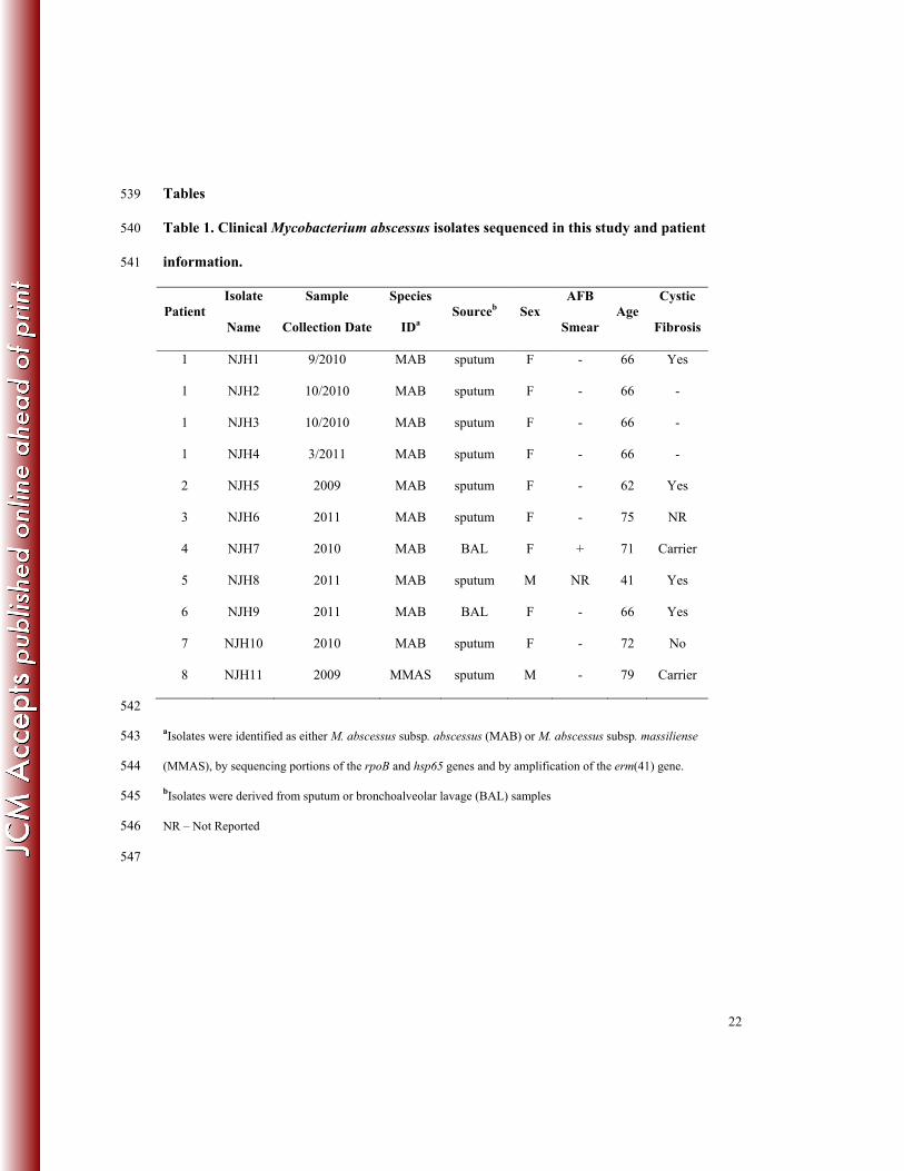
Table 1. Clinical Mycobacterium abscessus isolates sequenced in this study and patient 540
information. 541
Patient Isolate
Name
Sample
Collection Date
Species
IDa Sourceb Sex
AFB
Smear Age
Cystic
Fibrosis
1 NJH1 9/2010 MAB sputum F - 66 Yes
1 NJH2 10/2010 MAB sputum F - 66 -
1 NJH3 10/2010 MAB sputum F - 66 -
1 NJH4 3/2011 MAB sputum F - 66 -
2 NJH5 2009 MAB sputum F - 62 Yes
3 NJH6 2011 MAB sputum F - 75 NR
4 NJH7 2010 MAB BAL F + 71 Carrier
5 NJH8 2011 MAB sputum M NR 41 Yes
6 NJH9 2011 MAB BAL F - 66 Yes
7 NJH10 2010 MAB sputum F - 72 No
8 NJH11 2009 MMAS sputum M - 79 Carrier
542
aIsolates were identified as either M. abscessus subsp. abscessus (MAB) or M. abscessus subsp. massiliense 543
(MMAS), by sequencing portions of the rpoB and hsp65 genes and by amplification of the erm(41) gene. 544
bIsolates were derived from sputum or bronchoalveolar lavage (BAL) samples 545
NR – Not Reported 546
547
23
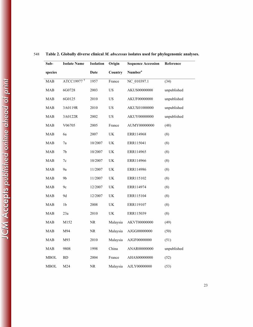
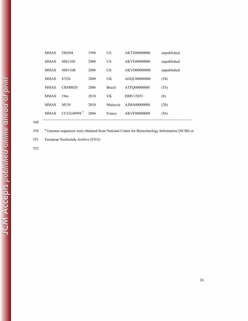
Table 2. Globally diverse clinical M. abscessus isolates used for phylogenomic analyses. 548
Sub-
species
Isolate Name Isolation
Date
Origin
Country
Sequence Accession
Numbera
Reference
MAB ATCC19977 T 1957 France NC_010397.1 (34)
MAB 6G0728 2003 US AKUS00000000 unpublished
MAB 6G0125 2010 US AKUF00000000 unpublished
MAB 3A0119R 2010 US AKUX01000000 unpublished
MAB 3A0122R 2002 US AKUY00000000 unpublished
MAB V06705 2005 France AUMY00000000 (48)
MAB 6a 2007 UK ERR114968 (8)
MAB 7a 10/2007 UK ERR115041 (8)
MAB 7b 10/2007 UK ERR114965 (8)
MAB 7c 10/2007 UK ERR114966 (8)
MAB 9a 11/2007 UK ERR114986 (8)
MAB 9b 11/2007 UK ERR115102 (8)
MAB 9c 12/2007 UK ERR114974 (8)
MAB 9d 12/2007 UK ERR115104 (8)
MAB 1b 2008 UK ERR119107 (8)
MAB 23a 2010 UK ERR115039 (8)
MAB M152 NR Malaysia AKVT00000000 (49)
MAB M94 NR Malaysia AJGG00000000 (50)
MAB M93 2010 Malaysia AJGF00000000 (51)
MAB 9808 1998 China ANAR00000000 unpublished
MBOL BD 2004 France AHAS00000000 (52)
MBOL M24 NR Malaysia AJLY00000000 (53)
24
MMAS 5S0304 1998 US AKTX00000000 unpublished
MMAS 4S0116S 2008 US AKVE00000000 unpublished
MMAS 4S0116R 2008 US AKVD00000000 unpublished
MMAS 47J26 2009 UK AGQU00000000 (54)
MMAS CRM0020 2006 Brazil ATFQ00000000 (55)
MMAS 19m 2010 UK ERR115051 (8)
MMAS M154 2010 Malaysia AJMA00000000 (20)
MMAS CCUG49998 T 2004 France AKVF00000000 (56)
549
a Genome sequences were obtained from National Center for Biotechnology Information (NCBI) or 550
European Nucleotide Archive (ENA) 551
552
25
Table 3. Drug susceptibility of NJH M. abscessus clinical isolates to a 20-drug panel 553
reported as minimum inhibitory concentrations (MIC; μg/ml) 554
NJH M. abscessus isolates
Drug 1 4 5 6 7 8 9 10 11 % Ra
Amikacin 16 <=8 <=8 <=8 >64 32 16 <=8 32 1/9
Kanamycin 16 <=8 <=8 <=8 >64 32 16 <=8 16 1/9
Tobramycin 16 16 8 8 >16 16 8 16 >16 6/9
Gentamicin >16 8 4 8 >16 >16 16 8 >16 5/9
Imipenem 16 >16 >16 8 8 8 16 >16 >16 6/9
Cefoxitin 64 64 64 32 64 64 32 64 64 9/9
Ceftriaxone >64 >64 >64 >64 >64 >64 >64 >64 >64 9/9
Cefotaxime >64 >64 >64 >64 >64 >64 >64 >64 >64 9/9
Cefepime 32 >32 >32 >32 >32 32 >32 >32 >32 9/9
Clarithromycin 0.5 0.5 1 2 1 1 1 >32 0.5 1/9
Azithromycin <=16 <=16 32 32 32 32 <=16 >256 <=16 1/9
Augmentin >32/16 >32/16 >32/16 >32/16 >32/16 >32/16 >32/16 >32/16 >32/16 9/9
Ciprofloxacin 8 >8 8 4 >8 >8 8 >8 8 9/9
Moxifloxacin >4 >4 >4 >4 >4 >4 >4 >4 >4 9/9
Trimethoprim
/Sulfa >4/76 >4/76 >4/76 >4/76 >4/76 >4/76 >4/76 >4/76 >4/76 9/9
Doxycycline 16 >16 >16 >16 >16 >16 >16 >16 >16 9/9
Minocycline 8 >8 >8 >8 >8 >8 >8 >8 >8 9/9
Tigecycline 1 0.5 <=0.25 2 1 1 0.5 2 1 0/9
Linezolid >16 16 >16 16 >16 >16 16 >16 >16 9/9

26
aProportion of isolates with resistant (R) phenotypes based on previously defined thresholds of MIC 555
values specific to each drug. 556
557
27
Table 4. Large sequence polymorphisms between 19 clinical M. abscessus isolates and the 558
reference type strain, ATCC 19977T. 559
Isolate
Name
Country
of Origin
No. of genomic windows
< 2x coverage (Kb)a
% of
Genome
Genetic group b
NJH1 US 141 2.8% MAB-clustered
NJH2 US 142 2.8% MAB-clustered
NJH3 US 142 2.8% MAB-clustered
NJH4 US 142 2.8% MAB-clustered
NJH5 US 82 1.6% MAB-clustered
NJH6 US 15 0.3% MAB-clustered
NJH9 US 25 0.5% MAB-clustered
NJH10 US 16 0.3% MAB-clustered
7a UK 85 1.7% MAB-clustered
7b UK 85 1.7% MAB-clustered
7c UK 85 1.7% MAB-clustered
NJH7 US 71 1.4% MAB-clustered
NJH8 US 297 5.9% MAB non-clustered
1b UK 317 6.3% MAB non-clustered
9b UK 360 7.1% MAB non-clustered
9c UK 359 7.1% MAB non-clustered
9d UK 359 7.1% MAB non-clustered
9a UK 360 7.1% MAB non-clustered
NJH11 US 419 8.3% MMAS
560
28
a Genomic windows with significantly low sequence read coverage (less than 2x) compared to the 561
reference strain. 562
b The three genetic groups identified in our phylogenomic analysis; clustered M. abscessus ssp. abscessus 563
(MAB) isolates, non-clustered MAB isolates and M. abscessus ssp. massiliense (MMAS). 564
29
Figure Legends 565
Figure 1. Phylogenomic comparison of the National Jewish Health (NJH) M. abscessus clinical 566
isolates from the United States, compared to global strains. A. Genotype data at 2,479 core genome 567
positions, relative to the M. abscessus ssp. abscessus ATCC 19977T reference genome, were used to 568
estimate the phylogeny among 41 M. abscessus isolates using the Neighbor Joining algorithm. The 569
phylogenomic tree supports previously recognized subspecies as monophyletic groups. Isolates 570
highlighted by gray bars and asterisks indicate isolates acquired from the same patients. B. A higher-571
resolution phylogeny of the 18-isolate US/European M. abscessus ssp. abscessus cluster was created 572
using genotype information at 128,074 core genome positions. Isolation dates are included. Only 320 of 573
128,074 (0.25%) core genome sites vary among these isolates. 574
575
Figure 2. Large-scale genomic variation among M. abscessus genomes. Large-scale genomic 576
variations, including deletions and duplications, were identified by sequence read mapping and coverage 577
assessment along a reference genome. Chromosome coordinates of three regions of the reference genome 578
are indicated on the x-axis. Next generation sequence read counts were assessed along non-overlapping 579
1Kb sliding windows of the reference genome (5,068 total windows). Contiguous windows with z-scores 580
(y-axes) less than -2.0 indicate putative genomic deletions or highly divergent regions, while contiguous 581
stretches of high z-scores, greater than 2.0, indicate putative genomic duplications. The dendrogram 582
represents the result of average linkage hierarchical clustering of genome wide coverage values, using a 583
Pearson correlation metric. Arrows labeled A – F indicate large-scale genomic differences (>30Kb) 584
among the clinical M. abscessus isolates. 585
586
Figure 3. Correlations of genome wide coverage patterns between 11 M. abscessus isolates collected 587
from three patients. Correlations of genome wide sequence coverage patterns were performed for all 588
pairwise combinations of 11 isolates (n = 55). Each pair was binned as between patient isolates (n = 40) 589
or within patient isolates (n = 15). Isolates obtained from the same patient have much higher correlation 590

30
than isolates from different patients, suggesting genetically homogeneous infecting populations in 591
individual patients. 592
593