Embed Size (px)
Citation preview

J Oral Maxillofac Surg70:e683-e699, 2012
Is Entire Removal of a Post-TraumaticTemporomandibular Joint Ankylotic Site
Necessary for an Optimal Outcome?Dorrit W. Nitzan, DMD,* Jawad Abu Tair, DMD,† and
Hadas Lehman, DMD, MSc‡
Purpose: Temporomandibular joint (TMJ) ankylosis that occurs after TMJ condylar fracture constitutesa treatment challenge. The purpose of the present study is shed light on an alternative treatmentapproach for certain such cases where the displaced condylar head or part of it can be detected incomputed tomography. The leading principle of this protocol is accurate removal of the ankylotic massonly, leaving the condyle–disc apparatus un-touched.
Patients and Methods: The study is based on such cases of post trauma ankylosis where the displacecondyle was detected. Thirteen cases are reported (10 unilateral and 3 bilateral) age ranged from 8 to 51years (mean 20). All patients were treated according to the presented protocol that emphasizes thesignificance of preserving the condyle–disc apparatus while accurately removing the ankylotic mass. Toachieve the required precision, 3-dimensional computed tomography was used. An integral part of thetreatment plan is intensive guided physiotherapy, which is intended to re-establish normal joint function,the original occlusion and facial symmetry (in growing individuals).
Results: The patients were followed up for 6 to more than 60 months. After guided physiotherapy, allpatients had significant postoperative improvement in maximal mouth opening from a mean of 18.4 mm(range 8 to 28) to a mean of 41.2 mm (range 35 to 50). All patients had returned to their originalocclusion. In all the growing patients, a marked improvement in facial symmetry was observed.
Conclusions: In post trauma ankylosis the displaced head of the condyle and disc should be searchedfor using computed tomography. If detected we recommend its preservation while accurately removeonly the ankylotic mass. Using this approach, in addition to achieving adequate mandibular motion, goodocclusion, and normal facial growth, major surgery, with all its inconveniences and potential complica-tions, is avoided.Crown Copyright © 2012 Published by Elsevier Inc on behalf of the American Association of Oral andMaxillofacial Surgeons. All rights reserved.J Oral Maxillofac Surg 70:e683-e699, 2012
Temporomandibular joint (TMJ) ankylosis often oc-curs after misdiagnosis, delayed treatment, inade-quate surgery, prolonged immobilization, or insuffi-cient physiotherapy for intracapsular or subcondylarfractures.1,2
TMJ ankylosis affects facial growth potential2 and,therefore, can lead to facial deformity; it is also asso-ciated with difficulties in breathing, biting, chewing,and swallowing and is commonly accompanied by
poor oral hygiene.1,3-7 This condition is challenging totreat, because it requires complete release of ankylo-sis followed by restoration of the condylar height tofacilitate a full range of motion and good occlusion. Anumber of surgical techniques have been describedfor TMJ ankylosis, including gap arthroplasty8-13 inter-positional arthroplasty,8,11,12,14-17 and after excisionof the ankylotic mass, a variety of interpositional mate-rials have been used.9,13,18-23 Furthermore, for recon-
Received from the Department of Oral and Maxillofacial Surgery,
Hebrew University Hadassah School of Dental Medicine, Jerusalem,
Israel.
*Full Professor.
†Attending Professor.
‡Attending Professor.
Address correspondence and reprint requests to Professor Nitzan:
Department of Oral and Maxillofacial Surgery, Hadassah Faculty of
Dental Medicine, Hebrew University Hadassah School of Dental Med-
icine, PO Box 12272, Jerusalem 91120 Israel; e-mail: dorrit.
Crown Copyright © 2012 Published by Elsevier Inc on behalf of the
American Association of Oral and Maxillofacial Surgeons. All rights reserved.
0278-2391/12/7012-0$36.00/0
http://dx.doi.org/10.1016/j.joms.2012.08.007
e683

struction of the ramus–condyle unit, assorted methodshave been used, such as autogenous bone (includingcostochondral graft,15,24-26 fibula,27,28 clavicle,29 iliaccrest,30,31 and metatarsal head32,33), alloplastic materi-als,34-39 and distraction osteogenesis.40-43 No consensushas been reached regarding the optimal strategy. Lim-ited range of motion and reankylosis are the most fre-quently reported long-term complications.11,44 Facialasymmetry, facial nerve paresis, occlusal changes, over-growth of the costochondral graft, unpredicted resorp-tion of autogenous bone, and foreign body reactions toalloplastic materials are some of the complications thatoccur, clearly dependent on the surgical and reconstruc-tive method used.45
To achieve optimal outcomes, Kaban et al46 estab-lished guidelines for the management of TMJ ankylo-sis. They emphasized that reankylosis occurs becauseof inadequate release of the ankylotic mass, which isassociated with deficient passive mouth opening.2,46
Their 7-step protocol suggests aggressive excision of
the ankylotic mass and emphasizes the importance ofthe medial resection.2,46 In ankylosis that occurs aftera displaced condylar fracture, the displaced fragmentis bound to the medial aspect of the ankylotic site.When this occurs, their protocol strongly recom-mends removal of the entire ankylotic site, includingthe medial part, to enable free, passive jaw move-ment2,46 (Fig 1A,B). Fine-cut 3-dimensional computedtomography (3DCT) allows the identification of thelocation, extent, and anatomic relations of the area ofankylosis.2
The protocol from Kaban et al46 has substantiallyimproved the success rate of TMJ ankylosis treatment.We hypothesize that in cases with Sawhney’s ankylo-sis type III,21 preserving the medially located con-dyle–disc apparatus will maintain inherent joint func-tion, conserve the vertical height of the ramus, and, inchildren, protect the growth site. It is important tonote that the disc is the optimal interpositional mate-rial, serving as a barrier and preventing fusion of the
FIGURE 1. 3D-CT scan showing ankylosis of right TMJ. A, Lateral view of ankylotic site (A). Dotted line shows inferior surgical cut asrecommended by Kaban protocol. B, Anterior view of ankylotic site (A) on lateral aspect and displaced condyle (C) in medial anterior inferiordirection. Solid line shows inferior surgical cut recommended by presented protocol, a sharp angle separates the proper cut (solid line) froma potential improper cut (broken line) through the condyle. Dotted line indicates inferior surgical cut as recommended by Kaban protocol.(Figure 1 continued on next page.)
Nitzan, Abu Tair, and Lehman. Post Traumatic TMJ Ankylosis - An Alternative Treatment. J Oral Maxillofac Surg 2012.
e684 POST TRAUMATIC TMJ ANKYLOSIS - AN ALTERNATIVE TREATMENT
distal fragment with the glenoid fossa (ie, ankylosisand bony overgrowth are less likely to occur).47 Anadditional advantage of this approach is that all thecomplications associated with reconstructive tech-niques are prevented.
In the presented protocol (which was previouslydescribed in 199848), the guiding principle is preciseremoval of the ankylotic mass only, leaving the con-dyle–disc apparatus untouched (Figs 1B,C, 3). Theprocedure is followed by intensive physiotherapy,with the aim of rehabilitating normal joint functionand occlusion and facilitating the re-establishment offacial symmetry.
Patients and Methods
A total of 13 patients (4 males and 9 females, agerange 8 to 51 years), who had been referred to theoral and maxillofacial surgery department with TMJankylosis with the displaced condyle bound to the
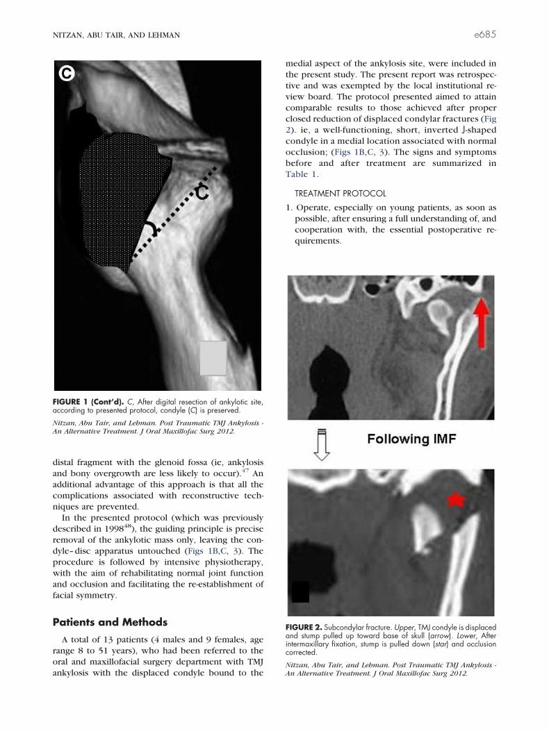
medial aspect of the ankylosis site, were included inthe present study. The present report was retrospec-tive and was exempted by the local institutional re-view board. The protocol presented aimed to attaincomparable results to those achieved after properclosed reduction of displaced condylar fractures (Fig2). ie, a well-functioning, short, inverted J-shapedcondyle in a medial location associated with normalocclusion; (Figs 1B,C, 3). The signs and symptomsbefore and after treatment are summarized inTable 1.
TREATMENT PROTOCOL
1. Operate, especially on young patients, as soon aspossible, after ensuring a full understanding of, andcooperation with, the essential postoperative re-quirements.
FIGURE 1 (Cont’d). C, After digital resection of ankylotic site,according to presented protocol, condyle (C) is preserved.
Nitzan, Abu Tair, and Lehman. Post Traumatic TMJ Ankylosis -An Alternative Treatment. J Oral Maxillofac Surg 2012.
FIGURE 2. Subcondylar fracture. Upper, TMJ condyle is displacedand stump pulled up toward base of skull (arrow). Lower, Afterintermaxillary fixation, stump is pulled down (star) and occlusioncorrected.
Nitzan, Abu Tair, and Lehman. Post Traumatic TMJ Ankylosis -An Alternative Treatment. J Oral Maxillofac Surg 2012.
NITZAN, ABU TAIR, AND LEHMAN e685
2. Use fine-cut 3DCT to plan the incision between theankylotic site and displaced condyle (Fig 1B), con-sidering the limited surgical field, that the displacedcondyle and disc are hidden medially (Fig 1 to 3),and that a small error in the angle of the incisioncan cut through the condyle (Figs 1B,C).
3. Resect the fibrous and/or bony ankylotic mass onthe lateral aspect and leave about 2 cm distancefrom the base of the skull to the neck of condyle(Figs 1B,C, 3).
4. Leave the condyle and disc untouched (Figs 1C, 3).5. Perform coronoidectomy on the affected side using
either an intra- or an extraoral approach.
6. Perform coronoidectomy on the contralateral side,using an intraoral approach, if steps 3 and 5 do notresult in free maximal mouth opening (MMO).
7. Suture the native disc (to cover the condyle) to thecapsule, in a posterolateral direction. If, as is rarelyobserved (our experience included only 1 suchcase), the disc cannot be safely sutured to thecapsule, the condyle should be separated from the“previous” (what used to be) fossa by an interposi-tional material. This can be done, for example,using a 2- to 3-mm-thick silicone sheet sutured tothe presently unloaded fossa, applying stainlesssteel ligature wire 0.016 in.
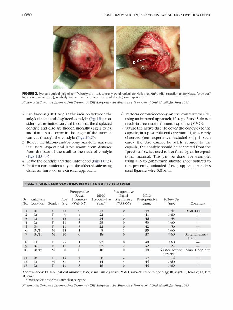
FIGURE 3. Typical surgical field of left TMJ ankylosis. Left, Lateral view of typical ankylotic site. Right, After resection of ankylosis, “previous”fossa and eminence (E), medially located condylar head (C), and disc (D) are exposed.
Nitzan, Abu Tair, and Lehman. Post Traumatic TMJ Ankylosis - An Alternative Treatment. J Oral Maxillofac Surg 2012.
Table 1. SIGNS AND SYMPTOMS BEFORE AND AFTER TREATMENT
Pt.No.
AnkylosisLocation Gender
Age(yr)
PreoperativeFacial
Symmetry(VAS 0-5)
MMOPreoperative
(mm)
PostoperativeFacial
Asymmetry(VAS 0-5)
MMOPostoperative
(mm)Follow-Up
(mo) Comment
1 Rt F 23 0 23 0 39 41 Deviation2 Lt F 9 4 22 1 41 �60 —3 Lt F 12 2 24 0 46 53 —4 Lt F 11 3 28 0 50 �60 —5 Rt F 11 3 22 0 42 56 —6 Rt/Lt M 23 1 8 1 35 �60 —7 Rt/Lt M 40 0 18 0 37 �60 Anterior cross-
bite8 Lt F 25 1 22 0 40 �60 —9 Rt F 11 4 22 2 42 24 —
10 Rt/Lt M 8 0 10 0 38 6 since secondsurgery*
2-mm Open bite
11 Rt F 15 4 8 2 37 16 —12 Lt M 51 3 14 3 44 �60 —13 Lt F 11 3 18 0 44 �60 —
Abbreviations: Pt. No., patient number; VAS, visual analog scale; MMO, maximal mouth opening; Rt, right; F, female; Lt, left;M, male.
*Twenty-four months after first surgery.
Nitzan, Abu Tair, and Lehman. Post Traumatic TMJ Ankylosis - An Alternative Treatment. J Oral Maxillofac Surg 2012.
e686 POST TRAUMATIC TMJ ANKYLOSIS - AN ALTERNATIVE TREATMENT

8. Early aggressive mobilization of the jaw to achievenormal mouth closing and opening and the bestpossible occlusion.8,48,49 In addition to existingprinciples,50 we emphasize the following points.First, in securing the gained mouth opening, thepatient should leave the operating room with anopen mouth secured by a mouth probe for 24hours. This enables the patient to see the results ofthe surgery and internalize his/her goal. Further-more, the efficiency of the exercises is enhanced bystarting from this position rather than from a closedmouth position. Second, and for the same reason,these patients should sleep with an open mouth(applying a mouth probe) as long as they are notable to freely open their mouth. Finally, during theday, the patients should exercise their mouth open-ing according to established guidelines.
Rehabilitation of Mouth ClosureIn long-duration ankylosis, the unused closing mus-
cles weaken. On release of the ankylosis, the man-dible drops, and the patient is unable to closehis/her mouth. Therefore, guidance for propermouth closure should be provided.
Rehabilitation of OcclusionA contralateral open bite is often observed immedi-
ately after surgery. Erich arch bars with rubberbands should be used during the night to guide thelower jaw to the desirable occlusion.
Case Reports
Of the 13 cases, 4 are reported in detail, withdistinctive aspects of the pathologic features or treat-ment approach provided.
CASE 1
An 8-year-old girl was diagnosed with a left TMJcondylar fracture after being in a motor vehicle acci-dent (in Table 1, this patient is listed as patient 5).Physiotherapy was recommended. Six months later,she was referred to the oral and maxillofacial surgerydepartment at Hadassah Hospital because of progres-sive facial asymmetry. On clinical examination, facialasymmetry with the chin deviating to the left wasnoted (Fig 4A). Her MMO was 22 mm with deviationto the left (Fig 4B) and limited movements to theright. Forced mouth opening was not painful butshowed resistance in the left TMJ. Protrusion waslimited, as was deviation to the left. CT scanning(coronal sections) showed left TMJ ankylosis. Carefulevaluation showed that the ankylosis was located onthe lateral aspect of the TMJ, between the stump andthe base of skull, and the displaced fractured condylewas facing medially toward the base of skull and itsbase was ankylosed to the medial aspect of the stump(Fig 4C).
FIGURE 4. Case 1 (patient 5 in Table 1) 6 months after untreated leftTMJ fracture. A, Facial asymmetry with deviation of mandible to left. B,Limited mouth opening with deviation to left. C, CT scan (coronalsections) showing left TMJ ankylosis (A) on lateral aspect of left TMJbetween stump and base of skull. Displaced fractured condyle (C)faces medially.
Nitzan, Abu Tair, and Lehman. Post Traumatic TMJ Ankylosis - AnAlternative Treatment. J Oral Maxillofac Surg 2012.
NITZAN, ABU TAIR, AND LEHMAN e687

To prevent further progress of her facial asymme-try, surgery was performed immediately. The anky-lotic site on the lateral aspect was aggressively re-moved, leaving 1.5 to 2 cm between the stump andthe base of skull (Figs 1B, 3). In this condition, thecondyle, covered by the disc, was observed on themedial aspect (Figs 1B, 3). The disc was then pulledand sutured to the lateral aspect of the displaced
condyle. No interpositional material was placed in theresected area. Because the movement was not yetfree, ipsi- and contralateral coronoidectomies wereperformed. Free movement of the mandible with nodeviation was then noted.
After 6 months of intensive physiotherapy, the pa-tient presented with substantial improvement in facialsymmetry with minor deviation of the mandible to the
FIGURE 5. Case 1 (patient 5 in Table 1) 1 year after protocol-directed surgery. A, Marked improvement in facial symmetry. B, Markedimprovement in MMO to 44 mm without deviation. (Figure 5 continued on next page.)
Nitzan, Abu Tair, and Lehman. Post Traumatic TMJ Ankylosis - An Alternative Treatment. J Oral Maxillofac Surg 2012.
e688 POST TRAUMATIC TMJ ANKYLOSIS - AN ALTERNATIVE TREATMENT
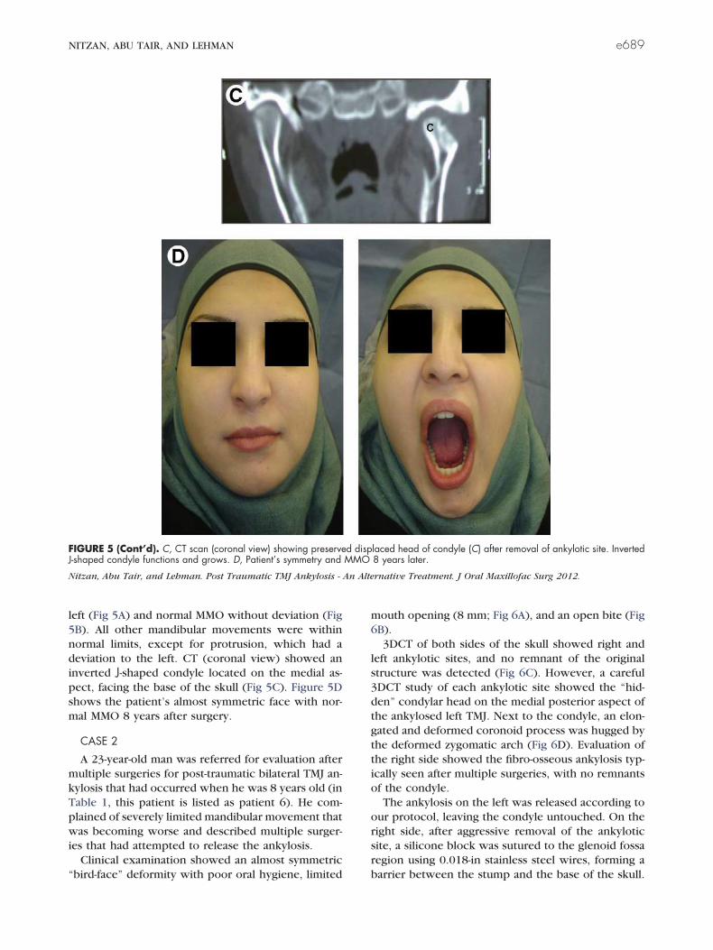
left (Fig 5A) and normal MMO without deviation (Fig5B). All other mandibular movements were withinnormal limits, except for protrusion, which had adeviation to the left. CT (coronal view) showed aninverted J-shaped condyle located on the medial as-pect, facing the base of the skull (Fig 5C). Figure 5Dshows the patient’s almost symmetric face with nor-mal MMO 8 years after surgery.
CASE 2
A 23-year-old man was referred for evaluation aftermultiple surgeries for post-traumatic bilateral TMJ an-kylosis that had occurred when he was 8 years old (inTable 1, this patient is listed as patient 6). He com-plained of severely limited mandibular movement thatwas becoming worse and described multiple surger-ies that had attempted to release the ankylosis.
Clinical examination showed an almost symmetric“bird-face” deformity with poor oral hygiene, limited
mouth opening (8 mm; Fig 6A), and an open bite (Fig6B).
3DCT of both sides of the skull showed right andleft ankylotic sites, and no remnant of the originalstructure was detected (Fig 6C). However, a careful3DCT study of each ankylotic site showed the “hid-den” condylar head on the medial posterior aspect ofthe ankylosed left TMJ. Next to the condyle, an elon-gated and deformed coronoid process was hugged bythe deformed zygomatic arch (Fig 6D). Evaluation ofthe right side showed the fibro-osseous ankylosis typ-ically seen after multiple surgeries, with no remnantsof the condyle.
The ankylosis on the left was released according toour protocol, leaving the condyle untouched. On theright side, after aggressive removal of the ankyloticsite, a silicone block was sutured to the glenoid fossaregion using 0.018-in stainless steel wires, forming abarrier between the stump and the base of the skull.
FIGURE 5 (Cont’d). C, CT scan (coronal view) showing preserved displaced head of condyle (C) after removal of ankylotic site. InvertedJ-shaped condyle functions and grows. D, Patient’s symmetry and MMO 8 years later.
Nitzan, Abu Tair, and Lehman. Post Traumatic TMJ Ankylosis - An Alternative Treatment. J Oral Maxillofac Surg 2012.
NITZAN, ABU TAIR, AND LEHMAN e689

Surgery was followed by intensive physiotherapy forboth mouth opening and closure to the original oc-clusion.
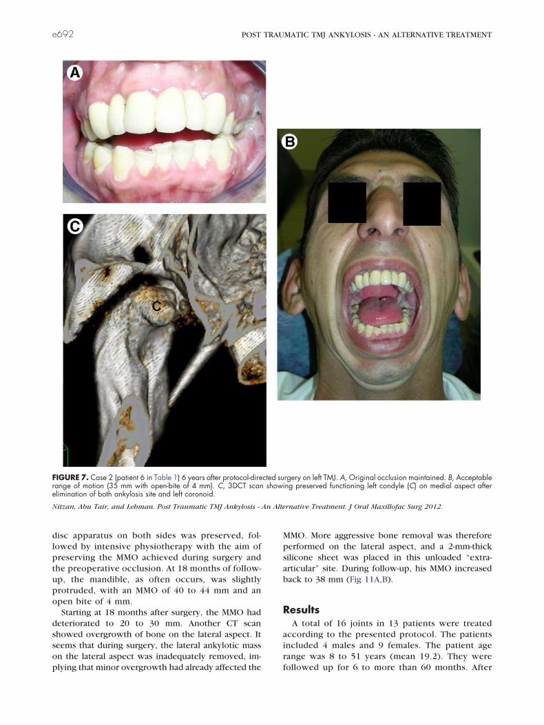
At 6 years of follow-up, the patient presented withan acceptable range of motion (MMO of 35 mm; Fig
7A) and unchanged occlusion (Fig 7B), taking intoaccount previous multiple interventions and his longcrowns. The patient was spared reconstructive sur-gery with its potential complications.
Postoperative 3DCT of the isolated preserved con-dyle showed the condylar head on the medial-poste-rior aspect of the stump. This condyle maintainedfunction and vertical height of the ramus (Fig 7C).
CASE 3
A 9-year-old girl was referred to the oral and max-illofacial surgery department for evaluation of pro-gressing facial asymmetry. The medical historyshowed that she had fallen and hurt her chin 2 yearsearlier with no further complaints (in Table 1, thispatient is listed as patient 9). Extraoral examinationshowed facial asymmetry with chin deviation to theright (Fig 8A). The MMO was limited to 22 mm, withmarked deviation to the right (Fig 8B). Lateral move-ments to the left were limited, and protrusion wasalso associated with deviation to the right. Intraoralexamination showed a right cross bite with markeddeviation of the mandibular midline to the right (Fig8C). A panoramic radiograph vaguely showed theankylosis (Fig 8D).
3DCT scan of the right ankylotic TMJ only showeda small part of the displaced condyle on the medialaspect of the ankylotic site (Fig 8E). We assumed thatthis remnant would be sufficient to act as a growthsite and, therefore, decided to proceed according toour protocol. For optimal performance, a 3D modelwas used to carefully plan and perform the surgerywithout damage to the condyle.
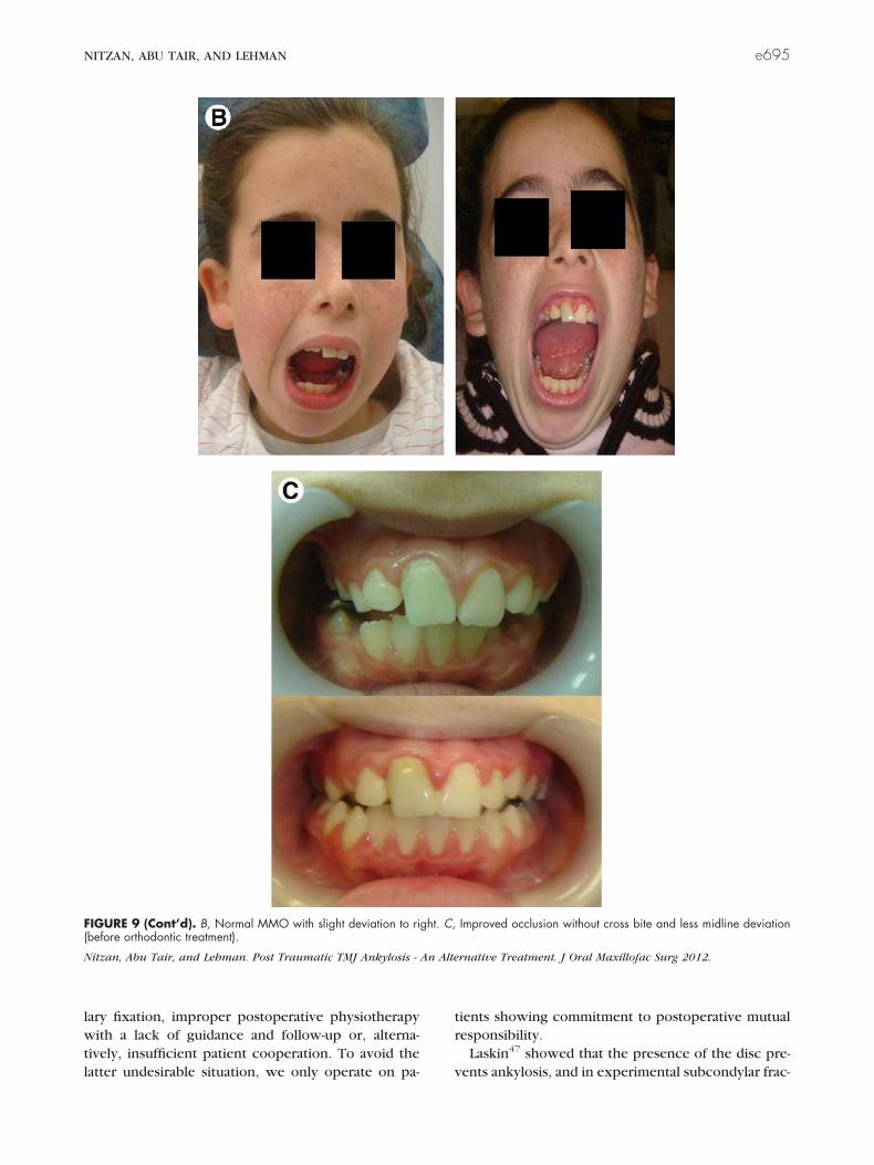
At 2 years after surgery, the patient presented withimproved facial symmetry (Fig 9A) and significantimprovement in both mouth opening (Fig 9B) andocclusion (Fig 9C).
CASE 4
An 8-year-old boy had severely limited mouthopening with a slight anterior open bite (Fig 10A)and a symmetric face (Fig 10B) owing to bilateralankylosis after trauma when he was 6 years old (thispatient is listed as patient 10 in Table 1). CTshowed bilateral bony TMJ ankylosis. The posteriorview of the 3DCT model showed both condylarheads on the medial aspect (Fig 10C). Detailedstudy of the left TMJ showed complicated ankylosiswith the damaged condyle in a medial anteriorlocation, adjacent to the base of the skull. The anglebetween the accurate surgical line (Fig 10D, solidline) and the line (Fig 10D, interrupted line) thatcuts through the condyle is narrow.
Despite the questionable prognosis, the patientwas treated according to the protocol to maintainthe growth potential of the condyles. The condyle–
FIGURE 6. Case 2 (patient 6 in Table 1), with long-durationbilateral TMJ ankylosis. Patient presented with, A, severely limitedmouth opening (8 mm including open-bite of 4 mm) and, B, anterioropen bite and poor dental health. (Figure 6 continued on nextpage.)
Nitzan, Abu Tair, and Lehman. Post Traumatic TMJ Ankylosis -An Alternative Treatment. J Oral Maxillofac Surg 2012.
e690 POST TRAUMATIC TMJ ANKYLOSIS - AN ALTERNATIVE TREATMENT

FIGURE 6 (Cont’d). C, 3DCT scan showing severe bilateral ankylosis with no indication of original TMJ structure. D, Careful study of 3DCTscan uncovered presence of condylar head (C) and elongated deformed coronoid (CR) process embraced by zygomatic arch on left side.
Nitzan, Abu Tair, and Lehman. Post Traumatic TMJ Ankylosis - An Alternative Treatment. J Oral Maxillofac Surg 2012.
NITZAN, ABU TAIR, AND LEHMAN e691
disc apparatus on both sides was preserved, fol-lowed by intensive physiotherapy with the aim ofpreserving the MMO achieved during surgery andthe preoperative occlusion. At 18 months of follow-up, the mandible, as often occurs, was slightlyprotruded, with an MMO of 40 to 44 mm and anopen bite of 4 mm.
Starting at 18 months after surgery, the MMO haddeteriorated to 20 to 30 mm. Another CT scanshowed overgrowth of bone on the lateral aspect. Itseems that during surgery, the lateral ankylotic masson the lateral aspect was inadequately removed, im-plying that minor overgrowth had already affected the
MMO. More aggressive bone removal was thereforeperformed on the lateral aspect, and a 2-mm-thicksilicone sheet was placed in this unloaded “extra-articular” site. During follow-up, his MMO increasedback to 38 mm (Fig 11A,B).
ResultsA total of 16 joints in 13 patients were treated
according to the presented protocol. The patientsincluded 4 males and 9 females. The patient agerange was 8 to 51 years (mean 19.2). They werefollowed up for 6 to more than 60 months. After
FIGURE 7. Case 2 (patient 6 in Table 1) 6 years after protocol-directed surgery on left TMJ. A, Original occlusion maintained. B, Acceptablerange of motion (35 mm with open-bite of 4 mm). C, 3DCT scan showing preserved functioning left condyle (C) on medial aspect afterelimination of both ankylosis site and left coronoid.
Nitzan, Abu Tair, and Lehman. Post Traumatic TMJ Ankylosis - An Alternative Treatment. J Oral Maxillofac Surg 2012.
e692 POST TRAUMATIC TMJ ANKYLOSIS - AN ALTERNATIVE TREATMENT

guided physiotherapy, all patients had significantimprovement in the MMO from a mean of 18.4 mm(range 8 to 28) to a mean of 41.2 mm (range 35 to50) postoperatively. All the growing patientsachieved good facial symmetry. In all the patients,except for one (patient 10 in Table 1), the occlu-sion returned to the original form after intensivephysiotherapy period (Table 1). One patient under-went removal of aggressive bone growth at the
lateral aspect owing to insufficient bone resectionat the first intervention (case 4 in the present reportand patient 10 in Table 1).
Discussion
TMJ ankylosis is most commonly caused by a mis-diagnosed or mistreated mandibular condylar frac-ture. In most of these cases, the fractured condyle or
FIGURE 8. Case 3 (patient 9 in Table 1) was a 9-year-old girl complaining of developing facial asymmetry 2 years after minor trauma. A,Facial asymmetry, with deviation of chin to right. B, Limited mouth opening (22 mm), with deviation to right. C, Malocclusion with right crossbite. D, Panoramic view showing right TMJ ankylosis. (Figure 8 continued on next page.)
Nitzan, Abu Tair, and Lehman. Post Traumatic TMJ Ankylosis - An Alternative Treatment. J Oral Maxillofac Surg 2012.
NITZAN, ABU TAIR, AND LEHMAN e693

a part of it is displaced, and ankylosis (Sawhney’s typeIII21) occurs between the stump and the base of theskull. In contrast, the displaced condyle is protected
from ankylosis by the displaced disc47 (Fig 3). Thepresent study focused on this group.
To clarify the rationale of the presented treatment,one needs to consider the entire sequence of events,starting with the fracture itself. In most cases of dis-placed subcondylar fractures, the head of the con-dyle, as described by Laskin,47 is displaced, togetherwith the disc, in an anteromedial-inferior direc-tion.7,51-53 At the same time, the stump is pulled up bythe ipsilateral closing muscles (masseter and medialpterygoid) toward the base of skull (Fig 2), resultingin typical malocclusion (ie, ipsilateral premature con-tact and a contralateral open bite).54
Two main approaches have been used to correctthis condition: open reduction, in which the condyleis exposed surgically, reduced, and fixed to its ana-tomic location; and closed reduction, in which theteeth are brought back into occlusion by intermaxil-lary fixation, and the stump is retracted away from thebase of the skull. The condyle, however, is not re-duced to the correct position owing to the attachedpulling muscles (Fig 2). Intermaxillary fixation is lim-ited to 10 to 14 days and is followed by intensivephysiotherapy with the aim of rehabilitating normaljaw movement and ensuring stable normal occlusion.Both are achieved, although the inverted J-shapedcondyle is located medially with some loss of verticalheight (Fig 2).55
Ankylosis typically develops because of incorrecttreatment. This includes extended use of intermaxil-
FIGURE 8 (Cont’d). E, 3DCT scan, medial view, showing rightTMJ ankylosis (Ank); only a small part of the condyle (C) was foundto be displaced medially.
Nitzan, Abu Tair, and Lehman. Post Traumatic TMJ Ankylosis -An Alternative Treatment. J Oral Maxillofac Surg 2012.
FIGURE 9. Case 3 (patient 9 in Table 1) 2 years postoperatively. A, Improved facial symmetry. (Figure 9 continued on next page.)
Nitzan, Abu Tair, and Lehman. Post Traumatic TMJ Ankylosis - An Alternative Treatment. J Oral Maxillofac Surg 2012.
e694 POST TRAUMATIC TMJ ANKYLOSIS - AN ALTERNATIVE TREATMENT
lary fixation, improper postoperative physiotherapywith a lack of guidance and follow-up or, alterna-tively, insufficient patient cooperation. To avoid thelatter undesirable situation, we only operate on pa-
tients showing commitment to postoperative mutualresponsibility.
Laskin47 showed that the presence of the disc pre-vents ankylosis, and in experimental subcondylar frac-
FIGURE 9 (Cont’d). B, Normal MMO with slight deviation to right. C, Improved occlusion without cross bite and less midline deviation(before orthodontic treatment).
Nitzan, Abu Tair, and Lehman. Post Traumatic TMJ Ankylosis - An Alternative Treatment. J Oral Maxillofac Surg 2012.
NITZAN, ABU TAIR, AND LEHMAN e695

FIGURE 10. Case 4 (patient 10 in Table 1) was an 8-year-old boy with bilateral TMJ ankylosis 2 years after trauma. A, Severely limited mouth opening(10 mm, including open bite). B, Good facial symmetry. C, Surgical planning with close view at left ankylotic site from lateral, anterior, and posterior views.Note, damaged condyle located close to base of skull. D, 3D model showing bilateral TMJ ankylosis (Ank) with condyles (C) displaced medially.
Nitzan, Abu Tair, and Lehman. Post Traumatic TMJ Ankylosis - An Alternative Treatment. J Oral Maxillofac Surg 2012.
e696 POST TRAUMATIC TMJ ANKYLOSIS - AN ALTERNATIVE TREATMENT

tures with meniscectomy and immobilization, ankylo-sis of the condylar head was noted.
The presented protocol aimed to reverse the con-dition of ankylosis between the stump and the base ofthe skull and achieve results similar to those achievedwith closed reduction (ie, a well-functioning short,inverted J-shaped condyle in a medial location associ-ated with normal occlusion; Fig 2). In addition tothe avoidance of unnecessary, high-risk, deep dis-section and osteotomies to remove the displacednative condyle, this approach is particularly impor-tant for growing individuals, because it maintainsthe growth site and allows normal and symmetricdevelopment of the mandible. Other alternative sur-gical interventions are extensive, associated with aloss of the growth site and vertical height, andrequire reconstruction with its associated compli-cations.
It is important to note that postoperatively themandible is not always as free as was recommendedby Kaban et al,46 especially regarding contralateralmovements, owing to the location of the condyle;therefore, long-term postoperative physiotherapy isrequired.
We have reported on 13 cases (10 unilateral and 3bilateral), of which 4 were described in detail. Allwere treated according to the protocol presented. Inbrief, releasing the ankylosis, avoiding damage to thedisplaced condyle, guidance of the saved condyle toenable a normal range of joint function, good occlu-sion, and, in children, growth and establishment ofgood facial symmetry. This method challenges the
approach recently suggested by Long et al14 to releasethe ankylosis, redesign a form of condyle at the anky-lotic site, remove the medially displaced condyle, andmobilize the disc over the preserved lateral stump. Italso challenges the approach of Li et al,17 who usedthe stump to reconstruct a new TMJ with the discrepositioned over it. These approaches might provideproper vertical height; however, the first is associatedwith the undesirable removal of the growth site inchildren and involves removal of the invisible con-dyle, which is demanding and risky. Also, the secondapproach recommends a pointless repositioning ofthe disc. When our protocol is followed, althoughsome loss of vertical height can occur, normal func-tion and occlusion are restored, and the condyleserves as an articulation and a growth site.
Considering that ankylosis occurs as a result ofnumerous fracture types, various types of ankylosisdevelop. For optimal evaluation, 3DCT imaging isrequired (Figs 1, 6D, 7C, 8E, 10D). Occasionally, a 3Dmodel (case 3 in Fig 8E and case 4 in Fig 10C) isbeneficial to show the exact condyle location andshape and enable adequate surgical planning to avoiddamaging the condyle or the disc, especially becausethe surgical field is limited and the condyle is hiddenon the medial aspect of the ankylosis. Figures 1 and 3show the risk of damaging the condylar head. In case2, after 15 years of ankylosis and 5 surgical interven-tions, 3DCT showed the hidden undamaged condyle,which was saved (Figs 6D, 7C) and provided satisfac-tory results: sufficient vertical height, good occlusion,
FIGURE 11. Case 4 (patient 10 in table 1) 18 months after protocol-directed surgery. A, Normal MMO (�40 mm). B, Increased anterioropen bite that developed from rotation of free mandible after surgery corrected gradually with guided physiotherapy.
Nitzan, Abu Tair, and Lehman. Post Traumatic TMJ Ankylosis - An Alternative Treatment. J Oral Maxillofac Surg 2012.
NITZAN, ABU TAIR, AND LEHMAN e697
and proper function, preventing the need for jointreplacement.
There are 4 aims of surgical intervention for TMJankylosis that we desire to accomplish:
1. Preserve the range of motion accomplished atthe end of surgery. All the present patients hadsignificant improvement in MMO (35 to 50 mm,mean 41.2). Owing to the medial location of thedisplaced condyle, the contralateral movementsmight be limited.
2. Attain mouth closure. Longstanding ankylosiscauses a lack of tonus in the closing muscles,because the mandible was supported by theankylosis. Thus, the mandible often falls forwardwhen the ankylosis is released. Physiotherapy,therefore, includes rehabilitation of the ability toclose the mouth, as well as to chew and biteeffectively.
3. Return to the original occlusion. Usually, afterrelease of longstanding ankylosis, changes occurin occlusion. When bilateral ankylosis is re-leased, the mandible moves forward and rotatesclockwise, resulting in an increased anterioropen bite. Release of unilateral ankylosis cancause a contralateral open bite because of theloss of vertical height owing to the relativelyinferior location of the displaced condyle. Thesesituations are solved by guided physiotherapy.
4. Enable growth of the mandible/facial structures(Table 1, patients 2, 3, 4, 5, 9, 10, and 13). Ofthose reported in detail (cases 1 and 3), surgerywas performed during the growth period. Thus,restoration of symmetric function and resump-tion of facial, almost symmetric, growth rapidlyensued (Figs 5A, 5D, 9A).
The details of case 3 highlight that growth cancontinue with maintenance of the vertical height,even when only a part of the condyle is saved, whichcan occur in cases of split condylar fractures (Figs 8,9). This case was treated according to our protocol,and the remaining part of the condyle functioned as agrowth site. At 2 years of follow-up, the facial sym-metry had improved, the occlusion was satisfactory,and the MMO was normal and almost symmetric (Fig9). The surgical intervention in this case was alsoguided by 3DCT, emphasizing, once again, the impor-tance of imaging to avoid damaging the hidden con-dyle.
In summary, the present study reports on a com-plementary protocol to that of Kaban et al, suggestingthat not all cases of TMJ ankylosis require aggressivetreatment. Surgeons should evaluate the option ofreleasing the lateral ankylosis while preserving thecondyle and the disc. This procedure will allow the
patient to avoid major surgery, with all its inconve-niences and complications, but still gain adequatemandibular motion and facial growth.
References1. Zimmermann CE, Troulis MJ, Kaban LB: Pediatric facial frac-
tures: Recent advances in prevention, diagnosis and manage-ment. Int J Oral Maxillofac Surg 34:823, 2005
2. Kaban LB, Bouchard C, Troulis MJ: A protocol for managementof temporomandibular joint ankylosis in children. J Oral Max-illofac Surg 67:1966, 2009
3. Lee CY, McCullon C III, Blaustein DI, et al: Sequelae of unrec-ognized, untreated mandibular condylar fractures in the pedi-atric patient. Ann Dent 52:5, 1993
4. Anantanarayanan P, Narayanan V, Manikandhan R, et al: Pri-mary mandibular distraction for management of nocturnal de-saturations secondary to temporomandibular joint (TMJ) anky-losis. Int J Pediatr Otorhinolaryngol 72:385, 2008
5. Tellez RJ, Carvalho TD, Belmont LF: Osteogenic distraction andorthognathic surgery to correct sequelae of ankylosis of thetemporomandibular joint: A case report. Int J Adultorthodonor-thognathsurg 17:291, 2002
6. Myall RW: Management of mandibular fractures in children.Oral Maxillofac Surg Clin North Am 21:197, vi, 2009
7. He D, Ellis E III, Zhang Y: Etiology of temporomandibular jointankylosis secondary to condylar fractures: The role of concom-itant mandibular fractures. J Oral Maxillofac Surg 66:77, 2008
8. Zhi K, Ren W, Zhou H, et al: Management of temporomandib-ular joint ankylosis: 11 years’ clinical experience. Oral SurgOral Med Oral Pathol Oral Radiol Endod 108:687, 2009
9. Güven O: A clinical study on temporomandibular joint ankylo-sis. Auris Nasus Larynx 27:27, 2000
10. Roychoudhury A, Parkash H, Trikha A: Functional restorationby gap arthroplasty in temporomandibular joint ankylosis: Areport of 50 cases. Oral Surg Oral Med Oral Pathol Oral RadiolEndod 87:166, 1999
11. Topazian RG: Comparison of gap and interposition arthroplastyin the treatment of temporomandibular joint ankylosis. J OralSurg 24:405, 1966
12. Topazian RG: Gap versus interposition arthroplasty for ankylo-sis of the temporomandibular joint. Oral Surg Oral Med OralPathol Oral Radiol Endod 91:388, 2001
13. Raveh J, Vuillemin T, Lädrach K, et al: Temporomandibularjoint ankylosis: Surgical treatment and long-term results. J OralMaxillofac Surg 47:900, 1989
14. Long X, Li X, Cheng Y, et al: Preservation of disc for treatmentof traumatic temporomandibular joint ankylosis. J Oral Maxil-lofac Surg 63:897, 2005
15. He D, Yang C, Chen M, et al: Traumatic temporomandibularjoint ankylosis: Our classification and treatment experience.J Oral Maxillofac Surg 69:1600, 2011
16. Bulgannawar BA, Rai BD, Nair MA: Use of temporalis fascia asan interpositional arthroplasty in temporomandibular joint an-kylosis: Analysis of 8 cases. J Oral Maxillofac Surg 69:2076,2011
17. Li Z, Li ZB, Li JR: Surgical management of posttraumatic tem-poromandibular joint ankylosis by functional restoration withdisk repositioning in children. Plast Reconstr Surg 119:1311,2007
18. Chossegros C, Guyot L, Cheynet F, et al: Full-thickness skingraft interposition after temporomandibular joint ankylosis sur-gery. A study of 31 cases. Int J Oral Maxillofac Surg 28:330,1999
19. Meyer RA: The autogenous dermal graft in temporomandibularjoint disc surgery. J Oral Maxillofac Surg 46:948, 1988
20. Erdem E, Alkan A: The use of acrylic marbles for interpositionarthroplasty in the treatment of temporomandibular joint an-kylosis: Follow-up of 47 cases. Int J Oral Maxillofac Surg 30:32,2001
21. Sawhney CP: Bony ankylosis of the temporomandibular joint:Follow-up of 70 patients treated with arthroplasty and acrylicspacer interposition. Plast Reconstr Surg 77:29, 1986
e698 POST TRAUMATIC TMJ ANKYLOSIS - AN ALTERNATIVE TREATMENT
22. Garrett WR, Abbey PA: The use of fat grafts to prevent heter-otopic bone formation in the temporomandibular joint. J OralMaxillofac Surg 55:1037, 1997
23. Paterson AW, Shepherd JP: Fascia lata interpositional arthro-plasty in the treatment of temporomandibular joint ankylosiscaused by psoriatic arthritis. Int J Oral Maxillofac Surg 21:137,1992
24. Topazian RG: Etiology of ankylosis of temporomandibularjoint: Analysis of 44 cases. J Oral Surg Anesth Hosp Dent Serv22:227, 1964
25. Lindqvist C, Pihakari A, Tasanen A, et al: Autogenous costo-chondral grafts in temporo-mandibular joint arthroplasty: Asurvey of 66 arthroplasties in 60 patients. J Maxillofac Surg14:143, 1986
26. Guralnick WC, Kaban LB: Surgical treatment of mandibularhypomobility. J Oral Surg 34:343, 1976
27. Phillips JH, Rechner B, Tompson BD: Mandibular growth fol-lowing reconstruction using a free fibula graft in the pediatricfacial skeleton. Plast Reconstr Surg 116:419, 2005
28. Iconomou TG, Zuker RM, Phillips JH: Mandibular reconstruc-tion in children using the vascularized fibula. J Reconstr Micro-surg 15:83, 1999
29. Wolford LM, Cottrell DA, Henry C: Sternoclavicular grafts fortemporomandibular joint reconstruction. J Oral MaxillofacSurg 52:119, 1994
30. Kummoona R: Chondro-osseous iliac crest graft for one stagereconstruction of the ankylosed TMJ in children. J MaxillofacSurg 14:215, 1986
31. Matukas VJ, Szymela VF, Schmidt JF: Surgical treatment of bonyankylosis in a child using a composite cartilage-bone iliac crestgraft. J Oral Surg 38:903, 1980
32. Dattilo DJ, Granick MS, Soteranos GS: Free vascularized wholejoint transplant for reconstruction of the temporomandibularjoint: A preliminary case report. J Oral Maxillofac Surg 44:227,1986
33. Landa LE, Gordon C, Dahar N, et al: Evaluation of long-termstability in second metatarsal reconstruction of the temporo-mandibular joint. J Oral Maxillofac Surg 61:65, 2003
34. Saeed N, Hensher R, McLeod N, et al: Reconstruction of thetemporomandibular joint autogenous compared with alloplas-tic. Br J Oral Maxillofac Surg 40:296, 2002
35. Machon V, Hirjak D, Beno M, et al: Total alloplastic temporo-mandibular joint replacement: The Czech-Slovak initial experi-ence. Int J Oral Maxillofac Surg 41:514, 2012
36. Mercuri LG, Swift JQ: Considerations for the use of alloplastictemporomandibular joint replacement in the growing patient.J Oral Maxillofac Surg 67:1979, 2009
37. Pearce CS, Cooper C, Speculand B: One stage management ofankylosis of the temporomandibular joint with a custom-madetotal joint replacement system. Br J Oral Maxillofac Surg 47:530, 2009
38. Manemi RV, Fasanmade A, Revington PJ: Bilateral ankylosis ofthe jaw treated with total alloplastic replacement using the TMJconcepts system in a patient with ankylosing spondylitis. Br JOral Maxillofac Surg 47:159, 2009
39. Müller-Richter UD, Roldán JC, Mörtl M, et al: SAPHO syndromewith ankylosis of the temporomandibular joint. Int J Oral Max-illofac Surg 38:1335, 2009
40. Schwartz HC, Relle RJ: Distraction osteogenesis for temporo-mandibular joint reconstruction. J Oral Maxillofac Surg 66:718,2008
41. Cheung LK, Lo J: The long-term effect of transport distractionin the management of temporomandibular joint ankylosis. PlastReconstr Surg 119:1003, 2007
42. Stucki-McCormick SU: Reconstruction of the mandibular con-dyle using transport distraction osteogenesis. J Craniofac Surg8:48, 1997
43. Yoon HJ, Kim HG: Intraoral mandibular distraction osteogen-esis in facial asymmetry patients with unilateral temporoman-dibular joint bony ankylosis. Int J Oral Maxillofac Surg 31:544,2002
44. Padgett EC, Robinson DW, Stephenson KL: Ankylosis of thetemporomandibular joint. Surgery 24:426, 1948
45. Vasconcelos BC, Porto GG, Bessa-Nogueira RV, et al: Surgicaltreatment of temporomandibular joint ankylosis: Follow-up of15 cases and literature review. Med Oral Patol Oral Cir Bucal14:E34, 2009
46. Kaban LB, Perrott DH, Fisher K: A protocol for management oftemporomandibular joint ankylosis. J Oral Maxillofac Surg 48:1145, 1990
47. Laskin DM: Role of the meniscus in the etiology of posttrau-matic temporomandibular joint ankylosis. Int J Oral Surg 7:340,1978
48. Nitzan DW, Bar-Ziv J, Shteyer A: Surgical management of tem-poromandibular joint ankylosis type III by retaining the dis-placed condyle and disc. J Oral Maxillofac Surg 56:1133, 1998
49. Nitzan DW, Azaz B, Constantini S: Severe limitation in mouthopening following transtemporal neurosurgical procedures: Di-agnosis, treatment, and prevention. J Neurosurg 76:623, 1992
50. Salter RB: The physiologic basis of continuous passive motionfor articular cartilage healing and regeneration. Hand Clin 10:211, 1994
51. Rowe NL: Ankylosis of the temporomandibular joint. J R CollSurg Edinb 27:67, 1982
52. Zachariades N, Mezitis M, Mourouzis C, et al: Fractures of themandibular condyle: A review of 466 cases. Literature review,reflections on treatment and proposals. J Craniomaxillofac Surg34:421, 2006
53. Nussbaum ML, Laskin DM, Best AM: Closed versus open reduc-tion of mandibular condylar fractures in adults: A meta-analysis.J Oral Maxillofac Surg 66:1087, 2008
54. Ward Booth P, Schendel SA, Hausamen JE: Surgical manage-ment of mandibular, condylar neck and atrophic mandiblefracture, in Dolan J (ed): Maxillofacial Surgery, ed 2. St. Louis,MO, Elsevier, 2007, pp 62-102
55. Ellis E, Throckmorton GS: Treatment of mandibular condylarprocess fractures: Biological considerations. J Oral MaxillofacSurg 63:115, 2005
NITZAN, ABU TAIR, AND LEHMAN e699