Embed Size (px)
Citation preview
1
2
Jagiellonian University
in Krakow
Institute of Catalysis and
Surface Chemistry PAS
International Union of
Crystallography, Crystallography in Art and
Cultural Heritage Commission
3
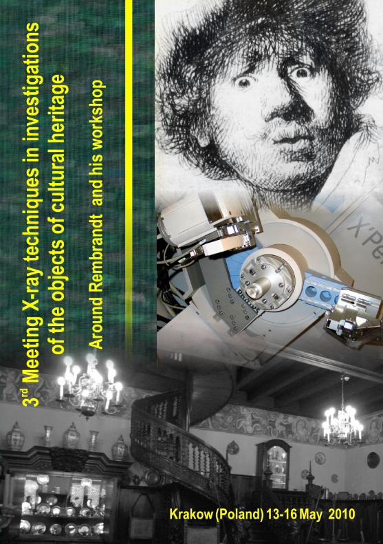
3rd MEETING
X-RAY TECHNIQUES IN INVESTIGATIONS
OF THE OBJECTS OF CULTURAL HERITAGE
AROUND REMBRANDT AND HIS WORKSHOP
Krakow, 13-16 May, 2010
4
The aim of the Meeting is to promote the development
and use of X-ray techniques in order to extract information
from the objects of cultural heritage. It is also a forum to
bring together scientists, whose major expertise is in a high-
tech field, and museum professionals (conservators and
curators) whose particular responsibility is the organization
and preservation of collections, and to spread the newest
results in the area of scientific investigations of art objects.
First day of the meeting will be focused on the investigations
of Rembrandt paintings.
5
Organizers:
Faculty of Chemistry Jagiellonian University
30-060 Krakow, ul. Ingardena 3, POLAND www.chemia.uj.edu.pl
Jagiellonian University Museum 31-010 Krakow, ul. Jagiellooska 15, POLAND www.maius.uj.edu.pl
Institute of Catalysis and Surface Chemistry PAS 30-239 Krakow, ul. Niezapominajek 8, POLAND http://atom.ik-pan.krakow.pl
International Union of Crystallography, Crystallography in Art and Cultural Heritage Commission http://www.iucr.org/iucr/commissions/ccach
6
International Scientific Committee:
Prof. Henk Schenk, Amsterdam University, the Netherlands,
Dr Eric Dooryhee, Centre National de la Recherche Scientifique,
France,
Dr Thomas Wroblewski, Deutsches Elektronen-Synchrotron,
Germany.
Organizing Committee:
Prof. Grażyna Stochel Prof. Stanisław Waltoś
Prof. Małgorzata Witko Prof. Wiesław Łasocha
Prof. Roman Kozłowski Alicja Rafalska-Łasocha
Anna Jasioska Jolanta Pollesch
Secretary of the Meeting:
Alicja Rafalska-Łasocha
Sponsors:
PANalytical B.V. Branch Office Poland
Testchem
7
Programme
13 May 2010, Thursday 14.00 - 17.00
Registration: Collegium Maius – Jagiellonian University
Museum, Jagiellooska str. No. 15, Krakow.
17.00 - 17.15
Opening of the Meeting
17.15 - 18.15
"Climate change effects on Europe's cultural heritage: challenges
and possibilities" Roman Kozłowski, Polish Academy of Sciences,
Krakow, Poland
18.30 - 21.00 Get together party in the Cellars of Collegium
Maius
14 May 2010, Friday
Around Rembrandt and his workshop Chair: Prof. Roman Kozłowski, Polish Academy of Sciences
9.30 - 10.30
“Neutron-Activation-Autoradiography of Paintings by
Rembrandt at the Berlin Picture Gallery” Claudia Laurenze-
Landsberg, Gemäldegalerie der Staatlichen Museen, Berlin,
Germany
10.30 – 11.30
”Two of Rembrandt’s paintings: ‘Girl in a picture frame’ and
‘Scholar at his writing table’ from the Warsaw Royal Castle
Collection – history, examination and conservation” Joanna
Czernichowska, Regina Dmowska, Anna Nowicka, Royal Castle in
Warsaw, Poland
8
11.30 - 12.00 Coffee break
12.00 - 13.00
”Rembrandt’s ‘Landscape with Good Samaritan’ from the
Czartoryski Collection – observation and technical information
after restoration” Anna Grochowska-Angelus, Katarzyna
Novljakovic, Dorota Dec, Maria Rogóż, Krakow National Museum,
Poland
13.00 - 14.00
Poster Session and Visit to the Conservation Workshop in
Collegium Maius
14.00 - 15.00 Lunch
Chair: Prof. Zbigniew Sojka, Faculty of Chemistry Jagiellonian
University
15.00 – 16.00
”Scientific analyses in determining original appearance. Studio
and Rembrandt, Saul and David, c. 1660, oil on canvas (H: 131 x
L: 164 cm)” Petria Noble, Royal Picture Gallery Mauritshuis, The
Hague, The Netherlands
16.00 – 16.45
“Is portrait of ‘Young Man’ from the Cracow Royal Castle
Collection by Jan Lievens?” Joanna Winiewicz-Wolska, Ewa
Wiłkojd, Wawel Royal Castle, Krakow, Poland
9
16.45 - 17.30
“Portrait of Joost van den Vondel’ by Philips Koninck from the
Jagiellonian University Museum Collection. Attribution and
identification” Anna Jasioska, M. Krąpiec, Jolanta Pollesch, Beata
Skalmierska, Jagiellonian University Museum, Krakow, Poland
17.30 - 17. 45 Coffee break
17.45 - 19.00
Visit to the Collegium Maius Collection
15 May 2010, Saturday
X-ray techniques in investigations of art objects
Chair: Prof. Henk Schenk, Amsterdam University, the Netherlands,
9.00 - 10.00 ”Use of X-ray powder micro-diffraction for identification of local sources of painting materials” Petr Bezdička, Silvie Švarcová, David Hradil, Janka Hradilová, Academic Materials Research Laboratory of Painted Artworks (ALMA) Institute of Inorganic Chemistry of the ASCR, Husinec-Rez, Czech Republic 10.00 - 10.45 ”Portable digital X-ray radiography system for studies of historical objects, Piotr Frączek, Joanna Sobczyk, Łukasz Bratasz, Janusz Czop, National Museum in Krakow, Poland 10.45 - 11.15 Coffee break
10
11.15 - 12.00 “X-RAY POWDER DIFFRACTOMETRY FOR STUDIES OF HISTORICAL OBJECTS - A
NEW EQUIPMENT AT FACULTY OF CHEMISTRY JAGIELLONIAN UNIVERSITY” M. Oszajca, M. Grzesiak, K. Podulka, A. Rafalska-Lasocha, W. Lasocha, Faculty of Chemistry Jagiellonian University, Krakow, Poland 12.00 - 13.00 Sponsors’ presentations 13.00 - 14.30 Lunch
Chair: Prof. Wieslaw Lasocha, Faculty of Chemistry, Jagiellonian University, Krakow, Poland 14.30 - 15.15
“Recovering Erased Scripts from Palimpsests: First results from
X-Ray flourescence element mapping experiments” L. Glaser, D.
Deckers , G. Falkenberg , C. Mackert, C. Brockmann and D.
Harlfinger, Deutsches Elektronen-Synchrotron DESY, Hamburg,
Germany
15.15 - 16.15 Closing lecture “Rare Silverpoint Drawings by Rembrandt in the Focus of SR-XRF” Ina Reiche, Silke Merchel, Martin Radtke, Heinrich Riesemeier, Holm Bevers, Centre de Recherche et de Restauration des Musées de France - CNRS UMR 171, PARIS
16.15 -16.30 Coffee break 16.30 - 18.00 Science Festival in Krakow
11
19.00 - 23.00
Meeting Dinner in the cellars of Collegium Maius
16 May 2010, Sunday
Visit to the historic Salt Mine in Wieliczka
Poster presentations:
1. REMBRANDT IN LABORATORY. FACTORS OF
DETERIORATION OF OIL PAINTINGS CONTAINING LEAD WHITE.
Dawid Popławski, Maria Poksioska, Nicolaus Copernikus
University, Torun, Poland
2. DIGITAL PROCESSING OF X-RAY RADIOGRAMS FOR
CONSERVATION PURPOSES, Joanna Sobczyk, Piotr Frączek,
Jakub S. Prauzner-Bechcicki, Łukasz Bratasz, Janusz Czop,
National Museum In Krakow, Poland
3. MULTI-METALLIC ARTEFACTS FROM WROCŁAW -EDXRF
STUDIES, Beata Miazga, University of Wrocław, Institute of
Archaeology, Poland
4. APPLICATION OF X-RAY METHODS FOR PIGMENTS
IDENTIFICATION IN COLLECTION OF RAFAŁ HADZIEWICZ
PAINTINGS, A. KLISIOSKA-KOPACZ, A. RAFALSKA-ŁASOCHA,
E. ZYGIER and J. CZOP, National Museum in Krakow, Poland,
12
5. X-RAY BASED ANALYSIS OF THE METAL THREADS IN
HISTORICAL TEXTILES,
A.Klisioska-Kopacz, M. Włodarczak, A. Prokopowicz and Ł.
Bratasz, National Museum in Krakow, Krakow, Poland
6. WITKACY’S PASTELS ANALYSIS FOR SAFE ANOXIA STORAGE,
A. Klisioska-Kopacz, J. Sobczyk, J. Thomas, Piotr Frączek, D.
Godyo, Ł. Bratasz, J. Bagniuk, T. Łojewski, National Museum in
Krakow, Krakow, Poland
7. XRD and SEM/EDX INVESTIGATIONS OF THE CORROSION
PRODUCTS OF METAL THREADS FROM THE TAPESTRY OF
SIGISMUND AUGUSTUS, Alicja Rafalska-Łasocha, Jerzy Holc,
Anna Bielaoska, Wiesław Łasocha , Faculty of Chemistry
Jagiellonian University, Krakow, Poland
8. XRD INVESTIGATIONS OF COMMERCIALLY AVAILABLE
PRUSSIAN BLUE PIGMENTS AND OIL PAINTS, Alicja Rafalska-
Łasocha, Katarzyna Podulka, Roman Dziembaj, Wiesław Łasocha,
Faculty of Chemistry Jagiellonian University, Krakow, Poland
9. SYNCHROTRON X-RAY POWDER MICRO-DIFFRACTION and
MICRO-FLUORESCENCE FOR IDENTIFICATION OF GREEN
PIGMENTS IN the GOTHIC ALTAR PAINTINGS FROM
MALOPOLSKA REGION, Alicja Rafalska-Łasocha, Marta Grzesiak,
Zofia Kaszowska, Thomas Wróblewski, Dariusz Zając, Andre
Rothkirch, Wiesław Łasocha, Faculty of Chemistry Jagiellonian
University, Krakow, Poland
13
10. CONSERVATIONAL ANALYSIS OF THE SIGISMUND
BELL, Marcin Biborski1 and Andrzej Biborski, Institute of
Archeology, Faculty of History, Jagiellonian University, Krakow,
Poland
11. AN INTERESTING DILEMMA – A SMALL LANDSCAPE SIGNED
REMBRANDT F. 1627. A DEPOSIT IN THE JAGIELLONIAN
UNIVERSITY MUSEUM COLLECTION, Anna Jasioska, Jolanta
Pollesch, Beata Skalmierska, Jagiellonian University Museum,
Krakow, Poland
14
A B S T R A C T S
15
CLIMATE CHANGE EFFECTS ON EUROPE’S CULTURAL
HERITAGE: CHALLENGES AND POSSIBILITIES
Roman Kozłowski
Institute of Catalysis and Surface Chemistry Polish Academy of
Sciences, Krakow, Poland.
The climate parameters in the environment of cultural
heritage objects can have a profound effect on changes
occurring in the historic materials and consequently on their
preservation. Objects expand and contract as the temperature
or humidity change, are mechanically damaged by the melting
and freezing of water, or dissolution and crystallisation of salts
in response to impacts of the outdoor environment. The rates
of some important chemical reactions, such as the
degradation of cellulose in paper and textiles, increase with
rising temperature. Temperature and time of wetness
influence the activity of fungi and insects responsible for the
biodeterioration of organic materials.
Climate change is now widely recognized as the major
environmental problem facing the globe. Addressing climate
16
change is also central to the work of the European Union
which has launched research initiatives aiming at the
assessment of damage potential of climate change on
European cultural heritage, as well as the development of
possible mitigation strategies. The Noah’s Ark project,
implemented between 2004- 2007, focused on the impact of
different climate parameters on broadly defined heritage and
sought to improve the protection strategies of heritage
materials, structures and infrastructures. In November 2009,
the “Climate for Culture” project was initiated. It will last five
years and will develop high resolution climate evolution
scenarios and couple them with whole building simulation
models to identify the most urgent risks for various European
regions.
The presentation will identify different climate
parameters that pose risk to cultural heritage, as well as
provide information on the vulnerabilities of the European
regions to climate change based on the climate change
scenarios. The impact of climate on outdoor and indoor
heritage materials will be discussed. Historic wooden
structures and decorated wooden objects will be selected as

17
an example of vulnerable historic objects both outdoors due
to the biological attack by wood destroying fungi and indoors
due to physical damage caused by variations mainly in
ambient relative humidity.
Principles of mitigation and adaptation will be
discussed. The cultural heritage sector shares responsibility for
the environment, and it will need to reduce the impact on the
environment, primarily using energy more efficiently.
Measures that are necessary to adapt museums and historic
buildings to a changing climate will be analysed. The capacity
of the built heritage to adapt to climate change should be
maximised by changes in both the management of cultural
heritage as well as physical changes.
18
AROUND REMBRANDT AND HIS WORKSHOP
19
NEUTRON-AUTORADIOGRAPHY OF PAINTINGS BY
REMBRANDT IN THE GEMÄLDEGALERIE BERLIN
Claudia Laurenze-Landsberg1, C.O. Fischer2
1Staatliche Museen Gemäldegalerie, Berlin, Germany,
2Helmholtz-Zentrum für Materialien und Energie, Berlin,
Germany.
The research using the Neutron-Autoradiography
method is in collaboration with the Helmholtzzentrum für
Materialien und Energie, Berlin, formerly called the Hahn-
Meitner-Institute, and the Gemäldegalerie of the Staatliche
Museen Berlin. The Gemäldegalerie Berlin is the only institute
world wide, which systematically employs the method of
Neutron-Activation-Autoradiography to analyse paintings.
Today we have investigated about 70 mainly 17th century
works.
The paintings to be investigated are scanned by means
of neutron activation. The isotopes arising during this process
have specific half-lives and emit gamma and beta energies.
For a period of up to six weeks after activation x-ray films are
20
placed on the painting exposing them to the radiation.
Hereby, paint layers, which vary in colour, can be separated
on different films and supply valuable additional data to x-
radiographs.
Deeper paint layers are made visible. In this way, it is
possible to gain insight into the work process and the artistic
approach of the painter. A hidden composition might reveal
another artist’s influence on the painter, which might be
important for the dating. The elucidation of the brushstroke
can be read as the handwriting of the artist responsible for the
painting. Examples for these various findings by using non-
destructive autoradiography will be shown.
Furthermore the development of Rembrandt’s painting
technique will be demonstrated. With the assistance of
autoradiography it is now possible to be witness to
Rembrandt‘s rejection of the contemporary practice of under
modelling the whole composition in brown paint. This form of
preliminary draft was only adhered to in the earliest painting
we investigated. A year later he designs the composition on
the panel, the umbra is already thinner and only partly but still
21
flatly applied. Finally the flat design is replaced completely by
a linear, evolving sketch which is broadly laid out. Although
initially executed in umbra Rembrandt later preferred
boneblack for these compositional sketches. This change
might be related to his adoption of darker grounds. The
distribution of the pigment smalt in the later works is also
highly significant and attests to Rembrandt‘s understanding of
the characteristic features of materials. While in his early
works Rembrandt employed smalt as a coloured pigment only,
he later used the special qualities of this pigment by exploiting
its optical effects.
J.Kelch (GG), C.-O.Fischer (HMI), C.Laurenze (GG), W.Leuther
(HMI), G.Pieh (GG), K.Slusallek (RF), Bilder im Blickpunkt: Der
Mann mit dem Goldhelm. Eine Dokumentation der
Gemäldegalerie in Zusammenarbeit mit dem
Rhatgenforschungslabor und dem Hahn-Meitner-Institut
Berlin, 1986
C.-O.Fischer, Claudia.Laurenze.Landsberg, et al., Neues zur
Neutronen-Aktivierungs-Autoradiographie, Tizians „Mädchen
mit Fruchtschale“ und die Verwendung von Neapelgelb,
Restauro 6/99 pp 426
22
Claudia Laurenze-Landsberg, Neutron activation
autoradiography of paintings by Rembrandt at the Berlin
Picture Gallery, Conservation Science 2002, papers from the
conference held in Edinburgh, Scotland 22 – 24 May 2002 pp
254;
Claudia Laurenze-Landsberg, “Neutron-Autoradiography of
Two Paintings by Jan Vermeer in the Gemäldegalerie Berlin”,
Wolfgang Lefèvre, Inside the Camera Obscura – Optics and Art
under the Spell of the Projected Image, Reprint 333, pp 211
Katja Kleinert und Claudia Laurenze-Landsberg, Auf der Suche
nach einer optimalen Bildlösung. Zur Bildgenese von
Rembrandts Gemälde „Simson und Delila“, Rembrandt –
Wissenschaft auf der Suche, Beiträge des internationalen
Symposiums Berlin, 04. – 05.11.2006, Jahrbuch der Berliner
Museen, Band 51, 2009, pp 147
23
TWO REMBRANDT PAINTINGS: GIRL IN A PICTURE FRAME
and SCHOLAR AT HIS WRITING DESK
FROM THE ROYAL CASTLE COLLECTION IN WARSAW
–HISTORY, EXAMINATION AND CONSERVATION
Joanna Czernichowska1, Regina Dmowska2, Anna Nowicka1
1Academy of Fine Arts, Warsaw, Poland,
2 Royal Castle in Warsaw, Warsaw, Poland.
In 1994, Warsaw Royal Castle was honored with a gift
from the Lancoronsky Family; a priceless art collection with
two panel paintings – formerly in the XVII c. royal collection –
attributed to Rembrandt van Rijn (1606-1660): Girl in a picture
frame, Inv. no. ZKW/3906, Scholar at his desk, Inv. no. ZKW
3905, both: oil on panel, c.105.5 x 76.5 cm., signed:
Rembrandt f/1641.
In 2004-2006 both panels were examined, treated and
reattributed by Prof. Ernst van de Wetering, head of the
Rembrandt Research Project. The comprehensive examination
programme concerning the technology of the paintings and
the artist’s work technique was carried out by laboratories in
Warsaw and Krakow.
24
Both panels are single pieces of poplar, vertical grain, back
bevelled along four sides, very little wraped; thicknesses vary:
20-16 mm (Girl), 14-19 mm (Scholar), trimmed to create
pendants. Face surface not very smooth, back – stained; some
numbers, seals and traces of framing. Panels sized and
covered with ground layers: Girl- two layers: chalk in fat
medium and lead white with chalk in fat medium too, smooth
finished; Scholar- one layer: lead white in fat medium; light
ochre imprimatura on both, no drawings or preliminary
sketches prior to the present tronies.
The original paint layer pigments – examined by a cross-
section and microscope analysis (also seen by K. Groen, co.
RRP) – identified as: lead white mixed with chalk, red and
yellow ochre, red lake, green earth, bone black, bituminous
brown; also lead-tin yellow, smalt and malachite in the male
portrait and cinnabar in the female portrait. The binding
medium, typical for Rembrandt’s works, was examined (using
analytical instrumental methods: SEM-EDX, GC-MS, FT-IR):
linseed and walnut oil – walnut oil has been found in both
paintings. The paint layers in major parts of both paintings
were in a good condition with the exception of the red dress
25
of the Girl portrait – here the paint layer had developed a
prominent raised craquelure, some of which was poorly
attached to the panel, some had created tents, and some had
flaked off; in this part some pentimenti were visible through
the red colours. Surface coated with heavy layers of varnish,
badly discoloured and surface grime – very disturbing,
partially blanched, with poor saturation in dark areas; also
some small losses, changed retouching, a few dents and
scratches.
X-ray and IR examination as well as the cleaning process
revealed the existence of an initial composition – a sketch for
the portrait of another woman executed with a few black
brushstrokes – which shows up in the background around the
girl’s head and chest (on the present portrait) as a result of the
growing transparency of the upper paint layers. Most likely
the appearance of this first sketch was the reason for the later
overpainting, restoration and damage. Scholar signature –
solid, done in wet paint, Girl – most likely added later, partially
damaged.
Two varnish layers with old retouching and some
overpainting were removed from both panels. In the case of
26
the Girl this process was combined with local consolidation of
the red dress cupping and cleavage area. Large scale
overpainting of the upper background with the added column,
frame, extremely heavy dress (in the most part) were
removed as well as some smaller retouches on the hat, face,
ears and right earring, hair, necklaces, belt and some on the
hands – some overpainting had to be left due to damaging the
original or the impossibility of distinguishing or removing it.
Aquarelle, then Movilith AYAB in ethanol and Gamblin
retouching was done over the dammar varnish.
Afterwards they were exhibited with great success at the
most important Rembrandt Year, 2006, two venue exhibition
Rembrandt. Quest of a genius in the Rembrandthuis
(Amsterdam) and Gëmaldegalerie (Berlin).
27
REMBRANDT'S LANDSCAPE WITH GOOD SAMARITAN FROM
CZARTORYSKI MUSEUM - OBSERVATION AND TECHNICAL
INFORMATION AFTER RESTORATION
Anna Grochowska-Angelus1, Katarzyna Novljakovic1, Dorota
Dec1, Maria Rogóż2,
1Krakow National Museum, Krakow, Poland,
2Academy of Fine Arts, Krakow, Poland.
Painting collections of Western Art in Poland are
privileged to include the Rembrandt masterpiece Landscape
with Good Samaritan owned by Cracow’s Prince Czartoryski
Museum. In Rembrandt Year, 2006, this distinguished painting
was exhibited at the Museum Stedelijk De Lakenhal, Leiden,
with other landscapes of the Master that have survived to this
day, along with prints and designs by him.
The Czartoryski painting opened the show, having been
a main attraction for many years behind the “Iron Curtain”
during the communist era. The painter, Jean-Pierre Norblin de
la Gourdaine, brought the painting from France in 1774, and
has remained in the hands of the Czartoryski family since
28
1813. Throughout the last 200 years of the dramatic history of
Poland, the painting was moved many times but also with
great care and respect so that it remains in good condition
despite the instability.
In 2003, the painting underwent treatment by our
Conservatory to rediscover and recognize the work done by
the Rembrandt Research Project in its A Corpus of Rembrandt
Paintings. After removing two layers of varnish, including one
layer two hundred years old, the painting’s original surface
was examined in great detail using this ingenious technique.
This was also possible because of the painting’s excellent
condition. One of the discoveries made under the microscope
was to find that the signature had been executed in wet paint.
Among one of the Great Masters of Painting and Prints
is a famous technique that is widely known. This new method
helps to dispel the “glazing-myth” by Rembrandt with resin
varnish or mediums most characteristic of his work. The
mistake of seeing translucent paint is not a result of this
glazing, but the result of very fluid paint with a lot of oil. This
makes certain browns and black colours semi-transparent.
29
Also the light parts of impasto details were created in
wet paint. The significance of the oil medium used was the
way it had been prepared which shows the extent of
knowledge in the workshop at that time. Four hundred years
had not substantially affected the layer of paint, with almost
imperceptible craquelure, and only very soft cracks.
The “Golden Age” of Dutch painting has been taken to
new heights with these technologies. In 2007 the French
company Lumière Technology completed photographic
examinations of Lady with Ermine with a multispectral
camera, as well as other important paintings owned by the
Czartoryski Foundation, including Landscape with Good
Samaritan. Thanks to these photographs we have full
documentation of the results after restoration.
30
SCIENTIFIC ANALYSES IN DETERMINING ORIGINAL
APPEARANCE. STUDIO AND REMBRANDT, SAUL AND DAVID,
C. 1660, OIL ON CANVAS (H: 131 X L: 164 CM)
Petria Noble
Royal Picture Gallery Mauritshuis, The Hague, The
Netherlands.
Dating from around 1660 this controversial picture in
the Mauritshuis (inv. 621) depicting the figures of Saul and
David portrayed against a dark background, was considered
for a long time to be one of Rembrandt’s most important late
paintings. At some point in the past the two figures were cut
apart and reassembled, at the same time replacing a large
piece of missing canvas with a modern insert. For a long time
the true condition of the painting has been unclear. Recent
investigation of the picture using a range of technologies
involving the use of X-rays: high resolution scanning of the X-
ray and analyses of the paint layers with light microscopy, X-
ray fluorescence (XRF) and SEM coupled with energy-
dispersive X-ray microanalyses (SEM-EDX) provides important
31
new information about the painting’s condition and original
appearance.
Detailed examination and manual thread counts of the
X-ray assembly (24 films) makes clear that the painting is
comprised of ten separate pieces of canvas and that the four
pieces of linen on which Saul and David are painted are
identical. Furthermore, the later additions of narrow strips at
the upper, lower and right edges are clearly visible. High
resolution scanning of the X-ray (600 dpi) and a novel
computer-assisted thread counting software developed by
Rick Johnson (Cornell University), also makes thread counts of
the narrow strips possible. As a result we now know for
instance, that one of the added strips at the lower edge
originates from the lost segments of the painting from either
above or below the figure of David. Discontinuity of weave
faults in the linen support also makes it possible to prove that
a section of canvas is missing along the vertical notched join
between the two figures. This is significant, since in the Paris
auction catalogue of 1830, when the picture first appeared, it
was listed as 16 cm wider that it is now. Other features in the
X-ray, such as deep cusping along three edges, strainer bar
32
marks and the presence of (part) of the original right tacking
edge, make it possible to deduce the picture’s original format.
Unfortunately little or nothing is known about the
restoration history of the picture. It can be deduced, however,
from comparison of measurements cited in the auction
catalogues that the painting was fashioned into its current
state between 1830 and 1869. The painting was last restored
in Berlin in 1900 by the German restorer, Alois Hauser. In
Dutch newspaper articles that appeared at the time it was
stated that Hauser gave the insert its present dark tone.
Microscopic examination of the paint surface and analyses of
the paint involving light microscopy, hand held XRF and SEM-
EDX, in collaboration with Conservation Scientist Annelies van
Loon, have demonstrated the presence of overpaint in much
of the background. Characterization of the pigment
composition and paint layer build-up of several paint cross-
sections from the background makes it possible to discern the
extent of the overpaint, although in some cases, this proved
challenging due to the use of similar pigments. The original
dark brown paint of the background comprises several layers
containing bone black and Kassel earth, as well as red/yellow
33
earth pigments. The folds in the original curtain are applied
with a compact red paint containing red lake, red earth and
smalt. Notable is the presence of intermediate fluorescing
(varnish) layers, suggesting different campaigns of painting. It
can be concluded that a curtain is part of the original design,
although it is difficult to assess its original appearance and
whether other features/ figures in the background may be
obscured by the overpaint. It would therefore seem that
Hauser not only gave the insert its present dark tone, but to a
large extent overpainted the original curtain between the two
figures in order to camouflage the disfiguring joins and the
abraded condition of the paint.
Through the use of both novel and standard analytical
methods, new insights have been made regarding this famous
painting. The detailed results of this investigation will be
presented and consequences and options for treatment will
be discussed.
34
IS THE PORTAIT OF A YOUNG MAN, IN THE ROYAL WAWEL
COLLECTION THE WORK OF JOHN LIEVENS?
Joanna Winiewicz-Wolska, Ewa Wiłkojd
Wawel Royal Castle, Krakow, Poland.
In 1910 at the Amsterdam auction a professor of the
history of art, George Mycielski, bought The Portrait of a
Young Man, considered as the work of a Dutch painter John
Lievens. After Mycielski’s death, in 1929, The Portrait
,together with his complete collection, were donated to the
Wawel Royal Collection according to his will. In 1917 Hans
Schneider published an article that comprised the analogy
with The Portrait of a Young Man by Rafael in the Czartoryski
Family collection.
The authenticity of Lievens work, the disciple of
Rembrandt, has not been undermined yet, although some
scientists and researchers have spotted similarities to works of
Juriaen Ovens or Govaert Flinck.
The painting represents the so called “international”
portrait style, that has been formed under Flemish art,
35
particularly portraits by Anton van Dyck. Thus the author of
the painting should be searched among the artists for whom
van Dyck works constituted an important source of artistic
inspiration and the proper perspective. Those could have
perceived The Portrait by Rafael both original and copy or
graphic form. Van Dyck knew Rafael’s work as he saw it during
his journey to Italy in 1623 and sketched it.
The stylistic analysis of the Wawel collection The
Portrait apart from claiming the precise dependence both on
Rafael masterpiece as well as van Dyck heritage cannot
provide an answer to the dilemma of authorship. The essential
notion seems to be reference to technological research,
particularly that Lievens works have been methodically
analysed due to the last monographic exhibition at the
National Gallery in Washington and Rembrandthuis in
Amsterdam.
The fact that the Wawel painting was not enlisted in
Lievens oeuvre presented at the exhibition mentioned above
makes it very adequate that it is the masterpiece of Flinck or
Ovens. Flinck’s portraits based on Flemish tradition constitute
36
a mere margin and the artist did not manage to escape some
typisation. They do not bear any traces of van Dyck painting
fascination or premises to base the conclusion that he knew
Rafael’s work. He could get acquainted with its replica while
collecting or art dealing. His fascination with van Dyck
paintings as well as familiarity with his works find prove in
many sketches he based on the Flemish artist’s works. Some
details, such as clothing folds or background in the Wawel
Portrait also seem to be to his advantage.
37
PORTRAIT OF JOOST VAN DEN VONDEL’ BY PHILIPS KONINCK
FROM THE JAGIELLONIAN UNIVERSITY MUSEUM
COLLECTION. ATTRIBUTION AND IDENTIFICATION.
Anna Jasioska1, Danuta Skowron1, Marek Krąpiec2, Maria
Ligęza3, Jan Rutkowski3, Jolanta Pollesch1, Paweł
Karaszkiewicz3
1Jagiellonian University Museum, Krakow, Poland,
2AGH University of Science and Technology, Krakow, Poland,
3Academy of Fine Arts, Krakow, Poland.
The cleaning of the painting "The scholar in his study"
provided for an opportunity to reveal the long-forgotten
signature of the artist, previously hidden under the dirty varnish.
It was already earlier that the painting captured and held a
considerable interest of the investigators. But only upon the
disclosing of the signature, which was that of Ph. Koninck, the
painting became the object of research, Koninck being either
the disciple or, if not, then certainly a close friend of
Rembrandt.
38
The objective of the present contribution is the
establishment of the authenticity of the painting and its author.
To arrive at this goal complex conservators studies of the
painting were carried out. The painting became also an object of
stylistic analysis as well as manifold examinations. It was viewed
in the light of ultraviolet, infrared and sodium rays. The painting
was X-rayed, its pigments were analysed by means of spectral
emission while its support was exposed to dendrological as well
as dendrochronological investigation. In addition, a series of
macrophotographs were made.
While stylistically analysing the painting, the investigators
drew above all upon the only monograph written about this
artist by H. Gerson: "Philips Koninck" (Berlin 1936). Among
other materials exploited for the analysis also the recent
research by W. Sumowski published in his "Gemälde der
Rembrandt Schüler" (London 1990) were used.
The discussed painting may be either the portrait of a specific
individual with the accentuated vanitative motif (still life) or it
may be an allegorical representation of Vanitas. As regards the
potential portrait of a specific individual, one may bring forward
39
a hypothesis that the painting is the portrait, the seventeenth
one, of poet Joost van den Yondel who was Koninck's close
friend. Both Koninck and the poet stayed in Amsterdam at the
time when the painting came to being.
Upon the stylistic analysis we may arrive at a few
fundamental conclusions decisive of the authenticity of the
painting and its author:
1) considering its theme the painting fits the scope of interests
of the artist,
2) there may be found a few significant details: types of faces,
the handling of the painted individuals, which allow for the
association of this painting with other works of the artist,
3) the composition of the painting, the use of pigments and
the manner of applying them complies with the modus
procedendi of the artist as described in the monograph by H.
Gerson,
4) the coloristic scale applied in the painting corresponds,
according to what Gerson observed, to the early period of the
artist's creativity,
40
5) the influence of Rembrandt, so typical of the early
period of Koninck's output, is easily detectable in the painting,
6) the fact, emphasized by the author of the monograph,
that only the early works of the artist, as affected by Rembrandt,
were painted on the board. While supported by dendrological
and dendrochronological investigation, this fact is additionally
determinative of the dating and authenticity of the discussed
work,
7) in the light of conservator's examination and stylistic
analysis, also the artist's signature, previously unknown to the
owner of the painting, seems to confirm the authenticity of the
authorship of the picture.
While drawing on the conclusions listed above, it is possible
to state that the painting is one of the early works produced by
Philips Koninck. The work must have come to being around
1645. Later it sank into oblivion while its signature for a long
time remained hidden under the varnish. The recent research
made by Piotr Oczko ( Opuscula Musealia 2008) confirmed the
hypothesis, that portrait is the image of Joost van Vondel.
41
X-RAY TECHNIQUES IN INVESTIGATIONS OF
ART OBJECTS
42
USE OF X-RAY POWDER MICRO-DIFFRACTION FOR
IDENTIFICATION OF LOCAL SOURCES OF PAINTING
MATERIALS
Petr Bezdička1,2, Silvie Švarcová1,2, David Hradil1,2,
Janka Hradilová2
1Academic Materials Research Laboratory of Painted Artworks
(ALMA)
joint laboratory of the Institute of Inorganic Chemistry of the
ASCR, v.v.i.,
Husinec-Rez, Czech Republic,
2Academy of Fine Arts, Prague, Czech Republic.
Combined use of flexible and non-destructive methods
together with the knowledge of historic painting techniques,
the mineralogy and the knowledge of geological and/or
geochemical processes may generate a reliable basis to
uncover local sources of materials that could help to attribute
the provenance, dating or authorship of painted artworks.
Among the modern non-destructive (with respect to the
sample) micro-analytical methods the laboratory x-ray powder
micro-diffraction becomes more and more widely used as an
43
effective tool for such a direct analysis especially of inorganic
and mineral phases without any special sample pretreatment.
Clay-based (earthy) pigments represent an interesting
group of materials widely used for painting and ground layers.
Their elemental composition, similar from one mineral to
another, is not sufficient for their reliable identification
without the use of another instrumental technique. Direct
phase analysis by X-ray diffraction plays unsubstitutable role
in the identification of clay minerals structures. Similarly, the
identification of specific minerals like vivianite in paint layers
together with the knowledge of only few deposits around
Europe could also put the light into the attribution of an
artwork.
Kaolinite containing ground layer found in wall painting
in St. Maria Magdalena Church in Bor (district Karlovy Vary)
represents a technological rarity. Calcareous (lime wash)
ground layer was usually used for that purpose. Here
identified kaolinite comes most probably from local kaolin
deposits. Also the presence of accompanying minerals such as
fluorite, titanium oxide and phosphates may lead to an idea of

44
the use of material sources which are typical for Western-
Bohemian region around Karlovy Vary.
45
PORTABLE DIGITAL X-RAY RADIOGRAPHY SYSTEM FOR
STUDIES OF HISTORICAL OBJECTS
Piotr Frączek, Joanna Sobczyk, Łukasz Bratasz, Janusz Czop
National Museum, Krakow, Poland.
The use of X-ray radiography for art research, and
particularly for paintings has a long history. X-ray photographs
of paintings, sculptures or other pieces of art are used as one
of the basic techniques in physico-chemical analysis. The
image previously caught on light sensitive films is nowadays
stored via digital sensors caries a lot of information about the
structure of an object, its build-up and state of preservation.
This presentation is about the use of a portable digital
X-ray radiography system during research at the Laboratory of
Analysis and Non-destructive Examinations of Historical
Objects at the National Museum in Krakow.
The system used by the Laboratory is based on a
wireless detector in a form of a flat container sized 35 x 43 cm
1,9 cm thick. The set consists also of a radiation source – a
46
lamp with variable voltage and dose, computers used for
acquisition and image processing and a server for data
storage.
The PORTABLE DIGITAL X-RAY RADIOGRAPHY SYSTEM
is used for technological research, to identify the construction
of paintings as well as for other tests being part of our own
research projects. This presentation show chosen examples of
the practical use of radiography for various types of objects
from the MNK collections. Its’ vital advantages are mobility
combined with the wireless data transfer from the detector
and size allowing for tests outside the Laboratory, for example
in the galleries or storage rooms. This is particularly
considerable in a situation when MNK has 10 separate
Departments.
47
“X-RAY POWDER DIFFRACTOMETRY FOR STUDIES OF HISTORICAL OBJECTS - A NEW EQUIPMENT AT FACULTY OF
CHEMISTRY JAGIELLONIAN UNIVERSITY”
Marcin Oszajca, Marta Grzesiak, Katarzyna Podulka,
Alicja Rafalska-Lasocha, Wieslaw Lasocha,
Faculty of Chemistry Jagiellonian University, Krakow, Poland
The powder diffraction technique in studies of historical
objects can be applied to the investigations of such as
substances as pigments, corrosion products of metals and other
crystalline artistic materials.. Unlike techniques such as X-ray
fluorescence (XRF) and other methods of chemical analysis
that provide information on elemental composition, XRD
enables identification and differentiation of materials with
similar or even identical chemical compositions. Shell and
limestone are chemically the same (calcium carbonate), but the
atoms are arranged differently in each of them. It would be
difficult to tell these materials apart using elemental analysis.
Some techniques, however, such as X-ray diffraction (XRD),
provide information on the way atoms are arranged in a given
sample.
48
As other examples, one can mention several pigments; e.g.
two types of lead-tin yellow, Pb2SnO4 and PbSnO3
polymorphic modifications of TiO2,; or different kinds of
verdigris. Such information is sometimes of great importance
in dating and authentication a work of art, and in studying the
origin of historical materials. Moreover, a description of
secondary changes in the phase composition enables us to
study the signs and causes of damage produced by
environmental conditions and is vital to the proper
conservation of the object, whether through preventive
measures or restorative treatment. X-ray diffraction analysis is
particularly useful in the study of museum objects because it
requires a very small sample (in micro-diffraction
measurements, often much less than the size of a pinhead).
When X-rays are fired at a crystalline sample, a part of
them are diffracted by the regular crystal structure. These
diffracted X-rays produce a diffraction pattern whose nature
depends on the crystal structure of the sample. This pattern
can be used as a kind of 'fingerprint' to identify a wide variety
of materials. Such an identification can be performed with the
49
use of reference powder diffraction data (PDF Files), which are
prepared and distributed by the International Centre for
Diffraction Data (Pennsylvania, USA).
X-ray micro-diffraction is used for the study of very
small amounts of powder samples, or of small single-crystals.
The X-ray beam, with a high intensity in this case, is
concentrated on a very small area (often smaller than 50
microns) in order to produce a sufficiently clean signal so that
the data may be collected in a reasonable time.
Micro-diffraction can be applied for many diffraction
studies where local information of the sample is needed.
Such studies include:
small samples such as paint flakes from ancient
masterpieces
small spots on samples with strong gradients in
composition, stress and texture, such as worked pieces of
metal or mineralogical samples
50
The new X'Pert PRO MPD diffractometer at Faculty of
Chemistry Jagiellonian University is equipped with a
theta/theta goniometer, ceramic Cu X-ray tube, the
position sensitive detector PSD PIXCEL, crystal
monochromator, programmable incident radiation slit and
focusing mirror.
With appropriate additional equipment (special
cameras) with the possibility of high-temperature and low-
temperature measurements, our diffractometer can be used
for research focused on phase analysis, structural surveys and
studies of phase transitions in a wide temperature range. The
proper set of collimators and zero-background sample holders
allows micro-diffraction measurements important in the study
works of art.
In conclusion, the purchased equipment creates
opportunities for research of small amount of samples,
volatile and reactive compounds, enables phase analysis and
structural surveys, important for chemists, physicists,
archeologists and art conservators.
51
RECOVERING ERASED SCRIPTS FROM PALIMPSESTS: FIRST
RESULTS FROM X-RAY FLOURESCENCE ELEMENT MAPPING
EXPERIMENTS
L. Glaser1, D. Deckers2 , G. Falkenberg1 , C. Mackert3, C.
Brockmann2 and D. Harlfinger2
1Hamburger Synchrotronstrahlungslabor HASYLAB at
Deutsches Elektronen-Synchrotron DESY, Hamburg, Germany,
2Universität Hamburg, Institut für Griechische und Lateinische
Philologie, Hamburg, Germany,
3Universitätsbibliothek Leipzig, Leipzig, Germany.
In the Middle Ages, literary texts where frequently
copied on parchment before the use of paper prevailed. As
the availability of affordable parchment was at times limited,
it was not uncommon to reuse parchment from older books to
write upon, creating what we call a palimpsest. The original
writing was erased by chemical or mechanical means, and the
repristined parchment leaves from one or several former
books were written upon once more. Today, the erased
underlying texts on these palimpsests are often at least as
52
interesting to the scholar as the upper text, and there are
even some texts from Classical Antiquity which have only been
preserved through such copies.
The inks used where of the iron gall variety, and in those
palimpsests created by chemical erasure are often still (or
again) partially visible to the naked eye. Various methods have
been used to make these better readable. Most recently X-ray
fluorescence spectroscopy was used in an experiment similar
to the one presented here, to map the text on four pages from
the Archimedes palimpsest [1]. X-ray techniques pose no
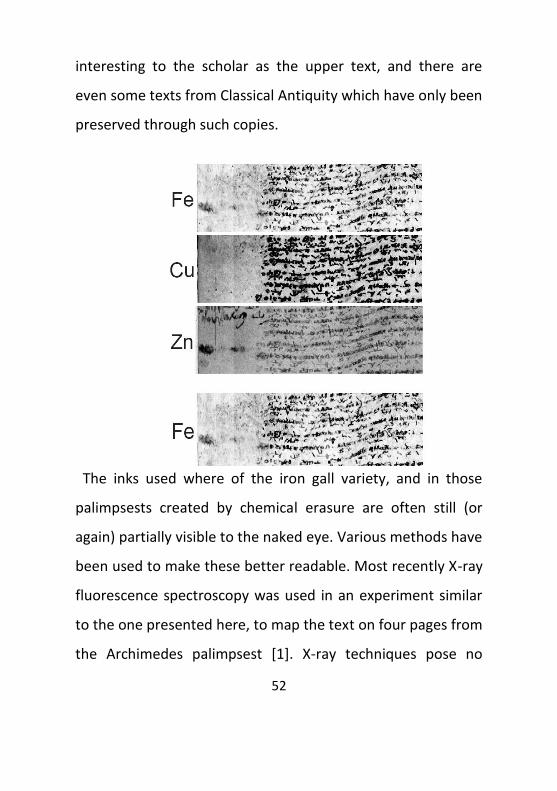
Figure 1: The element maps of iron, copper
and zinc show quite different contrast and
partially even different portions of the text.
53
significant danger to a parchment’s preservation, as radiation
damage is negligible [2].
The project presented here is a collaboration of
Teuchos – Zentrum für Handschriften- und Text-forschung
(Universität Hamburg, DFG), Uni-versitätsbibliothek Leipzig
and HASYLAB showing the possibilities of X-ray fluorescence
element mapping to enhance the contrast of upper and lower
writing concentrating on the scant impurities of the iron used
in the manufacture of the iron gall ink. Element maps as
shown in figure 1 for iron, copper and zinc have been
recorded for various trace elements in regions of visible and
erased writing. While reproduction of part of the text is often
possible using the iron contrast only, additional information
can be obtained from the contrast maps based on the iron
impurities.
References
[1] U.Bergmann, X-Ray Fluorescene Imaging of the Archimedes Palimpsest:
A Technical Summary,
<http://www.slac.stanford.edu/gen/com/slac_techrpt.html>
[2] G.Young, Effect of High Flux X-radiation on Parchment (Canadian
Conservation Institute Report No. Proteus 92195,
http://www.archimedespalimpsest.org/pdf/archimedes_f.pdf
54
RARE SILVERPOINT DRAWINGS BY REMBRANDT
IN THE FOCUS OF SR-XRF
Ina Reiche1, Silke Merchel2, Martin Radtke3, Heinrich
Riesemeier3 & Holm Bevers4
1Laboratoire du C2RMF UMR 171 CNRS, Paris, France,
2 Forschungszentrum Dresden-Rossendorf, Dresden, Germany,
3 Bundesanstalt für Materialforschung und –prüfung (BAM),
Berlin ,Germany,
4 Kupferstichkabinett Staatliche Museen zu Berlin, Berlin,
Germany.
The silverpoint drawing technique had its cumulating
period in the late Middle Ages and the Renaissance. However,
some undoubtful Rembrandt drawings were made on
prepared vellum by the master with this already obsolete
technique at the Golden Age. Among these drawings is the
best-known portrait of his wife Saskia, 1633 (KdZ1152, Berlin).
It is thus interesting to investigate these drawings. In addition
to art historic studies, it was also important to get new
insights into the graphical material employed in order to know
whether it was different from that used in former periods,
55
which in turn can give information on the genesis and dating
of the drawings.
Silverpoint drawings belong to the most valuable
treasures of graphical art collections. They are generally very
precise drawings of excellent quality. Therefore, only
completely non-destructive analytical methods are applicable.
Moreover, they need to be very sensitive because of the low
quantity of matter present in the strokes (less than some
hundreds of μg/cm2). Several preliminary tests showed that
only Particle Induced X-ray Emission (PIXE) spectroscopy and
Synchrotron radiation induced X-ray fluorescence analyses
(SR-XRF) fulfil the analytical requirements for the
investigations of these drawings meaning that they are
sensitive enough, feasible in air and require no sampling.
Synchrotron radiation induced X-ray fluorescence
results obtained at the BAMline, BESSY II, HZB, Berlin on three
Rembrandt silverpoint drawings of the collection of the
Kupferstichkabinett Staatliche Museen zu Berlin will be
presented (Reiche et al. 2006).
The chosen method will be explained as well as the
requirements for studying non-destructively valuable works of
56
art such as these silver point drawings. The main part of the
presentation focuses on the meaning of the results and
illustrates how SR-XRF analysis can reinforce art historical
assumptions on the genesis, the dating of the drawings and
their connection. Additional information can be gained from
such analytical studies on the conservation state of the
drawings.
The results will also be compared to those available on
other silverpoint drawings by Van Eyck, Dürer and the Holbein
family (Reiche und Roth, 2009, Ketelsen et al. 2005, Reiche et
al. 2004).
References:
I. Reiche und M. Roth, BBA (2009)
I. Reiche et al. Appl Phys. A (2006)
T. Ketelsen et al. The Burlington Magazine (2005)
I. Reiche et al. NIMB (2004)

57
POSTERS
58
REMBRANDT IN LABORATORY.
FACTORS OF DETERIORATION OF OIL PAINTINGS
CONTAINING LEAD WHITE
Dawid Popławski, Maria Poksioska
Department of the History of Art and Culture, Faculty of
History,
Nicolaus Copernicus University, Toruo, Poland.
The ageing processes that take place in the structure
of historical objects have not been fully recognized yet. As an
example we can give untypical damage of the paint layers
observed on the face of some oil paintings containing lead
white. A very characteristic effect of "micro-craters" was
observed. 100-200 um cavities are spread over the whole
surface of the painting and go inside as deep as the ground.
This effect was first observed in 1997 by Dutch researchers on
"The anatomy lesson of Dr Nicolaes Tulp" by Rembrandt.
The borders of the mysterious holes were
surrounded or filled with white fluorescent products of
reactions that took place in course of time in the paint layer
and the lead white ground. Their formation is connected with
59
the presence of chlorine. The compound formed – Pb3Cl4(OH)2
was identified as fiedlerite by means of a mass spectrometer
SIMS-TOF and XRD methods (P. Noble, J.Wadum, K. Groen,
R.Heeren, K.J. van den Berg, Aspects of 17th century binding
medium: Inclusions in Rembrandt's Anatomy Lesson of Dr
Nicolaes Tulp, in Art et Chimie, la couleur. Actes du congrés,
Paris 2000, s. 126-129).
Independently, unexplained fluorescence of lead
compounds was also observed in case of Gothic paintings
during restoration works in 1996. XRD diffraction allowed to
classify it as laurionite - Pb(OH)Cl (M. Poksioska, Przyczyny
szarzenia minii w malowidłach ściennych in Gotyckie
malowidła ścienne w kościele św. Jakuba w Toruniu, Toruo
2001, s. 73-83).
The present work reports the many-year experience
of the authors in the area of interpretation of this
phenomenon of significant danger to historic works of art
which appears quite common and affects not only oil
paintings. It also concerns other lead pigments. Hitherto
research on the chemical nature of these compounds and the
60
role of chlorine in their formation inspired us for laboratory
investigation in order to verify this theory.
The authors hope that drawing attention to self-
destruction processes of the works of art, in some cases the
masterpieces of the world's heritage, and the research results
will accelerate actions leading to reduction of these effects
and to provision of a restoration program for this type of
deterioration.
61
DIGITAL PROCESSING OF X-RAY RADIOGRAMS FOR
CONSERVATION PURPOSES
Joanna Sobczyk1, Piotr Frączek1, Jakub S. Prauzner-Bechcicki2,
Łukasz Bratasz1, Janusz Czop1
1 National Museum in Krakow, Krakow,Poland,
2 Centre for Nanometer-Scale and Advanced Materials
(NANOSAM) Faculty of Physics, Astronomy and Applied
Computer Science, Jagiellonian University, Krakow,Poland.
Radiography is a very useful tool in the identification,
care and understanding of historical objects. Here we would
like to report on application of digital processing and analysis
of radiographs for conservation purposes. It is shown that the
use of a computer may ease or even in some cases enable an
interpretation of the x-ray photograph. One may point, at
least, two aspects of digital processing of radiographs [1].
Namely, removal of some limitations that appear upon
preparation of analogue x-ray radiograms and gain of an
additional information about the historical object. Both
aspects are analysed further basing on examples.
A typical problem appearing in analogue x-ray
photography is film blackening (called fog) [2]. The fog
62
emerges mainly due to long film storing periods or too long
exposition times. In both cases, taking a digital photograph of
the radiograph seems to be a good solution. Human eye
adapts to the amount of light that is present at the moment of
observation, while a CCD camera sums up the light as long as
the shutter is open. Therefore with long exposure times it is
possible to read out the dark radiograph. Moreover, use of
graphical tools such as a histogram or tone curves may allow
further correction of a picture [1].
Additional information may be acquired from the
radiograph by making it more legible. It may be achieved by
adding particular sets of colours to the picture originally made
in a grey scale. The method is based on knowledge of human
visual perception. For example presenting an x-ray
photograph as a duotone yellow-to-black or white-to-blue
picture makes the observer to involve more emotions and
causes him to see more details [1].
More advanced digital image processing and computer
analysis techniques are also presented [3]. These methods not
only make visible details that can not be seen in a normal way
63
due to limits of the human eye, but also upgrade a regular
qualitative analysis into quantitative one [4]. Two examples
are presented in detail. Firstly, extraction of the painting layer
of a part of Matejko’s painting Joan D’Arc (from the National
Museum in Krakow collection) [5] is shown. Secondly,
penetration of wood strengthening substances injected into
wooden object is analysed in a quantitative manner.
[1] Digital processing of X-Ray radiograms in conservation –
perspectives, Joanna Sobczyk, Jakub S. Prauzner-Bechcicki,
Journal of Conservation-Restoration, Vol. 19 No 1-4 (72-75)
2008
[2] X-rays in Art, A. Gilardoni, R.A. Orsini, S. Taccani, Gilardoni
S.p.A. Mandello Lario (Como), Italy 1977
[3] Zastosowanie komputerowej analizy obrazu w
nieniszczących badaniach obiektów zabytkowych – prezentacja
na wybranych przykładach, Piotr Frączek, Jacek Sobczyk,
Joanna Sobczyk, Bogusław Obara, Acta Universitatis Nicolai
Copernici, Zabytkoznawstwo i Konserwatorstwo XXXVI, Zeszyt
386,Toruo 2008 (in polish only)
64
[4] Computer Image Analysis as a Method to Calculate the
Original Surface Area of the Banner, Joanna Sobczyk, Series:
Conservation Work at the National Museum in Krakow,
Krakow 2008
[5] Zastosowanie analizy obrazu w nieniszczących badaniach
obiektów zabytkowych. Wybrane przykłady. Jacek Sobczyk,
Bogusław Obara, Piotr Frączek, Joanna Sobczyk, Ochrona
Zabytków, Nr 2, 2006 (in polish only)
65
MULTI-METALLIC ARTEFACTS FROM WROCŁAW -EDXRF
STUDIES
Beata Miazga
University of Wrocław, Institute of Archaeology, Wrocław,
Poland.
Metallic artefacts from archaeological excavation in
Wrocław were analyzed by energy dispersive X-ray
fluorescence in order to establish the type of material used.
The investigated object (mostly utensils fragments) were
made using many different metals, also some artefacts are
multi-metal composition. Because of high historic value, the
artefacts need to be analyzed with non-destructive or non-
invasive methods, preferably before conservation works.
There are two main reasons for studying artefacts prior to
their conservation or restoration. One of them is the
determination of chemical composition. It is necessary to
choose the appropriate conservation program (materials and
procedures adequate for each metals). Second reason is
possibility of appearance of negative effects during
conservation (surface contamination or modification by tools
66
and chemicals). The EDXRF is an appropriate research tool for
this investigation: universal, fast, inexpensive and the analyses
do not cause damages of artefacts. The opportunity of “point-
analysis” (area - 1mm2) is very useful in the analysis of small
parts of objects (e.g. incrustation, decoration, solders, rivets).
The main disadvantage of EDXRF is a surface character, but
using simple sample preparation it is possible to overcome the
limitation of the method.
67
APPLICATION OF X-RAY METHODS FOR PIGMENTS
IDENTIFICATION IN COLLECTION OF RAFAŁ HADZIEWICZ
PAINTINGS
A. Klisioska - Kopoacz1, A. Rafalska - Łasocha2, E. Zygier1,
J. Czop1
1National Museum in Krakow,Krakow, Poland,
2Faculty of Chemistry, Jagiellonian University, Krakow, Poland.
The classical X-ray analytical methods include X-ray
fluorescence (XRF) and X-ray diffractometry (XRD). First one
gives qualitative identification of chemical elements, while
second delivers crystallographic structural analysis. During the
last decades both of them have been widely applied in
historical object studies. In this work capabilities of these
analytical techniques have been used to collect information on
the painting technique of Rafał Hadziewicz.
Rafał Hadziewicz was a Polish 19th century painter,
author of many religious paintings and portraits. He was the
best known for historic compositions, which were often
compared to art of the Italian Renaissance and European
Baroque.
68
National Museum in Krakow posses broad collection of that
author paintings, which have been conserved last year.
Identification of compounds used by Hadziewicz was
necessary before conservation treatment and it was
performed with X-ray fluorescence analysis (XRF) in
combination with X-ray diffraction analysis (XRD). Three
women portraits, painted in various life periods of painter
were chosen for analysis. The study was focused on the
pigments used by the artist in the paint layers. The inorganic
components of the ground were also analyzed.
Three paintings were studied: “Portrait of Stefania
Jarooska” (ca.1850), “Portrait of young lady” (1845) and
“Portrait of Julia Hadziewicz” (1860). Before the cleaning of
the objects, micro-samples were taken in order to study the
materials used by the painter. For XRD analysis a very small
amount of painted materials (four samples, each the size of a
few mm2) were taken from damaged areas, without damaging
the picture further. All XRD measurements were carried out
with the use of X’PERT PRO MPD diffractometer. CuK
radiation at 40kV and 30mA, a graphite monochromator and
scintillation or X’Celerator detectors were used. The
69
divergence of the beam was 0.5° or 1°, zero-background
holders were used for samples available in very small
amounts. The measurements were performed in the range
5-80° with a step size of 0.02°. The obtained diffraction
patterns were interpreted with the use of diffractometer
software (XMenu: Philips Diffraction Software) and PDF-2 or
PDF-4 databases.
Elemental compositions of all objects were found by using an
ArtTAX® μXRF spectrometer (Bruker AXS Microanalysis,
Germany) equipped with a Rhodium X-ray tube (50kV, 500μA,
50 keV, in air, 300s).
Performed analysis indicated that white lead, naples
yellow, vermilion, prussian blue and natural iron oxide
pigments were used for all three paintings. Additionally cobalt
blue was detected in the case of “Portrait of Stefania
Jarooska” and “Portrait of Julia Hadziewicz”, while chromium
green was applied for “Portrait of Stefania Jarooska” and
“Portrait of young lady”. Besides, manganese violet was found
in “Portrait of Julia Hadziewicz”.
70
The combined use of XRF and XRD techniques enabled
us to identify inorganic components of the painting materials
used by Rafał Hadziewicz. Performed analysis allowed to
understand deeply the craftsmanship and technology used by
this painter.
71
X-RAY BASED ANALYSIS OF THE METAL THREADS
IN HISTORICAL TEXTILES
A. Klisioska- Kopacz, M. Włodarczak, A. Prokopowicz,
Ł.Bratasz
National Museum in Krakow, Krakow, Poland.
Historical textiles were decorated by metal threads,
since thousands years. The term metal thread includes every
type of thin, like-yarn textile decoration as strips and wires,
made of solid metals, metal-coated organic materials or the
combination of these with natural or man-made fibers. Metal
strips can be made of pure gold, gold alloyed with silver,
gilded or gild-silvered copper and gold-like copper alloys. Pure
gold, silver or gilded silver were used from early times in
manufacture of valuable materials, signifying wealth and
social status. Copper, brass or tin were often coated with a
thin layer of silver or gold [1, 2].
The knowledge about the composition of the metal threads in
old textile objects gives very valuable information about the
ancient manufacturing techniques and also about the
appropriate treatment for cleaning and conservation. The aim
72
of the study was to determine the composition of the various
metal threads of textiles dated to different epochs (between
15th and 19th centuries) and originating from Europe and Asia.
Performed study allows to create metal composition database
for identification and classification of museum textiles of
various provenience.
The investigation of textiles was carried out by
analytical non-destructive methods, as XRF and Raman
spectroscopy. Elemental compositions were found by using an
ArtTAX® μXRF spectrometer (Bruker AXS Microanalysis,
Germany) equipped with a Rhodium X-ray tube (50kV, 500μA,
50 keV, in air, 120s). Raman spectra were acquired by using a
portable Raman spectrometer, DeltaNu, equipped with a
diode 785 nm laser.
Performed analysis indicated that flax and silk textiles
were decorated by metal threads. In the case of European
textiles the elemental composition of metal threads changed
through the ages from gold in 15th century, gold with silver in
17th century and finally gilded copper in 18th century. Similar
trend was observed for Asiatic historical objects. Gold and
73
gilded silver metal strips were used in 18th century, while tin
strips were found in 19th century Japanese hanging scroll.
Beside, additional elements as iron, antimony and nickel were
detected.
Non-invasive μXRF technique was found suitable for
characterization of the elements in the metal strip
decorations. The compositional analysis of the metal threads
in historical textiles provided significant information as
regards the technology of their manufacture as well as the
economical value and consequently the social rank that they
represent.
This work was partially supported by a grant “Direct monitoring of strain for protection of historic textiles and paintings on canvas” from Iceland, Liechtenstein and Norway through the EEA Financial Mechanism
*1+ M. Jaró, Metal threads in historical textiles, in: Molecular and
Structural Archeology: Cosmetics and Therapeutic Chemicals, G.
Tsoucaris and J. Lipkowski (eds.), 2003, Kluwer Academic Publishers,
pp. 163-178
[2] A.G. Nord, K. Tronner, A note on the analysis of gilded metal
embroidery threads, Studies in Conservation, 45 (2000) 274-279
74
WITKACY’S PASTELS ANALYSIS FOR SAFE ANOXIA STORAGE
A. Klisioska-Kopacz1, J. Sobczyk1, J. Thomas, Piotr Frączek1, D.
Godyo1, Ł. Bratasz1,
J. Bagniuk2, T. Łojewski2
1National Museum in Krakow, Krakow, Poland, 2Faculty of Chemistry, Jagiellonian University, Krakow, Poland
Due to photofading, pastels on coloured-paper supports are
some of the most sensitive and unstable museum objects. The
National Museum in Krakow possesses a broad collection of
this type of light sensitive object, and their preservation as
well as accessibility through exhibition are co-concerns.
Oxygen-free storage and display have been proposed as a
means to achieve both goals. However, before application of
anoxic conditions, identification of the pigments used in art
object is required.
This work presents the results of studies performed on three
Witkacy pastels. Objects created in different years were
chosen: “Portrait of Władysław Dunin-Borkowski” (1920),
“Portrait of Stefan Szuman” (1929) and “Portrait of Anna
Friedrichowa” (1936). Pastels by Witkacy are interesting due
75
to their coloured-paper supports, which are very light
sensitive.
Analysis of pigments was performed by X-ray fluorescence
spectroscopy (ArtTAX, Bruker), while the paper support colour
changes were measured by UV/VIS spectrophotometer
(OceanOptics USB 4000). Additionally, the objects were also
studied in analytical light (VIS, UV).
Colour measurements indicate that Witkacy used the same
paper support in the case of “Portrait of Władysław Dunin-
Borkowski” and “Portrait of Stefan Szuman”. Analysis of
exposed and unexposed regions of the support indicates that
significant colour change has occurred in the past. XRF analysis
showed that painter used both inorganic and organic pastels,
however only inorganic pigments could be recognize by this
method. Chrome yellow was detected in all objects, while
vermillion was found in “Portrait of Władysław Dunin-
Borkowski” and “Portrait of Stefan Szuman”, and Schelle’s
green was present in “Portrait of Stefan Szuman” and “Portrait
of Anna Friedrichowa”.
76
XRD INVESTIGATIONS OF COMMERCIALLY AVAILABLE
PRUSSIAN BLUE PIGMENTS AND OIL PAINTS
Alicja Rafalska-Łasocha1, Katarzyna Podulka1, Roman
Dziembaj1, Wiesław Łasocha1,2
1Faculty of Chemistry, Jagiellonian University, Krakow, Poland,
2Institute of Catalysis and Surface Chemistry PAS, Krakow,
Poland.
Prussian blue is thought to be the earliest of the
modern synthetic pigments. Discovered in 1704 by Diesbach in
Berlin, it was soon manufactured in France (Paris), England
and other countries. It was used as a pigment in oil paintings,
printing inks, typewriter ribbons, carbon paper and sometimes
also cosmetics. Since the middle of the twentieth century it
has been replaced by phthalocyanine blue.
The chemical formula of Prussian blue is Fe4[Fe(CN)6]3
xH2O or KFe[Fe(CN)6] xH2O. However, Kremer Pigments
Company produces this pigment as an ammonium complex
with the formula NH4Fe[Fe(CN)6] . xH2O.
77
The aim of the presented study was to perform the
phase analysis of commercially available Prussian blue
pigments and oil paints. The obtained results may be helpful
for identification of this pigment in the scientific analysis of
works of art.
We have investigated Prussian blue powder pigments
produced by the Maimeri, Schmincke and Kremer companies.
The results showed that the pigment produced by Schmincke
was Fe[Fe(CN)6+·4H2O [PDF 74-9174] whereas the powder
patterns of the other two samples were similar to each other.
We have found no reference data for these patterns in the
PDF 4 file, that is why the XRPD data for phase
characterization of NH4Fe[Fe(CN)6].H2O (Prussian blue
produced by Kremer Company) were elaborated. The
compound crystallizes in the cubic system, with lattice
parameters a=b=c=10.232(1)Å and Fm3m space group.
In commercially available blue oil paints the pigment
Prussian blue, due to its high tinting strength, is mixed with
various, usually white, compounds. We performed XRD phase
analysis of blue oil paints named Prussian blue produced by
78
Pollena-Astra, Maimeri, Daler-Rowney, Renesans, Royal
Talens, and Phoenix and established their phase compositions.
It was found that only Pollena-Astra produces Prussian
blue oil paint which contains Fe4[Fe(CN)6]3 . 14H2O. [PDF 73-
0689] without any crystaline fillers. In other paints ferric
ferrocyanide hydrate was accompanied by CaCO3 [PDF 83-
0578], (Ba,Pb)SO4 [PDF 77-0432] or Al2(Si2O5)·(OH)4 [PDF 89-
6538].
We have also investigated a blue powder pigment
named Prussian blue (no inw.MNK IV - V 1024/ 1- 16) which
belonged to well-known Polish painter Henryk Siemiradzki. It
was found that the sample contained Fe4[Fe(CN)6]3 . 4H2O
[PDF 74-9174] and gypsum CaSO4·2H2O [PDF 06-0046].
All XRD measurements were carried out with the use of an
X’PERT PRO MPD diffractometer. CuK radiation at 40kV and
30mA, a graphite monochromator and scintillation or X’Celerator
detectors were used. The divergence of the beam was 0.5°. The
measurements were performed in the 2range 3-80° with a step
size of 0.025° or 0.02°. The obtained diffraction patterns were
interpreted with the use of diffractometer software (X’Menu: Philips

79
Diffraction Software) and PDF-2 or PDF-4 databases. Details of the
performed studies will be presented during the Meeting.
80
XRD and SEM/EDX INVESTIGATIONS OF THE CORROSION
PRODUCTS OF METAL THREADS FROM TAPESTRY OF
SIGISMUND AUGUSTUS
Alicja Rafalska-Łasocha1, Jerzy Holc2, Anna Bielaoska3, Wiesław
Łasocha1,3
1Faculty of Chemistry Jagiellonian University, Krakow, Poland,
2Wawel Royal Castle, Krakow, Poland,
3Institute of Catalysis and Surface Chemistry PAS, Krakow, Poland.
The Sigismund Augustus collection, partially exhibited in
Wawel Castle in Krakow, consists of 136 tapestries, commissioned in
the middle of the 16th century from the most prominent workshops
in Brussels on the occasion of the king’s wedding *1+. The materials
used were wool, silk and metal threads for the weft. Some areas of
the design (mainly those depicting clothing) contain precious metal
threads with a fibrous, usually silk core.
The Wawel collection of tapestries includes 19 pieces with
subjects taken from the Book of Genesis. Seven pieces depict The
Story of the First Parents, eight pieces The Story of Noah and four
pieces The Story of the Building of the Tower of Babel. The
tapestries are about 4.8 meters high and up to 8.8 meters wide.
81
During the conservation work after cleaning The Building of
the Tower of Babel tapestry it was found that the metal threads
were partially corroded. Samples of damaged threads as well as
corrosion products (black powder) were investigated with the use of
XRD powder diffractometry and SEM techniques.
All XRD measurements were carried out with the use of an
X’PERT PRO MPD diffractometer. CuK radiation at 40kV and
30mA, a graphite monochromator and scintillation or X’Celerator
detectors were used. The divergence of the beam was 0.5°, or 1° for
samples available in very small amounts. The measurements were
performed in the 2range 4-80° with a step size of 0.02°. The
obtained diffraction patterns were interpreted with the use of
diffractometer software (X’Menu: Philips Diffraction Software) and
PDF-2 or PDF-4 databases.
Scanning electron microscopic studies of the metal threads
were carried out by means of a JEOL JSM – 7500F field emission
scanning electron microscope.
The obtained results allowed us to establish the elemental
composition of the metal threads as alloys of silver and gold, or
silver, gold and copper. As one might have expected the main
component of the corrosion product in the case of such
82
composition was Ag2S. The details of the investigations described
above will be presented in the poster.
[1] M. Piwocka, The tapestries of Sigismund Augustus, Wawel Royal
Castle State Art Collections, Krakow 2007
[2] A.M. Hacke, C.M.Carr., A. Brown, D. Howell, Investigation into
the nature of metal threads in a Renaissance tapestry and the
cleaning of tarnished silver by UV/Ozone (UVO) treatment.,
JOURNAL OF MATERIALS SCIENCE 38 (2003) 3307 – 3314
83
SYNCHROTRON X-RAY POWDER MICRO-DIFFRACTION and
MICRO-FLUORESCENCE FOR IDENTIFICATION OF GREEN
PIGMENTS IN the GOTHIC ALTAR PAINTINGS FROM THE
MALOPOLSKA REGION
Alicja Rafalska-Łasocha1, Marta Grzesiak1, Zofia Kaszowska2,
Thomas Wróblewski3, Dariusz Zając3, Andre Rothkirch3 and
Wiesław Łasocha1,4
1Faculty of Chemistry Jagiellonian University, Krakow, Poland,
2 Academy of Fine Arts, Krakow, Poland,
3Hamburger Synchrotronstrahlungslabor HASYLAB at
Deutsches Elektronen-Synchrotron DESY, Notkestr. 85,
Hamburg, Germany,
4Institute of Catalysis and Surface Chemistry PAS, Krakow,
Poland.
The aim of the presented work was to investigate the
samples of green paint taken from five Gothic altars from the
Malopolska region. The oldest paintings in this group are the
altar wings of a triptych from Zator (one of the wings is in the
84
parish church in Zator, the other in the National Museum in
Krakow). They were painted in the second half of the 15th
century by an unknown master. Holly virgins depicted in the
painting represent the end of the early years of Malopolska
panel paintings, in which the tradition of international style of
the end of the 15th century (soft arrangement of drapery) is
present. The painting workshop where the wings from Zator
were executed is recognizable by the physiognomy of the
depicted characters (swollen eyes and thick necks).
All the investigated samples were collected from
unobtrusive areas of the paintings. Each sample was divided
into two similar pieces, one of which was then embedded in
polyester resin. X-ray diffraction and micro X-ray fluorescence
measurements were performed at DORIS III of
Synchrotronstrahlungslabor HASYLAB in Deutsches
Elektronen-Synchrotron DESY (Hamburg, Germany).
On the basis of optical microscopic study the
conservators suspected the presence of verdigris in the
samples. Verdigris is a name used to describe a group of
copper salts of acetic acid which range in colour from green,
85
via green blue and blue green, to blue. Different kinds of
verdigris may be divided into two groups: basic verdigris and
neutral verdigris.
Basic verdigris may contain one or a mixture of the following
acetates: [Cu(CH3COO)2]2Cu(OH)2.5H2O (blue),
Cu(CH3COO)2Cu(OH)2.5H2O (blue), Cu(CH3COO)2[Cu(OH)2]2
(blue) and Cu(CH3COO)2[Cu(OH)2]3.2H2O (green). The formula
of neutral verdigris is Cu(CH3COO)2.H2O [1]. In powder
diffraction data file PDF 4+ one can find 10 entries for copper
acetate; however, none is a basic copper acetate.
Results obtained from XRF analysis show that all
green pigments in the investigated samples are copper
compounds (high intensities of copper bands) and also
indicate the presence of lead and tin.
In our investigation we prepared verdigris in the lab
according to a historical recipe, to provide a reliable reference
pattern. Comparison of diffraction patterns of prepared
verdigris with the patterns of investigated samples shows that
most peaks fit verdigris. In the case of the sample from
Grywałd there are some peaks which could belong to
86
malachite. Additionally in the sample from Łącko some peaks
may indicate that lead tin yellow type I might have been used
by the painter.
1. Kühn H. Identyfication of the materials of paintings:
Verdigris and copper resinate, Studies in Conservation 15
(1970), 12-36
87
CONSERVATIONAL ANALYSIS OF THE SIGISMUND BELL
Marcin Biborski1 and Andrzej Biborski2
1 Institute of Archeology, Faculty of History, Jagiellonian
University, Krakow, Poland
2Faculty of Physics, Astronomy and Informatics, Jagiellonian
University, Krakow, Poland
The Sigmund Bell was cast of bronze in 1520 in Krakow
by a nurembergian bell-founder Hans Bohem. The height of
the bell is equal to 2.58 m, its diameter at the bottom is 2.42
m, and the periphery in the widest place extends to almost 8
m. The thickness of the walls of its body is in the range
between 7 and 21 cm. The length of the clapper is 218.5 cm
and its weight is 323 kg. The body itself weights about 10 t and
the total weight of the whole bell with the clapper and yoke is
almost 13 t.
In the year 2000 Sigismund Bell became silent for over
three months because of the fracture of its steel clapper.
Upon preliminary investigations, it was decided build a new
clapper. It has been agreed that the clapper ought to be
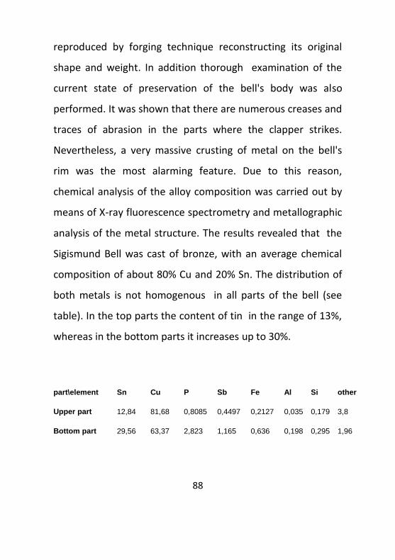
88
reproduced by forging technique reconstructing its original
shape and weight. In addition thorough examination of the
current state of preservation of the bell's body was also
performed. It was shown that there are numerous creases and
traces of abrasion in the parts where the clapper strikes.
Nevertheless, a very massive crusting of metal on the bell's
rim was the most alarming feature. Due to this reason,
chemical analysis of the alloy composition was carried out by
means of X-ray fluorescence spectrometry and metallographic
analysis of the metal structure. The results revealed that the
Sigismund Bell was cast of bronze, with an average chemical
composition of about 80% Cu and 20% Sn. The distribution of
both metals is not homogenous in all parts of the bell (see
table). In the top parts the content of tin in the range of 13%,
whereas in the bottom parts it increases up to 30%.
part\element Sn Cu P Sb Fe Al Si other
Upper part 12,84 81,68 0,8085 0,4497 0,2127 0,035 0,179 3,8
Bottom part 29,56 63,37 2,823 1,165 0,636 0,198 0,295 1,96
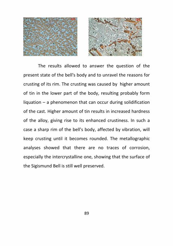
89
The results allowed to answer the question of the
present state of the bell's body and to unravel the reasons for
crusting of its rim. The crusting was caused by higher amount
of tin in the lower part of the body, resulting probably form
liquation – a phenomenon that can occur during solidification
of the cast. Higher amount of tin results in increased hardness
of the alloy, giving rise to its enhanced crustiness. In such a
case a sharp rim of the bell's body, affected by vibration, will
keep crusting until it becomes rounded. The metallographic
analyses showed that there are no traces of corrosion,
especially the intercrystalline one, showing that the surface of
the Sigismund Bell is still well preserved.
90
AN INTERESTING DILEMMA – A SMALL LANDSCAPE SIGNED
REMBRANDT F. 1627. A DEPOSIT IN THE JAGIELLONIAN
UNIVERSITY MUSEUM COLLECTION
Anna Jasioska, Jolanta Pollesch, Paweł Karaszkiewicz, Beata
Skalmierska
Jagiellonian University Museum, Krakow, Poland.
In 1995 the Jagiellonian University Museum in Cracow
received for long-term deposit a painting that depicts a
Landscape with a mill; oil on oak board; dimensions: 23.2 x
31.5 cm. The painting has a signature on the bottom edge,
almost in the centre: Rembrandt f.1627. The Landscape was
bought by its owner at an antiquarian market in Stockholm. At
the time of purchase, it had a different frame. The frame had
covered the signature, which was discovered only when the
frame was changed for the present one. On the face of the
painting, there is a clearly visible print of the old frame that
overlaps with the signature.
The scene shows a large mill with a granary with a mill
wheel and gates in front. When seen in large close-up, the
91
painting reveals a rich diversity of detail which cannot be seen
with the naked eye. The work has survived in good condition.
It was painted on an oak board cut radially. The reverse side of
the board is unbevelled. In the right upper part, a piece of
paper with information printed in French is attached and
reads that the painting was a part of the Duke of Nemours’s
collection, and then was kept in Hombourg (name of the
owner illegible). Next to it, there is a notice written in red pen:
by Rembrandt. At the bottom, centrally, there is a barely
visible text written in pen (?): "Rembrandt était agé de 21 ans
quand il fit ce tableau, a l'age de 18 ans il était déja celébre."
A painting with a signature like this obviously aroused
concern and interest, the more so because it seemed to be an
interesting Dutch period piece already during the initial
examination. The decision was taken to investigate it closely.
On account of that, a number of examinations were carried
out to determine the authenticity of the painting as a Dutch
work of art from the 17th century and investigate it in terms of
technological and graphological aspects against works by
Rembrandt. The examinations were carried out in the
Conservation Workroom of the Jagiellonian University
92
Museum in 1997 and 1998, with the assistance of the
Academy of Fine Arts in Cracow and experts from Warsaw, the
Hague and Germany.
The painting was examined in sodium light, UV light, and
infrared light (the observation under the IM 3
infrastereoscopic microscope revealed a sketch of the
composition; particularly compelling is the outline of the
perspective sketch) and subjected to an X-ray inspection,
dendrological and dendrochronological examinations, an
examination of pigments and graphological analyses
performed by I.H. Hardy.
The performed examinations prove the authenticity of the
painting as a Dutch work of art from the 17th century. None of
the technological examinations radically excluded the
Landscape as a potential early work by Rembrandt. It only
remains to conduct a thorough stylistic and comparative
analysis, which – based on the results of the completed
examinations – should give a final answer to the questions:

93
- is it a work by the Master?
- is it a work by one of the apprentices from his studio or his
followers?
- is it a forgery created later than the signature suggests?
- is it a work by an unknown artist which reflects the so-called
Zeitgeist?

94
LIST OF PARTICIPANTS
95
1. Petr Bezdicka, Institute of Inorganic Chemistry of the ASCR, v.v.i., c. p. 1001, CZ-25068 Husinec-Rez, Czech Republic
e-mail: [email protected]
2. Andrzej Biborski Faculty of Physics,Astronomy and Informatics, ul. Reymonta 4, 30-059 Krakow, Poland
3. Marcin Biborski Institute of Archeology, Faculty of History, Jagiellonian
University, ul. Gołębia 11, 31-007 Krakow, Poland
4. Fatima-Zahra BOUJRHAL, Faculté des Sciences et Techniques, B.P. 523, Béni Béni Mellal, Marocco,
e-mail: [email protected]
5. Joanna Czernichowska, Academy of Fine Arts, Warsaw, Faculty of Conservation and Restoration of Works of Art, Al.Wojska Polskiego 2A, 01-524 Warsaw, Poland
e-mail: [email protected]
6. Janusz Czop, National Museum in Krakow, al. 3 Maja 1, 30-062 Krakow, Poland
e-mail: [email protected]

96
7. Dorota Dec , The Princes Czartoryski Museum -the Branch of National Museum in Kraków , ul.Św.Jana 19 ,31-017 Kraków , Poland
e-mail: [email protected]
8. Regina Dmowska, Royal Castle in Warsaw, Plac Zamkowy 4, 00-277 Warsaw, Poland
e-mail: [email protected]
9. Piotr Fraczek, National Museum in Krakow, 1, 3 Maja Avenue, 30-062 Krakow, Poland
e-mail: [email protected]
10. Leif Glaser, HASYLAB/DESY, Notkestr.85 - 22607 Hamburg - Germany,
e-mail: [email protected]
11. Anna Grochowska-Angelus, Cracow National Museum, Cracow, Św. Jana 19, Poland
e-mail: [email protected]
97
12. Marta Grzesiak, Faculty of Chemistry Jagiellonian University, 30-060 Krakow, Ul. Ingardena 3, Poland,
e-mail: [email protected]
13. Jerzy Holc, Zamek Królewski na Wawelu, Wawel 5 31-001 Kraków, Poland
e-mail: [email protected]
14. Anna Jasioska, Museum of the Jagiellonian University, ul. Jagiellooska 15, 31-010 Kraków, Poland
e-mail: [email protected]
15. Zofia Kaszowska, Academy of Fine Arts, 30-052Kraków ul. L ea 27-29, Poland
e-mail: [email protected]
16. Regina Klincewicz Krupinska, Muzeum Narodowe w Krakowie, Krakow, Pałac bpa E. Ciołka, ul. Kanonicza 17, Poland
e-mail: [email protected]
17. Anna Klisinska-Kopacz, National Museum in Krakow, al. 3 Maja 1, 30-062 Krakow, Poland,
e-mail: [email protected]
98
18. Roman Kozłowski, Institute of Catalysis and Surface Chemistry, Polish Academy of Sciences, ul. Niezapominajek 8, 30-239 Kraków, Poland
e-mail: [email protected]
19. Wieslaw Lasocha, Faculty of Chemistry Jagiellonian University, 30-060 Krakow, Ul. Ingardena 3, Poland,
e-mail: [email protected]
20. Claudia Laurenze-Lansberg, Staatliche Museen Berlin. Gemäldegalerie, Germany,
e-mail: [email protected]
21. Beata Miazga, University of Wrocław, Institute of Archaeology, Szewska 48, 50-139 Wrocław, Poland
e-mail: [email protected]
22. Petria Noble, Royal Picture Gallery Mauritshuis, Korte Vijverberg 8, 2513 AB The Hague, The Netherlands,
e-mail: [email protected]
23. Katarzyna Novljakovid, Cracov National Museum , Cracov , Św.Jana 19, Poland
e-mail: [email protected]
99
24. Anna Nowicka, Akademy of Fine Arts in Warsaw, Warszawa, ul. Krakowskie Przedmieście 5, Poland
e-mail: [email protected]
25. Marcin Oszajca, Faculty of Chemistry Jagiellonian University, 30-060 Krakow, Ul. Ingardena 3, Poland,
e-mail:[email protected]
26. Katarzyna Podulka, Faculty of Chemistry Jagiellonian University, 30-060 Krakow, Ul. Ingardena 3, Poland,
e-mail: [email protected]
27. Maria Poksioska, Department of the History of Art and Culture, Faculty of History, Nicolaus Copernicus University, Toruo, ul. Mickiewicza 121, Poland
e-mail: [email protected]
28. Jolanta Pollesch, Museum of the Jagiellonian University, ul. Jagiellooska 15, 31-010 Kraków, e-mail: [email protected]
29. Dawid Popławski, Freelance Conservator, Copenhagen, Denmark
e-mail: [email protected]
100
30. Alicja Rafalska-Łasocha, Faculty of Chemistry Jagiellonian University, 30-060 Krakow, Ul. Ingardena 3, Poland,
e-mail: [email protected]
31. Ina Reiche, Laboratoire du C2RMF UMR 171 CNRS, Paris, France,
e-mail : [email protected]
32. Maria Gabriela Rogóż, Academy of Fine Arts, 30-052Kraków ul. L ea 27-29, Poland
e-mail: [email protected]
33. Henk Schenk University of Amsterdam, Laboratory for Crystallography, Nieuwe Achtergracht 166, 1018 WV Amsterdam, The Netherlands,
e-mail: [email protected]
34. Beata Skalmierska, Museum of the Jagiellonian University, ul. Jagiellooska 15, 31- 010 Kraków, Poland
e-mail: [email protected]
35. Joanna Sobczyk, National Museum in Krakow, 1, 3 Maja Avenue, 30-062 Krakow , Poland
e-mail: [email protected]
101
36. Zbigniew Sojka, Faculty of Chemistry Jagiellonian University, 30-060 Krakow, Ul. Ingardena 3, Poland,
e-mail: [email protected]
37. Grażyna Stochel Faculty of Chemistry Jagiellonian University, 30-060 Krakow, Ul. Ingardena 3, Poland,
e-mail: [email protected]
38. Szymon Stolarek, PANalytical B.V. Branch Office Poland, Al. Jerozolimskie 195B 02-222 Warsaw, Poland
e-mail: [email protected]
39. Silvie Svarcova, Institute of Inorganic Chemistry of the ASCR, v.v.i., c. p. 1001, CZ-25068 Husinec-Rez, Czech Republic
e-mail: [email protected]
40. Stanislaw Waltos, Museum of the Jagiellonian University, ul. Jagiellooska 15, 31-010 Kraków, Poland
41. Andrzej Węsek, Testchem, 44-370 Pszów, ul.Niepodległości 82d, Poland
e-mail: [email protected]
102
42. Joanna Winiewicz-Wolska, Zamek Królewski na Wawelu, Wawel 5 31-001 Kraków, Poland
e-mail: [email protected]
43. Elżbieta Zygier National Museum in Krakow, 1, 3 Maja Avenue, 30-062 Krakow , Poland
44. Anke Zürn, ETH Zurich, Wolfgang-Pauli-Strasse 10, CH-8049 Zürich, Switzerland, e-mail: [email protected]
45. Barbara Krajewska Faculty of Chemistry Jagiellonian University,
30-060 Krakow, Ul. Ingardena 3, Poland,
e-mail: [email protected]
46. Anna Latosinska Faculty of Chemistry Jagiellonian University, 30-060 Krakow, Ul. Ingardena 3, Poland,
47. Jacek Kumański,
Museum of the Jagiellonian University, ul. Jagiellooska 15, 31-010 Kraków, Poland
103
48. Matusz Biborski,
Museum of the Jagiellonian University, ul. Jagiellooska 15, 31-010 Kraków, Poland
49. Magdalena Markowicz
Faculty of Chemistry Jagiellonian University, 30-060 Krakow, Ul. Ingardena 3, Poland,
50. Łukasz Bratasz
Institute of Catalysis and Surface Chemistry, Polish Academy of Sciences, ul. Niezapominajek 8, 30-239 Kraków, Poland
51. Anna Mikołajska
Academy of Fine Arts, 30-052Kraków ul. L ea 27-29, Poland

104
-Notes -

105
-Notes -

106
-Notes -

107
-Notes -
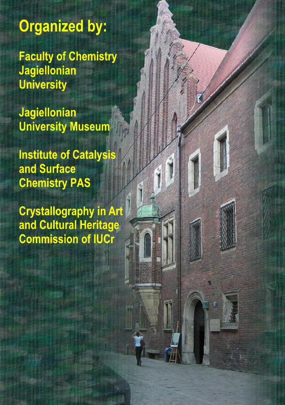
108