Embed Size (px)
Citation preview
1770
Abstract. – OBJECTIVE: The study was de-signed to investigate the JAK2/STAT3 signaling pathway in pancreatitis and its association with inflammation and cell death to provide a poten-tial treatment method for pancreatitis.
MATERIALS AND METHODS: The rat pancre-atic acinar AR42J cells were used for the study, and they were transfected with JAK2 and STAT3 siRNAs to mimic knockdown condition. Cerulein was used to treat AR42J cells. Western blot and ELISA were employed to detect the expression of related proteins. Flow cytometry was done to analysis the necrosis of AR42J cells.
RESULTS: In this study, we found that cell death and the secretion of IL-6 and TGF-β1 were significantly increased, and the JAK2/STAT3 signaling pathway was activated in cerulein-in-duced AP. To determine the role of JAK2 and STAT3, JAK2 siRNA and STAT3 siRNA were used to block JAK2 and STAT3, respectively. The levels of IL-6 and TGF-β1 levels in the me-dium were lower in JAK2 siRNA and STAT3 siR-NA-treated cells compared with controls. Flow cytometry analysis showed that the level of cell death, expression of cleaved caspase-3, and the release of LDH were decreased following JAK2 siRNA and STAT3 siRNA treatment.
CONCLUSIONS: These findings point to a novel role for the JAK2/STAT3 signaling path-way in the progression of cerulein-induced AP.
Key Words:Acute pancreatitis, JAK2, STAT3, Inflammation, Cell
death.
Introduction
Acute pancreatitis (AP) is one of the most com-mon disorders of the exocrine pancreas which is characterized by the dysregulation of digestive enzyme production and secretion1,2. Although
the underlying mechanisms have not been fully elucidated and there is no specific and effective therapy3,4, AP is believed to originate in the injured acinar cells due to uncontrolled inflam-mation, which contributes to the death of acinar cells4. Indeed, inflammation and parenchymal cell death are prominent pathological phenotypes in patients with AP5.
Cerulein is an ortholog of the intestinal hor-mone cholecystokinin and is one of the most-char-acterized and widely used agents in experimental animal models of pancreatitis5-7. Accumulating evidence demonstrates that cerulein induces the death of acinar cells, the formation of edema, and the infiltration of inflammatory cells into the pancreas in mice and rats8,9. The mechanism of cerulein action in vivo involves the promotion of oxidative stress and the release of pro-inflam-matory cytokines10-12. However, the effects and molecular mechanisms of cerulein have not been extensively explored through in vitro models.
It is well-known that the Janus kinase 2 (JAK2) signal transducer and the activator of transcrip-tion 3 (STAT3) pathway plays a key role in modu-lating immune responses in several cell types13,14. This pathway generally transduces signals from activated receptors or intracellular kinases to the nucleus, thereby activating and regulating gene transcription. Its target genes include cy-tokines, adhesion molecules, and other inflam-matory mediators13,15,16. On the other hand, JAK2 and STAT3 proteins are stimulated and activated by cytokines17,18. Considering that AP is caused by the production and release of various pro-in-flammatory cytokines, the JAK2/STAT3 pathway is an important regulator of inflammation and cell death in the pancreas when exposed to an inflammatory stimulus like cerulein19,20. In this
European Review for Medical and Pharmacological Sciences 2019; 23: 1770-1777
W.-D. CHEN1, J.-L. ZHANG1, X.-Y. WANG1, Z.-W. HU1, Y.-B. QIAN2
1Department of Emergency Surgery, The 1st Affiliated Hospital of Anhui Medical University, Hefei, China2Department of Hepatopancreatobiliary Surgery, The 1st Affiliated Hospital of Anhui Medical University, Hefei, China
Corresponding Author: Yeben Qian, MD; e-mail: [email protected]
The JAK2/STAT3 signaling pathway is required for inflammation and cell death induced by cerulein in AR42J cells
JAK2/STAT3 signaling pathway is required for cerulein in AR42J cells
1771
study, we investigated the effect of cerulein on AR42J cells and the specific role of the JAK2/STAT3 pathway in the AP model. We found that cerulein directly activated the JAK2/STAT3 pathway leading to the production of IL-6 and TGF-β1. Downregulating the expression of JAK2 and STAT3 by siRNAs abolished the cerulein-in-duced production of IL-6 and TGF-β1, and in-hibited cell death in AR42J cells. These results suggest that the JAK2/STAT3 signaling pathway mediates the cerulean-induced cell death and in-flammation in AR42J cells.
Methods and Materials
Cell Culture and TransfectionThe rat pancreatic acinar AR42J cells were
cultured in F-12K medium supplemented with 20% fetal bovine serum (FBS, Gibco, Rockville, MD, USA) and antibiotics (100 U/mL penicil-lin and 100 μg/mL streptomycin) at 37°C in a humidified atmosphere containing 5% CO2. To develop an in vitro model of acute pancreatitis, AR42J cells were treated with cerulein (10-8 M) and the expression of JAK2, phospho-JAK2, STAT3, and phospho-STAT3 were determined at a different time-points after cerulein treatment. Transfection of AR42J cells with siRNAs was do-ne using Lipofectamine 2000 (11668019, Thermo Fisher Scientific, Waltham, MA, USA) according to the manufacturer’s instructions. The plasmid constructs encoding JAK2 and STAT3 siRNAs were used for the transfection. The sequence of JAK2 siRNA (sc-270385) and STAT3 siRNA (sc-270027) and non-targeting siRNA pool (sc-36869) used as a control were purchased from Santa Cruz Biotechnology (Santa Cruz, CA, USA). 48 h after transfection, the effect of siRNAs silencing was tested by Western blot.
Western BlotFor Western blot analysis, the cells were ho-
mogenized in lysis buffer (1% Nonidet P-40, 20 mM Tris, pH 8.0, 137 mM NaCl, 10% glycerol, 1 mM phenylmethylsulfonyl fluoride (PMSF), sodium butyrate 1 mM, and protease inhibitors) at 4°C. After removal of cellular debris by cen-trifugation, the supernatant was collected, and protein concentration in the lysate was mea-sured by the Bradford assay (Bio-Rad, Hercules, CA, USA). 50 μg of each sample was boiled in the presence of sample buffer for 5 minutes be-fore separation on 10% sodium dodecyl sulphate
(SDS) polyacrylamide gel. Thereafter, the pro-teins were transferred to polyvinylidene diflu-oride (PVDF) membranes (Millipore, Billerica, MA, USA). The immunoblots were blocked with 5% nonfat dry milk dissolved in phosphate-buff-ered saline (PBS) for 60 min. The membranes were then incubated overnight at 4°C with pri-mary antibody: rabbit anti-JAK2 (1:500, Santa Cruz Biotechnology, Santa Cruz, CA, USA), rabbit anti-phospho-JAK2 (1:1000, Cell Signal-ing, Danvers, MA, USA), rabbit anti-STAT3 (1:1000, Upstate Biotechnology, Lake Placid, NY, USA), rabbit anti-phospho-STAT3 (1:1000, Upstate Biotechnology, Lake Placid, NY, USA), rabbit anti-caspase-3 (1:1000, Upstate Biotech-nology, Lake Placid, NY, USA), rabbit anti-β-ac-tin (1:2500, Abcam, Cambridge, MA, USA). Pri-mary antibody incubation was followed by three washes (5 min, rocking, at room temperature) in PBST (PBS containing 0.2% Tween 20) before incubation with goat anti-rabbit secondary anti-body conjugated to horseradish peroxidase. The blots were washed three times and the protein expression was detected with Bio-Rad Imaging Systems (Hercules, CA, USA). β-actin was used as the loading control, and the optical densities of protein bands were quantitatively analyzed with Quantity One software (Bio-Rad, Hercu-les, CA, USA).
Biochemical AssaysThe levels of IL-6, TGF-β1, and LDH in the
medium were determined by enzyme-linked im-munosorbent assay (ELISA) kits (IL-6: R&D System, Minneapolis, MN, USA; TGF-β1: R&D System, Minneapolis, MN, USA). All the pro-cedures were performed according to the manu-facturer’s instructions. At the end of the assays, fluorescence intensities of the 96 well microplates were read by an assay reader (Tecan, Männedorf, Switzerland). After averaging the results of dupli-cate wells, the value of IL-6 and TGF-β1 of each sample was calculated as pg/mL medium.
Quantification of Cell DeathNecrosis of AR42J cells was determined by
flow cytometry analysis after incubation with cerulein for 24 h using Cell Death Detection kit (BD Biosciences, San Jose, CA, USA) according to the manufacturer’s protocol.
Statistical AnalysisAll values were expressed as mean ± SEM.
The data were analyzed using one-way ANOVA
W.-D. Chen, J.-L. Zhang, X.-Y. Wang, Z.-W. Hu, Y.-B. Qian
1772
and New-Keul’s test, followed by post hoc test. A p-value < 0.05 was considered statistically significant.
Results
Cerulein Increased Cell Death and Production of Inflammatory Cytokines in AR42J Cells
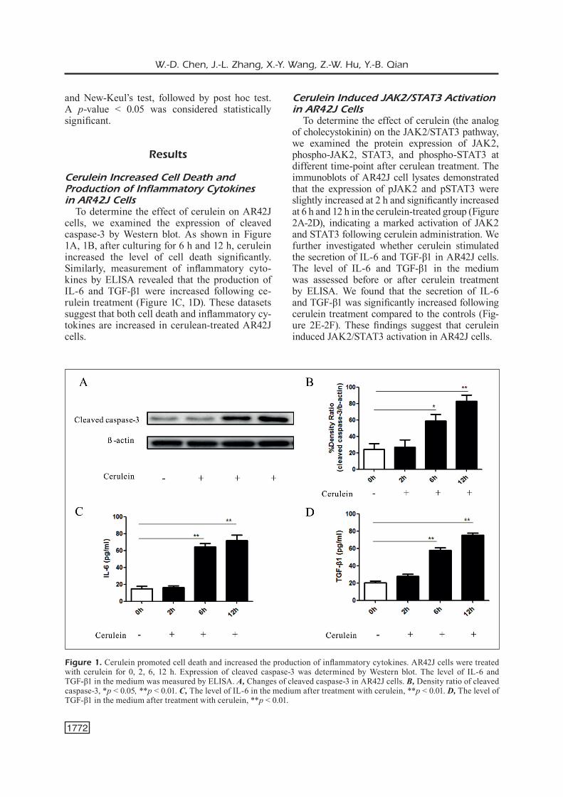
To determine the effect of cerulein on AR42J cells, we examined the expression of cleaved caspase-3 by Western blot. As shown in Figure 1A, 1B, after culturing for 6 h and 12 h, cerulein increased the level of cell death significantly. Similarly, measurement of inflammatory cyto-kines by ELISA revealed that the production of IL-6 and TGF-β1 were increased following ce-rulein treatment (Figure 1C, 1D). These datasets suggest that both cell death and inflammatory cy-tokines are increased in cerulean-treated AR42J cells.
Cerulein Induced JAK2/STAT3 Activation in AR42J Cells
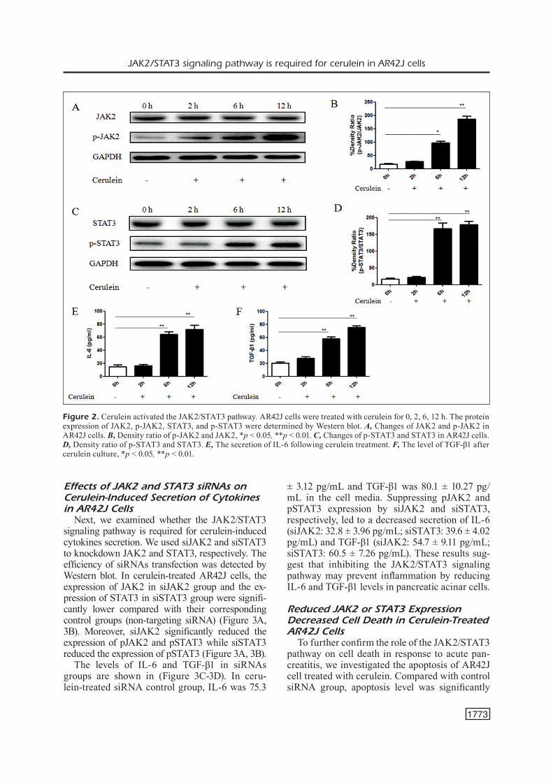
To determine the effect of cerulein (the analog of cholecystokinin) on the JAK2/STAT3 pathway, we examined the protein expression of JAK2, phospho-JAK2, STAT3, and phospho-STAT3 at different time-point after cerulean treatment. The immunoblots of AR42J cell lysates demonstrated that the expression of pJAK2 and pSTAT3 were slightly increased at 2 h and significantly increased at 6 h and 12 h in the cerulein-treated group (Figure 2A-2D), indicating a marked activation of JAK2 and STAT3 following cerulein administration. We further investigated whether cerulein stimulated the secretion of IL-6 and TGF-β1 in AR42J cells. The level of IL-6 and TGF-β1 in the medium was assessed before or after cerulein treatment by ELISA. We found that the secretion of IL-6 and TGF-β1 was significantly increased following cerulein treatment compared to the controls (Fig-ure 2E-2F). These findings suggest that cerulein induced JAK2/STAT3 activation in AR42J cells.
Figure 1. Cerulein promoted cell death and increased the production of inflammatory cytokines. AR42J cells were treated with cerulein for 0, 2, 6, 12 h. Expression of cleaved caspase-3 was determined by Western blot. The level of IL-6 and TGF-β1 in the medium was measured by ELISA. A, Changes of cleaved caspase-3 in AR42J cells. B, Density ratio of cleaved caspase-3, *p < 0.05, **p < 0.01. C, The level of IL-6 in the medium after treatment with cerulein, **p < 0.01. D, The level of TGF-β1 in the medium after treatment with cerulein, **p < 0.01.
JAK2/STAT3 signaling pathway is required for cerulein in AR42J cells
1773
Effects of JAK2 and STAT3 siRNAs on Cerulein-Induced Secretion of Cytokines in AR42J Cells
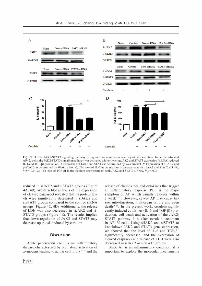
Next, we examined whether the JAK2/STAT3 signaling pathway is required for cerulein-induced cytokines secretion. We used siJAK2 and siSTAT3 to knockdown JAK2 and STAT3, respectively. The efficiency of siRNAs transfection was detected by Western blot. In cerulein-treated AR42J cells, the expression of JAK2 in siJAK2 group and the ex-pression of STAT3 in siSTAT3 group were signifi-cantly lower compared with their corresponding control groups (non-targeting siRNA) (Figure 3A, 3B). Moreover, siJAK2 significantly reduced the expression of pJAK2 and pSTAT3 while siSTAT3 reduced the expression of pSTAT3 (Figure 3A, 3B).
The levels of IL-6 and TGF-β1 in siRNAs groups are shown in (Figure 3C-3D). In ceru-lein-treated siRNA control group, IL-6 was 75.3
± 3.12 pg/mL and TGF-β1 was 80.1 ± 10.27 pg/mL in the cell media. Suppressing pJAK2 and pSTAT3 expression by siJAK2 and siSTAT3, respectively, led to a decreased secretion of IL-6 (siJAK2: 32.8 ± 3.96 pg/mL; siSTAT3: 39.6 ± 4.02 pg/mL) and TGF-β1 (siJAK2: 54.7 ± 9.11 pg/mL; siSTAT3: 60.5 ± 7.26 pg/mL). These results sug-gest that inhibiting the JAK2/STAT3 signaling pathway may prevent inflammation by reducing IL-6 and TGF-β1 levels in pancreatic acinar cells.
Reduced JAK2 or STAT3 Expression Decreased Cell Death in Cerulein-Treated AR42J Cells
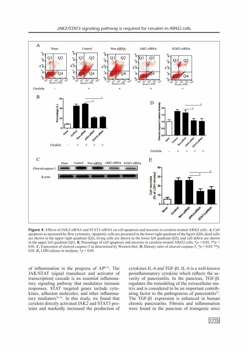
To further confirm the role of the JAK2/STAT3 pathway on cell death in response to acute pan-creatitis, we investigated the apoptosis of AR42J cell treated with cerulein. Compared with control siRNA group, apoptosis level was significantly
Figure 2. Cerulein activated the JAK2/STAT3 pathway. AR42J cells were treated with cerulein for 0, 2, 6, 12 h. The protein expression of JAK2, p-JAK2, STAT3, and p-STAT3 were determined by Western blot. A, Changes of JAK2 and p-JAK2 in AR42J cells. B, Density ratio of p-JAK2 and JAK2, *p < 0.05, **p < 0.01. C, Changes of p-STAT3 and STAT3 in AR42J cells. D, Density ratio of p-STAT3 and STAT3. E, The secretion of IL-6 following cerulein treatment. F, The level of TGF-β1 after cerulein culture, *p < 0.05, **p < 0.01.
W.-D. Chen, J.-L. Zhang, X.-Y. Wang, Z.-W. Hu, Y.-B. Qian
1774
reduced in siJAK2 and siSTAT3 groups (Figure 4A, 4B). Western blot analysis of the expression of cleaved caspase-3 revealed that its protein lev-els were significantly decreased in siJAK2 and siSTAT3 groups compared to the control siRNA groups (Figure 4C, 4D). Additionally, the release of LDH was also decreased in siJAK2 and si-STAT3 groups (Figure 4E). The results implied that down-regulation of JAK2 and STAT3 may decrease apoptosis induced by cerulein.
Discussion
Acute pancreatitis (AP) is an inflammatory disease characterized by premature activation of zymogens leading to acinar cell injury21-24 and the
release of chemokines and cytokines that trigger an inflammatory response. Pain is the major symptom of AP which usually resolves within 1 week25-27. However, severe AP may cause tis-sue auto-digestion, multiorgan failure and even death28-30. In the present work, cerulein signifi-cantly induced cytokines (IL-6 and TGF-β1) pro-duction, cell death and activation of the JAK2/STAT3 pathway 6 h after cerulein treatment in AR42J cells. Using siJAK2 and siSTAT3 to knockdown JAK2 and STAT3 gene expression, we showed that the level of IL-6 and TGF-β1 significantly decreased, and the expression of cleaved caspase-3 and release of LDH were also decreased in siJAK2 or siSTAT3 groups.
Since AP is an inflammatory condition, it is important to explore the molecular mechanisms
Figure 3. The JAK2/STAT3 signaling pathway is required for cerulein-induced cytokines secretion. In cerulein-treated AR42J cells, the JAK2/STAT3 signaling pathway was activated while silencing JAK2 and STAT3 expression (siRNA) reduced IL-6 and TGF-β1 production. A, Expression of JAK2 and STAT3 as determined by Western blot. B, Expression of p-JAK2 and p-STAT3 as determined by Western blot. C, The level of IL-6 in the medium after treatment with JAK2 and STAT3 siRNA, **p < 0.01. D, The level of TGF-β1 in the medium after treatment with JAK2 and STAT3 siRNA, **p < 0.01.
JAK2/STAT3 signaling pathway is required for cerulein in AR42J cells
1775
of inflammation in the progress of AP5,31. The JAK/STAT (signal transducer and activator of transcription) cascade is an essential inflamma-tory signaling pathway that modulates immune responses. STAT targeted genes include cyto-kines, adhesion molecules, and other inflamma-tory mediators32-34. In this study, we found that cerulein directly activated JAK2 and STAT3 pro-teins and markedly increased the production of
cytokines IL-6 and TGF-β1. IL-6 is a well-known proinflammatory cytokine which reflects the se-verity of pancreatitis. In the pancreas, TGF-β1 regulates the remodeling of the extracellular ma-trix and is considered to be an important contrib-uting factor to the pathogenesis of pancreatitis35. The TGF-β1 expression is enhanced in human chronic pancreatitis. Fibrosis and inflammation were found in the pancreas of transgenic mice
Figure 4. Effects of JAK2-siRNA and STAT3-siRNA on cell apoptosis and necrosis in cerulein-treated AR42J cells. A, Cell apoptosis as measured by flow cytometry: apoptotic cells are presented in the lower right quadrant of the figure (Q4), dead cells are shown in the upper right quadrant (Q2), living cells are shown in the lower left quadrant (Q3), and cell debris are shown in the upper left quadrant (Q1). B, Percentage of cell apoptosis and necrosis in cerulein-treated AR42J cells, *p < 0.05, **p < 0.01. C, Expression of cleaved caspase-3 as determined by Western blot. D, Density ratio of cleaved caspase-3, *p < 0.05, **p 0.01. E, LDH release in medium, *p < 0.05.
W.-D. Chen, J.-L. Zhang, X.-Y. Wang, Z.-W. Hu, Y.-B. Qian
1776
overexpressing TGF-β1. Therefore, the expres-sion of IL-6 and TGF-β1 is important to the de-velopment of pancreatitis36. Interestingly, block-ing JAK2 and STAT3 decreased the expression of IL-6 and TGF-β1 significantly. Hence, we demonstrated that the JAK2/STAT3 pathway is required for inflammatory responses in cerule-an-treated AR42J cells.
The development of acute pancreatitis is a complex process that is characterized by inflam-mation and parenchymal cell death5,37. It has been shown38,39 that the occurrence of cell death in different inflammatory diseases is not similar. As for acute pancreatitis, the severity of inflam-mation is directly correlated with necrosis and inversely correlated with apoptosis. Therefore, shifting death responses from necrosis to apop-tosis has a therapeutic value40. In this research, we demonstrated that necrosis and apoptosis of AR42J cells were increased following cerulein treatment in addition to the increased expression of cleaved caspase-3 and release of LDH. Ad-ministration of JAK2 siRNA and STAT3 siRNA showed a therapeutic effect on cerulein-induced pancreatitis.
Conclusions
The activation of JAK2/STAT3 signaling path-way increased the production of cytokines and cell death which were eliminated by blocking JAK2 and STAT3 expression. Our findings indi-cates that the JAK2/STAT3 signaling pathway is a potential therapeutic target in the management of acute pancreatitis.
Conflict of InterestThe Authors declare that they have no conflict of interests.
References
1) Whitcomb Dc. Clinical practice. Acute pancreati-tis. N Engl J Med 2006; 354: 2142-2150.
2) Li h, Qian Z, Liu Z, Liu X, han X, Kang h. Risk fac-tors and outcome of acute renal failure in patients with severe acute pancreatitis. J Crit Care 2010; 25: 225-229.
3) Papachristou gi, cLermont g, sharma a, YaDav D, Whitcomb Dc. Risk and markers of severe acute pancreatitis. Gastroenterol Clin North Am 2007; 36: 277-296.
4) sarmiento n, sancheZ-bernaL c, aYra m, pereZ n, hernanDeZ-hernanDeZ a, caLvo JJ, sancheZ-Yague J. Changes in the expression and dynamics of SHP-1 and SHP-2 during cerulein-induced acute pancreatitis in rats. Biochim Biophys Acta 2008; 1782: 271-279.
5) Kim h. Cerulein pancreatitis: oxidative stress, in-flammation, and apoptosis. Gut Liver 2008; 2: 74-80.
6) Lerch mm, goreLicK Fs. Models of acute and chronic pancreatitis. Gastroenterology 2013; 144: 1180-1193.
7) sarmiento n, sancheZ-bernaL c, pereZ n, sarDina JL, mangas a, caLvo JJ, sancheZ-Yague J. Rolipram and SP600125 suppress the early increase in PTP1B expression during cerulein-induced pancreatitis in rats. Pancreas 2010; 39: 639-645.
8) mareninova oa, sung KF, hong p, Lugea a, panDoL sJ, guKovsKY i, guKovsKaYa as. Cell death in pan-creatitis: caspases protect from necrotizing pan-creatitis. J Biol Chem 2006; 281: 3370-3381.
9) garg pK, maDan K, panDe gK, Khanna s, sathYanaraYan g, bohiDar np, tanDon rK. Associ-ation of extent and infection of pancreatic necro-sis with organ failure and death in acute necrotiz-ing pancreatitis. Clin Gastroenterol Hepatol 2005; 3: 159-166.
10) bettaieb a, Xi Y, hosein e, coggins n, bachaaLanY s, WieDe F, pereZ s, griFFeY sm, sastre J, tiganis t, haJ Fg. Pancreatic T cell protein-tyrosine phos-phatase deficiency ameliorates cerulein-induced acute pancreatitis. Cell Commun Signal 2014; 12: 13.
11) vaQuero e, guKovsKY i, Zaninovic v, guKovsKaYa as, panDoL sJ. Localized pancreatic NF-kappaB activation and inflammatory response in tauro-cholate-induced pancreatitis. Am J Physiol Gas-trointest Liver Physiol 2001; 280: G1197-G1208.
12) Kubisch ch, sans mD, arumugam t, ernst sa, WiLLiams Ja, LogsDon cD. Early activation of endoplasmic reticulum stress is associated with arginine-induced acute pancreatitis. Am J Physiol Gastrointest Liver Physiol 2006; 291: G238-G245.
13) Liu F, Zhao X, perna F, Wang L, KoppiKar p, ab-DeL-Wahab o, harr mW, Levine rL, Xu h, teFFeri a, DebLasio a, hatLen m, menenDeZ s, nimer sD. JAK2V617F-mediated phosphorylation of PRMT5 downregulates its methyltransferase activity and promotes myeloproliferation. Cancer Cell 2011; 19: 283-294.
14) Yu Jh, Kim Kh, Kim h. SOCS 3 and PPAR-gamma ligands inhibit the expression of IL-6 and TGF-be-ta1 by regulating JAK2/STAT3 signaling in pan-creas. Int J Biochem Cell Biol 2008; 40: 677-688.
15) griFFiths Ds, Li J, DaWson ma, trotter mW, cheng Yh, smith am, mansFieLD W, Liu p, KouZariDes t, nichoLs J, bannister aJ, green ar, gottgens B. LIF-independent JAK signalling to chromatin in embryonic stem cells uncovered from an adult stem cell disease. Nat Cell Biol 2011; 13: 13-21.
JAK2/STAT3 signaling pathway is required for cerulein in AR42J cells
1777
16) berishaJ m, gao sp, ahmeD s, LesLie K, aL-ahmaDie h, geraLD WL, bornmann W, bromberg JF. Stat3 is tyrosine-phosphorylated through the inter-leukin-6/glycoprotein 130/Janus kinase path-way in breast cancer. Breast Cancer Res 2007; 9: R32.
17) bromberg J, Wang tc. Inflammation and cancer: IL-6 and STAT3 complete the link. Cancer Cell 2009; 15: 79-80.
18) grivenniKov s, Karin e, terZic J, muciDa D, Yu gY, vaLLabhapurapu s, scheLLer J, rose-John s, cheroutre h, ecKmann L, Karin m. IL-6 and Stat3 are required for survival of intestinal epithelial cells and devel-opment of colitis-associated cancer. Cancer Cell 2009; 15: 103-113.
19) Zhu s, Zhang c, Weng Q, Ye b. Curcumin protects against acute renal injury by suppressing JAK2/STAT3 pathway in severe acute pancreatitis in rats. Exp Ther Med 2017; 14: 1669-1674.
20) shieLDs bJ, WieDe F, gurZov en, Wee K, hauser c, Zhu hJ, moLLoY tJ, o’tooLe sa, DaLY rJ, sutherLanD rL, mitcheLL ca, mcLean ca, tiganis t. TCPTP reg-ulates SFK and STAT3 signaling and is lost in tri-ple-negative breast cancers. Mol Cell Biol 2013; 33: 557-570.
21) baron th, morgan De. Acute necrotizing pancre-atitis. N Engl J Med 1999; 340: 1412-1417.
22) Jin Y, Xu h, chen Y, Wu J, Jin F, Wu Q, Yao Xm. Therapeutic effect of Bifidobacterium combined with early enteral nutrition in the treatment of se-vere acute pancreatitis: a pilot study. Eur Rev Med Pharmacol Sci 2018; 22: 4018-4024.
23) YaDav D, LoWenFeLs ab. The epidemiology of pan-creatitis and pancreatic cancer. Gastroenterology 2013; 144: 1252-1261.
24) roberts se, aKbari a, thorne K, atKinson m, evans pa. The incidence of acute pancreatitis: impact of social deprivation, alcohol consumption, season-al and demographic factors. Aliment Pharmacol Ther 2013; 38: 539-548.
25) singh vK, moran ra, aFghani e, De-maDaria e. Treating acute pancreatitis: what’s new? Expert Rev Gastroenterol Hepatol 2015; 9: 901-911.
26) saLuJa aK, Donovan ea, YamanaKa K, Yamaguchi Y, hoFbauer b, steer mL. Cerulein-induced in vitro activation of trypsinogen in rat pancreatic acini is mediated by cathepsin B. Gastroenterology 1997; 113: 304-310.
27) naruse s. Molecular pathophysiology of pancreati-tis. Intern Med 2003; 42: 288-289.
28) Lerch mm, goreLicK Fs. Models of acute and chronic pancreatitis. Gastroenterology 2013; 144: 1180-1193.
29) Jensen rt, WanK sa, roWLeY Wh, sato s, garD-ner JD. Interaction of CCK with pancreatic acinar cells. Trends Pharmacol Sci 1989; 10: 418-423.
30) WiLLemer s, eLsasser hp, aDLer g. Hormone-induced pancreatitis. Eur Surg Res 1992; 24 Suppl 1: 29-39.
31) Yu Jh, Kim Kh, Kim h. Suppression of IL-1beta ex-pression by the Jak 2 inhibitor AG490 in ceru-lein-stimulated pancreatic acinar cells. Biochem Pharmacol 2006; 72: 1555-1562.
32) Yang X, he g, hao Y, chen c, Li m, Wang Y, Zhang g, Yu Z. The role of the JAK2-STAT3 pathway in pro-inflammatory responses of EMF-stimulated N9 microglial cells. J Neuroinflammation 2010; 7: 54.
33) Yu Jh, Kim h. Role of janus kinase/signal trans-ducers and activators of transcription in the pathogenesis of pancreatitis and pancreatic can-cer. Gut Liver 2012; 6: 417-422.
34) horiguchi a, asano t, KuroDa K, sato a, asaKuma J, ito K, haYaKaWa m, sumitomo m, asano t. STAT3 inhibitor WP1066 as a novel therapeutic agent for renal cell carcinoma. Br J Cancer 2010; 102: 1592-1599.
35) branDs mW, banes-berceLi aK, inscho eW, aL-aZaWi h, aLLen aJ, LabaZi h. Interleukin 6 knockout pre-vents angiotensin II hypertension: role of renal vasoconstriction and janus kinase 2/signal trans-ducer and activator of transcription 3 activation. Hypertension 2010; 56: 879-884.
36) Ju KD, Lim JW, Kim Kh, Kim h. Potential role of NA-DPH oxidase-mediated activation of Jak2/Stat3 and mitogen-activated protein kinases and ex-pression of TGF-beta1 in the pathophysiology of acute pancreatitis. Inflamm Res 2011; 60: 791-800.
37) guKovsKaYa as, guKovsKY i, Jung Y, mouria m, pan-DoL sJ. Cholecystokinin induces caspase activa-tion and mitochondrial dysfunction in pancreatic acinar cells. Roles in cell injury processes of pan-creatitis. J Biol Chem 2002; 277: 22595-22604.
38) gunther c, neumann h, neurath mF, becKer c. Apoptosis, necrosis and necroptosis: cell death regulation in the intestinal epithelium. Gut 2013; 62: 1062-1071.
39) JiLLing t, Lu J, JacKson m, capLan ms. Intestinal ep-ithelial apoptosis initiates gross bowel necrosis in an experimental rat model of neonatal necrotizing enterocolitis. Pediatr Res 2004; 55: 622-629.
40) mareninova oa, sung KF, hong p, Lugea a, panDoL sJ, guKovsKY i, guKovsKaYa as. Cell death in pan-creatitis: caspases protect from necrotizing pan-creatitis. J Biol Chem 2006; 281: 3370-3381.