Embed Size (px)
Citation preview

Jamie Groh PGY-6
PBCGME/West Palm Hospital
Objectives
Clinical Presentation
Histology
Current Treatments
Future Treatment
Case 72 yo Caucasian male
Subcutaneous nodule right posterior arm ~1year
PCP told patient it was likely a lipoma
Lesion began to grow and patient requested further evaluation
Patient was referred to dermatology
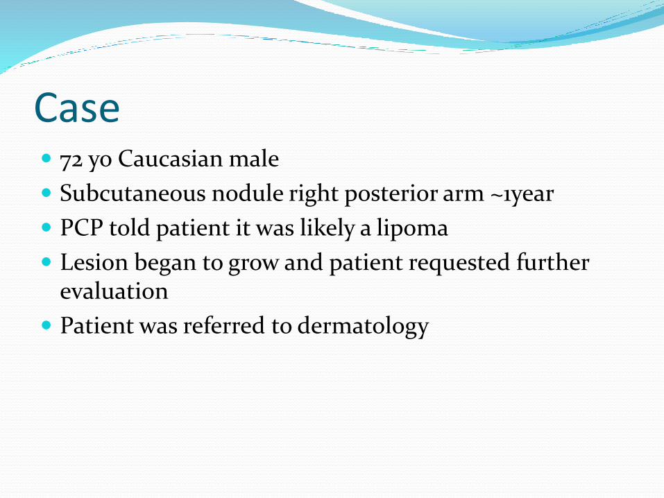
Right posterior arm 11/1/13. 1.2cm x 2.5 cm subcutaneous, mobile, skin colored nodule fixed to overlying epidermis
Case Patient presented to dermatology 5 months after
initial referral
Lesion had continued to grow rapidly
Two weeks prior to visit patient noted swelling in the right axilla.
ROS: Intentional 40 pound weight loss last several month. All other ROS non-contributory.
Case PMHx: DMII
PSHx: non-contributory
Fam Hx: Father deceased from pancreatic ca
Social hx:
Tobacco: quit 50 years ago
ETOH: hx of social use, currently none
Illicit drugs: denies

Case Physical Exam
Gen: A&O x 3 NAD
Right posterior arm: 6 cm erythematous moist, mobile, subcutaneous nodule, fixed to overlying epidermis.
Right axilla LAD

Right posterior arm 4/11/15.
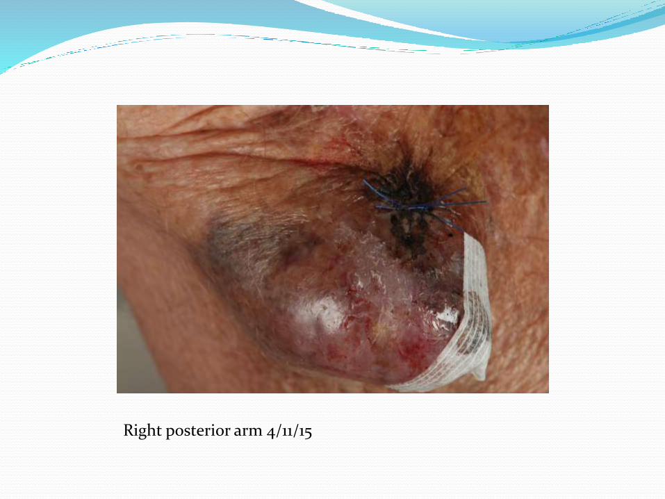
Right posterior arm 4/11/15
Case-work up 5mm lesional punch biopsy for H&E
CT Chest and Right upper extremity with and without contrast.
Case CT- subcutaneous mass of posterior right arm with
evidence of necrosis. Right axillary LAD, largest lymph node 3 cm
Histology- Poorly differentiated neuroendocrinecarcinoma
Immunohistochemistry-
+CK 20 perinuclear dot, synaptophysin
Negative TTF-1 and CK7
Diagnosis: Merkel Cell Carcinoma
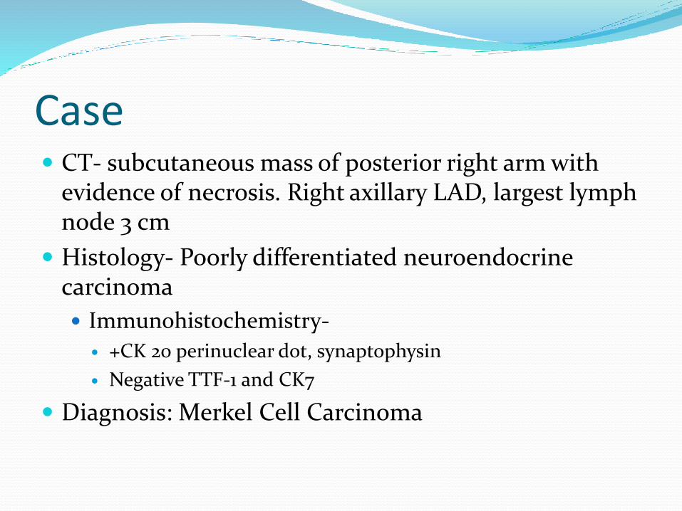
Right posterior arm. H&E 2x magnification
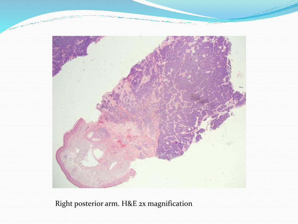
H&E 20x magnification
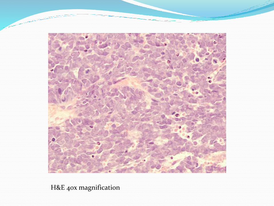
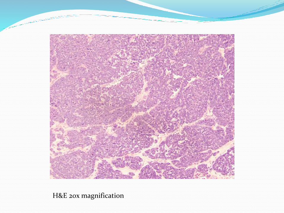
H&E 40x magnification
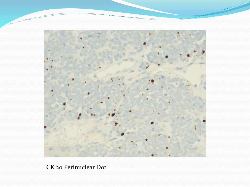
CK 20 Perinuclear Dot
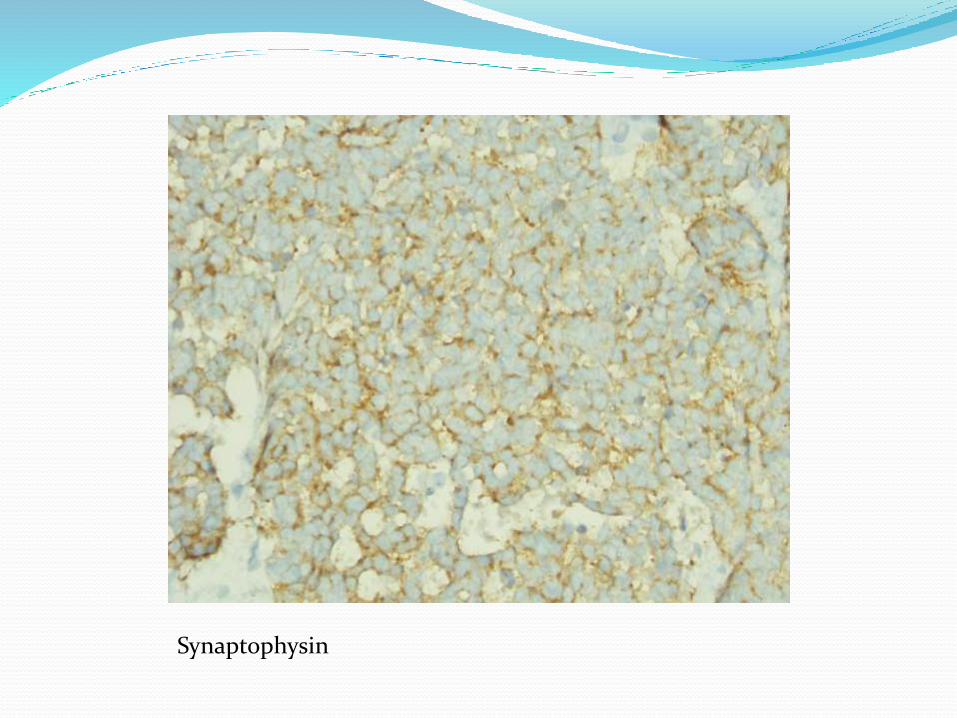
Synaptophysin
Case General Surgery consult
WLE of Right posterior arm MCC and axillary lymph node dissection
Despite WLE margins were still positive
21 of 29 lymph nodes positive for MCC
Pathologic preliminary staging was T3N1
Patient was referred to oncology
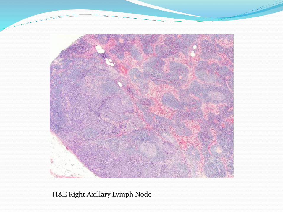
H&E Right Axillary Lymph Node

Case PET scan was ordered by oncology
Metabolic areas in right distal arm, right supraclavicularlymph nodes, right axillary lymph nodes and b/l hilarlymph
Due to extent of disease radiation was not an option
Oncology recommended chemotherapy with platinum and etopiside.
Patient chose palliative care.
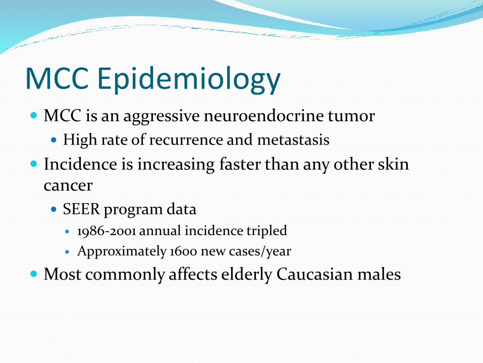
MCC Epidemiology MCC is an aggressive neuroendocrine tumor
High rate of recurrence and metastasis
Incidence is increasing faster than any other skin cancer
SEER program data
1986-2001 annual incidence tripled
Approximately 1600 new cases/year
Most commonly affects elderly Caucasian males
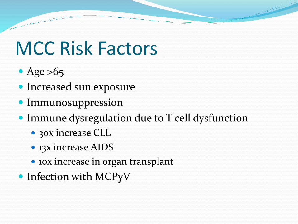
MCC Risk Factors Age >65
Increased sun exposure
Immunosuppression
Immune dysregulation due to T cell dysfunction
30x increase CLL
13x increase AIDS
10x increase in organ transplant
Infection with MCPyV

MCC Clinical Presentation Painless, solitary, rapidly growing nodule/indurated
plaque, arising on chronically sun exposed skin
Most common location face and neck followed by upper limb
Acronym AEIOU for clinical parameters Asymptomatic
Expanding Rapidly
Immune Suppressed
Older than 50 years old
UV exposed areas
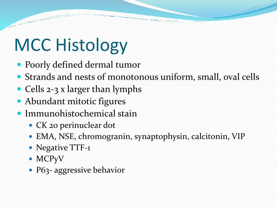
MCC Histology Poorly defined dermal tumor
Strands and nests of monotonous uniform, small, oval cells
Cells 2-3 x larger than lymphs
Abundant mitotic figures
Immunohistochemical stain CK 20 perinuclear dot
EMA, NSE, chromogranin, synaptophysin, calcitonin, VIP
Negative TTF-1
MCPyV
P63- aggressive behavior
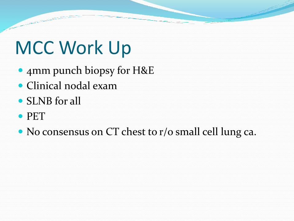
MCC Work Up 4mm punch biopsy for H&E
Clinical nodal exam
SLNB for all
PET
No consensus on CT chest to r/o small cell lung ca.
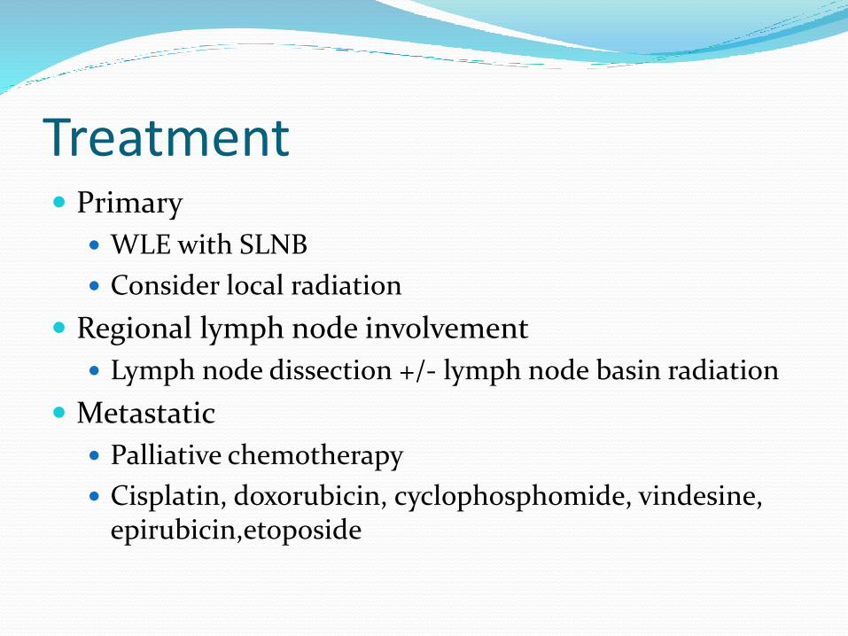
Treatment Primary
WLE with SLNB
Consider local radiation
Regional lymph node involvement
Lymph node dissection +/- lymph node basin radiation
Metastatic
Palliative chemotherapy
Cisplatin, doxorubicin, cyclophosphomide, vindesine, epirubicin,etoposide
Possible Future Treatment Targeted Molecular therapy
Ipilimumab
YM-155
Pazopanib
Lanetreotide
ILGFR-1 and mTOR inhibitor
Oblimersen
Possible Future Therapies Immunotherapy
Recombinant IL-2
Intra-lesional interferon
PD-1 inhibitor
IL-12 DNA Electroporation
Bortezomib
T-cell immunotransfer
References 1.Agelli M, Clegg LX, Becker JC, Rollison DE. The etiology and epidemiology of merkel cell carcinoma. Curr Probl Cancer. 2010;34(1):14–37. 2.Albores-Saavedra J, Batich K, Chable-Montero F, Sagy N, Schwartz AM, HensonDE. Merkel cell carcinoma demographics, morphology, and survival based on 3870 cases: a
population based study. J Cutan Pathol. Jan 2010;37(1):20-27. 3.Hodgson NC. Merkel cell carcinoma: changing incidence trends. J Surg Oncol.2005;89(1):1-4. 4.Heath M, Jaimes N, Lemos B, et al. Clinical characteristics of Merkel cell carcinoma at diagnosis in 195 patients: the AEIOU features. J Am Acad Dermatol.2008;58(3):375-381. 5. Engels EA, Frisch M, Goedert JJ, Biggar RJ, Miller RW. Merkel cell carcinoma andHIV infection. Lancet. 2002;359(9305):497-498. 6. Dowlatshahi M, Huang V, Gehad AE, et al. Tumor-specific T cells in human Merkelcell carcinomas: a possible role for Tregs and T-cell exhaustion in reducing T-cell
responses. J Invest Dermatol. 2013;133(7):1879-1889. 7. Tilling T, Moll I. Which are the cells of origin in merkel cell carcinoma?J Skin Cancer. 2012;2012:680410. 8. Swann MH, Yoon J. Merkel cell carcinoma. Semin Oncol. 2007; 34(1):51 9. Goessling W, McKee PH, Mayer RJ. Merkel cell carcinoma. J Clin Oncol. 2002;20(2):588-598. 10.Heath M, Jaimes N, Lemos B, Mostaghimi A, Wang LC, Peñas PF, Nghiem P. Clinical characteristics of Merkel cell carcinoma at diagnosis in 195 patients: the AEIOU features.
J Am Acad Dermatol. 2008;58(3):375. 11.Sibley RK, Dehnew LP, Rosai J. Primary neuroendocrine (Merkel cell?) carcinoma of the skin. I. AM J Surg Pathol. 1985; 9:95-108 12.Poulsen M. Merkel-cell carcinoma of the skin. Lancet Oncol. Oct 2004;5(10):593- 599. 13. Smith, DF, Messina JL, Perrott R, Berman CG, Reintgen DS, Cruse, CW, Glass, FL,
Fenske NA, Deconti RC, Trotti A. Clinical approach to neuroendocrine carcinoma of the skin (Merkel cell carcinoma). Cancer Control. 2000;7(1):72. 14. Cheuk W, Kwan MY, Suster S, Chan JK. Immunostaining for thyroid transcription face 1 and cytokeratin 20 aids in the distinction of small cell carcinoma from Merkel cell
carcinoma, but not pulmonary form of extra pulmonary small cell carcinomas. Atch Pathol Lab Med. 2001;125:228-231. 15. Duncavage EJ, Le BM, Wang D, Pfeifer JD. Merkel cell polyomavirus: a specific marker for Merkel cell carcinoma in histologically similar tumors. Am J Surg Pathol. 2009;
33:1771-1777 16. Asioli S, Righi A, Volante M, et al. p63 expression as a new prognostic marker in Merkel cell carcinoma. Cancer. 2007: 110:640-647. 17. Edge SB, Byrd DR, Compton CC, et al., eds. Merkel cell carcinoma. AJCC Cancer Staging Manual. 7th ed. New York, NY: Springer, 2010, pp 315-23. 18.Tarantola TI, Vallow LA, Halyard MY, et al. Prognostic factors in Merkel cell carcinoma: analysis of 240 cases. J Am Acad Dermatol. Mar 2013;68(3):425-432. 19. Lemos BD, Storer BE, Iyer JG, et al. Pathologic nodal evaluation improves prognostic accuracy in Merkel cell carcinoma: analysis of 5823 cases as the basis of the first
consensus staging system. J Am Acad Dermatol. 2010;63(5):751-761. 20. Iyer JG, Storer BE, Paulson KG, et al. Relationships between primary tumor size, number of involved nodes and survival among 8,044 cases of Merkel cell carcinoma. J Am
Acad Dermatol. 2014;70(4):637-643. 21.Howle JR, Hughes TM, Gebski V, Veness MJ. Merkel cell carcinoma: an Australianperspective and the importance of addressing the regional lymph nodes in clinically node
negative patients. J Am Acad Dermatol. 2012;67(1):33-40. 22. Treglia G, Kakhki VR, Giovanella L, Sadeghi R. Diagnostic performance of fluorine-18-fluorodeoxyglucose positron emission tomography in patients with Merkel cell
carcinoma: a systematic review and meta-analysis. Am J Clin Dermatol. 2013;14(6):437.
Refrences 23.Gupta SG, Wang LC, Penas PF, Gellenthin M, Lee SJ, Nghiem P. Sentinel lymph node biopsy for evaluation and treatment of patients with
Merkel cell carcinoma: The Dana-Farber experience and meta-analysis of the literature. Arch Dermatol. 2006;142(6):685-690. 24. Prieto Munoz I, Pardo Masferrer J, Olivera Vegas J, Medina Montalvo MS, Jover Diaz R, Perez Casas AM. Merkel cell carcinoma from 2008
to 2012: reaching a new level of understanding. Cancer Treat Rev. 2013;39(5):421-429. 25.Tarantola TI, Vallow LA, Halyard MY, et al. Unknown primary Merkel cell carci- noma: 23 new cases and a review. J Am Acad Dermatol. 2013;68(3):433-440. 26.Chen KT, Papavasiliou P, Edwards K, et al. A better prognosis for Merkel cell carcinoma of unknown primary origin. Am J Surg.
2013;206(5):752-757. 27.Touze A, Le Bidre E, Laude H, et al. High levels of antibodies against merkel cell polyomavirus identify a subset of patients with merkel
cell carcinoma with betterclinical outcome. J Clin Oncol. 2011;29(12):1612-1619. 28. Bichakjian CK, Alam M, Anderson J, et al. Merkel cell carcinoma, version 1.2014.J Natl Compr Canc Netw. 2014;12(3):410-424. 29. Nghiem P, James N: Merkel cell carcinoma. In: Wolff K, Goldsmith LA, Katz SI, et al., eds.: Fitzpatrick's Dermatology in General Medicine.
7th ed. New York, NY: McGraw-Hill , 2008, pp 1087-94. 30.Allen PJ, Bowne WB, Jaques DP, et al.: Merkel cell carcinoma: prognosis and treatment of patients from a single institution. J Clin Oncol
23 (10): 2300-9, 2005. 31.Nghiem P, McKee PH, Haynes HA: Merkel cell (cutaneous neuroendocrine) carcinoma. In: Sober AJ, Haluska FG, eds.: Skin Cancer.
Hamilton, Ontario: BC Decker Inc., 2001, pp 127-141. 32.Boyer JD, Zitelli JA, Brodland DG, et al.: Local control of primary Merkel cell carcinoma: review of 45 cases treated with Mohs
micrographic surgery with and without adjuvant radiation. J Am Acad Dermatol. 47 (6): 885-92, 2002. 33. Wilson LD, Gruber SB: Merkel cell carcinoma and the controversial role of adjuvant radiation therapy: clinical choices in the absence of
statistical evidence. J Am Acad Dermatol. 50 (3): 435-7; discussion 437-8, 2004. 34. Gollard R, Weber R, Kosty MP, et al.: Merkel cell carcinoma: review of 22 cases with surgical, pathologic, and therapeutic considerations.
Cancer. 8 (8) 1842-51, 2000. 35.Allen PJ, Bowne WB, Jaques DP, et al.: Merkel cell carcinoma: prognosis and treatment of patients from a single institution. J Clin Oncol 23
(10): 2300-9, 2005. 36. Senchenkov A, Barnes SA, Moran SL. Predictors of survival and recurrence in the surgical treatment of merkel cell carcinoma of the
extremities. J Surg Oncol. 2007;95(3):229-234. 37.Veness MJ, Perera L, McCourt J, et al. Merkel cell carcinoma: improved outcome with adjuvant radiotherapy. ANZ J Surg. 2005;75(5):275-
281.

Refrences 38. Mojica P, Smith D, Ellenhorn JD: Adjuvant radiation therapy is associated with improved survival in Merkel cell
carcinoma of the skin. J Clin Oncol 25 (9): 1043-7, 2007. 39.Pape E, Rezvoy N, Penel N, et al. Radiotherapy alone for Merkel cell carcinoma: a comparative and retrospective
study of 25 patients. J Am Acad Dermatol. 2011;65(5):983-990. 40. Cotter SE, Devlin PM, Sahni D, et al. Treatment of cutaneous metastases of Merkel cell carcinoma with surface-
mold computer-optimized high-dose-rate brachytherapy. J Clin Oncol. 2010;28(27):e464-466. 41. Maza S, Trefzer U, Hofmann M, et al.: Impact of sentinel lymph node biopsy in patients with Merkel cell carcinoma:
results of a prospective study and review of the literature. Eur J Nucl Med Mol Imaging 33 (4): 433-40, 2006. 42.Mehrany K, Otley CC, Weenig RH, et al.: A meta-analysis of the prognostic significance of sentinel lymph node
status in Merkel cell carcinoma. Dermatol Surg 28 (2): 113-7; discussion 117, 2002. 43.Krasagakis K, Almond-Roesler B, Zouboulis CC, et al. Merkel cell carcinoma:report of ten cases with emphasis on
clinical course, treatment, and in vitro drug sensitivity. J Am Acad Dermatol.1997;36(5 Pt 1):727-732. 44.Kearsley JH, Hurst T, Khoo SK. Chemosensitivity testing of primary cultures of Merkel cell cancer. Anticancer
Drugs. 1993;4(5):571-575. 45.Garneski KM, Nghiem P. Merkel cell carcinoma adjuvant therapy: current data support radiation but not
chemotherapy. J Am Acad Dermatol. 2007;57(1) 166-169. 46. Donepudi S, DeConti RC, Samlowski WE. Recent advances in the understandingof the genetics, etiology, and
treatment of Merkel cell carcinoma. Semin Oncol.2012;39(2):163-172. 47. Aldabagh B, Joo J, Yu SS. Merkel cell carcinoma: current status of targeted and future potential for
immunotherapies. Semin Cutan Med Surg. 2014 Jun;33(2):76-82. 48. Miller N, Bhatia S, Parvathaneni U, Iyer J, Nghiem P. Emerging and Mechanism-Based therapies for recurrent of
metastatic merkel cell carcinoma. Curt Treat Options Oncol. 2013 June; 14(2):249-263.


![· Web view[18F]-Fluorodeoxyglucose positron emission tomography in children with neurofibromatosis type 1 and plexiform neurofibromas: correlation with malignant transformation.J](https://img.pdfslide.net/doc/110x75/5b1c5e287f8b9a37258fdaa9/-web-view18f-fluorodeoxyglucose-positron-emission-tomography-in-children-with.jpg)

![Package ‘pgirmess’ - The Comprehensive R Archive Network · Package ‘pgirmess’ ... David Pleydell [ctb], Mike Treglia [ctb] Maintainer Patrick Giraudoux](https://img.pdfslide.net/doc/110x75/5c04658409d3f29b388ba722/package-pgirmess-the-comprehensive-r-archive-network-package-pgirmess.jpg)


![Quantifying [ F]fluorodeoxyglucose uptake in the arterial ...pinlab.hcuge.ch/pdf/EJNMMI2015.pdf · ORIGINAL ARTICLE Quantifying [18F]fluorodeoxyglucose uptake in the arterial wall:](https://img.pdfslide.net/doc/110x75/5b540f517f8b9a575f8c76c5/quantifying-ffluorodeoxyglucose-uptake-in-the-arterial-original-article.jpg)


![Pharmacokinetic modeling of [18F]fluorodeoxyglucose (FDG](https://img.pdfslide.net/doc/110x75/61886b54df681277ae16a602/pharmacokinetic-modeling-of-18ffluorodeoxyglucose-fdg-.jpg)