Embed Size (px)
Citation preview

EFFECTS OF TEMPORARY INACTIVATION OF DORSAL HIPPOCAMPUS ON
EXPLICITLY NONSPATIAL, UNIMODAL, CONTEXTUAL FEAR LEARNING
By
Teresa Camille Parsons
A thesis submitted to the
Graduate School-New Brunswick
Rutgers, The State University of New Jersey
in partial fulfillment of the degree requirements
for the degree of
Master of Science
Graduate Program in Psychology
written under the direction of
Timothy Otto, Ph.D
And approved by
_____________________
_____________________
_____________________
New Brunswick, New Jersey
January 2008
ii
ABSTRACT OF THE THESIS
Effects of temporary inactivation of dorsal hippocampus on explicitly nonspatial,
unimodal, contextual fear learning
by TERESA CAMILLE PARSONS
Thesis Director:
Timothy Otto, Ph.D
Several studies have reported that dorsal hippocampal damage attenuates the
acquisition (Kim et al., 1993; Phillips & LeDoux, 1992; Young et al., 1994) or expression
(Anagnostaras et al., 1999; Holt & Maren, 1999) of recently acquired contextual fear
conditioning. "Context" is often operationalized as the conditioning chamber in which
CS-US pairings occurred. However, the hippocampus is known to participate in spatial
learning, presenting interpretative difficulties regarding the role of dorsal hippocampus in
learning and memory. The current study examined the effects of temporary inactivation
of DH on freezing, rearing, ambulating, grooming, and whisking behavior in an explicitly
nonspatial contextual fear conditioning paradigm, where olfactory stimuli served as
temporally and spatially diffuse contexts. Results indicate that temporary inactivation of
DH produced both anterograde and retrograde deficits in contextually conditioned
freezing, while sparing the acquisition and expression of freezing to a discrete auditory
CS. Further, animals with DH inactivation froze modestly and similarly to the unsafe and
safe contextual stimuli, while intact animals froze robustly to the unsafe, but not the safe,
contextual stimulus. These data indicate that there is a decidedly nonspatial component
to the role of DH in contextual conditioning, and suggest that olfactory contextual
conditioning is a fruitful means of further exploring this function.
iii
Table of Contents
I. Page ii Abstract
II. Page iii Table of Contents
III. Page 1 Introduction
IV. Page 5 Method
V. Page 12 Results
VI. Page 17 Discussion
VII. Page 24 Appendix
a. Page 25 Table 1. Experimental design
b. Page 26 Figure 1. Cannula placement in dorsal hippocampus for
experimental groups
c. Page 27 Figure 2. Freezing behavior during unsafe context test
d. Page 28 Figure 3. Freezing behavior during safe context test
e. Page 29 Figure 4. Difference between unsafe and safe context
f. Page 30-31 Figure 5. Behavior allocation during relevant periods of
unsafe context test
VIII. Page 32 References
1
1. Introduction
For organisms from rodents to primates, the ability to associate events with
one another in time and space appears to rely heavily on the hippocampus
(Anagnostaras, Gale, & Fanselow, 2001; Eichenbaum, Otto, & Cohen, 1994; Kim &
Fanselow, 1992). Mounting evidence suggests that within this large bilateral
structure, dissociable regions exist with respect to their cytoarchitecture and
connections (Brun et al., 2002; Eichenbaum et al., 1994; Pitkänen, Pikkarainen,
Nurminen, & Ylinen, 2000). Based on these considerations, the hippocampus may
be divided along its septotemporal axis into ventral and dorsal portions (Hargreaves,
Rao, Lee, & Knierim, 2005; Ishikawa & Nakamura, 2006; Kusljic & van den Buuse,
2004; Moser & Moser, 1998) that may subserve unique or overlapping memory
processes (Moser & Moser, 1998; Richmond et al., 1999).
Among the many fruitful behavioral paradigms used to identify neural
substrates of learning and memory is Pavlovian fear conditioning. In one version of
this paradigm known as delay fear conditioning, presentations of a neutral stimulus
(henceforth referred to as a conditioned stimulus, or CS) co-terminate with
presentations of a stimulus such as footshock (an unconditioned stimulus, or US).
Repeated CS-US pairings eventually result in the development of a learned
association between the CS and US. Use of this paradigm has led many researchers
to conclude that while the hippocampus is normally not critically involved in
temporally discrete CS-US associations (Kim & Fanselow, 1992; Phillips & LeDoux,
1992), it does participate in the acquisition of the context in which tone-shock
pairings take place (Kim, Rison, & Fanselow, 1993; Phillips & LeDoux, 1992; Young,
Bohenek, & Fanselow, 1994). Further, while lesions of the entire hippocampus
generally produce some disruption in contextual but not auditory fear conditioning

2
(Kim & Fanselow, 1992; Otto & Poon, 2006; Philips & LeDoux, 1992), attempts to
clarify the respective roles of ventral hippocampus and dorsal hippocampus in
contextual fear conditioning have been inconclusive. In some cases, electrolytic or
excitotoxic dorsal hippocampus lesions made prior to contextual fear conditioning
training were found to interrupt acquisition of contextual fear conditioning (Kim et al.,
1993; Phillips & LeDoux, 1992; Young et al., 1994), while other studies reported that
dorsal hippocampus lesions produced no impairment in the acquisition of contextual
fear conditioning (Maren, Aharonov, & Fanselow, 1997; Richmond et al., 1999).
In the event of permanent damage to a structure, surrounding areas may be
compromised, recruited or otherwise affected, leading some researchers to adopt
instead of lesions the reversible technique of temporary inactivation. The fact that
most hippocampal neurons contain the GABAα receptor (Chan-Palay, 1978) makes
the GABAα agonist muscimol a potentially useful means for temporarily inactivating
components of the hippocampus. Using muscimol to reversibly inactivate the dorsal
hippocampus, several researchers have found dorsal hippocampus to be critically
involved in the acquisition or retrieval of contextual fear memory and context-specific
fear expression in paradigms such as latent inhibition and facilitated extinction,
without affecting contextual discrimination or performance of the freezing response
(Corcoran & Maren, 2001; Holt & Maren, 1999; Matus-Amat, Higgins, Barrientos, &
Rudy, 2004).
Almost without exception, studies investigating the role of dorsal
hippocampus in contextual learning have defined �context� as the behavioral
chamber where CS-US pairings took place. However, it is well established that the
dorsal hippocampus participates in spatial conditioning (Bannerman et al., 2002;
Eichenbaum et al., 1994; Moser, Moser, & Andersen, 1993). Thus, equating space
with context presents a potential confound. Recently, Bannerman, Rawlins, and
3
Good (2006) referred to as �context� the features of an environment that collectively
define the place or situation in which the animal is located at the time learning
occurred. Conceptual definitions of context typically emphasize the nature of the
temporal and spatial relationship between the CS and other aspects of the
environment (see Balsam & Tomie, 1985). Stimuli ranging from background color of
a response key (Thomas, 1985), the presence or absence of unimodal stimuli (Otto
& Poon, 2006), tones of specific pitch (Hulse, Cynx, & Humpal, 1994), and internal
states resulting from drug administration or food deprivation (Davidson & Jarrard,
1993; Hock & Bunsey, 1998; Overton, 1964) have been experimentally manipulated
as contextual variables. In the light of evidence that dorsal hippocampus participates
in certain aspects of contextual conditioning, such variety within the class of events
that may function as contextual stimuli underscores the importance of examining
neural substrates of explicitly nonspatial forms of contextual conditioning.
Recently, Otto and Poon (2006) described a unique paradigm for dissociating
the role of dorsal hippocampus in contextual fear conditioning from its involvement in
spatial conditioning. In an olfactory contextual conditioning procedure, the presence
of strawberry odor and 15% pyridine, presented in alternation, constituted different
�safe� and �unsafe�, temporally and spatially diffuse contexts. During a single delay
fear conditioning session in one behavioral chamber, tone-CS/footshock-US pairings
occurred during presentations of the �unsafe� context. At the time of testing 24 hr
later, intact rats froze significantly during presentation of the �unsafe� but not the
�safe� contextual stimulus, while animals with lesions of dorsal hippocampus were
dramatically impaired in freezing during the �unsafe� contextual stimulus (but froze
normally with respect to the CS). Even as these results evince a function for dorsal
hippocampus in a form of contextual fear conditioning that is explicitly nonspatial, the
permanent nature of the dorsal hippocampus damage precluded examination of the
4
role of dorsal hippocampus in olfactory contextual fear memory retrieval independent
of its involvement in acquisition. To this end, the current study replicates use of the
olfactory contextual conditioning paradigm (Otto & Poon, 2006) while adopting
temporary inactivation of dorsal hippocampus at various time points, affording
examination of the role of dorsal hippocampus during acquisition and retrieval of fear
conditioned to contextual and discretely presented stimuli. Further, because both
muscimol and hippocampal lesions may produce motor deficits or hyperactivity,
respectively (see Anagnostaras et al., 2001), the present study examined a host of
other behavioral responses in addition to the more commonly measured freezing and
immobility (Fanselow, 1980; Fendt & Fanselow, 1999).
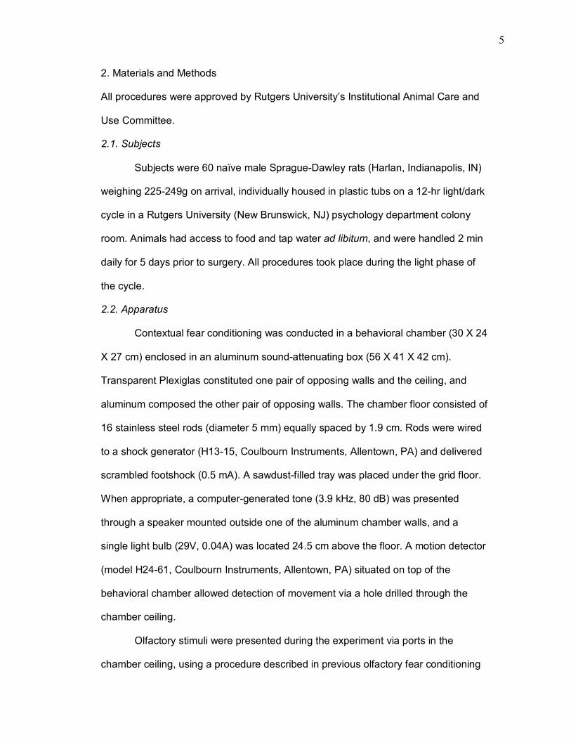
5
2. Materials and Methods
All procedures were approved by Rutgers University�s Institutional Animal Care and
Use Committee.
2.1. Subjects
Subjects were 60 naïve male Sprague-Dawley rats (Harlan, Indianapolis, IN)
weighing 225-249g on arrival, individually housed in plastic tubs on a 12-hr light/dark
cycle in a Rutgers University (New Brunswick, NJ) psychology department colony
room. Animals had access to food and tap water ad libitum, and were handled 2 min
daily for 5 days prior to surgery. All procedures took place during the light phase of
the cycle.
2.2. Apparatus
Contextual fear conditioning was conducted in a behavioral chamber (30 X 24
X 27 cm) enclosed in an aluminum sound-attenuating box (56 X 41 X 42 cm).
Transparent Plexiglas constituted one pair of opposing walls and the ceiling, and
aluminum composed the other pair of opposing walls. The chamber floor consisted of
16 stainless steel rods (diameter 5 mm) equally spaced by 1.9 cm. Rods were wired
to a shock generator (H13-15, Coulbourn Instruments, Allentown, PA) and delivered
scrambled footshock (0.5 mA). A sawdust-filled tray was placed under the grid floor.
When appropriate, a computer-generated tone (3.9 kHz, 80 dB) was presented
through a speaker mounted outside one of the aluminum chamber walls, and a
single light bulb (29V, 0.04A) was located 24.5 cm above the floor. A motion detector
(model H24-61, Coulbourn Instruments, Allentown, PA) situated on top of the
behavioral chamber allowed detection of movement via a hole drilled through the
chamber ceiling.
Olfactory stimuli were presented during the experiment via ports in the
chamber ceiling, using a procedure described in previous olfactory fear conditioning
6
studies (Cousens & Otto, 1998; Herzog & Otto, 1997, 1998; Otto, Cousens, &
Rajewski, 1997). Operation of a solenoid valve caused clean air (1.5 L/min) to be
pumped to a 20-ml bottle containing 3 ml of either 15% pyridine in propylene glycol
or strawberry extract (McCormick, Hunt Valley, MD). Odorized air was then directed
to the conditioning chamber through Tygon tubing (1/8 in inner diameter) connected
to a ceiling outlet port. An exhaust fan mounted on the chamber provided ventilation,
directing the odorized air out to a vacuum pump; odor was eliminated from the inner
behavioral chamber within 20s of the solenoid closure. Training chambers were
cleaned between sessions with cage cleaner.
The testing apparatus consisted of a separate behavioral chamber in a distal
room. While it contained dimensions identical to those of the training chamber and
was capable of presenting olfactory and auditory stimuli, the testing apparatus also
contained the distinguishing features of a diagonally striped back wall and a solid
black Plexiglas floor. A video camera was positioned in a corner of the sound-
attenuating outer chamber. Alcohol wipes were used to clean the inner testing
chamber, further differentiating it from the chamber used during training.
2.3. Procedure
2.3.1. Surgery
Subjects were anesthetized with a Ketamine/Xylazine solution (80 ml/kg
Ketamine, 12 ml/kg Xylazine, ip). The subject�s head was then shaved and mounted
in a stereotaxic frame (Kopf Instruments, Tujunga, CA), and the scalp cleaned with
20% Nolvasan solution. Bupivicaine (0.15 ml) was injected in multiple subcutaneous
sites along the scalp midline. The scalp was incised, retracted, and fascia was
removed from the skull. Bregma and lambda were located, and the position of dorsal
hippocampus (A/P -3.8, M/L +-2.5 from bregma) was marked in bilateral locations on
the skull. Six burr holes were drilled through the skull, and screws were inserted and
7
tightened in four of the holes. A double guide cannula (Plastics One, Roanoke, VA)
was implanted into the two bilateral holes above dorsal hippocampus, reaching a
depth of 2.2 mm ventral to dura. Dental acrylic and cement were applied to secure
the cannula. After the acrylic/cement structure was secure, the incision was sutured
with stainless-steel surgical staples. An obturator was inserted into the guide
cannula. After surgery subjects were placed in home cages where they were closely
monitored for two days.
2.3.2. Drug infusion
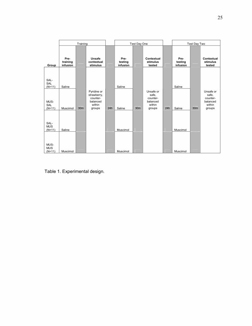
Subjects were randomly assigned to one of four groups. Each group was
assigned to receive either saline or muscimol 30m prior to a training session, and
either saline or muscimol prior to two subsequent test sessions on consecutive days.
Thus, each group received infusions on three occasions, separated by 24hr. The first
group (SAL-SAL, n = 15) received saline prior to both training and testing sessions.
Group MUS-SAL (n = 15) received muscimol infusions prior to training, and saline
infusions prior to testing sessions. A third group (SAL-MUS, n = 15) received saline
prior to training and muscimol prior to testing. A fourth group (MUS-MUS, n = 15)
received muscimol prior to both training and testing sessions.
Between surgery and the onset of behavioral procedures, each subject was
brought to the infusion room for 2 min every 2 days, and the pump was run in the
background in order to acclimate subjects to the infusion room and noise associated
with infusion. Infusions began 10 days after surgery, when subjects were brought
individually in clear plastic boxes to the infusion room. The obturator was removed
and replaced with 30-gauge injection cannula, attached by polyethylene tubing (PE-
10) to 10-µL Hamilton syringes mounted in an infusion pump (Harvard Apparatus,
South Natick, MA). Bilateral microinfusion of saline (0.9%, pH=7.4) or muscimol
(1µg/µL dissolved in 0.9% saline; Sigma Aldrich, St. Louis, MO) occurred over a 1.5-
8
min period. Each bilateral infusion introduced volumes of 0.25µL saline or muscimol
per hemisphere, for a total volume of 0.5µL into dorsal hippocampus. Rats were held
during the infusion period to prevent dislodging tubing, and held for an additional 2
min to allow diffusion of the drug or vehicle prior to removal of the injection cannula.
Dummy cannula and cap were then replaced, and the subject was returned to the
holding box for 28 min before transporting to the training or testing room so that 30
min separated infusions from training or testing sessions.
2.3.3. Olfactory contextual delay fear conditioning
Training took place 10 days after surgery in a single session, in one of two
identical behavioral chambers (A and B) in the same room. Whether an animal
received training in Chamber A or B was counterbalanced for each group. Each
subject received an infusion of either saline or muscimol 30 min prior to training (see
2.3.2., Drug infusion). During the 35-min training session, two olfactory contexts were
presented in alternation, each for 5 min and separated from each other by 1-min
inter-context-intervals. Thus, each context was presented three times. During context
periods referred to as �safe�, no auditory or footshock stimuli were presented; during
each presentation of the �unsafe� context, subjects were exposed to three pairings of
a 3.9 kHz-pure tone (~80dB, 20s) co-terminating with a footshock (2 sec, 0.5mA).
Successive CS-US pairings were separated by a 1 min ITI. Therefore, a total of nine
CS-US pairings was presented during training, each against the background of an
�unsafe� unimodal olfactory contextual stimulus (see Fig. 1). Strawberry extract and
pyridine, 1.5 L/min flow rate, served as olfactory contextual stimuli and were
counterbalanced for all groups.
2.3.4. Testing
9
Testing began 24 h after training in a separate room and chamber, as
described in Section 2.2 (Apparatus) above. Thirty min after an infusion of either
saline or muscimol, subjects were placed in the testing chamber for 10 min. In each
testing session, one contextual stimulus was presented during Min 3, and the tone
CS was presented during Min 10. No other stimuli were explicitly presented.
Responses to unsafe and safe contextual stimuli were assessed on consecutive
days, with unsafe day counterbalanced for each group (see Table 1). That is, for a
subject in group SAL-MUS assigned to unsafe test Day 1, muscimol infusion first
occurred prior to unsafe context testing on Day 1. Muscimol was infused 24 hr later
on test Day 2, and the safe context was tested. Each subject therefore received two
testing sessions separated by 24 hr, with each test session preceded by the same
drug infusion. For all test sessions, behavioral measures were scored by a human
observer, other data were compiled by a computer, and a videotape was made for
offline analysis.
2.3.5. Behavioral measures
2.3.6. Primary measure of conditioned fear
The primary dependent measure of fear was freezing behavior, scored
continuously by a human observer blind to the infusion condition of each subject. To
record the onset and offset of freezing (the adoption of rigid posture unaccompanied
by body, extremity, head, or whisker movement except that required for respiration),
the observer held a hand switch sampled by the computer each second. Depression
of the switch recorded freezing onset, while release of the switch constituted offset.
For each minute, freezing observations were transformed to a percentage of total
observations, yielding the percent time spent freezing during each minute.
Simultaneously, an infrared motion detector (see section 2.2, Apparatus) registered
10
immobility and was sampled by the computer controlling stimuli presentation, yielding
the percent time spent immobile during each minute of the testing session.
2.3.7. Secondary behavioral measures
Previous studies have found a high and positive correlation between freezing
behavior assessed by human subjects and immobility registered by computerized
means (Anagnostaras et al., 2001; Yoon & Otto, 2001), and freezing behavior is
generally accepted to be a valid measure of conditioned fear (Fanselow, 1980;
Maren, 1998). However, interpretations of freezing data can potentially be
confounded by performance accounts (Anagnostaras et al., 2001; Holt & Maren,
1999). The testing paradigm used in the current study allowed the observation of
behavior conditioned to a �safe� context on a day different from the �unsafe� test,
perhaps introducing other relevant dependent variables. In addition to freezing, the
occurrence of whisking, grooming, ambulating, and rearing behavior were measured
during test sessions. Data were collected offline from videotapes using a momentary
time-sampling technique. Briefly, tapes were scored by trained human observers,
who categorized behavior during each 5-s epoch of a session as one of the following:
rearing (standing on hind legs with the upper limbs above midline), ambulating
(consecutive grossmotor movements that resulted in displacement of the rat in the
horizontal plane), whisking (visible movement of vibrissae in the air or contacting
surfaces), grooming (body part to body part contact), or freezing (rigid and
motionless posture, except for respiration-related movement). These data were
coded by an observer unaware of the condition of subjects, and were converted into
percentage of time spent engaged in freezing, rearing, ambulating, whisking or
grooming during relevant testing periods including pre-context, context, post-context,
pre-CS, and CS presentation for analysis.
2.3.8. Histology
11
Following the last testing session, each subject was anesthetized with sodium
pentobarbitol (100 mg/kg, ip) and perfused transcardially with 0.9% saline and 10%
buffered formalin solution. Brains were removed and held in 30% sucrose solution
(wt/vol) for at least 48 hr, then frozen and sliced into coronal sections of 50µm
thickness. Slices were mounted on glass slides, stained with cresyl violet, and
examined visually via a light microscope for verification of cannula placement in
dorsal hippocampus.
2.3.9. Statistical analysis
Freezing behavior and immobility during testing sessions were statistically
analyzed using two-way repeated measures analyses of variance (ANOVAS), with
treatment group as the between-subjects factor and testing minute as the within-
subjects factor. Subsequent multiple pairwise comparisons were conducted using
Student-Newman-Keuls (SNK) post-hoc tests. A one-way ANOVA was used to
analyze treatment groups� difference between conditioned freezing to safe versus
unsafe unimodal contextual stimuli, and subsequent post-hoc analyses were
conducted using Dunnett�s test (α=.05). Data from offline video analysis, including
rearing, ambulating, grooming, whisking, and freezing, were analyzed using separate
two-way ANOVAs, with treatment group as the between-subjects factor and testing
period the within-subjects factor. A one-way ANOVA was performed on unsafe test
data of the control group (SAL-SAL), to analyze effects of testing the unsafe
contextual stimulus on Day 1 versus Day 2. Likewise, a one-way ANOVA was
performed within the SAL-SAL group on unsafe odor, to analyze the effects of
different olfactory contextual stimuli (strawberry versus pyridine odor) on contextually
conditioned freezing.
3. Results
3.1. Cannula placement
12
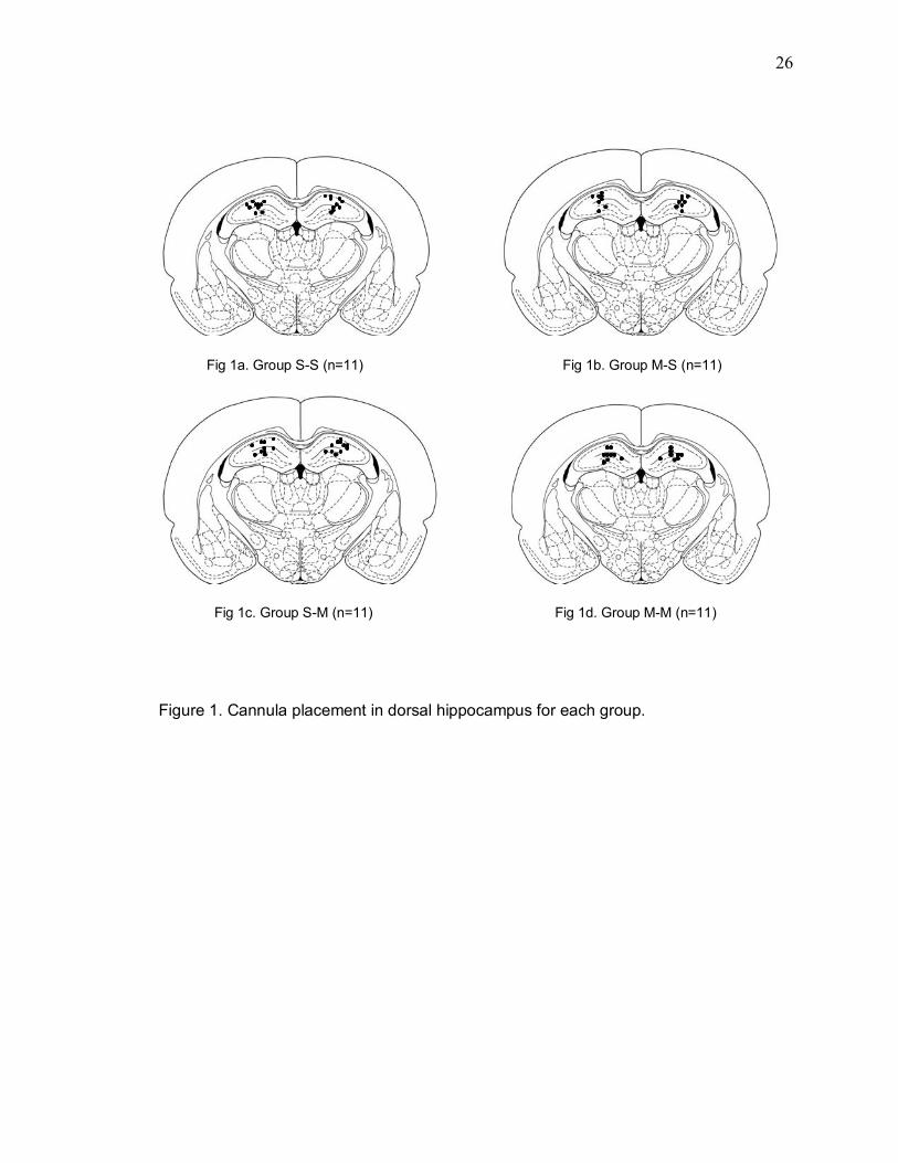
Three subjects were excluded from statistical analyses due to post-surgical
complications. Muscimol is expected to spread at least 1 mm below the site of
infusion (Martin, 1991); thus, subjects were retained for statistical analyses only if
cannula tips were localized within the dorsal hippocampus, above or within (but not
below) area CA3. Following histological examination of the location of cannula tracks
in dorsal hippocampus, four subjects were removed from group SAL-SAL and three
subjects were removed from the MUS-SAL, SAL-MUS, and MUS-MUS groups,
resulting in final group sizes of 11 subjects in each group for statistical analyses.
Figure 1 illustrates cannula placement in the dorsal hippocampus for each group.
3.1.2. Correlation between hand-scored freezing behavior and computer-detected
immobility
Correlational analyses compared freezing data obtained by human observers
to the computerized detection of immobility during the contextual conditioning test.
That is, for relevant periods during testing, the number of seconds that each subject
was scored as �freezing� by a human observer was examined against the number of
seconds during which the motion detector recorded immobility. Two correlation
coefficients were obtained. First, the unsafe context correlation coefficient was 0.91
(p < 0.0001), between the number of seconds recorded by a human observer as
freezing and the number of seconds recorded as immobile by the computerized
motion detection system, during the unsafe context presentation. Second, the safe
context correlation coefficient was 0.76 (p = 0.20), between the human observer-
scored freezing and computer-scored immobility during the safe contextual stimulus
presentation. In general, these correlations are similar to those previously reported in
the literature comparing human observer scores of freezing; the figures within the
text to follow show freezing data only.
3.1.3. Effects of particular contextual stimuli and day of unsafe context test

13
As mentioned previously, the odor used as unsafe context was
counterbalanced within groups. In order to determine the effects of particular odor
(strawberry odor versus pyridine odor) as unsafe contextual stimulus on conditioned
contextual freezing, a one-way ANOVA was performed on freezing during unsafe
context (odor). For group SAL-SAL, results of this one-way ANOVA revealed no
difference between subjects contextually conditioned with strawberry, and subjects
for whom the unsafe contextual stimulus was pyridine (F(1,9) = 0.88, p > 0.05).
Therefore, subsequent analyses collapsed data across unsafe contextual stimulus
(odor). Also counterbalanced within groups was unsafe context testing day, with
some subjects tested for unsafe contextual conditioning on Day 1, and others on Day
2. To evaluate the effects of unsafe context testing day on contextual freezing for
SAL-SAL subjects, a one-way ANOVA was performed for freezing on unsafe context
testing day one versus unsafe context testing day 2. This analysis did not reveal a
difference between unsafe context testing days (F(1,9) = 0.49, p > 0.05). Data are
thus collapsed across unsafe context test day.
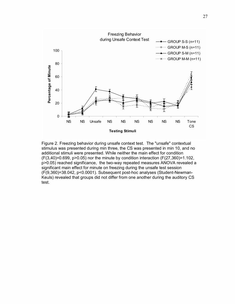
3.1.4. Freezing to unsafe olfactory contexts and auditory CS
Data from the unsafe context testing session 24 or 48 hr after training are
presented in Figure 2. The effects of infusions of muscimol into dorsal hippocampus
on contextually conditioned freezing were analyzed using a two-way repeated-
measures ANOVA with one repeated measure (Min). Neither the main effect of
condition (F(3,40) = 0.699, p > 0.05) nor the minute by condition interaction
(F(27,360) = 1.102, p > 0.05) reached significance. There was a significant main
effect of minute on freezing during the unsafe test session (F(9,360) = 38.042, p <
0.0001). Pairwise comparisons (Student-Newman-Keuls) showed that regardless of
infusion condition, all groups froze similarly to the auditory CS (p>0.05).
14
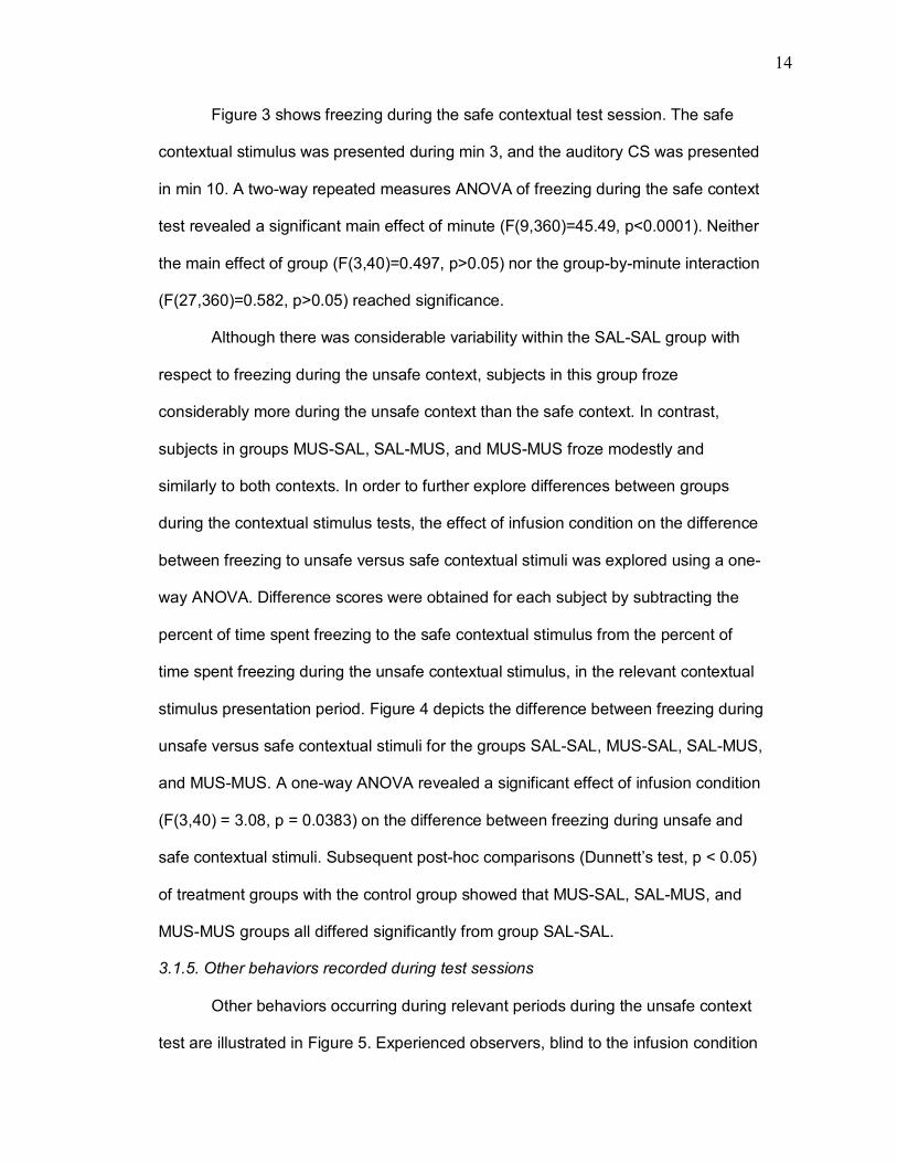
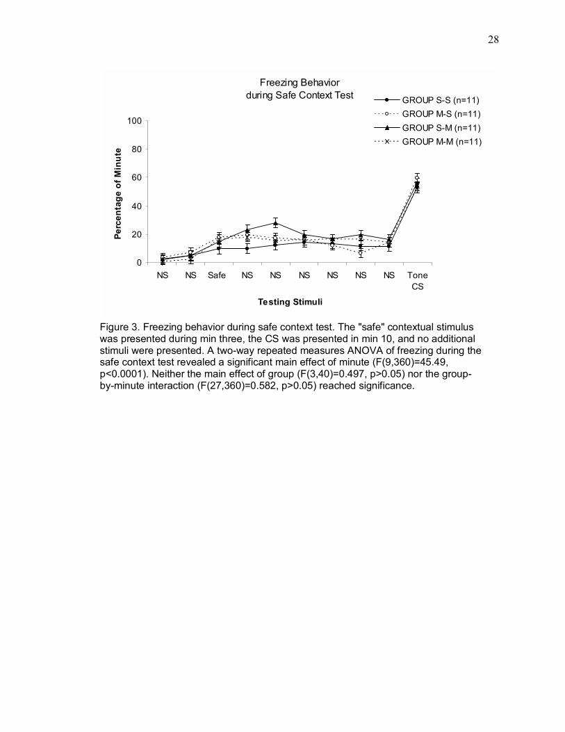
Figure 3 shows freezing during the safe contextual test session. The safe
contextual stimulus was presented during min 3, and the auditory CS was presented
in min 10. A two-way repeated measures ANOVA of freezing during the safe context
test revealed a significant main effect of minute (F(9,360)=45.49, p<0.0001). Neither
the main effect of group (F(3,40)=0.497, p>0.05) nor the group-by-minute interaction
(F(27,360)=0.582, p>0.05) reached significance.
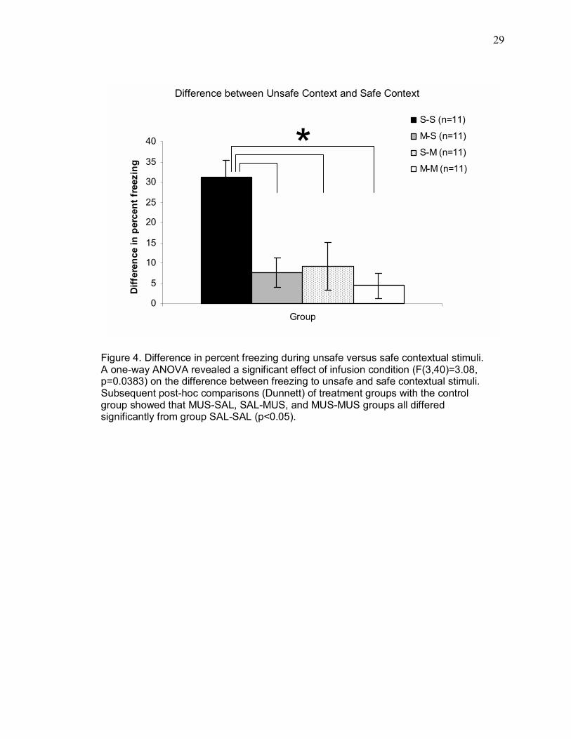
Although there was considerable variability within the SAL-SAL group with
respect to freezing during the unsafe context, subjects in this group froze
considerably more during the unsafe context than the safe context. In contrast,
subjects in groups MUS-SAL, SAL-MUS, and MUS-MUS froze modestly and
similarly to both contexts. In order to further explore differences between groups
during the contextual stimulus tests, the effect of infusion condition on the difference
between freezing to unsafe versus safe contextual stimuli was explored using a one-
way ANOVA. Difference scores were obtained for each subject by subtracting the
percent of time spent freezing to the safe contextual stimulus from the percent of
time spent freezing during the unsafe contextual stimulus, in the relevant contextual
stimulus presentation period. Figure 4 depicts the difference between freezing during
unsafe versus safe contextual stimuli for the groups SAL-SAL, MUS-SAL, SAL-MUS,
and MUS-MUS. A one-way ANOVA revealed a significant effect of infusion condition
(F(3,40) = 3.08, p = 0.0383) on the difference between freezing during unsafe and
safe contextual stimuli. Subsequent post-hoc comparisons (Dunnett�s test, p < 0.05)
of treatment groups with the control group showed that MUS-SAL, SAL-MUS, and
MUS-MUS groups all differed significantly from group SAL-SAL.
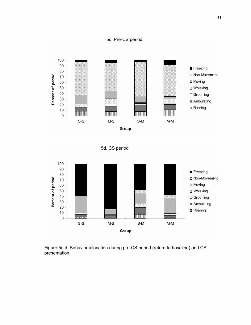
3.1.5. Other behaviors recorded during test sessions
Other behaviors occurring during relevant periods during the unsafe context
test are illustrated in Figure 5. Experienced observers, blind to the infusion condition
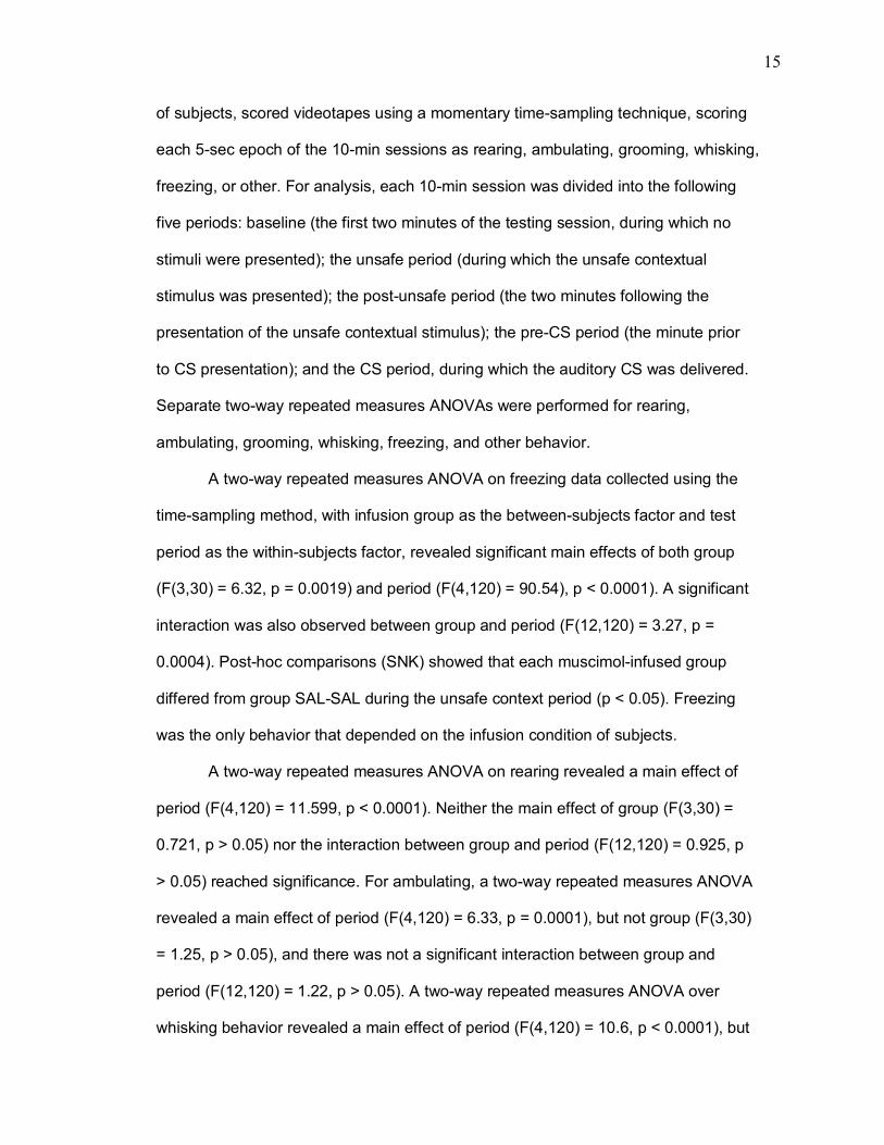
15
of subjects, scored videotapes using a momentary time-sampling technique, scoring
each 5-sec epoch of the 10-min sessions as rearing, ambulating, grooming, whisking,
freezing, or other. For analysis, each 10-min session was divided into the following
five periods: baseline (the first two minutes of the testing session, during which no
stimuli were presented); the unsafe period (during which the unsafe contextual
stimulus was presented); the post-unsafe period (the two minutes following the
presentation of the unsafe contextual stimulus); the pre-CS period (the minute prior
to CS presentation); and the CS period, during which the auditory CS was delivered.
Separate two-way repeated measures ANOVAs were performed for rearing,
ambulating, grooming, whisking, freezing, and other behavior.
A two-way repeated measures ANOVA on freezing data collected using the
time-sampling method, with infusion group as the between-subjects factor and test
period as the within-subjects factor, revealed significant main effects of both group
(F(3,30) = 6.32, p = 0.0019) and period (F(4,120) = 90.54), p < 0.0001). A significant
interaction was also observed between group and period (F(12,120) = 3.27, p =
0.0004). Post-hoc comparisons (SNK) showed that each muscimol-infused group
differed from group SAL-SAL during the unsafe context period (p < 0.05). Freezing
was the only behavior that depended on the infusion condition of subjects.
A two-way repeated measures ANOVA on rearing revealed a main effect of
period (F(4,120) = 11.599, p < 0.0001). Neither the main effect of group (F(3,30) =
0.721, p > 0.05) nor the interaction between group and period (F(12,120) = 0.925, p
> 0.05) reached significance. For ambulating, a two-way repeated measures ANOVA
revealed a main effect of period (F(4,120) = 6.33, p = 0.0001), but not group (F(3,30)
= 1.25, p > 0.05), and there was not a significant interaction between group and
period (F(12,120) = 1.22, p > 0.05). A two-way repeated measures ANOVA over
whisking behavior revealed a main effect of period (F(4,120) = 10.6, p < 0.0001), but

16
neither the main effect of group (F(3,30) = 0.346, p > 0.05) nor the group-by-period
interaction (F(12,120) = 0.895, p > 0.05) reached significance. Finally, a two-way
repeated measures ANOVA over grooming behavior revealed no main effects of
group (F(3,30) = 0.192, p > 0.05) or period (F(4,120) = 1.57, p > 0.05), and no
significant interaction (F(4,120) = 0.479, p > 0.05).
17
4. Discussion
The present results suggest that the temporary inactivation of dorsal
hippocampus, whether occurring prior to training, prior to subsequent testing, or
both, dramatically impairs contextual conditioning. In contrast, inactivation of dorsal
hippocampus spared subjects� abilities to acquire and express conditioned freezing
to an explicit auditory CS and had no effect on ambulation, grooming, rearing, and
whisking behavior. The selective deficit in olfactory contextual conditioning is likely
not attributable to stimulus, means of dorsal hippocampus inactivation, day of testing,
or the specific odor used as the �unsafe� contextual stimulus. These data provide
further evidence that dorsal hippocampus participates in the association of a US with
contextual stimuli that are relatively ambiguous and temporally and spatially diffuse.
That is, when context is operationalized as a unimodal cue both temporally and
spatially diffuse from the US, dorsal hippocampus is critically involved in the
acquisition and expression of contextual fear.
The role of dorsal hippocampus in contextual fear conditioning has been
examined using both temporary inactivation (Corcoran & Maren, 2001; Holt & Maren,
1999) and multiple means of producing permanent damage (Maren 1999; Maren &
Fanselow, 1997; Otto & Poon, 2006). Methods of producing both temporary
(Bellgowan & Helmstetter, 1995) and permanent (Douglas & Isaacson, 1964; Maren
& Fanselow, 1997) hippocampal damage have been reported to affect rats� ability to
perform the freezing response. In a related vein, McNish, Gewirtz, and Davis (1997)
have found discrepancies in the effects of hippocampal damage on freezing versus
fear-potentiated startle. Therefore, the current study employed multiple measures
both in order to rule out potential performance accounts of any deficits observed in
animals with dorsal hippocampal inactivation, and to assess whether behaviors other
than freezing were systematically related to contextual fear memory. These data
18
show no group differences with respect to rearing, ambulating, grooming, or
whisking, indicating that the ability of animals to physically execute the responses of
interest was not likely compromised by inactivation of dorsal hippocampus. This
conclusion is strengthened by the fact that even for groups that did not appreciably
freeze during presentation of contextual stimuli, robust freezing occurred later in the
same test session, during CS presentation.
It might legitimately be questioned whether the pattern of results seen here
could be attributed to the specific modality of contextual stimuli used in the present
paradigm. However, recent work by Otto and Poon (2006) has shown that while
lesions of dorsal hippocampus impaired freezing to an olfactory contextual stimulus,
the identical olfactory stimulus elicited freezing when serving as a discrete,
temporally punctate CS. Thus, stimulus modality alone cannot account for the failure
of animals with dorsal hippocampal damage to acquire contextual fear conditioning.
Furthermore, the current experiment counterbalanced odors used as the unsafe
context across subjects. Regardless of the specific stimulus serving as the unsafe
contextual stimulus during training, subjects effectively acquired contextual fear
conditioning when dorsal hippocampus was intact during training and testing, while
subjects infused with muscimol did not.
Dissociating the potentially functional difference of spatial learning and
contextual fear conditioning has been complicated by the fact that context is typically
defined as the chamber in which conditioning occurred (Kim, Rison, & Fanselow,
1993; Phillips & LeDoux, 1992; Maren & Fanselow, 1997; Young et al., 1994),
although it is widely acknowledged that stimuli other than spatially defined cues can
serve as effective contextual variables (Otto et al., 1997; Otto & Poon, 2006;
Overton, 1964; Randrich & Ross, 1985). But are spatial and olfactory contexts
functionally dissociable? Recent research by Waxler and Otto (2004) suggests that

19
they are: in their study, hippocampal place fields were observed to remap during
exploration of a novel spatial environment�but not to novel odors�and to remain
stable during olfactory contextual conditioning even though subjects acquired
freezing responses to an �unsafe� but not a �safe� context within the same space.
Similarly, other researchers have suggested that a neural representation of space is
distributed across the hippocampus, with certain place fields remapping completely
when an animal enters a novel spatial environment (Muller, Kubie, & Ranck Jr.,
1987), while other fields only �partially remap� when aspects of the environment
other than space are changed (Jeffrey, Anderson, Hayman, & Chakraborty, 2004).
Results of the current study are consistent with the idea that spatial and other forms
of contextual conditioning may be dissociable processes (Bannerman et al., 2004;
Richmond et al., 1999) with overlapping neural substrates (Anderson et al., 2006;
Barry et al., 2006).
While hippocampal damage typically impairs spatial learning (Jarrard, 1983;
Morris, Schenk, Tweedie, & Jarrard, 1990; Olton, Becker, & Handelmann, 1979;
Sutherland et al., 1983), research suggests that the mere exploration of a novel
environment does not depend on an intact dorsal hippocampus (Gaskin, Chai &
White, 2005). This possibility is supported by results of the current study, in which
testing occurred in a separate room and chamber, positioned at a different
orientation, and containing tactile, visual, and olfactory stimuli different from those
used during training. If an intact dorsal hippocampus is required for the exploration of
novel spatial contexts, behavioral correlates of exploration such as rearing
(Anderson et al., 2006; Gaskin et al., 2005; Lever, Burton, & O�Keefe, 2006) would
be expected to be impaired by inactivation of dorsal hippocampus. In fact, neither
rearing nor ambulating (see Fig. 5) was impaired during testing sessions, which
occurred in a novel spatial environment. It is suggested that spatial and olfactory
20
contexts are not only topographically but also functionally dissociable, and that
dorsal hippocampus integrity may participate differently in their processing.
The results of several studies suggest that dorsal hippocampal lesions prior
to training may impair contextual learning (Maren & Fanselow, 1997; Phillips &
LeDoux, 1992), although other studies have detected only retrograde�and not
anterograde�impairments in contextual learning after such damage (Anagnostaras
et al., 2001; Maren et al., 1997; Richmond et al., 1999). Discrepancies such as these
have led some to conclude that contextual fear conditioning is more susceptible to
damage inflicted between training and testing than to lesions performed before
training (Bannerman et al., 2004; Richmond et al., 1999). Attempting to explain
cases in which an anterograde deficit was observed following dorsal hippocampal
damage, it has been suggested that fibers of passage between ventral and dorsal
hippocampus may have been responsible (Bannerman et al., 2004; Maren et al.,
1997). The current study speaks specifically to this controversy, in that reversible
inactivation of dorsal hippocampus produced both anterograde and retrograde
deficits in one form of contextual fear conditioning.
In studies of temporary inactivation, one precondition for interpreting effects is
first to rule out state-dependent learning (Bannerman et al., 2004; Overton, 1964). To
this end, the present experiment included one group (MUS-MUS) receiving muscimol
prior to both training and testing. If acquiring olfactory contextual fear conditioning
depends on being in the same drug state at the time of original conditioning and
subsequent testing, group MUS-MUS would be expected to effectively acquire and
express contextual fear conditioning (much like that seen in group SAL-SAL, Fig. 2).
However, the present results indicate an actual and dramatic impairment in group
MUS-MUS, suggesting state-dependent learning cannot account for effects.
21
The current paradigm afforded the assessment of conditioned responses to
the safe and the unsafe contextual stimuli on separate testing days. Freezing evoked
by two separate olfactory stimuli could be compared without potential confounds that
might have resulted from evaluating both stimuli in a single test session. Of course,
this design raises the possibility that for subjects presented with one contextual
stimulus on the first testing day, extinction could account for reduced freezing to the
second tested stimulus presented on day two. Importantly, an analysis of freezing to
the unsafe contextual stimulus as a function of test day indicated that for group SAL-
SAL, freezing was unrelated to day of testing.
The conditioning paradigm employed in the current study departs from that
typically used to assess conditioned responses to stimuli (Kim & Fanselow 1992).
Here, conditioning to contextual stimuli was assessed on consecutive days, with the
CS presented during the final minute of each session. Results of CS presentation are
consistent with the existing body of literature suggesting that auditory fear
conditioning is not disrupted by inactivation of dorsal hippocampus or hippocampal
damage (Kim & Fanselow, 1992; Otto & Poon, 2006), irrespective of both testing day
serial order and additional stimuli presented during the test session.
Anagnostaras et al. (1999) report that lesions of dorsal hippocampus severely
affected recently acquired, but not remote, (spatial) contextual fear memory. The
current study assessed olfactory contextual fear either 24 or 48 hr after original
training, therefore not addressing the time-course of dorsal hippocampal involvement
in contextual conditioning. A crucial and interesting follow-up to this study concerns
the participation of dorsal hippocampus in memory for olfactory contextual fear
conditioning at multiple time points after training.
Even as mounting evidence suggests that the hippocampus is involved in
encoding contextual representations (Frankland, Cestari, Filipkowski, McDonald, &
22
Silva, 1998; Good et al. 1998; Kim and Fanselow 1992; Maren et al. 1997; Phillips &
LeDoux 1992; Rudy & O'Reilly 2001), the extent to which dorsal hippocampus
normally participates in discrimination between contexts remains unclear (Frankland
et al., 1998; Holt & Maren, 1999). Holt and Maren (1999) demonstrated that in
contrast to saline-infused rats, in which the expression of latent inhibition to a tone
after pre-exposure was context-specific, animals with dorsal hippocampus
inactivation exhibited low levels of freezing to a tone independent of where tone pre-
exposure had occurred. This result seems incongruent with evidence that animals
with inactivation of dorsal hippocampus discriminated between spatial contexts,
freezing more in spatial contexts which had been associated with shock than in
neutral spatial contexts. To explain this discrepancy, the authors suggest that
muscimol disrupts contextual retrieval only in cases involving ambiguous cues or
contexts (Holt & Maren, 1999). In fact, there is some precedent for the idea that
hippocampal damage may reduce the ability of subjects to discriminate between
contexts. The computational model presented by O�Reilly and Rudy (2001) predicts a
degree of context generalization in subjects with hippocampal damage. Possible
evidence for a tendency to generalize between contexts was observed in the current
study in that animals with inactivation of dorsal hippocampus froze modestly and
similarly to both the unsafe and safe contextual stimuli, while control animals froze
robustly to the unsafe but not to the safe contextual stimuli (see Fig. 4). Assessing
the different contextual stimuli in the same spatial environment eliminated the ability
of subjects to utilize spatial cues to disambiguate the meaning of olfactory contextual
stimuli. Taken together, these results indicate that there is a decidedly nonspatial
component to the role of the dorsal hippocampus in contextual conditioning, and that
olfactory contextual conditioning is a fruitful means of examining such a function.
23
While several studies have concluded that selective damage of dorsal
hippocampus does not produce anterograde deficits in contextually conditioned
freezing (Richmond et al., 1999; Maren et al., 1997), compelling evidence suggests
that space is not the only contextual variable encoded by the hippocampus
(Anderson et al., 2006; Otto & Poon, 2006). Results of the present study support this
notion, and further suggest that theories of hippocampal function need to account for
forms of explicitly nonspatial forms of learning. The time-course and exact nature of
dorsal hippocampus� normal participation in such explicitly nonspatial forms of
contextual learning remains to be seen.

24
5. Appendix
25
Training Test Day One Test Day Two
Group
Pre-training infusion
Unsafe contextual stimulus
Pre-testing
infusion
Contextual stimulus
tested
Pre-testing
infusion
Contextual stimulus
tested
SAL-SAL (N=11) Saline Saline Saline
MUS-SAL (N=11) Muscimol 30m
Pyridine or strawberry,
counter-balanced
within groups 24h Saline 30m
Unsafe or safe,
counter-balanced
within groups 24h Saline 30m
Unsafe or safe,
counter-balanced
within groups
SAL-MUS (N=11) Saline Muscimol Muscimol
MUS-MUS (N=11) Muscimol Muscimol Muscimol
Table 1. Experimental design.
26
Figure 1. Cannula placement in dorsal hippocampus for each group.
Fig 1a. Group S-S (n=11) Fig 1b. Group M-S (n=11)
Fig 1c. Group S-M (n=11) Fig 1d. Group M-M (n=11)
27
Freezing Behavior during Unsafe Context Test
0
20
40
60
80
100
NS NS Unsafe NS NS NS NS NS NS ToneCS
Testing Stimuli
Perc
enta
ge o
f Min
ute
GROUP S-S (n=11)GROUP M-S (n=11)GROUP S-M (n=11)GROUP M-M (n=11)
Figure 2. Freezing behavior during unsafe context test. The "unsafe" contextual stimulus was presented during min three, the CS was presented in min 10, and no additional stimuli were presented. While neither the main effect for condition (F(3,40)=0.699, p>0.05) nor the minute by condition interaction (F(27,360)=1.102, p>0.05) reached significance, the two-way repeated measures ANOVA revealed a significant main effect for minute on freezing during the unsafe test session (F(9,360)=38.042, p<0.0001). Subsequent post-hoc analyses (Student-Newman-Keuls) revealed that groups did not differ from one another during the auditory CS test.
28
Freezing Behavior during Safe Context Test
0
20
40
60
80
100
NS NS Safe NS NS NS NS NS NS ToneCS
Testing Stimuli
Perc
enta
ge o
f Min
ute
GROUP S-S (n=11)GROUP M-S (n=11)GROUP S-M (n=11)GROUP M-M (n=11)
Figure 3. Freezing behavior during safe context test. The "safe" contextual stimulus was presented during min three, the CS was presented in min 10, and no additional stimuli were presented. A two-way repeated measures ANOVA of freezing during the safe context test revealed a significant main effect of minute (F(9,360)=45.49, p<0.0001). Neither the main effect of group (F(3,40)=0.497, p>0.05) nor the group-by-minute interaction (F(27,360)=0.582, p>0.05) reached significance.
29
Figure 4. Difference in percent freezing during unsafe versus safe contextual stimuli. A one-way ANOVA revealed a significant effect of infusion condition (F(3,40)=3.08, p=0.0383) on the difference between freezing to unsafe and safe contextual stimuli. Subsequent post-hoc comparisons (Dunnett) of treatment groups with the control group showed that MUS-SAL, SAL-MUS, and MUS-MUS groups all differed significantly from group SAL-SAL (p<0.05).
0
5
10
15
20
25
30
35
40
Group
Diff
eren
ce in
per
cent
free
zing
S-S (n=11)
M-S (n=11)
S-M (n=11)
M-M (n=11)*
Difference between Unsafe Context and Safe Context

30
0
10
20
30
40
50
60
70
80
90
100
S-S M-S S-M M-M
Group
Perc
ent o
f per
iod
Freezing
Non-Movement
Moving
Whisking
Grooming
Ambulating
Rearing
0102030405060708090
100
S-S M-S S-M M-M
Group
Perc
ent o
f per
iod
Freezing
Non-Movement
Moving
Whisking
Grooming
Ambulating
Rearing
Figure 5a-b. Behavior allocation during baseline and unsafe context presentation. Separate two-way repeated measures ANOVAs for freezing, rearing, ambulating, grooming, and whisking revealed a significant interaction between infusion condition (group) and testing period only for freezing behavior (F(12,120) = 3.27,p = 0.0004). Subsequent post-hoc comparisons (Student-Newman Keuls) revealed that for freezing behavior, group SAL-SAL differed from group MUS-SAL, group SAL-MUS, and group MUS-MUS during the unsafe context presentation (Fig. 5b).
5a. Baseline
5b. Unsafe Context Presentation
*
31
0102030405060708090
100
S-S M-S S-M M-M
Group
Perc
ent o
f per
iod
Freezing
Non-Movement
Moving
Whisking
Grooming
Ambulating
Rearing
0102030405060708090
100
S-S M-S S-M M-M
Group
Perc
ent o
f per
iod
Freezing
Non-Movement
Moving
Whisking
Grooming
Ambulating
Rearing
Figure 5c-d. Behavior allocation during pre-CS period (return to baseline) and CS presentation.
5c. Pre-CS period
5d. CS period
32
References Anagnostaras, S.G., Gale, G.D., & Fanselow, M.S. (2001). The hippocampus and
contextual fear conditioning: Recent controversies and advances. Hippocampus, 11, 8-17.
Anderson, M.I., Killing, S., Morris, C., O'Donoghue, A., Onyiagha, D., Stevenson, R.,
Verriotis, M., & Jeffrey, K.J. (2006). Behavioral correlates of the distributed coding of spatial context. Hippocampus, 16, 730-742.
Balsam, P.D., & Tomie, A. (1985). Context and learning. Hillsdale, NJ: Erlbaum. Bannerman, D.M., Deacon, R.M.I., Offen, S., Friswell, J., Grubb, M., Rawlins, J.N.P.
(2002). A double dissociation of function within the hippocampus: spatial memory and hyponeophagia. Behavioral Neuroscience, 116, 884-901.
Bannerman, D.M., Rawlins, J.N.P., McHugh, S.B., Deacon, R.M.J., Yee, B.K., Bast,
T., Zhang, W-N., Pothuizen, H.J., & Feldon, J. (2004). Regional dissociations within the hippocampus- memory and anxiety. Neuroscience and Biobehavioral Reviews, 28, 273�283.
Bannerman, D.M., Rawlins, J.N.P., & Good, M.A. (2006). The drugs don't work-or
do they? Pharmacological and transgenic studies of the contribution of NMDA and GluR-A-containing AMPA receptors to hippocampal-dependent memory. Psychopharmacology, 188, 552-566.
Barry, C., Lever, C., Hayman, R., Hartley, R., Burton, S., O'Keefe, J., Jeffrey, K.J., &
Burgess, N. (2006). The boundary vector cell model of place cell firing and spatial memory. Reviews in Neuroscience, 17, 71-97.
Bellgowan, P.S.F., & Helmstetter, F.J. (1995). Effects of muscimol applied to the
dorsal hippocampus on the acquisition and expression of cued versus contextual fear conditioning. Society for Neuroscience Abstracts 21:1219.
Bouton, M.E., & Bolles, R.C. (1985). Context, event-memories, and extinction.
Hillsdale, NJ: Lawrence Erlbaum Associates. Brun, V.H., Otnass, M.K., Molden, S., Steffenach, H.A., Witter, M.P., Moser, M.B., &
Moser, E.I. (2002). Place cells and place recognition maintained by direct entorhinal hippocampal circuitry. Science, 296, 2243�2246.
Chan-Palay, V. (1978). Quantitative visualization of gamma-aminobutyric acid
receptors in hippocampus and area dentata demonstrated by 3H muscimol autoradiography. Proceedings of the National Academy of Science, 75, 2516-2520.
Corcoran, K.A., & Maren, S. (2001). Hippocampal inactivation disrupts contextual
retrieval of fear memory after extinction. Journal of Neuroscience, 21, 1720-1728.
33
Cousens, G., & Otto, T. (1998). Both pre- and post-training lesions of the basolateral amygdala abolish the expression of olfactory and contextual fear conditioning. Behavioral Neuroscience, 112, 1092-1103.
Davidson, T.L., and Jarrard, L.E. (1993). A role for the hippocampus is the utilization
of hunger signals. Behavioral and Neural Biology, 59, 167-171. Douglas, R.J., & Isaacson, R.L. (1964). Hippocampal lesions and activity.
Psychological Science, 1, 187-188. Eichenbaum, H., Otto, T., & Cohen, N.J. (1994). Two functional components of the
hippocampal memory system. Behavioral and Brain Sciences, 17, 449-517. Eichenbaum, H., Schoenbaum, G., Young, B., & Bunsey, M. (1996). Functional
organisation of the hippocampal memory system. Proceedings of the National Academy of Science, 93, 13500-13507.
Fanselow, M. (1980). Conditional and unconditional components of post shock
freezing in rats. Pavlovian Journal of Biological Science, 15, 177-182. Fendt, M., & Fanselow, M. S. (1999). The neuroanatomical and neurochemical basis
of conditioned fear. Neuroscience and Biobehavioral Reviews, 23, 743-760. Frankland, P.W., Cestari, V., Filipkowski, R., McDonald, R.J., & Silva, A.J. (1998).
The dorsal hippocampus is essential for context discrimination but not for contextual conditioning. Behavioral Neuroscience, 112, 863-874.
Gaskin, S., Chai, S., & White, N. (2005). Inactivation of the dorsal hippocampus does
not affect learning during exploration of a novel environment. Hippocampus, 15, 1085-1093.
Hargreaves, E.L., Rao, G., Lee, I., & Knierim, J.J. (2005) Major dissociation between
medial and lateral entorhinal input to dorsal hippocampus. Science, 308, 1792-1794.
Herzog, C., & Otto, T. (1997). Odor-guided fear conditioning. II. Lesions of anterior
perirhinal cortex disrupt fear conditioned to the explicit CS but not to the training context. Behavioral Neuroscience, 111, 1265-1274.
Herzog, C. D. & Otto, T. (1998). Contributions of anterior perirhinal cortex to olfactory
and contextual fear conditioning. NeuroReport, 9, 1855-1859. Hock, B.J., & Bunsey, M.D. (1998). Differential effects of dorsal and ventral
hippocampal lesions. Journal of Neuroscience, 18, 7027-7032. Holt, W., & Maren, S. (1999). Muscimol inactivation of the dorsal hippocampus
impairs contextual retrieval of fear memory. Journal of Neuroscience, 19, 9054-9062.

34
Hulse, S.H., Cynx, J., & Humpal, J. (1984). Absolute and relative pitch discrimination in serial pitch perception by birds. Journal of Experimental Psychology: General, 113, 38�54.
Ishikawa, A., & Nakamura, S. (2006). Ventral hippocampal neurons project axons
simultaneously to the medial prefrontal cortex and amygdala in the rat. Journal of Neurophysiology, 96, 2134-2138.
Jarrard, L.E. (1983). Selective hippocampal lesions and behavior: Effects of kainic
acid lesions on performance of place and cue tasks. Behavioral Neuroscience, 97, 251-259.
Jeffrey, K.J., Anderson, M. I., Hayman, R., & Chakraborty, S. (2004). A proposed
architecture for the neural representation of spatial context. Neuroscience and Biobehavioral Reviews, 28, 201-218.
Kim, J.J. & Fanselow, M.S. (1992). Modality-specific retrograde amnesia of fear.
Science, 256, 657-677. Kim, J.J., Rison, R.A., & Fanselow, M.S. (1993). Effects of amygdala, hippocampus,
and periaqueductal gray lesions on short- and long-term contextual fear. Behavioral Neuroscience, 197, 1093-1098.
Kusljic, S., & van den Buuse, M. (2004). Functional dissociation between
serotonergic pathways in dorsal and ventral hippocampus in psychotomimetic drug-induced locomotor hyperactivity and prepulse inhibition in rats. European Journal of Neuroscience, 20, 3424-3432.
Lever, C., Burton, S., & O'Keefe, J. (2006). Rearing on hind legs, environmental
novelty, and the hippocampal formation. Reviews in the Neurosciences, 17, 111-133.
Maren, S. (1999). Neurotoxic basolateral amygdala lesions impair learning and
memory but not the performance of conditional fear in rats. Journal of Neuroscience, 19, 8696-8703.
Maren, S., & Fanselow, M.S. (1997). Electrolytic lesions of the dorsal hippocampus,
fimbria-fornix, or entorhinal cortex produce anterograde deficits in contextual fear conditioning in rats. Neurobiolopgy of Learning and Memory, 67, 142-149.
Maren, S., Aharonov, G., & Fanselow, M.S. (1997). Neurotoxic lesions of the dorsal
hippocampus and Pavlovian fear conditioning in rats. Behavioral Brain Research, 88, 261-274.
Martin, J.H. (1991). Autoradiographic estimation of the extent of reversible
inactivation produced by microinjection of lidocaine and muscimol in the rat. Neuroscience Letters, 127, 160-164.
35
Matus-Amat, P., Higgins, E. A., Barrientos, R. M., & Rudy, J. (2004). The role of the dorsal hippocampus in the acquisition and retrieval of context memory representations. Journal of Neuroscience, 24, 2431-2439.
McNish, K.A., Gewirtz, J.C., & Davis, M. (1997). Evidence of contextual fear after
lesions of the hippocampus: A disruption of freezing but not fear-potentiated startle. Journal of Neuroscience, 17, 9353-9360.
Morris, R.G.M. (1981). Spatial location does not require the presence of local cues.
Learning and Motivation, 12, 317-338. Morris, R.G.M., Schenk, F., Tweedie, F., & Jarrard, L.E. (1990). Ibotenate lesions of
hippocampus and/or subiculum: Dissociating components of allocentric spatial learning. European Journal of Neuroscience, 2, 1016-1028.
Moser, M.B., and Moser, E.I. (1998). Distributed encoding and retrieval of spatial
memory in the hippocampus. Journal of Neuroscience, 18, 7535-7542. Moser, E., Moser, M.B., & Andersen, P. (1993). Spatial learning impairment parallels
the magnitude of dorsal hippocampal lesions, but it is hardly present following ventral lesions. Journal of Neuroscience, 13, 3916-3925.
Muller, R.U., Kubie, J.L., & Ranck Jr., J.B. (1987). Spatial firing patterns of
hippocampal complex-spike cells in a fixed environment. Journal of Neuroscience, 7, 1935-1950.
Olton, D.S., Becker, J.T., & Handelmann, G.E. (1979). The hippocampus, space, and
memory. Behavioral and Brain Sciences, 2, 313-365. O'Reilly, R.C., & Rudy, J.W. (2001). Conjunctive representations in learning and
memory: Principles of cortical and hippocampal function. Psychological Review, 108, 311-345.
Otto T, Cousens G.A., & Rajewski, K. (1997). Odor-guided fear conditioning in rats:
Acquisition, retention, and latent inhibition. Behavioral Neuroscience, 111, 1257-1264.
Otto, T. & Poon, P. (2006). Dorsal hippocampal contributions to unimodal contextual
conditioning. The Journal of Neuroscience, 26, 6603-6609. Overton, D.A. (1964). State-dependent or "dissociated" learning produced with
pentobarbital. Journal of Comparative and Physiological Psychology, 5, 3-12. Phillips, R. & LeDoux, J. (1992). Differential contribution of amygdala and
hippocampus to cued and contextual fear conditioning. Behavioral Neuroscience, 106, 274-285.
Pitkänen, A., Pikkarainen, M., Nurminen, N., & Ylinen, A. (2000). Reciprocal
connections between the amygdala and the hippocampal formation, perirhinal cortex, and postrhinal cortex in rat: A review. Annals Of The New York Academy Of Sciences, 911, 369-391.
36
Randrich, A., & Ross, R.T. (1985). Contextual stimuli mediate the effects of pre- and
post-exposure to the unconditioned stimulus on conditioned suppression. In P. D. Balsam & A. Tomie (Eds). Context and learning (pp. 105�132). Hillsdale, NJ: Erlbaum.
Richmond, M.A., Yee, B.K., Pouzet, B., Veenan, L., Rawlins, J.N.P., Feldon, J., &
Bannerman, D.M. (1999). Dissociating context and space within the hippocampus: Effects of complete, dorsal, and ventral excitotoxic hippocampal lesions on conditioned freezing and spatial learning. Behavioral Neuroscience, 113, 1189-1203.
Thomas, D.R. (1985). Contextual stimulus control of operant responding in pigeons.
In: Context and learning (Balsam, P.D., Tomie, A. eds), pp 295-321. Hillsdale, NJ: Erlbaum.
Waxler, D., and Otto, T. (2004). Olfactory contexts are dissociable from spatial
contexts in the rat hippocampus [abstract]. In: Pavlovian Society Abstracts; 2004 Sept 16-18, Baltimore. The Pavlovian Society.
Yoon, T., & Otto, T. (2001). Contributions of the dorsal and ventral hippocampus to
trace fear conditioning in rats. Society for Neuroscience Abstracts 89:14. Young, S.L., Bohenek, D.L., & Fanselow, M.S. (1994). NMDA processes mediate
anterograde amnesia of contextual fear conditioning induced by hippocampal damage: Immunization against amnesia by context preexposure. Behavioral Neuroscience, 108, 19-29.


![2008] Sophia Gershman - Rutgers University](https://img.pdfslide.net/doc/110x75/626d72f053347e6bb044a836/2008-sophia-gershman-rutgers-university.jpg)