Embed Size (px)
Citation preview

1
Applying the Neuromuscular Principles in TMD and Orthodontics
Clayton A. Chan, D.D.S., M.I.C.C.M.O.
Neuromuscular Dentistry = “Dentistry” The term “Neuromuscular” dentistry has evolved in meaning to specifically distinguish itself from the traditional approach to clinical dentistry, since our traditional concept of dentistry has approached the functioning of the masticatory system from a mechanical perspective.<1,2> Nature does not think mechanically, but rather obeys the laws of bio-physiology as it pertains to the posture of the mandible to the skull. Neuromuscular emphasizes the innervation of the nerves to the masticatory muscles as they pertain to the “physiologic posture” when establishing an occlusion. Neuromuscular dentistry also recognizes that generic rules of physiology and patho-physiology which apply to the skeletal muscles in other parts of the body also apply to the masticatory muscles. The TMJ is a joint that follows the generic principles of any body articulation. It allows freedom of movement of the mandible allowing the teeth to come together at a terminal contact position for stable occlusion. The neuromuscular principles follow all known parameters of medicine.<3> Today, clinicians are recognizing these profound neuromuscular concepts are nothing less than what our pioneering occlusion forefathers have acknowledged years ago. “Some very innovative and forward thinking dentists who forged a broader perspective to dentistry in the early 1920’s, challenged the present beliefs and views of the time by broadening the perspective of what dentistry was about and refining mechanical measuring and recording instruments to understand mandibular movements as they related to occlusion”. “They emphasized that a thorough diagnosis and understanding of the mouth as a functioning unit was the basis of gnathological principles.” <4> In the past the pioneering gnathologist were also criticized among their peers who distinguished themselves as ones that realized occlusions roll in treating dental disease, something more than what the majority at that time realized. The same is happening today. The foregoing statements reflect the same views and focus we clinicians are aiming for in today’s clinical dentistry. Today’s neuromuscular clinicians are doing nothing less than fulfilling the past gnathologist dream, broadening our perspective of what dentistry is about and refining our understanding of mandibular movements as they related to occlusion, muscles and the jaw joints with today’s computerized measuring and recording instrumentation, being physicians of the mouth. We have continued this journey to better understand our clinical dilemmas we all face by seeking scientific and rationale answers to better serve our patients. Today’s neuromuscular dentist is nothing less than a gnathologist, a physician of the
2
mouth, A DENTIST fulfilling the dream of doing quality comprehensive aesthetic dentistry and what is best for our patients. Literature suggests that the basis for TMD symptomology is a disturbance of proprioception at a mid brain level. <5> The cerebral cortex is the site of perception, thought, and ability to respond to a stimulus with anything more than a simple reflex reaction. Magoun and Moruzzi recognized the arousal site of the cerebral cortex was the reticular activating center (reticular formation), a center that that does not relay any specific message, but simply an arousal site in the brain. <6,>. The reticular activating system (RAS) is the site of slow component nocioceptive reflex relays from the dental propioceptive receptors. Nocioceptive relays have reciprocity with the extensor reflexes. Extensor and flexor effects on the mandible can be elicited by stimulating the areas in the reticular formation. <7> Griffin noted the slow component reflex is totally blocked with TMJ dysfunction patients.<8> Clinical result is trismus caused by bilateral inhibition of the slow component reflex (lingual mandibular reflex) with resultant hypertonicity of the mandibular elevators. This prolonged firing explains the usual finding that the prerelaxation habitual resting position of the mandible is closer to occlusal contact than after relaxation therapy with the Myomonitor. <9> The tonic activity of the reticular formation is disturbed with the resultant hypertonicity of muscle tone. Dentist should all be “Physicians of the Mouth” who diagnose and treat the problems associated with the trigeminal nerve system, since this cranial nerve (V) is the dentist nerve. Unfortunately muscles and nerves are the least diagnosed and often overlooked especially with the dental profession which pay little attention to muscles and how they affect our patient’s dental health.<10> Studies have indicated that 80-90% of all TMD problems are muscle related.<11,12> Muscles and muscle health cannot be clearly seen on radiographic analysis neither diagnosed with an x-ray alone. The importance of muscles and its effect on dental occlusion (micro-occlusion) and the temporomandibular joints have been highly under-rated although we superficially discuss the importance of the masticatory muscles in our profession.
Today, clinicians acknowledge the fundamental bio-physiologic principles in nature as they pertain to the neuromuscular occlusion arena. These basic principles apply to all areas of dentistry including restorative dentistry, orthodontics/ orthopedics and TMJ. The following are five points that must be considered:<13>
1. There exists numerous Musculoskeletal Occlusal Signs and Symptoms. 2. Identify an optimal starting point for diagnosis and treatment -
“PHYSIOLOGIC REST” - without manual intervention.
3
3. Recognize a physiologic mandibular opening and closing NEUROMUSCULAR TRAJECTORY along an isotonic path for STABILITY at a terminal contact position.
4. MICRO-OCCLUSION- Eliminates the afferent and efferent noxious proprioceptive stimuli of occlusion during mandibular closure with FREEDOM OF ENTRY and EXIT.
5. Can accurately OBJECTIVELY MEASURE and RECORD muscle and postural responses of the mandible in establishing an occlusion.
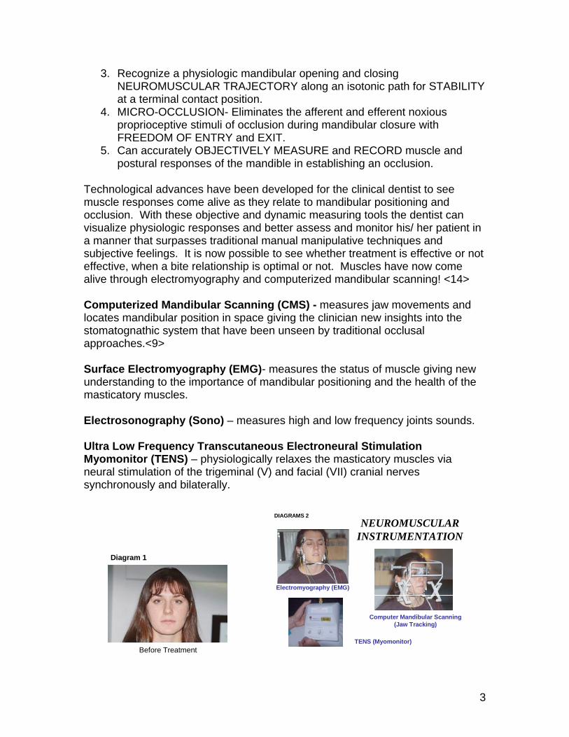
Technological advances have been developed for the clinical dentist to see muscle responses come alive as they relate to mandibular positioning and occlusion. With these objective and dynamic measuring tools the dentist can visualize physiologic responses and better assess and monitor his/ her patient in a manner that surpasses traditional manual manipulative techniques and subjective feelings. It is now possible to see whether treatment is effective or not effective, when a bite relationship is optimal or not. Muscles have now come alive through electromyography and computerized mandibular scanning! <14> Computerized Mandibular Scanning (CMS) - measures jaw movements and locates mandibular position in space giving the clinician new insights into the stomatognathic system that have been unseen by traditional occlusal approaches.<9> Surface Electromyography (EMG)- measures the status of muscle giving new understanding to the importance of mandibular positioning and the health of the masticatory muscles. Electrosonography (Sono) – measures high and low frequency joints sounds. Ultra Low Frequency Transcutaneous Electroneural Stimulation Myomonitor (TENS) – physiologically relaxes the masticatory muscles via neural stimulation of the trigeminal (V) and facial (VII) cranial nerves synchronously and bilaterally.
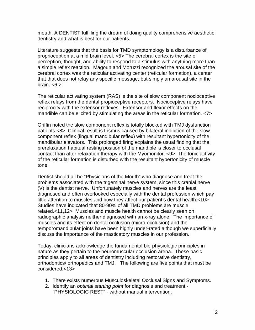
Before Treatment
Diagram 1
Electromyography (EMG)
TENS (Myomonitor)
Computer Mandibular Scanning (Jaw Tracking)
NEUROMUSCULAR INSTRUMENTATION
DIAGRAMS 2
4
CASE STUDY A Caucasian female age 19 experienced the following:
• Excruciating pain in the muscles around her jaw bilaterally for the past three years.
• Previous orthodontic therapy. • She has seen two previous dentists, specialists, and message therapy
with limited improvement. • She has had her teeth adjusted. • Doctors concluded that muscle spasms were due to teeth grinding. She
was placed on a ‘soft food diet’. She said she was eating with tongue her tongue between her teeth.
• Isometric exercises were recommended, no sleeping on pillows. A hard splint was made with limited help.
• Another dentist prescribed muscle relaxants, ibuprofen, and sleeping pills, along with hot packs with no relief of pain.
• Fifth doctor reshaped teeth and made a soft splint for lower jaw (worn at night), symptoms worsened.
A thorough evaluation was completed which included:
• Health History • TMJ Screening Questionnaire • Initial Consultation • Clinical Examination • Diagnostic records - study models, intra oral and extra oral photographs
along with a complete K7 Neuromuscular work up including EMG scans 9, 10, 11 and CMS scans 2, 6, 13, and 4/5. (Myotronics K7 Kineseograph, Tukwila Washington, USA).
• Delivery of a lower mandibular orthosis was indicated for stabilization and verification was made using the Myotronics K7 Kineseograph.
5
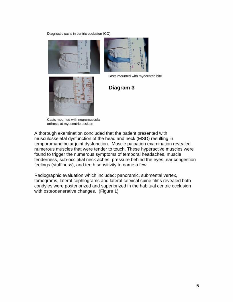
Diagnostic casts in centric occlusion (CO)
Casts mounted with myocentric bite
Casts mounted with neuromuscular orthosis at myocentric position
Diagram 3
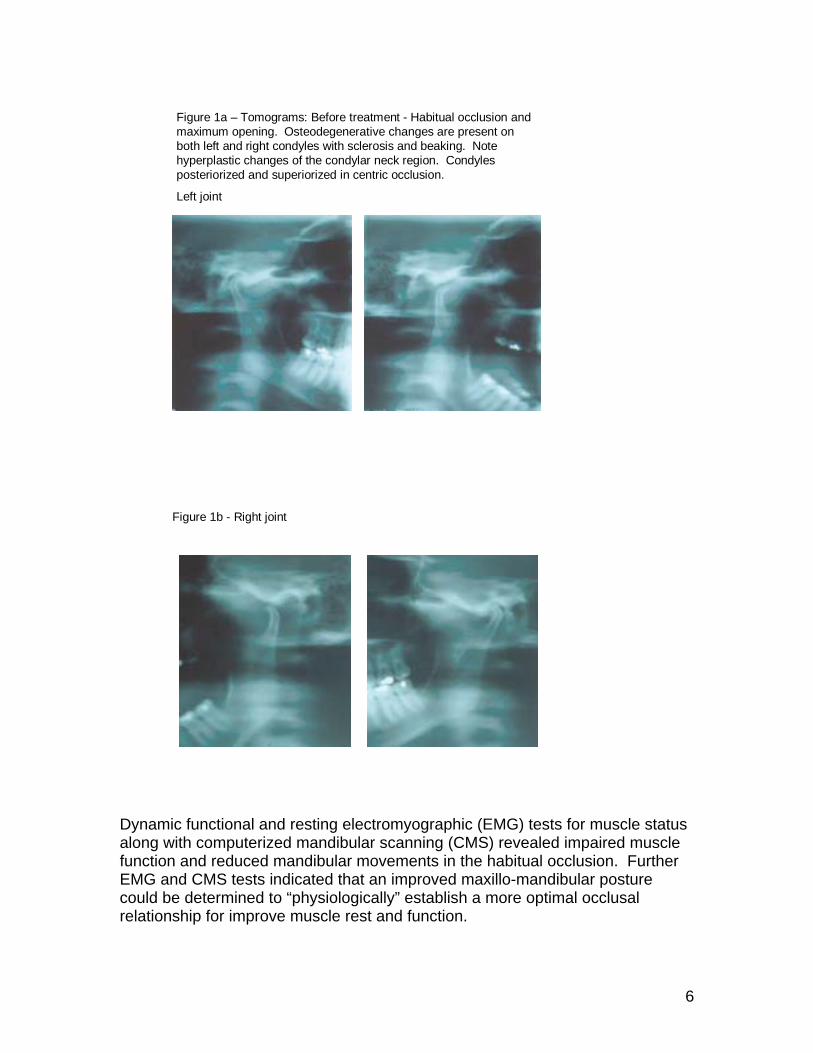
A thorough examination concluded that the patient presented with musculoskeletal dysfunction of the head and neck (MSD) resulting in temporomandibular joint dysfunction. Muscle palpation examination revealed numerous muscles that were tender to touch. These hyperactive muscles were found to trigger the numerous symptoms of temporal headaches, muscle tenderness, sub-occiptial neck aches, pressure behind the eyes, ear congestion feelings (stuffiness), and teeth sensitivity to name a few. Radiographic evaluation which included: panoramic, submental vertex, tomograms, lateral cephlograms and lateral cervical spine films revealed both condyles were posteriorized and superiorized in the habitual centric occlusion with osteodenerative changes. (Figure 1)
6
Figure 1a – Tomograms: Before treatment - Habitual occlusion and maximum opening. Osteodegenerative changes are present on both left and right condyles with sclerosis and beaking. Note hyperplastic changes of the condylar neck region. Condyles posteriorized and superiorized in centric occlusion.
Left joint
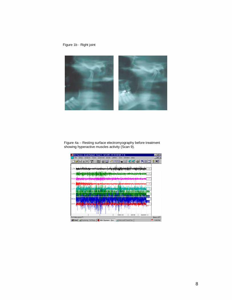
Figure 1b - Right joint
Dynamic functional and resting electromyographic (EMG) tests for muscle status along with computerized mandibular scanning (CMS) revealed impaired muscle function and reduced mandibular movements in the habitual occlusion. Further EMG and CMS tests indicated that an improved maxillo-mandibular posture could be determined to “physiologically” establish a more optimal occlusal relationship for improve muscle rest and function.
7
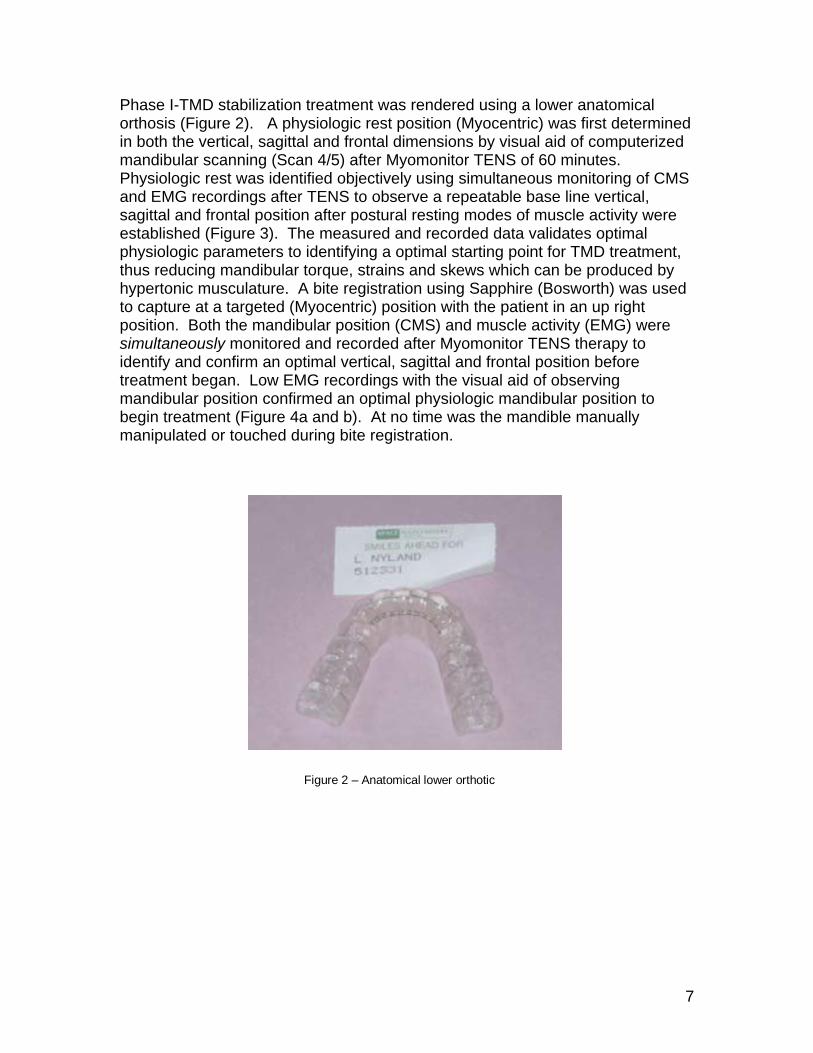
Phase I-TMD stabilization treatment was rendered using a lower anatomical orthosis (Figure 2). A physiologic rest position (Myocentric) was first determined in both the vertical, sagittal and frontal dimensions by visual aid of computerized mandibular scanning (Scan 4/5) after Myomonitor TENS of 60 minutes. Physiologic rest was identified objectively using simultaneous monitoring of CMS and EMG recordings after TENS to observe a repeatable base line vertical, sagittal and frontal position after postural resting modes of muscle activity were established (Figure 3). The measured and recorded data validates optimal physiologic parameters to identifying a optimal starting point for TMD treatment, thus reducing mandibular torque, strains and skews which can be produced by hypertonic musculature. A bite registration using Sapphire (Bosworth) was used to capture at a targeted (Myocentric) position with the patient in an up right position. Both the mandibular position (CMS) and muscle activity (EMG) were simultaneously monitored and recorded after Myomonitor TENS therapy to identify and confirm an optimal vertical, sagittal and frontal position before treatment began. Low EMG recordings with the visual aid of observing mandibular position confirmed an optimal physiologic mandibular position to begin treatment (Figure 4a and b). At no time was the mandible manually manipulated or touched during bite registration.
Figure 2 – Anatomical lower orthotic
8
Figure 1b - Right joint
Figure 4a – Resting surface electromyography before treatment showing hyperactive muscles activity (Scan 9).
9
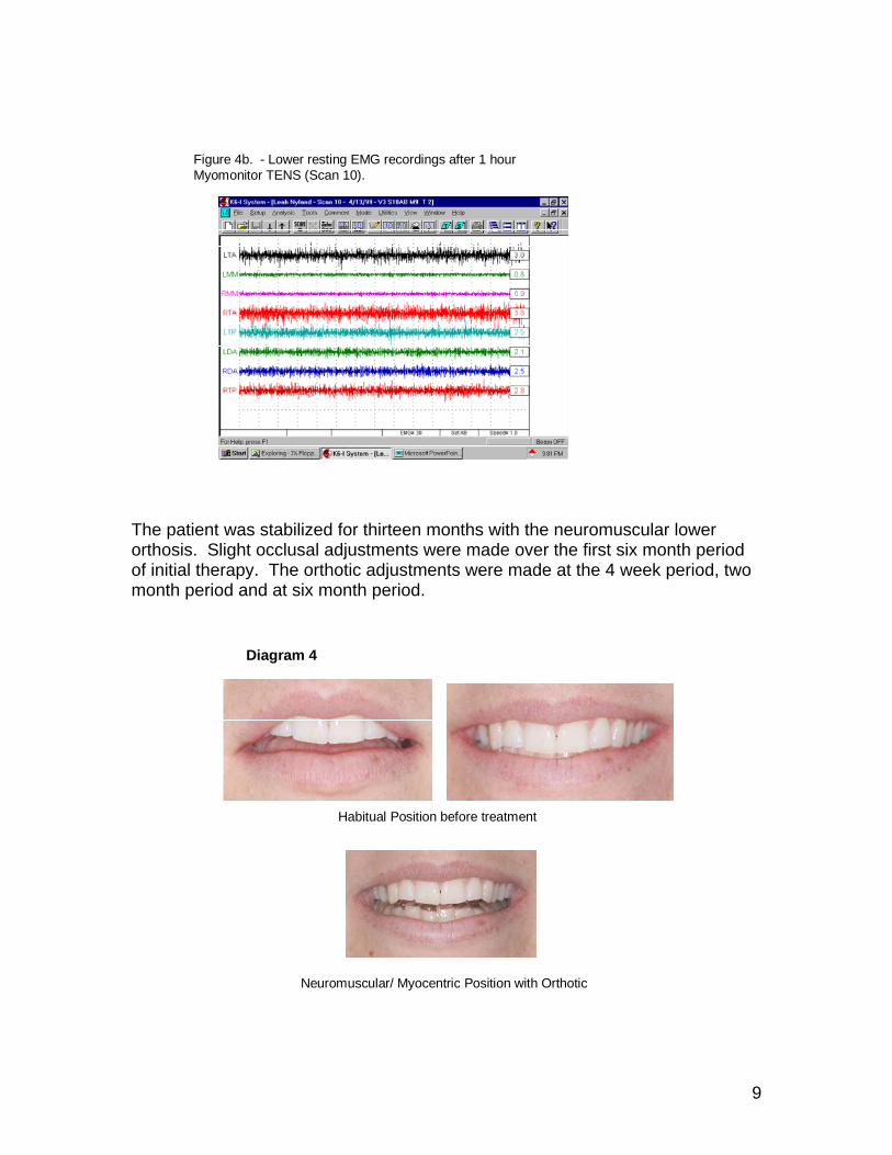
Figure 4b. - Lower resting EMG recordings after 1 hour Myomonitor TENS (Scan 10).
The patient was stabilized for thirteen months with the neuromuscular lower orthosis. Slight occlusal adjustments were made over the first six month period of initial therapy. The orthotic adjustments were made at the 4 week period, two month period and at six month period.
Habitual Position before treatment
Neuromuscular/ Myocentric Position with Orthotic
Diagram 4
10
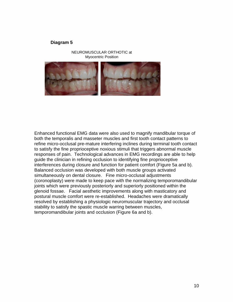
NEUROMUSCULAR ORTHOTIC at Myocentric Position
Diagram 5
Enhanced functional EMG data were also used to magnify mandibular torque of both the temporalis and masseter muscles and first tooth contact patterns to refine micro-occlusal pre-mature interfering inclines during terminal tooth contact to satisfy the fine proprioceptive noxious stimuli that triggers abnormal muscle responses of pain. Technological advances in EMG recordings are able to help guide the clinician in refining occlusion to identifying fine proprioceptive interferences during closure and function for patient comfort (Figure 5a and b). Balanced occlusion was developed with both muscle groups activated simultaneously on dental closure. Fine micro-occlusal adjustments (coronoplasty) were made to keep pace with the normalizing temporomandibular joints which were previously posteriorly and superiorly positioned within the glenoid fossae. Facial aesthetic improvements along with masticatory and postural muscle comfort were re-established. Headaches were dramatically resolved by establishing a physiologic neuromuscular trajectory and occlusal stability to satisfy the spastic muscle warring between muscles, temporomandibular joints and occlusion (Figure 6a and b).
11
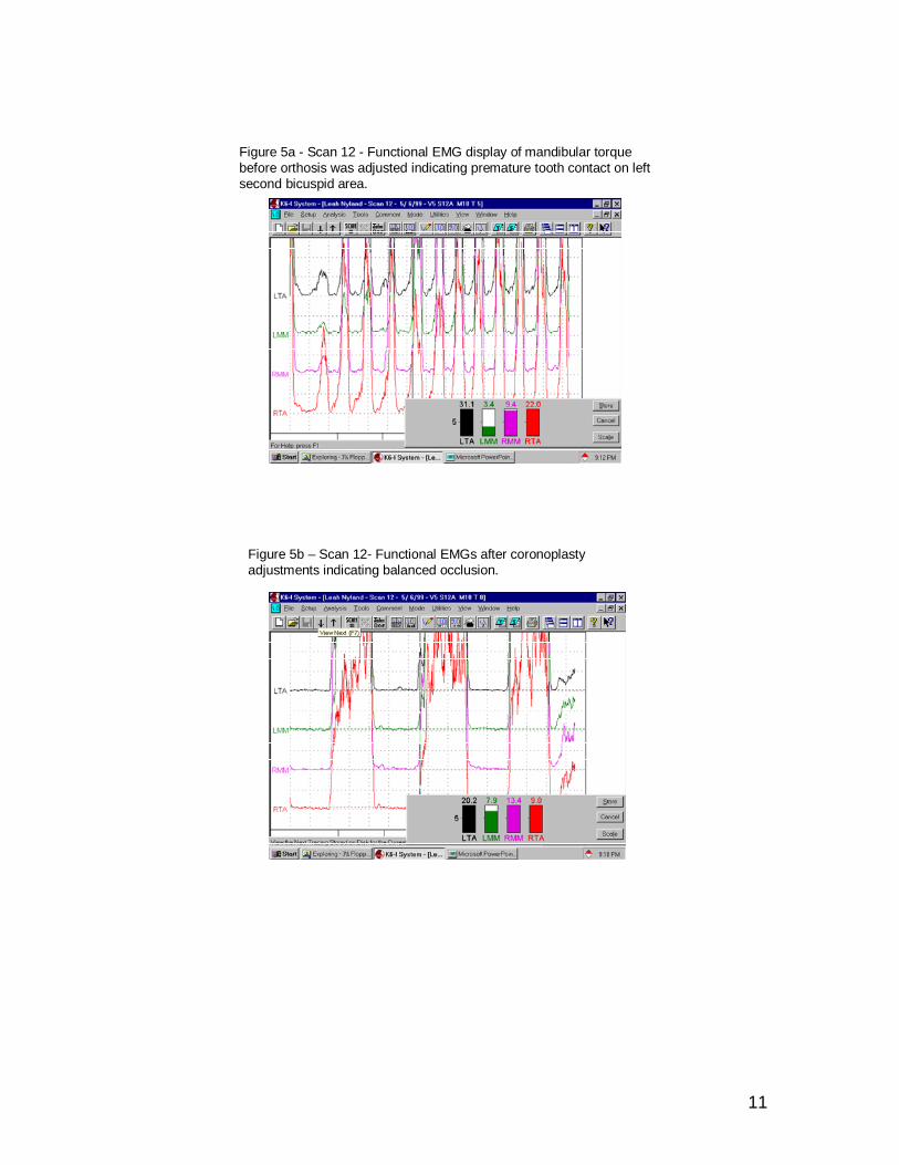
Figure 5a - Scan 12 - Functional EMG display of mandibular torque before orthosis was adjusted indicating premature tooth contact on left second bicuspid area.
Figure 5b – Scan 12- Functional EMGs after coronoplasty adjustments indicating balanced occlusion.
12
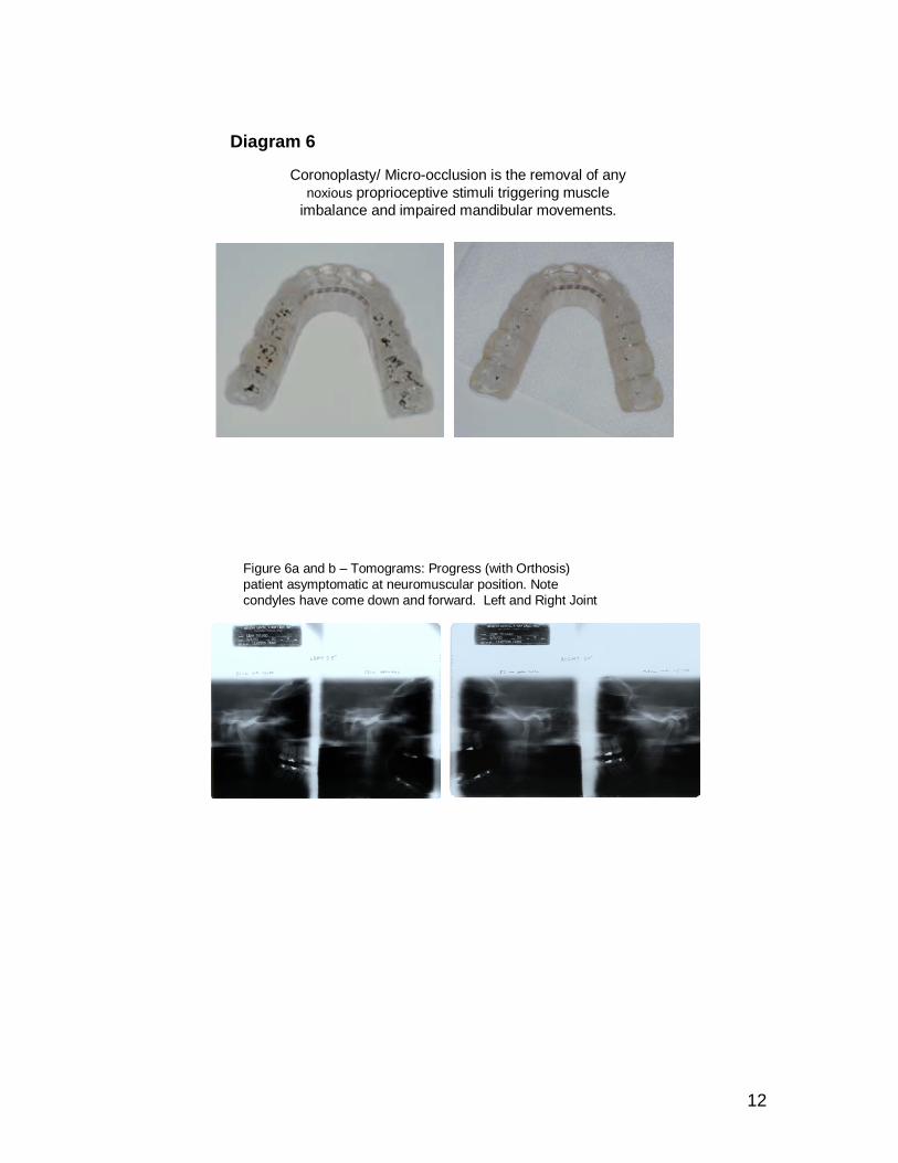
Coronoplasty/ Micro-occlusion is the removal of any noxious proprioceptive stimuli triggering muscle
imbalance and impaired mandibular movements.
Diagram 6
Figure 6a and b – Tomograms: Progress (with Orthosis) patient asymptomatic at neuromuscular position. Note condyles have come down and forward. Left and Right Joint
13
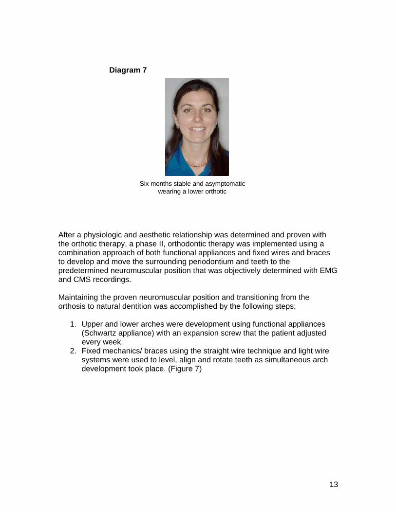
Six months stable and asymptomatic wearing a lower orthotic
Diagram 7
After a physiologic and aesthetic relationship was determined and proven with the orthotic therapy, a phase II, orthodontic therapy was implemented using a combination approach of both functional appliances and fixed wires and braces to develop and move the surrounding periodontium and teeth to the predetermined neuromuscular position that was objectively determined with EMG and CMS recordings. Maintaining the proven neuromuscular position and transitioning from the orthosis to natural dentition was accomplished by the following steps:
1. Upper and lower arches were development using functional appliances (Schwartz appliance) with an expansion screw that the patient adjusted every week.
2. Fixed mechanics/ braces using the straight wire technique and light wire systems were used to level, align and rotate teeth as simultaneous arch development took place. (Figure 7)
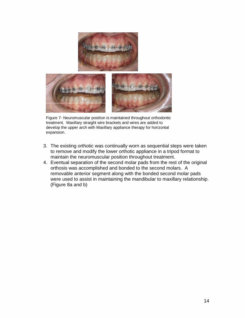
14
Figure 7- Neuromuscular position is maintained throughout orthodontic treatment. Maxillary straight wire brackets and wires are added to develop the upper arch with Maxillary appliance therapy for horizontal expansion.
3. The existing orthotic was continually worn as sequential steps were taken to remove and modify the lower orthotic appliance in a tripod format to maintain the neuromuscular position throughout treatment.
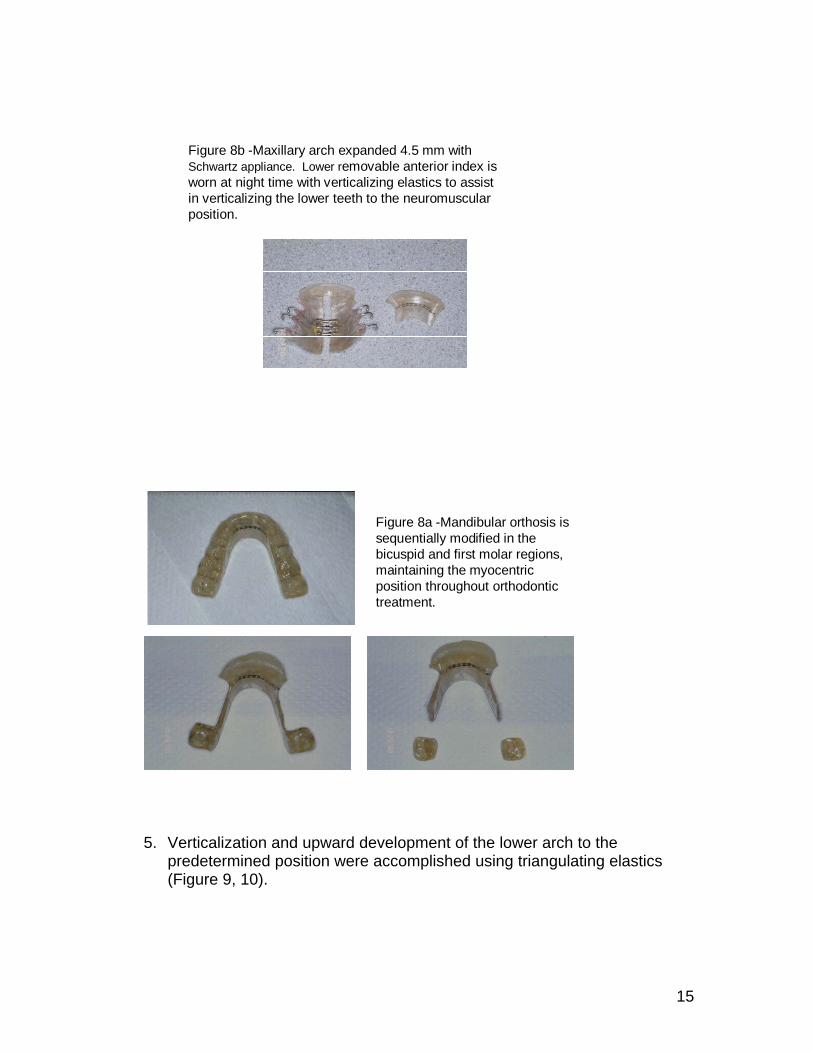
4. Eventual separation of the second molar pads from the rest of the original orthosis was accomplished and bonded to the second molars. A removable anterior segment along with the bonded second molar pads were used to assist in maintaining the mandibular to maxillary relationship. (Figure 8a and b)
15
Coronoplasty orthotic while maxillary arch is expanded.
Figure 8b -Maxillary arch expanded 4.5 mm with Schwartz appliance. Lower removable anterior index is worn at night time with verticalizing elastics to assist in verticalizing the lower teeth to the neuromuscular position.
Figure 8a -Mandibular orthosis is sequentially modified in the bicuspid and first molar regions, maintaining the myocentric position throughout orthodontic treatment.
Bond molar acrylic to second molars
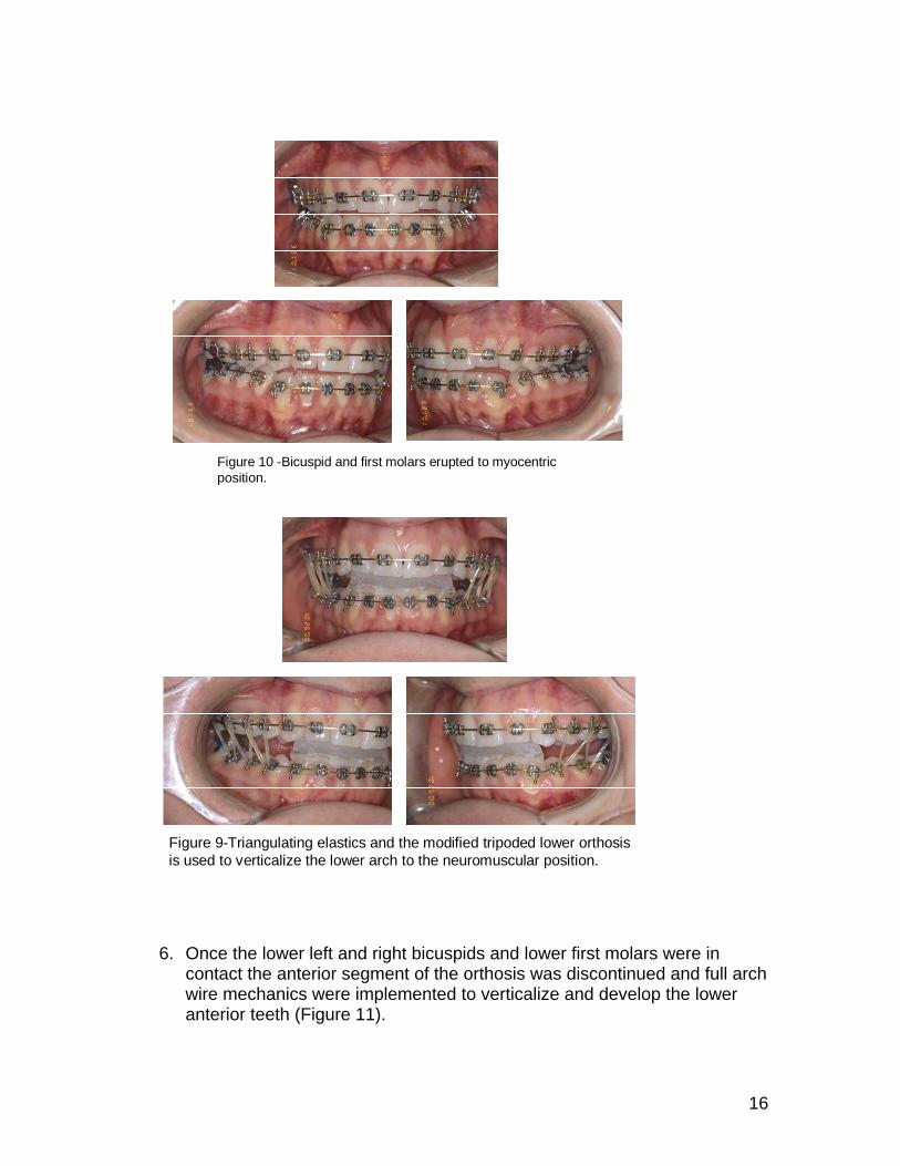
5. Verticalization and upward development of the lower arch to the predetermined position were accomplished using triangulating elastics (Figure 9, 10).
16
Figure 10 -Bicuspid and first molars erupted to myocentric position.
Figure 9-Triangulating elastics and the modified tripoded lower orthosis is used to verticalize the lower arch to the neuromuscular position.
6. Once the lower left and right bicuspids and lower first molars were in contact the anterior segment of the orthosis was discontinued and full arch wire mechanics were implemented to verticalize and develop the lower anterior teeth (Figure 11).

17
Figure 11- Arches are horizontally developed and lower bicuspids and molar teeth are now at myocentric position.
7. Once a stable occlusion was established and the patient continued to be
asymptomatic the remaining second molar pads were removed and verticalizing elastics were used to erupt the second molars to complete the orthodontics treatment (Figure 12).
Figure 12 - Second molars are verticalized using up and down elastics and Hawley retainers to complete the treatment.
18
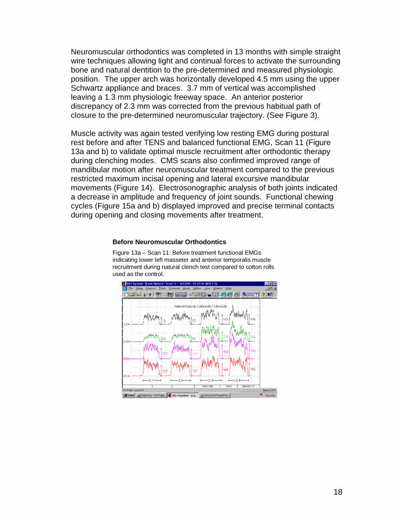
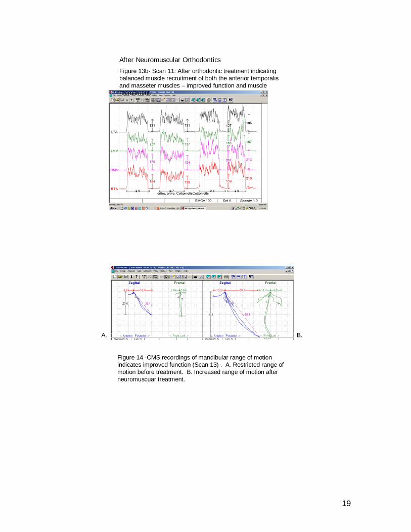
Neuromuscular orthodontics was completed in 13 months with simple straight wire techniques allowing light and continual forces to activate the surrounding bone and natural dentition to the pre-determined and measured physiologic position. The upper arch was horizontally developed 4.5 mm using the upper Schwartz appliance and braces. 3.7 mm of vertical was accomplished leaving a 1.3 mm physiologic freeway space. An anterior posterior discrepancy of 2.3 mm was corrected from the previous habitual path of closure to the pre-determined neuromuscular trajectory. (See Figure 3). Muscle activity was again tested verifying low resting EMG during postural rest before and after TENS and balanced functional EMG, Scan 11 (Figure 13a and b) to validate optimal muscle recruitment after orthodontic therapy during clenching modes. CMS scans also confirmed improved range of mandibular motion after neuromuscular treatment compared to the previous restricted maximum incisal opening and lateral excursive mandibular movements (Figure 14). Electrosonographic analysis of both joints indicated a decrease in amplitude and frequency of joint sounds. Functional chewing cycles (Figure 15a and b) displayed improved and precise terminal contacts during opening and closing movements after treatment.
Before Neuromuscular OrthodonticsFigure 13a – Scan 11: Before treatment functional EMGsindicating lower left masseter and anterior temporalis muscle recruitment during natural clench test compared to cotton rolls used as the control.
19
After Neuromuscular Orthodontics
Figure 13b- Scan 11: After orthodontic treatment indicating balanced muscle recruitment of both the anterior temporalis and masseter muscles – improved function and muscle recruitment.
Figure 14 -CMS recordings of mandibular range of motion indicates improved function (Scan 13) . A. Restricted range of motion before treatment. B. Increased range of motion after neuromuscuar treatment.
A. B.
20
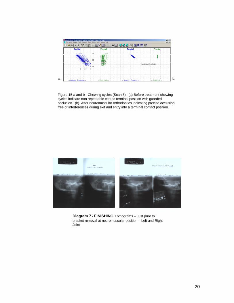
Figure 15 a and b - Chewing cycles (Scan 8)– (a) Before treatment chewing cycles indicate non repeatable centric terminal position with guarded occlusion. (b). After neuromuscular orthodontics indicating precise occlusion free of interferences during exit and entry into a terminal contact position.
a. b.
Diagram 7 - FINISHING Tomograms – Just prior to bracket removal at neuromuscular position – Left and Right Joint
21
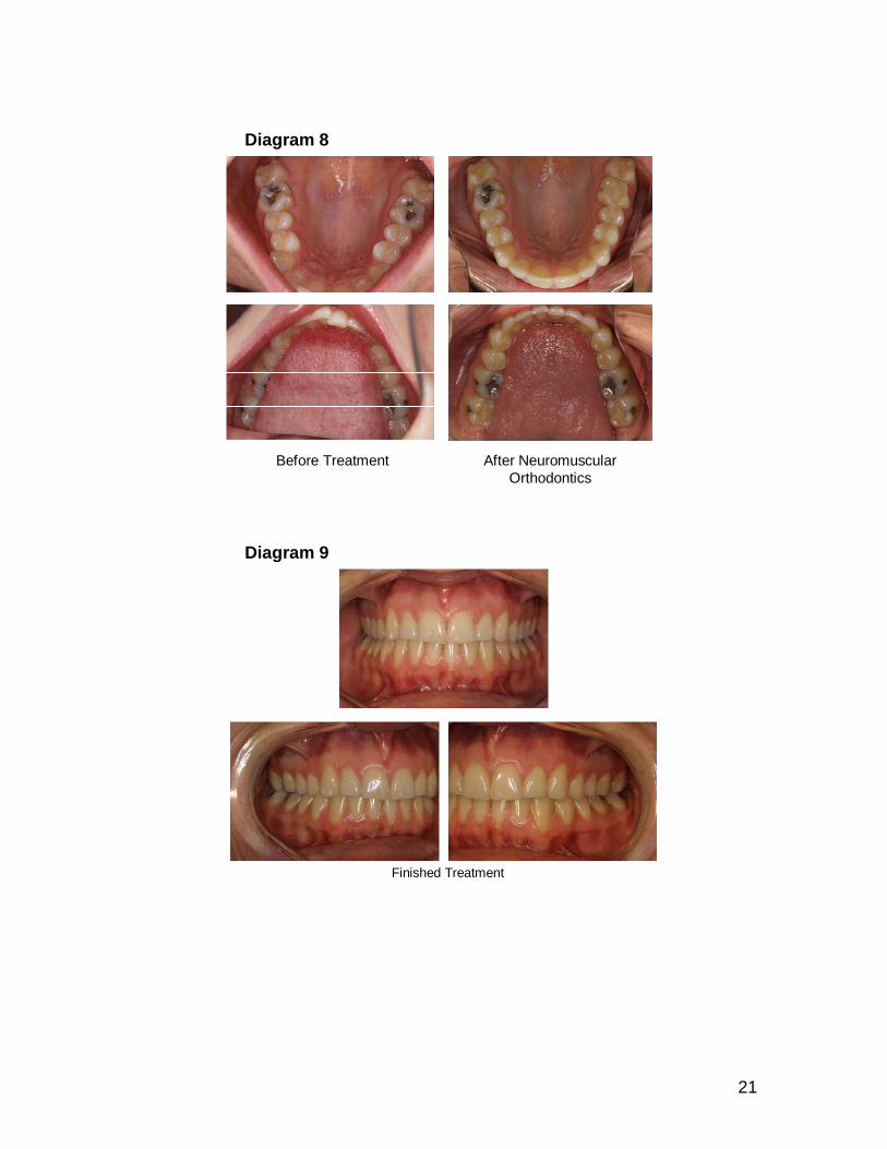
Before Treatment After Neuromuscular Orthodontics
Diagram 8
6-30-02
Finished Treatment
Diagram 9
22
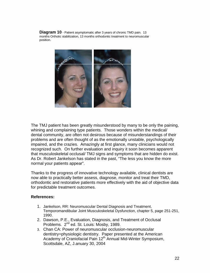
Diagram 10 - Patient asymptomatic after 3 years of chronic TMD pain. 13 months Orthotic stabilization, 13 months orthodontic treatment to neuromuscular position.
The TMJ patient has been greatly misunderstood by many to be only the paining, whining and complaining type patients. Those wonders within the medical/ dental community, are often not desirous because of misunderstandings of their problems and are often thought of as the emotionally unstable, psychologically impaired, and the crazies. Amazingly at first glance, many clinicians would not recognized such. On further evaluation and inquiry it soon becomes apparent that musculoskeletal occlusal/ TMJ signs and symptoms that are hidden do exist. As Dr. Robert Jankelson has stated in the past, “The less you know the more normal your patients appear”.
Thanks to the progress of innovative technology available, clinical dentists are now able to practically better assess, diagnose, monitor and treat their TMD, orthodontic and restorative patients more effectively with the aid of objective data for predictable treatment outcomes.
References:
1. Jankelson, RR: Neuromuscular Dental Diagnosis and Treatment, Temporomandibular Joint Musculoskeletal Dysfunction, chapter 5, page 251-251, 1990.
2. Dawson, P.E., Evaluation, Diagnosis, and Treatment of Occlusal Problems. 2nd ed. St. Louis: Mosby, 1989.
3. Chan CA: Power of neuromuscular occlusion-neuromuscular dentistry=physiologic dentistry. Paper presented at the American Academy of Craniofacial Pain 12th Annual Mid-Winter Symposium, Scottsdale, AZ, J.anuary 30, 2004
23
4. Stuart CE, Golden IB: The History of Gnathology. C.E. Stuart Gnathological Instruments, 1981:14-15.
5. Griffin, CJ: Tempormandibular joint dysfunction and the brain stem reticular formation. Aust Dent J, 9(6):524, 1964.
6. Moruzzi G, Magoon HW.: Brain stem reticular formation and activation of the EEG, EEG Clinical Neurophsiology,1:455, 1949.
7. Hampson, JL., Harrison, CR., Woolsey, CN.: Cerebro-cerebellar
projections and the somatotopic localization reflex. J Comp Neurol, 102(3):565, 1955.
8. Griffin, CJ: Tempormandibular joint dysfunction and the brain stem reticular formation. Aust Dent J, 9(6):524, 1964.
9. Myotronics-Noromed, Inc., J 4 Myomonitor and the K6-I/K7, Kinesiograph, Tukwila, Washington.
10. Travell JG. Myofascial pain and dysfunction the trigger point manual, Vol. 1, Baltimore, MD: Williams & Wilkins; 1983.
11. Garry JF.: Seminar on diseases of the temporomandibular apparatus. White Memorial Otolaryngology Foundation and TMJ Foundation, Communication between Dr. Janet Travel and Dr. James Garry, White Memorial Hospital, Los Angeles, California, , October 7-9, 1986.
12. Grummons D. Orthodontics for the TMJ-TMD patient, Second printing. Scottsdale, AZ: Wright & Co.; 1997:14-16.
13. Chan CA. Common myths of neuromuscular dentistry and the five basic principles of neuromuscular occlusion. September/October Vol.2, Number 5. LVI Dental Vision; 2002:10-11.
14. Cooper B: The role of bioelectrical instrumentation in the documentation Path oral Radiol and Management of temporomandibular disorders. Oral Surg Oral Med Oral Endod 1997;83:91-100.
Brief Biography:
Clayton A. Chan, D.D.S. is a general dentist who practices neuromuscular dentistry in San Diego, California and Las Vegas, Nevada. He is the Director of the Neuromuscular Dental Center at the Las Vegas Institute for Advanced Dental Studies. He lectures extensively on neuromuscular occlusion and bio-instrumentation in the United States and abroad. He has focused his care in the areas of dental orthopedic rehabilitation involving musculoskeletal occlusal disorders of the head, neck and face, restorative and orthodontic problems. He is a trained gnathologist. He holds Mastership status in the International College of Craniomandibular Orthopedics.