Embed Size (px)
Citation preview
Sequence requirements within collagen II for recognition by MMP-13
1
The recognition of collagen and triple-helical Toolkit peptides by MMP-13:
Sequence specificity for binding and cleavage
Joanna-Marie Howes1, Dominique Bihan
1, David A. Slatter
1, Samir W. Hamaia
1, Len C.
Packman1, Vera Knauper
2, Robert Visse
3, Richard W. Farndale
1*.
1From the Department of Biochemistry, University of Cambridge, Downing Site, Cambridge, CB2
1QW, UK.
2Cardiff University Dental School, Dental Drive, CF14 4XY, Cardiff, UK.
3Kennedy Institute of Rheumatology, 65 Aspenlea Road, Hammersmith, London W6 8LH, UK.
Running title: Sequence requirements within collagen II for recognition by MMP-13
*To whom correspondence should be addressed: Richard W. Farndale, Department of Biochemistry,
University of Cambridge, Downing Site, Cambridge, CB2 1QW, UK.
Tel: +44(0)1223 766111; Fax: +44(0)1223 333345; E-mail: [email protected].
Acknowledgment: This work was supported by a British Heart Foundation programme grant,
RG/009/003/27122, and peptide synthesis, by grants from Medical Research Council and Wellcome
Trust.
Keywords: MMP-13; protease; matrix metalloproteinase (MMP); collagen; collagenolysis; Toolkit
peptides; protein conformation, substrate specificity; hemopexin domain
Background: MMP-13 recognizes poorly-
defined sequences in collagens.
Results: MMP-13 binds key residues in the
canonical cleavage site and another site near the
collagen N-terminus.
Conclusion: MMP-1 and MMP-13 differ in
their recognition and cleavage of collagen,
which is regulated primarily through the Hpx
domain of MMP-13.
Significance: Our data explain the preference of
MMP-13 for collagen II.
Remodeling of collagen by matrix
metalloproteinases (MMPs) is crucial to tissue
homeostasis and repair. MMP-13 is a
collagenase with a substrate preference for
collagen II over collagens I and III. It
recognizes a specific, well-known site in the
tropocollagen molecule where its binding
locally perturbs the triple helix, allowing the
catalytic domain of the active enzyme to
cleave the collagen α chains sequentially, at
Gly775
-Leu776
in collagen II. However, the
specific residues upon which collagen
recognition depends within and surrounding
this locus have not been systematically
mapped. Using our triple-helical peptide
Collagen Toolkit libraries in solid-phase
binding assays, we found that MMP-13 shows
little affinity for Collagen Toolkit III, but
binds selectively to two triple-helical peptides
of Toolkit II. We have identified the residues
required for the adhesion of both proMMP-
13 and MMP-13 to one of these, Toolkit
peptide II-44, which contains the canonical
collagenase cleavage site (at helix residues
775-6). MMP-13 was unable to bind to a
linear peptide of the same sequence as II-44.
We also discovered a second binding site near
the N-terminus of collagen II (starting at helix
residue 127) in Toolkit peptide II-8. The
pattern of binding of the free hemopexin
domain of MMP-13 was similar to that of the
full-length enzyme, but the free catalytic
subunit bound none of our peptides. The
susceptibility of Toolkit peptides to
proteolysis in solution was independent of the
very specific recognition of immobilized
peptides by MMP-13; the enzyme proved able
to cleave a range of dissolved collagen
peptides.
http://www.jbc.org/cgi/doi/10.1074/jbc.M114.583443The latest version is at JBC Papers in Press. Published on July 9, 2014 as Manuscript M114.583443
Copyright 2014 by The American Society for Biochemistry and Molecular Biology, Inc.
by guest on March 28, 2020
http://ww
w.jbc.org/
Dow
nloaded from
Sequence requirements within collagen II for recognition by MMP-13
2
Collagens are comprised of three α
chains containing repeating Gly-X-X' triplets
(where X and X' are often Pro and Hyp
respectively). This primary structure allows the
formation of a right-handed collagen superhelix
which endows the molecule with resistance to
degradation by most proteases. The fibrillar
collagens I, II and III contain a conserved triple-
helical COL domain of 1014 residues, with
short, non-helical extensions at each end, which
constitutes a tropocollagen molecule. Such
collagens assemble side-by-side to form a three-
dimensional staggered array (a fibril) with a
regular offset of 234 residues (one D-period)
between adjacent tropocollagens. This
organization gives rise to the striated structure of
the collagen fiber observed by transmission
electron microscopy, where the 67 nm
periodicity reflects the dimensions of one D-
period(1).
Collagenolytic MMPs are members of
the zinc-dependent peptidase family whose
tightly-regulated proteolytic activities play a
pivotal role in extracellular matrix homeostasis,
cell migration and wound healing. Under
pathological conditions, uncontrolled tissue
remodeling by collagenases such as MMP-13 is
associated with the potentiation of tumor
progression and metastasis, atherosclerotic
plaque remodeling and arthritic disease(2). The
preferred substrate for MMP-13 is collagen II
which is cleaved five times faster than collagen I
and six times faster than collagen III(3), and
more readily by MMP-13 than by other
collagenases.
As with other MMPs, collagenases are
secreted as pro-enzymes with an organized
domain structure comprising a pro-peptide
region (cleaved to yield the active mature
enzyme), a globular, Zn2+
-binding catalytic
domain (Cat), a linker region and a C-terminal 4-
bladed -propeller hemopexin-like domain
(Hpx)(4,5). The Cat and Hpx are thought to
cooperate to recognize and bind to the cleavage
site, in collagen D-period 4 (D4), three-quarters
of the way along the tropocollagen
molecule(6,7). Binding of the Hpx is considered
to facilitate local relaxation of the helix,
allowing the Cat domain of the MMP to
hydrolyze specific Gly-X bonds within Gly-X-
X', where X is either Leu or Ile, and X' is either
Ala or Leu (i.e., within the triplet G-[I/L]-
[A/L])(8). Deletion of the Hpx domain therefore
results in a loss of collagenase activity(9,10). To
date, two cleavage sites have been identified for
MMP-13 within collagen II. The first at Gly775
-
Leu776
(numbering refers to position within
collagen II helical domain) is shared by MMPs-
1 and -8; the second site, Gly778
-Gln779
(11) is
three amino acids from the N-terminus of the
newly-cleaved quarter fragment(12). Several G-
[I/L]-[A/L] triplets are present in native
collagen, indicating that, in principle, other
scissile bonds may exist. However, with the
exception of MMP-13, most MMPs primarily
cleave the collagen helix at a single location,
reflecting the importance of the unique sequence
which surrounds the cleavage site(13,14).
To simplify structural research, a mutant
species, MMP-13(E204A), has been produced.
The mutation lies within the active site, and
MMP-13(E204A) lacks catalytic activity whilst
retaining the same conformation. The specific
collagen residues required for recognition of the
collagen triple helix and the residues
within/surrounding the canonical collagenase
site described above have not yet been
systematically identified, and with this in view,
we have investigated both wild type MMP-13
and MMP-13(E204A) in the present study. The
use of both wild type and mutant active forms,
together with their corresponding pro-forms and
a free Hpx domain allows us to investigate the
contribution to binding of all three component
domains.
To facilitate mapping studies we
synthesized a library of overlapping
homotrimeric host-guest peptides, in which 27
residues of primary collagen (guest) sequence is
placed between [GPP]5 hosts that ensure triple-
helical conformation. The last nine guest amino
acids are the same as the first nine amino acids
in each successive peptide, and these peptide
libraries (referred to as Collagen II and III
Toolkits respectively) encompass the entire
triple helical domains of collagens II and
III(15,16), reviewed by Farndale et al.(17).
Here, we used these Toolkits to map MMP-13
binding to collagens II and III, and we proceeded
to synthesize subsidiary peptides to identify
those residues surrounding the cleavage site that
are required for the binding of MMP-13. This
systematic approach has also allowed us to
identify a new MMP-13 binding site on collagen
type II.
The recent elucidation(7) of the structure
of MMP-1 in complex with a triple-helical
peptide derived from the use of the Toolkits
allows us to compare the binding activity of both
collagenases to their common cleavage site, and
by guest on March 28, 2020
http://ww
w.jbc.org/
Dow
nloaded from

Sequence requirements within collagen II for recognition by MMP-13
3
to show that there are marked differences
between the binding activities of the two
enzymes that may account for their differing
specificities for collagens I, II and III.
EXPERIMENTAL PROCEDURES
MMP-13 expression, purification and
activation–Recombinant proMMP-13 was
expressed and purified as previously
described(18). Where required, proMMP-13
was activated to yield MMP-13 by incubation in
1 mM 4-amino-phenylmercuric acetate (APMA)
for 1 h at 37 °C. ProMMP-13(E204A) was
expressed in E. coli BL21(DE3), refolded,
purified, and activated essentially as described
for proMMP-1(E200A)(19,20). The Cat domain
of MMP-13 (249-451) was expressed and
purified from NSO mouse myeloma cells as
previously described(12). MMP-13 GST-Hpx
domain was expressed in E. coli using the
pGEX-2T expression vector, the forward primer
TCCGCGTGGATCCCTCTATGGTCCAGGAG
ATGAA and the reverse primer GCAA-
ATTCCATTTTGTGGTGTTGAAGAATTCAT,
which contain BamHI and EcoRI restriction sites
respectively, as previously described(16).
Peptide Synthesis–Collagen Toolkit II
and III and other peptides were generated using
an AB Systems Pioneer automated synthesizer
and N-(9-fluorenyl)methoxycarbonyl (Fmoc)
chemistry as previously described(15,16). All
peptides were verified using MALDI-TOF mass
spectrometry and their triple-helical
conformation confirmed by polarimetry.
Peptide Design–Unless stated otherwise,
all peptides were triple-helical, a structure
maintained by the flanking sequences,
GPC(GPP)5- and -(GPP)5-GPC-amide, at their
N- and C-terminus respectively. For simplicity,
peptides are referred to by their specific guest
sequence. A negative control peptide, (GPC-
(GPP)10-GPC-amide) is referred to as GPP10. A
linear version of Toolkit peptide II-44 contained
the same guest sequence between disordered
flanking host sequences, thus:
GCPP(GPPP)2GGPPPG– II-44 guest sequence –
P(GPPP)2GGPPPGCPG-amide.
MMP-13 Toolkit Solid-Phase Binding
Assays (SPBA)–Immulon 2 HB 96-well plates
(Nunc, Langenselbold, Germany) were coated
with Collagen Toolkit or other peptides, fibrillar
or monomeric collagen at a saturating
concentration (10 µg/ml in 0.01 M acetic acid)
overnight at 4 °C. Fibrous bovine type I collagen
was a gift from Ethicon Corporation
(Somerville, NJ). Monomeric collagen was
obtained from Devro (Bathurst, Australia). All
further incubations were performed at room
temperature, 24 °C, for 1 h unless otherwise
stated. The wells were washed three times with
adhesion buffer (1 mg/ml BSA in Tris-Buffered
Saline (TBS) containing 0.1 % (v/v) Tween-20)
between each incubation step. The wells were
then blocked with 50 mg/ml BSA in TBS prior
to the addition of MMP at a concentration of 83
nM (unless otherwise stated) in adhesion buffer.
Where indicated, increasing amounts of Toolkit
peptides II-8 and II-44 were pre-incubated for 20
min with the MMP prior to adhesion assays.
Rabbit anti-MMP-13, raised against MMP-13
hinge region (Abcam, Cambridge, UK), and goat
anti-rabbit HRP (Dako, Ely, UK) were added at
a dilution of 1:2000 in adhesion buffer prior to
the addition of TMB substrate system (Sigma)
and the plates read at 450 nm. Rabbit HRP-
linked anti-GST (GE Healthcare; dilution
1:10,000) was used to detect GST-Hpx. To
confirm affinity of the anti-hinge antibody for
MMP-13 and MMP-13(E204A), increasing
concentrations of MMP were coated onto an
ELISA plate prior to blocking and detected as
previously described. Binding curves were fitted
using Prism 5.0 software (GraphPad, San Diego,
USA), allowing total binding (Bmax) and
equilibrium dissociation constant (KD) to be
determined.
Biotinylation of MMP-13(E204A) and
the Cat domain–Proteins were C-terminally
biotinylated using an EZ-Link Micro Sulfo-
NHS-Biotinylation Kit (Pierce) according to the
manufacturer’s instructions. Successful biotinyl-
ation was detected via Western Blotting using an
ultrasensitive Streptavidin-Peroxidase Polymer
(Sigma).
Peptide digestion assays–To determine
the likelihood of Toolkit peptide clipping by
MMP-13 during SPBA experiments, Toolkit
peptides at a final concentration of 80 mM were
incubated with a high (4.4 µM final)
concentration of MMP-13, MMP-13(E204A) or
the equivalent volume of Tris buffer pH 7.4 for 1
h at room temperature. Proteolytic activity of
MMP-13 was assessed following incubation of
Toolkit peptides with 250 nM MMP for 16 h at
24 °C and 37 °C respectively. The samples were
then either examined by electrophoresis under
reducing conditions, with silver staining, or
submitted for MALDI mass spectrometry. SDS-
PAGE electrophoresis was performed on 4-12 %
by guest on March 28, 2020
http://ww
w.jbc.org/
Dow
nloaded from
Sequence requirements within collagen II for recognition by MMP-13
4
NuPage® bis-tris gels (Invitrogen) according to
the manufacturer’s instructions.
Mass Spectrometry Analysis–Peptides in
TBS buffer, pH 7.4, were reduced with 5 mM
tris(2-carboxyethyl)phosphine (Thermo
Scientific-Pierce) for 10 min at 35 °C and then
desalted using uC18ZipTips (Millipore)
equilibrated and washed with 5 % (v/v) acetic
acid. Peptides were eluted with 2 l ferulic acid
matrix (Sigma; 10 mg/ml in 50 % (v/v) aqueous
acetonitrile), spotted to the MALDI target plate,
dried and washed once with 2 l 5 % (v/v) acetic
acid. Mass spectra were collected on a MALDI
MicroMX Instrument (Waters, UK) in reflectron
mode at threshold laser power. Spectra were
calibrated externally with polyethyleneglycol
1000-2000-3000 (Sigma) and then adjusted by
lockmass to one of two known peptides present
in all samples from the autodigestion of MMP-
13 (20-42 & 21-42, confirmed by ms/ms as
below). Mass accuracies were generally better
than 20 ppm, allowing only a single
interpretation of the cleavage site for most
peptides. Any ambiguities were resolved by
ms/ms fragmentation on a separately desalted
sample, eluted in 70 % MeOH/ 0.2 % (v/v)
formic acid and analyzed on a Thermo LCQ
Classic ion-trap instrument using static
nanospray delivery. This confirmed the
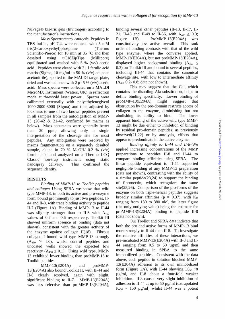
sequence identity. RESULTS Binding of MMP-13 to Toolkit peptides
and collagen–Using SPBA we show that wild
type MMP-13, in both its active and pro-enzyme
form, bound prominently to just two peptides, II-
44 and II-8, with trace binding activity to peptide
II-7 (Figure 1A). Binding of MMP-13 to II-44
was slightly stronger than to II-8 with A450
values of 0.7 and 0.6 respectively. Toolkit III
showed uniform absence of binding (data not
shown), consistent with the greater activity of
the enzyme against collagen II(18). Fibrous
collagen I bound wild type MMP-13 strongly
(A450 ≥ 1.0), whilst control peptides and
uncoated wells showed the expected low
reactivity (A450 ≤ 0.1). Using wild type, MMP-
13 exhibited lower binding than proMMP-13 to
Toolkit peptides.
MMP-13(E204A) and proMMP-
13(E204A) also bound Toolkit II, with II-44 and
II-8 clearly resolved, again with slight,
significant binding to II-7. MMP-13(E204A)
was less selective than proMMP-13(E204A),
binding several other peptides (II-13, II-17, II-
21, II-45 and II-49 to II-56, with A450 ≥ 0.3;
Figure 1B). ProMMP-13(E204A) was
constitutively less active overall. This rank
order of binding contrasts with that of the wild
type enzyme, where the converse applied.
MMP-13(E204A), but not proMMP-13(E204A),
displayed higher background binding (A450 ≤
0.3) on Toolkit III and bound to several peptides,
including III-44 that contains the canonical
cleavage site, with low to intermediate affinity
(A450 0.2- 0.8; data not shown).
This may suggest that the Cat, which
contains the disabling Ala substitution, helps to
define binding specificity. Lower binding of
proMMP-13(E204A) might suggest that
obstruction by the pro-domain restricts access of
collagen to the enzyme, diminishing but not
abolishing its ability to bind. The lower
apparent binding of the active wild type MMP-
13 might be due either to inhibition of binding
by residual pro-domain peptides, as previously
observed(21,22) or by autolysis, effects that
appear to predominate in the active enzyme.
Binding affinity to II-44 and II-8–We
applied increasing concentrations of the MMP
preparations to peptides II-8 and II-44 to
compare binding affinities using SPBA. The
linear peptide equivalent to II-44 supported
negligible binding of any MMP-13 preparation
(data not shown), contrasting with the ability of
a similar peptide(23,24) to support the binding
of fibronectin, which recognizes the same
site(25,26). Comparison of the pro-forms of the
enzyme on both triple-helical peptides suggests
broadly similar affinities (p = 0.37), with KD
ranging from 130 to 380 nM, the latter figure
(the only outlying value) being the estimate for
proMMP-13(E204A) binding to peptide II-8
(data not shown).
Our Toolkit and SPBA data indicate that
both the pro and active forms of MMP-13 bind
more strongly to II-44 than II-8. To investigate
the relative affinities of these interactions, we
pre-incubated MMP-13(E204A) with II-8 and II-
44 ranging from 0.5 to 50 µg/ml and then
measured binding in SPBA to the same
immobilized peptides. Consistent with the data
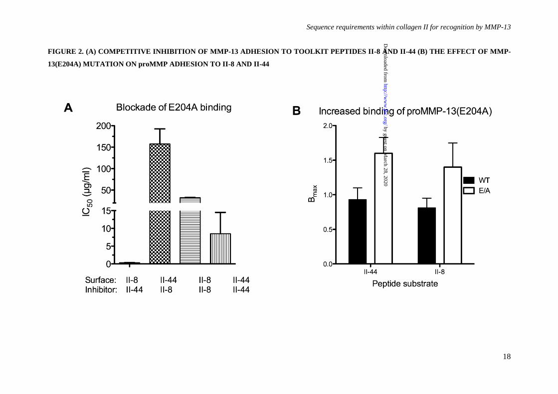
above, each peptide in solution blocked MMP-
13(E204A) adhesion to its own immobilized
form (Figure 2A), with II-44 showing IC50 ~8
µg/ml, and II-8 about a four-fold weaker
inhibition. II-8 caused very slight inhibition of
adhesion to II-44 at up to 50 µg/ml (extrapolated
IC50 ~ 150 µg/ml) whilst II-44 was a potent
by guest on March 28, 2020
http://ww
w.jbc.org/
Dow
nloaded from

Sequence requirements within collagen II for recognition by MMP-13
5
inhibitor of binding to II-8 (IC50 < 0.5 µg/ml).
These data confirm II-44 as a more potent ligand
for MMP-13 than II-8 (p = 0.017, Kruskal-
Wallis test).
It was noted that Bmax was dependent on
the presence of the active site mutation, being
roughly double in the proMMP-13(E204A) and
MMP-13(E204A) mutant forms compared with
their wild type counterparts (p < 0.02, 2-way
ANOVA, Figure 2B). This was not due to
different recognition of wild type and MMP-
13(E204A) by the detecting antibody (data not
shown).
Binding to full-length collagen I–Active
forms of MMP-13 and MMP-13(E204A) were
used in similar assays to establish the ability of
the enzyme to bind fibrillar and monomeric
collagens (Figure 3). Both enzyme preparations
exhibited much lower binding to saturated
coatings of monomeric collagen I than to II-44.
This may reflect the lower density of binding
sites in monomeric collagen, where there are two
sites per 1000 residues, compared with one site
per 63 residues in these Toolkit peptides. The
two substrates are directly comparable, since
rotary shadowing/transmission electron
microscopy (27) suggests that monomeric
collagens lie flat upon a surface, as would also
be anticipated for the much shorter Toolkit
peptides.
Our data suggest that the binding of the
MMP to collagen is greatly facilitated by
fibrillar conformation (KD ~ 40 nM). Pugh et al.
have shown that collagen fibers as used here
form a complex meshwork that extends tens of
microns above the coated surface(28), offering a
large binding area. The observed higher affinity
implies co-operative binding to discrete sites
within the fiber network. Possibly, a new
composite binding site comprising the canonical
site and sequences in the adjacent tropocollagen
molecules within a single fiber might also
generate a larger footprint on the fiber surface,
and hence higher affinity.
Binding of Cat and Hpx Toolkit peptides
II-8 and II-44–To determine whether the MMP-
13 Cat and Hpx domains were individually able
to recognize Toolkit peptides, their binding was
assessed in SPBA. Though the Cat domain was
able to recognize collagen, it was unable to bind
to any Toolkit II peptides (data not shown),
whilst Hpx bound well to peptide II-44 (A450
0.8) and weakly to II-8 (A450 0.2; Figure 1C).
Both domains bound fibrillar collagen I, albeit
weakly in the case of the Cat domain.
Ala-scanning of II-44–To explore the
primary sequence determinants of MMP-13
binding to II-44 in SPBA, we made a set of
truncated and alanine-substituted ('Ala-scanned')
triple-helical peptides of the same general host-
guest form. The data are shown in Figure 4.
MMP-13(E204A), and to a lesser extent wild-
type MMP preparations, proMMP-13(E204A)
and Hpx were supported by an 18-residue triple-
helical peptide, II-44A, which lacked only the
three C-terminal triplets of II-44. Peptide 44B,
containing only these missing triplets,
GFOGLOGPS, supported no binding.
Successive omissions of triplets (peptides II-44C
to II-44I) from both the N- and C-terminus of II-
44A each modulated the binding of MMP-
13(E204A), indicating that further truncation
was inappropriate. These findings were used to
define the C-terminus of the Ala-scanned
sequence, culminating in the addition of a
single-triplet native sequence extension, GPQ, to
the N-terminal boundary of II-44, thus creating a
21-residue peptide (44J). Using the
nomenclature of Schechter and Berger to define
the distance from the cleavage site, peptide 44J,
GPQG~LAGQRGIVGLOGQRGER, stretched
from P4 to P17' where P1, preceding the scissile
bond, is the first (Gly) residue of the guest
sequence of peptide II-44.
As with II-44, binding to 44J differed
amongst MMP-13 preparations, with MMP-
13(E204A) binding most strongly (A450 0.8),
then proMMP-13, then proMMP-13(E204A),
then active MMP-13, and lastly the Hpx domain
alone (A450 0.4). All MMP-13 preparations
yielded data that allowed us to discriminate
between the Ala-substituted peptides.
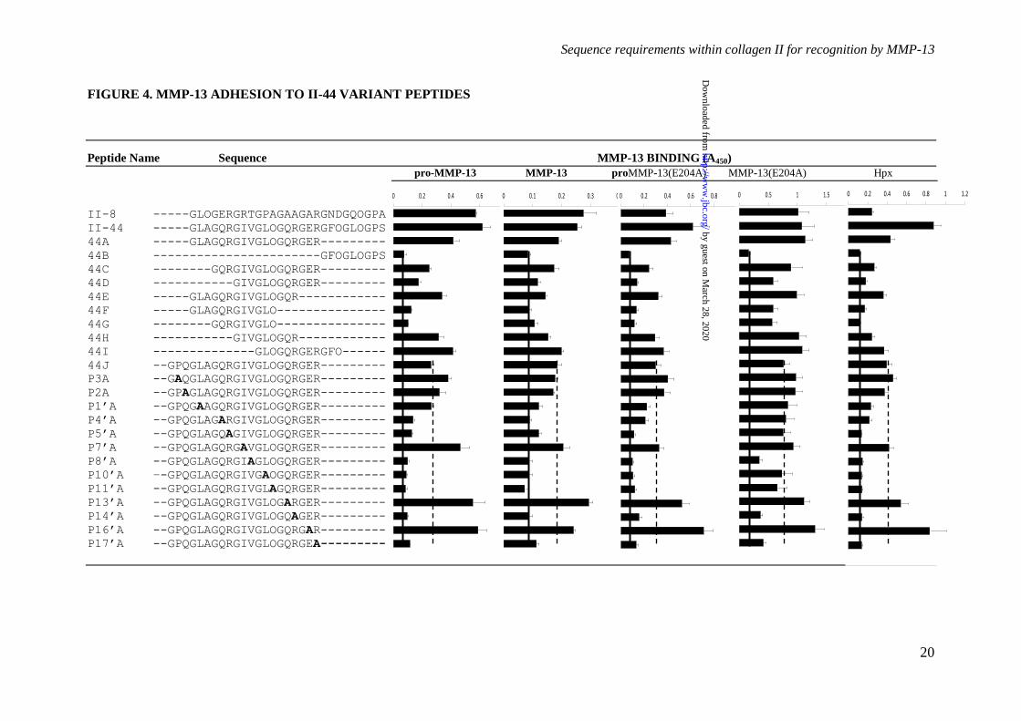
Three residues were uniformly essential
for binding, P8'(V), P14'(R) and P17'(R). No other
Ala-substitutions perturbed the binding of MMP-
13(E204A), consistent with less rigorous
determinants of peptide binding to this mutant
form, but several residues were important for the
binding of the other MMP preparations
including the Hpx domain. P4'(Q), P5'(R),
P10'(L), and P11'(O) all fall into this category,
whilst P1'(L) was similarly important for the
binding of Hpx, and to a lesser extent MMP-13
and proMMP-13(E204A).
Two residues in particular in the parent
peptide, P13'(Gln) and P16'(Glu), appeared to
exert a restrictive effect, since MMP-13 binding
increased after Ala-substitution. Further
residues, P3(Pro), P2(Gln) and P7'(Ile), showed a
similar tendency with some but not all MMP-13
by guest on March 28, 2020
http://ww
w.jbc.org/
Dow
nloaded from
Sequence requirements within collagen II for recognition by MMP-13
6
preparations. These data suggest that
recognition of the native sequence is poised
between firm binding and release of the enzyme,
perhaps allowing some freedom to re-organize
or to permit relaxation of the triple-helical
structure prior to cleavage, or to ensure release
of the enzyme once the substrate is cleaved.
Like the intact forms of the enzyme,
Hpx only bound to Toolkit peptides II-8 and II-
44 in the static adhesion assays (Figure 1C). The
adhesion profile of the Hpx domain to II-44 Ala-
scanned peptides reflected that of the intact
enzyme(s), indicating that it is primarily the Hpx
domain which defines the recognition of the
intact enzyme to the collagenase cleavage site of
collagen II.
Proteolysis of Toolkit peptides–We first
considered the possibility that poor binding of
active MMP-13 to peptides or collagen might
reflect proteolysis and release from the 96-well
surface during SPBA, leading to loss of apparent
binding activity. Our binding data also
suggested that peptide II-8 may offer a novel
cleavage site for MMP-13. We therefore
investigated the ability of MMP-13 to cleave
several triple-helical peptides, selected by their
differing ability to bind MMP-13(E204A).
Peptides II-44 and II-8 (high-affinity), III-5 and
III-40 (intermediate affinity) and II-24 and II-28
(non-binding), were incubated with a high
concentration (4.4 µM) MMP-13 for 1 h at 24
°C, as in our adhesion assays, then examined
using SDS gel electrophoresis as described. The
migration of the peptides was slower than their
mass would suggest, an observation which is
well known. The actual masses of the peptides
are shown in Table 1. No proteolysis of any
Toolkit peptide was observable after 1 h at 24 °C
(data not shown), despite the rather high enzyme
concentration used, allowing us to conclude that
our binding assays were not compromised by
substrate degradation. As expected, MMP-
13(E204A) (and buffer control) proved unable to
degrade any peptide under any condition tested
in the present study.
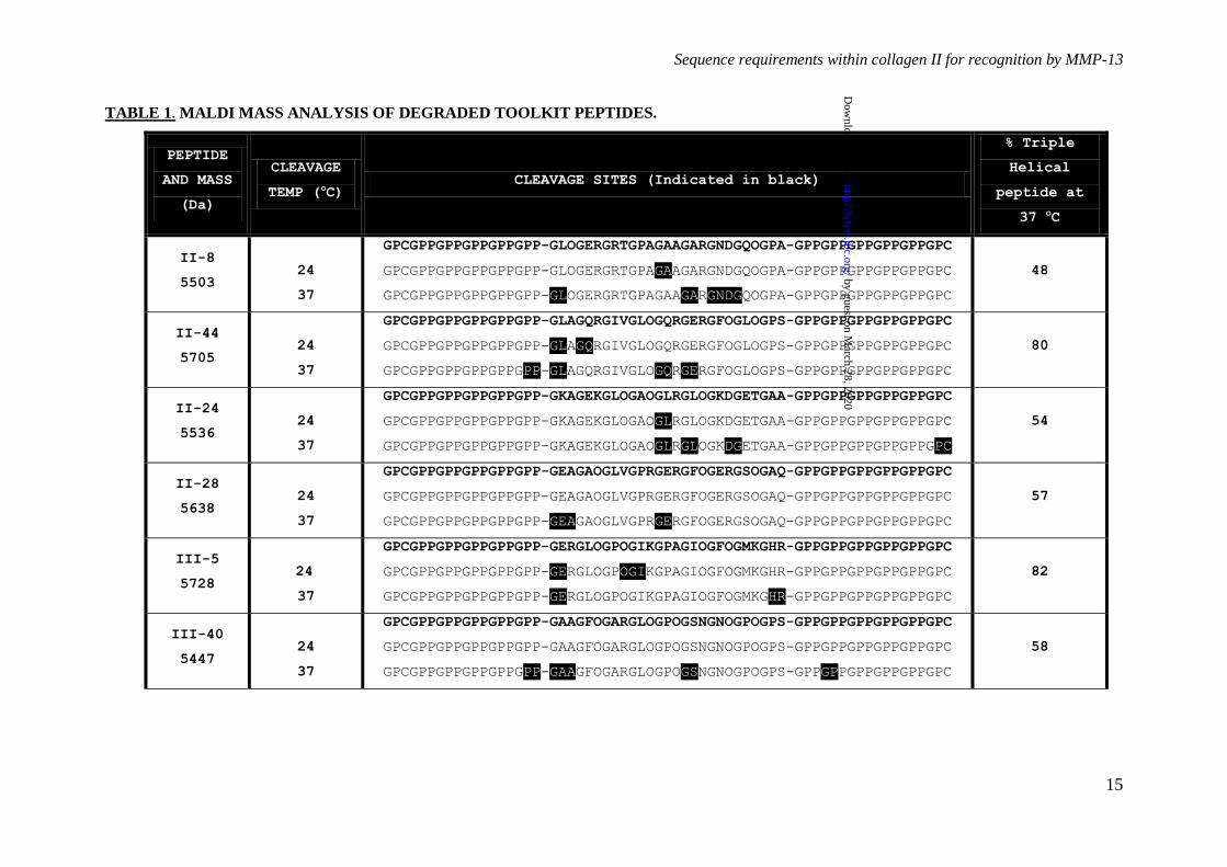
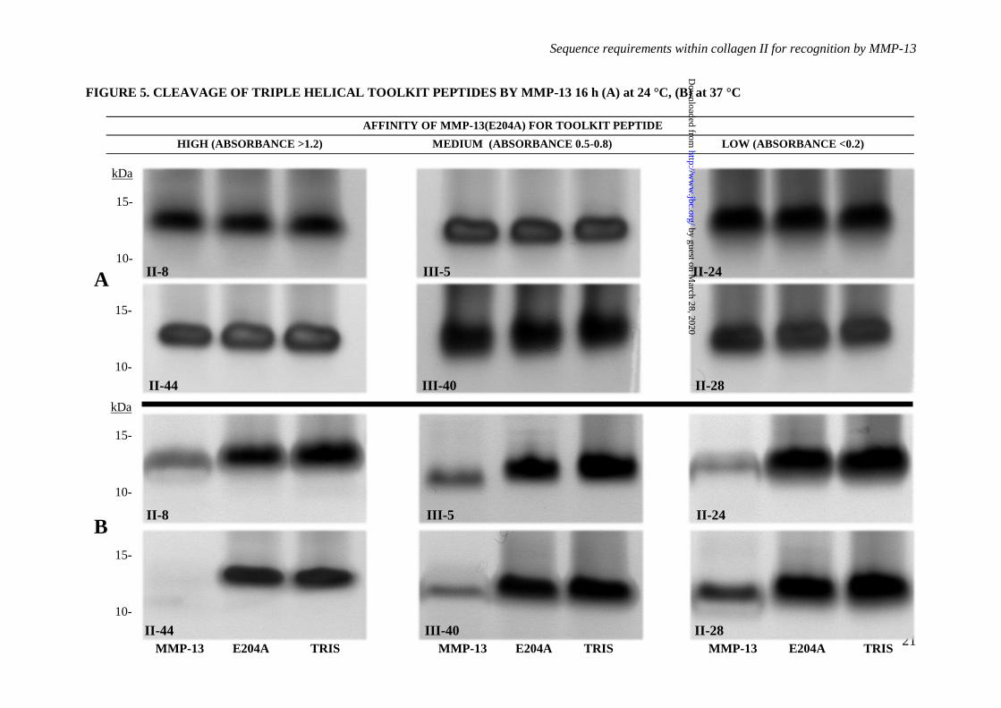
However, following incubation of the
peptides with 250 nM MMP-13 for 16 h at 24
°C, a temperature at which the majority of each
peptide is folded as a triple helix, proteolysis of
II-8, II-44, II-24 and III-5 was detectable by
mass spectrometry. Cleavage sites are shown in
Table 1. Degradation was slight and not
detectable using SDS-PAGE (Figure 5A). At 37
°C, proteolysis was clearly observable by SDS-
PAGE for all peptides tested (Figure 5B).
Cleavage sites were subsequently confirmed by
MALDI mass spectrometry (Table 1). At 24 °C,
peptide II-44 was specifically cleaved at both its
primary and secondary collagenase cleavage
sites (GL and GQ respectively). Peptide II-8
was cleaved, but at a G~A bond downstream
from the expected GL cleavage site. At 37 °C
however, the degradation of II-44 was
increasingly marked and less specific, most
likely due to the gelatinase activity of MMP-13.
DISCUSSION
We used four full-length MMP-13
preparations to screen the Toolkit peptides; the
wild type pro-form expressed and purified from
mouse myeloma cells, and recombinant MMP-
13(E204A) and proMMP-13(E204A) expressed
in pET3 cells as previously described (18,20).
The active wild type MMP-13 was prepared by
APMA-induced cleavage of its pro-form (18,29).
In addition, we tested the binding properties of
recombinant Cat and Hpx domains. The present
work shows that collagen and two triple-helical
Toolkit peptides, II-44 and II-8, support firm
adhesion of MMP-13 in both its active and pro-
forms, and of the equivalent catalytically
inactive mutant forms, proMMP-13(E204A) and
MMP-13(E204A). Cat bound weakly to
collagen, but not to any Toolkit peptide, whilst
Hpx bound to II-8 and to a greater extent II-44,
much like proMMP-13(E204A).
The fidelity of MMP-13(E204A)
substrate recognition–The use of MMPs that are
alanine-substituted at the catalytic site simplifies
structural and binding studies, since both
autolysis and digestion of adhesive substrates are
avoided. Here, we observe differences in
behaviour of the active conformation of MMP-
13(E204A) compared with either its own pro-
form or both the active or pro-forms of the wild
type enzyme. We find MMP-13(E204A) to bind
less specifically to Toolkits, with extra hits
observed in Toolkit II and a noisy pattern of
binding to Toolkit III. MMP-13(E204A) was
less sensitive to Ala-scanning of peptide II-44,
notably at P5', P10' and P11'. In contrast, pro-
MMP-13(E204A) behaved exactly as pro- or
active wild type. We also found a two-fold
higher Bmax for MMP-13(E204A), which implies
twice the number of molecules binding per
peptide. In turn, this observation suggests
dimerisation in situ, the first immobilized copy
of MMP-13(E204A) recruiting a second
molecule, a process mediated by interactions
remote from the peptide-binding surface of the
by guest on March 28, 2020
http://ww
w.jbc.org/
Dow
nloaded from
Sequence requirements within collagen II for recognition by MMP-13
7
enzyme. Crystallographic analysis by Stura et
al.(30) indicates close agreement between the
structures of native and mutant forms, but they
also report MMP-13(E204A) to be monomeric
in solution despite observing dimerization in
their crystal structure, mediated by the Hpx
domain. Reconciling these findings suggests
that the E204A substitution subtly modulates the
relationship between the Cat and Hpx domains
in the active conformation of the enzyme, in a
way that renders the Hpx less selective in
recognising collagenous substrates.
Collagen residues critical for
recognition by MMP-13–Binding of MMP-13 to
peptide II-44 was expected, since it contains the
known collagenase cleavage site near the N-
terminus of collagen D-period 4, whereas the
prominent but lower affinity of MMP-13 for II-8
was unsuspected; its sequence occurring in the
centre of D-period 1 (see Figure 6). Equilibrium
binding studies suggest similar affinities of all
the MMP forms tested for II-44, with KD on the
order of 200-400 nM. Ala-scanning of 44J, a
shorter peptide derived from II-44, revealed
several residues that make important
contributions to the binding of MMP-13, i.e. that
Ala-substitution diminishes binding. Five of
these lie within the C-terminal half of the guest
sequence of the peptide, P8'(V), P10'(L), P11'(O),
P14'(R) and P17'(R). Conversely, substitution of
two further residues in this region P13'(Q) and
P16'(E) enhanced the binding of all MMP-13
preparations. These observations underline the
importance of this tract of the peptide in
securing MMP-13 binding, and clarify the basis
for its interaction with peptide II-8. II-8 shares
just three critical residues with II-44,
corresponding to P1'(L), P5'(R) and P17'(R), but in
addition, contains two alanine residues,
corresponding to P13' and P16'. Ala-substitution
of both residues in peptide II-44J enhanced
MMP-13 binding. Good binding of II-8,
especially in the region predicted to bind Hpx,
might therefore be anticipated.
The importance of Hpx in MMP–
collagen interaction–The distance of many of
these residues from P1~P1' suggests that the Hpx
domain is the main contributor to MMP-13
binding, investigated further using isolated Hpx
which bound to both II-8 and II-44, exactly
echoing the adhesion profile of proMMP-
13(E204A) on the Ala-scanned peptide set
(Figure 4). These data suggest a second site of
interaction for Hpx in II-44. In the first,
“productive” mode of binding, Hpx engages
residues P10' to P17', GLOGQRGER, which
would align the scissile P1~P1' (G~L) bond close
to the catalytic site. The inhibition of Hpx
binding by Ala-scanning at P1', P4' and P5'
indicates that these conserved residues,
G~LAGQRGIV, can also interact directly with
the Hpx domain, offering a second locus that can
recruit MMP-13 to the environs of its site of
action. In this case however, alignment of the
Cat with P10–P9 would not be productive, as the
proline-rich collagen sequence at this location
would restrict local unwinding by the MMP-13
enzyme.
Comparison of MMP-13 with MMP-1–
Our interpretation is informed by the crystal
structure of the complex between MMP-1 and a
triple-helical peptide derived from II-44J.
Inspection of the MMP-1–peptide co-crystal
shows that P8' and P10' interact with the proximal
rim of the Hpx -propeller, binding to the
interface between blades 1 and 2, with the distal
rim of Hpx tilted away from the axis of the
peptide. No interactions were observed beyond
P11'. Only two peptide residues, P1'(L) and
P10'(L), had measurable effects on binding of
MMP-1 using a very similar SPBA, whilst
effects of substitutions distal to P10' were
negligible. In contrast, using MMP-13, there
were obvious effects of Ala-substitutions from
P10'(L) to P17'(R), suggesting that its Hpx enjoys
a closer interaction with regions of the peptide
extending further away from the scissile G~L
bond.
Another marked difference between the
two enzymes is that proMMP-1 did not bind to
collagen or Toolkit peptides, with the pro-
domain apparently impeding access to the Cat
(7,31), preventing the critical P1' interaction with
substrate. How can MMP-13 avoid this
constraint? Superposition of the Cat domains
from the crystal structures of full-length MMP-1
and MMP-13 by Stura et al. illustrates the
different Cat–Hpx interface in the two enzymes.
The two domains move closer together in MMP-
13, with Hpx rotated relative to Cat by 30°.
The effect is to move the collagen-binding S10'
pocket of MMP-13 by about 7 Å relative to that
of MMP-1(22). This might facilitate collagen–
proMMP-13 binding without displacement of
pro-domain from the S1' site. Like MMP-13,
both proMMP-3 and proMMP-9 can bind
collagen (32). Alternatively, although activation
of MMPs is achieved through proteolytic
cleavage, interaction with substrate may also
displace the pro-domain from the active center
by guest on March 28, 2020
http://ww
w.jbc.org/
Dow
nloaded from

Sequence requirements within collagen II for recognition by MMP-13
8
of the enzyme(33). Nonetheless, our SPBA data
suggests that the pro-domain does impede
collagen binding to some degree: whilst the two
full-length pro-forms of MMP-13 showed
similar binding, the catalytically dead MMP-
13(E204A), lacking pro-domain, showed highest
binding to Toolkit peptides.
The significance of the II-8 binding site–
The presence of this site was not expected, and
its potential as a substrate for the collagenase
activity of MMP-13 appears restricted to the
G~A clip observed at 24 °C. It is located at the
centre of II-8, four triplets C-terminal to the
more promising GLOGER motif that aligns well
with the canonical site in II-44; both sites occur
at the start of the guest sequence in their
respective peptides, and in nature, P4 to P1 are
GPRG and GPQG respectively. Q, preceding
the canonical (II-44) site, is well represented at
P2 in the MEROPS database(34), whilst R,
preceding the optimally aligned II-8 site, occurs
just 4 times in 41 observations of MMP-13
activity against collagenous substrates. This
locus in II-8 was clipped at 37 °C, and should
not be discounted as an authentic site for the
collagenolytic activity of MMP-13.
The two sites may cooperate in
recruiting MMP-13 to collagen. In 2007,
Overall and Butler(20) proposed an "inchworm"
model of MMP translation along the collagen
molecule, based on the "burnt bridges"
molecular ratchet model (for MMP-1) of
Saffarian et al.(33), and the atomic force study
of Rosenblum et al.(34) on MMP-9. For this
model to be generally applicable, the MMPs
would need to have affinity for long stretches of
sequence within the collagen helix, as well as the
capacity for sequence-independent cleavage of
triple-helical collagen. Manka et al.(6) found
that MMP-1 lacked the ability to bind well to
any site other than II-44 in collagen II, and we
show here that MMP-13 is only slightly less
selective, binding to II-8 as well as to the second
site in II-44. The "inchworm" model may
therefore have limited validity for MMP-13,
applying to translocation of Hpx binding from
the P1' to P5' tract to P10' to P17'. The "quarter-
stagger" assembly of the collagen fiber dictates
that any D-period is aligned side by side with all
others (see figure 6). Thus, a molecule that
bound at the start of D1 would be quite close to
the start of D2, D3, D4 and of the truncated D5
(the gap region) in adjacent tropocollagen
molecules. The simple model of fiber assembly
allows us to calculate the proximity of any two
sites. GLAGQR in II-44 represents residues
775-780 of the tropocollagen helix, or 73-78 of
the D-period, whereas GLOGER in II-8 is
residues 127-132 of both the helix and D-period.
Within a fiber, therefore, there is an axial offset
of 54 residues between the two sites,
corresponding to about 15 nm, close enough,
perhaps, to support some co-operativity.
Inspection of the co-crystal of MMP-1 and a II-
44-derived triple-helical peptide(7) suggests that
an MMP can span about 25 collagen helix
residues, less than 10 nm. Although not
contiguous, therefore, it is plausible that
hydrolysis of the canonical site might lead to
anchorage of MMP-13 by its Hpx domain to the
clipped helix, with sufficient flexibility in the
clipped collagen to relocate via its Cat to nearby
II-8 sites. This "ball and chain" model would
also apply to adjacent canonical sites in D4. The
three-dimensional structure may be more
complex than the simple model(35,36), but in
either of these two, reciprocal, renderings of the
fiber, hydrolysis at one site could reveal the
other site beneath.
Finally, whether it is a cleavage site or
not, it is plausible that II-8 fulfils a depot
function, acting to sequester proMMP-13 in the
collagen matrix to be activated as required.
MMP-13 differs from MMP-1 in this respect, in
that only the active form of MMP-1 was found
to bind to Toolkit II(7).
The selectivity of MMP-13 for different
collagens–Comparison of the collagenase
cleavage sites of the various fibrillar collagens
with the sequences of the MMP-13-binding
peptides provides a basis for their relative
recognition and subsequent cleavage. High
identity occurs between 1(I) and 1(II), with
only two non-identical but conserved amino
acids within the sequence equivalent to II-44.
The reported 6-fold lower activity of MMP-13
on collagen I may therefore reside in the
different sequence at P4' and P5' in 2(I), where
A and O occur in place of Q and R, both of
which, when replaced with A in peptide II-44J,
support reduced binding of active MMP-13. In
collagen III, only three (P5'(R), P11'(O) and
P17'(R)) of the eight critical amino acids are
identical with those in collagen II. We therefore
anticipate a lower affinity interaction of MMP-
13 with collagen III, consistent with its relative
resistance to proteolysis.
Toolkit peptides as substrates for
proteolysis by MMP-13–We tested the ability of
MMP-13 to digest six peptides during 16 h at
by guest on March 28, 2020
http://ww
w.jbc.org/
Dow
nloaded from

Sequence requirements within collagen II for recognition by MMP-13
9
either 24 °C or 37 °C by examining peptide
fragments by mass spectrometry. After
incubation at 24 °C, we observed both expected
clips in II-44, at P1~P1' and P3'~P4', at the
canonical and secondary sites that are well-
known(12). At this temperature, only five other
clips were located in the six peptides tested,
including a G~A bond in II-8. We had
anticipated that the G~L bond at the start of the
guest sequence of II-8, equivalent to P1~P1' in II-
44, might be susceptible, best aligning with the
catalytic site according to our interpretation of
Ala-scan data, but it proved otherwise. The
MEROPS database(34) describes the preferred
MMP-13 cleavage sequence as being
GPxG~LxGx, with Proline (bold) occurring at
P3 in 92 of 147 sites, P being the most abundant
P3 residue in both collagenous and non-
collagenous substrates. The sequence cleaved in
II-8 conforms well, the site being GPAG~AA,
with no other imino acids occurring within 9
residues, which may contribute to a more
relaxed structure that facilitates proteolysis(37).
At 37 °C, all Toolkit peptides tested
were cleaved by MMP-13, irrespective of their
ability to bind MMP-13(E204A) in SPBA, and
20 different sites were identified in addition to
the 7 found at 24 °C. We found four cleaved
G~L sites, from a total of ten occurrences of GL
in the six peptides tested. Five of these ten were
within G~LO triplets, only one of which was
cleaved, suggesting that the hydroxyproline at
P2' confers a protective effect, which might
account for the resistance of the GL bond in II-8
to hydrolysis. We also found four G~E sites, of
only eight GEx' triplets within the peptides
tested. The MEROPS database(34) describes the
preferred MMP-13 cleavage sequence as being
GPxG~LxGx, with Proline (bold) occurring at
P3 in 92 of 147 sites, P being the most abundant
P3 residue in both collagenous and non-
collagenous substrates. Deng et al. report that
the hydrophobic MMP-13 S3 subsite has a
preference for Pro in P3(38), and propose a
consensus motif, PLG~MR. Our limited data
suggest a novel consensus sequence: PPG~ER,
with POG~Ax' and POG~LA as alternatives.
Our data concur with that compiled in
MEROPS, although G~E cleavage is only
recorded twice therein, as proline occurs as P3 in
15 of the 27 cleavage sites (~55 %) we report
here, regardless of temperature.
We considered why elevating
temperature might increase the number of
resolvable cleavage sites. Firstly, reaction rates
will increase, but secondly, our peptides have
limited thermal stability, with melting
temperatures typically of 45 °C. Thus at 37 °C a
substantial proportion (18-52 %), estimated for
each specific peptide and shown in Table 1,
exists in unfolded, single-stranded form(39)
where, unlike native collagens, tertiary structure
no longer governs substrate specificity and
MMP-13 functions as a gelatinase(40).
Digestion of any single-stranded material that is
susceptible to proteolysis by MMP-13 will draw
the equilibrium in favor of unfolded peptide,
leading to gradual depletion of intact triple helix.
Mass spectrometry revealed that few of the
common peptide fragments were generated by
cleavage within the GPP hosts. This may reflect
the greater thermal stability of the relatively
proline-poor host sequence, which unfolds less
readily and so presents a less frequent target for
proteolysis by MMP-13 than the guest sequence.
In summary, here we show that, unlike
MMP-1, both the pro- and active forms of
MMP-13 are able to bind collagen, at both the
canonical MMP cleavage site, and at a second,
lower-affinity site located in Toolkit peptide II-8
(corresponding to D-period 1 of the collagen
monomer). We show that the recognition of II-
44 by MMP-13 is primarily mediated by the Hpx
domain and is specified by GLxGQR motifs at
either P1 - P5' or P9' - P14'. By examining the
cleavage of selected collagen peptides by MMP-
13, we show that hydrolysis is not confined to
motifs recognized by the Hpx domain, but is
likely to include peptide strands that are
unfolded at 37 °C. These data provide a
rationale for the structural regulation of collagen
proteolysis and the resistance of peptide II-8 to
cleavage by MMP-13 at 24 oC.
by guest on March 28, 2020
http://ww
w.jbc.org/
Dow
nloaded from

Sequence requirements within collagen II for recognition by MMP-13
10
REFERENCES
1. Steplewski, A., Hintze, V., and Fertala, A. (2007) Molecular basis of organization of collagen
fibrils. J Struct Biol 157, 297-307
2. Hijova, E. (2005) Matrix metalloproteinases: their biological functions and clinical
implications. Bratisl Lek Listy 106, 127-132
3. Mitchell, P. G., Magna, H. A., Reeves, L. M., Lopresti-Morrow, L. L., Yocum, S. A., Rosner,
P. J., Geoghegan, K. F., and Hambor, J. E. (1996) Cloning, expression, and type II
collagenolytic activity of matrix metalloproteinase-13 from human osteoarthritic cartilage. J
Clin Invest 97, 761-768
4. Leeman, M. F., Curran, S., and Murray, G. I. (2002) The structure, regulation, and function of
human matrix metalloproteinase-13. Crit Rev Biochem Mol Biol 37, 149-166
5. Li, J., Brick, P., O'Hare, M. C., Skarzynski, T., Lloyd, L. F., Curry, V. A., Clark, I. M., Bigg,
H. F., Hazleman, B. L., Cawston, T. E., and et al. (1995) Structure of full-length porcine
synovial collagenase reveals a C-terminal domain containing a calcium-linked, four-bladed
beta-propeller. Structure 3, 541-549
6. Fields, G. B. (1991) A model for interstitial collagen catabolism by mammalian collagenases.
J Theor Biol 153, 585-602
7. Manka, S. W., Carafoli, F., Visse, R., Bihan, D., Raynal, N., Farndale, R. W., Murphy, G.,
Enghild, J. J., Hohenester, E., and Nagase, H. (2012) Structural insights into triple-helical
collagen cleavage by matrix metalloproteinase 1. Proc Natl Acad Sci U S A 109, 12461-12466
8. Lauer-Fields, J. L., Tuzinski, K. A., Shimokawa, K., Nagase, H., and Fields, G. B. (2000)
Hydrolysis of triple-helical collagen peptide models by matrix metalloproteinases. J Biol
Chem 275, 13282-13290
9. Clark, I. M., and Cawston, T. E. (1989) Fragments of human fibroblast collagenase.
Purification and characterization. Biochem J 263, 201-206
10. Knauper, V., Osthues, A., DeClerck, Y. A., Langley, K. E., Blaser, J., and Tschesche, H.
(1993) Fragmentation of human polymorphonuclear-leucocyte collagenase. Biochem J 291 (
Pt 3), 847-854
11. Lauer-Fields, J. L., Nagase, H., and Fields, G. B. (2000) Use of Edman degradation sequence
analysis and matrix-assisted laser desorption/ionization mass spectrometry in designing
substrates for matrix metalloproteinases. J Chromatogr A 890, 117-125
12. Knauper, V., Cowell, S., Smith, B., Lopez-Otin, C., O'Shea, M., Morris, H., Zardi, L., and
Murphy, G. (1997) The role of the C-terminal domain of human collagenase-3 (MMP-13) in
the activation of procollagenase-3, substrate specificity, and tissue inhibitor of
metalloproteinase interaction. J Biol Chem 272, 7608-7616
13. Salsas-Escat, R., and Stultz, C. M. (2010) Conformational selection and collagenolysis in type
III collagen. Proteins 78, 325-335
14. Han, S., Makareeva, E., Kuznetsova, N. V., DeRidder, A. M., Sutter, M. B., Losert, W.,
Phillips, C. L., Visse, R., Nagase, H., and Leikin, S. (2010) Molecular mechanism of type I
collagen homotrimer resistance to mammalian collagenases. J Biol Chem 285, 22276-22281
15. Konitsiotis, A. D., Raynal, N., Bihan, D., Hohenester, E., Farndale, R. W., and Leitinger, B.
(2008) Characterization of high affinity binding motifs for the discoidin domain receptor
DDR2 in collagen. J Biol Chem 283, 6861-6868
16. Raynal, N., Hamaia, S. W., Siljander, P. R., Maddox, B., Peachey, A. R., Fernandez, R.,
Foley, L. J., Slatter, D. A., Jarvis, G. E., and Farndale, R. W. (2006) Use of synthetic peptides
to locate novel integrin alpha2beta1-binding motifs in human collagen III. J Biol Chem 281,
3821-3831
17. Farndale, R. W., Lisman, T., Bihan, D., Hamaia, S., Smerling, C. S., Pugh, N., Konitsiotis,
A., Leitinger, B., de Groot, P. G., Jarvis, G. E., and Raynal, N. (2008) Cell-collagen
interactions: the use of peptide Toolkits to investigate collagen-receptor interactions. Biochem
Soc Trans 36, 241-250
18. Knauper, V., Lopez-Otin, C., Smith, B., Knight, G., and Murphy, G. (1996) Biochemical
characterization of human collagenase-3. J Biol Chem 271, 1544-1550
19. Kennedy, A. M., Inada, M., Krane, S. M., Christie, P. T., Harding, B., Lopez-Otin, C.,
Sanchez, L. M., Pannett, A. A., Dearlove, A., Hartley, C., Byrne, M. H., Reed, A. A., Nesbit,
by guest on March 28, 2020
http://ww
w.jbc.org/
Dow
nloaded from
Sequence requirements within collagen II for recognition by MMP-13
11
M. A., Whyte, M. P., and Thakker, R. V. (2005) MMP13 mutation causes
spondyloepimetaphyseal dysplasia, Missouri type (SEMD(MO). J Clin Invest 115, 2832-2842
20. Chung, L., Dinakarpandian, D., Yoshida, N., Lauer-Fields, J. L., Fields, G. B., Visse, R., and
Nagase, H. (2004) Collagenase unwinds triple-helical collagen prior to peptide bond
hydrolysis. Embo J 23, 3020-3030
21. Overall, C. M., and Butler, G. S. (2007) Protease yoga: extreme flexibility of a matrix
metalloproteinase. Structure 15, 1159-1161
22. Stura, E. A., Visse, R., Cuniasse, P., Dive, V., and Nagase, H. (2013) Crystal structure of full-
length human collagenase 3 (MMP-13) with peptides in the active site defines exosites in the
catalytic domain. FASEB J 27, 4395-405
23. Erat, M. C., Slatter, D. A., Lowe, E. D., Millard, C. J., Farndale, R. W., Campbell, I. D., and
Vakonakis, I. (2009) Identification and structural analysis of type I collagen sites in complex
with fibronectin fragments. Proc Natl Acad Sci U S A 106, 4195-4200
24. Erat, M. C., Schwarz-Linek, U., Pickford, A. R., Farndale, R. W., Campbell, I. D., and
Vakonakis, I. (2010) Implications for collagen binding from the crystallographic structure of
fibronectin 6FnI1-2FnII7FnI. J Biol Chem 285, 33764-33770
25. Ingham, K. C., Brew, S. A., and Migliorini, M. (2002) Type I collagen contains at least 14
cryptic fibronectin binding sites of similar affinity. Arch Biochem Biophys 407, 217-223
26. Dzamba, B. J., Wu, H., Jaenisch, R., and Peters, D. M. (1993) Fibronectin binding site in type
I collagen regulates fibronectin fibril formation. J Cell Biol 121, 1165-1172
27. Giudici, C., Raynal, N., Wiedemann, H., Cabral, W. A., Marini, J. C., Timpl, R., Bachinger,
H. P., Farndale, R. W., Sasaki, T., and Tenni, R. (2008) Mapping of SPARC/BM-
40/osteonectin-binding sites on fibrillar collagens. J Biol Chem 283, 19551-19560
28. Pugh, N., Simpson, A. M., Smethurst, P. A., de Groot, P. G., Raynal, N., and Farndale, R. W.
(2010) Synergism between platelet collagen receptors defined using receptor-specific
collagen-mimetic peptide substrata in flowing blood. Blood 115, 5069-5079
29. Nagase, H., Enghild, J. J., Suzuki, K., and Salvesen, G. (1990) Stepwise activation
mechanisms of the precursor of matrix metalloproteinase 3 (stromelysin) by proteinases and
(4-aminophenyl)mercuric acetate. Biochemistry 29, 5783-5789
30. Stura, E. A., Visse, R., Cuniasse, P., Dive, V., and Nagase, H. (2013) Crystal structure of full-
length human collagenase 3 (MMP-13) with peptides in the active site defines exosites in the
catalytic domain. FASEB J 27, 4395-4405
31. Jozic, D., Bourenkov, G., Lim, N. H., Visse, R., Nagase, H., Bode, W., and Maskos, K.
(2005) X-ray structure of human proMMP-1: new insights into procollagenase activation and
collagen binding. J Biol Chem 280, 9578-9585
32. Zhen, E. Y., Brittain, I. J., Laska, D. A., Mitchell, P. G., Sumer, E. U., Karsdal, M. A., and
Duffin, K. L. (2008) Characterization of metalloprotease cleavage products of human
articular cartilage. Arthritis Rheum 58, 2420-2431
33. Bannikov, G. A., Karelina, T. V., Collier, I. E., Marmer, B. L., and Goldberg, G. I. (2002)
Substrate binding of gelatinase B induces its enzymatic activity in the presence of intact
propeptide. J Biol Chem 277, 16022-16027
34. Rawlings, N. D. (2009) A large and accurate collection of peptidase cleavages in the
MEROPS database. Database (Oxford) 2009, bap015
35. Perumal, S., Antipova, O., and Orgel, J. P. (2008) Collagen fibril architecture, domain
organization, and triple-helical conformation govern its proteolysis. Proc Natl Acad Sci U S A
105, 2824-2829
36. Herr, A. B., and Farndale, R. W. (2009) Structural Insights into the Interactions between
Platelet Receptors and Fibrillar Collagen. J Biol Chem 284, 19781-19785
37. Williams, K. E., and Olsen, D. R. (2009) Matrix metalloproteinase-1 cleavage site recognition
and binding in full-length human type III collagen. Matrix Biol 28, 373-379
38. Deng, S. J., Bickett, D. M., Mitchell, J. L., Lambert, M. H., Blackburn, R. K., Carter, H. L.,
3rd, Neugebauer, J., Pahel, G., Weiner, M. P., and Moss, M. L. (2000) Substrate specificity of
human collagenase 3 assessed using a phage-displayed peptide library. J Biol Chem 275,
31422-31427
by guest on March 28, 2020
http://ww
w.jbc.org/
Dow
nloaded from
Sequence requirements within collagen II for recognition by MMP-13
12
39. Slatter, D. A., Bihan, D. G., Jarvis, G. E., Stone, R., Pugh, N., Giddu, S., and Farndale, R. W.
(2012) The properties conferred upon triple-helical collagen-mimetic peptides by the presence
of cysteine residues. Peptides 36, 86-93
40. O'Farrell, T. J., Guo, R., Hasegawa, H., and Pourmotabbed, T. (2006) Matrix
metalloproteinase-1 takes advantage of the induced fit mechanism to cleave the triple-helical
type I collagen molecule. Biochemistry 45, 15411-15418
by guest on March 28, 2020
http://ww
w.jbc.org/
Dow
nloaded from

Sequence requirements within collagen II for recognition by MMP-13
13
FIGURE LEGENDS
Table 1. Mass analysis of degraded Toolkit peptides. Following degradation for 16 h at 24 °C
and 37 °C as described for Figures 9B and C respectively, Toolkit peptides incubated with MMP-13
or Tris buffer pH 7.4 (as a negative control) were subjected to MALDI mass spectrometric analysis to
determine the peptide cleavage site(s) of MMP-13. Cleavage sites are summarized with the site of
proteolysis after the first residue in the sequence of six. Triple helical abundance of each peptide at 37
°C is also listed.
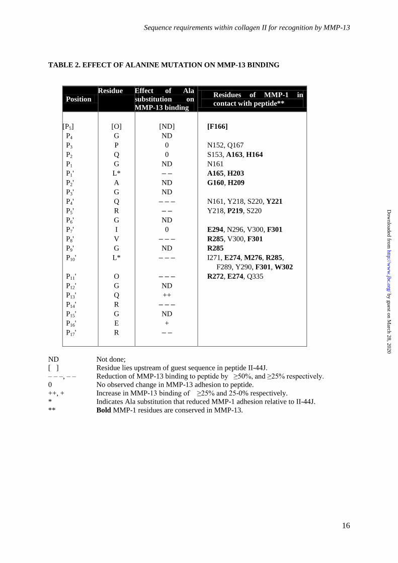
Table 2. Summary of the effect of alanine substitution on MMP-13 binding. Residues of
Toolkit peptide II-44J are listed as are the residues of MMP-1 that interact with these amino acids
(MMP-1 residues in bold are conserved in MMP-13). The effect of alanine substitution of each
residue is annotated as ND = Not done; [ ] residue lies upstream of guest sequence in peptide II-44J;
– – –, – – Reduction of MMP-13 binding to peptide by ≥ 50 %, and ≥ 25 % respectively; 0 = No
observed change in MMP-13 adhesion to peptide; +, ++ Increase in MMP-13 binding of 25-0 % and
≥ 25 % respectively; *Ala substitution that reduced MMP-1 adhesion relative to II-44J.
Figure 1. Adhesion of MMP-13 preparations to Collagen II Toolkit peptides. Plates were coated
with 10 µg/ml Toolkit II peptides, fibrillar type I collagen and the negative binding peptide GPP10.
(A) proMMP-13 (black bars) and MMP-13 (grey bars), (B) proMMP-13(E204A; black bars) and
MMP-13(E204A; grey bars), (C) Hpx at a concentration of 83 nM were allowed to adhere to the
peptides for 1 h at room temperature. Full-length MMP-13 preparations were then detected using an
antibody directed at the MMP-13 linker region, and Hpx with an anti-GST antibody as described in
Experimental Procedures. Data represent mean A450 ± S.E. of four experiments. For reference,
collagen D-periods corresponding to these peptides are shown as grey bars below, and the collagenase
cleavage site indicated by an arrow.
Figure 2. (A) Competitive inhibition of MMP-13 adhesion to Toolkit peptides II-8 and II-44.
(A) MMP-13 or MMP-13(E204A) were pre-incubated with increasing amounts of Toolkit peptides II-
8 and II-44 prior to adhesion assays on II-8 and II-44 respectively. Absorbance was measured at 450
nm. Data represents the mean ± S.E. of four experiments. (B) The effect of MMP-13(E204A)
mutation on increased proMMP-13 adhesion to Toolkit peptides II-8 and II-44. Binding of MMP-13
and MMP-13(E204A) to collagen coated ELISA wells was performed as described in Figure 1. Both
pro- and active forms of the enzyme exhibited broadly similar affinities (p = 0.37), with KD ranging
from 130 to 380 nM, the latter figure (the only outlying value) being the estimate for proMMP-
13(E204A) binding to peptide II-8Data represents the mean ± S.E. of four experiments.
by guest on March 28, 2020
http://ww
w.jbc.org/
Dow
nloaded from

Sequence requirements within collagen II for recognition by MMP-13
14
Figure 3. Binding of MMP-13 and MMP-13(E204A) to monomeric and fibrillar type I
collagens. Binding of MMP-13 and MMP-13(E204A) to collagen coated ELISA wells was performed
as described in Figure 1. For MMP-13 binding to fibres and monomers, KD values were 33 and 118
nM, respectively, and for MMP-13(E204A), 52 and 26 nM, respectively. Data points represents the
mean ± S.E. of four experiments, and differences were not significant.
Figure 4. Adhesion of proMMP-13, MMP-13, proMMP-13(E204A), MMP-13(E204A) and the
Hpx domain to II-44 variant Toolkit peptides was carried out as described for Figure 1. The dashed
line represents MMP adhesion level on peptide II-44J, and the solid line MMP baseline adhesion on
peptide II-44B. Data represents the mean ± S.E. of four experiments.
Figure 5. Analysis of possible degradation of Toolkit peptides by active MMP-13. Toolkit
peptides at a final concentration of 1200 µg/ml to which MMP-13(E204A) displayed high binding (II-
8 and II-44; A450 > 1.2); intermediate binding (Toolkit peptides III-5 and III-40; A450 0.5-0.8) and low
binding (Toolkit peptides II-24 and II-28; A450 < 0.2) were incubated with Tris buffer, MMP-13 or
MMP-13(E204A) at 250 nM final concentration were incubated at (A) 24 °C for 16 h and (B) 37 °C
for 16 h. After incubation, samples were subjected to electrophoresis under reducing conditions and
silver stained. Images are representative of three experiments.
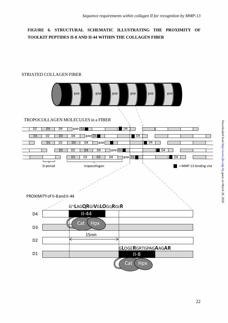
Figure 6. Schematic illustration of a collagen fiber. Above shows the alternating gap (pale)
and overlap (dark) banding, derived from the staggered assembly of the fiber from tropocollagen
molecules. Middle, the "quarter stagger" between tropocollagen molecules is shown, with labeled D-
periods alternating (gray and white) with the MMP-13 binding sites mapped onto them (black).
Below the linear separation between the II-44 and II-8 sites is shown, and their sequences provided.
Key binding residues in II-44 and II-8 are larger, bold font, whilst restrictive residues (II-44) are
smaller font. Lateral separation between the axes of the tropcollagen monomers (not indicated) is of
the order of 3 nm.
by guest on March 28, 2020
http://ww
w.jbc.org/
Dow
nloaded from
Sequence requirements within collagen II for recognition by MMP-13
15
TABLE 1. MALDI MASS ANALYSIS OF DEGRADED TOOLKIT PEPTIDES.
PEPTIDE
AND MASS
(Da)
CLEAVAGE
TEMP (oC) CLEAVAGE SITES (Indicated in black)
% Triple
Helical
peptide at
37 oC
II-8
5503
24
37
GPCGPPGPPGPPGPPGPP-GLOGERGRTGPAGAAGARGNDGQOGPA-GPPGPPGPPGPPGPPGPC
GPCGPPGPPGPPGPPGPP-GLOGERGRTGPAGAAGARGNDGQOGPA-GPPGPPGPPGPPGPPGPC
GPCGPPGPPGPPGPPGPP-GLOGERGRTGPAGAAGARGNDGQOGPA-GPPGPPGPPGPPGPPGPC
48
II-44
5705
24
37
GPCGPPGPPGPPGPPGPP-GLAGQRGIVGLOGQRGERGFOGLOGPS-GPPGPPGPPGPPGPPGPC
GPCGPPGPPGPPGPPGPP-GLAGQRGIVGLOGQRGERGFOGLOGPS-GPPGPPGPPGPPGPPGPC
GPCGPPGPPGPPGPPGPP-GLAGQRGIVGLOGQRGERGFOGLOGPS-GPPGPPGPPGPPGPPGPC
80
II-24
5536
24
37
GPCGPPGPPGPPGPPGPP-GKAGEKGLOGAOGLRGLOGKDGETGAA-GPPGPPGPPGPPGPPGPC
GPCGPPGPPGPPGPPGPP-GKAGEKGLOGAOGLRGLOGKDGETGAA-GPPGPPGPPGPPGPPGPC
GPCGPPGPPGPPGPPGPP-GKAGEKGLOGAOGLRGLOGKDGETGAA-GPPGPPGPPGPPGPPGPC
54
II-28
5638
24
37
GPCGPPGPPGPPGPPGPP-GEAGAOGLVGPRGERGFOGERGSOGAQ-GPPGPPGPPGPPGPPGPC
GPCGPPGPPGPPGPPGPP-GEAGAOGLVGPRGERGFOGERGSOGAQ-GPPGPPGPPGPPGPPGPC
GPCGPPGPPGPPGPPGPP-GEAGAOGLVGPRGERGFOGERGSOGAQ-GPPGPPGPPGPPGPPGPC
57
III-5
5728
24
37
GPCGPPGPPGPPGPPGPP-GERGLOGPOGIKGPAGIOGFOGMKGHR-GPPGPPGPPGPPGPPGPC
GPCGPPGPPGPPGPPGPP-GERGLOGPOGIKGPAGIOGFOGMKGHR-GPPGPPGPPGPPGPPGPC
GPCGPPGPPGPPGPPGPP-GERGLOGPOGIKGPAGIOGFOGMKGHR-GPPGPPGPPGPPGPPGPC
82
III-40
5447
24
37
GPCGPPGPPGPPGPPGPP-GAAGFOGARGLOGPOGSNGNOGPOGPS-GPPGPPGPPGPPGPPGPC
GPCGPPGPPGPPGPPGPP-GAAGFOGARGLOGPOGSNGNOGPOGPS-GPPGPPGPPGPPGPPGPC
GPCGPPGPPGPPGPPGPP-GAAGFOGARGLOGPOGSNGNOGPOGPS-GPPGPPGPPGPPGPPGPC
58
by guest on March 28, 2020
http://ww
w.jbc.org/
Dow
nloaded from
Sequence requirements within collagen II for recognition by MMP-13
16
TABLE 2. EFFECT OF ALANINE MUTATION ON MMP-13 BINDING
Position Residue Effect of Ala
substitution on
MMP-13 binding
Residues of MMP-1 in
contact with peptide**
[P5]
P4
P3
P2
P1
P1'
P2'
P3'
P4'
P5'
P6'
P7'
P8'
P9'
P10'
P11'
P12'
P13'
P14'
P15'
P16'
P17'
[O]
G
P
Q
G
L*
A
G
Q
R
G
I
V
G
L*
O
G
Q
R
G
E
R
[ND]
ND
0
0
ND
– –
ND
ND
– – –
– –
ND
0
– – –
ND
– – –
– – –
ND
++
– – –
ND
+
– –
[F166]
N152, Q167
S153, A163, H164
N161
A165, H203
G160, H209
N161, Y218, S220, Y221
Y218, P219, S220
E294, N296, V300, F301
R285, V300, F301
R285
I271, E274, M276, R285,
F289, Y290, F301, W302
R272, E274, Q335
ND Not done;
[ ] Residue lies upstream of guest sequence in peptide II-44J.
– – –, – – Reduction of MMP-13 binding to peptide by ≥50%, and ≥25% respectively.
0 No observed change in MMP-13 adhesion to peptide.
++, + Increase in MMP-13 binding of ≥25% and 25-0% respectively.
* Indicates Ala substitution that reduced MMP-1 adhesion relative to II-44J.
** Bold MMP-1 residues are conserved in MMP-13.
by guest on March 28, 2020
http://ww
w.jbc.org/
Dow
nloaded from

Sequence requirements within collagen II for recognition by MMP-13
17
FIGURE 1. ADHESION OF MMP-13 PREPARATIONS AND Hpx TO TOOLKIT II
0
0.2
0.4
0.6
0.8
1
1.2
1.4
1.6
1.8
2
1 2 3 4 5 6 7 8 91
01
11
21
31
41
51
61
71
81
92
02
12
22
32
42
52
62
72
82
93
03
13
23
33
43
53
63
73
83
94
04
14
24
34
44
54
64
74
84
95
05
15
25
35
45
55
6C
OLL
GP
P1
0B
SA
A4
50
MMP-13(E204A) Adhesion to Toolkit II
0
0.2
0.4
0.6
0.8
1
1.2
1 2 3 4 5 6 7 8 9 10 11 12 13 14 15 16 17 18 19 20 21 22 23 24 25 26 27 28 29 30 31 32 33 34 35 36 37 38 39 40 41 42 43 44 45 46 47 48 49 50 51 52 53 54 55 56CO
LLG
PP10
BSA
A45
0
MMP-13 Adhesion to Toolkit II
D1 D2 D3 D4 D5
D-periods of collagen
0
0.2
0.4
0.6
0.8
1
1.2
1.4
1.6
1.8
1 2 3 4 5 6 7 8 91
01
11
21
31
41
51
61
71
81
92
02
12
22
32
42
52
62
72
82
93
03
13
23
33
43
53
63
73
83
94
04
14
24
34
44
54
64
74
84
95
05
15
25
35
45
55
6C
OLL
GP
P1
0B
SA
A4
50
Hpx Adhesion to Toolkit II
A
B
C
proMMP
MMP
proMMP
MMP
by guest on March 28, 2020
http://ww
w.jbc.org/
Dow
nloaded from
Sequence requirements within collagen II for recognition by MMP-13
18
FIGURE 2. (A) COMPETITIVE INHIBITION OF MMP-13 ADHESION TO TOOLKIT PEPTIDES II-8 AND II-44 (B) THE EFFECT OF MMP-
13(E204A) MUTATION ON proMMP ADHESION TO II-8 AND II-44
by guest on March 28, 2020
http://ww
w.jbc.org/
Dow
nloaded from

Sequence requirements within collagen II for recognition by MMP-13
19
FIGURE 3. ADHESION OF MMP-13(E204A) AND MMP-13 TO TYPE I COLLAGEN MONOMERS AND FIBERS
by guest on March 28, 2020
http://ww
w.jbc.org/
Dow
nloaded from
Sequence requirements within collagen II for recognition by MMP-13
20
0 0.2 0.4 0.6 0.80 0.1 0.2 0.3 0.40 0.2 0.4 0.6 0.8 10 0.5 1 1.5 2
FIGURE 4. MMP-13 ADHESION TO II-44 VARIANT PEPTIDES
Peptide Name Sequence MMP-13 BINDING (A450)
pro-MMP-13 MMP-13 proMMP-13(E204A) MMP-13(E204A) Hpx
II-8 -----GLOGERGRTGPAGAAGARGNDGQOGPA
II-44 -----GLAGQRGIVGLOGQRGERGFOGLOGPS
44A -----GLAGQRGIVGLOGQRGER---------
44B -----------------------GFOGLOGPS
44C --------GQRGIVGLOGQRGER---------
44D -----------GIVGLOGQRGER---------
44E -----GLAGQRGIVGLOGQR------------
44F -----GLAGQRGIVGLO---------------
44G --------GQRGIVGLO---------------
44H -----------GIVGLOGQR------------
44I --------------GLOGQRGERGFO------
44J --GPQGLAGQRGIVGLOGQRGER---------
P3A --GAQGLAGQRGIVGLOGQRGER---------
P2A --GPAGLAGQRGIVGLOGQRGER---------
P1’A --GPQGAAGQRGIVGLOGQRGER---------
P4’A --GPQGLAGARGIVGLOGQRGER---------
P5’A --GPQGLAGQAGIVGLOGQRGER---------
P7’A --GPQGLAGQRGAVGLOGQRGER---------
P8’A --GPQGLAGQRGIAGLOGQRGER---------
P10’A –-GPQGLAGQRGIVGAOGQRGER---------
P11’A --GPQGLAGQRGIVGLAGQRGER---------
P13’A --GPQGLAGQRGIVGLOGARGER---------
P14’A --GPQGLAGQRGIVGLOGQAGER---------
P16’A --GPQGLAGQRGIVGLOGQRGAR---------
P17’A --GPQGLAGQRGIVGLOGQRGEA---------
0 0.2 0.4 0.6 0.8 1 1.2
by guest on March 28, 2020
http://ww
w.jbc.org/
Dow
nloaded from
Sequence requirements within collagen II for recognition by MMP-13
21
FIGURE 5. CLEAVAGE OF TRIPLE HELICAL TOOLKIT PEPTIDES BY MMP-13 16 h (A) at 24 °C, (B) at 37 °C
AFFINITY OF MMP-13(E204A) FOR TOOLKIT PEPTIDE
HIGH (ABSORBANCE >1.2) MEDIUM (ABSORBANCE 0.5-0.8) LOW (ABSORBANCE <0.2)
kDa
15-
10-
15-
10-
II-8 III-5 II-24
II-44 III-40 II-28
15-
10-
II-8 III-5 II-24
II-44 III-40 II-28
MMP-13 E204A TRIS MMP-13 E204A TRIS MMP-13 E204A TRIS
kDa
15-
10-
A
B
by guest on March 28, 2020
http://ww
w.jbc.org/
Dow
nloaded from
Sequence requirements within collagen II for recognition by MMP-13
22
STRIATED COLLAGEN FIBER
TROPOCOLLAGEN MOLECULES in a FIBER
G~LAGQRGIVGLOGQRGER
II-44
II-8
D4
Cat Hpx
Cat Hpx
15nm
PROXIMITY of II-8 and II-44
D4D1D1 D2 D3 D4 gap
D1 D4D1 D2 D3 D4 gap
D1 D4D1 D2 D3 D4 gap
D1 D4D1 D2 D3 D4 gap
D2 D3 D4 gap D4D1
gapgapgapgap gap gap
tropocollagenD-period = MMP-13-binding site
D1
D3
D2
GLOGERGRTGPAGAAGAR
FIGURE 6. STRUCTURAL SCHEMATIC ILLUSTRATING THE PROXIMITY OF
TOOLKIT PEPTIDES II-8 AND II-44 WITHIN THE COLLAGEN FIBER
by guest on March 28, 2020
http://ww
w.jbc.org/
Dow
nloaded from
Packman, Vera Knauper, Robert Visse and Richard W. FarndaleJoanna-Marie Howes, Dominique Bihan, David A. Slatter, Samir W. Hamaia, Len C.
Sequence specificity for binding and cleavageThe recognition of collagen and triple-helical Toolkit peptides by MMP-13:
published online July 9, 2014J. Biol. Chem.
10.1074/jbc.M114.583443Access the most updated version of this article at doi:
Alerts:
When a correction for this article is posted•
When this article is cited•
to choose from all of JBC's e-mail alertsClick here
by guest on March 28, 2020
http://ww
w.jbc.org/
Dow
nloaded from