Embed Size (px)
Citation preview

1
Title: Methylation of Human Papillomavirus Type 16 E2BS#1, E2BS#2 and Sp1 1
binding site CpGs in cervical cancer samples determined by High Resolution 2
Melting PCR 3
4
5
Running title: HPV16 LCR methylation in cervical cancer samples 6
7
8
9
Elise Jacquin1,2, Alice Baraquin3, Rajeev Ramanah3, Xavier Carcopino4,5, Adrien Morel1,2, Séverine 10
Valmary-Degano1,2,3, Ignacio G. Bravo6, Silvia de Sanjosé6, Didier Riethmuller1,2,3, Christiane 11
Mougin1,2,3, Jean-Luc Prétet * 1,2,3 12
13
14
15 1Univ. Franche-Comte, F-25000 Besancon, France 16
2EA 3181, FED4234, CIC-BT 506, F-25000 Besancon, France 17
3CHU Besancon, F-25000, France 18
4Department of Obstetrics and Gynecology, Assistance Publique des Hôpitaux de Marseille (APHM), 19
Hôpital Nord, Marseille, France 20
5Aix-Marseille Université (AMU), 13385 Marseille, France 21
6Unit of Infections and Cancer, Cancer Epidemiology Research Program, Catalan Institute of 22
Oncology (ICO), Barcelona, Spain 23
24
*Corresponding author: 25
Jean-Luc Prétet, Laboratoire de Biologie Cellulaire et Moléculaire, FED4234, Université de Franche-26
Comté, Centre Hospitalier Universitaire, Boulevard A Fleming, 25030 Besançon cedex, France. 27
email: [email protected], tel: +33.3.81.66.91.12, fax: +33.3.81.66.93.70 28
29
Copyright © 2013, American Society for Microbiology. All Rights Reserved.J. Clin. Microbiol. doi:10.1128/JCM.01106-13 JCM Accepts, published online ahead of print on 17 July 2013
on June 6, 2018 by guesthttp://jcm
.asm.org/
Dow
nloaded from

2
Abstract 30
High-risk (HR) HPV-associated carcinogenesis is mainly driven by the overexpression of E7 and E6 31
oncoproteins following the viral DNA integration and the concomitant loss of the E2 ORF However, 32
integration of HR-HPV DNA is not systematically observed in cervical cancers. The E2 protein acts a 33
transcription factor that governs viral oncogenes expression. Methylation of CpGs in the E2-binding 34
sites (E2BS) in the viral long control region abrogates E2 binding, thus impairing the E2-mediated 35
regulation of E7/E6 transcription. Here, a High Resolution Melting (HRM) PCR was developed to 36
quantitatively analyze the methylation status of E2BS#1, E2BS#2 and Sp1-binding site in 119 HPV16-37
positive cervical smears. This is a rapid assay, suitable for the analysis of cervical samples. The 38
proportion of cancer samples with methylated E2BS#1, E2BS#2 and Sp1 binding site CpGs was 47% 39
whereas the vast majority of samples diagnosed as normal, LSIL or HSIL harbored unmethylated 40
CpGs. Methylation levels varied widely since some cancer samples harbored up to 60% of methylated 41
HPV16 genomes. A pyrosequencing approach was used as a confirmation test and highlighted that 42
quantitative measurement of methylation can be achieved by HRM PCR. Its prognostic value 43
deserves to be investigated alone or in association with other biomarkers. The reliability of this single-44
tube assay offers great opportunities for the investigation of HPV16 methylation in other HPV-related 45
cancer such as head and neck cancers, a major public health burden. 46
47
on June 6, 2018 by guesthttp://jcm
.asm.org/
Dow
nloaded from

3
Introduction 48
Certain Alphapapillomaviruses classified as “high-risk” (HR-HPVs) have been recognized as the 49
etiologic factors of invasive cervical carcinoma worldwide (1, 2). HR-HPV infections are frequent in 50
young women but they are generally transient, with an estimated mean duration of incident infection of 51
16 months (3). Only persistent HR-HPV infections are associated with an increased risk of developing 52
high grade cervical lesions or cancer of the cervix (4-6). Persistent HR-HPV infections may be 53
associated with microscopic abnormalities called low-grade squamous intraepithelial lesions (LSIL). 54
These LSIL present a high rate of regression following HPV clearance, but may however progress 55
towards high-grade squamous intraepithelial lesions (HSIL) if HR-HPV infection persists. Upon 56
diagnosis, HSIL are treated mostly by excisional procedures to avoid the risk of progression towards 57
invasive cancer (for review see(7)). 58
Among HR-HPVs, HPV16 exhibits the highest persistence rate (8). Moreover, HPV16 is associated 59
with an increased risk of precancerous and cancerous lesion development (9) and is considered as 60
the most carcinogenic PV in humans (10). This is probably why HPV16 prevalence increases with the 61
severity of cervical lesions (11) reaching 61% in invasive cervical carcinoma worldwide (12). 62
HPV-associated carcinogenesis requires the continuous overexpression of the two main viral 63
oncoproteins E7 and E6, which interact with many cellular proteins leading to the induction and the 64
maintenance of a transformed phenotype in infected cells (for review see (13)). E7 and E6 expression 65
is regulated by the viral E2 protein through the early promoter (named p97 for HPV16) located in the 66
long control region (LCR) of the PV genome. This promoter harbors four specific E2-binding sites 67
(E2BS) sharing the consensus sequence 5’-ACCG(N)4CGGT-3’ (14, 15). The three sites proximal to 68
the TATA box have been shown to be involved in E2-mediated repression of the promoter activity and 69
the E2BS#1 and E2BS#2 sites are probably the main sites to achieve this effect (for review see (16)). 70
From a mechanistic point of view, the binding of E2 to E2BS#1 and E2BS#2 induces a displacement 71
of transcription activators such as Sp1 and TATA Binding Protein (TBP) from their binding sites 72
leading to a repression of E7 and E6 expression from the early promoter (17, 18). HR-HPV DNA 73
integration has been described as a key step in the carcinogenesis process since it generally results in 74
the disruption of the E2 ORF (19, 20). 75
Epigenetic alterations and particularly aberrant DNA methylation are frequently associated with tumor 76
progression. Methylation of DNA mainly occurs on cytosines located in CpG dinucleotides and lead to 77
on June 6, 2018 by guesthttp://jcm
.asm.org/
Dow
nloaded from

4
gene silencing after chromatin structure remodeling. Both HPV16 E7 and E6 proteins seem to 78
increase DNA methyltransferase (DNMT) activity in vitro (21). Moreover, increased methylation of 79
cellular tumor suppressor genes such as rarb (retinoic acid receptor beta), dapk1 (death-associated 80
protein kinase 1) and camd1 (cell adhesion molecule 1) is frequently observed in cervical cancer 81
samples (22). Viral DNA sequences including HPV genomes can also undergo methylation by cellular 82
DNMT (23, 24). 83
The HPV16 genome contains 112 CpG and its methylome has been described (25-27). The CpG 84
dinucleotides located in the L1/L2 region of HPV16 genome are preferentially methylated during 85
disease progression (25, 26, 28, 29) and a recent study suggested the clinical value of methylation for 86
the identification of prevalent and incident high grade lesions (27). 87
Several studies aimed to describe methylation on the HPV16 LCR and the therein-contained E2BS in 88
clinical samples, but the results are very conflicting. Some studies reported a decreased E2BS 89
methylation during lesion progression (30-32) or a trend of lower risk of incident precancerous and 90
cancerous lesions for patients with high EB2S methylation (33, 34). On the contrary, the methylation of 91
the five CpGs located in the E2BS#1 and E2BS#2 and Sp1-binding site (Sp1BS) has frequently been 92
associated with cervical cancer (28, 29, 35-38) and a gradual increase of methylation during lesion 93
progression toward cancer has been reported in some studies (29, 36, 37, 39). Nevertheless, most of 94
these studies did not include the full spectrum of cervical disease progression, from normal to cancer. 95
Furthermore, various quantitative and non-quantitative techniques have been used to study HPV16 96
DNA methylation. Some studies have been conducted on fresh-frozen tissues or microdissected FFPE 97
biopsies whereas others have been performed on exfoliated cervical cells that may explain discrepant 98
results. 99
We report here a simple and robust High Resolution Melting (HRM) PCR that permits the quantitation 100
of the methylation levels of the five CpGs located in the two most important HPV16 E2BS involved in 101
E7/E6 regulation, namely E2BS#1 and E2BS#2, and in the Sp1BS. After optimization and validation 102
steps, this single-tube assay was used to explore E2BS#1, E2BS#2 and Sp1BS methylation in 103
samples previously identified to harbor HPV16 and representative of the natural history of cervical 104
cancer. The HRM analysis reveals that methylation of the target CpG is specific for cancer samples. 105
106
on June 6, 2018 by guesthttp://jcm
.asm.org/
Dow
nloaded from

5
Material and Methods 107
108
Cell lines 109
The human HPV16-positive Ca Ski and SiHa cell lines were obtained from the ATCC (Manassas, VA, 110
USA) and maintained at 37°C (5% CO2) in complete RPMI or DMEM respectively (Lonza, Levallois-111
Perret, France) supplemented with 10% fetal bovine serum (FBS; Lonza), 5x10⁴ U/L 112
penicillin/streptomycin (Lonza) and 2 mM L-glutamine (Lonza). 113
114
Clinical samples 115
Samples had been collected at the endo-ectocervix junction with a cytobrush and conserved in 116
specimen transport medium (Digene, QIAGEN, Courtaboeuf, France) or in PreservCyt (Cytyc, 117
Marlborough, MA) until routine HR-HPV testing with the Digene HC2 High-Risk HPV DNA Test 118
(QIAGEN). Samples were stored into a biobank for which a declaration of preparation and storage of 119
human samples for research use has been sent to the “Ministère de l'Enseignement Supérieur et de la 120
Recherche” (n° DC-2008-850). 121
122
Samples positive for HR-HPVs DNA were used for DNA extraction with the QIAamp DNA Mini Kit 123
(QIAGEN). The presence of HPV16 DNA was assessed by quantitative PCR as previously described 124
(40). 125
One hundred and nineteen HPV16 positive samples with cytological abnormalities representative of 126
the natural history of cervical cancer were selected. When available, the histological diagnosis 127
obtained from a subsequent biopsy sample or cervical resection was retrieved from medical record. 128
For 13 samples the histological and cytological diagnoses were discrepant and the histological 129
diagnosis was retained. Thirty-seven samples were finally diagnosed as normal (WNL), 30 as low-130
grade lesions (LSIL), 35 as high-grade lesions (HSIL) and 17 as invasive carcinomas. Among 131
carcinomas, five were diagnosed as adenocarcinomas and eleven as squamous cell carcinomas 132
(SCC); all of them were early stage tumors (stage I-II). 133
134
DNA bisulfite conversion 135
on June 6, 2018 by guesthttp://jcm
.asm.org/
Dow
nloaded from

6
Bisulfite conversion of DNA (500 ng) was performed using the Cells-to-CpG™ Bisulfite Conversion Kit 136
(Life Technologies, Villebon-sur-Yvette, France). Briefly, DNA was denatured at 50°C for 10 min with 137
the Denaturation Reagent. Then, 100 µL of reconstituted Conversion Reagent was added to the 138
denatured DNA and the solution was incubated for 2 cycles of 30 min at 65°C and 1.5 min at 95°C 139
followed by 30 min at 65°C. Bisulfite converted DNA was purified on columns provided with the kit. 140
Several washes were realized to remove salts and sulfonic groups. Converted DNA was eluted in 40 141
µL of Elution Buffer. 142
143
HPV16 E2BS High Resolution Melting (HRM) PCR 144
We set up a methylation-independent PCR that targets a sequence of HPV16 3’LCR localized from nt 145
6 to nt 115 (NCBI reference sequence NC_001526.2, (41)) This stretch contains two CpGs located in 146
the proximal E2BS#1 (nt 52 and 58), two CpGs located in the proximal E2BS#2 (nt 37 and 43) and 147
one CpG in the Sp1BS site (nt 31). Primers 16E2BS_For and 16E2BS_Rev are listed in Table 1 and 148
were purchased from Eurogentec (Angers, France). For convenience, this assay is referred to as 149
HPV16 E2BS HRM PCR. 150
HRM PCR experiments were run on the 7500 Fast Real Time PCR System (Life Technologies). The 151
reaction was performed in a final volume of 20 µL containing Melt Doctor™ HRM Master Mix 1X (Life 152
Technologies) and 300 nM of each 16E2BS primer. After an initial denaturation step at 95°C for 10 153
min, 40 amplification cycles were performed (95°C-15 sec, 52°-20 sec, 60°C-1 min). Then, the high-154
resolution melting curve stage was performed with a 1% ramping from 50°C to 95°C. HRM curves 155
were analyzed using the HRM 2.0 Software (Life Technologies). Raw data were normalized for 156
fluorescence intensity by the software algorithms in order to establish normalized HRM curves. 157
Serial dilutions (1:10) of Ca Ski cell DNA were also used to evaluate the repeatability and efficiency of 158
the PCR. 159
160
Construction of pE2BS.0/5 and pE2BS.5/5 plasmids 161
Bisulfite converted DNA from SiHa cells (harboring unmethylated HPV16 E2BS) and from Ca Ski cells 162
(harboring mostly methylated HPV16 E2BS) was amplified by PCR in a final volume of 50 µL 163
containing 1X Green Go Taq Flexi Buffer, 1.25 mM MgCl2, 200 nM dNTPs, 500 nM each primer and 164
1.25 U Go Taq Hot Start Polymerase (Promega, Charbonnières, France). The used thermal cycling 165
on June 6, 2018 by guesthttp://jcm
.asm.org/
Dow
nloaded from

7
was an initial denaturation step of 5 min at 94°C followed by 40 amplification cycles (94°C-30 sec, 166
50°C-30 sec, 72°C-30 sec) and a final elongation step of 7 min at 72°C. After purification with the 167
QIAEXII Gel Extraction Kit (QIAGEN), PCR products were cloned into the pGEM®-T Easy Vector 168
(Promega). Subcloning Efficiency™DH5α™ Competent Cells (Life Technologies) were transformed 169
with 15 ng of recombinant vector by heat shock and grown in a selection medium. Plasmids from 170
multiple colonies were sequenced to confirm the presence of the insert. The plasmid with five T 171
residues at positions 31, 37, 43, 52 and 58 (corresponding to unmethylated DNA) was named 172
pE2BS.0/5 and the plasmid with five C at positions 31, 37, 43, 52 and 58 (corresponding to methylated 173
DNA) was named pE2BS.5/5. 174
pE2BS.0/5 and pE2BS.5/5 were mixed in different proportions to mimic samples harboring 0 to 100% 175
of methylated CpGs with 10% increments. Six replicates of each mix were analyzed to assess 176
sensitivity and repeatability of the technique. 177
178
Standards for methylation quantification 179
Standards consisted of artificial samples containing known amounts of methylated E2BS DNA in a 180
background of unmethylated target. To obtain fully methylated DNA matrix, 1 µg of Ca Ski cell DNA 181
was treated with 4 units of Methylase SssI (New England Biolabs, Evry, France) in a final volume of 50 182
µL containing 1X NE Buffer and 160 µM S-adenosylmethionine during 4 h at 37°C followed by 20 min 183
at 65°C. The QIAEXII Gel Extraction Kit (QIAGEN) was used to purify and concentrate the modified 184
DNA. For unmethylated matrix pBR322-HPV16 plasmids that contained the HPV16 whole genome 185
were used. Standards were prepared to mimic samples with 0, 10, 25, 50, 75 and 100% of methylated 186
target. They were used to validate the overall HRM PCR method, including the bisulfite conversion 187
step. 188
Standards were run in triplicate in parallel with clinical samples to quantify the level of methylation of 189
the five CpG dinucleotides of interest. Considering the unmethylated standard (0%) as the reference, 190
difference plots were calculated in order to obtain a linear regression of the standard curves that can 191
be used to quantify methylation (42). 192
193
HPV16 E2BS pyrosequencing for methylation analysis 194
on June 6, 2018 by guesthttp://jcm
.asm.org/
Dow
nloaded from

8
Bisulfite-converted DNA from Ca Ski cells, standards and clinical samples (5 µL) were amplified by 195
PCR in a final volume of 25 µL containing PyroMark PCR Master Mix (1X, QIAGEN), CoralLoad (1X, 196
QIAGEN), 200 nM of 16E2BS_For primer and 200 nM of 16E2BS_Rev primer tagged with biotin at 197
their 5’-end (Biotin TEG 569.61, Eurogentec). After an initial denaturation step at 95°C for 15 min, 40 198
amplification cycles (95°C-20 sec, 52°C-30 sec, 72°C-20 sec) were performed followed by a final 199
elongation step at 72°C for 5 min. Pyrosequencing was applied to each sample in duplicate. Ten 200
microliters of PCR product were immobilized on Streptavidin Sepharose HP Beads (QIAGEN) and 201
purified using the PyroMark Q24 Vacuum Workstation (QIAGEN) according to the manufacturer’s 202
instruction. After denaturation (80°C-2 min), PCR products were annealed to 300 nM of 16E2BS_For 203
primer, used as sequencing primer, and subjected to pyrosequencing using the PyroMark Q24 204
Instrument (QIAGEN) and the PyroMark Gold Q24 Reagents (QIAGEN). The nucleotide dispensation 205
order and the analysis sequence (Table 1) were determined with the PyroMark Q24 Software 2.0.6. 206
Negative control nucleotides were automatically incorporated and a cytosine was added at position 8 207
to check the efficiency of bisulfite conversion. Pyrograms were analyzed in “CpG Assay” mode in 208
order to quantify the methylation percentage of the 5 CpGs. The cut-off value used to distinguish 209
methylated samples and unmethylated samples was calculated as the mean methylation percentage 210
plus three standard deviations from samples that clearly harbor unmethylated targets. The cutoff value 211
for the global methylation of the five CpGs was set at 4.1%. 212
213
Data analyses 214
The intra-assay repeatability HPV16 E2BS PCR was assessed by calculating mean, standard 215
deviation (SD) and variance from cycles of quantification (Cq) from six or eight replicates. The 216
reproducibility of HPV16 E2BS PCR for the accurate quantification of methylation was assessed by 217
calculating mean, SD and coefficients of variation (CV) of standard difference plot peak height from 218
eight independent experiments. 219
220
221
on June 6, 2018 by guesthttp://jcm
.asm.org/
Dow
nloaded from

9
Results 222
Analytical characteristic of the HPV16 E2BS PCR 223
Ca Ski and SiHa cellular DNA treated with sodium bisulfite was successfully amplified by the 224
HPV16 E2BS PCR, rendering a unique amplicon after gel electrophoresis (data not shown). 225
Furthermore, no amplification product was observed when the HPV16 E2BS PCR was carried out with 226
DNA from HPV negative cells (C-33A) or from cells harboring HPV18, HPV31, HPV33 or HPV45 227
indicating that the PCR was specific (data not shown). 228
The analysis of eight replicates of bisulfite-converted Ca Ski cell DNA diluted from 105 to 1 229
copies/µL showed that the HPV16 E2BS PCR was highly repeatable. Indeed, Cq variances were 230
below 0.2, independently of HPV16 DNA concentration. Moreover, the linear dynamic range covered 231
four orders of magnitude: from 105 to 10 copies/µL. The efficiency calculated from these experiments, 232
was 1.94 ± 0.06 (mean ± SD). Finally, six out of eight replicates with the lowest concentration (1 233
copy/µL) were amplified and the analytical sensitivity was set at 10 copies/µL, corresponding to a Cq 234
of 33 (Table 2). Since HRM analysis required enough fluorescence intensity and high repeatability 235
between replicates, the Cq limit for HRM analysis was set at 33 to avoid misinterpretation of HRM 236
curves. 237
238
Performances of the HPV16 E2BS HRM PCR for methylation assessment 239
Mixes with different proportions of pE2BS.0/5 (corresponding to 0% of methylated target) and 240
pE2BS.5/5 (corresponding to 100% of methylated target) were run to assess the performance of the 241
HPV16 E2BS HRM PCR to discriminate between differentially methylated targets. 242
As expected, each derivative HRM curve obtained from pE2BS.0/5 and pE2BS.5/5 exhibited a 243
single peak with a Tm equal to 68°C and 70°C respectively (Fig. 1A) contrary to derivative curves 244
obtained from other mixes that exhibited the two peaks at 68°C and 70°C with variable fluorescence 245
intensity (data not shown). Blinded HRM analysis of mixes defined six profiles according to the shape 246
of normalized melting curves (Fig. 1B). Samples with 0% and 10% of methylated targets were clearly 247
discriminated as they presented different melting curves (profile 1 and profile 2 in Fig. 1B). The third 248
profile grouped samples with 20% and 30% of methylated targets; the fourth profile grouped those 249
with 40%, 50% and 60% of methylated targets. The fifth and the sixth profiles grouped mixes with 250
on June 6, 2018 by guesthttp://jcm
.asm.org/
Dow
nloaded from

10
70%-80% and 90%-100% of methylated targets respectively. Furthermore, the six replicates of each 251
mix were classified in the same group suggesting a good repeatability of the HRM analysis (Fig. 1B). 252
253
HRM analysis of standardized DNA mixes permits the methylation level to be quantified 254
In order to quantify methylation levels, standards with known proportions of methylated target 255
DNA were run in parallel with clinical samples. These standards consisted in unmethylated pBR322-256
HPV16 and in vitro methylated Ca Ski cell DNA whose methylation patterns had been previously 257
confirmed by HRM and cloning-sequencing (data not shown). 258
After bisulfite conversion, eight replicates of standards containing 0%, 10%, 25%, 50%, 75% 259
and 100% of methylated target were assayed with the HPV16 E2BS HRM PCR. Again, six HRM 260
profiles were defined corresponding to the six standards and the eight replicates were correctly 261
assigned to their corresponding profiles showing the good repeatability of the technique (Fig. 2A). 262
The peak-height difference (Fig. 2B) derived from standards run in eight independent 263
experiments were plotted against methylation percentage in order to generate a linear regression (Fig. 264
2C) (42). The correlation coefficient, R2 = 0.998, indicates a perfect linearity across 10 and 100% of 265
methylation. Moreover, minimal variations were observed between the eight experiments. 266
Furthermore, reproducibility of the HPV16 E2BS HRM PCR was very good with CV below 11% for all 267
standards tested (Table 3). Linear regression analysis can thus be applied for accurate determination 268
of DNA methylation levels in clinical samples exhibiting difference plot heights comprised between 269
12.8 and 49.6 arbitrary fluorescence units. The average methylation level of Ca Ski cell DNA, 270
calculated according to this method, was 87%. 271
272
E2BS#1, E2BS#2 and Sp1BS are specifically methylated in cervical cancers 273
We quantitatively analyzed methylation status differences in HPV16 E2BS#1, E2BS#2 and 274
Sp1BS from 119 HPV16 positive cervical smears with different cytological diagnoses, globally 275
representative of the natural history of cervical cancer. The HRM analysis failed for 33 samples due to 276
a low quantity of target DNA. Indeed Cq values following HPV16 E2BS PCR were above 33 in these 277
samples. For the remaining 86 cervical samples, bisulfite-converted DNA was successfully analyzed 278
by HRM PCR. Based on the comparison of HRM profiles, 77 samples were assigned to the 0% 279
methylation profile, five to the 10% methylation profile, two to the 25% methylation profile, and two to 280
on June 6, 2018 by guesthttp://jcm
.asm.org/
Dow
nloaded from

11
the 75% methylation profile (Table 4). Interestingly, most if not all samples with a normal, LSIL or HSIL 281
cytology did not exhibit methylation of the targeted CpG sites since only one LSIL sample harbored 282
methylated target CpGs. Conversely, 47% of the cancer samples exhibited methylated CpG sites. For 283
samples exhibiting difference peak height comprised in the linear regression range [12.8 - 49.6], 284
methylation levels were accurately calculated and varied from 10% to 60% (Table 4). Moreover, for 285
five out of eight cancer samples with methylated CpG, the subsequent biopsies revealed histological 286
features of adenocarcinoma. 287
To confirm our results obtained by HRM PCR, we used a pyrosequencing approach to re-analyze the 288
standards and the 86 cervical samples. Among clinical samples, 16 could not be analyzed by 289
pyrosequencing. Thirteen samples were not amplified by the PCR and three samples were not 290
properly converted with sodium bisulfite that may lead to an erroneous assessment of methylation 291
levels. Apart for these samples that have not been reassessed because no more DNA was available, 292
and with a cut-off value set at 4.1% for pyrosequencing positivity, a hundred percent agreement was 293
obtained between the two techniques. Furthermore, quantitative data obtained with the two techniques 294
were very similar (Table 5). Nevertheless, for three samples with the highest methylation level the 295
HPV16 E2BS HRM PCR resulted in a lower estimation of methylation than pyrosequencing. 296
Interestingly, we noted that methylation level was very consistent between the five CpG sites with no 297
position effect. In a clinical point of view, with a cut-off set at 10%, targeted CpG site methylation 298
determined with HPV16 E2BS HRM PCR allowed the identification of women with a cervical cancer 299
with a specificity of 97% and a sensitivity of 44%. 300
301
on June 6, 2018 by guesthttp://jcm
.asm.org/
Dow
nloaded from

12
Discussion 302
303
HPV16 E2BS HRM PCR is a reliable method for methylation assessment in clinical samples. 304
Several studies have described methylation status of the HPV16 E2BS in cervical samples. Different 305
assays have been used, more or less quantitative, more or less labor-intensive, and requiring 306
sometimes large amount of DNA. Here, we report a rapid, quantitative and reliable single-tube assay 307
based on DNA bisulfite conversion and E2BS methylation-independent real-time PCR followed by 308
HRM analysis allowing the determination of methylation level of five CpGs localized in HPV16 309
E2BS#1, E2BS#2 and Sp1BS. We show that the HPV16 E2BS PCR is specific and highly 310
reproducible with Cq variances below 0.2 independently of the target DNA concentration (from 105 to 311
10 copies of HPV16 genome per reaction). 312
As for the methylation assessment if the five CpGs in the long control region, HPV16 E2BS HRM PCR 313
permits to accurately discriminate between methylated and unmethylated targets. Indeed, following 314
HRM analysis, the eight replicates of pE2BS.0/5 and pE2BS.5/5 mixed in different proportions were 315
systematically attributed to the same group. Furthermore, the use of standards comprising known 316
proportions of methylated target (Ca Ski cell DNA treated with Methylase SssI) mixed with 317
unmethylated pBR322-HPV16 plasmid permitted to achieve a linear regression plot of standard curves 318
from 10% to 100% of methylation. Reproducibility of the HPV16 E2BS HRM PCR is very good since 319
CV never exceed 11% for each standard. Thus, the peak height plot derived from standards can be 320
readily applied for an accurate quantification of the global methylation of the five CpGs. To further 321
demonstrate the quantitative aspect of the HPV16 E2BS HRM PCR, all standards were also tested 322
using pyrosequencing as a confirmation test. An excellent agreement between HRM analysis and 323
pyrosequencing results confirms the robustness of our assay. Finally, we set a limit Cq value at 33 for 324
HRM analysis. Indeed, for amplification with Cq higher than 33, methylated and unmethylated targets 325
cannot be discriminated by HRM anymore. This needs to be taken into account when the amount of 326
starting DNA is low (i.e. from small number of cells or from microdissected tissues), especially 327
because the bisulfite modification frequently leads to the degradation of DNA (43). 328
329
Methylation of HPV16 E2BS#1, E2BS#2 and Sp1BS is specifically associated with cervical 330
carcinomas 331
on June 6, 2018 by guesthttp://jcm
.asm.org/
Dow
nloaded from

13
Analysis of HPV16 DNA methylation in our samples with the HPV16 E2BS HRM PCR reveals a 332
specific methylation pattern in cancers. Indeed, 47% of smears with a diagnosis of cancer exhibited a 333
methylation of the five target CpGs, whereas virtually all targeted CpGs in WLN, LSIL and HSIL 334
samples were unmethylated. As for the only LSIL sample harboring methylated CpGs, no histological 335
diagnosis or follow-up data could be retrieved from the medical record. We cannot thus exclude that 336
the lesion severity could have been underestimated by cytological analysis. 337
Taken together, our results confirm data from previous studies reporting a higher frequency of HPV16 338
promoter methylation in cancer samples than in precancerous lesions or normal samples (28, 35-37). 339
However, the proportion of cancer samples with methylated E2BS varied from 20% to 90% likely 340
highlighting the variability in the methodology used in the different studies (44). For example, 341
Snellenberg and collaborators used a Luminex® xMAP™ system coupled to methylation-independent 342
PCR approach and communicated a methylation frequency of 69% for the E2BS#1 and of 90% for the 343
E2BS#2 in SCC samples (37). These high frequencies are probably related to the extremely high 344
sensitivity of their technique (0.5-1%). In the same line, Bhattacharjee and collaborators found up to 345
85% of cancer samples with methylated E2BS#1 and E2BS#2 by performing a direct sequencing of 346
bisulfite treated DNA (35). This contrasts with the study by Kalantari and colleagues using a cloning 347
sequencing approach, in which they reported that only one out of five cancers presented a methylated 348
promoter (28). As for precancerous lesions, we found a low methylation frequency (2%) of the five 349
target CpGs and normal samples harbored only unmethylated CpGs. Several studies also reported a 350
very low methylation frequency in cervical dysplasia (28, 30, 32, 33, 36). This contrasts with the work 351
of Mazumder and collaborators who describe methylated CpG in 50% of LSIL-HSIL. These authors 352
used microdissected histological samples to select tissue with at least 60% of abnormal epithelial 353
cells, an approach which is however not well suited for a routine use (31). 354
355
The HPV16 E2BS HRM PCR described here also allows for a quantitative assessment of HPV16 356
multiple copy methylation. In our sample series, the average methylation level was low (2.3%), with 357
only two out of nine samples showing more than 50% of HPV16 E2BS#1, E2BS#2 and Sp1BS 358
carrying methylated CpGs. The overall results are consistent with previously published data reporting 359
average methylation levels from 4.3% to 8% (32) and from 0.2% to 5% (45) regardless of the 360
on June 6, 2018 by guesthttp://jcm
.asm.org/
Dow
nloaded from

14
cytological diagnosis. Even with a highly sensitive method, Snellenberg and collaborators also found 361
methylation levels below 10% in the majority of samples except for cervical carcinoma biopsies (37). 362
Interestingly, the comparison between the quantitative assessment of the all five CpG methylation 363
level by HRM and of each individual CpG by pyrosequencing revealed no difference. This confirms the 364
robustness of HRM for quantitative methylation analysis. We propose that HPV16 E2BS HRM PCR 365
alone can be sufficient to evaluate the methylation status of HPV16 E2BS#1, E2BS#2 and Sp1BS 366
from cervical smears. Pyrosequencing analysis also showed that the five CpGs always displayed a 367
similar methylation level, indicating no position effect, as previously described (37, 45). This suggests 368
a concerted regulation of DNA methylation at E2BS#1, E2BS#2 and Sp1BS CpG sites. Nevertheless, 369
differential methylation pattern of the HPV16 genome has been described, with the HPV16 L1 ORF 370
being 5-10 times more methylated than the LCR in high grade lesions and cancer samples (27, 28, 371
45). Turan and collaborators hypothesized that L1 ORF may be preferentially methylated while the 372
accessibility of CpGs located in the LCR to DNMTs may be hampered by transcription factors (46). 373
Furthermore it has been shown that transcription factors might also favor the recruitment of DNMT to 374
specific gene loci. For example, the phosphorylation of RelA/p65 promotes DNMT-1 recruitment to 375
chromatin and represses transcription of the tumor metastasis suppressor gene BRMS1 (47). 376
377
HPV16 E2BS#1, E2BS#2 and Sp1BS methylation in the carcinogenesis process 378
The analytical importance of the E2BS methylation status arises from its involvement in E7 and E6 379
expression regulation. The overexpression of E7/E6 during HPV-associated transforming infection 380
may be associated to the integration of the viral DNA into the cellular genome. Integration usually 381
occurs in the early-late boundary of the PV genome and often implies the partial or complete ablation 382
of the E2 ORF (19, 20). Consequently, transcription from early promoter is no longer repressed, 383
resulting in an overexpression of E7/E6. Integration seems to be an early phenomenon as it has been 384
documented in cervical smears with no cellular abnormality (48, 49). Nevertheless, the proportion of 385
samples harboring integrated viral genomes (pure or mixed with episomal forms) increases with the 386
severity of lesions (40, 50, 51) and integration is associated with an increased risk of lesion 387
progression (50, 52). Surprisingly, some studies revealed that cervical cancer samples can harbor no 388
integrated forms but only HPV16 episomes (40, 50, 53). Moreover, multiple copies of the viral genome 389
can integrate as head to tail concatemers. This integration pattern allows for functional E2 mRNA 390
on June 6, 2018 by guesthttp://jcm
.asm.org/
Dow
nloaded from

15
expression in cells, as observed in the Ca Ski cell line (54). Taken together, these data suggest that 391
other mechanisms than integration can also lead to E7/E6 overexpression and cellular transformation. 392
Since methylation of the CpG dinucleotides contained in HPV16 E2BS inhibits E2 binding to viral DNA 393
and impairs its regulatory functions (26, 55, 56), their methylation may be involved in E7/E6 394
expression deregulation. The technique here developed will also allow to address these questions at 395
the basic level of PV biology, for instance the hitherto poorly understood mechanistic interplay 396
between integration and methylation, that ultimately controls oncogene expression. In our samples, 397
the integration level has been estimated by measuring E2/E6 ratio as described by Peitsaro and 398
collaborators (49). Unfortunately, we do not observe an association between methylation and 399
integration even if none of the samples harboring fully integrated HPV16 genomes - including cancer 400
samples - exhibited methylated target CpGs (data not shown). 401
In the present study, the methylation status of the E2BS#3 and E2BS#4 was not examined because 402
these sites do not appear to play a major role in transcriptional repression induced by E2 (for review 403
see(16)). However, a recent study by Vinokurova and von Knebel Doeberitz showed a specific and 404
high methylation of E2BS#4 (named E2BS#1 in their paper) linked with a transforming infection 405
characterized by p16 overexpression whereas no methylated CpG was observed in permissive 406
infection allowing the HPV life cycle (38). 407
408
HPV16 E2BS#1, E2BS#2 and Sp1BS methylation clinical value 409
HPV16 E2BS#1, E2BS#2 and Sp1BS methylation appears to be a late event in cervical 410
carcinogenesis, as it was observed only in invasive cancer samples. We demonstrate here that the 411
methylation of HPV16 E2BS#1, E2BS#2 and Sp1BS analyzed from cervical smears by HRM permits 412
to identify women with cervical cancers among those who are infected by HPV16 with a good 413
specificity and a limited sensitivity. Several previous studies showed that methylation was higher in 414
high grade lesions than in normal samples and/or low grade lesions (29, 36, 37). The results reported 415
here support the limited interest of HPV16 E2BS#1, E2BS#2 and Sp1BS methylation in cervical 416
cancer screening since neither prevalent nor incident high grade lesions can be identified with this 417
assay. 418
Nevertheless, the prognostic value of HPV16 E2BS methylation likely deserves to be explored, either 419
alone or in association with other biomarkers in cancer samples. For example Mazumder and 420
on June 6, 2018 by guesthttp://jcm
.asm.org/
Dow
nloaded from

16
collaborators showed that in addition to integration, the absence of HPV16 LCR methylation was 421
associated with a poor prognosis in cancer patients (31). In our series, all the cancer samples were 422
early stage tumors (FIGO stage I or II) known to have a better outcome than late stage tumors. 423
Remarkably five out seven of the cancers with methylated CpGs were adenocarcinomas. Whether this 424
apparent association between histological type and methylation status of E2BS#1, E2BS#2 and 425
Sp1BS may be due to the small series of samples analyzed, it also may be hypothesized that specific 426
methylation patterns drives specific mechanism of HPV16 associated carcinogenesis according to 427
HPV-infected epithelium types. 428
429
In conclusion, HRM PCR permits to accurately assess methylation status of HPV16 E2BS#1, E2BS#2 430
and Sp1BS CpG sites. This versatile approach may be easily adapted to target other DNA loci and 431
other PV genotypes. It would be interesting to transfer this method to the HPV16 L1 gene which is a 432
good biomarker candidate for differential methylation status, as reviewed by Clarke and collaborators 433
(44). In both males and females, HR-HPVs infection is associated with several ano-genital cancers, 434
and with a subset of head and neck cancers for which few is known about viral DNA methylation. We 435
propose here that HRM PCR could allow for an efficient screening of samples to rapidly determine the 436
clinical relevance of HPV DNA methylation in terms of diagnostic, prognostic and predictive values. 437
438
Acknowledgements: 439
The authors are indebted to Dr K Le Bail Carval for providing us with some clinical samples and to F. 440
Poncet (Genomic platform, FED4234, Besançon, France) who performed the Sanger sequencing 441
analysis. They thank E. M. de Villiers (DKFZ, Heidelberg, Germany) for kindly providing us with the 442
HPV16 plasmid. 443
E. Jacquin was recipient of a predoctoral scholarship from the Conseil Régional de Franche-Comté. 444
Part of this work was supported by grants from La Ligue Contre le Cancer (Comité du Doubs and 445
CCIR-GE), the Conseil Régional de Franche-Comté and the University of Franche-Comte (BQR) and 446
L’association Française des Femmes diplômées des Universités (AFFDU, Besançon group). 447
448
449
on June 6, 2018 by guesthttp://jcm
.asm.org/
Dow
nloaded from
17
References 450
1. Bosch FX, Manos MM, Munoz N, Sherman M, Jansen AM, Peto J, Schiffman MH, Moreno V, 451 Kurman R, Shah KV. 1995. Prevalence of human papillomavirus in cervical cancer: a 452 worldwide perspective. International biological study on cervical cancer (IBSCC) Study Group. 453 J Natl Cancer Inst 87:796-802. 454
2. Walboomers JM, Jacobs MV, Manos MM, Bosch FX, Kummer JA, Shah KV, Snijders PJ, Peto 455 J, Meijer CJ, Munoz N. 1999. Human papillomavirus is a necessary cause of invasive cervical 456 cancer worldwide. J Pathol 189:12-19. 457
3. Richardson H, Kelsall G, Tellier P, Voyer H, Abrahamowicz M, Ferenczy A, Coutlee F, Franco 458 EL. 2003. The natural history of type-specific human papillomavirus infections in female 459 university students. Cancer Epidemiol Biomarkers Prev 12:485-490. 460
4. Ho GY, Burk RD, Klein S, Kadish AS, Chang CJ, Palan P, Basu J, Tachezy R, Lewis R, Romney 461 S. 1995. Persistent genital human papillomavirus infection as a risk factor for persistent 462 cervical dysplasia. J Natl Cancer Inst 87:1365-1371. 463
5. Dalstein V, Riethmuller D, Pretet JL, Le Bail Carval K, Sautiere JL, Carbillet JP, Kantelip B, 464 Schaal JP, Mougin C. 2003. Persistence and load of high-risk HPV are predictors for 465 development of high-grade cervical lesions: a longitudinal French cohort study. Int J Cancer 466 106:396-403. 467
6. Woodman CB, Collins S, Winter H, Bailey A, Ellis J, Prior P, Yates M, Rollason TP, Young LS. 468 2001. Natural history of cervical human papillomavirus infection in young women: a 469 longitudinal cohort study. Lancet 357:1831-1836. 470
7. Moscicki AB, Schiffman M, Kjaer S, Villa LL. 2006. Chapter 5: Updating the natural history of 471 HPV and anogenital cancer. Vaccine 24 Suppl 3:S3/42-51. 472
8. Bulkmans NW, Berkhof J, Bulk S, Bleeker MC, van Kemenade FJ, Rozendaal L, Snijders PJ, 473 Meijer CJ. 2007. High-risk HPV type-specific clearance rates in cervical screening. Br J Cancer 474 96:1419-1424. 475
9. Khan MJ, Castle PE, Lorincz AT, Wacholder S, Sherman M, Scott DR, Rush BB, Glass AG, 476 Schiffman M. 2005. The elevated 10-year risk of cervical precancer and cancer in women 477 with human papillomavirus (HPV) type 16 or 18 and the possible utility of type-specific HPV 478 testing in clinical practice. J Natl Cancer Inst 97:1072-1079. 479
10. Bouvard V, Baan R, Straif K, Grosse Y, Secretan B, El Ghissassi F, Benbrahim-Tallaa L, Guha 480 N, Freeman C, Galichet L, Cogliano V. 2009. A review of human carcinogens--Part B: 481 biological agents. Lancet Oncol 10:321-322. 482
11. Guan P, Howell-Jones R, Li N, Bruni L, de Sanjose S, Franceschi S, Clifford GM. 2012. Human 483 papillomavirus types in 115,789 HPV-positive women: a meta-analysis from cervical infection 484 to cancer. Int J Cancer 131:2349-2359. 485
12. de Sanjose S, Quint WG, Alemany L, Geraets DT, Klaustermeier JE, Lloveras B, Tous S, Felix 486 A, Bravo LE, Shin HR, Vallejos CS, de Ruiz PA, Lima MA, Guimera N, Clavero O, Alejo M, 487 Llombart-Bosch A, Cheng-Yang C, Tatti SA, Kasamatsu E, Iljazovic E, Odida M, Prado R, 488 Seoud M, Grce M, Usubutun A, Jain A, Suarez GA, Lombardi LE, Banjo A, Menendez C, 489 Domingo EJ, Velasco J, Nessa A, Chichareon SC, Qiao YL, Lerma E, Garland SM, Sasagawa T, 490 Ferrera A, Hammouda D, Mariani L, Pelayo A, Steiner I, Oliva E, Meijer CJ, Al-Jassar WF, 491 Cruz E, Wright TC, Puras A, Llave CL, Tzardi M, Agorastos T, Garcia-Barriola V, Clavel C, Ordi 492 J, Andujar M, Castellsague X, Sanchez GI, Nowakowski AM, Bornstein J, Munoz N, Bosch FX. 493 2010. Human papillomavirus genotype attribution in invasive cervical cancer: a retrospective 494 cross-sectional worldwide study. Lancet Oncol 11:1048-1056. 495
13. McLaughlin-Drubin ME, Munger K. 2009. Oncogenic activities of human papillomaviruses. 496 Virus Res 143:195-208. 497
14. Phelps WC, Howley PM. 1987. Transcriptional trans-activation by the human papillomavirus 498 type 16 E2 gene product. J Virol 61:1630-1638. 499
on June 6, 2018 by guesthttp://jcm
.asm.org/
Dow
nloaded from

18
15. Romanczuk H, Thierry F, Howley PM. 1990. Mutational analysis of cis elements involved in 500 E2 modulation of human papillomavirus type 16 P97 and type 18 P105 promoters. J Virol 501 64:2849-2859. 502
16. Thierry F. 2009. Transcriptional regulation of the papillomavirus oncogenes by cellular and 503 viral transcription factors in cervical carcinoma. Virology 384:375-379. 504
17. Tan SH, Leong LE, Walker PA, Bernard HU. 1994. The human papillomavirus type 16 E2 505 transcription factor binds with low cooperativity to two flanking sites and represses the E6 506 promoter through displacement of Sp1 and TFIID. J Virol 68:6411-6420. 507
18. Bellanger S, Tan CL, Xue YZ, Teissier S, Thierry F. 2011. Tumor suppressor or oncogene? A 508 critical role of the human papillomavirus (HPV) E2 protein in cervical cancer progression. Am 509 J Cancer Res 1:373-389. 510
19. Kalantari M, Karlsen F, Kristensen G, Holm R, Hagmar B, Johansson B. 1998. Disruption of 511 the E1 and E2 reading frames of HPV 16 in cervical carcinoma is associated with poor 512 prognosis. Int J Gynecol Pathol 17:146-153. 513
20. Arias-Pulido H, Peyton CL, Joste NE, Vargas H, Wheeler CM. 2006. Human papillomavirus 514 type 16 integration in cervical carcinoma in situ and in invasive cervical cancer. J Clin 515 Microbiol 44:1755-1762. 516
21. Leonard SM, Wei W, Collins SI, Pereira M, Diyaf A, Constandinou-Williams C, Young LS, 517 Roberts S, Woodman CB. 2012. Oncogenic human papillomavirus imposes an instructive 518 pattern of DNA methylation changes which parallel the natural history of cervical HPV 519 infection in young women. Carcinogenesis 33:1286-1293. 520
22. Wentzensen N, Sherman ME, Schiffman M, Wang SS. 2009. Utility of methylation markers in 521 cervical cancer early detection: appraisal of the state-of-the-science. Gynecol Oncol 112:293-522 299. 523
23. Danos O, Katinka M, Yaniv M. 1980. Molecular cloning, refined physical map and 524 heterogeneity of methylation sites of papilloma virus type 1a DNA. Eur J Biochem 109:457-525 461. 526
24. Fernandez AF, Esteller M. 2010. Viral epigenomes in human tumorigenesis. Oncogene 527 29:1405-1420. 528
25. Brandsma JL, Sun Y, Lizardi PM, Tuck DP, Zelterman D, Haines GK, 3rd, Martel M, Harigopal 529 M, Schofield K, Neapolitano M. 2009. Distinct human papillomavirus type 16 methylomes in 530 cervical cells at different stages of premalignancy. Virology 389:100-107. 531
26. Fernandez AF, Rosales C, Lopez-Nieva P, Grana O, Ballestar E, Ropero S, Espada J, Melo SA, 532 Lujambio A, Fraga MF, Pino I, Javierre B, Carmona FJ, Acquadro F, Steenbergen RD, Snijders 533 PJ, Meijer CJ, Pineau P, Dejean A, Lloveras B, Capella G, Quer J, Buti M, Esteban JI, Allende 534 H, Rodriguez-Frias F, Castellsague X, Minarovits J, Ponce J, Capello D, Gaidano G, Cigudosa 535 JC, Gomez-Lopez G, Pisano DG, Valencia A, Piris MA, Bosch FX, Cahir-McFarland E, Kieff E, 536 Esteller M. 2009. The dynamic DNA methylomes of double-stranded DNA viruses associated 537 with human cancer. Genome Res 19:438-451. 538
27. Mirabello L, Schiffman M, Ghosh A, Rodriguez AC, Vasiljevic N, Wentzensen N, Herrero R, 539 Hildesheim A, Wacholder S, Scibior-Bentkowska D, Burk RD, Lorincz AT. 2013. Elevated 540 methylation of HPV16 DNA is associated with the development of high grade cervical 541 intraepithelial neoplasia. Int J Cancer 132:1412-1422. 542
28. Kalantari M, Calleja-Macias IE, Tewari D, Hagmar B, Lie K, Barrera-Saldana HA, Wiley DJ, 543 Bernard HU. 2004. Conserved methylation patterns of human papillomavirus type 16 DNA in 544 asymptomatic infection and cervical neoplasia. J Virol 78:12762-12772. 545
29. Sun C, Reimers LL, Burk RD. 2011. Methylation of HPV16 genome CpG sites is associated 546 with cervix precancer and cancer. Gynecol Oncol 121:59-63. 547
30. Badal V, Chuang LS, Tan EH, Badal S, Villa LL, Wheeler CM, Li BF, Bernard HU. 2003. CpG 548 methylation of human papillomavirus type 16 DNA in cervical cancer cell lines and in clinical 549 specimens: genomic hypomethylation correlates with carcinogenic progression. J Virol 550 77:6227-6234. 551
on June 6, 2018 by guesthttp://jcm
.asm.org/
Dow
nloaded from
19
31. Mazumder Indra D, Singh RK, Mitra S, Dutta S, Chakraborty C, Basu PS, Mondal RK, 552 Roychoudhury S, Panda CK. 2011. Genetic and epigenetic changes of HPV16 in cervical 553 cancer differentially regulate E6/E7 expression and associate with disease progression. 554 Gynecol Oncol 123:597-604. 555
32. Patel DA, Rozek LS, Colacino JA, Van Zomeren-Dohm A, Ruffin MT, Unger ER, Dolinoy DC, 556 Swan DC, Onyekwuluje J, DeGraffinreid CR, Paskett ED. 2012. Patterns of cellular and HPV 557 16 methylation as biomarkers for cervical neoplasia. J Virol Methods 184:84-92. 558
33. Piyathilake CJ, Macaluso M, Alvarez RD, Chen M, Badiga S, Edberg JC, Partridge EE, 559 Johanning GL. 2011. A higher degree of methylation of the HPV 16 E6 gene is associated with 560 a lower likelihood of being diagnosed with cervical intraepithelial neoplasia. Cancer 117:957-561 963. 562
34. Xi LF, Jiang M, Shen Z, Hulbert A, Zhou XH, Lin YY, Kiviat NB, Koutsky LA. 2011. Inverse 563 association between methylation of human papillomavirus type 16 DNA and risk of cervical 564 intraepithelial neoplasia grades 2 or 3. PLoS One 6:e23897. 565
35. Bhattacharjee B, Sengupta S. 2006. CpG methylation of HPV 16 LCR at E2 binding site 566 proximal to P97 is associated with cervical cancer in presence of intact E2. Virology 354:280-567 285. 568
36. Ding DC, Chiang MH, Lai HC, Hsiung CA, Hsieh CY, Chu TY. 2009. Methylation of the long 569 control region of HPV16 is related to the severity of cervical neoplasia. Eur J Obstet Gynecol 570 Reprod Biol 147:215-220. 571
37. Snellenberg S, Schutze DM, Claassen-Kramer D, Meijer CJ, Snijders PJ, Steenbergen RD. 572 2012. Methylation status of the E2 binding sites of HPV16 in cervical lesions determined with 573 the Luminex(R) xMAP system. Virology 422:357-365. 574
38. Vinokurova S, von Knebel Doeberitz M. 2011. Differential methylation of the HPV 16 575 upstream regulatory region during epithelial differentiation and neoplastic transformation. 576 PLoS One 6:e24451. 577
39. Hong D, Ye F, Lu W, Hu Y, Wan X, Chen Y, Xie X. 2008. Methylation status of the long control 578 region of HPV 16 in clinical cervical specimens. Mol Med Rep 1:555-560. 579
40. Saunier M, Monnier-Benoit S, Mauny F, Dalstein V, Briolat J, Riethmuller D, Kantelip B, 580 Schwarz E, Mougin C, Pretet JL. 2008. Analysis of human papillomavirus type 16 (HPV16) 581 DNA load and physical state for identification of HPV16-infected women with high-grade 582 lesions or cervical carcinoma. J Clin Microbiol 46:3678-3685. 583
41. Seedorf K, Krammer G, Durst M, Suhai S, Rowekamp WG. 1985. Human papillomavirus type 584 16 DNA sequence. Virology 145:181-185. 585
42. Tse MY, Ashbury JE, Zwingerman N, King WD, Taylor SA, Pang SC. 2011. A refined, rapid and 586 reproducible high resolution melt (HRM)-based method suitable for quantification of global 587 LINE-1 repetitive element methylation. BMC Res Notes 4:565. 588
43. Warnecke PM, Stirzaker C, Song J, Grunau C, Melki JR, Clark SJ. 2002. Identification and 589 resolution of artifacts in bisulfite sequencing. Methods 27:101-107. 590
44. Clarke MA, Wentzensen N, Mirabello L, Ghosh A, Wacholder S, Harari A, Lorincz A, 591 Schiffman M, Burk RD. 2012. Human papillomavirus DNA methylation as a potential 592 biomarker for cervical cancer. Cancer Epidemiol Biomarkers Prev 21:2125-2137. 593
45. Sun LL, Cao DY, Yang JX, Li H, Zhou XR, Song ZQ, Cheng XM, Chen J, Shen K. 2012. 594 Population-based case-control study on DAPK1, RAR-beta2 and MGMT methylation in liquid-595 based cytology. Arch Gynecol Obstet 285:1433-1439. 596
46. Turan T, Kalantari M, Calleja-Macias IE, Cubie HA, Cuschieri K, Villa LL, Skomedal H, 597 Barrera-Saldana HA, Bernard HU. 2006. Methylation of the human papillomavirus-18 L1 598 gene: a biomarker of neoplastic progression? Virology 349:175-183. 599
47. Liu Y, Mayo MW, Nagji AS, Smith PW, Ramsey CS, Li D, Jones DR. 2012. Phosphorylation of 600 RelA/p65 promotes DNMT-1 recruitment to chromatin and represses transcription of the 601 tumor metastasis suppressor gene BRMS1. Oncogene 31:1143-1154. 602
on June 6, 2018 by guesthttp://jcm
.asm.org/
Dow
nloaded from

20
48. Kulmala SM, Syrjanen SM, Gyllensten UB, Shabalova IP, Petrovichev N, Tosi P, Syrjanen KJ, 603 Johansson BC. 2006. Early integration of high copy HPV16 detectable in women with normal 604 and low grade cervical cytology and histology. J Clin Pathol 59:513-517. 605
49. Peitsaro P, Johansson B, Syrjanen S. 2002. Integrated human papillomavirus type 16 is 606 frequently found in cervical cancer precursors as demonstrated by a novel quantitative real-607 time PCR technique. J Clin Microbiol 40:886-891. 608
50. Tonon SA, Picconi MA, Bos PD, Zinovich JB, Galuppo J, Alonio LV, Teyssie AR. 2001. Physical 609 status of the E2 human papilloma virus 16 viral gene in cervical preneoplastic and neoplastic 610 lesions. J Clin Virol 21:129-134. 611
51. Cricca M, Morselli-Labate AM, Venturoli S, Ambretti S, Gentilomi GA, Gallinella G, Costa S, 612 Musiani M, Zerbini M. 2007. Viral DNA load, physical status and E2/E6 ratio as markers to 613 grade HPV16 positive women for high-grade cervical lesions. Gynecol Oncol 106:549-557. 614
52. Hudelist G, Manavi M, Pischinger KI, Watkins-Riedel T, Singer CF, Kubista E, Czerwenka KF. 615 2004. Physical state and expression of HPV DNA in benign and dysplastic cervical tissue: 616 different levels of viral integration are correlated with lesion grade. Gynecol Oncol 92:873-617 880. 618
53. Fujii T, Masumoto N, Saito M, Hirao N, Niimi S, Mukai M, Ono A, Hayashi S, Kubushiro K, 619 Sakai E, Tsukazaki K, Nozawa S. 2005. Comparison between in situ hybridization and real-620 time PCR technique as a means of detecting the integrated form of human papillomavirus 16 621 in cervical neoplasia. Diagn Mol Pathol 14:103-108. 622
54. Schmitt M, Pawlita M. 2011. The HPV transcriptome in HPV16 positive cell lines. Mol Cell 623 Probes 25:108-113. 624
55. Thain A, Jenkins O, Clarke AR, Gaston K. 1996. CpG methylation directly inhibits binding of 625 the human papillomavirus type 16 E2 protein to specific DNA sequences. J Virol 70:7233-626 7235. 627
56. Kim K, Garner-Hamrick PA, Fisher C, Lee D, Lambert PF. 2003. Methylation patterns of 628 papillomavirus DNA, its influence on E2 function, and implications in viral infection. J Virol 629 77:12450-12459. 630
631 632
633
on June 6, 2018 by guesthttp://jcm
.asm.org/
Dow
nloaded from

21
Figure legends 634
635
FIGURE 1: Intra-assay validation of HPV16 E2BS HRM analysis 636
HRM curves were obtained after analysis of pE2BS.0/5 and pE2BS.5/5 alone or mixed in different 637
proportions. A: Derivative melt curves derived from pE2BS.0/5 representing unmethylated target and 638
from pE2BS.5/5 representing fully methylated target. (A.U.: arbitraty units) B: Normalized HRM curves 639
derived from mixes of pE2BS.0/5 and pE2BS.5/5 and their assignation to six HRM profiles. For clarity 640
only representative triplicates of the 10 mixes are shown Profile 1: 0% of methylation; Profile 2: 10% of 641
methylation; Profile 3: 20% to 30% of methylation; Profile 4: 40% to 60% of methylation; Profile 5: 70% 642
to 90% of methylation; Profile 6: 100% of methylation 643
644
FIGURE 2: Standards to quantify the methylation level of HPV16 E2BS#1, E2BS#2 and Sp1BS 645
A: Normalized HRM curves obtained from eight replicates of standard mixes (Std) containing 0, 10, 646
25, 50, 75 and 100% of methylated target. For clarity, only four out of eight representative replicates 647
are shown. B: Representative difference plots derived from four replicates of standards defining Std 648
0% as the reference. C: A linear regression plot of the standard curves 649
against percent methylation. Data were plotted as mean of difference peak height obtained in eight 650
independent experiments ± SD. 651
652
on June 6, 2018 by guesthttp://jcm
.asm.org/
Dow
nloaded from
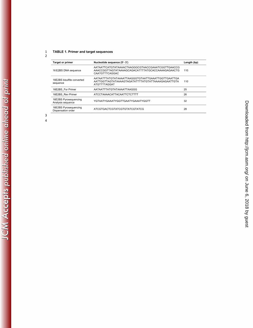
TABLE 1. Primer and target sequences 1 2
Target or primer Nucleotide sequence (5’- 3’) Length (bp)
16 E2BS DNA sequence AATAATTCATGTATAAAACTAAGGGCGTAACCGAAATCGGTTGAACCGAAACCGGTTAGTATAAAAGCAGACATTTTATGCACCAAAAGAGAACTGCAATGTTTCAGGAC
110
16E2BS bisulfite converted sequence
AATAATTTATGTATAAAATTAAGGGTGTAATTGAAATTGGTTGAATTGAAATTGGTTAGTATAAAAGTAGATATTTTATGTATTAAAAGAGAATTGTAATGTTTTAGGAT
110
16E2BS_For Primer AATAATTTATGTATAAAATTAAGGG 25
16E2BS_Rev Primer ATCCTAAAACATTACAATTCTCTTTT 26
16E2BS Pyrosequencing Analysis sequence
YGTAATYGAAATYGGTTGAATYGAAATYGGTT 32
16E2BS Pyrosequencing Dispensation order
ATCGTGACTCGTATCGTGTATCGTATCG 28
3
4
on June 6, 2018 by guesthttp://jcm
.asm.org/
Dow
nloaded from

TABLE 2: Intra-assay repeatability of HPV16 E2BS PCR 1 2
Theoretical concentration of HPV16 genome (copies/µL)
105 104 103 102 10 1
Cq mean 18.53 22.45 26 29.26 32.83 36.09
Cq SD 0.42 0.31 0.19 0.39 0.30 0.30
Cq variance 0.18 0.1 0.03 0.15 0.09 0.09 3
Mean, SD and variance of Cq were calculated from 8 replicates of serial dilutions of bisulfite converted Ca Ski cell DNA (or 6 4
replicates for the dilution theoretically containing 1 copy/µL). 5
on June 6, 2018 by guesthttp://jcm
.asm.org/
Dow
nloaded from

TABLE 3. Inter-assay reproducibility of the HPV16 E2BS HRM PCR for methylation level 1
determination 2
3
DNA sample Std 10% Std 25% Std 50% Std 75% Std 100% Ca Ski
Mean difference peak heights 12.8 20.0 30.1 39.1 49.6 43.9
SD 1.5 1.1 1.4 1.4 2.3 2.0
CV 11% 6% 5% 4% 5% 5%
4
Triplicates of standards were analyzed in 8 independent experiments. Difference plots were generated for each standard using 5
the unmethylated standard (Std 0%) as the reference. Mean, SD and CV were calculated from difference peak heights to 6
evaluate the reproducibility of the method. 7
on June 6, 2018 by guesthttp://jcm
.asm.org/
Dow
nloaded from

TABLE 4: Clinical samples with unmethylated or methylated E2BS#1, E2BS#2 and Sp1BS 1
according to cytological diagnosis 2
3
E2BS methylation level
0% ]0-10%] ]10-25%] ]25-50%] ]50-75%]
WNL (n=21) 21 0 0 0 0
LSIL (n=23) 22 0 1 0 0
HSIL (n=25) 25 0 0 0 0
Carcinomas (n=17) 9 5 1 0 2
4
HRM profiles derived from clinical sample DNA were compared to those derived from standards and assigned to the most 5
similar profile. The number of samples assigned to each profile among WNL, LSIL, HSIL and carcinomas is reported in the 6
table. 7
8
on June 6, 2018 by guesthttp://jcm
.asm.org/
Dow
nloaded from

TABLE 5: Methylation levels of HPV16 E2BS#1, E2BS#2 and Sp1BS determined 1
by HRM PCR and pyrosequencing 2
3
Sample type Diagnosis (n) HPV16 E2BS
HRM PCR Pyrosequencing
Methylation level (%)
Individual methylation level of CpG sites (%)
nt 31 nt 37 nt 43 nt 52 nt 58
Standard 0% - - 0 2 2 2 3 3
Standard 10% - - 10 10 10 10 12 12
Standard 25% - - 25 24 24 25 26 26
Standard 50% - - 50 47 47 49 50 50
Standard 75% - - 75 67 67 70 70 71
Standard 100% - - 100 94 95 97 97 98
Ca Ski cell DNA - - 87 93 89 98 96 98
Cervical smears WNL (13) 0 2.2 1.2 1.3 2 1.5
Cervical smears LSIL (21) 0 2.1 1.4 1.5 2.1 1.8
(1) 24 25 25 28 28 28
Cervical smears HSIL (20) 0 1.8 1.8 1.5 2.6 2.2
Cervical smears Carcinoma
(9) 0 2.2 1.7 1.9 2.4 2.4
(1) ]0-10] 6 4 5 6 7
(1) ]0-10] 8 4 5 10 11
(1) ]0-10] 18 17 20 21 22
(1) ]0-10] 7 7 8 9 8
(1) ]0-10] 3 3 4 7 7
(1) 25 40 38 37 39 35
(1) 59 80 80 89 84 89
(1) 61 72 72 78 78 79 4
on June 6, 2018 by guesthttp://jcm
.asm.org/
Dow
nloaded from