Embed Size (px)
Citation preview

84
Mas
anor
i Non
aka
Jewels of the Deep Sea - Precious Corals
Masanori Nonaka1 & Katherine Muzik2
1 Okinawa Churaumi Aquarium, 424 Ishikawa Motobu-Cho, Okinawa 900-0206, Japan
Contact e-mail: [email protected]
2 Japan Underwater Films, 617 Yamazato, Motobu-cho, Okinawa 905-0219, Japan
IntroductionPrecious Corals belong to the Family Coralliidae (Anthozoa: Octocorallia) and are well-known for
their red or pink skeletons that have been used since antiquity for ornament, medicine, talismans and
currency. In Okinawa, they are found living at depths from 200 to about 300m, but in northern Japan they
are found in shallower waters, generally about 150m deep.
We have been keeping and displaying several local species of Coralliidae since the opening of the
Okinawa Churaumi Aquarium on November 1, 2002 (Nonaka et al., 2006). One of the most difficult groups
of coral to keep, we have thus far succeeded in keeping individuals in captivity for only about two years.
Nevertheless, we can gather valuable data from living precious corals being kept in a tank.
Although many people are familiar with the word “coral”, there are relatively few people who have ever
seen coral alive. Even fewer people have seen deep-sea species of precious coral alive. Public aquariums
can provide an excellent opportunity to introduce living corals from both shallow and deep water to
visitors, and to encourage interest in them and other marine creatures too. But, although aquariums can and
should serve as educational facilities, it is difficult to get visitors to notice these tranquil, quiet creatures!
We have tried and failed to attract attention to precious corals by special signs and lighting, so now we at
the Churaumi Aquarium are planning a special exhibit about the relationship of precious coral to cultural
anthropology.
What are coralsIn the past, the words “coral” and “sango” (coral in Japanese) generally meant jewelry coral to most
people, but recently, “coral” has increasingly been understood to refer to “coral reef” corals. Both the
precious corals and the reef-building corals belong to phylum Cnidaria, but they are in different taxonomic
groups. Most reef-building corals are in the Order Scleractinia of the Subclass Hexacorallia, and most
precious corals are in the Order Alcyonacea of the Subclass Octocorallia. Both are members of the Phylum
Cnidaria (animals with stinging cells, “cnidae”) which contains four taxonomic Classes: Scyphozoa,
Hydrozoa, Cubozoa and finally, Anthozoa, which includes the “sea anemones” and the reef, soft and

85
Masanori N
onaka
precious “corals” being discussed here. Each Class is defined by its life cycle: Scyphozoans and Cubozoans
have short-term “polyp” generations (attached to or resting on the substratum) and long-term “medusa”
(floating “jellyfish”) generations. Hydrozoans have both medusa and polyp generations, but Anthozoans
have only the polyp stage. Therefore species in Scyphozoa, Cubozoa and some species in Hydrozoa are all
commonly referred to as jellyfish. Anthozoans lack a medusoid stage, and so are generally called corals or
sea-anemones. Species in the Class Anthozoa are further differentiated into two Subclasses based on the
number of tentacles in their polyps: Octocorallia (having eight tentacles) and Hexacorallia (having six or
multiples-of-six tentacles). Uchida (1992) suggested that members of the Hexacorallia, having more varied
shapes and numbers of tentacles forming their polyps, are likely not as closely related to each other as are
members of the Octocorallia, all of whose polyps have eight pinnate tentacles.
What is the definition of coral? Both words, “coral” in English and “sango” in Japanese, were originally quite likely referring to the
Mediterranean precious coral, an octocoral named Corallium rubrum. Wood (1983) indicated that coral is
a general term for species which belong to the Phylum Coelenterata (Cnidaria) and have skeletons. In Fig.
1, the red letters indicate the different taxonomic groups which have members considered to be “corals”. Almost all are species belonging to the Class Anthozoa, but “Fire corals” and “False precious coral” are in
the Class Hydrozoa. As Cairns (2007)
explained, it is not surprising that there is
no consistent definition of the word “coral”, since it is a layman's term that refers to
animals in several taxonomic groups.
According to his definition, “coral” includes
members of seven taxa: Order Scleractinia
(hard corals), Order Zoanthidea (in part, the
zoanthids known as gold corals), Order
Antipatharia (black corals), Subclass
Octocorallia (soft corals, gorgonians, red
and pink corals, bamboo corals, blue coral, sea pens), and three Families in the Class Hydrozoa, the
Stylasteridae (lace corals, stylasters), Hydractiniidae (in part, the longhorn hydrozoans) and Milleporidae
(hydrocorals, fire corals).
What are the precious corals?The skeletons (axes, axial skeletons) of some coral species are so hard that they can be beautifully
polished for use in sculptures and jewelry. These are the species called “precious corals.” In general, the
polished products are also called just “coral” or “sango” (in Japanese). Almost all commercial species of
precious corals belong to the Subclass Octocorallia, Order Alcyonacea, Family Coralliidae. So far, the
Fig.1:Gene alogical tree of Cnidaria, with“corals” in red

86
Mas
anor
i Non
aka
described species include one species from the Mediterranean Sea, three species from Japan, three species
from Hawaii, and one unidentified species from
Midway. In Hawaii and Mexico, the “black corals” harvested for jewelry are not octocorals but
antipatharians, in the Order Antipatharia, in a different
Subclass, Hexacorallia. The less well-known “gold
corals” include zoanthids with golden skeletons
harvested from Hawaii (several species of
Savalia/Gerardia spp., again in Subclass
Hexacorallia), and also the so-called “gold corals” from Alaska, which in fact are several species in
Subclass Octocorallia, Family Primnoidae. The “angel
corals” too, harvested from shallow waters of the
Caribbean, are skeletons of members of the Subclass
Octocorallia, likely species in the Family Plexauridae.
The skeletons of two Pacific shallow-water octocorals,
“blue coral” (Heliopora coerulea) and “sponge coral” (Melithaea ochracea), have also been worked into
beads for necklaces. However, for this document,
“precious coral” is a rather narrow term, referring only
to members of the octocoral family Coralliidae.
History of the coral fishery in JapanPrecious corals have been harvested routinely from the Mediterranean Sea for at least 5,000 years
and were taken even as long as 30,000 years ago or more! Products made from Corallium rubrum are
recorded from an Old Stone Age Monument about 25,000 years old in Germany (Kosuge, 1987), and
precious corals and shells were found in a ruins roughly 30,000 years old in Lausanne, Switzerland
(Liverino, 1986). Coral products were first imported to Japan via the Silk Road, and coral products are
known to have been stored over 1,200 years ago at Nara's Sho-so-in, one of the oldest storage facilities in
Japan (Suzuki, 1999b). Thereafter, products of Mediterranean precious coral were distributed in many cities
in Japan. During the Edo-era (1603-1867), as they became less expensive, even some daimyos (a kind of
governor), samurais and rich dealers could own them. Near the end of the Edo-era, even ordinary people
were using coral beads as accessories.
The first record of collecting precious coral in Japan is in 1812, when a fisherman found a precious
coral entangled in his net off Muroto, Kochi Prefecture. Reportedly, he gave the Daimyo of Kochi the coral
colony he had collected (Shozakai, 1983, Suzuki, 1999a). In 1815, in a book entitled “Nan-ro-shi”, or
Fig. 2: Primitive coral net (Kitahara, 1903)

87
Masanori N
onaka
“Book of the Southern Way”, referring
to the southern, Kochi area of Japan,
reddish pieces of coral were reported
found occasionally, if rarely, hung-up on
fishing nets (Kosuge, 1987, Suzuki,
1999a). At a store in Kochi City named
Tachibana-Ya, there is a receipt from
1835 for the purchase of precious corals.
A similar record from 1848, reports a
coral colony found stuck on a fishing
hook (Kosuge, 1987). Thus, it appears that Kochi fishermen at that time were familiar with precious corals,
and very likely recognized their commercial value. In 1938, the Kochi Prefectural government prohibited
collection of red corals (Suzuki, 1999a) and tried to conceal information about the corals from the central
government (Shozakai, 1983). Perhaps information was hidden by the fishermen because they were afraid
of incurring penalties or high taxes. But, Kosuge (1987) suggests that the Kochi government was actually
not so strict, because there are some records showing government approval for fishermen to harvest corals.
For example, 225g of coral were sold in Kochi in 1835, and the local government asked fishermen for
estimates of the fishing boats and nets being used for coral harvesting.
With the arrival of the Meiji-Era (1868-1912), there were no longer any limits on the precious coral
fishery. Kosuge (1987) reports that coral harvesting was begun in the Muroto region at Kochi in 1871, but
the fishery techniques had been already been developed well before that date. Konojo Ebisuya, a coral
fisherman, invented coral nets (Fig. 2) for efficient harvest during the Edo-Era. His invention consisted of a
large weight with long nets attached, to be dragged across the sea floor. (He was honored in the scientific
name of “white coral”, named Corallium konojoi by Prof. Kishinouye in 1903a,b.) By then, fishermen were
fishing large quantities of precious corals. Kitahara (1904) reported a total of more than 16t of corals
collected from coral beds off Kochi, Kagoshima and Nagasaki in 1901 (Fig. 3). Suddenly, Japan went from
being an importing country to an
exporting country (Fig. 4). Because of
overfishing and consequent reduction of
harvest in the Mediterranean area,
dealers there were forced to import
supplies, mostly from Japan.
The impact of the coral trade
could be seen in the many, expensive,
tile-roofed houses built in the Kochi
fishing villages, because the corals were
worth so much money. Although the
Fig. 3: Initial amounts of coral harvest (data from Kitahara, 1904)
Fig. 4: Export and Import of precious corals in Japan during the Meiji-Era (data from Kitahara, 1904)

88
Mas
anor
i Non
aka
Fig. 5: Modern coral fishing areas around Japan
prices seemed unbelievably high to the fishermen, dealers in the big city sold the coral at even higher
prices. One interesting episode was reported in the Kochi Newspaper by Shozakai, 1983: the captain of a
large ship transporting rice met a fisherman with his small boat filled with recently harvested coral of
excellent quality. Knowing the extremely high value of coral, the captain made an offer to the fisherman to
trade a boatload of rice for his boatload of coral. The fisherman accepted the proposal, thinking it very
favorable to him, even too high, and as soon as he had exchanged his coral for a boatload of rice, fled to his
home port. Meanwhile, the captain waited in vain for the fisherman to return to get the remaining rice,
meaning his entire shipload, due him. The fisherman did not return, thinking that “the boatload” referred
just to his small fishing boat, and so the captain thereafter also fled, with the corals and remaining rice, to
his own port, Muroto. This story illustrates the incredible price difference between coral in a fishing boat
and coral eventually sold in the city market in those days. Even though selling their catch at a fraction of
the ultimate price, the corals still gave the fishermen a get-rich-quick opportunity, and they took many risks
to collect them. Many coral boats were sunk by typhoons (Kosuge, 1987). He reports that in just the
western area of Kochi, 125 fishermen died in 1909. He also reports that in the Dan-jo Islands, Nagasaki,
300 fishermen died in 1895; 10 fishermen died, 209 were missing and 155 boats sank in 1905; 119
fishermen died, 615 were missing and 173 boats sank in 1906; and 64 men died and 30 boats were missing
in 1914.
Despite these numerous accidents, the fishery did not cease, instead it grew! It spread to Izu, the
Bonins (Ogasawara Archipelago) and even eventually to Okinawa. In 1924, 10 fishing boats obtained
permission to harvest coral in
Okinawa Prefecture. Fishermen
collected corals off the southern
coast (Chinen) in 1937, perhaps the
first record of a coral fishery in
Okinawa. A coral bed was also
found offshore the island of
Yonaguni, the most western island
in Okinawa Prefecture (and Japan).
After World War II, the discovery
of a huge coral fishing ground at
Miyako Island, Okinawa brought
general awareness of the coral fishery in the Prefecture (Kosuge, 1987). Beds of momo-iro-sango
(Corallium elatius) were found in the Ogasawara Archipelago (the Bonin Islands south of Tokyo) in 1918,
and some harvesting has continued there ever since, except when interrupted during war-time (Kosuge,
1987). One of the largest coral grounds in the Pacific was found around Midway Is. by a Japanese fishing
ship from Fukushima Prefecture in 1965. Thereafter, not only Japanese but also Chinese ships came to
Midway to harvest coral.

89
Masanori N
onaka
In the early days, fishermen used a “coral net,” a kind of dredge, to harvest corals. Consisting of long nets
attached to a weight, this coral net would be dragged across the bottom, entangling all benthic animals in its
path. Because of this destructive method, not just organisms but entire ecosystems were damaged or
destroyed. As a result the coral harvest declined worldwide, and alternative, less-destructive methods of
harvest were increasingly sought, such as by submarine and R,O,V, (remotely-operated vehicle). In 1979,
both Kagoshima and Okinawa Prefectures banned harvesting of precious corals by “coral net,” permitting
collection only by submarine or ROV. In fact, only one company, a big one wealthy enough to have a
submarine, now has a permit to collect in Okinawa. In Kochi, however, the traditional coral-net method is
still being permitted, with little expectation of government prohibition, because the local fishermen simply
cannot afford an ROV or submarine.
Resource conservation of precious coralsResources of precious corals are known to have been reduced everywhere in the Mediterranean and
the Pacific. In the Mediterranean, where remaining precious corals are found living in water shallow
enough to be readily observed by SCUBA, there have been many reports about their ecology, reproduction,
resources, etc. There are also several reports on precious corals in Hawaii (Grigg, 1974 etc.). But there are
almost no reports on precious coral resources in Japan.
The necessary data for resource conservation include such factors as growth rate, age at maturity
and reproductive ecology (Table 1).
Information from the Mediterranean species, Corallium rubrum, indicates they can grow 0.2-2 cm in a
year, reach reproductive maturity 7-10 years after attachment, and are dioecious (sexes are separate)
brooders. Vighi (1972) reported they reproduce from July to October. Colonies of a Hawaiian species, C.
secundum, can grow 0.9 cm per year in length (Grigg, 1976), can mature in about 13 years (Grigg, 1976) or
12-13 years (Grigg, 1993), and are dioecious spawners (Grigg, 1976). But there is only one report about
growth in Japanese species. Grigg (1974) briefly mentioned, from a personal communication of
observations of tagged colonies of Paracorallium japonicum recovered off Japan, that colony growth was
0.3 mm/ year. Kishinouye (1904a) reported only that gonads were more developed in March than in
area Mediterranean Hawaii Japan
species C. rubrum C. secundum P. japonicum
0.2-2cm/yr. (Marchal et al., 2004) 0.9cm/yr. (Grigg, 1976) 0.3mm/yr. ?(Grigg, 1974)growth rate length
diameter 0.24-1.32mm/yr. (Marchal et al., 2004) no data no data
no data
no data
no data
sex separate (Vighi, 1972) separate (Grigg, 1976)
maturity 7-10yrs (Torrents et al., 2006) 13yrs (Grigg, 1976)
reproduction style brooder (Lacaze-Duthiers, 1864 etc.) spawner (Grigg, 1993)
Table 1 Biological and ecological data of precious corals
90
Mas
anor
i Non
aka
September, without any further details.
We are now researching the precious coral resource in Okinawa Prefecture. We hope to obtain
instructive data to analyze for the purpose of precious coral conservation.
Taxonomic studies of Japanese precious coralsWhen we started our precious coral conservation research we soon found it imperative to study their
taxonomy. The last taxonomic
studies of Japanese precious corals
were published more than 100 years
ago by Kishinouye (1904b). More
than five decades later, in a study of
Hawaiian species, Bayer (1956)
devised a taxonomic key to all
known Pacific species, including the
ones described from Japan. As our
observations and our collections
continue to improve with use of an
ROV, we are finding that the
Japanese fauna needs much more
study. For example, according to our
recent observations, fishermen in
Okinawa are using the same name “Shiro-Sango” for what may be two different species with white axes;
one with rounded branches and thick coenenchyme, and one with thin sharp branches and thin
coenenchyme (see Nonaka et al., 2006; figs 12-14). Kishinouye (1903a,b) gave the name “Shiro-Sango” to
C. konojoi, which is certainly the one with “rounded” branches. The “thin sharp one” is likely a
still-undescribed species, new to science.
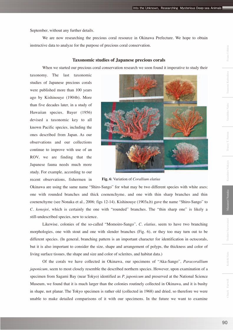
Likewise, colonies of the so-called “Momoiro-Sango”, C. elatius, seem to have two branching
morphologies, one with stout and one with slender branches (Fig. 6), or they too may turn out to be
different species. (In general, branching pattern is an important character for identification in octocorals,
but it is also important to consider the size, shape and arrangement of polyps, the thickness and color of
living surface tissues, the shape and size and color of sclerites, and habitat data.)
Of the corals we have collected in Okinawa, our specimens of “Aka-Sango”, Paracorallium
japonicum, seem to most closely resemble the described northern species. However, upon examination of a
specimen from Sagami Bay (near Tokyo) identified as P. japonicum and preserved at the National Science
Museum, we found that it is much larger than the colonies routinely collected in Okinawa, and it is bushy
in shape, not planar. The Tokyo specimen is rather old (collected in 1968) and dried, so therefore we were
unable to make detailed comparisons of it with our specimens. In the future we want to examine
Fig. 6: Variation of Corallium elatius

91
Masanori N
onaka
alcohol-preserved, or better still, fresh specimens from Sagami Bay, to determine if these indeed are two
different species. (The original holotype material described and figured by Kishinouye, 1903a, is lost.)
In 2003, Bayer & Cairns suggested a new genus of Coralliidae, which they named Paracorallium,
and they put “Aka-Sango (Paracorallium japonicum)” in this new genus. Their description of the genus is
“colonies with autozooids seated in deep pits in the solid axis, pits with prominently beaded margins.” But,
the Okinawan Paracorallium japonicum may in fact belong back in “Corallium”, because we have not
found any “deep pits” in the axis of samples collected here. Indeed, examination of the Sagami Bay P.
japonicum also revealed no deep pits, so we are also calling into question the validity of the genus
Paracorallium.
To further our studies, we are now adding DNA analysis to our repertoire of taxonomic techniques.
Taxonomy of Indo-Pacific species of CoralliidaeIn the Indo-Pacific, there are 25 known species in the family Coralliidae. So far in the studies on
this family, the most important characters for identification are shape and size of sclerites. We hope our
future DNA studies will help to reveal so-far difficult to determine taxonomic relationships in the family. In
the section below, their known taxonomic characters (according to Bayer, 1956) are summarized, species
by species, and the original descriptions in the literature have been translated and condensed. All figures are
copies of the original published illustrations, reprinted here with permission of the publishers. We hope this
compilation of the scattered literature contributes to helping identify precious corals from Japanese waters
in the future.
The precious corals known from Japanese waters are presented first, in a group arranged
alphabetically by genus and species, followed by the group from Hawaii, also arranged alphabetically, and
finally a group formed by the rest of the known Indo-Pacific species, again arranged in alphabetical order.
Precious Corals from Japanese Waters (Depth ranges reported include those from both the early literature and our recent collection data from Okinawa.)
Corallium boshuensis Kishinouye, 1903 (Figs. 7-9)Corallium boshuensis Kishinouye, 1903a: 624; Kishinouye, 1903b: 104; Kishinouye, 1904a: 23, pl.3, fig.
4; pl.7, fig.2; pl.8, fig.20; (in Japanese); Kishinouye, 1904b: 22, pl.3, fig. 4; pl.7, fig.2; pl.8, fig.20;
Kukenthal, 1924: 51.
Corallium boshuense,-- Bayer, 1956: 75 (in key); Pasternak, 1981: 43.
Colony form: Colony finely branched in one plane. Terminal twigs sharply pointed. Main branches laterally
compressed when viewed in cross-section.
Coenenchyme: Coenenchyme thin, light yellow in color with small granulated furrows on the surface.

92
Mas
anor
i Non
aka
Polyps: Contracted autozooids prominent, cylindrical, with eight radial grooves, taller than wide.
Distributed on one side only on twigs, rare on the stem, not in clusters.
Axis: Axis smooth, without striation or pits on the surface. Many large burrows of some commensal animal
on the front side of the axis. Burrows about 30mm in length, some of them buried within axis. Axis entirely
cream-white in color.
Sclerites: Five kinds of sclerites: 8-radiate, cruciform, long-warty spindles, double clubs and irregular
forms, 8-radiates most numerous. Large irregular forms may be fused sclerites.
Distribution: Japan, 550 m(?).
Remarks
Only one specimen, recorded from Chiba Pref., from perhaps approximately 600m deep. Colony about 200
mm in height, about 300 mm in width and 20 mm in diameter near holdfast. Coenenchyme lost basally, a
feature sometimes seen in mature colonies, suggesting this was a mature colony at time of collection.
Closely resembles C. sulcatum, possibly a synonym. The two species can be distinguished in color,
branching and coenenchyme thickness, but sclerites are similar. Type specimen described by Kishinouye is
missing. Specimen number NSMT-CoR951 labeled “Corallium boshuensis” preserved at the National
Science Museum in Tokyo is actually a specimen of C. konojoi (personal observation).
Fig. 8: Surface detail. (from Kishinouye, 1904).
Fig. 9: Sclerites. (from Kishinouye, 1904)Fig. 7: Whole colony of Corallum boshuensis. (from Kishinouye, 1904)

93
Masanori N
onaka
Corallium elatius (Ridley, 1882) (Figs.10-14)Pleurocorallium secundum var. elatior Ridley, 1882: 228, pl.9, figs.6-11
Corallium elatior,-- Kishinouye, 1903a: 625; Kishinouye, 1903b: 104 (in Japanese).
Corallium elatius,--Kishinouye, 1904a:25, pl.1, fig.3; pl.3, figs.1,2; pl.7, fig.6; pl.8, figs.7-15; (in
Japanese); Kishinouye, 1904b: 24, pl.1, figs.3; pl.3, figs.1,2; pl.7, fig.6; pl.8, figs.7-15; Kukenthal, 1924:
49; Bayer, 1956: 76 (in key).
Corallium sp.1,--Nonaka et al., 2006: 1823, figs.9-11.
Colony form: Colony branches in one plane, more or less recurved, some branches anastomosing. Terminal
branches very fine.
Coenenchyme: Coenenchyme thick and firm, with fine projections on the surface. Scarlet or vermilion in
color, but near the free end of growing branches light red or colorless. Sometimes all light-yellow variation.
Polyps: Autozooids arranged in about four rows and generally on one face of the colony only. Large, 1.5 to
2.0 mm in diameter, sub-hemispherical.
Axis: Axis finely striated. Normally red to pink in color with a white center, rarely all white with a yellowish
center. (Kishinouye (1903) described small pits in the axis generally beneath every polyp, but our
specimens of what may be this species lack this feature. The specimen he observed is unfortunately
missing.)
Sclerites: Three kinds of sclerites: 6-radiates,
7-radiates and double clubs, the 7-radiate form rare.
8-radiates not present. Autozooids with many small
6-radiates. No spindles in autozooid verrucae.
Distribution: Japan, 50-330 m.
Remarks
Ridley (1882) recorded this species as a subspecies
of C. secundum named Pleurocorallium secundum
elatior, but Kishinouye (1903) redescribed it as a
distinct species of Corallium. This species can
grow to an enormous size. Kishinouye (1904a)
described some specimens of about one meter in
height, 20kg in weight. Fig. 10: Colony, axis and sclerites of Corallium elatius. (from Ridley, 1882)

94
Mas
anor
i Non
aka
Fig. 12: Colony of Corallium elatius. (from Kishinouye, 1904)
Fig. 11: Terminal branch. (from Kishinouye, 1904)
Fig. 13: Sclerites. (from Kishinouye, 1904)
Corallium konojoi Kishinouye, 1903 (Figs.15, 16)Corallium konojoi Kishinouye, 1903a: 625; Kishinouye, 1903b: 105; Kishinouye, 1904a: 27, pl.1, fig. 4;
pl.7, fig.5; pl.8, figs.16,17(in Japanese); Kishinouye, 1904b: 26, pl.1, fig. 4; pl.7, fig.5; pl.8, figs.16,17;
Kukenthal, 1924: 50; Bayer, 1956: 76 (in key); Nonaka et al., 2006: 1824, figs.6-8.
Colony form: Colony sparingly branched, generally growing in one plane. Branches often anastomosing,
branch tips blunt and rounded.
Coenenchyme: Coenenchyme thick and firm, surface smooth. Yellowish to reddish in color, becoming
lighter in color towards the holdfast.
Polyps: Autozooids unevenly distributed on one face of branches, crowded on prominences and at the tips.
Autozooids clustered in groups. Autozooids large, 2-3 mm in diameter, a little elevated.
Fig. 14: Detail of surface. (from Kishinouye, 1904)

95
Masanori N
onaka
Fig. 16: Sclerites. (from Kishinouye, 1904)
Axis: Axis weakly striated, milky white in color with pinkish center.
Sclerites: Four kinds of sclerites: 8-radiate, 6-radiate, 7-radiate and double clubs. Crosses very rare.
6-radiates, 0.09mm in length, most abundant. 8-radiates not common. Autozooids have 8-radiates,
6-radiates and small irregular forms.
Distribution: Japan, 50-250 m.
Remarks
Colonies about 300 mm in height and width. Axis a beautiful white, but there is little demand. The species
name honors the fisherman, Konojo Yebisuya, who in 1836 invented a net for collecting corals and began
harvesting them for commercial purposes.
Unfortunately the type specimen Kishinouye described has been lost.
Corallium pusillum Kishinouye, 1904 (Figs. 17-19)Corallium pusillum Kishinouye, 1904a: 29, pl.5, figs.3,4; pl,7, fig.4; Kishinouye,1904b: 27, pl.5, figs.3,4;
pl,7, fig.4; Kukenthal, 1924: 50; Bayer, 1956: 76 (in key).
Colony form: Colony is dichotomously branched in one plane, which is more or less recurved. Colony
width exceeds height. Terminal branches rounded in section.
Coenenchyme: Coenenchyme thick and firm, granulated on the surface. Many commensal polychaetes on
one surface, their burrows with I or T-shaped openings. Main stem orangeish to yellow-orange but
branchlets becoming grayish yellow.
Fig. 15: Whole colony of Corallium konojoi. (from Kishinouye, 1904)

96
Mas
anor
i Non
aka
Polyps: Large, hemispherical autozooids, 1.5 mm high
and in diameter, distributed on one surface of the
branches only, becoming more abundant on the
terminal twigs, sometimes making clusters.
Axis: Surface rough with fine grooves. Commensal
burrows sometimes are shallow hollows. Axis white
and partly pink.
Sclerites: Three kinds of sclerites: 8-radiates, crosses
and double clubs, 8-radiates and double clubs
abundant. Autozooids with 8-radiates, rods with
irregular projections. Sclerites the largest reported in
Japanese Corallium, about 0.06-0.09 mm long.
Distribution: Japan.
Remarks
Specimen from Izu-Ohshima Island 70 mm high, 210
mm wide and 15 mm in diameter near the holdfast, but
may not represent maximum size. The only
type-specimen was lost, and no further specimens have
been found since.
Corallium sulcatum Kishinouye, 1903 (Figs. 20-22)Corallium sulcatum Kishinouye, 1903a: 624; Kishinouye, 1903b: 104; Kishinouye, 1904a: 24, pl.4,
figs.1,2; pl.7, fig.3; pl.8, fig.19; Kishinouye, 1904b: 23, pl.4, figs.1,2; pl.7, fig.3; pl.8, fig.19; Kukenthal,
1924: 52; Bayer, 1956: 75 (in key).
Colony form: Colony branched in one plane, some branches anastomosing. Terminal twigs are slender and
sharp.
Coenenchyme: Coenenchyme thin. Small spines distributed in longitudinal rows on the coenenchyme.
Light red, twigs yellow.
Polyps: Verrucae of autozooids are cylindrical, with eight conspicuous grooves, on one surface of colony,
Fig. 17: Whole colony of Corallum pusillum. (from Kishinouye, 1904)
Fig. 18: Tip of colony. (from Kishinouye, 1904)
Fig. 19: Sclerites. (from Kishinouye, 1904)

97
Masanori N
onaka
numerous on terminal twigs, fewer on stem. Autozooids about 0.9-0.18 mm in height, 0.9 mm in diameter.
Siphonozooids rather projecting.
Axis: Axis smooth, rounded in section. Many shallow longitudinal grooves with prickly margins on one
surface of the branches. On smaller branches these grooves become cavities, quite similar to the burrows
found in C. boshuensis. Axis variegated, with lighter and darker pinkish areas.
Sclerites: Five kinds of sclerites: 8-radiates,
crosses, double club, rods with some projections
and many irregular forms. 8-radiates most
abundant.
Distribution: Japan, 180-550 m(?).
Remarks
A fine specimen, about 300 mm in height, 230 mm
in width and 23 mm in basal diameter, collected
from 180-550 m off Chiba Pref. (depth data not
exact). Only two specimens known, both
subsequently lost. This species resembles P.
japonicum and C. boshuensis, especially the latter,
but differs in several features, including color,
grooved branches, etc.
Fig. 20: Whole colony of Corallum sulcatum. (from Kishinouye, 1904)
Fig. 21: Branch of colony. (from Kishinouye, 1904) Fig. 22: Sclerites. (from Kishinouye, 1904)

98
Mas
anor
i Non
aka
Paracorallium inutile (Kishinouye, 1902) (Figs. 23-25)Pleurocorallium inutile, Kishinouye, 1902: 419
Corallium inutile, -- Kishinouye, 1903a: 626; Kishinouye, 1903b: 105; Kishinouye, 1904a: 28, pl.5,
figs.1,2; pl,7, fig.7; pl.8, fig.18; Kishinouye, 1904b: 27, pl.5, figs.1,2; pl,7, fig.7; pl.8, fig.18; Kukenthal,
1924: 48; Bayer, 1956: 76 (in key).
Paracorallium inutile, -- Bayer & Cairns, 2003: 224.
Colony form: Main branches planar, but smaller
branches branching in all directions, often
anastomosing, net-like.
Coenenchyme: Coenenchyme thin but firm, light
red.
Polyps: Autozooids small, 0.8-1.0 mm in diameter,
slightly elevated and distributed over all parts of the
branches.
Axis: Axis brittle, finely striated. Small but deep pit
in axis under autozooids. Axis entirely white,
slightly tinged with yellow.
Sclerites: Two kinds of sclerites: 6-radiates and
double clubs. 6-radiates few, double clubs
predominant in coenenchyme. Double clubs mainly
smooth. Autozooids with rod-like six-radiates with
small projections.
Distribution: Japan.
Remarks
Specimen described was 120 mm high and wide
base of the stem is 21 mm in diameter, collected at
Kashiwa-jima Island in Kochi Pref. (depth
unknown). Rare and not a beautiful color, therefore
without commercial value.
The type-specimen Kishinouye described was lost.
Fig. 24: Branch of colony. (from Kishinouye, 1904)Fig. 24: Branch of colony.
Fig. 23: Whole colony of Paracorallum inutile. (from Kishinouye, 1904)
Fig. 25: Sclerites of P. inutile. (from Kishinouye, 1904)

99
Masanori N
onaka
Fig. 26: Tip of colony.(from Kishinouye, 1904)
Paracorallium japonicum (Kishinouye, 1903) (Figs. 26-29)Corallium japonicum Kishinouye, 1903a: 623; Kishinouye, 1903b: 103; Kishinouye, 1904a: 22, pl.1,
figs.1,2; pl,2; pl,4, fig.3; pl.7, fig.1; pl.8, figs.1-6; Kishinouye, 1904b: 21, pl.1, figs.1,2; pl,2; pl,4, fig.3;
pl.7, fig.1; pl.8, figs.1-6; Kukenthal, 1924: 50; Bayer, 1956: 76 (in key).
Paracorallium japonicum, Bayer & Cairns, 2003: 225; Nonaka et al., 2006: 1823, figs.3-5.
Colony form: Colony abundantly branched in one plane. Short, prickly branchlets grow on one surface and
on the sides of the branches.
Coenenchyme: Coenenchyme thin (in Okinawan specimens papillate). Generally dark red but growing tips,
pinkish to white.
Polyps: Autozooids small, about 0.7 mm in diameter, and when contracted only a little elevated. Distributed
in four or five rows, generally on one surface only.
Axis: Axial surface striated, a small pit in the axis underneath each polyp. In cross section, axis round or
oval. Normally dark red in color with white center, Y or X-shaped when viewed in cross section.
Sclerites: Two kinds of sclerites: 8-radiate and crosses. 8-radiates numerous, 0.05 mm long, crosses smaller
and fewer. Autozooids almost all 8-radiates.
Distribution: Japan, 100-300 m.
Remarks
The famous coral known as “aka-sango” red coral. Height and width both about 300 mm, base of the stem
120 mm in diameter. Kishinouye (1904a) reported that this species was the most abundant, forming two
thirds of the entire coral harvest. Resembling C. stylasteroides, but differing in color and distribution of
polyps. The type Kishinouye described is lost.
Fig. 27: Autozooid with tentacles extending. (from Kishinouye, 1904)

100
Mas
anor
i Non
aka
Precions corals from Hawaiian waters
Corallium abyssale Bayer, 1956 (Figs. 30-32)Corallium abyssale Bayer, 1956: 76, figs.4,5a, 7a-d.
Colony form: Colony branched in one plane in an asymmetrically dichotomous manner, alternate branches
dominating to produce a zigzag, sympodial main stem but twigs in upper part of colony branch
symmetrically and quite regularly.
Coenenchyme: Coenenchyme of the type extremely thin and rubbed off in places. Surface of coenenchyme
with scattered, prominent papillae smaller than the wart-like siphonozooids. Specimen preserved in alcohol
pale brown in color, probably discolored, “pink” when fresh. according to original field label.
Polyps: Autozooids very widely separated and few, essentially biserial although an occasional individual
out of line. Verrucae 2 mm in height, cylindrical, with eight longitudinal grooves in the distal half,
corresponding to the septal insertions. Siphonozooids low, wart-like protuberances in groups around the
autozooids.
Axis: Axis solid, round, and smooth, 4 mm in diameter at the lowest point, tapering to 2.5 mm at the top.
Pale pink with center somewhat darker.
Sclerites: Three kinds of sclerites in the coenenchyme: stubby crosses and 8-radiates often coarse and
clumsy-looking; double clubs with wide, depressed, weakly sculptured heads and short handles with
radiating processes. Sclerites of the autozooid verrucae long, blunt rods 0.12-0.13 mm long in addition to
the forms found in the coenenchyme proper. In the oral disk and pharyngeal region spiny rodlets that
appear to be derived from the 8-radiate type. Sclerites colorless.
Fig. 29: P. japonicum sclerites. (from Kishinouye, 1904)
Fig. 28: Whole colony of Paracorallum japonicum. (from Kishinouye, 1904) 1. Autozooid side. 2. Opposite side. 3. Branch tip.

101
Masanori N
onaka
Fig. 30: Tip of branch of Corallinm abyssale. (from Bayer, 1956)
Fig. 32: Sclerites. (from Bayer, 1956)a. From tentacles. b. From pharynx. c. Double-clubs from coenenchyme. d. Crosses and 8-radiates from coenenchyme.
Fig. 31: Detail of polyps. (from Bayer, 1956)
Distribution: Hawaii, 1830-2400 m.
Remarks
C. abyssale does not closely resemble any other known species of the genus, although it is like C. sulcatum
and several other species in having tall sulcate verrucae with long blunt rods.
Corallium kishinouyei Bayer, 1996 (Figs.33-35)Corallium kishinouyei Bayer, 1996: 218, figs.11-19.
Colony form: Colony sparingly branched in one plane, openly dichotomous or lateral, the smaller terminal
branchlets more or less clavate. The stoutest main branch nearly round, 7.2 mm in diameter. Terminal
branchlets about 4 mm in diameter, with a tendency to flattening in the plane of branching.
Coenenchyme: Colonies initially distinctly yellowish white but the yellowish tint soon faded in alcohol.
Polyps: Autozooids situated biserially and directed slightly toward one face of the colony (the “front”),
with occasional individuals also on the front face roughly between bilateral pairs, becoming more generally
scattered on the stoutest branches. Retracted to form low, moundlike verrucae about 3 mm in diameter and
at most 1mm in height, the orifices with marginal lobes not necessarily as many as 8 but depending upon
the degree of contraction. Numerous small, bluntly conical papillae less than 0.5 mm in diameter, each with

102
Mas
anor
i Non
aka
Fig. 33: Branches of colony of Corallium Kishinouyei. (from Bayer, 1996)
Fig. 34: Tip of branch; stereo pair. (from Bayer, 1996) Fig. 35: Sclerites. (from Bayer, 1996)
an apical pore, cover the surface of the coenenchyme around and between the autozooids, interpreted as
siphonozooids but not confirmed by histological examination.
Axis: Axis longitudinally striated but no deep, smooth pits beneath the autozooids. Depressions in the axis
accommodating the autozooids are confined to apical regions, where they are irregular and do not have
smooth bottoms nor prominent beaded margins.
Sclerites: Coenenchymal sclerites predominantly
8-radiates from about 0.05 mm up to 0.13 mm in length;
6-radiates present also, and 7-radiates uncommon;
crosses present, and a few irregular forms. The tentacles
of autozooids slender, bluntly pointed rods up to about
0.09 mm in length, derived from the predominant
8-radiate form. Sclerites colorless, those of the larger
branches predominantly opaque white, those of the
terminal branches glass-clear.
Distribution: Hawaii, 1145 m.
Remarks
Sclerites are the largest of any known species. Species
named in honor Dr. Kamakichi Kishinouye who studied
many marine animals, and pioneered scientific study of
precious corals in Japan.

103
Masanori N
onaka
Fig. 36: Tip of branch.(from Bayer, 1956)
Corallium laauense Bayer, 1956 (Figs.36, 37)Corallium laauense Bayer, 1956: 78, figs.5e-f, 7h-j.
Colony form: Colony with some terminal twigs giving off lateral branchlets in one plane.
Coenenchyme: Coenenchyme longitudinally costate, bearing small, conical papillae. Sometimes the
coenenchyme extending as a thin, membranous expansion along the two edges of the twigs, where the
margins of opposite sides may recurve and join to form closed tunnels. White or faintly pink.
Polyps: Autozooids distributed on two sides and one face of the branches, forming cylindrical,
longitudinally-grooved verrucae about 1mm tall. Siphonozooids appearing as small warts with an apical
pore, near the autozooid bases.
Axis: Axis is practically round, with broad, longitudinal grooves in the largest parts preserved. White.
Sclerites: Two kinds of coenenchymal sclerites, crosses and
8-radiates. Spinose rods, up to 0.145 mm long in the
autozooid verrucae. Small rods in the pharyngeal region and
oral disk. Sclerites colorless.
Distribution: Hawaii, 365-584 m.
Remarks
In the form of its calyces and its finely divided branching, C.
laauense resembles C. sulcatum, but the latter has double
clubs and massive, irregular forms among its sclerites.
Fig. 37: Sclerites. (from Bayer, 1956) h. Pharyngeals. i. Coenenchymal 8-radiates and crosses. j. Tentacle scales.

104
Mas
anor
i Non
aka
Corallium niveum Bayer, 1956 (Figs.38, 39)Corallium niveum Bayer, 1956: 84, figs. 5g, 8h-k.
Colony form: Colonies irregularly branched in one plane, the major branches often diverging strongly from
near the base. The branches stout, the largest ones 5-10 mm in diameter at the base, tapering to about 2mm
toward the clavate tips. Small twigs arise from the front of the colonies, each ending in a recurved cluster of
polyps directed toward the base of the colony.
Coenenchyme: The surface of the coenenchyme finely wrinkled or corrugated, but no papillae. White.
Polyps: Autozooids form rather large, hemispherical verrucae with 8-rayed orifices, clustered in groups
occurring almost exclusively on one face of the colony. Each twig tip ends in a cluster of autozooids and
thus assumes a clavate form. The siphonozooids are inconspicuous, appearing as simple pores in the thick
coenenchyme.
Axis: Axis solid, round, or oval, and longitudinally grooved. No pitting beneath autozooids. White.
Sclerites: Sclerites of both verrucae and coenenchyme 6-, 7-, and 8-radiates, crosses and double clubs. In
the pharyngeal region and oral disk of the polyps small
spiny rods and crosses. Sclerites colorless.
Distribution: Hawaii, 232-282 m.
Remarks
C. niveum is related to C. pusillum from Japan, which
differs in lacking 6- and 7-radiate sclerites and in color
(orange).
Fig. 38: Corallium niveum detail of polyps. (from Bayer, 1956)
Fig. 39: Sclerites. (from Bayer, 1956) h. Crosses, 6-, 7-, and 8-radiates from coenenchyme. i. Double-clubs from coenenchyme. j. From pharynx. k. From pharynx (0.05mm scale).

105
Masanori N
onaka
Corallium porcellanum Pasternak, 1981 (Fig.40)Corallium porcellanum Pasternak, 1981: 43, fig.2.
Colony form: Speimens injured during dredging, only numerous fragments of twigs available, about 50
mm long, 3.6 mm maximum diameter. Branching in one plane. Twigs compressed in section, 2.1 mm by
1mm in diameter.
Coenenchyme: Coenenchyme white to yellow-white.
Polyps: Autozooids laterally arranged in double lines, with 4-5 autozooids in bundles on terminal twigs, 5-6
polyps in 30 mm of each row, at nearly equal intervals. Verrucae of autozooids low and wide, up to 2 mm in
diameter at the base, 8 radial grooves not reaching summit. Siphonozooids not verrucae-shaped, with small
opening only, some concentrated on the base of autozooids, but others scattered on one surface of the
colony.
Axis: Axis of main stem is solid, rounded in cross-section, but twigs are brittle, compressed. Surface of the
stem with precision longitudinal lines, rough to the touch. Twigs are comparatively soft, 1mm thick in
lateral direction. Deep pits absent beneath the autozooids. Axis white or slightly transparent and
reminiscent of quality porcelain.
Sclerites: Sclerites of coenenchyme and autozooids mostly 8-radiate, decorating several mounds and
conical projections. Size variable, but less than 0.08 mm in length. 6- and 7-radiate and compressed crosses
few. Double-club sclerites absent. Sclerites colorless, transparent.
Distribution: Hawaii, (Marcus-Necker; 13 27’.4N, 173 27’.3W) 1213-1292 m.
Remarks
Sclerites of C. porcellanum similar to those of P. japonicum mostly, but distinguished from japonicum by
lack of axial pits. Distinguised from C. laauense by wide autozooid verrucae, and sclerites, predominantly
8-radiates. Similar to C. niveum in size of autozooids, but autozooids of C. porcellanum mainly in double
rows, and without double clubs.

106
Mas
anor
i Non
aka
Fig. 40: Corallium porcellanum. (from Pasternak, 1981) a. Tip of branch. b. Detail of polyps. c. Sclerites from tentacles. d. Sclerites (6-, and 8-radiates) from coenenchyme. e. Crosses from coenenchyme.
Corallium regale Bayer, 1956 (Figs.41,42)Corallium regale Bayer, 1956: 77, figs.5c,7e-g; Pasternak, 1981: 43.
Colony form: Specimen only one branch with a few short twigs, insufficient to show the pattern of
branching. Branching probably in one plane, as indicated by the twigs originating from the sides of the
branch, and furthermore probably pinnate.
Coenenchyme: Surface not papillate, although surface irregular. Coenenchyme moderately thick, and in
places expanded from the sides of the branches as recurved flaps supported by thin, calcareous outgrowths
of the axis, forming tunnels inhabited by commensal polychaetes.
Polyps: Autozooids along two sides and one face, leaving one face bare, forming tall, cylindrical verrucae
1.5-2.0 mm in height and 1.5 mm in diameter, 8-lobed and grooved toward the distal ends.
Siphonozooids small pores, sometimes in small, wart-like protuberances, some distributed on bases of
autozooids and some dispersed on the coenenchyme.
Axis: Axis solid and rounded in section, surface smooth. Pink in color.

107
Masanori N
onaka
Sclerites: Three kinds of sclerites in both verrucae and coenenchyme: 6-, 7-, and 8-radiates, a few crosses,
and double clubs with rudely sculptured heads. The 6-radiates usually short, often with the radii much
reduced, 7-radiates the usual form, quite uncommon, 8-radiates also usual form, common.
Autozooid verrucae with long, irregular rods 0.09-0.12 mm in length. Small, spinose rodlets in the oral disk
and pharyngeal region. Sclerites pale pink by reflected light.
Distribution: Hawaii, 365-723 m.
Remarks
C. regale is most closely related to C. sulcatum and C. imperiale, but differs from both in having the
peculiar, almost spherical 6-radiates found also in C. maderense and C. tricolor of the Atlantic.
Of all the Hawaiian precious corals, C. regale has the best color and might be of commercial value if it
could be harvested in quantity.
Fig. 41: Corallium regale, tip of branch. (from Bayer, 1956)
Fig. 42: Sclerites. (from Bayer, 1956) e. From tentacles. f. From pharynx. g. 6-, 7-, and 8-radiates, double-clubs from coenenchyme.
108
Mas
anor
i Non
aka
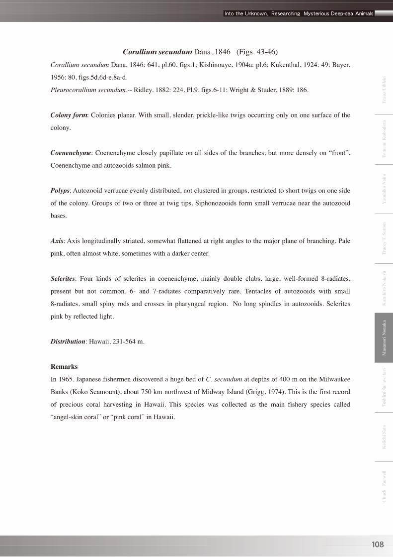
Corallium secundum Dana, 1846 (Figs. 43-46)Corallium secundum Dana, 1846: 641, pl.60, figs.1; Kishinouye, 1904a: pl.6; Kukenthal, 1924: 49; Bayer,
1956: 80, figs.5d,6d-e,8a-d.
Pleurocorallium secundum,-- Ridley, 1882: 224, Pl.9, figs.6-11; Wright & Studer, 1889: 186.
Colony form: Colonies planar. With small, slender, prickle-like twigs occurring only on one surface of the
colony.
Coenenchyme: Coenenchyme closely papillate on all sides of the branches, but more densely on “front”.
Coenenchyme and autozooids salmon pink.
Polyps: Autozooid verrucae evenly distributed, not clustered in groups, restricted to short twigs on one side
of the colony. Groups of two or three at twig tips. Siphonozooids form small verrucae near the autozooid
bases.
Axis: Axis longitudinally striated, somewhat flattened at right angles to the major plane of branching. Pale
pink, often almost white, sometimes with a darker center.
Sclerites: Four kinds of sclerites in coenenchyme, mainly double clubs, large, well-formed 8-radiates,
present but not common, 6- and 7-radiates comparatively rare. Tentacles of autozooids with small
8-radiates, small spiny rods and crosses in pharyngeal region. No long spindles in autozooids. Sclerites
pink by reflected light.
Distribution: Hawaii, 231-564 m.
Remarks
In 1965, Japanese fishermen discovered a huge bed of C. secundum at depths of 400 m on the Milwaukee
Banks (Koko Seamount), about 750 km northwest of Midway Island (Grigg, 1974). This is the first record
of precious coral harvesting in Hawaii. This species was collected as the main fishery species called
“angel-skin coral” or “pink coral” in Hawaii.

109
Masanori N
onaka
Fig. 43: Whole colony of Corallum secundum. (from Kishinouye, 1904)
Fig. 45: Tip of branch. (from Bayer, 1956)Fig. 44: Detail of polyps. (from Bayer, 1956)
Fig. 46: Sclerites. (from Bayer, 1956) a. 8-radiates from coenenchyme. b. Double-clubs from coenenchyme. c. From tentacles. d. From pharynx.

110
Mas
anor
i Non
aka
Paracorallium tortuosum (Bayer, 1956) (Figs. 47, 48)Corallium tortuosum Bayer, 1956: 82, figs.5b, 6c, 8e-g.
Paracorallium salomonense tortuosum.-- Bayer, 1993: 16, Pls.10,14-16.
Paracorallium tortuosum.-- Bayer & Cairns, 2003: 225.
Colony form: Colonies irregularly branched, but showing definite tendency to remain in one plane.
Largest specimens about 7.5 cm high. Although branching mainly in one plane, twigs here and there grow
out in various directions. The branches twisted and tortuous with numerous swellings, cysts, tunnels and
other deformities caused by the many infesting epizooic and commensal animals.
Coenenchyme: Coenenchyme exceedingly thin with few sclerites, except between the longitudinal cortical
solenia, each of which follows a groove in the axis. It therefore appears to have lines of sclerites running
through it longitudinally. Pale pink or salmon pink in color, the region surrounding the autozooids darker.
Polyps: Autozooids raised areas on the trunk and branches; each set in a depression surrounded by a raised
rim. Rim usually highest above and open toward the base of the colony, so that the calycular margin forms
a projecting shelf over the zooid. They do not form projecting verrucae, but retract flush across the
calycular pit, and have the usual 8-rayed orifice at center. Siphonozooids occur as tiny verrucae between
the lines of sclerites in the coenenchyme, i.e., along the solenia, especially based from the autozooids.
Axis: Axis round or oval in cross section, about 10 mm in diameter and longitudinally grooved; the smaller
branches basically round in cross section but more or less distorted by the autozooid calyces which indent
the solid axis. The calyces usually have the best developed rims, and since two are often opposed at the
twigs tips, a cross section of the axis there assumes a roughly x-shaped outline. Pinkish. The projecting
calycular rims strongly beaded and often darker in color than the surrounding areas.
Sclerites: Two types of sclerites, the same in both coenenchyme and verrucae: 8-radiates and numerous
crosses. In the pharyngeal region and oral disk of the anthocodiae minute, irregular rodlets and crosses.
Sclerites pink.
Distribution: Hawaii, 168-408 m.
Remarks
All specimens of P. tortuosum are infested with a small zoanthid which pits and distorts the axis. The
depressions caused by the zoanthid are distinguishable from those formed by the polyps of the
Paracorallium itself by their larger size and lack of raised, beaded margin. The coral is also host to a
polynoid polychaete, to which the tunnels and cavities in the axis are due. One specimen has in the main

111
Masanori N
onaka
stem some chambers filled with a
sponge, which may either be directly
responsible for the cavities or merely
occupying space left by some other
inhabitant. P. inutile is similarly
infested with actinians but, the axis is
not affected.
P. stylasteroids is very similar in
general appearance to P. tortuosum, but
lacks the numerous cross sclerites so
conspicuous in the Hawaiian material.
P. tortuosum appears to be the most
abundant precious coral in Hawaiian
waters but, due to its small size and
usually deformed axis, it probably has
no commercial possibilities. Bayer
(1993) treated P. tortuosum as a
subspecies of P. salomonense but later
as a seperated species pending
discovery of more materials for
comparisons. (Bayer and Cairns, 2003)
a
b d
c
Fig. 47: Paracorallium tortuosum. (from Bayer, 1993) a,b Whole colonies. c,d Axis of branchlet.
b,c Tip of branches. e. Coenenchymal sclerites (8-radiates) f. Coenenchymal crosses. g. Pharyngeal sclerites.
Fig. 48:Paracorallium tortuosum.(from Bayer, 1956)

112
Mas
anor
i Non
aka
Precious Corals from other Indo-Pacific areas (Descriptions were condensed from the original publications)
Corallium borneense Bayer, 1950 (Fig. 49)Corallium borneense Bayer, 1950: 59, fig.1; Bayer, 1956: 76 (in key).
Colony form: Branches on all sides of the stem, projecting at right angles or inclined a little upward. Large
branches irregularly subdivided into thick, clavate twigs bearing at their ends clusters of several autozooids.
Small branches mostly simple and of about the same caliber as the secondary twigs of the large branches.
Type specimen 5.5cm in height, possibly only a branch of a large colony.
Coenenchyme: Coenenchyme highly colored, being nearest to Ridgway's “salmon” and the verrucae
darker, approximating “flame scarlet.”
Polyps: Autozooids are low verrucae about 2 mm in diameter, very few on the main stem, but numerous on
the branches. Twigs and branches with autozooids in groups, No predominance of autozooids on any one
side. Siphonozooids small warts with simple orifices, often of a lighter color than the surrounding
coenenchyme, situated around the bases of the autozooids.
Axis: Axis obscurely striated, round except at the twig tips, X- or Y-shaped in cross section. The main axis
is about 9 mm in diameter near the base, decreasing to 3.5 mm at the top. About 15 mm from the base there
is a round hole 3 mm in diameter leading to a cavity within the stem; at the base of a branch 6mm above the
first hole there is a small but deep pit not connected with the axial cavity. These deformities were probably
caused by a polychaete commensal. The white axis has a pink center.
Sclerites: Four kinds of sclerites in both coenenchyme and autozooids, predominant sclerite the double
club. Less common are the radiate forms with 6-, 7-, or 8-radiate sclerites, such as the malformed 8-radiate,
and simple crosses. Small rods with conical processes occur only in the autozooids; this form may be
derived from the multi-radiate type by suppression of the rays.
Distribution: Borneo, 534 m.
Remarks
Those with rather large, dome-like verrucae often clustered in groups and also not set into the axis (e.g., C.
konojoi). The manner of branching is similar in appearance to C. konojoi. Microscopically the latter differs
greatly in the predominance of 6-radiates, the more irregular double clubs, and the presence of autozooids
only on one surface of the colony.

113
Masanori N
onaka
Fig. 49: Corallium borneense. (from Bayer, 1950) a. Whole colony. b-i. Sclerites from autozooid verrucae. j-q. Sclerites from coenenhcyme.
Corallium ducale Bayer, 1955 (Fig. 50)Corallium ducale Bayer, 1955: 210, Pls.1; Bayer, 1956: 75 (in key).
Colony form: Colony spread in one plane, openly branched laterally and dichotomously. Branches round or
slightly compressed at right angles to the plane of branching, the largest nearly 10 mm in diameter.
Terminal twigs 1.5-2.0 mm in diameter.
Coenenchyme: Coenenchyme of the “back” face of the colony wrinkled by an anastomosing reticulum of
narrow ridges marking the presence of the solenial system, sinuous grooves present on the coenenchyme
between the autozooids. Colony dark pink in alcohol.
Polyps: Autozooids restricted to one face of the colony, their calyces short cylindrical or blunt conical,
distinctly 8-ribbed. Tentacles fully retractile and none exsert in preservation. Verrucae of autozooids 1.5
mm or less in height, and up to 2.0 mm in diameter at the base, more or less tapering apically.
Siphonozooids small, hemispherical or irregular calyces near the autozooids.
Axis: Axis faintly striated; in the terminal portions with low surface irregularities and distinct granulation.
The axis is of a richer and deeper color than the coenenchyme.
Sclerites: Four kinds of sclerites in the coenenchyme: abundant double clubs derived from radiate forms by
asymmetrical development of two radii, measuring 0.060-0.085 mm in length, and 6-, 7-, and 8-radiates up
to 0.1 mm in length, some of which may show a considerable subdivision of the radii or are otherwise
misshapen. Sclerites in the pharyngeal region and oral disk: crosses, massive, irregular bodies and slender
spinous rods in the pharyngeal region and oral disk, and abundant stouter rods in the tentacles.
Distribution: East-Pacific Mexico.
Remarks

114
Mas
anor
i Non
aka
The massive, irregular sclerites of C. ducale resemble those of C. boshuensis and C. sulcatum, but C.
ducale differs widely from both those species in its open, lateral dichotomous plan of ramification, lower
autozooid calyces, and presence of both 6- and 7-radiates as well as the usual 8-radiate forms.
Corallium halmaheirense Hickson, 1907 (Figs. 51-53)Corallium halmaheirense Hickson, 1907: 6, figs.5,6,9; Kukenthal, 1924: 51; Bayer, 1956: 76, (in key).
Colony form: Only broken parts are preserved, suggesting branching mainly in one plane. Sample 15 mm
in height and axis diameter 2.25 mm. with four lateral branches ranging from 5-8 mm length with two
small branches on one face about 3 mm in length.
Coenenchyme: Coenenchyme orange-red and much darker than axis. Distortion of growth due to
commensal polychaete worm.
Polyps: Verrucae of autozooids conical in shape, very prominent, 2-3 mm in height, and marked externally
by eight deep longitudinal grooves. Not all turned in the same direction but nevertheless tend to turn
towards “anterior” surface of the colony.
Between verrucae of autozooids a number of minute white specks visible, certainly siphonozooids.
Axis: Main axis half-moon shaped in section, very pale pink, almost white, but with an eccentrics darker
pink core.
Sclerites: Coenenchyme with numerous crowded sclerites of the 8-radiate type 0.06-0.07 mm long.
Sclerites of verrucae of autozooids flattened spindles or rods (about 0.09 x 0.015 mm) with many tubercles.
Distribution: Celebes Sea, 1089 m.
Fig. 50: Corallium ducale. (from Bayer, 1955) a. Double-club sclerites. b. 6-radiate sclerite. c. 7-radiate sclerite. d. 8-radiate sclerite e-g. Irregular radiates. h. Cross sclerites. i-k. Irregular sclerites. l. Sclerites from oral disk and pharynx. m. Sclerite from tentacle. n. Tip of branch.

115
Masanori N
onaka
Remarks
A characteristic feature of this species seems to be many of the terminal branchlets terminate in a pair of
opposite verrucae so that these branchlets have the shape of a capital T. It is difficult to separate from C.
sulcatum, C. tricolor and C. maderense, but in C. sulcatum, the sclerites are of three kinds, six-radiates,
seven-radiates and double clubs. In C. tricolor and C. maderense, there are three forms of sclerites in the
cortex, double clubs being the most numerous. Sclerites of C. halmaheirense very similar in form and
shape to the sclerites of C. reginae but decidedly smaller.
Corallium imperiale Bayer, 1955 (Fig. 54)Corallium imperiale Bayer, 1955: 209, Pls.2c-h; Bayer, 1956: 75, 76 (in key).
Colony form: Colony large, spread in one plane, abundantly branched in a subpinnate fashion. Main
branches practically circular in cross section, about 5mm in diameter; end twigs slender, about 1.5mm in
diameter.
Coenenchyme: Coenenchyme with a predominantly longitudinal and parallel system of narrow ridges, here
and there densely anastomosing cross-connections marking the presence of the coenenchymal solenial
network. One face (“back”) of colony bare. Both coenenchyme and axis a rich pink in color.
Polyps: Autozooids restricted to one face of the colony, their calyces tall, cylindrical, 8-ribbed; the tentacles
fully retractile, but in preservation may remain exsert. The calyces about 2.5 mm tall, up to 3 mm if the
tentacles not fully retracted, and 1.5 mm in diameter. Siphonozooids forming small, irregular verrucae
between the autozooids.
Axis: Axis very weakly and obscurely striated, rich pink.
Fig. 51: Branchlet of Corallium halmaheirense. (from Hickson, 1907)a. Tunnel containing a commensal polychaete. b and c. Typical T-shape terminal branch.
Fig. 52: Detail of polyps. (from Hickson, 1907)
Fig. 53: Sclerites. (from Hickson, 1907) a. 8-radiate from coenenchyme. Others from autozooids.
x4 x7 a

116
Mas
anor
i Non
aka
Sclerites: Two kinds of sclerites predominant in the general coenenchyme and autozooids: 8-radiates and
double clubs. Double clubs very abundant; averaging 0.05-0.06 mm in length. Typical 8-radiates to
0.08-0.09 mm long, and occasional atypical examples 0.1 mm. Crosses not uncommon. In the distal part of
the autozooids a few rods 0.10-0.11 mm in length may be found, and this type of sclerite is the predominant
one in the tentacles. Small 8-radiate and irregularly spinous rods also occur in the tentacles, where the
sclerites are irregularly packed, extending as points into the bases of the pinnules.
Distribution: East- Pacific Mexico southwest of Guadalupe Is.
Remarks
Corallium imperiale seems to be closely allied to C. boshuensis and C. sulcatum, but differs from both in
the absence of massive, irregular sclerites and the predominance of double clubs rather than 8-radiates. C.
impteriale differs further from C. sulcatum in its less profuse branching and more prominent autozooids,
and from C. boshuensis in its lack of compression of the branches and its rich pink color.
Corallium reginae Hickson, 1905 (Figs. 55-59)Corallium reginae Hickson, 1905: 270; Hickson, 1907: 4, figs.1-4,8,10; Kukenthal, 1924: 50; Bayer, 1956:
76 (in key).
Colony form: Colony branching irregularly but principally in one plane.
Specimens considerably broken, 37-80 mm in height, base of attachment 3-6 mm in diameter and the main
branches are 4-5 mm in diameter at their bases.
Coenenchyme: Pink.
Fig. 54: Corallium imperiale. (from Bayer, 1955) c. Tip of branch. d. Sclerites (double-clubs) from coenenchyme. e. Sclerites (8-radiates) from coenenchyme. f. Cross from coenenchyme. g. Atypical sclerite from coenenchyme. h. Sclerites from tentacles.

117
Masanori N
onaka
x1.5
x1.5
ax
A
S
Ccoe
Polyps: Autozooids situated on one surface only except at branch extremities. Autozooids in retraction
forming irregularly scattered hemispherical verrucae projecting about 1.5 m from the surface, about 1.4mm
in diameter, scored externally by eight radiating grooves. All the larger branches have few autozooids.
Siphonozooids very small, in some cases indicated by minute papillae or tubular pores but usually
inconspicuous, numerous, unevenly distributed.
Axis: Axis very hard and takes a polish. In transverse section it shows a pale central spot and one or two
white rings arranged concentrically around the center. Color of axis a rather dark pink. Some fistulose
places due to the action of a commensal polychaete.
Sclerites: Sclerites very variable in form, the largest 8-radiates about 0.075-0.08 mm in length not
exceeding 0.085 mm, the double-club shape comparatively rare. The great majority of sclerites irregular
8-radiates but a considerable number approximating the typical 8-radiate sclerites may be found.
Distribution: Flores Sea, 122 m.
Remarks
Although Hickson first mentioned this new species in 1905,
a more complete description is in Hickson 1907. x6
Fig. 55: A basal fragment of a colony of C. reginae. (from Hickson, 1907) b. Base of attachment.
Fig. 56: Whole colony from anterior surface (left) and posterior surface (right). (from Hickson, 1907)
Fig. 58: Sclerites. (from Hickson, 1907) a. Irregular 8-radiate sclerite from coenenchyme. b. Double-club from coenenchyme. c and d. Spindles from autozooid verrucae.
Fig. 59: Diagram of branch.(from Hickson, 1907) A. Autozooid. S. Siphonozooid bearing the gonads. C. Canals. coe. coenenchyme. ax. Axis.
Fig. 57: Detail of polyps. (from Hickson, 1907)

118
Mas
anor
i Non
aka
Corallium variabile (Thomson & Henderson, 1906) (Figs. 60, 61)Pleurocorallium variabile Thomson & Henderson, 1906: 24, Pl.1 fig.9. Pl.5 fig.6. Pl.9 fig.13.
Corallium variabile. -- Kukenthal, 1924: 51, Bayer, 1956: 76 (in key).
Colony form: Complete colony form still unknown, only some broken pieces available, the largest one 13.5
cm in height and 6.2 cm in width. Colony very profusely branched in one plane, branches tortuous, with
little, if any sign of lateral compression. Branching from the antero-lateral surfaces of the stem, diminishing
gradually in thickness towards their tips. Numerous short branchlets on the sides of the large branches and
the stem.
Coenenchyme: Coenenchyme thin, creamy white in color, and full of closely packed small sclerites
resembling glistening sand grains. Also a few fragments from the same locality rosy-red with a
yellowish-brown coenenchyme with the same polyp arrangement and details of sclerites and axis as above.
Polyps: Polyps irregular on the anterior surface of the stem and branches, the salmon pink verrucae a fine
contrast to the creamy white of the general coenenchyme and the yellow of the tentacles. Verrucae
prominent, almost cylindrical in shape, marked by eight longitudinal ridges, 2.7 mm high. The tentacles
about 0.5 mm in length, yellowish in color, and closely covered by small sclerites. With tentacles retracted,
the apices of the verrucae resemble eight-rayed stars.
Axis: Axis hard, not easily indented with a knife, solid, almost cylindrical in section in some parts and
slightly oval in others. White in color, surface marked by very fine striations, often very faint.
Sclerites: Coenenchymal sclerites two kinds:
(1) 8-radiate sclerites, with a short shaft and terminal tubercles. Several tubercles project at each end of the
sclerite at right angles to the shaft. The large tubercles may themselves be covered with smaller tubercles.
These sclerites from 0.06-0.08 mm long, and 0.04-0.05 mm in diameter.
(2) Double-clubs consisting of two globes masses somewhat flattened at one end, and at the other end
bearingshort processes, variable in shape, with several tubercles. On average 0.06 mm in length, and 0.04
mm in width.
Also a very few rough spiny spindles from 0.06-0.09 mm in length, and 0.02 mm in diameter.
Distribution: Ceylon, 926 m.
Remarks
This species differs from C. johnsoni, in being more profusely branched, and in having more prominent and
differently arranged verrucae.

119
Masanori N
onaka
Fig. 60: Detail of polyps. (from Thomson & Henderson, 1906)
Fig. 61: Sclerites of holotype specimens. (from Thomson & Henderson, 1906)
Paracorallium nix (Bayer, 1996) (Figs. 62-64)Corallium nix Bayer, 1996: 213, figs.7-10.
Paracorallium nix.-- Bayer & Cairns, 2003: 225.
Colony form: Specimen only a branch so colony form unknown. Branch sinuously curved, thick,
approximately round in cross section, and 6.2 mm in greatest diameter at the base, shape suggesting
branching roughly in one plane. Short terminal branchlets produced mostly from two sides in the plane of
curvature, but a few small ones arise from the “front” of the branch.
Coenenchyme: Coenenchyme very thin, generally smooth but inconspicuously papillate in areas protected
from abrasion, suggesting that elsewhere the minute papillae may either have been rubbed off during
collection, or have contracted completely as the result of contact with other objects in the trawl. In
conspicuous sinuous longitudinal grooves indicate the location of the principal coenenchymal canals.
Coenenchyme white.
Polyps: Autozooids on all sides of the branch and terminal branchlets, retracted flush within the rim of the
axial pits, the bases of the infolded tentacles forming an 8-lobed margin of the closed verrucal apertures.
Their distribution most clearly revealed by staining with crystal violet, which stains the structures within
the gastric cavities more darkly than the surrounding coenenchyme so that they contrast sharply with the
paler cortex as the highly soluble stain dissipates.
Siphonozooids not distinct small verrucae and therefore very difficult to detect, but commonly one in the
tapered groove extending from the autozooid, and scattered individuals in the coenenchyme between and
around the autozooids.
Axis: The surface of the axis longitudinally grooved, as usual covered with minute axial protuberances
ornamented with thorny projections. Axis a distinct rounded pit 0.75 mm wide and 1.05 mm long at the

120
Mas
anor
i Non
aka
position of each autozooid, bordered by a beaded rim commonly interrupted where a coelenteric canal
passes through to connect with the gastrobascular cavity of the autozooid. White.
Sclerites: Four kinds of coenenchymal sclerites, predominantly
short 8-radiates reaching lengths of 0.06-0.07 mm. Very short,
wide 6- and 7-radiates 0.04-0.06 mm long, some weakly
asymmetrical but not clearly modified as double clubs, the
shortest of them forming tuberculate spheroids. Crosses absent
or rare. The tentacles contain blunt rods derived from
8-radiates, as shown by numerous intermediate forms.
Sclerites colorless, translucent.
Distribution: New Caledonia, 240 m.
Remarks
There are distinct axial pits with beaded margins in the
autozooids of P. stylasteroides, P. salomonense, and P. thrinax.
However the sclerites of P. stylasteroides are smaller and not so
sharply sculptured, and those of P. salomonense are both larger
and more acutely sculptured. P. thrinax differs conspicuously
in its small, dichotomously branched uniplanar growth form
and its strongly asymmetrical double-club sclerites.
Fig. 62: Whole specimen of P. nix from front surface. 5.75cm in height. (from Bayer, 1996)
Fig. 63: Axis of branch.(from Bayer, 1996)
Fig. 64: Sclerites of P. nix. (from Bayer, 1996)

121
Masanori N
onaka
Paracorallium salomonense (Thomson & Mackinnon, 1910) (Figs. 65, 66)Sympodium salomonense Thomson & Mackinnon, 1910: 168, Pls.12,13, figs.11,12,15a,15b.
Corallium salomonense salomonense. -- Bayer, 1993: 15, Pls.10 upper,11,12,13.
Paracorallium salomonense. -- Bayer & Cairns, 2003: 225.
Colony form: Complete colony form still unknown, only some broken pieces available. The short, rounded
branches resemble a digitiform massive alcyonacean, therefore Thomson & Mackinnon originally (1910)
put this species in the genus Sympodium (family Xeniidae).
Coenenchyme: Coenenchyme thin.
Polyps: Autozooids flat or only slightly raised in contracted state, apertures closed by tentacle bases in the
form of an 8-rayed star. Siphonozooids scattered, obscure.
Axis: Axis with autozooids seated in distinct pits in the calcareous axis, the apical ones bordered by a
distinctly raised, horse-shoe shaped rim.
Sclerites: Two kinds of sclerites: 8-radiates and crosses. Not markedly asymmetrical, having a distinctly
thorny aspect with tubercular ornamentation complex and sharp, reaching more than 0.09 mm (largest 0.11
mm) in length by 0.058-0.06 mm in width.
Distribution: Chagos Archipelago, 217-272 m.
Remarks
Skeletal characters agree so closely with those of P. tortuosum that Bayer (1993) suggested they be
considered representatives of one species, the senior synonym being salomonense. However, in view of the
wide geographic separation of the type localities of P. tortuosum (Hawaii) and P. salomonense (Chagos),
Bayer & Cairns (2003) treated them as speperate species, pending the discovery of more specimens for
comparisons.
Fig. 65: Whole colony of syntype specimens. (from Bayer, 1993)

122
Mas
anor
i Non
aka
Paracorallium stylasteroides (Ridley, 1882) (Figs. 67, 68) Corallium stylasteroides Ridley, 1982: 225, Pl.9, figs.1-4; Kukenthal, 1924: 48, figs.39,40; Bayer, 1956: 76
(in key); Bayer, 1993: 17, Pl.17.
Paracorallium stylasteroides,-- Bayer & Cairns, 2003: 225.
Colony form: Colony dichotomously branched in one plane, dichotomous, but subject to considerable
variation. Branches tortuous, decidedly compressed laterally in the case of all but the peripheral members,
arising from the antero-lateral rather than the postero-lateral aspects of the stem or branches from which
they may be derived, diminishing gradually in thickness towards the peripheral part. A few small branchlets
scattered on sides of the larger branches irregularly, and together with the terminal branchlets, generally
subclavate in form, consisting of a slightly contracted basal portion and an enlarged, pointed, and
polyhedric terminal portion. “Posterior” aspect of main branches very convex, but that of lesser branches
less so. Maximum transverse breadth of the whole colony 135 mm, maximum height 105 mm. Stem stout,
irregular in transverse section.
Coenenchyme: Coenenchyme very thin, with minute projecting points (visible only with the aid of a lens)
arranged along the axial striae. Color of coenenchyme extremely pale orange.
Polyps: Verrucae of autozooids in the grooves of axis, not projecting beyond their margins, or in slight
depressions distributed over all parts of the colony, but most abundantly on the lateral aspects of the main
branches, and on the small terminal and lateral projecting branchlets. Peripheral part a pale pink collar,
slightly darker than the general crust. The eight valves of autozooids very pale yellow in color. Diameter
about 0.75 mm.
Fig. 66: Some variations of sclerites in P. salomonense. (from Bayer, 1993)

123
Masanori N
onaka
Axis: Axis hard, compact. Longitudinal striae on the fine surface, about four to 1 mm. Grooves for
autozooids generally with a narrow raised lip on each side; length of grooves 1.5 to 2 mm long (in direction
of branches) by about 0.75 mm broad, and about 0.5 mm deep in the center, a smooth-walled hemispherical
pit. Axis perforated by a few canals (normal, or due to parasites?) of about 1mm in diameter, which issue
distally at various points on the lateral aspects of the branches, the openings being often covered by a
curved lamina of hard material. Pure white throughout.
Sclerites: Ridley (1882) described sclerites of coenenchyme as only8-radiates, from 0.053 to 0.058 mm
long by 0.035 mm broad (including the tubercles); shaft, excluding tubercles, about 0.02 mm broad.
However, Bayer (1993) illustrated three kinds of sclerites prepared from the type: 6-radiates, incomplete
double clubs with rough surface, and crosses (rare), all with rather smooth tubercles. Length approximately
0.05 mm, breadth 0.03 mm.
Distribution: Mauritius, 137 m.
Remarks
The present species agrees in the mode of branching in one plane with C. secundum, and also in that many
of the polyps are borne on small lateral branchlets; but differs from it in having polyps on the posterior as
well as the anterior surface, and also in the very pale pinkish color of the coenenchyme (that of C.
secundum being darker pink), and the pure white of the hard axis (that of C. secundum being white and
pink). Bayer and Cairns (2003) suggested that when new specimens are discovered, P. stylasteroides, P. nix
(from New Caledonia) and P. tortuosum (Hawaii) may be a single variable species.
Fig. 67: Pacorallium stylasteroides. (from Ridley, 1882) 1. Whole colony. 2. Detail of autozooids. 3. Part of axis with coenenchyme removed. 4. Sclerites from coenenchyme.
Fig. 68: Sclerites of P. stylasteroides. (from Bayer, 1993)

124
Mas
anor
i Non
aka
Paracorallium thrinax (Bayer & Stefani, 1996) (Figs. 69-71)Corallium thrinax Bayer 1996: 206, figs.1-5.
Paracorallium thrinax Bayer & Cairns, 2003: 224.
Colony form: Specimens small, from 3.7 to 6 cm in height, and between 3 and 6 cm in width. Sparingly
branched dichotomously, often with a few short lateral twigs. The holdfast a narrow basal expansion of the
trunk. For the most part branching in one plane, but in some colonies a branch may stand at nearly 90
degrees from the plane of the principal fan. No anastomoses. In most cases a short main trunk, oval in cross
section, about 0.6 x 1.2 cm in diameter, which bifurcates to produce principal branches approximately
round in section, which in turn bifurcate as many as two or three times; a few short, blunt lateral twigs
sometimes arise from one or more of the internodes between bifurcations. In some cases, principal branches
2-4 mm in diameter arise directly from the holdfast.
Coenenchyme: Coenenchyme so thin that the axial sculpture is exposed in areas subject to abrasion. The
longitudinal axial striations are clearly indicated through the coenenchyme, most clearly toward branch tips
where the autozooids are closer together. Colonies preserved in ethanol are white.
Polyps: Autozooids low, inconspicuous verrucae distributed predominantly on one face (the “front”) of the
colony, with only an occasional stray on the back surface, indistinct unless artificially stained with crystal
violet. Tentacles retracted flush with the coenenchyme, their bases forming an 8-lobed margin of the
verrucal orifice. Siphonozooids scarce and distributed randomly in the coenenchyme, so inconspicuous that
they can be distinguished only by staining with crystal violet.
Axis: The surface of the axis longitudinally grooved and covered with minute tubercles ornamented with
thorny projections. At the position of each autozooid, the axis a distinct rounded pit 0.75 mm wide and 1.05
m long bordered by a beaded rim that in some cases is interrupted by a narrow gap for passage of a
coelenteric canal. Axis is white.
Sclerites: Four kinds of coenenchymal sclerites. Numerous 6-radiates up to 0.04 mm in length. Some
modified as globose double clubs by asymmetrical hypertrophy of two of the rays to form a pair of smooth,
spheroidal processes. Surface of the coenenchyme crowded, with the spheroidal processes directed
outward. Sclerites intermediate between double clubs and 6-radiates with the hypertrophied rays more or
less strongly modified not uncommon. Irregular forms occur but extremely rare. No crosses or 8-radiates
observed, and only a single small 7-radiate found in 3 preparations for SEM observations.
Small rods and 6-radiates present in the tentacles, the distal ones decreasing in size to about 0.03 mm.
Sclerites colorless.

125
Masanori N
onaka
Fig. 69: P. thrinax, Whole colony from anterior surface. (from Bayer, 1996)
Fig. 70: Axis of branch. (from Bayer, 1996)
Fig. 71: Sclerites of P. thrinax. (from Bayer, 1996)
Distribution: New Caledonia, 240 m.
Remarks
The colonial form of P. thrinax is immediately distinguished from salomonense, inutile and nix. Among
those with beaded pits beneath the autozooid, P. stylasteroides, salomonense and tortuosum lack double
clubs. P. inutile has double clubs of somewhat different outline and only 6-radiates but no 7- and 8-radiates,
and differs further in its colonial form, pale pink axis and reddish cortex.
ACKNOWLEDGMENTS We thank Dr. Stephen Cairns, Curator of Invertebrates at the National Museum of Natural History,
Smithsonian Institution, for permission to reprint the original illustrations and photos in Smithsonian
publications. Dr. Sadao Kosuge approved the reprinting of the original photos in the Journal of “Precious
Coral and Octocoral Research.” We thank Dr. Masashi Yamaguchi who provided us literature about
126
Mas
anor
i Non
aka
precious coral biology and cultural history. Mr. Yukimitsu Imahara, Researcher of Wakayama Laboratory,
Biological Institute on Kuroshio, Kuroshio Biological Research Foundation, shared much useful knowledge
about octocorals with us. Dr. Nozomu Iwasaki, Kochi University, and Mr. Fumihito Iwase, curator of
Kuroshio Biological Laboratory, reviewed the manuscript. Mr. Kei Kajihara, underwater photographer,
kindly offered the jellyfish photograph for publication in figure 1. We thank the staff of Okinawa Churaumi
Aquarium, especially Dr. Keiichi Sato, for editing this contribution to the Proceedings. Special thanks is
due Dr. Senzo Uchida, Director of Okinawa Churaumi Aquarium, for management of this research.
ReferencesBayer, F.M. 1950. A new precious coral from North Borneo. Journal of the Washington Academy of
Science 40 (2): 59-61.
Bayer, F.M. 1955. Contributions to the nomenclature, systematics, and morphology of the Octocorallia.
Proceedings of the United States National Museum 105: 207-220, plates 1-8.
Bayer, F.M. 1956. Descriptions and redescriptions of the Hawaiian octocorals collected by the U.S. Fish
Commission steamer "Albatross" (2. Gorgonacea: Scleraxonia). Pacific Science 10 (1): 67-95.
Bayer, F.M. 1993. Generic reassignment and affinities of Sympodium salomonense Thomson and
Mackinnon (Coelenterata: Octocorallia). Precious Corals and Octocoral Research 1: 14-19, plates
10-17.
Bayer, F.M. 1996. Three new species of precious coral (Anthozoa: Gorgonacea, genus Corallium) from
Pacific waters. Proceedings of the Biological Society of Washington 109 (2): 205-228.
Bayer, F.M. and S.D. Cairns. 2003. A new genus of the scleraxonian family Coralliidae (Octocorallia:
Gorgonacea) Proceedings of the Biological Society of Washington 116 (1): 222-228.
Cairns, S. D. 2007. Deep-Water Corals: An Overview with special Reference to Diversity and Distribution
of Deep- Water Scleractinian Corals. Bull Mar Sci 81(3): 311-322.
Grigg, R.W. 1974. Distribution and abundance of precious corals in Hawaii. Proceedings of the 2nd
International Coral Reef Symposium. 2, Great Barrier Reef Committee, Australia, p. 235-340.
Grigg, R.W. 1976. Fishery management of precious and stony corals in Hawaii. Sea Grant Technical
Report. UNIHI-SEAGRANT-TR-77-03 HIMB Contribution No. 490. 48pp.
Grigg, R.W. 1993. Precious coral fisheries of Hawaii and the U.S. Pacific Islands - Fisheries of Hawaii and
U.S.- associated Pacific Islands. Marine Fisheries Review 55 (2): 50-60
Hickson, S. J. 1905. On a new species of Corallium from Timor. Proceedings of the Meeting of Saturday
September 30: 268-271.
Hickson, S. J. 1907. Die Alcyoniden der Siboga-Expedition I. Coralliidae. Siboga-Expeditie Monogragh.
13C:1-8, plate 1.
Kitahara, T. 1904. Sango gyogyo chosa hokoku. Suisan Chosa Hokoku 13 (3): 1-24, plates 1-5. (in
Japanese)
Kishinouye, K. 1902. Honpou san sango no ichi shinshu Doubutsu Gaku Zasshi 14: 419-420. (in Japanese)

127
Masanori N
onaka
Kishinouye, K. 1903a. Preliminary note on the Coralliidae of Japan. Zoologischer Anzeiger 26 (705):
623-626.
Kishinouye, K. 1903b. Honpou san no sango. Doubutsu Gaku Zasshi 15: 103-106. (in Japanese)
Kishinouye, K. 1904a. Sango no kenkyu. Suisan Chosa Hokoku 14 (1): 1-31, pls. 1-9. (in Japanese)
Kishinouye, K. 1904b. Notes on the natural history of corals. Journal of Imperial Fishery Bureau 14 (1):
1-32, pls. 1-9.
Kosuge, S.1987. The CORALS -Captivating and charming gift from Neptune- Institute of Malacology of
Tokyo. Special publication No. 2, 174pp. (in Japanese)
Kukenthal, W. 1924. Gorgonaria. Das Tierreich 47: i-xxviii + 1- 478, 209 figs. Berlin and Leipzig, Walter
de Gruyter and Co.
Uchida H. 1992. Hexacorallia. In: Guide to seashore animals of Japan with color pictures and keys. 1:
118-120. (in Japanese)
Vighi M. 1972. Etude sur la reproduction du Corallium rubrum (L.) Vie Milieu 23 (1.A): 21-32
Lacaze-Duthiers, H.D. 1864. On the formation of coral (Corallium rubrum). Quarterly Journal science 1:
614-623.
Liverino, B. 1986. The CORALS -Cultural Tourist Itineraries in the south of Italy- Industrial History
Series. Analisi Co. Ltd, Rome, Italy, 200pp.
Marschal C., J. Garrabou, J.G. Harmelin and M. Pichon. 2004. A new method for measuring growth and
age in the precious red coral Corallium rubrum (L.). Coral Reefs 23: 423-432.
Nonaka M., K. Muzik and S. Uchida. 2006 Culture, Study and Display of Precious Corals. Proceedings of
the 10th International Coral Reef Symposium., Japanese Coral Reef Society, Japan: 1821-1831.
Pasternak, F.A. 1981. Alcyonacea and Gorgonacea. In: Kuznetsov, A.P. and A.N. Mironov [Eds.], Benthos
of the submarine mountains Marcus-Necker and adjacent Pacific regions. Moscow: Akademiya
Nauk: 40-55.
Ridley, S.O. 1882. On the arrangement of the Coralliidae, with descriptions of new or rare species.
Proceedings of the Zoological Society of London: 221-233, plate 9.
Shozakai K. 1983. Sango no umi. The Kochi Newspaper. (in Japanese)
Suzuki K. 1999a. SANGO. Cultural history of tools and human-beings, No.91. University of Housei Press,
Tokyo, 362pp. (in Japanese)
Suzuki K. 1999b. About the corals preserved in Syo-so-in. Syo-so-in Sango Cho-sa Houkokusyo, 31-39. (in
Japanese)
Tsounis G., S. Rossi, M. Aranguren, J-M. Gili and W. Arntz. 2006. Effects of spatial variability and colony
size on the reproductive output and gonadal development cycle of the Mediterranean red coral
(Corallium rubrum L.). Marine Biology 148: 513-527.
Wood E. M. 1983. Corals of the World. T.F.H. Publications, Inc., Ltd. 256pp.