Embed Size (px)
Citation preview
Joint Pain in Children:Initial Evaluation and When to Refer
C. Egla Rabinovich MD, MPHProfessor of PediatricsDivision Chief, Duke Pediatric Rheumatology

Disclosures
• No relevant disclosures
Educational Objective
• Develop a practical approach to joint pain in children
• Understand differential, emphasis on common causes
• Discuss rheumatologic causes of arthralgia
• Develop plans for initial work-up
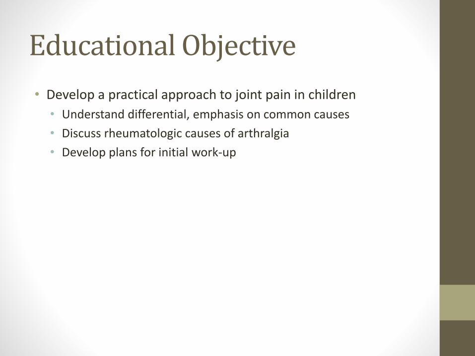
Musculoskeletal Pain in Children
Arthralgia is extremely common in childhood
5-30% of children have chronic pain complaints
More common in girls
May last years
Most have benign etiology
Differential of joint pain
• Age dependent
• 2 year old will not have repetitive use injuries
• 15 year old will not develop growing pains
• Some gender dependence
• Adolescent females and knee pain
• Need to keep a broad differential in mind
• Think thru the differential
• The work up
• And then possible referral
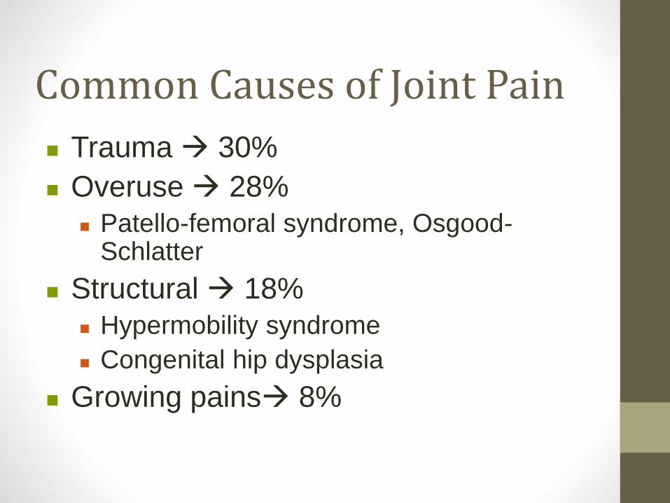
Common Causes of Joint Pain
Trauma 30%
Overuse 28%
Patello-femoral syndrome, Osgood-Schlatter
Structural 18%
Hypermobility syndrome
Congenital hip dysplasia
Growing pains 8%
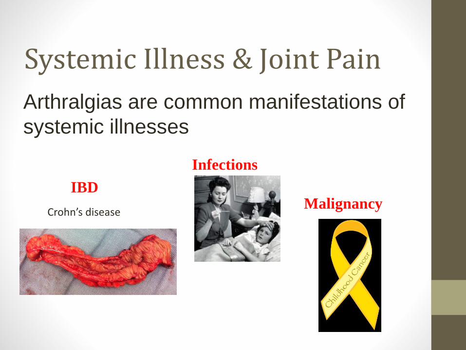
Systemic Illness & Joint Pain
Arthralgias are common manifestations of
systemic illnesses
Infections
IBDMalignancy
Crohn’s disease
Arthralgias: systemic disease
• Malignancy• Bone/soft tissue tumor• Bone marrow cancer (ALL/neuroblastoma)
• Endocrine disease• Thyroid disease• IDDM
• Gastrointestinal disease• Celiac disease• IBD• Autoimmune hepatitis
• Infectious disease• Viral (EBV/parvovirus)• Fever• Streptococcus
• Genetic • Fabry• Lysosomal storage disorders
• Rheumatologic
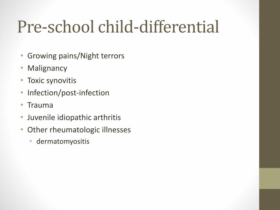
Pre-school child-differential
• Growing pains/Night terrors
• Malignancy
• Toxic synovitis
• Infection/post-infection
• Trauma
• Juvenile idiopathic arthritis
• Other rheumatologic illnesses
• dermatomyositis
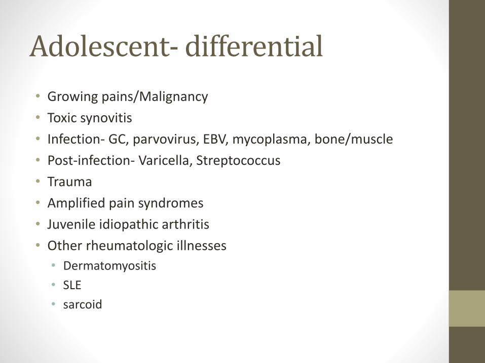
Adolescent- differential
• Growing pains/Malignancy
• Toxic synovitis
• Infection- GC, parvovirus, EBV, mycoplasma, bone/muscle
• Post-infection- Varicella, Streptococcus
• Trauma
• Amplified pain syndromes
• Juvenile idiopathic arthritis
• Other rheumatologic illnesses
• Dermatomyositis
• SLE
• sarcoid
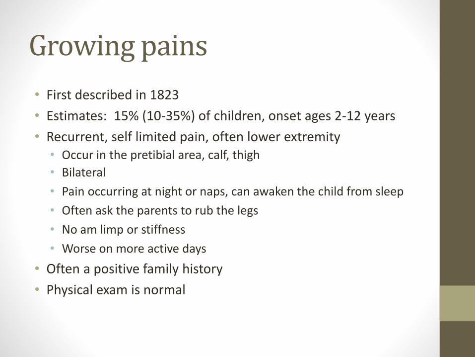
Growing pains
• First described in 1823
• Estimates: 15% (10-35%) of children, onset ages 2-12 years
• Recurrent, self limited pain, often lower extremity• Occur in the pretibial area, calf, thigh
• Bilateral
• Pain occurring at night or naps, can awaken the child from sleep
• Often ask the parents to rub the legs
• No am limp or stiffness
• Worse on more active days
• Often a positive family history
• Physical exam is normal
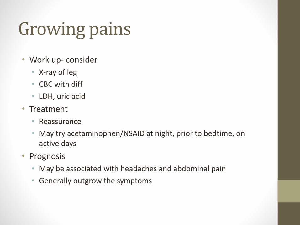
Growing pains
• Work up- consider
• X-ray of leg
• CBC with diff
• LDH, uric acid
• Treatment
• Reassurance
• May try acetaminophen/NSAID at night, prior to bedtime, on active days
• Prognosis
• May be associated with headaches and abdominal pain
• Generally outgrow the symptoms
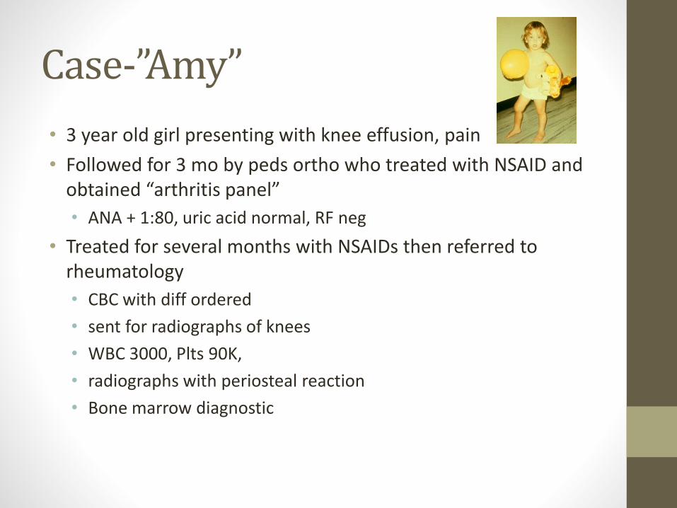
Case-”Amy”
• 3 year old girl presenting with knee effusion, pain
• Followed for 3 mo by peds ortho who treated with NSAID and obtained “arthritis panel”
• ANA + 1:80, uric acid normal, RF neg
• Treated for several months with NSAIDs then referred to rheumatology
• CBC with diff ordered
• sent for radiographs of knees
• WBC 3000, Plts 90K,
• radiographs with periosteal reaction
• Bone marrow diagnostic
Malignancies presenting with joint pain-younger children
• Bone marrow occupying lesions
• Acute lymphocytic leukemia
• Neuroblastoma
• Tumors of bone, muscle, soft tissues (rare)
• Work up
• CBC with differential
• Imaging of affected area
• Referral to peds oncology
Malignancies- older child
• Lymphoma
• Marrow occupying lesions
• ALL
• Neuroblastoma (in younger age group)
• Tumors of bone and muscle
• Consider:
• CXR
• Imaging of affected area
• CBC with diff
• LDH/uric acid
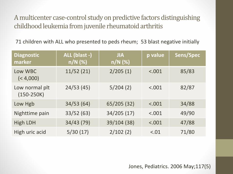
A multicenter case-control study on predictive factors distinguishing childhood leukemia from juvenile rheumatoid arthritis
Diagnosticmarker
ALL (blast -)n/N (%)
JIAn/N (%)
p value Sens/Spec
Low WBC (< 4,000)
11/52 (21) 2/205 (1) <.001 85/83
Low normal plt(150-250K)
24/53 (45) 5/204 (2) <.001 82/87
Low Hgb 34/53 (64) 65/205 (32) <.001 34/88
Nighttime pain 33/52 (63) 34/205 (17) <.001 49/90
High LDH 34/43 (79) 39/104 (38) <.001 47/88
High uric acid 5/30 (17) 2/102 (2) <.01 71/80
Jones, Pediatrics. 2006 May;117(5)
71 children with ALL who presented to peds rheum; 53 blast negative initially
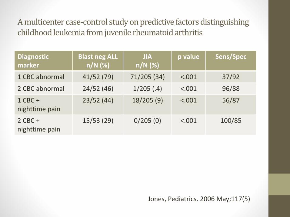
A multicenter case-control study on predictive factors distinguishing childhood leukemia from juvenile rheumatoid arthritis
Diagnostic marker
Blast neg ALLn/N (%)
JIAn/N (%)
p value Sens/Spec
1 CBC abnormal 41/52 (79) 71/205 (34) <.001 37/92
2 CBC abnormal 24/52 (46) 1/205 (.4) <.001 96/88
1 CBC + nighttime pain
23/52 (44) 18/205 (9) <.001 56/87
2 CBC + nighttime pain
15/53 (29) 0/205 (0) <.001 100/85
Jones, Pediatrics. 2006 May;117(5)
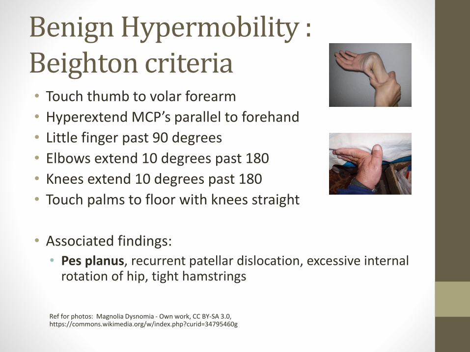
Benign Hypermobility : Beighton criteria• Touch thumb to volar forearm
• Hyperextend MCP’s parallel to forehand
• Little finger past 90 degrees
• Elbows extend 10 degrees past 180
• Knees extend 10 degrees past 180
• Touch palms to floor with knees straight
• Associated findings:• Pes planus, recurrent patellar dislocation, excessive internal
rotation of hip, tight hamstrings
Ref for photos: Magnolia Dysnomia - Own work, CC BY-SA 3.0, https://commons.wikimedia.org/w/index.php?curid=34795460g
Benign hypermobility
• Can be associated with Marfan’s, Stickler’s syndrome, Ehler’s-Danlos, Down’s syndrome
• 8-20% of population, + family history
• More common in girls, Chinese, West Africans
• Decreases with age (peak ages 3-10 years)
• Symptoms often intermittent
• No disability
• Reassurance, PT for joint protection and stretching program, arch supports
Overuse syndromes
• Patellofemoral syndrome
• Plica syndromes
• Stress fractures
• Apophyseal injuries
• Pelvic
• Osgood-Schlatter
• Sinding-Larsen-Johansson
• Sever’s Disease
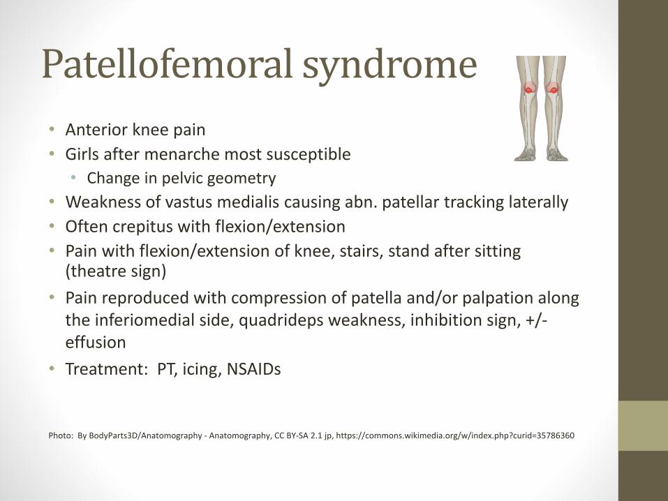
Patellofemoral syndrome
• Anterior knee pain
• Girls after menarche most susceptible
• Change in pelvic geometry
• Weakness of vastus medialis causing abn. patellar tracking laterally
• Often crepitus with flexion/extension
• Pain with flexion/extension of knee, stairs, stand after sitting (theatre sign)
• Pain reproduced with compression of patella and/or palpation along the inferiomedial side, quadrideps weakness, inhibition sign, +/-effusion
• Treatment: PT, icing, NSAIDs
Photo: By BodyParts3D/Anatomography - Anatomography, CC BY-SA 2.1 jp, https://commons.wikimedia.org/w/index.php?curid=35786360
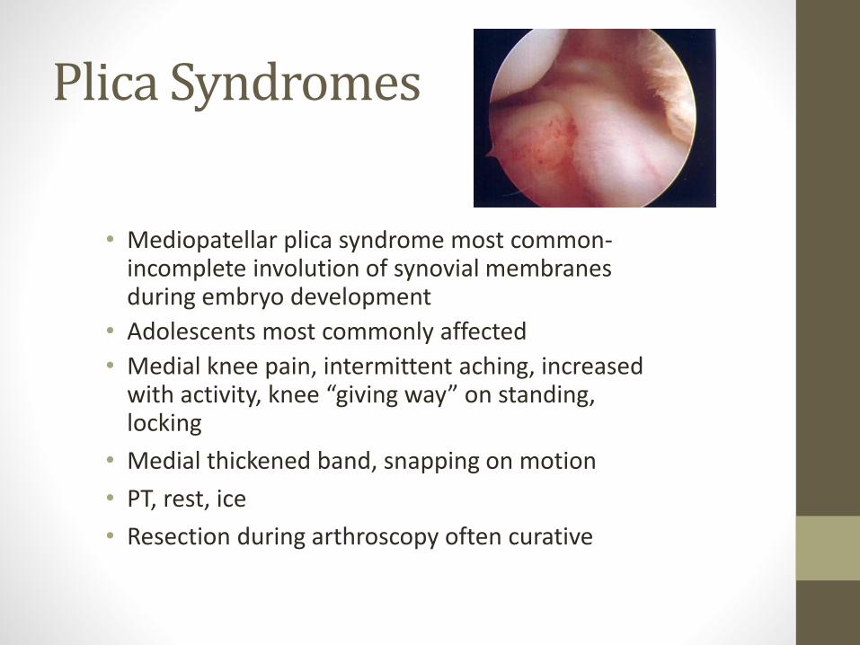
Plica Syndromes
• Mediopatellar plica syndrome most common-incomplete involution of synovial membranes during embryo development
• Adolescents most commonly affected
• Medial knee pain, intermittent aching, increased with activity, knee “giving way” on standing, locking
• Medial thickened band, snapping on motion
• PT, rest, ice
• Resection during arthroscopy often curative
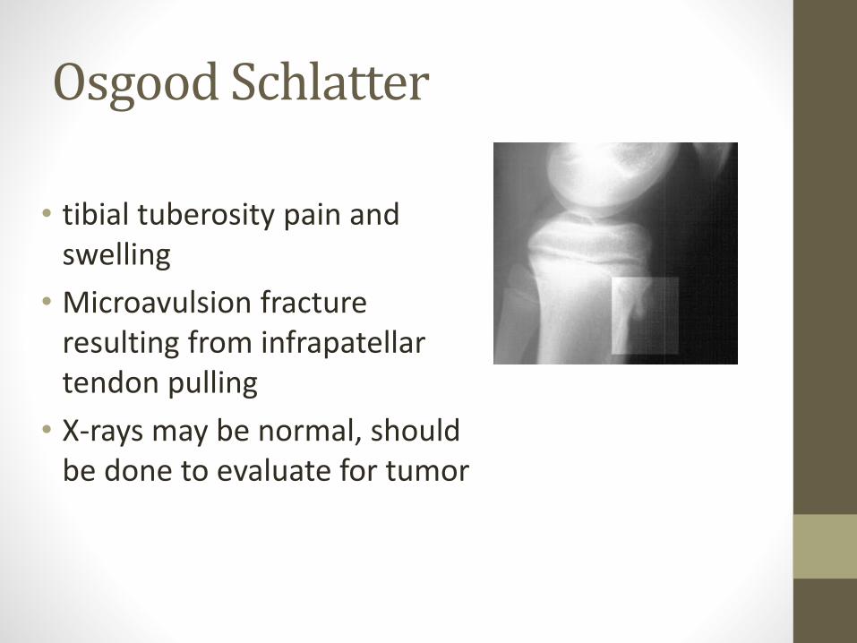
Osgood Schlatter
• tibial tuberosity pain and swelling
• Microavulsion fracture resulting from infrapatellar tendon pulling
• X-rays may be normal, should be done to evaluate for tumor
Amplified Pain syndromes
• Fibromyalgia/ Amplified pain Syndrome
• Chronic Regional Pain Syndrome
• RSD
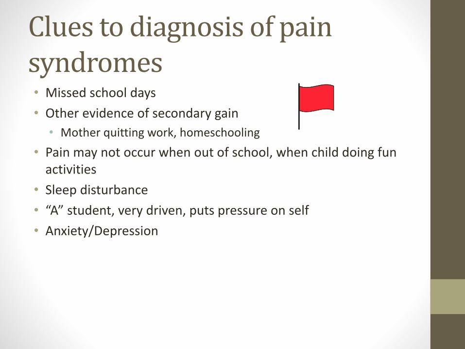
Clues to diagnosis of pain syndromes• Missed school days
• Other evidence of secondary gain
• Mother quitting work, homeschooling
• Pain may not occur when out of school, when child doing fun activities
• Sleep disturbance
• “A” student, very driven, puts pressure on self
• Anxiety/Depression
Fibromyalgia-epidemiology
• 2% of population (3-6 million)• 5-10% of general practice
• 15% of rheumatology practice
• 20% of university practice
• 20% of new ped. rheum. patients
• 85% female
• All ages (mean age onset 50 yrs)
• Family history
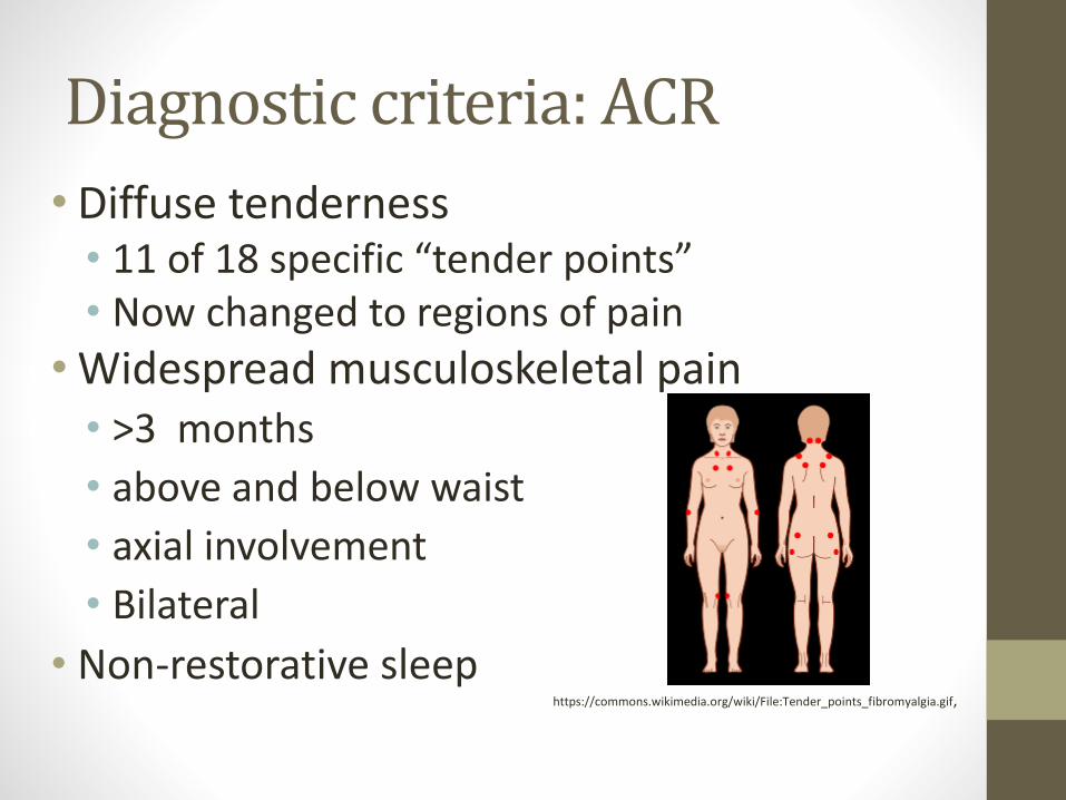
Diagnostic criteria: ACR
• Diffuse tenderness• 11 of 18 specific “tender points” • Now changed to regions of pain
• Widespread musculoskeletal pain • >3 months
• above and below waist
• axial involvement
• Bilateral
• Non-restorative sleephttps://commons.wikimedia.org/wiki/File:Tender_points_fibromyalgia.gif,
Other Features
Fatigue
Paresthesias
Headaches
Cognitive dysfunction
Subjective hand swelling
Overachievers/perfectionist personality
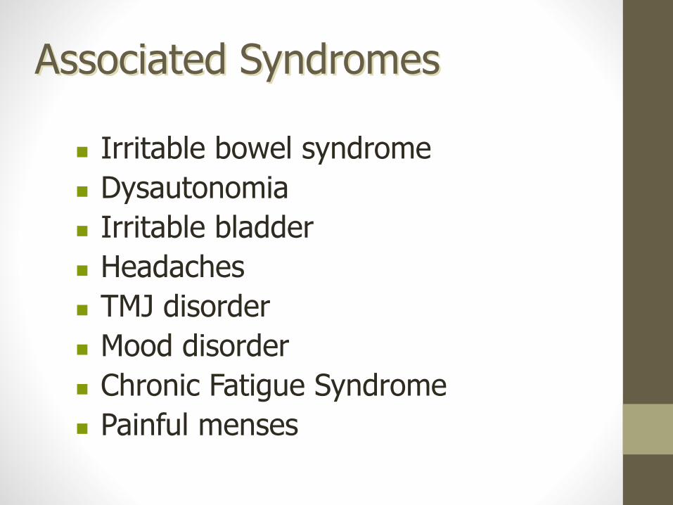
Associated Syndromes
Irritable bowel syndrome
Dysautonomia
Irritable bladder
Headaches
TMJ disorder
Mood disorder
Chronic Fatigue Syndrome
Painful menses
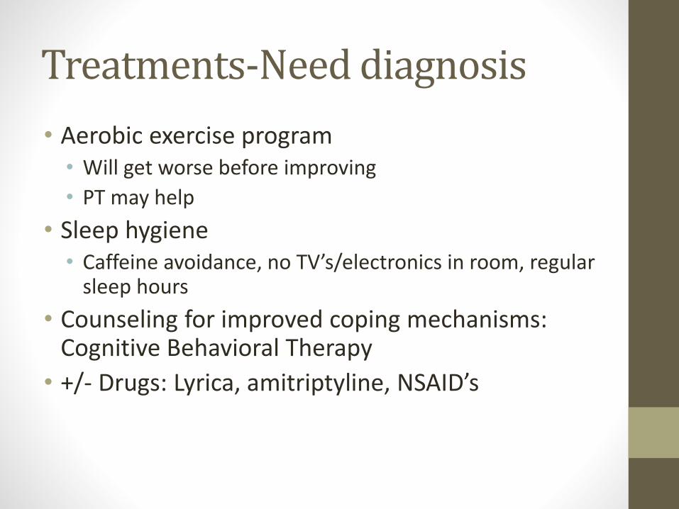
Treatments-Need diagnosis
• Aerobic exercise program• Will get worse before improving
• PT may help
• Sleep hygiene• Caffeine avoidance, no TV’s/electronics in room, regular
sleep hours
• Counseling for improved coping mechanisms: Cognitive Behavioral Therapy
• +/- Drugs: Lyrica, amitriptyline, NSAID’s
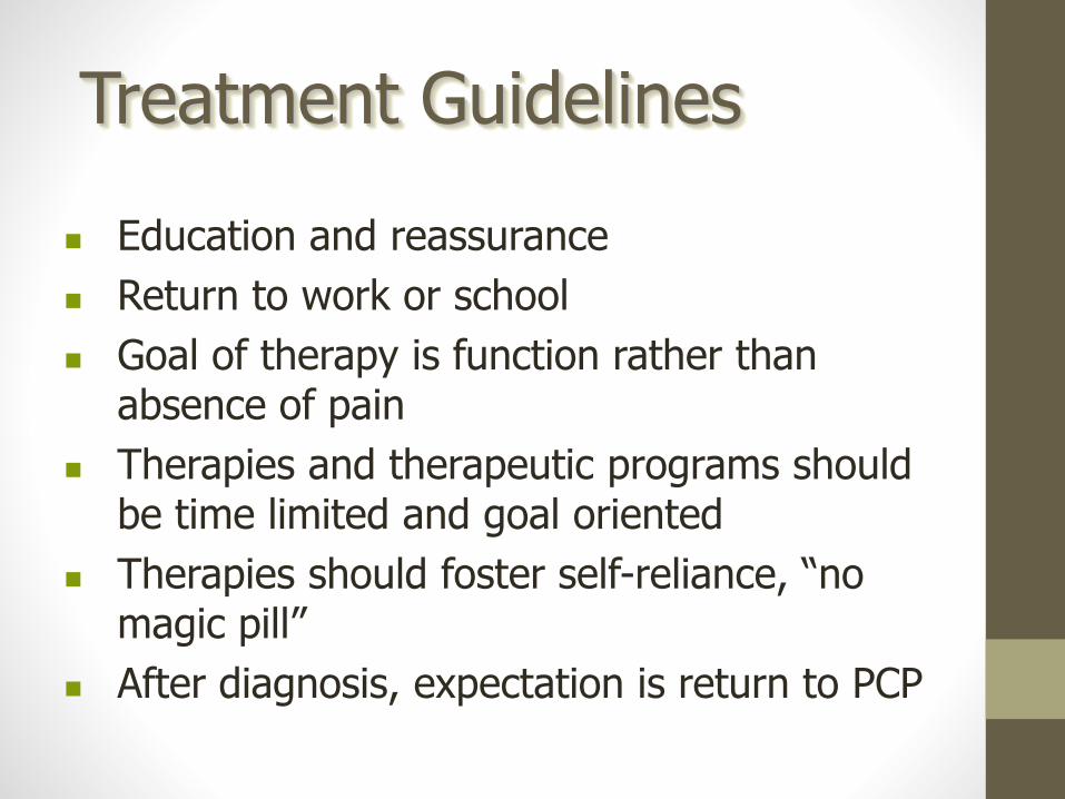
Treatment Guidelines
5.
Education and reassurance
Return to work or school
Goal of therapy is function rather than absence of pain
Therapies and therapeutic programs should be time limited and goal oriented
Therapies should foster self-reliance, “no magic pill”
After diagnosis, expectation is return to PCP
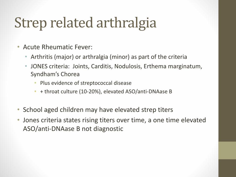
Strep related arthralgia
• Acute Rheumatic Fever:
• Arthritis (major) or arthralgia (minor) as part of the criteria
• JONES criteria: Joints, Carditis, Nodulosis, Erthema marginatum, Syndham’s Chorea
• Plus evidence of streptococcal disease
• + throat culture (10-20%), elevated ASO/anti-DNAase B
• School aged children may have elevated strep titers
• Jones criteria states rising titers over time, a one time elevated ASO/anti-DNAase B not diagnostic
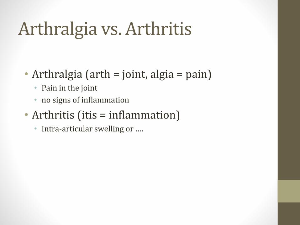
Arthralgia vs. Arthritis
• Arthralgia (arth = joint, algia = pain)• Pain in the joint
• no signs of inflammation
• Arthritis (itis = inflammation)• Intra-articular swelling or ….
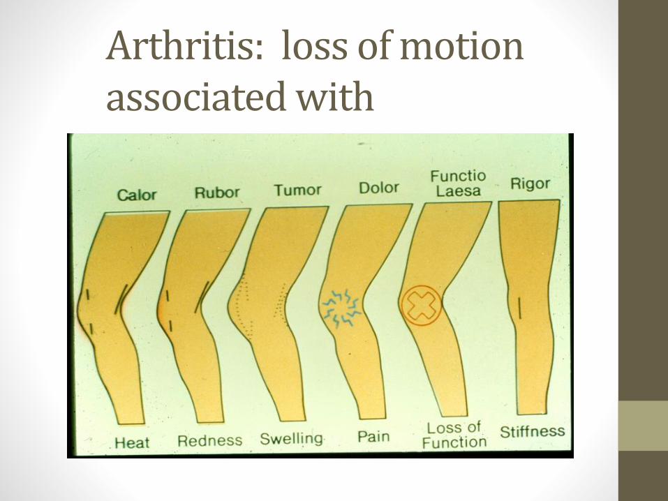
Arthritis: loss of motion associated with
Juvenile Idiopathic Arthritis
• Arthritis lasting at least 6 weeks in a child less than 16 years of age
• No other cause found for the arthritis
• No diagnostic blood tests
• Physical exam crucial for diagnosis
• Essentially a Dx of exclusion
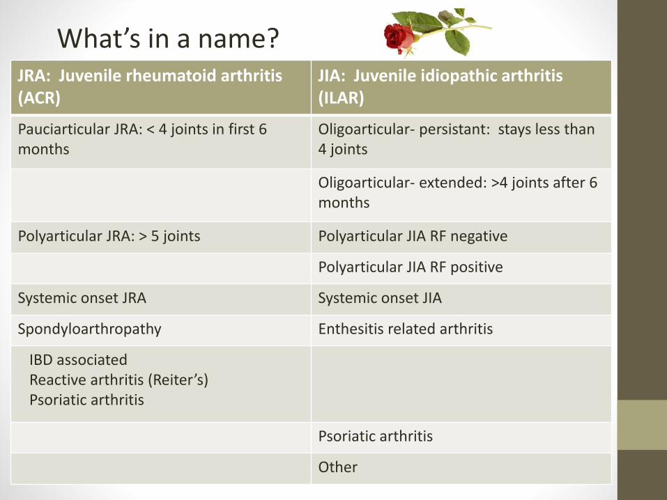
JRA: Juvenile rheumatoid arthritis (ACR)
JIA: Juvenile idiopathic arthritis (ILAR)
Pauciarticular JRA: < 4 joints in first 6 months
Oligoarticular- persistant: stays less than4 joints
Oligoarticular- extended: >4 joints after 6 months
Polyarticular JRA: > 5 joints Polyarticular JIA RF negative
Polyarticular JIA RF positive
Systemic onset JRA Systemic onset JIA
Spondyloarthropathy Enthesitis related arthritis
IBD associatedReactive arthritis (Reiter’s)Psoriatic arthritis
Psoriatic arthritis
Other
What’s in a name?
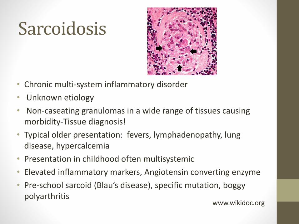
• Chronic multi-system inflammatory disorder
• Unknown etiology
• Non-caseating granulomas in a wide range of tissues causing morbidity-Tissue diagnosis!
• Typical older presentation: fevers, lymphadenopathy, lung disease, hypercalcemia
• Presentation in childhood often multisystemic
• Elevated inflammatory markers, Angiotensin converting enzyme
• Pre-school sarcoid (Blau’s disease), specific mutation, boggy polyarthritis
Sarcoidosis
www.wikidoc.org
• Sites of involvement:• thoracic lymph nodes
• lungs
• liver/ spleen
• eyes
• bones/joints
• salivary and lacrimal glands
• central nervous system
• skin (papular rash)
• Boys = girls
• Incidence (US) 5/100,000 Caucasians, 40/100,000 in African-Americans
• Southeastern United States is an endemic area.
Sarcoidosis
DeRemee RA: Concise review for primary care physicians: sarcoidosis, Mayo Clin Proc, 70:177-181, 1995.
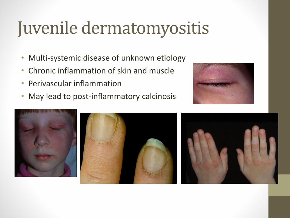
• Multi-systemic disease of unknown etiology
• Chronic inflammation of skin and muscle
• Perivascular inflammation
• May lead to post-inflammatory calcinosis
Juvenile dermatomyositis
Criteria of Bohan and Peter
Characteristic rash
Symmetric proximal weakness
Elevation of one or more of muscle derived serological enzymes
EMG demonstration of myopathy and denervation
Muscle biopsy with necrosis, and inflammation
MUST HAVE CUTANEOUS + 3/4 OTHER CRITERIA - (rarely no cutaneous, all 4 other criteria = polymyositis)
Bohan A, Peter JB. NEJM 292: 344, 403, 1975
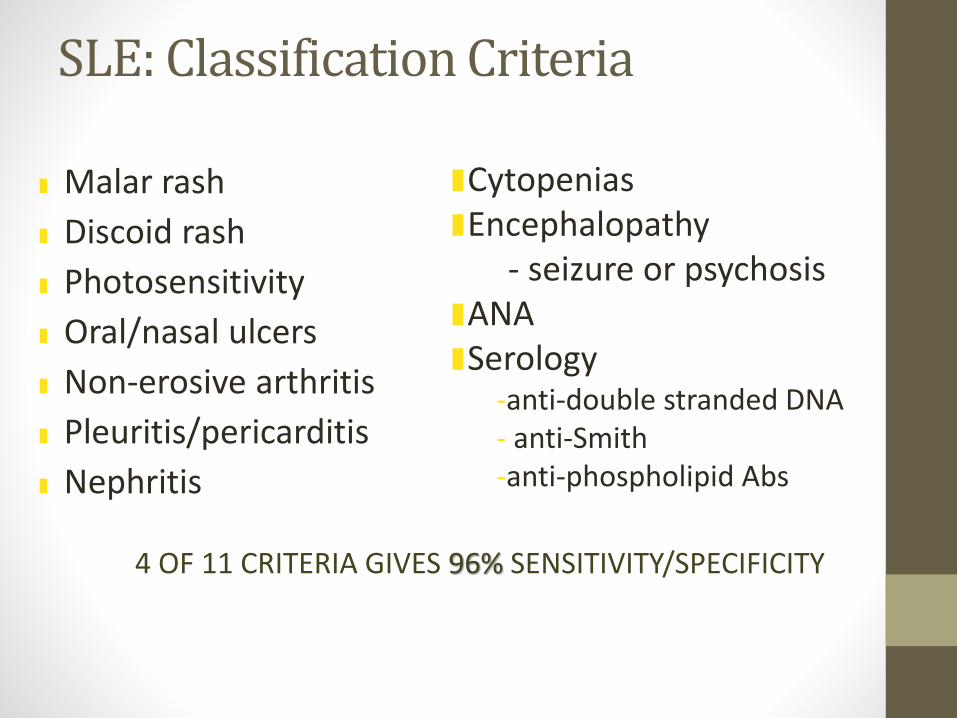
SLE: Classification Criteria
▮ Malar rash
▮ Discoid rash
▮ Photosensitivity
▮ Oral/nasal ulcers
▮ Non-erosive arthritis
▮ Pleuritis/pericarditis
▮ Nephritis
▮Cytopenias▮Encephalopathy
- seizure or psychosis▮ANA▮Serology
-anti-double stranded DNA- anti-Smith-anti-phospholipid Abs
4 OF 11 CRITERIA GIVES 96% SENSITIVITY/SPECIFICITY
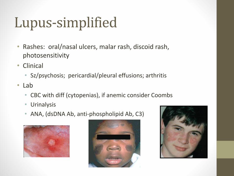
Lupus-simplified
• Rashes: oral/nasal ulcers, malar rash, discoid rash, photosensitivity
• Clinical
• Sz/psychosis; pericardial/pleural effusions; arthritis
• Lab
• CBC with diff (cytopenias), if anemic consider Coombs
• Urinalysis
• ANA, (dsDNA Ab, anti-phospholipid Ab, C3)
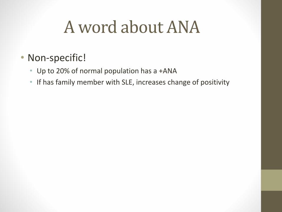
A word about ANA
• Non-specific!• Up to 20% of normal population has a +ANA
• If has family member with SLE, increases change of positivity
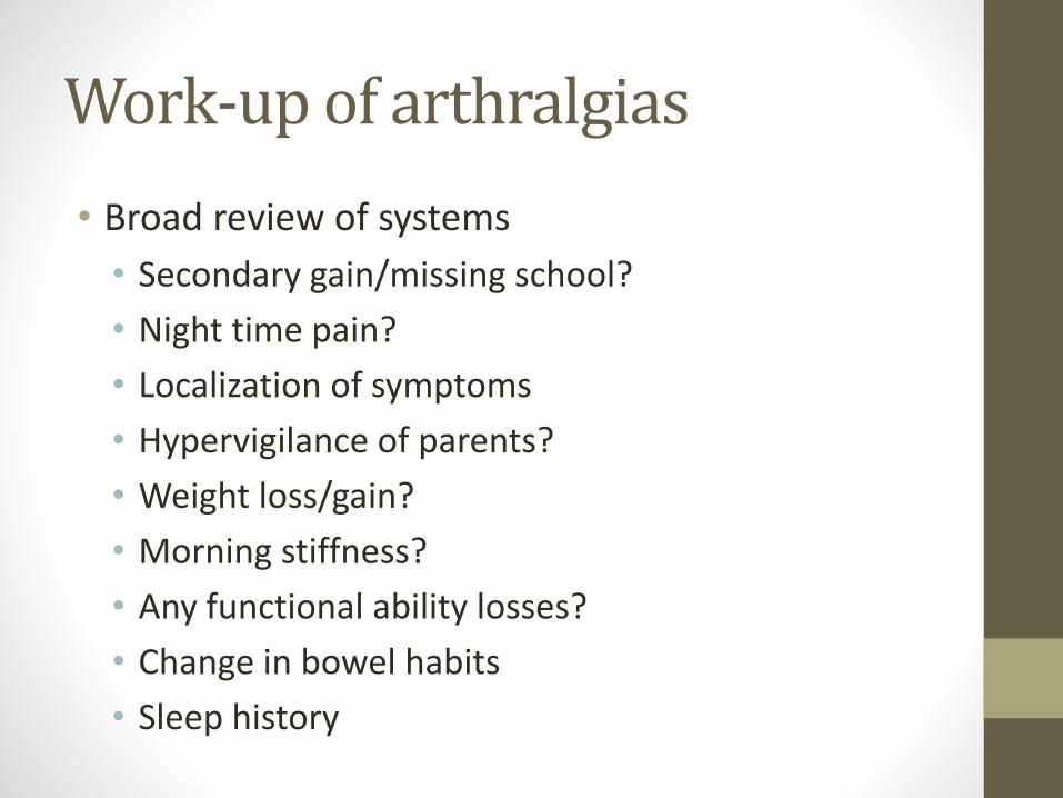
Work-up of arthralgias
• Broad review of systems
• Secondary gain/missing school?
• Night time pain?
• Localization of symptoms
• Hypervigilance of parents?
• Weight loss/gain?
• Morning stiffness?
• Any functional ability losses?
• Change in bowel habits
• Sleep history
Complete physical exam
• Vitals: Weight loss, poor growth
• Point tenderness
• Weakness? Quick strength exam
• Rash?
• Affect of child (la belle indifference, hypersensitivity, allodynia)
• Arthritis?
Lab work up of arthralgias
• CBC with diff
• Sed rate, CRP: CRP up compared to sed rate- infection!
• Consider: urinalysis
• Consider: uric acid/LDH
• Consider: ASO/anti-DNAse B
• Consider: Thyroid studies, Celiac panel
• Consider: Infectious work up
• EBV, parvo, varicella, measles all arthogenic infections
Lab work up of arthralgias
• Consider: Angiotensin converting enzyme (sarcoid)
• Consider: ANA, dsDNA Ab, C3, ENA’s (Ro, La, Sm, RNP), anti-cardiolipin Ab, U/A (for work up of lupus)
• Remember, no diagnostic tests for JIA
• ANA- <50% sensitivity, prognostic marker for uveitis in JIA
• RF present in <10% of children with arthritis
• Obtain ANA if suspicious for SLE
Refer when:
• There is arthritis on exam
• History, physical and labs consistent with rheumatic disease
• Lupus, dermatomyositis, JIA, sarcoid
• You need help with an amplified pain syndrome patient
• We are always available for phone consultation!

QUESTIONS?