Embed Size (px)
Citation preview
Ceramic Endocrown vs Ceramic Onlay with Resin Core in Endodontically Treated Teeth: A Finite Element Analysis
João Nuno Campante Moreira Prina
Orientador: Professor Doutor Paulo Jorge Palma
Coorientador: Mestre Dr. Rui Isidro Falacho
Mestrado Integrado em Medicina Dentária
Faculdade de Medicina da Universidade de Coimbra
Coimbra, 2016

CeramicEndocrownvsCeramicOnlaywithResinCoreinEndodonticallyTreatedTeeth:AFiniteElementAnalysis
JoãoNunoCampanteMoreiraPrina 1
Ceramic Endocrown vs Ceramic Onlay with Resin Core in Endodontically Treated Teeth: A Finite Element Analysis
Prina, J*, Falacho, RI**, Palma, PJ***
* Aluno do 5º ano do Mestrado Integrado em Medicina Dentária da Faculdade de Medicina
da Universidade de Coimbra
** Assistente Convidado do Mestrado Integrado em Medicina Dentária da Faculdade de
Medicina da Universidade de Coimbra
*** Professor Auxiliar Convidado do Mestrado Integrado em Medicina Dentária da Faculdade
de Medicina da Universidade de Coimbra
Endereço:
Área da Medicina Dentária da Faculdade de Medicina da Universidade de Coimbra
Avenida Bissaya Barreto, Bloco de Celas
3000-075 Coimbra
Telefone: +351 239484183 Fax: +351 239402910
Endereço de e-mail: [email protected]

CeramicEndocrownvsCeramicOnlaywithResinCoreinEndodonticallyTreatedTeeth:AFiniteElementAnalysis
JoãoNunoCampanteMoreiraPrina 2
Resumo
Introdução: Os dentes não vitais apresentam certas características que os fragilizam,
diminuindo deste modo a sua resistência. Esta fragilidade está intimamente ligada a perda
de tecido dentário, que pode ser resultante de trauma, cárie ou mesmo na terapêutica
endodôntica. Neste âmbito, no procedimento endodôntico pode haver a uma remoção
aumentada de tecido dentário, não só na zona coronária aquando da execução de acesso
coronário, mas também num acesso canalar direto ao 1/3 apical, podendo pode ser
necessário proceder a remoção de dentina no 1/3 cervical radicular. Posto isto, a
restauração deste tipo de dentes é altamente discutida na literatura, sendo que existem
várias abordagens possíveis, dentro das quais as Endocrowns e os Onlays. Este estudo
baseia-se numa análise de Elementos Finitos (EF) em modelo 3D de um primeiro prémolar
maxilar.
Objetivos: O objectivo deste estudo é comparar, num modelo de EF a distribuição de stress
entre duas possíveis abordagens para restauração de dentes com tratamento endodôntico,
Endocrown ou Onlay com um build-up em resina composta.
Materiais e métodos: O modelo do prémolar com dois canais radiculares foi isolado, tendo
sido feitos cortes de acordo com o tipo de cavidade necessária, com o objetivo de simular
um dente com uma grande destruição coronária, apenas com a parede vestibular e canais
obturados com guta-percha. Simulou-se posteriormente a restauração do dente com uma
Endocrown totalmente cerâmica e com um Onlay cerâmico com um core em resina
composta. Foram aplicadas três intensidades de força (200, 500 e 800 Newtons) com 2
inclinações diferentes (11º e 45º) em relação ao longo eixo do dente, na face oclusal do
modelo com uma esfera metálica de 4 mm.
Resultados: Neste estudo foram comparadas as distribuições de stress e os valores
máximos de stress no tecido dentário e nos materiais restauradores (esmalte, dentina,
cerâmica e resina composta) e apenas no tecido dentário (esmalte e dentina). Foi possível
observar uma maior concentração de stress em forças de maior intensidade com uma
inclinação de 45º em ambos os modelos. A Endocrown obteve maiores valores de stress em
todos os testes, excepto aquando da análise dos valores no esmalte e dentina com a
aplicação da força a 11º.
Conclusões: Apesar das limitações deste estudo podemos concluir que forças com um
ângulo de 45º com o longo eixo do dente geram maiores valores de stress no dente,
comparando com forças a 11º. É possível também concluir que quando estas forças mais
destrutivas são aplicadas, a restauração através de Onlays apresenta melhores resultados
do que a restauração com Endocrowns.
Palavras-chave: Endocrown ; Onlay ; Elementos Finitos ; Restodontics.
CeramicEndocrownvsCeramicOnlaywithResinCoreinEndodonticallyTreatedTeeth:AFiniteElementAnalysis
JoãoNunoCampanteMoreiraPrina 3
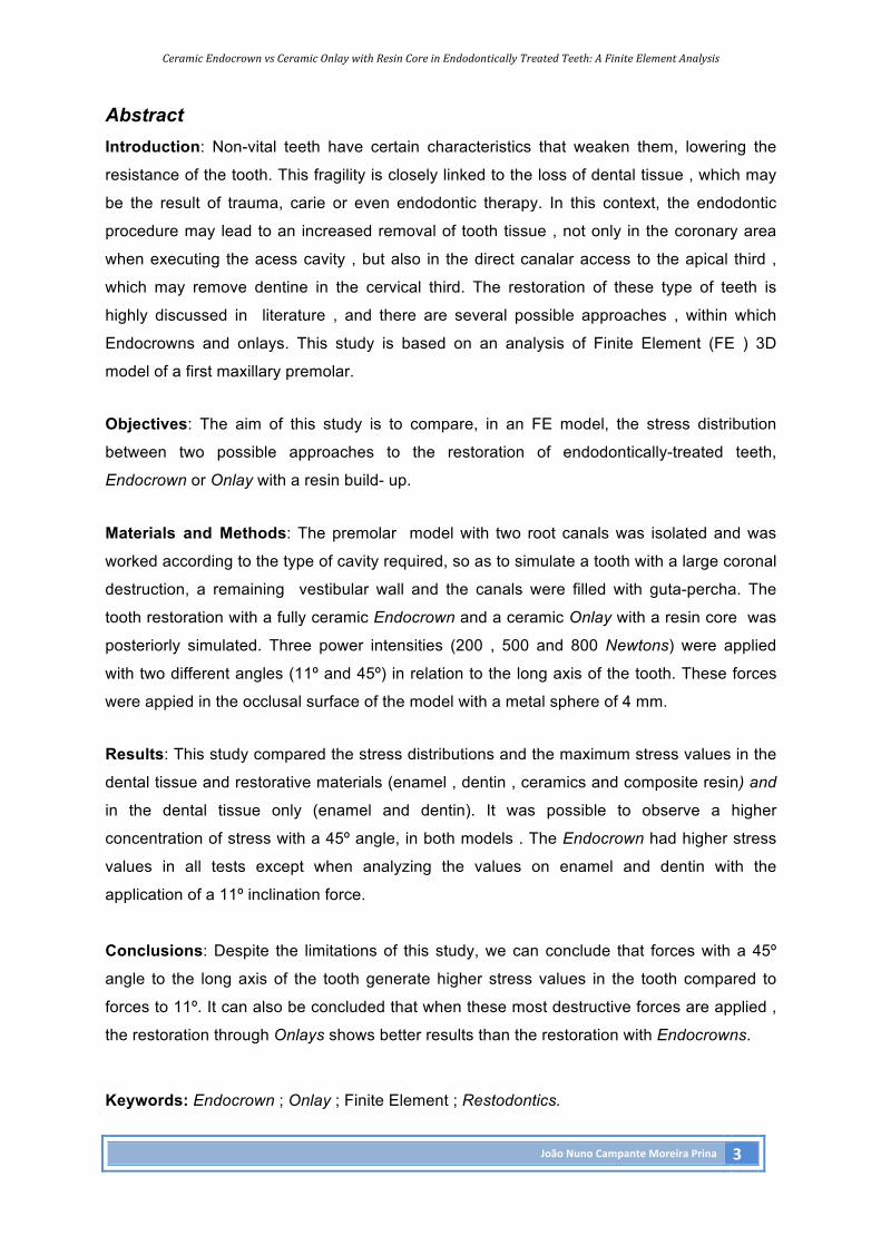
Abstract Introduction: Non-vital teeth have certain characteristics that weaken them, lowering the
resistance of the tooth. This fragility is closely linked to the loss of dental tissue , which may
be the result of trauma, carie or even endodontic therapy. In this context, the endodontic
procedure may lead to an increased removal of tooth tissue , not only in the coronary area
when executing the acess cavity , but also in the direct canalar access to the apical third ,
which may remove dentine in the cervical third. The restoration of these type of teeth is
highly discussed in literature , and there are several possible approaches , within which
Endocrowns and onlays. This study is based on an analysis of Finite Element (FE ) 3D
model of a first maxillary premolar.
Objectives: The aim of this study is to compare, in an FE model, the stress distribution
between two possible approaches to the restoration of endodontically-treated teeth,
Endocrown or Onlay with a resin build- up.
Materials and Methods: The premolar model with two root canals was isolated and was
worked according to the type of cavity required, so as to simulate a tooth with a large coronal
destruction, a remaining vestibular wall and the canals were filled with guta-percha. The
tooth restoration with a fully ceramic Endocrown and a ceramic Onlay with a resin core was
posteriorly simulated. Three power intensities (200 , 500 and 800 Newtons) were applied
with two different angles (11º and 45º) in relation to the long axis of the tooth. These forces
were appied in the occlusal surface of the model with a metal sphere of 4 mm.
Results: This study compared the stress distributions and the maximum stress values in the
dental tissue and restorative materials (enamel , dentin , ceramics and composite resin) and
in the dental tissue only (enamel and dentin). It was possible to observe a higher
concentration of stress with a 45º angle, in both models . The Endocrown had higher stress
values in all tests except when analyzing the values on enamel and dentin with the
application of a 11º inclination force.
Conclusions: Despite the limitations of this study, we can conclude that forces with a 45º
angle to the long axis of the tooth generate higher stress values in the tooth compared to
forces to 11º. It can also be concluded that when these most destructive forces are applied ,
the restoration through Onlays shows better results than the restoration with Endocrowns.
Keywords: Endocrown ; Onlay ; Finite Element ; Restodontics.
CeramicEndocrownvsCeramicOnlaywithResinCoreinEndodonticallyTreatedTeeth:AFiniteElementAnalysis
JoãoNunoCampanteMoreiraPrina 4
Index
Introduction 5
Materials and Methods 7 Finite Element model generation 7
Finite Element Analyses 11
Results 12 Stress distribution in enamel and dentin 13
Stress distribution on the tooth and restorative material 19
Discussion 26
Conclusion 29
Acknowledgements 30
Attachments 31
Bibliographic References 32
CeramicEndocrownvsCeramicOnlaywithResinCoreinEndodonticallyTreatedTeeth:AFiniteElementAnalysis
JoãoNunoCampanteMoreiraPrina 5
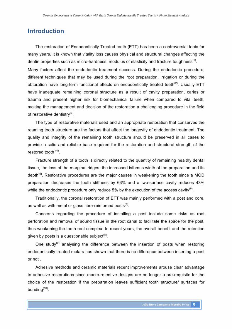
Introduction The restoration of Endodontically Treated teeth (ETT) has been a controversial topic for
many years. It is known that vitality loss causes physical and structural changes affecting the
dentin properties such as micro-hardness, modulus of elasticity and fracture toughness(1).
Many factors affect the endodontic treatment success. During the endodontic procedure,
different techniques that may be used during the root preparation, irrigation or during the
obturation have long-term functional effects on endodontically treated teeth(2). Usually ETT
have inadequate remaining coronal structure as a result of cavity preparation, caries or
trauma and present higher risk for biomechanical failure when compared to vital teeth,
making the management and decision of the restoration a challenging procedure in the field
of restorative dentistry(3).
The type of restorative materials used and an appropriate restoration that conserves the
reaming tooth structure are the factors that affect the longevity of endodontic treatment. The
quality and integrity of the remaining tooth structure should be preserved in all cases to
provide a solid and reliable base required for the restoration and structural strength of the
restored tooth (4).
Fracture strength of a tooth is directly related to the quantity of remaining healthy dental
tissue, the loss of the marginal ridges, the increased isthmus width of the preparation and its
depth(5). Restorative procedures are the major causes in weakening the tooth since a MOD
preparation decreases the tooth stiffness by 63% and a two-surface cavity reduces 43%
while the endodontic procedure only reduce 5% by the execution of the access cavity(6).
Traditionally, the coronal restoration of ETT was mainly performed with a post and core,
as well as with metal or glass fibre-reinforced posts(7).
Concerns regarding the procedure of installing a post include some risks as root
perforation and removal of sound tissue in the root canal to facilitate the space for the post,
thus weakening the tooth-root complex. In recent years, the overall benefit and the retention
given by posts is a questionable subject(8).
One study(9) analysing the difference between the insertion of posts when restoring
endodontically treated molars has shown that there is no difference between inserting a post
or not .
Adhesive methods and ceramic materials recent improvements arouse clear advantage
to adhesive restorations since macro-retentive designs are no longer a pre-requisite for the
choice of the restoration if the preparation leaves sufficient tooth structure/ surfaces for
bonding(10).
CeramicEndocrownvsCeramicOnlaywithResinCoreinEndodonticallyTreatedTeeth:AFiniteElementAnalysis
JoãoNunoCampanteMoreiraPrina 6
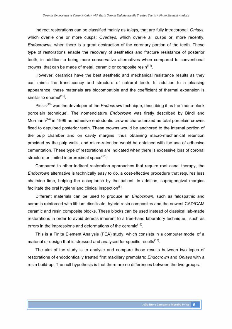
Indirect restorations can be classified mainly as Inlays, that are fully intracoronal; Onlays,
which overlie one or more cusps; Overlays, which overlie all cusps or, more recently,
Endocrowns, when there is a great destruction of the coronary portion of the teeth. These
type of restorations enable the recovery of aesthetics and fracture resistance of posterior
teeth, in addition to being more conservative alternatives when compared to conventional
crowns, that can be made of metal, ceramic or composite resin(11).
However, ceramics have the best aesthetic and mechanical resistance results as they
can mimic the translucency and structure of natrural teeth. In addition to a pleasing
appearance, these materials are biocompatible and the coefficient of thermal expansion is
similar to enamel(12).
Pissis(13) was the developer of the Endocrown technique, describing it as the ‘mono-block
porcelain technique’. The nomenclature Endocrown was firstly described by Bindl and
Mormann(14) in 1999 as adhesive endodontic crowns characterized as total porcelain crowns
fixed to depulped posterior teeth. These crowns would be anchored to the internal portion of
the pulp chamber and on cavity margins, thus obtaining macro-mechanical retention
provided by the pulp walls, and micro-retention would be obtained with the use of adhesive
cementation. These type of restorations are indicated when there is excessive loss of coronal
structure or limited interproximal space(15).
Compared to other indirect restoration approaches that require root canal therapy, the
Endocrown alternative is technically easy to do, a cost-effective procedure that requires less
chairside time, helping the acceptance by the patient. In addition, supragengival margins
facilitate the oral hygiene and clinical inspection(8).
Different materials can be used to produce an Endocrown, such as feldspathic and
ceramic reinforced with lithium dissilicate, hybrid resin composites and the newest CAD/CAM
ceramic and resin composite blocks. These blocks can be used instead of classical lab-made
restorations in order to avoid defects inherent to a free-hand laboratory technique, such as
errors in the impressions and deformations of the ceramic(16).
This is a Finite Element Analysis (FEA) study, which consists in a computer model of a
material or design that is stressed and analysed for specific results(17).
The aim of the study is to analyse and compare those results between two types of
restorations of endodontically treated first maxillary premolars: Endocrown and Onlays with a
resin build-up. The null hypothesis is that there are no differences between the two groups.
CeramicEndocrownvsCeramicOnlaywithResinCoreinEndodonticallyTreatedTeeth:AFiniteElementAnalysis
JoãoNunoCampanteMoreiraPrina 7
Materials and Methods
Finite Element model generation
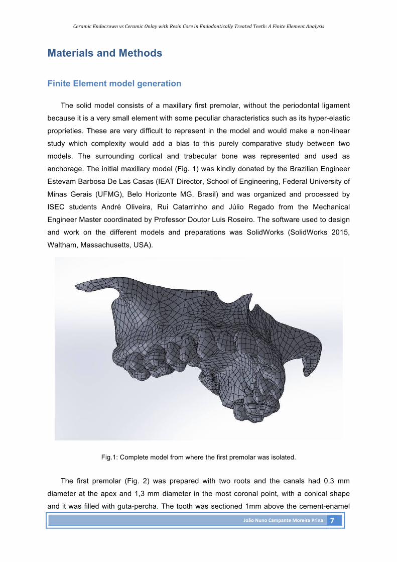
The solid model consists of a maxillary first premolar, without the periodontal ligament
because it is a very small element with some peculiar characteristics such as its hyper-elastic
proprieties. These are very difficult to represent in the model and would make a non-linear
study which complexity would add a bias to this purely comparative study between two
models. The surrounding cortical and trabecular bone was represented and used as
anchorage. The initial maxillary model (Fig. 1) was kindly donated by the Brazilian Engineer
Estevam Barbosa De Las Casas (IEAT Director, School of Engineering, Federal University of
Minas Gerais (UFMG), Belo Horizonte MG, Brasil) and was organized and processed by
ISEC students André Oliveira, Rui Catarrinho and Júlio Regado from the Mechanical
Engineer Master coordinated by Professor Doutor Luis Roseiro. The software used to design
and work on the different models and preparations was SolidWorks (SolidWorks 2015,
Waltham, Massachusetts, USA).
Fig.1: Complete model from where the first premolar was isolated.
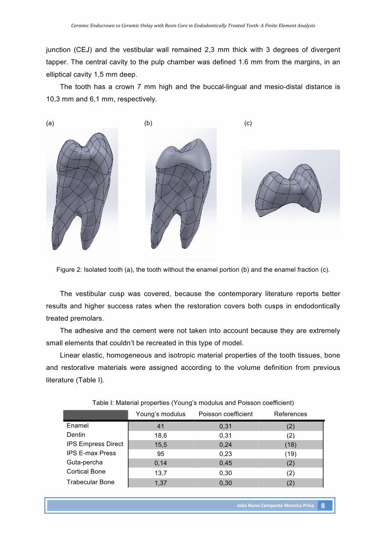
The first premolar (Fig. 2) was prepared with two roots and the canals had 0.3 mm
diameter at the apex and 1,3 mm diameter in the most coronal point, with a conical shape
and it was filled with guta-percha. The tooth was sectioned 1mm above the cement-enamel
CeramicEndocrownvsCeramicOnlaywithResinCoreinEndodonticallyTreatedTeeth:AFiniteElementAnalysis
JoãoNunoCampanteMoreiraPrina 8
junction (CEJ) and the vestibular wall remained 2,3 mm thick with 3 degrees of divergent
tapper. The central cavity to the pulp chamber was defined 1.6 mm from the margins, in an
elliptical cavity 1,5 mm deep.
The tooth has a crown 7 mm high and the buccal-lingual and mesio-distal distance is
10,3 mm and 6,1 mm, respectively.
(a) (b) (c)
Figure 2: Isolated tooth (a), the tooth without the enamel portion (b) and the enamel fraction (c).
The vestibular cusp was covered, because the contemporary literature reports better
results and higher success rates when the restoration covers both cusps in endodontically
treated premolars.
The adhesive and the cement were not taken into account because they are extremely
small elements that couldn’t be recreated in this type of model.
Linear elastic, homogeneous and isotropic material properties of the tooth tissues, bone
and restorative materials were assigned according to the volume definition from previous
literature (Table I).
Table I: Material properties (Young’s modulus and Poisson coefficient)
Young’s modulus Poisson coefficient References
Enamel 41 0,31 (2) Dentin 18,6 0,31 (2) IPS Empress Direct 15,5 0,24 (18) IPS E-max Press 95 0,23 (19) Guta-percha 0,14 0,45 (2) Cortical Bone 13,7 0,30 (2) Trabecular Bone 1,37 0,30 (2)

CeramicEndocrownvsCeramicOnlaywithResinCoreinEndodonticallyTreatedTeeth:AFiniteElementAnalysis
JoãoNunoCampanteMoreiraPrina 9
A convergence test was made resulting in a Solid Mesh model (Fig. 3) with a curvature
based mesh type with 4 Jacobian points. The size of the maximum element is 1,5 mm and
the minimum element is 0,3 mm with high quality and 3 degrees of freedom, finally resulting
in a model with 76,997 elements and 118,475 nodes. This model has a 96,1% element
percentage, which makes this a reliable study (Table II).
Fig.3: Mesh of the experimental model. Table II: Characteristics of the mesh model.
In this study 3 models were created:
Model 1: Sound tooth.
Enamel
Dentin
Pulp
Cortical Bone
Trabecular Bone
Fig.4: Scheme of model 1.
CeramicEndocrownvsCeramicOnlaywithResinCoreinEndodonticallyTreatedTeeth:AFiniteElementAnalysis
JoãoNunoCampanteMoreiraPrina 10
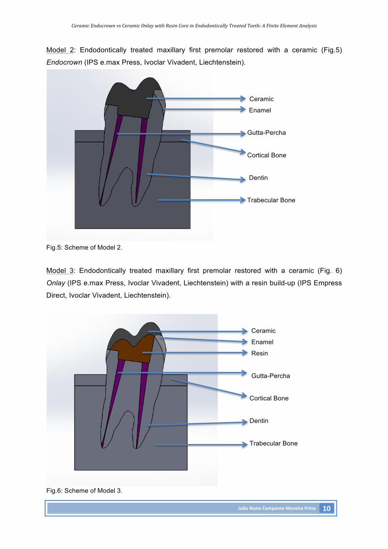
Model 2: Endodontically treated maxillary first premolar restored with a ceramic (Fig.5)
Endocrown (IPS e.max Press, Ivoclar Vivadent, Liechtenstein).
Ceramic
Enamel
Gutta-Percha
Cortical Bone
Dentin
Trabecular Bone
Fig.5: Scheme of Model 2.
Model 3: Endodontically treated maxillary first premolar restored with a ceramic (Fig. 6)
Onlay (IPS e.max Press, Ivoclar Vivadent, Liechtenstein) with a resin build-up (IPS Empress
Direct, Ivoclar Vivadent, Liechtenstein).
Ceramic
Enamel
Resin
Gutta-Percha
Cortical Bone
Dentin
Trabecular Bone
Fig.6: Scheme of Model 3.
CeramicEndocrownvsCeramicOnlaywithResinCoreinEndodonticallyTreatedTeeth:AFiniteElementAnalysis
JoãoNunoCampanteMoreiraPrina 11
Finite Element Analyses
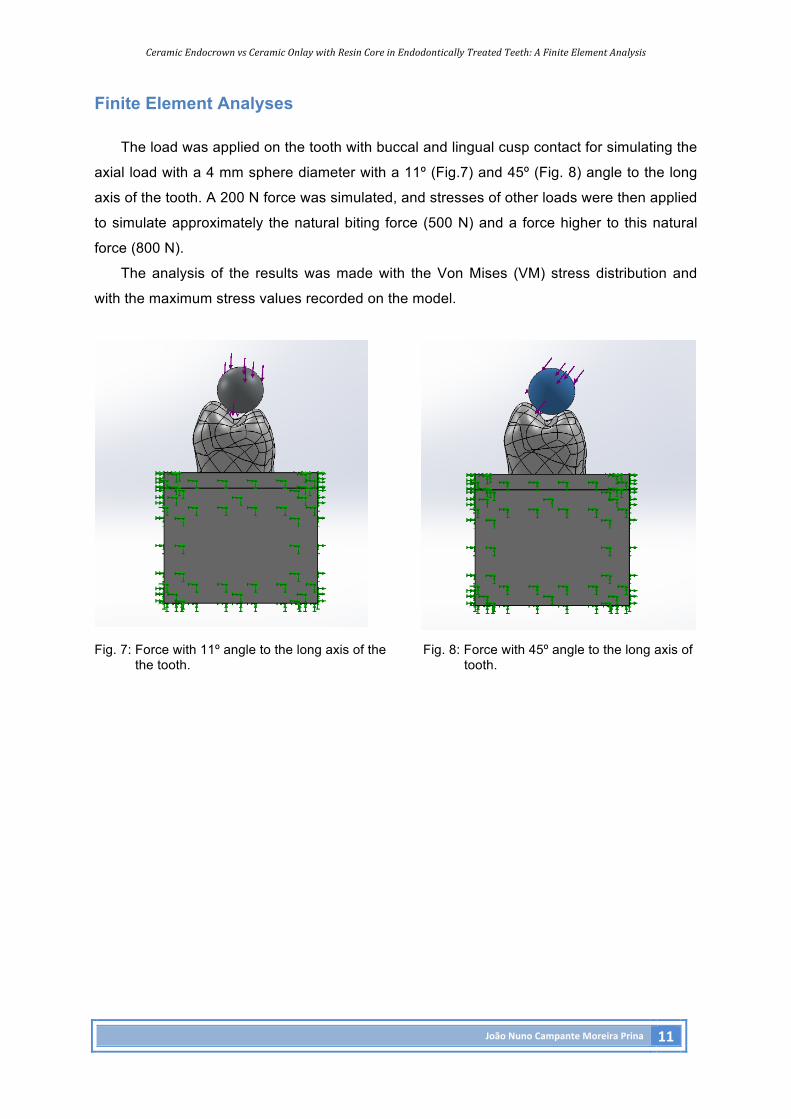
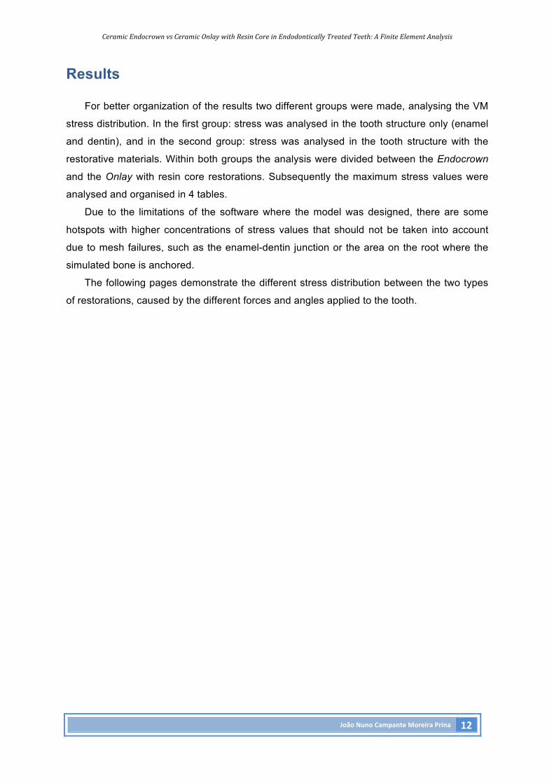
The load was applied on the tooth with buccal and lingual cusp contact for simulating the
axial load with a 4 mm sphere diameter with a 11º (Fig.7) and 45º (Fig. 8) angle to the long
axis of the tooth. A 200 N force was simulated, and stresses of other loads were then applied
to simulate approximately the natural biting force (500 N) and a force higher to this natural
force (800 N).
The analysis of the results was made with the Von Mises (VM) stress distribution and
with the maximum stress values recorded on the model.
Fig. 7: Force with 11º angle to the long axis of the Fig. 8: Force with 45º angle to the long axis of the tooth. tooth.
CeramicEndocrownvsCeramicOnlaywithResinCoreinEndodonticallyTreatedTeeth:AFiniteElementAnalysis
JoãoNunoCampanteMoreiraPrina 12
Results
For better organization of the results two different groups were made, analysing the VM
stress distribution. In the first group: stress was analysed in the tooth structure only (enamel
and dentin), and in the second group: stress was analysed in the tooth structure with the
restorative materials. Within both groups the analysis were divided between the Endocrown
and the Onlay with resin core restorations. Subsequently the maximum stress values were
analysed and organised in 4 tables.
Due to the limitations of the software where the model was designed, there are some
hotspots with higher concentrations of stress values that should not be taken into account
due to mesh failures, such as the enamel-dentin junction or the area on the root where the
simulated bone is anchored.
The following pages demonstrate the different stress distribution between the two types
of restorations, caused by the different forces and angles applied to the tooth.
CeramicEndocrownvsCeramicOnlaywithResinCoreinEndodonticallyTreatedTeeth:AFiniteElementAnalysis
JoãoNunoCampanteMoreiraPrina 13
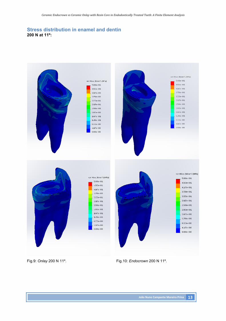
Stress distribution in enamel and dentin 200 N at 11º:
Fig.9: Onlay 200 N 11º. Fig.10: Endocrown 200 N 11º.
CeramicEndocrownvsCeramicOnlaywithResinCoreinEndodonticallyTreatedTeeth:AFiniteElementAnalysis
JoãoNunoCampanteMoreiraPrina 14
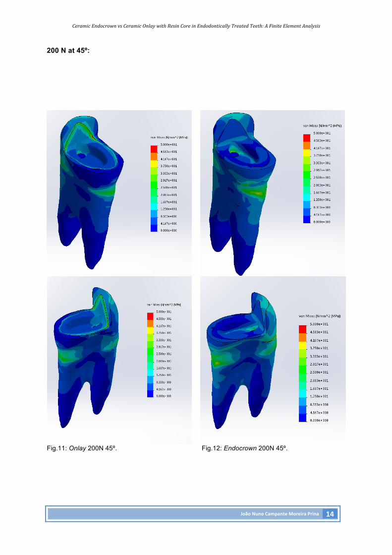
200 N at 45º:
Fig.11: Onlay 200N 45º. Fig.12: Endocrown 200N 45º.
CeramicEndocrownvsCeramicOnlaywithResinCoreinEndodonticallyTreatedTeeth:AFiniteElementAnalysis
JoãoNunoCampanteMoreiraPrina 15
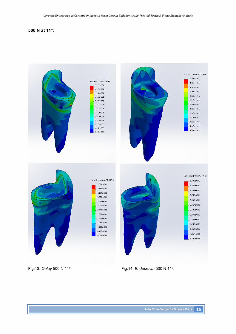
500 N at 11º:
Fig.13: Onlay 500 N 11º. Fig.14: Endocrown 500 N 11º.
CeramicEndocrownvsCeramicOnlaywithResinCoreinEndodonticallyTreatedTeeth:AFiniteElementAnalysis
JoãoNunoCampanteMoreiraPrina 16
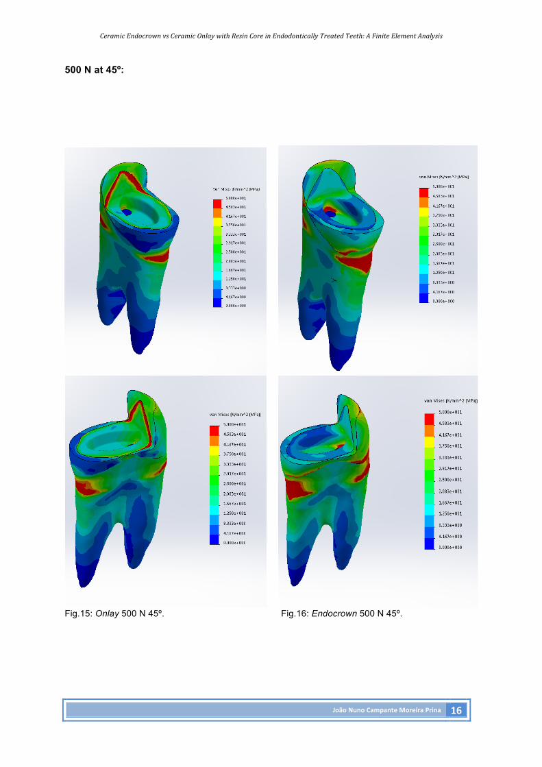
500 N at 45º:
Fig.15: Onlay 500 N 45º. Fig.16: Endocrown 500 N 45º.
CeramicEndocrownvsCeramicOnlaywithResinCoreinEndodonticallyTreatedTeeth:AFiniteElementAnalysis
JoãoNunoCampanteMoreiraPrina 17
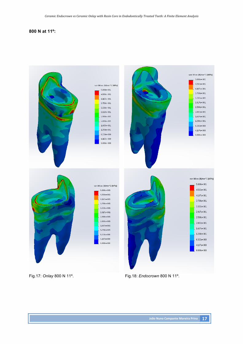
800 N at 11º:
Fig.17: Onlay 800 N 11º. Fig.18: Endocrown 800 N 11º.
CeramicEndocrownvsCeramicOnlaywithResinCoreinEndodonticallyTreatedTeeth:AFiniteElementAnalysis
JoãoNunoCampanteMoreiraPrina 18
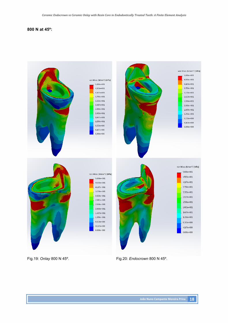
800 N at 45º:
Fig.19: Onlay 800 N 45º. Fig.20: Endocrown 800 N 45º.
CeramicEndocrownvsCeramicOnlaywithResinCoreinEndodonticallyTreatedTeeth:AFiniteElementAnalysis
JoãoNunoCampanteMoreiraPrina 19
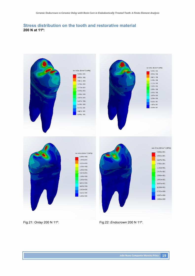
Stress distribution on the tooth and restorative material 200 N at 11º:
Fig.21: Onlay 200 N 11º. Fig.22: Endocrown 200 N 11º.
CeramicEndocrownvsCeramicOnlaywithResinCoreinEndodonticallyTreatedTeeth:AFiniteElementAnalysis
JoãoNunoCampanteMoreiraPrina 20
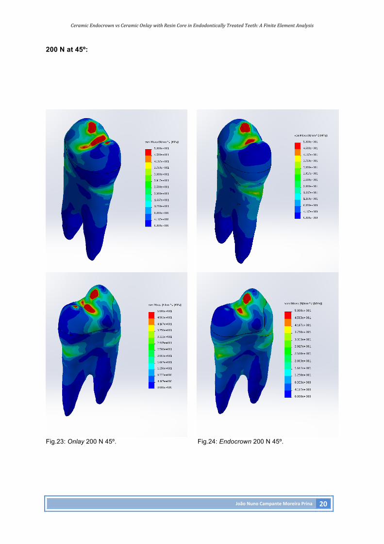
200 N at 45º:
Fig.23: Onlay 200 N 45º. Fig.24: Endocrown 200 N 45º.
CeramicEndocrownvsCeramicOnlaywithResinCoreinEndodonticallyTreatedTeeth:AFiniteElementAnalysis
JoãoNunoCampanteMoreiraPrina 21
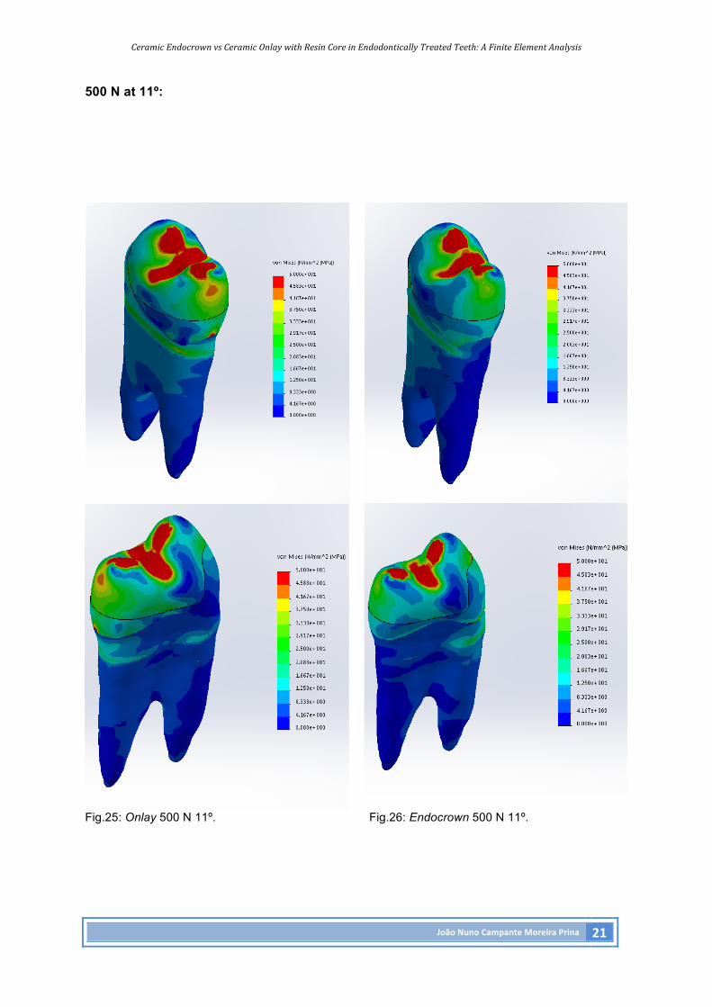
500 N at 11º:
Fig.25: Onlay 500 N 11º. Fig.26: Endocrown 500 N 11º.
CeramicEndocrownvsCeramicOnlaywithResinCoreinEndodonticallyTreatedTeeth:AFiniteElementAnalysis
JoãoNunoCampanteMoreiraPrina 22
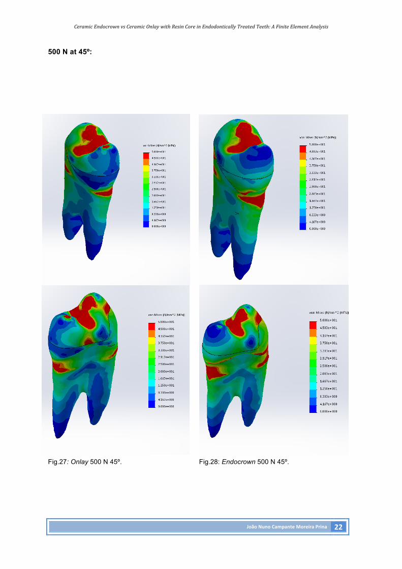
500 N at 45º:
Fig.27: Onlay 500 N 45º. Fig.28: Endocrown 500 N 45º.
CeramicEndocrownvsCeramicOnlaywithResinCoreinEndodonticallyTreatedTeeth:AFiniteElementAnalysis
JoãoNunoCampanteMoreiraPrina 23
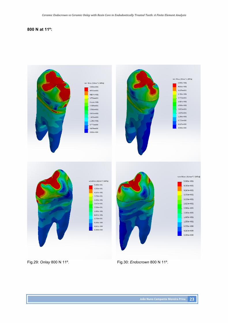
800 N at 11º:
Fig.29: Onlay 800 N 11º. Fig.30: Endocrown 800 N 11º.
CeramicEndocrownvsCeramicOnlaywithResinCoreinEndodonticallyTreatedTeeth:AFiniteElementAnalysis
JoãoNunoCampanteMoreiraPrina 24
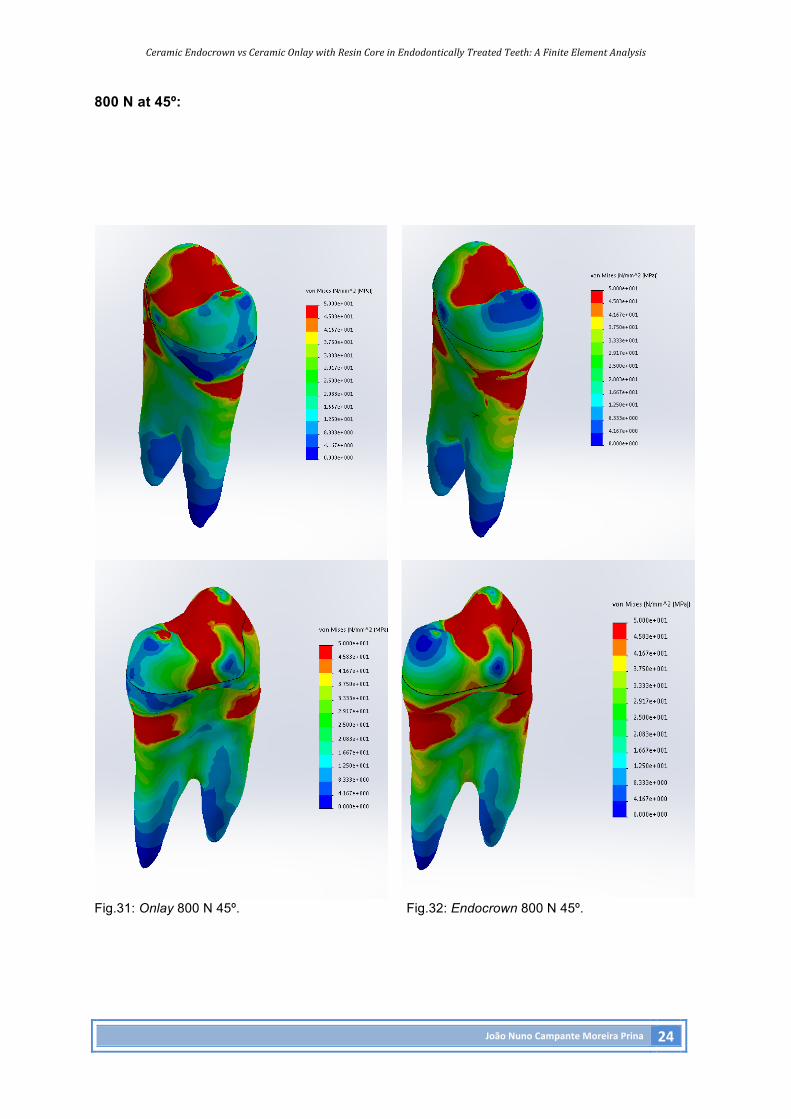
800 N at 45º:
Fig.31: Onlay 800 N 45º. Fig.32: Endocrown 800 N 45º.
CeramicEndocrownvsCeramicOnlaywithResinCoreinEndodonticallyTreatedTeeth:AFiniteElementAnalysis
JoãoNunoCampanteMoreiraPrina 25
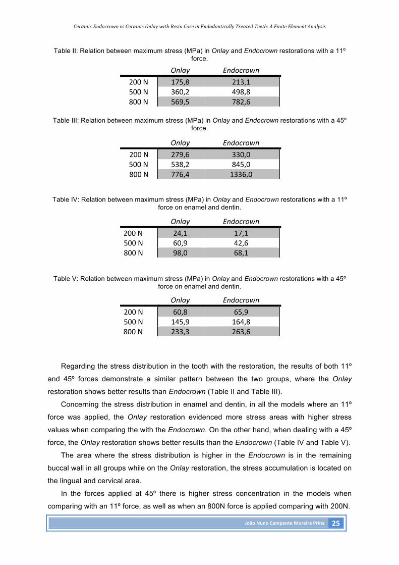
Table II: Relation between maximum stress (MPa) in Onlay and Endocrown restorations with a 11º force.
Table III: Relation between maximum stress (MPa) in Onlay and Endocrown restorations with a 45º
force.
Table IV: Relation between maximum stress (MPa) in Onlay and Endocrown restorations with a 11º
force on enamel and dentin.
Table V: Relation between maximum stress (MPa) in Onlay and Endocrown restorations with a 45º
force on enamel and dentin.
Regarding the stress distribution in the tooth with the restoration, the results of both 11º
and 45º forces demonstrate a similar pattern between the two groups, where the Onlay
restoration shows better results than Endocrown (Table II and Table III).
Concerning the stress distribution in enamel and dentin, in all the models where an 11º
force was applied, the Onlay restoration evidenced more stress areas with higher stress
values when comparing the with the Endocrown. On the other hand, when dealing with a 45º
force, the Onlay restoration shows better results than the Endocrown (Table IV and Table V).
The area where the stress distribution is higher in the Endocrown is in the remaining
buccal wall in all groups while on the Onlay restoration, the stress accumulation is located on
the lingual and cervical area.
In the forces applied at 45º there is higher stress concentration in the models when
comparing with an 11º force, as well as when an 800N force is applied comparing with 200N.
Onlay Endocrown200N 175,8 213,1500N 360,2 498,8800N 569,5 782,6
Onlay Endocrown200N 279,6 330,0500N 538,2 845,0800N 776,4 1336,0
Onlay Endocrown200N 24,1 17,1500N 60,9 42,6800N 98,0 68,1
Onlay Endocrown200N 60,8 65,9500N 145,9 164,8800N 233,3 263,6
CeramicEndocrownvsCeramicOnlaywithResinCoreinEndodonticallyTreatedTeeth:AFiniteElementAnalysis
JoãoNunoCampanteMoreiraPrina 26
Discussion
When an endodontic treatment is needed, the vitality loss has an impact in the physical
properties of dentin such as micro-hardness, modulus of elasticity and fracture resistance.
There are some changes in the tubule density that decrease towards the apex. The steps
from endodontic therapy such as the access cavity, the canal widening or the use of several
chemicals can, significantly, reduce the resistance of the tooth(1) and the literature reports the
absence of the marginal bridges as the main reason for the loss of structural strength(4).
Tooth fracture is a well-known concern for all dentists. This fracture can happen for two
reasons: iatrogenic causes such as loss of tooth structure, effect of chemicals or intra-canal
medication or problems in the restoration; and not iatrogenic causes such as anatomical
position of the tooth or the effect of age of tooth tissue(20).
According to what is reported in various studies, the conservation of remaining hard
tissue is crucial when dealing with non-vital teeth, as it improves the mechanical stability and
increases the available areas for making a good adhesion, which has a positive impact on
the long-term results of the treatment(21-23).
The introduction of adhesive techniques has revolutionized the restoration of
endodontically treated teeth, since it is no longer necessary to take the mechanical retention
into account , but instead rely on micromechanical and molecular retention provided by the
adhesive procedure. Bearing this in mind, the more area between the tooth and the
restoration (interface area), the higher probability of survival of the restoration(24).
It is reported in previous literature that, regardless of the restoration type used,
endodontically treated premolars can´t reach the fracture resistance of sound teeth.
However, there are ways to increase this resistance, such as cusp reduction and coverage
with the restoration. In a study made by Bitter et al. it is reported that the restoration of
cavities with remaining palatal and buccal wall using Onlays with cusp coverage is better
than with Inlays without it(21, 25).
The precision of the models is crucial for obtaining valid results in a Finite Element
Analysis (FEA) study. This type of study consists of a computer model of a material or design
in which a force is applied and analysed. FEA studies are an approximation to the reality,
since many details are idealized, simplified or ignored. The loading model is an
approximation of what happens in vivo in terms of boundary conditions or the material
properties. Still, FEA analysis models and simulations have been used for many years to
study the biomechanical behaviour of materials and structures where these variables are
impossible to measure directly. Moreover, many of the FEA studies already confirm the
laboratory ones. In this study, a 3-D model was created to evaluate the distribution of
functional stress between two types of restorations, Endocrown and an Onlay with a resin
CeramicEndocrownvsCeramicOnlaywithResinCoreinEndodonticallyTreatedTeeth:AFiniteElementAnalysis
JoãoNunoCampanteMoreiraPrina 27
build-up. This type of model is better than a 2-D model, which may not represent the tooth
irregularities and may neglect several important details(17).
The VM stress distribution was used in this study to analyse the images, as it is the
combination of the absolute values squared of all stresses and it is reported in most of the
previously published studies. This type of stress (VM) is widely used as an indicator of the
possible damage that can occur on a material. Another parameter also used in this study to
analyse the results was the maximum stress values, because when it comes to brittle
materials such as bone or ceramics, it is suitable for better indicating the magnitude of stress
concentrations and allowing the comparison with the ultimate compressive and ultimate
tensile strengths of a material(17, 26).
Analysing the results of this study regarding the stress distribution in the restored
models, the Onlay configuration showed better outcomes at 11º and 45º. The results
observed on enamel and dentin with a 45º force may occur because the resin build-up better
distributes or absorbs the stress caused by the force applied to the tooth than a ceramic
monoblock Endocrown would without this resin layer. In a in vitro study made by Magne et al.
it is concluded that the use of a small composite resin build-up may be useful because it can
provide enhanced geometry, remove undercuts from the endodontic preparation and
facilitate provisionalization when it is needed(27). Another study form Rocca et al.
demonstrates that the insertion of a resin-coating layer may reduce the risk of extensive
fractures and improve the success rate on non-vital teeth(28).
Since high stiffness materials like ceramics generate high stress values with a negative
influence in the biomechanical behaviour of the restorative system when used to replace
dentin, the use of low stiffness materials as composite resins that accompany the natural
flexure of the dentin, reduce the stress. This type of materials seems to be a reliable strategy
to generate low stress values when used a build-up(29).
Regarding the force application at 11º and the stress distribution on the enamel and
dentin, the Endocrown shows better results when comparing to he Onlay configuration,
which goes along with the results of a study made by Lin et al.(3), and it can be good when
restoring molars(30) because the angle of the forces applied on that type of teeth is closer to
this angle. These results may be due to the fact that the Endocrown presents some
advantages in reducing the effect of multiple interfaces of the restorative system or offering a
greater ceramic thickness resistant to compression forces. In one study made by Lin et al. it
is concluded that the Endocrown and the classical crown obtained the same results in the
failure probability and fatigue-load tests, showing that the Endocrown is a feasible option
because of its conservative preparation and aesthetic outcomes(15, 31). This conclusion was
also achieved by Durand et al., reporting that models only restored by ceramic material
bonded directly into the cavity showed better stress distribution than models restored with
CeramicEndocrownvsCeramicOnlaywithResinCoreinEndodonticallyTreatedTeeth:AFiniteElementAnalysis
JoãoNunoCampanteMoreiraPrina 28
composite bases (32). Endocrowns may have different materials, such as lithium dissilicate,
multiphase resin or leucite-reinforced ceramic. It is reported that under axial loading lithium
dissilacte (Li2Si2O5) and multiphase resin used as Endocrown material presented similar
results, but when it comes to lateral forces, lithium dissilicate shows better results(8, 33). For
Onlay configurations, lithium dissilicate showed significantly better performances than leucite
based ceramics(34).
The restoration of endodontically treated premolars is widely controversial in the
literature, since these teeth are under very aggressive forces. Some studies state that the
use of Endocrowns to restore this type of teeth is feasible or satisfactory(3, 11, 31), on the other
hand there are studies reporting that the addition of a pulp extension to the all ceramic
restorations such as Onlays or Inlays don’t bring any biomechanical advantage to the
restored teeth(35).
This study has several limitations such as the model mesh which has limitations related
to the software (SolidWorks) itself that couldn´t properly connect the nodes of the model,
affecting the results by creating spots on the model where a large concentration of stress
was seen without any actual points of stress concentration. One of these areas was the CEJ.
Other limitation is the fact that the load condition (200, 500 and 800 Newton), the angle and
the force application point in the model are only approximations to the complex balance
between the masticatory forces and their reactions. Since the occlusal forces are extremely
complex, they can´t be reproduced in numeric simulations and need to be simplified as
typical axial or lateral forces. The model used in this study did not represent the adhesive
materials (adhesive or cement) in the interfaces because these are very small components
and would require much more computing power and a different software approach. The
periodontal ligament wasn’t also taken in account because it is an hiper-elastic material and
would require this to be a non-linear analysis, which was not the objective of this purely
comparative study.
Reviewing the results of this study, the null hypothesis that there are no differences
between the two studied groups was rejected, since there are variations in the stress
distribution between the models restored with Onlays with a resin core and ceramic
Endocrowns, in endodontically treated teeth.
CeramicEndocrownvsCeramicOnlaywithResinCoreinEndodonticallyTreatedTeeth:AFiniteElementAnalysis
JoãoNunoCampanteMoreiraPrina 29
Conclusion Among the various options in restoring procedures, Endocrowns and Onlays are two
possible types of restorations for endodontically treated teeth vulnerable to the masticatory
forces that naturally occur in the oral cavity. These restorations try to restore the resistance
of these teeth, increasing their survival rates and fracture resistance.
Within the limitations of this study, the following conclusions were drawn:
I. A 45º force applied to the long axis of the tooth always generates higher stress
values in comparison with a force applied at 11º.
II. Endocrowns induce more stress in the remaining buccal wall, increasing the
probability of cusp fracture.
III. When it comes to more destructive loads, Onlays with a composite resin core seem
to present better results when compared to Endocrowns.
CeramicEndocrownvsCeramicOnlaywithResinCoreinEndodonticallyTreatedTeeth:AFiniteElementAnalysis
JoãoNunoCampanteMoreiraPrina 30
Acknowledgements
I would like to thank my supervisor and co-supervisor Professor Doutor Paulo Palma and
Mestre Dr. Rui Falacho for the assistance and total support during this study. A special
reference to Professor Doutor Luis Roseiro and to ISEC students, André Oliveira, Rui
Catarrinho and Júlio Regado for the support in the modelling studies. I would like to thank
Porfessor Doutor John Gurrea for the support in this research. This study couldn´t succeed
without all of them.
I would also like to thank all the teachers that helped me during my academic formation,
without their guidance I would not have reached this point.
A special thank to all my family, especially my parents, my sister and my uncle Ricardo,
who made me the person I am today, for always believing in me and in my abilities, for the
support during all these years and for making this possible. I couldn’t be more grateful to
them. I would also like to give a special thank to my girlfriend Mafalda for always being there
when I needed and for giving me her trust during these years.
Last but not the least, I would like to thank my friends for the unconditional support, which
impelled me to move on during the difficult moments and allowed me to pursuit my dreams.
CeramicEndocrownvsCeramicOnlaywithResinCoreinEndodonticallyTreatedTeeth:AFiniteElementAnalysis
JoãoNunoCampanteMoreiraPrina 31
Attachments
Abbreviations:
EF- Elementos Finitos.
FE- Finite Element.
ETT- Endodontically Treated Teeth.
CEJ – Cement-enamel junction.
VM- Von Mises.
FEA- Finite Element Analysis.
CeramicEndocrownvsCeramicOnlaywithResinCoreinEndodonticallyTreatedTeeth:AFiniteElementAnalysis
JoãoNunoCampanteMoreiraPrina 32
Bibliographic References 1. Dietschi D, Duc O, Krejci I, Sadan A. Biomechanical considerations for the restoration of
endodontically treated teeth: A systematic review of the literature, Part II (Evaluation of
fatigue behavior, interfaces, and in vivo studies). Quintessence Int. 2008;39(2):117-29.
2. Yjkjlgan I, Bala O. How can stress be controlled in endodontically treated teeth? A 3D
Finite Element Analysis. The Scientific World Journal. 2013;2013.
3. Lin C-L, Chang Y-H, Pa C-A. Estimation of the risk of failure for an endodontically treated
maxillary premolar with MODP preparation and CAD/CAM ceramic restorations. Journal of
Endodontics. 2009;35(10):1391-5.
4. Sevimli G, Cengiz S, Oruç Su. Endocrowns: Review. J Istanbul Univ Fac Dent.
2015;49(2):57-63.
5. Bianchi AA, Ghiggi PC, Mota EG, Borges GA, Júnior LH, Spohr AM. Influence of
restorative techniques on fracture load of endodontically treated premolars. Stomatologija,
Baltic Dental and Maxillofacial Journal. 2013;15:123-8.
6. Reeh ES, Messer HH, Douglas WH. Reduction in tooth stiffness as a result of endodontic
and restorative procedures. J Endod. 1989;15(11):512-6.
7. Ramírez-Sebastià As, Bortolotto T, Cattani-Lorente M, Giner L, Roig M, Krejci I. Adhesive
restoration of anterior endodontically treated teeth: influence of post length on fracture
strength. Clin Oral Invest. 2014;18:545-54.
8. Gresnigt MMM, Özcan M, Van den Houten MLA, Schipper L, Cune MS. Fracture strength,
failure type and Weibull characteristics of lithium disilicate and multiphase resin composite
endocrowns under axial and lateral forces. Dental Materials. 2016;32:607-14.
9. Keçeci AD, Heidemann D, Kurnaz S. Fracture resistance and failure mode of
endodontically treated teeth restored using ceramic onlays with or without fiber posts —an ex
vivo study. Dental Traumatology. 2015.
10. Lin C-L, Chang Y-H, Pai C-A. Evaluation of failure risks in ceramic restorations for
endodontically treated premolar with MOD preparation. Dental Materials. 2011;27:431-8.
11. Biacchi G, Tofano G, Tavares Filho A, Kina S. Restaurações estéticas cerâmicas e
endocrowns na reabilitação de dentes posteriores. Rev Dental Press Estét. 2012;9(4):98-
105.
12. Beier US, Kapferer I, Dumfahrt H. Clinical long-term evaluation and failure characteristics
of 1,335 All-ceramic restorations. The International Journal Of Prosthodontics.
2012;25(1):70-8.
13. Pissis P. Fabrication of a metal-free ceramic restoration utilizing the monobloc technique.
Practical periodontics and aesthetic dentistry : PPAD. 1995;7(5):83-94.
CeramicEndocrownvsCeramicOnlaywithResinCoreinEndodonticallyTreatedTeeth:AFiniteElementAnalysis
JoãoNunoCampanteMoreiraPrina 33
14. Bindl A, Mormann WH. Clinical evaluation of adhesively placed Cerec endo-crowns after
2 years--preliminary results. The journal of adhesive dentistry. 1999;1(3):255-65.
15. Biacchi GR, Basting RT. Comparison of fracture strength of Endocrowns and glass fiber
post- retained conventional crowns. Operative Dentistry. 2012;37(2):130-6.
16. Rocca GT, Saratti CM, Poncet A, Feilzer AJ, Krejci I. The influence of FRCs
reinforcement on marginal adaptation of CAD/CAM composite resin endocrowns after
simulated fatigue loading. Odontology. 2016;104:220-32.
17. Belli S, Eraslan O, Eskitascioglu G, Karbhari V. Monoblocks in root canals: a finite
elemental stress analysis study. International Endodontic Journal. 2011;44:817-26.
18. Vivadent I. IPS Empress Direct Scientific Documentation. In: Vivadent I, editor. Ivoclar
Vivadent Scientific Documentation2010.
19.Vivadent I. IPS e.max Press Scientific Documentation. In: Vivadent I, editor. Ivoclar
Vivadent Scientific Documentation2011.
20. Kishen A. Mechanisms and risk factors for fracture predilection in endodontically treated
teeth. Endodontic Topics. 2006;13(1):57-83.
21. ElAyouti A, Serry MI, Geis-Gerstorfer J, Lost C. Influence of cusp coverage on the
fracture resistance of premolars with endodontic access cavities. International Endodontic
Journal. 2011;44:543-9.
22. Mannocci F, Cowie J. Restoration of endodontically treated teeth. British Dental Journal.
2014;216(4):341-6.
23. Lander E, Dietschi D. Endocrowns: A clinical report. Quintessence Int. 2008;39:99-106.
24. Cheung W. A review of the management of endodontically treated teeth. Journal of the
American Dental Association. 2005;136:611-9.
25. Bitter K, Meyer-Lueckel H, Fotiadis N, Blunck U, Neumann K, Kielbassa AM, et al.
Influence of endodontic treatment, post insertion, and ceramic restoration on the fracture
resistance of maxillary premolars. International Endodontic Journal. 2010;43:469-77.
26. Moeen F, Nisar S, Dar N. A step by step guide to Finite Element Analysis in Dental
Implantology. Pakistan Oral & Dental Journal. 2014;31(1):164-9.
27. Magne P, Carvalho AO, Bruzi G, Anderson RE, Maia HP, Giannini M. Influence of no-
ferrule and no-post buildup design on the fatigue resistance of endodontically treated molars
Restored With Resin Nanoceramic CAD/CAM Crowns. Operative Dentistry. 2014;39(6).
28. Rocca GT, Rizcalla N, Krejci I. Fiber-reinforced resin coating for Endocrown
preparations: A Technical Report. Operative Dentistry. 2013;38(3):242-8.
29. Zarone F, Sorrentino R, Apicella D, Valentino B, Ferrari M, Aversa R, et al. Evaluation of
the biomechanical behavior of maxillary central incisors restored by means of endocrowns
compared to a natural tooth: A 3D static linear finite elements analysis. Dental Materials.
2006;22:1035-44.
CeramicEndocrownvsCeramicOnlaywithResinCoreinEndodonticallyTreatedTeeth:AFiniteElementAnalysis
JoãoNunoCampanteMoreiraPrina 34
30. Dejak B, Młotkowski A. 3D-Finite element analysis of molars restored with endocrowns
and posts during masticatory simulation. Dental Materials. 2013;29:309-17.
31. Lin C-L, Chang Y-H, Chang C-Y, Pai C-A, Huang S-F. Finite element and Weibull
analyses to estimate failure risks in the ceramic endocrown and classical crown for
endodontically treated maxillary premolar. Eur J Oral Sci. 2012;118(87-93).
32. Durand LB, Guimarães JC, Junior SM, Baratieri LN. Effect of ceramic thickness and
composite bases on stress distribution of Inlays - A Finite Element Analysis. Brazilian Dental
Journal. 2015;26(2):146-51.
33. Cunha L, Mondelli J, Auersvald C, Gonzaga C, Mondelli R, Correr G, et al. Endocrown
with Leucite-reinforced ceramic:
Case of Restoration of Endodontically Treated Teeth. Case Reports in Dentistry. 2015;2015.
34. Belli R, Petschelt A, Hofner B, Hajtó J, Scherrer S, Lohbauer U. Fracture rates and
lifetime estimations of CAD/CAM All-ceramic restorations. Journal of Dental Research.
2015;95(1):67-73.
35. Seow LL, Toh CG, Wilson NHF. Strain measurements and fracture resistance of
endodontically treated premolars restored with all-ceramic restorations. Journal of Dentistry.
2015;43:126-32.