Embed Size (px)
Citation preview
0
Jornades conjuntes de Biologia Molecular
i del Desenvolupament
Organitzades per
Secció de Biologia Molecular
Coordinador: Bernat Crosas
Secció de Biologia del Desenvolupament
Coordinador: Francesc Cebrià
Col·laboradors: Teresa Adell, Marta Morey i Berta Alsina
PROGRAMA I RESUMS DE LES COMUNICACIONS
Resums de les comunicacions
INSTITUT D´ESTUDIS CATALANS
Sala Prat de la Riba, Institut d’Estudis Catalans
Carrer del Carme, 47. Barcelona
Carrer del Carme 47
Barcelona
4 de maig de 2018
1
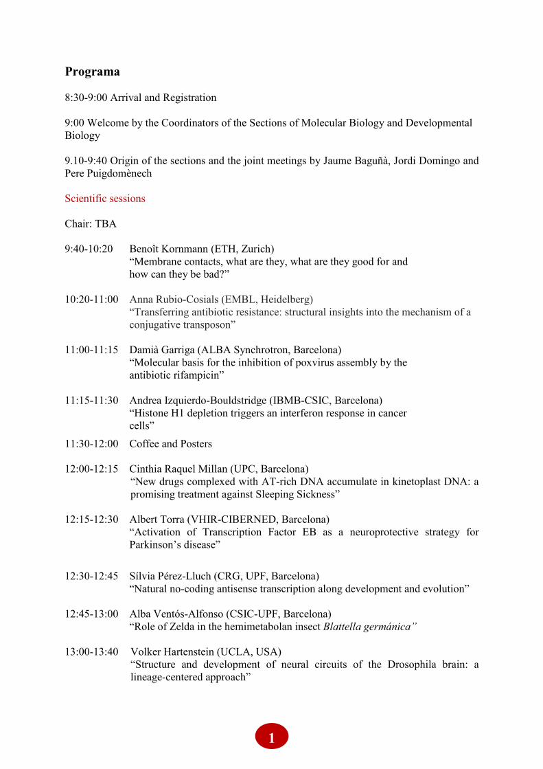
Programa
8:30-9:00 Arrival and Registration
9:00 Welcome by the Coordinators of the Sections of Molecular Biology and Developmental
Biology
9.10-9:40 Origin of the sections and the joint meetings by Jaume Baguñà, Jordi Domingo and
Pere Puigdomènech
Scientific sessions
Chair: TBA
9:40-10:20 Benoît Kornmann (ETH, Zurich)
“Membrane contacts, what are they, what are they good for and
how can they be bad?”
10:20-11:00 Anna Rubio-Cosials (EMBL, Heidelberg)
“Transferring antibiotic resistance: structural insights into the mechanism of a
conjugative transposon”
11:00-11:15 Damià Garriga (ALBA Synchrotron, Barcelona)
“Molecular basis for the inhibition of poxvirus assembly by the
antibiotic rifampicin”
11:15-11:30 Andrea Izquierdo-Bouldstridge (IBMB-CSIC, Barcelona)
“Histone H1 depletion triggers an interferon response in cancer
cells”
11:30-12:00 Coffee and Posters
12:00-12:15 Cinthia Raquel Millan (UPC, Barcelona)
“New drugs complexed with AT-rich DNA accumulate in kinetoplast DNA: a
promising treatment against Sleeping Sickness”
12:15-12:30 Albert Torra (VHIR-CIBERNED, Barcelona)
“Activation of Transcription Factor EB as a neuroprotective strategy for
Parkinson’s disease”
12:30-12:45 Sílvia Pérez-Lluch (CRG, UPF, Barcelona)
“Natural no-coding antisense transcription along development and evolution”
12:45-13:00 Alba Ventós-Alfonso (CSIC-UPF, Barcelona)
“Role of Zelda in the hemimetabolan insect Blattella germánica”
13:00-13:40 Volker Hartenstein (UCLA, USA)
“Structure and development of neural circuits of the Drosophila brain: a
lineage-centered approach”
2
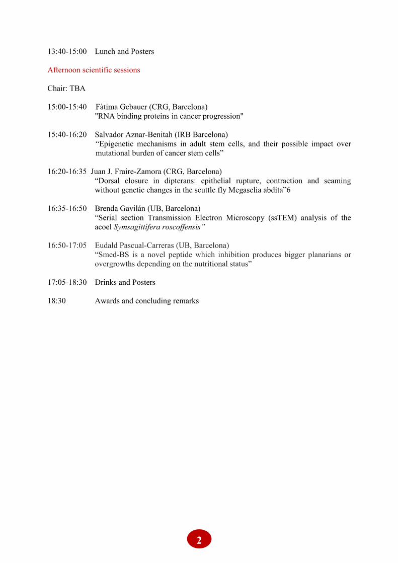
13:40-15:00 Lunch and Posters
Afternoon scientific sessions
Chair: TBA
15:00-15:40 Fàtima Gebauer (CRG, Barcelona)
"RNA binding proteins in cancer progression"
15:40-16:20 Salvador Aznar-Benitah (IRB Barcelona)
“Epigenetic mechanisms in adult stem cells, and their possible impact over
mutational burden of cancer stem cells”
16:20-16:35 Juan J. Fraire-Zamora (CRG, Barcelona)
“Dorsal closure in dipterans: epithelial rupture, contraction and seaming
without genetic changes in the scuttle fly Megaselia abdita”6
16:35-16:50 Brenda Gavilán (UB, Barcelona)
“Serial section Transmission Electron Microscopy (ssTEM) analysis of the
acoel Symsagittifera roscoffensis”
16:50-17:05 Eudald Pascual-Carreras (UB, Barcelona)
“Smed-BS is a novel peptide which inhibition produces bigger planarians or
overgrowths depending on the nutritional status”
17:05-18:30 Drinks and Posters
18:30 Awards and concluding remarks

3
Invited Speakers
4
Membrane contacts, what are they, what are they good for and how can they be bad?
Benoît Kornmann
ETH- Zurich, Switzerland
Intracellular organelles constitute dense and branched membrane networks that are under
constant remodeling. My lab is interested in how these organelle networks are generated,
distributed and regulated. We also investigate how this networked morphology is related to
the organelle's activity. These highly extensive and dynamic networks cohabit in the
extremely crowded cytoplasmic space. This situation leads to unwanted collisions and
entanglements that needs to be resolved. We show that, in the case of mitochondria, these
collisions and entanglements can be resolved by mitochondrial fission. Mechanical forces
applied to mitochondrial tubules lead to the recruitment and activation of the mitochondrial
fission machinery, leading to the resolution of entanglements. These results imply that a
biochemical response can be triggered by a mechanical stimulus and that forces within the
cells participate in the shaping of organelles. The extended morphology of several organelles
might allow them to contact each other to exchange lipid molecules. Because most of the
factors involved in lipid exchange are unclear or unknown, we developed a novel method that
uses transposons and next-generation sequencing to interrogate the yeast genome and map in
a single step all proteins and protein domains necessary for growth in a given condition. We
use it to identify redundancies in lipid exchange routes, but the power of the method finds
myriad of applications far beyond our usage.
5
Transferring antibiotic resistance: structural insights into the mechanism of a
conjugative transposon
Anna Rubio-Cosials, Eike C. Schulz, Lotte Lambertsen, Georgy Smyshlyaev, Carlos Rojas-
Cordova, Kristoffer Forslund, Ezgi Karaca, Aleksandra Bebel, Peer Bork, Orsolya Barabas
Structural and Computational Biology Unit, European Molecular Biology Laboratory
(EMBL), 69117 Heidelberg, Germany
Conjugative transposition drives the emergence of multidrug-resistance in diverse bacterial
pathogens, yet the mechanisms are poorly characterized. The Tn1549 conjugative transposon
propagates resistance to the antibiotic vancomycin used for severe drug-resistant infections.
Here, we present four high-resolution structures of the conserved Y-transposase of Tn1549
complexed with circular transposon DNA intermediates. The structures reveal individual
transposition steps and explain how specific DNA distortion and cleavage mechanisms enable
DNA strand exchange with an absolute minimum homology requirement. This appears to
uniquely allow Tn916-like conjugative transposons to bypass DNA homology and insert into
diverse genomic sites, expanding gene transfer. We further uncover a structural regulatory
mechanism that prevents premature cleavage of the transposon DNA before a suitable target
DNA is found, and generate a peptide antagonist that interferes with the transposase-DNA
structure to block transposition. Our results reveal mechanistic principles of conjugative
transposition, which could help control the spread of antibiotic resistance genes.
6
Structure and development of neuronal circuits of the Drosophila brain: A lineage-
centered approach
Volker Hartenstein
Department of Molecular Cell and Developmental Biology, University of California Los
Angeles, Los Angeles, CA 90095
Common features of the central nervous system encountered in all bilaterian animals are the
relatively high number of cells, diversity in neuronal cell types, and specificity in neuronal
connections. This puts a heavy burden on the developmental process generating the central
nervous system. In Drosophila, a fixed lineage mechanism plays a pivotal role in controlling
neuronal diversity and connectivity. The fly brain is composed of a relatively small number of
stereotyped neuronal lineages, groups of neurons descended from individual embryonic stem
cells, called neuroblasts. During the course of its proliferation, each neuroblast expresses
characteristic sets of regulatory genes. These genes control the differentiation of the neurons born
from that particular neuroblast during a particular time interval. Through this mechanism, a
lineage, or smaller subdivision of a lineage called sublineage, develops into a specific class of
neurons which share common wiring properties, including the projection of their axons, branching
pattern, and placement of synapses. Several discrete neuronal classes/lineages are put together
into a neuronal circuit. In the first part of my talk I will delineate the lineage mechanism,
concentrating on the example of a particular circuit, the anterior visual pathway (AVP). The AVP
conducts input form the optic lobe to the central complex, a brain center known to process and
store visual information in order to control fly locomotion. The central part of the AVP is formed
by three lineages, whose neurons form several classes of highly ordered parallel and sequential
elements.In the second part of my talk I will provide a progress report of our ongoing studies of
neuronal wiring at the single synapse level of resolution. We are following the hypothesis that the
wiring properties and choice of synaptic partners of a neuron is strictly correlated to its lineage
association. In collaboration with numerous other groups we use a dataset consisting of a
complete series of more than 5000 registered electron microscopic sections of an entire early
larval brain (“L1 EM stack”). The reconstructed neurons are morphologically complex 3D graphs
whose nodes are annotated with labels representing different types of synapses. By cross-
referencing the L1 EM stack with similarly oriented stacks of confocal sections we could identify
lineage identity of most neurons. The data allow one to study quantitatively the spatial
relationship between synapses with different partners, and formulate hypotheses regarding neural
function at the microcircuit level. We have designed software that generates 2D renderings of 3D
neurons in order to help biologists analyze the wealth of data that is now available. The renderings
are dendrograms that capture a neuron’s tree-like structure, and they realistically encode
morphological features, such as relative length and branching depth of a side branch, and synapse
locations. We hence refer to these neuron sketches as "morphological feature dendrograms"
(MFDs).
7
RNA binding proteins in cancer progression
Fátima Gebauer
CRG Barcelona
RNA binding proteins (RBPs) are gaining attention in the oncology field for their potential to
regulate essentially every hallmark of tumor development. However, to date only a few RBPs
have been shown to play roles in cancer progression, in large part because RNA metabolism
has been a poorly investigated aspect of cancer research. Fueled by our initial discovery that
the conserved RBP UNR/CSDE1 has dedicated roles in melanoma metastasis, we have
launched an unbiased genome-wide screen of RBPs for which cancer metastatic cells show
specific dependencies. My talk will focus on our efforts to untangle the RBP diversity of
metastatic cells.
8
Epigenetic mechanisms in adult stem cells, and their possible impact over mutational
burden of cancer stem cells
Salvador Aznar Benitah
ICREA Professor; IRB Barcelona
Mutations are not evenly distributed throughout the genome. However, what causes the
disparate genomic mutational distribution is still under debate. It has been proposed that a
factor contributing to the uneven distribution of cancer mutations is the open versus closed
chromatin distribution of their cell-of-origin. Importantly, mutations and expression changes
of epigenetic modifiers are pervasive in human tumours, making epigenetic factors attractive
as anti-tumour targets. However, if epigenetic alterations affect mutational burden, this raises
the concern that targeting epigenetic factors in cancer patients might alter mutability and
possibly aggravate disease progression in the long-term. Yet, a causal link between changes in
chromatin accessibility in tissues and the mutational landscape of their cognate tumours has
not yet been established. I will present functional data showing how altering chromatin
accessibility severely affects tumorigenesis in vivo. I will comment the effects that changing
chromatin openness has over tumor mutability. I will also discuss the implications of our
results on the effect that our lifestyle (i.e. diet) could have on the epigenetic landscape of adult
stem cells which might then influence the aggressiveness of their cognate tumors.

9
Selected Oral Presentations
10
Molecular basis for the inhibition of poxvirus assembly by the antibiotic rifampicin
Damià Garriga1,2, Cathy Accurso2, Stephen Headey3, Melissa Germany-Partarrieu2, Martin
Scanlon3 and Fasséli Coulibaly2
1ALBA Synchrotron, c/ de la Llum 2, Cerdanyola del Vallès, 08290.
2Biomedicine Discovery Institute and 3Monash Institute of Pharmaceutical Sciences, Monash
University, Melbourne, Victoria (Australia)
In contrast to most enveloped viruses, poxviruses produce infectious particles that do not
acquire their internal lipid membrane by budding through cellular compartments. Instead,
poxvirus immature particles are generated from atypical crescent-shaped precursors derived
from host ER membranes. Two key viral proteins participate to this process: A17 inserts in
the membrane and is proposed to induce curvature, while D13 forms a scaffold that remodels
membranes into a closed, spherical particle. The formation of these viral crescents can be
inhibited by the antibiotic rifampicin, although the mechanisms underlying such inhibition
remain unknown. Using a combination of X-ray crystallography, surface plasmon resonance
and CPMG, a ligand-detected NMR technique, we showed that rifampicin directly binds D13
and identified its binding site. We further proved that this inhibitor directly competes with
A17, evidencing an overlap of binding sites. We then used a classical fragment-based drug
design approach to target the A17/rifampicin binding site in D13, screening a library of 1137
compounds by STD and CPMG NMR. This allowed the identification of 25 fragments that
bind D13. Out of these, 2 molecules with unrelated structures were found to compete with
both rifampicin and A17. These two lead compounds were taken for optimisation towards the
design of assembly inhibitors against poxviruses.
11
Histone H1 depletion triggers an interferon response in cancer cells
Andrea Izquierdo-Bouldstridge1,#, Alberto Bustillos1,#, Carles Bonet-Costa1, Daniel García-
Gomis1, Albert Jordan1
1 Institut de Biologia Molecular de Barcelona (IBMB-CSIC), Barcelona, Catalonia, 08028
Spain
# These two authors contributed equally to this work.
Seven linker histone H1 variants exist in human somatic cells with distinct prevalence
depending on the cell type and along differentiation. H1 bind to linker DNA contributing to
higher order chromatin compaction. In addition, H1 seems to be actively involved in the
regulation of gene expression. It is not well known whether the different variants have
specific roles. We have shown that H1 variants are not distributed uniformly along the
genome and there are differences between variants, H1.2 being the one showing the most
specific pattern. We have explored functions of H1 variants by inducible shRNA-mediated
knock-down of each of the variants. Knock-down of each H1 variant alters expression of a
different, reduced subset of genes. Combined depletion of H1.2 and H1.4 has a strong
deleterious effect in the cancer cells examined, and induces a strong interferon (IFN) response
with up-regulation of many IFN-stimulated genes (ISGs). Although H1 participates to repress
ISG promoters, its activation upon H1 KD is mainly generated by the expression
of noncoding RNA generated from heterochromatic repeats including satellites. In conclusion,
redundant H1-mediated silencing of heterochromatin is important to maintain genome
stability and to avoid an unspecific growth-inhibiting IFN response.
12
New drugs complexed with AT-rich DNA accumulate in kinetoplast DNA: a promising
treatment against Sleeping Sickness
C.R. Millana, F.J. Acosta-Reyesab, L. Lagarterac, N. Saperasa, C. Dardonvillec, H. DeKoningd
& J.L. Camposa
aDepartment of Chemical Engeniering, Universitat Politecnica de Catalunya, E08019
Barcelona (Spain); [email protected]
bHoward Hughes Medical Institute, Columbia University, New York, NY 10032, USA
cInstituto de Química Médica, IQM–CSIC, 28006 Madrid, Spain; [email protected]
dInstitute of Infection, Immunity and Inflammation, College of Medical, Veterinary and Life
Sciences, University of Glasgow, Glasgow G12 8TA, UK; [email protected]
Approximately 70 million people distributed over a surface of 1.55 million km2 are estimated
to be at different levels of risk of contracting sleeping sickness. Trypanosoma brucei accounts
for 82.2% of the population at risk. About 6 million to 7 million people worldwide, mostly in
Latin America, are estimated to be infected with Trypansosoma cruzi, the parasite that causes
Chagas disease. We study drugs interacting with minor groove of DNA, such as the N-
phenylbenzamide bis(2-aminoimidazoline) derivatives 1 The main objective was to identify
their cellular target inside the parasite. We were able to demonstrate that the drugs have a
clear effect on the S-phase of T. brucei cell cycle by inflicting specific damage on the
mitochondrial DNA, a unique and complex structure called kinetoplast. The kinetoplast has
more than 70% of AT-DNA.
C.R. Millan, et al. “Functional and structural analysis of AT-specific minor groove binders
that disrupt DNA-protein interactions and cause disintegration of the Trypanosoma brucei
kinetoplas” Nucleic Acid Research (2017) vol.45, pag.8378-8391 Doi: 10.1093/nar/gkx521
Acknowledgements. We thank all the Staff for valuable assistance at the ALBA BL13-Xaloc
beamline and to Dr. Leandro Lemgruber for the SEM images.
13
Activation of Transcription Factor EB as a neuroprotective strategy for Parkinson’s
disease
Albert Torra1, Jordi Galiano1, Annabelle Parent1, Thais Cuadros1, Beatriz Rodríguez-
Galván1, Esther Ruiz-Bronchal2, Analía Bortolozzi2, Miquel Vila1,3,4 and Jordi Bové1
1Neurodegenerative Diseases Research Group, Vall d’Hebron Research Institute (VHIR)–
Center for Networked Biomedical Research on Neurodegenerative Diseases (CIBERNED),
Barcelona, Spain
2Department of Neurochemistry and Neuropharmacology, IIBB–CSIC, August Pi i Sunyer
Biomedical Research Institute (IDIBAPS)-Center for Networked Biomedical Research on
Mental Health (CIBERSAM), Barcelona, Spain
3Department of Biochemistry and Molecular Biology, Autonomous University of Barcelona,
Barcelona, Spain
4Catalan Institution for Research and Advanced Studies (ICREA), Barcelona, Spain
Postal address: Edifici Mediterrània, Hospital Vall d’Hebron, Passeig de la Vall d’Hebron,
119-129, 08035 Barcelona. Phone number: +3493 274 6899. Electronic address:
Neurotrophic factor-based therapy stands as one of the most promising disease-modifying
therapeutic approach for Parkinson’s disease (PD). However, axonal impairment and
downregulation of their receptors may account for the lack of therapeutic success in clinical
trials. An alternative to delivering neurotrophic factors to overcome these hurdles is to
directly activate the intracellular signaling pathways responsible for their effect. We
demonstrate that Transcription Factor EB (TFEB) overexpression in mice drives a previously
unknown bona fide neurotrophic effect that involves the activation of the MAPK1/3 and AKT
pro-survival pathways, giving rise to cell growth, and increased dopamine release. We also
demonstrate that TFEB overexpression prevents neuronal death, increases dopaminergic
function and counteracts atrophy and the associated protein synthesis decline in the MPTP
mouse model of PD. It has been suggested that TFEB neuroprotective effect may be due to its
capacity to boost the autophagy-lysosomal system for the clearance of protein aggregates.
However, we show that knocking down the master transcriptional repressor of autophagy
ZKSCAN3 is not sufficient to protect dopaminergic neurons in this model. Overall, our
results suggest that TFEB activation is an alternative neuroprotective/neurorestorative strategy
to neurotrophic factor-based therapies for PD and related disorders.
14
Natural no-coding antisense transcription along development and evolution
Sílvia Pérez-Lluch1,2, Alessandra Breschi1,2, Amaya Abad1,2, Cecilia Klein1,2, Marina Ruiz-
Romero1,2, Emilio Palumbo1,2, Carme Arnan1,2 and Roderic Guigó1,2,3.
1. Centre for Genomic Regulation (CRG), The Barcelona Institute of Science and
Technology, Dr. Aiguader 88, Barcelona 08003, Catalonia, Spain
2. Universitat Pompeu Fabra (UPF), Barcelona, Catalonia, Spain
3. Institut Hospital del Mar d’Investigacions Mediques (IMIM), Barcelona 08003, Catalonia,
Spain
e-mail: [email protected]
The genome of Drosophila melanogaster is estimated to encode over two thousand lncRNAs;
however, only few of them have a characterized function. Natural antisense transcripts
(NATs) are fully processed lncRNAs which overlap protein coding genes on the opposite
strand with or without exonic complementarity. Several roles in genomic regulation are
reported for NATs in metazoa, including gene expression regulation of the overlapping
protein coding gene, DNA methylation, chromatin modifications and RNA editing. Here, we
have identified 855 lncRNAs overlapping 873 protein coding genes in antisense orientation,
forming 953 sense-antisense (SA) pairs in the fruit fly genome. By analysing the
transcriptome of different imaginal tissues at 3rd instar larvae, we have explored the
relationship between NATs expression and alternative transcript usage across fly larval
samples. Of the 376 SA expressed pairs involving a protein coding gene with multiple
isoforms, blistered/CR44811 is the one showing a highest correlation between changes in
coding gene isoform usage and NAT expression. blistered (bs) gene encodes for two main
isoforms: a short one, expressed mainly in the wing where the NAT CR44811 is also
expressed, and a long one, expressed in the other tissues where the NAT is silent. CR44811
CRISPR mutant flies show a dramatic change in the bs isoform usage in larval and pupal
wings, as well as a strong phenotype in the adult, indicating impairment of the proper wing
development. Manual annotation of the bs locus using available RNAseq data from other
species has allowed us to align both isoforms of the coding gene along development,
suggesting a role of both isoforms in further species and its possible regulation through the
action of the lncRNA.
15
Role of Zelda in the hemimetabolan insect Blattella germanica
Alba Ventos-Alfonso, Xavier Belles
Institute of Evolutionary Biology (UPF-CSIC)
Passeig Marítim, 47, 08003 Barcelona, Tel. 93 230 95 00,
There are two modes of metamorphosis in insects: holometabolan and hemimetabolan. In the
hemimetabolan (which is the ancestral mode), the embryo develops the basic adult body
structure, whereas in the holometabolan, the adult body structure becomes completed in the
pupal stage. Therefore, to better understand the evolution of metamorphosis it is important to
study the differences in the mechanisms regulating embryo developmental processes in
hemimetabolan and holometabolan species. One of those processes is the maternal-to-zygotic
transition (MZT), where the maternal mRNAs are eliminated and the zygotic genome starts to
be transcribed. Zelda has been described as a key protein in the MZT in holometabolan
insects, such as Drosophila melanogaster, being involved in both maternal mRNA cleavage
and activation of the zygotic genome. Conversely, information about the role of Zelda in
hemimetabolan insects is very limited. In this work, we carried out a functional analysis of
Zelda in the embryo of the hemimetabolan insect Blattella germanica, using maternal RNAi.
We found that Zelda regulates the expression of early zygotic genes (involved in abdomen
formation and dorso-ventral patterning), and that of miR-309, a microRNA that plays a key
role in eliminating maternal mRNAs during the MZT. We have also observed that Zelda
regulates the expression of epigenetic factors, like DNMT1 or Nejire. The whole results
confirm that Zelda is a key protein in MZT in both hemimetabolan and in holometabolan
insects. However, a key difference between both metamorphosis modes is that Zelda is
expressed only in early embryogenesis in B.germanica, whereas in D. melanogaster it is
expressed all along the embryogenesis. This difference could explain the different output of
both types of embryo development.
16
Dorsal closure in dipterans: epithelial rupture, contraction and seaming without
genetic changes in the scuttle fly Megaselia abdita
Juan J. Fraire-Zamora, Johannes Jaeger and Jérôme Solon
Cell and Developmental Biology Program, Centre for Genomic Regulation (CRG).
Dr. Aiguader 88, 08003, Barcelona, Spain. [email protected]
The evolution of morphogenesis is generally assumed to be associated with changes
in genetic patterns that lead to the spatial reorganization of tissues. I will present a
case study where rearrangements of epithelial organization occur without major
changes in genetic patterning. In Drosophila melanogaster, the process of dorsal
closure consists of the fusion of opposing epithelial sheets where a contractile
extraembryonic amnioserosa and a JNK/Dpp-dependent epidermal actomyosin cable
result in a microtubule-dependent seaming of the epidermis. In the scuttle fly
Megaselia abdita, dorsal closure must occur in the presence of a separate serosa,
amnion and epidermis. It differs from Drosophila in morphogenetic rearrangements
despite conservation of the JNK/Dpp signaling pathway. Using a quantitative
approach in a non-model organism, we show that dorsal closure in Megaselia is
driven by the rupture and contraction of the serosa, an epidermal actomyosin cable
and the consecutive microtubule-dependent seaming of amnion and epidermis. Upon
rupture, serosa cells retract and reduce their size through actomyosin apical
accumulation. This process promotes the internalization of serosa cells bringing
together the opposing amniotic flanks, followed by the contractile activity of an
epidermal actomyosin cable that depends on JNK/Dpp signaling. The final process of
dorsal closure in Megaselia depends on two sequential microtubule-dependent
seaming events. First the amniotic flanks must seam at the dorsal midline, followed
by the seaming of the opposing epidermal flanks. Microtubule depolymerization
prevents amniotic seaming and rescue experiments resume Megaselia dorsal closure.
Using high-resolution time-lapse imaging, immunostaining and molecular tools, we
obtained evidence indicating that the evolutionary transition to a reduced system of
dorsal closure involves the simplification of the seaming process without changing the
signaling pathways of closure progression.
Fraire-Zamora JJ, Jaeger J and Solon J. (2018) Two consecutive microtubule-based
epithelial seaming events mediate dorsal closure in the scuttle fly Megaselia abdita.
eLife
17
Serial section Transmission Electron Microscopy (ssTEM) analysis of the acoel
Symsagittifera roscoffensis
Brenda Gavilán1, Pedro Martínez 1,2 and Volker Hartenstein3.
1Universitat de Barcelona (UB), Departament de Genètica, Microbiologia i Estadística.
2Institució Catalana de Recerca i Estudis Avançats (ICREA).
3University of California, Los Angeles (UCLA).
In collaboration with S. Sprecher and V. Hartenstein laboratories from University of Fribourg
(Switzerland) and University of California, Los Angeles (USA), respectively, we have
undertaken a thorough study of the acoel cellular organization leading towards the 3D
reconstruction of structures of major interest. Transmission Electron Microscopy serial
sections (ssTEM) of the most anterior part of a Symsagittifera roscofensis juvenile body were
taken, covering approximately the first half of the body, where the brain and beginning of the
nerve cords are located. These sections were aligned in the correct order forming a stack using
the ImageJ plugin TrakEM2 (developed by Albert Cardona, HHMI, Janelia Farm, USA). The
software allows us the 3D reconstruction of cells and cell groups. My work in this project
consists in analyzing the structure and organization of different cell types present, and how
they connect with each other. This is particularly challenging in this group of animals, since
the acoel body is not organized as well-delimited organs; the different cell types appear
intermingled and their membranes highly folded. As acoels (together with their entire group,
the phylum Xenacoelomorpha) are considered the sister group of the remaining bilaterians,
this study can help us to shed some light in the evolution of organs’ architectures. Our main
aim is to see if there are patterns of distribution of these cellular types, how they are
connected to each other and very especially to the central nervous system. We also took
particular interest in the different sensory receptors and gland necks located in the epidermis,
with a focus on their subtype classification, distribution and innervation. This systematic
approach gives us a unique opportunity of studying the nervous system in much more detail,
and aiming at a complete 3D reconstruction of the brain, nerve chords and main peripheral
nerve tracks.
18
Smed-BS is a novel peptide which inhibition produces bigger planarians or overgrowths
depending on the nutritional status
Pascual-Carreras, E1, de Sousa, N1, Marín, M2, Eckelt, K1, Saló, E1 and Adell, T1
1 Department of Genetics, Microbiology and Statistics, Universitat de Barcelona (UB) &
Institute of Biomedicine of Universitat de Barcelona (IBUB), Barcelona, Spain.
2 Norwich Research Park, Norwich NR4 7UH, UK.
Department of Genetics, Microbiology and Statistics, University of Barcelona
The control of cell number is crucial to define the final body size during animal development,
and also to restrict tumoral transformation in adult organisms. The cell number of any
organism results from the balance between cell proliferation and cell death. Although a large
number of genes are known to be involved in the control of both processes, the molecular
mechanisms relating cell number and body size remain poorly understood. To further
understand this relationship we study planarians, flatworms that continuously change their
body size depending of food viability. In this study, we present a novel secreted peptide,
Smed-Blitzschnell (Smed-BS), which inhibition produces an increase of proliferation and a
decrease in cell death, thus leading to an increase in cell number. Interestingly, Smed-bs RNAi
inhibition in starved planarians results in animals with more cells than controls but with the
same body size; thus, showing a higher density of cells that are smaller. Eventually, this
increase in cell density leads to overgrowths. In contrast, Smed-bs RNAi fed planarians
growth faster than controls, since they have more cells, but those cells are of the same size
than the control ones. Thus, the increase in cell number after Smed-bs RNAi is translated to:
1) a tumoral transformation in starved conditions, and 2) an increase in body size in animals
with a rich nutritional status. Thus, the impact of an increase in cell number depends on the
energetic state of cells

19
Posters
20
P1. Analysis of Nse1 function in maintenance of genomic stability in human cells
Sonia Apostolova, Marta Rafel, Jordi Torres-Rosell, Neus Colomina
Departament de Ciències Mèdiques Bàsiques
Universitat de Lleida/IRBLleida
Edifici Biomedicina. Av. Alcalde Rovira Roure 80, 25198 Lleida
During cell division multiple proteins are involved in replication and segregation of
chromosomes to ensure an accurate transmission and distribution of genetic material to
daughter cells. Failure in these key processes leads to the occurrence of aneuploidy and
genomic instability, a hallmark of cancer cells. The Smc5/6 protein complex is conserved
from yeast to humans and plays a crucial role in chromosome segregation and DNA repair.
One of the subunits of the complex - Nse1 - contains a RING domain with ubiquitin ligase
activity. Mutations in this domain impair DNA repair in budding yeast, causing
hypersensitivity to genotoxic drugs. To reveal the function of Nse1 in mammalian cells we
have created human cell lines carrying a deletion or a point mutation in the RING domain of
Nse1 by CRISPR/Cas9 system. Both type of mutants express extremely low levels of the
Nse1 protein, show slow growth and a higher death rate. Our preliminary results indicate that
there is an increase in gamma-H2AX phosphorylation, a slight increase in anaphase bridges
and micronuclei and a significantly increased number of BLM foci, compared to wild type
cells. Besides, FACS analysis shows a higher number of cells with less than G1 DNA content
in the Nse1 mutant population, suggesting a chromosome segregation defect.
Overall, we conclude that the integrity of the RING domain in the human Nse1 is
important for Nse1 protein stability and for the maintenance of genomic integrity.
21
P2. Tissue Engineering Unit at CRG. Services for Stem Cell and Developmental Biology
Laura Batlle Morera, Martin Gigirey, Marta Vila
CRG, Carrer Dr Aiguader n88, 08003, Barcelona. SPAIN
The goal for the Tissue Engineering Platform is to provide services to researchers in the
latests technologies used in the fields of stem cell biology, stem cell differentiation, organoid
formation and induced pluripotent stem cells (iPSCs). The Unit is constantly setting up new
technologies that are emerging in the above fields. The Tissue Engineering platform works in
collaboration with the Biomolecular Screening & Protein Technologies Unit at the CRG to
provide CRISPR/Cas9 genome editing technology service. We provide the following services
for stem cell and developmental biology researchers:
-Stem cells and iPS cells
-CRISPR/Cas9 gene editing to cell lines and directly to embryos
-Embryo micromanipulation
-2D and 3D (organoids) specific stem cell differentiation
22
P3. D-GADD45 as putative modulator of JNK pathway in regeneration
Carlos Camilleri1, Elena Vizcaya
1, Florenci Serras
1 and Montserrat Corominas
1
1Departament de Genètica, Microbiologia i Estadística, Facultat de Biologia and Institut de
Biomedicina (IBUB), Universitat de Barcelona. Barcelona, Catalonia, Spain
Av. Diagonal 643 (08028), Barcelona. [email protected]
Drosophila imaginal discs are a well established model system to study regeneration, both
after physical damage or cell-death induction. Although little is known about the early signals
driving the regenerative process, the activation of the Jun N-terminal kinase (JNK) pathway is
likely to play a decisive role. Studies on the gene expression profiles of imaginal discs at
different time points after cell death reveal several genes showing an expression burst right
after damage and returning to normal levels early in the process. We focus on the Drosophila
Growth arrest and DNA damage-inducible gene 45 (D-GADD45), which is a stress sensor
involved in DNA repair, apoptosis and cell cycle control. Downregulation of D-GADD45
after cell death blocks the activation of the JNK pathway and severely compromises the
regeneration process. We suggest that D-GADD45 activates the JNK pathway upstream of
basket, promoting the signaling cascade that activates the expression of key genes involved in
regeneration.
23
P4. How centrosome number influences collective cell migration
Rita Capella and Sofia J. Araújo
Department of Genetics, Microbiology and Statistics, University of Barcelona, Spain
Cell migration is a complex process that plays an important role both during the biological
development of organisms and in disease conditions. During cell migration, cell movement is
driven by the continuous reorganization and turnover of microtubules and the actin
cytoskeleton. The centrosome is the major microtubule-organizing center (MTOC) in
mammalian cells and centrosome abnormalities are associated with human tumours.
However, the role of centrosomes in cell migration and invasiveness is still not well
understood. To study cell migration during development, we use Drosophila melanogaster as
a model organism, focusing on the embryonic development of the tracheal system, an organ
whose development relies heavily in cell migration. In this study, we compare the migration
patterns of the tracheal cells in wild type flies to tracheal cell migration in different mutants
that have alterations in centrosome number. To approach this, we use confocal microscopy
both in fixed and in vivo embryos.
24
P5. Smed-cbp regulates stem cells commitment and differentiation in planarians
Sheila Cárcel1, Susanna Fraguas1, Thileepan Sekaran2, Kerstin Bartscherer2 and Francesc
Cebrià1
1Departament de Genètica, Microbiologia i Estadística, Universitat de Barcelona
2Max Planck Institute for Molecular Biomedicine, Münster, Germany
The plasticity and differentiation of stem cells into multiple specific lineages is an open basic
question in developmental biology. CBP (CREB-binding protein) is a conserved gene that
functions as transcriptional co-activator and histone acetyl transferase. In different organisms,
CBP plays an important role in wide range of cellular processes, including cell proliferation
and differentiation, cell death, DNA damage response and tumorigenesis. Here, we have
studied the function of a CBP homologue in planarians, which have astonishing cellular
plasticity thanks to their neoblasts (adult pluripotent stem cells). Because planarians have
remarkable regenerative abilities they provide us an ideal scenario to understand the
molecular mechanism underlying stem cell differentiation in vivo. Our data show that the
silencing of Smed-cbp in planarians results in apparently proper blastema formation but
severely impairs tissue differentiation. Analyses with specific molecular markers for neural
and eye lineage-committed progenitors indicate that neoblast specialization into these cell
types is largely blocked, which results in the absence of neural regeneration. In contrast, for
the epidermal lineage it seems that Smed-cbp is not necessary for the specialization of
epidermal progenitors, and opposite to what it is seen for neural lineages there is an increase
in the number of differentiated epidermal cells. Overall, our results suggest that Smed-cbp
could have a multiple function in regulating neoblast commitment and differentiation.
25
P6. Functional characterization of a mutation identified in an Opitz C syndrome patient
L. Castilla-Vallmanya, R. Urreizti, H. Franco-Valls, G. Cunill, A. Cueto-González, E.
Tizzano, J.M. Opitz, G. Neri, D. Grinberg, S. Balcells
Department of Genetics, Microbiology and Statistics, Faculty of Biology (UB)
Avinguda Diagonal, 643 Edifici Prevosti 08028 (Barcelona)
934021794
Opitz C syndrome (OTCS, MIM #211750) is an extremely rare genetic disorder characterized
by multiple malformations (e.g. trigonocephaly, congenital heart defects), contractures,
variable intellectual and psychomotor delay and a high mortality rate. In a cohort of 11
patients clinically diagnosed as OTCS, mutations in 9 different genes were identified as
disease-causing by whole exome sequencing (WES). Thus, OTCS could be causally
heterogeneous phenotype instead of a specific entity. In this project, we aim to functionally
characterize a de novo heterozygous missense variant found in a gene of the TRAF family in
one of the patients and to assess its pathogenicity. The protein encoded by this gene has a role
as E3 ubiquitin ligase in different signalling pathways mediated by Tumor Necrosis Factor
(TNF) family ligands, such as the NF-κB pathway.
We have analyzed cell viability by MTT assay and while a slight increase in the patient’s cell
viability could be observed, there is no significant difference. In vitro analysis of fibroblasts
from 3 patients bearing similar mutations were performed at mRNA level by qPCR and
mRNASeq. Compared to control fibroblasts, patient’s cells showed an altered basal
expression of several genes of the NFKB pathway and gene expression patterns in response to
TNFα stimulation. Results so far strongly suggest a pathogenic role of the mutation, however
they do not clarify if the effect is a loss or a gain of function of the protein. Future
experiments, such as co-immunoprecipitation assays with the major partner of the protein and
further analysis of the mRNASeq data, may help to elucidate the impact of these mutations.
26
P7. EGFR and ecdysone in Blattella germanica oogenesis.
Fleur Chelemen and Maria-Dolors Piulachs
Institut de Biologia Evolutiva - IBE- (CSIC-UPF)
Pg. Marítim de la Barceloneta 37-49. 08003 Barcelona. Tel. 932309648.
Oogenesis is a crucial process to ensure continuity of the species and, therefore, it needs to be
finely regulated. Even the regulation of this process is a complex network where different
genes and pathways participate, it is important to know each piece of the puzzle and its
function, and one of the main pieces of this puzzle is ecdysone that is already known in
Drosophila melanogaster for stimulating the germ stem cell proliferation. Our work is
focused on the epidermal growth factor receptor (EGFR) using as a model the cockroach
Blattella germanica, a species with panoistic ovaries, the most primitive ovary type. In
previous work, we found that the depletion of EGFR determines an increase in the number of
germinal cells and our objective is to unveil a possible interaction between EGFR signaling
and ecdysone, an interaction that must be regulating ecdysone biosynthesis or its signaling
gene cascade. To measure the activity of the ecdysone biosynthesis, we pay special attention
to the expression of Shadow an enzyme that triggers the final step in the biosynthesis of
ecdysone. To unveil these interactions, we use the RNAi methodologies to deplete the
expression levels of EGFR and Shadow, observing at microscopic levels how this depletion
could affect the differentiation of the germinal cells in the germarium and by qRT-PCR
quantifying the expression of those genes that can be implicated in this process.
27
P8. Proteasomal degradation of naturally occurring glutamine-rich peptides
B. Coll1, A. Zuin1, Y. Palau1, B. Crosas1
1. Molecular Biology Institute of Barcelona, CSIC, Barcelona, Spain
The ubiquitin proteasome system (UPS) is a central player in eukaryotic protein homeostasis.
In a cellular context, ubiquitin activating, conjugating and ligating enzymes act sequentially
marking the substrates for their degradation in the proteasome. In this way, ubiquitin serves as
a common recognition tag among most of the proteasome substrates. As part of the UPS
system, the proteasome acts as a protein degrading hub for the cell, integrating degradative
signals from multiple ubiquitin ligase enzymes. This tag, however, is not imperative for
protein degradation. Instead, it has been described that location to the proteasome is sufficient
for protein degradation, given that the substrate features an unstructured region able to
interact with the inner ATPases of the proteasome. When protein homeostasis fails, the whole
cell functioning is impaired. This is often associated with cell stress, toxicity and death. Here
we investigate the interaction between proteasome and disease related proteins such as
huntingtin or ataxin-3. These proteins have a common trait; they all feature glutamine rich
regions. In this project we propose that these characteristic domains can serve as signal
sequences able to direct the proteins to the proteasome in an ubiquitin independent way.
Direct targeting of toxic proteins to the proteasome serves as a model to study how
aggregation prone proteins affect the proteasome system. Additionally, it is to be studied how
replicating these proteasome modifications in eukaryotic cells could shed some light into new
treatment for the above-mentioned neurodegenerative diseases.
28
P9. In vivo and in vitro models in the study of the FGF23 regulation
Nuria Doladé1; Sandra Rayego-Mateos1; Jose Manuel Valdivielso1
1Institut de Recerca Biomèdica de Lleida (IRBLleida), Av. Alcalde Rovira Roure, 80, Lab.
1.10, Lleida, 25198, Spain. 973702952. [email protected], [email protected],
Introduction: Chronic kidney disease (CKD) is a major public health problem. The
fibroblastic growth factor 23 (FGF23) is a fosfaturic protein that is expressed predominantly
in osteocytes and whose levels are increased in patients with CKD. The possible regulation of
FGF23 by calcium and phosphorus independently remains unknown. The objective of this
study is to establish the models that allow us to study it. Materials and methods: A. Para-
thyroidectomy model (T-PTX): The animals underwent a para-thyroidectomy (T-PTX) and a
tyrosine hormone replacement therapy (T4). B. Hypercalcemia model: Intraperitoneal
injection of calcium gluconate monohydrate (250 mg/kg every 2 hours during 8h). C.
Hypocalcemia model: Intramuscular injection of EDTA (150 mg/kg). The animals were
sacrificed 6 hours later. In vitro: Primary cultures of bone marrow mesenchymal stem cells
(MSCs), differentiated by a conditioned medium. Results and conclusion: Mice subjected to
T-PTX showed a reduction in calcium levels, as well as an increase in serum phosphorus
levels. In mice undergoing hypercalcemia, a significant increase in the excretion of calcium in
urine was observed without changes in serum calcium levels. In mice subjected to
hypocalcemia, a decrease in calcium levels was observed in both serum and urine. Finally, in
in vitro differentiated cells (MSCs differentiated to osteocytes-like) there was an increase in
gene expression of mineral matrix markers such as OSC, OSX, RUNX2, OPN and FGF23, as
well as a decrease in mesenchymal markers such as COL I. We can conclude from the results
obtained that the T-PTX was successful and can be used for the study of the regulation of
FGF23 independently of PTH. On the other hand, models of hyper-and hypocalcemia in the
doses studied exert the desired effects on the regulation of calcium levels. The individual
combination of these two models with the T-PTX will allow us to study the role of calcium
independently of PTH in the regulation of FGF23.
29
P10. The effect of stress in the retinal cells of a Cerkl mouse model generated by
CRISPR/Cas9
Domènech, E. B.1, 2, Serra-Pascual, C.1, 2, Gonzàlez-Duarte, R.1, 2, 3, Marfany, G.1, 2, 3
1Departament de Genètica, Microbiologia i Estadística, Facultat de Biologia, Universitat de
Barcelona, Avinguda Diagonal 643, 08028 Barcelona, Spain.
2Centro de Investigación Biomédica en Red de Enfermedades Raras (CIBERER), Instituto de
Salud Carlos III, Barcelona, Spain.
3Institut de Biomedicina de la Universitat de Barcelona (IBUB-IRSJD), Barcelona, Spain.
Retinal neurodegeneration, characterized by the apoptosis of photoreceptor cells, is the major
cause of genetic blindness. So far, mutations in over 200 genes are associated with inherited
monogenic retinal diseases (1:3000 worldwide), but we are still far from completely
understanding their ethiopathology. Therefore, animal models and in vitro cell culture assays
are essential to characterize the precise role of CERKL, a causative gene of retinal
dystrophies. The physiological function of CERKL is yet to be determined, but it is related to
stress cell resilience since its overexpression protects cells from the apoptosis triggered by
oxidative stress. We generated a mouse model by causing the full deletion of the Cerkl gene
using CRISPR/Cas9. in order to investigate the retinal effects in the oxidative/light stress
response. Unexpectedly, complete ablation of Cerkl causes perinatal lethality in
homozygosity. Therefore, to approach the CERKL function in the retina, we have generated a
heterozygous knockdown/knockout mouse model (CerklKD/KO) in an albino background to
perform in vivo light stress experiments. This model is viable and fertile, and the expression
of Cerkl has been reduced to 18%. Our results showed that CERKL localized to the stress
granules formed the retina in response to stress. Moreover, the number of stress granules was
notably higher in the retinas with reduced CERKL expression compared to those of wild type
mice, thus supporting the CERKL role in the protection and maintenance of retinal cells.
30
P11. The role of ASK1 in Drosophila’s wing disc regeneration
José Esteban Collado, Giacomo Viola, Montserrat Corominas and Florenci Serras
Department of Genetics, Microbiology and Statistics, University of Barcelona, Barcelona
Recent work has strengthened Drosophila imaginal discs as a model system for regeneration
studies. Evidence is accumulating that oxidative stress drives the cellular responses for repair
and regeneration. Apoptotic cells result in a burst of reactive oxygen species (ROS) that can
propagate to neighboring cells. These ROS are known to activate JNK and p38 kinases for
regenerative growth.Two key issues arise from these observations. The first is how the ROS
are propagated. The second, what is the link between ROS and the stress activated protein
kinases p38 and JNK. Our results reveal that Ask1 senses ROS differently in apoptotic cells
and living surrounding cells. High levels of ROS are produced in apoptotic cells, which in
turn generates high activity of Ask1 that turns on JNK, which is known to enhance apoptosis.
Neighboring undamaged cells show low ROS levels, which are beneficial for the regenerating
tissue. In these, Ask1 is activated but its activity is attenuated by Pi3K dependent Akt1
phosphorylation, as a survival signal that results in beneficial levels of p38 and JNK. Our data
reveal a non-autonomously activated ROS sensing mechanism driven by Ask1 and Akt to
drive regeneration in the neighboring unstressed cells.
31
P12. Gene-Repair of point mutations at the endogenous locus using PolyPurine Reverse
Hoogsteen hairpins in mammalian cells
Alex J. Félix, Anna Solé, Véronique Noé and Carlos J. Ciudad
Departament of Biochemistry and Physiology, School of Pharmacy, University of Barcelona.
Av. Juan XXIII #27, [email protected]
Repair of point mutations in the DNA is important for the correction of monogenic diseases.
Repair-PPRHs are a new powerful tool within this field. A Repair-PPRH consists of a PPRH
hairpin core bearing an extension sequence at one end, which is homologous to the DNA
sequence to be repaired but containing the wild type nucleotide instead of the mutated one.
Previously, we designed Repair-PPRHs to correct deletions, insertions, substitutions and a
double substitution present in a collection of Chinese hamster ovary (CHO) cell mutants in
the endogenous locus of the dhfr gene. Surviving colonies in a DHFR selective medium
(lacking glycine-hypoxanthine-thymidine) were analyzed by DNA sequencing, mRNA and
protein levels and enzymatic measurements, confirming that all the dhfr mutants had been
corrected. To explore the generality of this methodology, we attempted to repair point
mutations in a different gene, that coding for adenosyl phosphoribosyl transferase (aprt). By
using different Repair-PPRHs we were able to correct nonsense mutations caused by a single
substitution. In this case, surviving colonies were obtained by applying the AAT (adenine-
aminoptherine-thymidine) selection and were analyzed by DNA sequencing and by mRNA
expression. No off-target effects were detected when comparing the S23 mutant and the S23
repaired with HpS23E1rep-L after sequencing with a mean coverage of 26x. These results
correspond to the set of 3158 contigs longer than 100Kb totaling around 90% of the genomic
sequence (2.2 Gb), indicating no major mutational differences between the two samples. We
did not see either any major bias when looking at the indels (insertion and deletions together)
or only at the insertions in the treated cells. Moreover, any of the insertions detected within
the variation set had similarity to the sequence present in the Repair-PPRH used for the
treatment. These results demonstrate that Repair-PPRHs are able to achieve a permanent
correction of point mutations in the DNA sequence of mammalian cells.
Acknowledgments: Work supported by grant SAF2014-51825-R
32
P13. Early hippo target genes in planarians
Daniel Font, Nídia da Sousa, Emili Saló and Teresa Adell
Department of Genetics, Microbiology and Statistics, University of Barcelona, Barcelona,
Spain
Regeneration and tissue renewal are essential processes of adult homeostasis that must be
tightly controlled, since its dysfunction may lead to cancer. The Hippo signaling pathway has
recently emerged as a key hub in the control of cellular renewal being found systematically
deregulated in tumoral processes. Several studies in animal models demonstrate the essential
role of the Hippo pathway in the control of cell proliferarion, cell death and cell
differentiation. However, the specific cellular function and molecular targets of the pathway
remain poorly understood. Recent studies in our lab have demonstrated that hippo inhibition
produces tumoral overgrowths in planarians, flatworms that endow a continuous tissue
renewal while changing their size according to nutrients availability. In this in vivo context,
we demonstrated that overgrowths are caused by the inability of hippoknockdown cells to
maintain the differentiated fate, to properly cycle and to die when required. In the present
study, we characterize the function of putative hippo target genes found deregulated in a
transcriptomic analysis of hippo RNAi animals. The RNAi inhibition of some of these early
target genes produced overgrowths similar to the ones observed after hippo inhibition. This
connection hinters potential effectors contributing to the tumorigenic mechanisms
upon hippo inhibition in planarians and, probably, in humans.
33
P14. Organ remodeling through the actomyosin cytoskeleton and programmed cell
death: the trachea of Drosophila melanogaster as a case study
Juan J. Fraire-Zamora, Jérôme Solon and Jordi Casanova
Cell and Developmental Biology Programs, Centre for Genomic Regulation (CRG)
and Institute for Research in Biomedicine (IRB), Barcelona. Dr. Aiguader 88, 08003
and Baldiri Reixac 10, 080028, Barcelona, Spain. [email protected]
Many cellular processes are in play during organ remodeling. Most of the attention has
focused on the role of progenitor cells and their transformation into adult tissues and organ
growth, however, little is known about the mechanisms of organ regression. An example of
this process is the removal of epithelial cells during mammary gland involution at weaning.
The fruit fly Drosophila melanogaster offers an advantageous system to study organ
remodeling at the cellular and molecular level. The larval trachea, a functional respiratory
organ, is a modular network of tubes that must remodel during metamorphosis to give rise to
the adult trachea. Our current work focuses on the cellular and molecular mechanisms that
orchestrate the reduction in size of the main tracheal tube, the dorsal trunk. Concomitant to
the migration of progenitor cells, the modules (i.e. metameres) of the dorsal trunk undergo a
sequential reduction in length that correlates with a reduction in apical cell area and the
appearance of actomyosin filaments along the longitudinal axis of the tube. Upon maximum
reduction in tracheal length, we observe caspase activation and a consequent loss of posterior
metameres of the dorsal trunk. These events result in a considerable reduction in size of the
larval trachea at the same time that progenitors migrate to form the abdominal adult trachea.
We are currently exploring the interplay between cell size reduction through actomyosin
contractility and the activation of programmed cell death through apoptosis. We believe that
such cellular and molecular interplay leads to the regression of the tubular dorsal trunk during
metamorphosis of Drosophila melanogaster, as it occurs in other flat epithelial monolayers.
34
P15. Generation of Sanfilippo C syndrome cellular models using CRISPR/Cas9 system
Edgar Creus-Bachiller (1)(*), Noelia Benetó (1)(*), María García-Morant (1), Daniel
Grinberg (1), Lluïsa Vilageliu (1), Isaac Canals (2)
(1)Department of Genetics, Microbiology and Statistics, Faculty of Biology, University of
Barcelona, CIBERER, IBUB, IRSJD, Barcelona, Spain; (2) Stem Cells, Aging and
Neurodegeneration Group, Lund Stem Cell Center, University Hospital, Lund, Sweden.(*)
Equal contributors.
Sección Biologia Molecular i Biologia del Desenvolupament. Facultat Biologia(UB):
Avinguda Diagonal, 643, 08028 Barcelona.
Sanfilippo syndrome is a rare lysosomal storage disorder (LSD) caused by mutations in genes
that encode enzymes involved in heparan sulfate (HS) degradation. The accumulation of HS,
due to mutations in these genes, causes a progressive and severe neurodegeneration in
patients, leading to an early death. There is currently no cure for this disease. The main
objective of this project is to generate cellular models for Sanfilippo C syndrome using
induced pluripotent stem cells (iPSC). For that purpose, we used CRISPR/Cas9 system to
knock-out (KO) the HGSNAT gene in iPSC cells, whose mutations cause Sanfilippo C
syndrome. Moreover, we optimized a protocol to differentiate iPSC into neurons, the most
affected cell type in this syndrome. Combining these two novel technologies, we will be able
to obtain KO and WT differentiated neurons in order to analyze differences between these
isogenic lines. With this approach, possible variations due to differences in the genetic
backgroundWe aim to compare levels of HS storage between lines by immunocytochemistry.
Furthermore, branching and spines measurements will be performed in GFP+ isolated
neurons. To refine the analysis, synapses will be quantified combining specific antibodies for
pre- and post-synaptic markers. To confirm that the phenotypes observed in the KO neurons
are only caused by HGSNAT disruption, we will carry out a rescue experiment transfecting
KO cells with a plasmid bearing the HGSNAT-WT cDNA. After transfection, we will perform
all the assays in order to test whether or not the WT phenotype has been restored. We are
confident that once this cellular model is validated, it will be valuable to test potential
treatments such as the use of shRNAs as a substrate reduction therapy.
35
P16. Role of Aquaporins in ROS diffusion following damage
Viola Giacomo, Prieto Cristina, Corominas Montserrat and Serras Florenci
Departament de Genètica and Institut de Biomedicina (IBUB), Universitat de Barcelona,
Barcelona, Spain
Drosophila wing imaginal discs are able to regenerate following different types of injury,
which leads to the reconstruction of normal adult wings. Recent studies pointed out the
importance of oxidative stress in driving the cellular response for wound repair and
regeneration. Upon apoptotic stimuli or physical injuries, a burst of reactive oxygen species
(ROS) is generated in the wing disc epithelium. This results in the activation of different
signalling pathways (like JNK and p38) that are required for regeneration. A key issue is to
unveil how reactive oxygen species (H2O2 in particular) spread from cells that are committed
to die to the surrounding ones, however the mechanism of ROS propagation is poorly
understood. In this study, we focus on two different Drosophila aquaporins: AQP and Drip as
putative mechanisms of oxidative stress propagation. Our results indicate that, in stressing
conditions, AQP is a key element of cell-to-cell communication facilitating ROS diffusion
across membranes, thus allowing the onset of the regenerative stimulus. Interestingly, while
AQP loss of function impairs the regenerative process, no significant defects were observed
with Drip, suggesting that the function in regeneration is not shared among all aquaporins.
36
P17. New Cyclin D1 cytoplasmic interactors
Marta Guasch, Tània Cemeli, Noel P. Fusté, Francisco Ferrezuelo, Neus Pedraza, Eloi Garí.
IRB Lleida, Universitat de Lleida, Avinguda Alcalde Rovira Roure 80, 25198, Lleida
The cell cycle is a set of well-ordered events that allows the cell to divide into two cells
genetically identical. The different stages of this cycle depends on the activity regulation of
cyclins and cyclin dependent kinases (Cdk) complexes. For instance, cyclin D1 (Ccnd1)-Cdk4
complex accumulates into the cell nucleus and phosphorylates and inactivates the
Retinoblastoma (RB1) transcriptional repressor. This allows the induction of a group of genes
that promote progression from G1 into S phase. For many years, export of Ccnd1 to the
cytoplasm was viewed as a mechanism to prevent the cell cycle entry and to reduce cellular
proliferation. More recently it has been described that Ccnd1 physically interacts with
proteins associated to the cell membrane, such as filamin A, PACSIN2, Ral GTPases and
paxillin. Furthermore, our group has described that Ccnd1-Cdk4 complex modulates cell
adhesion, migration and invasion through the regulation of some of those cytoplasmic
proteins. In our lab, we have recently carried out an iTRAQ-based protein analysis that has
produced a set of cytoplasmic proteins associated to the cell membrane that are new candidate
interactors of Ccnd1. Among these there are a number of proteins involved in cell signalling.
Here we report the validation of some of these candidates (PGRMC1, Dab2IP and Plekhh2)
by co-immunoprecipitation with Ccnd1. Specifically, PGRMC1 protein (Progesterone
Receptor Membrane Component 1) interacts with the N-terminal region of Ccnd1 and
promotes its stabilization. In experiments in HEK 293T cells with the protein synthesis
inhibitor cyclohexmide, we have observed that expressing PGRMC1 increases Ccnd1 half-
life. In conclusion, we show preliminary data pointing out to a possible function of
cytoplasmic Cyclin D1 in the regulation of cell signalling.
37
P18. Identifying wiring specificity mechanisms: what’s up with the mTOR pathway?
Camille Guillard Sirieix, Qi Zhu, Marta Morey
Section of Genetics, Faculty of Biology, and Institute of Biomedicine of the University of
Barcelona (IBUB)
A fundamental requirement in the assembly of neural circuits is that neurons establish
synaptic connections with their appropriate partners. In many systems, this involves extension
into a particular synaptic layer, and selection of the appropriate partner among all the cells in
the layer. Virtually nothing is known about the mechanisms governing the establishment of
specific connections in any system. Our hypothesis is that the molecular differences that exist
between neuronal subtypes, with similar developmental origin and function, contribute to
their distinct connectivity. We study the differential layer selection of the closely related R7
and R8 photoreceptors. Each eye contains 750 R7 and 750 R8 cells, and the entire population
of each subtype proceeds synchronously to their respective final synaptic layer during pupal
development. Taking advantage of such precise coordination, we have profiled the R7 and R8
transcriptomes right before this final extension. Our bioinformatic analysis has identified
differentially expressed genes between the R7 and the R8. We have focused on 229 R8
enriched genes and performed an RNAi screen. Out of 186 genes analyzed we have identified
44 candidate genes showing layer selection defects. One of them is 4E-BP, an inhibitor of
translation best known for its role in the mTOR pathway. Interestingly, the mTOR pathway
has been shown to have a neurogenic role uncoupled from its well-known control of cell
proliferation and growth. Following this lead we present future work exploring the role of 4E-
BP in wiring specificity through detailed characterization of the mutant phenotype and genetic
interactions with other members of the pathway.
38
P19. Study of sumoylation of TRIM28 variants associated with intellectual disability
Isabel Hinarejos, Roser Urreizti, MI Isabel Alvarez-Mora, Laura Domènech, Irene Madrigal,
Montserrat Milà, Xavier Estivill, Gemma Marfany, Daniel Grinberg, Raquel Rabionet
Universitat de Barcelona (Facultat de Biologia) Avinguda Diagonal, 643, 08028 Barcelona
Intellectual disability (ID) is characterized by high clinical and genetic heterogeneity.
Sporadic cases of ID are frequently related to de novo mutations in neurodevelopmental
genes. Our group has identified a de novo variant (p.P654L) in the TRIM28 gene in a sporadic
case of ID as the most likely causative variant; two additional ID patients harboring TRIM28
mutations (p.L708P and p.A180V) have been identified through Genematcher. TRIM28 is a
nuclear corepressor for KRAB domain-containing zinc finger proteins (KRAB-ZFPs)
expressed in several tissues, including the brain. TRIM28 mediates gene silencing by
recruiting CHD3, a subunit of the nucleosome remodeling and deacetylation (NuRD)
complex, and SETDB1, which specifically methylates histone H3 at 'Lys-9' (H3K9me), to the
promoter regions of KRAB target genes. Sumoylation/desumoylation events regulate
TRIM28-mediated transcriptional repression, as sumoylation is required for interaction with
CHD3 and SETDB1 and the corepressor activity. The mutation we detected in this patient is
located in the PHD-type zinc finger domain, which directs the sumoylation of the adjacent
bromodomain required for gene silencing. Two different mutations in the same domain
(p.C651A and p.L709A) have previously been shown to affect TRIM28 sumoylation. We
hypothesize that the mutations detected in the ID patients will affect TRIM28 sumoylation,
and thus its repression of gene expression. In order to prove this hypothesis, we are
performing directed mutagenesis and sumoylation analysis of the mutant forms of TRIM28.
39
P20. DNA activates the Nse2/Mms21 SUMO E3 ligase in the Smc5/6 complex
Eva Ibars, Nathalia Varejão, Neus Colomina, David Reverter, Jordi Torres-Rosell
Institut de Recerca Biomèdica de Lleida-Universitat de Lleida
IRBLLEIDA, Dept. Ciències Mèdiques Bàsiques, Universitat de Lleida. 973702414,
Modification of chromosomal proteins by conjugation to SUMO is a key step to cope with
DNA damage and to maintain the integrity of the genome. The recruitment of SUMO E3
ligases to chromatin may represent one layer of control on protein sumoylation. However, we
currently do not understand how cells upregulate the activity of E3 ligases on chromatin. Here
we show that the Nse2 SUMO E3 ligase in the Smc5/6 complex, a critical player during
recombinational DNA repair, is directly stimulated by binding to DNA. We have identified a
cluster of lysine residues in the coiled coil of Smc5 able to promote DNA-dependent
upregulation of the Nse2 SUMO ligase activity. To test the relevance of these residues in
budding yeast, we introduced different combinations of lysine to glutamic acid mutants to
countercharge binding to phosphate groups of DNA. Using these smc5-KE mutants we show
that compromising DNA binding to Smc5 sensitizes yeast cells to MMS-induced DNA
damage. Accordingly, sumoylation of Smc5 itself and the Nse2-target Sgs1 protein (a
homologue of the Bloom’s and Werner’s syndrome genes and a member of the STR complex)
is impaired in smc5-KE cells. These defects are not due to defective binding of Nse2 to Smc5
or to impaired loading of Smc5/6 onto chromatin in smc5-KE alleles. Overall, we conclude
that a positively charged patch in the Smc5 molecule acts as a DNA sensor in yeast, able to
interact with DNA and to promote the activity of the Nse2 SUMO ligase thus ensuing repair
of DNA damage. We propose that this mechanism restricts sumoylation in the vicinity of
those Smc5/6-Nse2 molecules directly engaged on DNA.
40
P21. Role of Myoglianin in metamorphosis of Blattella germanica
Orathai Kamsoi, Xavier Belles
Institute of Evolutionary Biology (UPF-CSIC)
Passeig Marítim 47, 08003 Barcelona, Spain,
[email protected], [email protected]
The onset of insect metamorphosis occurs when juvenile hormone (JH) titer decrease in the
pre-adult stage. In Blattella germanica, this is determined in the last nymphal instar (N6). At
the beginning of N6, JH production declines, the expression of Kr-h1 (a transducer of the JH
anti-metamorphic action) is concomitantly reduced, and that of E93 (an adult specifier factor)
starts increasing. However, it remains unclear what determines the initial decline of JH
production. In the cricket Gryllus bimaculatus, Ishimaru and coworkers (2016) demonstrated
that Myoglianin (myo), a homolog of Drosophila Myoglianin/vertebrate GDF8/11, suppresses
the expression of jhamt. jhamt is a gene that encodes the enzyme JH acid O-methyltransferase
(JHAMT), which catalyzes the last step of JH synthesis in the corpora allata (CA), which are
the JH producing glands. When myo expression is suppressed in last nymphal instar of the
cricket, expression of jhamt became activated and metamorphosis is inhibited. In B.
germanica, high expression levels of myo were observed in the CA of the penultimate
nymphal instar (N5). Moreover, using RNAi approaches, we showed that myo depletion in N5
up-regulated jhamt expression in N6, and metamorphosis became inhibited. The myo-depleted
insects molted to a supernumerary nymph (N7) instead to molt into adults. The results suggest
that high myo expression in N5 promotes the initiation of metamorphosis in N6 through the
cessation of JH synthesis. In addition, our results also suggest a conserved role of myo in
regulating JH production and metamorphosis, at least in hemimetabolan insects.
41
P22. The Spectraplakin Short-Stop is an essential microtubule regulator mediating
subcellular branching
Delia Ricolo, Miquel Lledós and Sofia J. Araújo
Department of Genetics, Microbiology and Statistics, University of Barcelona, Spain
Branching networks are a very common feature of multicellular animals and underlie the
formation and function of numerous organs including the nervous system, the respiratory
system, the vasculature and many internal glands. These networks vary from subcellular
structures such as dendritic trees to large multicellular tissues such as the lungs. The
production of branched structures by single cells, which has been better described in neurons
and in cells of the respiratory and vascular systems, involves complex cytoskeletal
remodelling events. In Drosophila, tracheal system terminal cells (TCs) and nervous system
dendrites are models for these subcellular branching processes. During tracheal embryonic
development, the generation of subcellular branches is characterized by extensive remodelling
of the microtubule network and actin cytoskeleton, followed by vesicular transport and
membrane dynamics. We have recently shown that centrosomes are key players in the
initiation of subcellular lumen formation where they act as microtubule organizing centres
(MTOCs) (Ricolo, et al. Cur. Biol. 2016). However, not much is known on the events that
trigger the formation of these subcellular branches or what makes them choose a particular
trajectory within the cytoplasm of the TC. We have identified that the spectraplakin Shortstop
(shot) is involved in the microtubule stabilisation events that lead to the formation and
extension of the subcellular lumen. We observed that an excess of shot induces more
branching points in the embryonic tracheal TC leading to cells with extra subcellular lumina
and that a shot loss-of-function leads to cells deficient in de novo subcellular lumen
formation. In addition, we show that shot expression is intimately linked to the tip-cell fate,
being modulated by the transcription factor DSRF.
42
P23. Differential expression of piRNAs during the ontogeny of Blattella germanica, a
short germ-band, hemimetabolan insect
Natalia Llonga, Guillem Ylla, Josep Bau, Xavier Belles, Maria-Dolors Piulachs
Institute of Evolutionary Biology (CSIC-Universitat Pompeu Fabra).
Passeig Maritim de la Barceloneta, 37. 08003 Barcelona, Spain.
Genomic stability is especially important in germ-line cells and in embryo stages for the
insect development. For this reason the insect needs to control transposable elements (TEs).
There is an evolutionary conserved mechanism, called piwi-interacting RNA (piRNA) system
to control TEs. This system is common within species among metazoans and insects. To study
piRNA system it is more suitable to use a long genome
with many TEs. Thus, we use the hemimetabolan insect Blattella germanica as a reference
model. In the present work we have identified and studied the expressed piRNA in B.
germanica across 11 developmental stages, with a stringent process that includes non-
fertilized egg, embryo, nymphs and adult females. We have been able to classify the piRNAs
from B. germanica based in their biogenesis (primary and secondary pathways). We found
that the majority piRNAs identified are generated from the primary pathway, although there
are a small but highly expressed set of piRNAs participating in the secondary (“Ping-Pong”)
reamplification pathway. Furthermore, we have analysed the expression pattern of all these
piRNAs identified, observing that the expression of piRNA generated in the “Ping-Pong”
pathway is quite restricted to early embryo stages. In addition, an important number of piRNA
clusters are exclusively expressed in late embryo and nymphal stages. This study contributes
to confirm the role
of the piRNA system controlling TEs in early embryogenesis, but also to show that the
function of piRNAs are wider than previously thought, as we found different expression of
piRNAs in different stages of development.
43
P24. Involvement of AGT1 in the cystinuria mouse model Slc7a9
Clara Mayayo-Vallverdú1, Miguel López de Heredia1, Isabel Cano-Morey1, Laura González1,
Esther Prat1,2, Virginia Nunes1,2
(1) U730, Human Molecular Genetics Laboratory, IDIBELL, L’Hospitalet de Llobregat,
Barcelona, Spain. (2) Genetic Unit, Physiological Sciences Departament, Health Science and
Medicine Faculty, University of Barcelona, Spain.
Cystinuria (OMIM #220100) is a rare inherited aminoaciduria characterized by urine
hyperexcretion of cystine and dibasic amino acids (lysine, arginine and ornithine). Its clinical
manifestation is cystine lithiasis in the urinary system due to the low solubility of cystine at
physiological urine pH that induces its precipitation and, as a consequence, stone formation.
Cystinuria, with a prevalence of 1:7000 births, is the most common primary inherited
aminoaciduria and, to date, genetic alterations in the renal amino acid transporters, SLC7A9
(b0,+AT) and SLC3A1 (rBAT) have been identified as responsible for this manifestation.
However, the lack of genotype/phenotype correlation in cystinuria patients justifies the search
for modulating genes. AGT1, the second renal cystine transporter encoded by SLC7A13,
heterodimerizes with rBAT in the renal apical membrane where mediates efflux of anionic
amino acids in exchange for cystine. Urine aminograms, renal expression of rBAT and AGT1
analyzed by western blot, analysis of Slc7a13 mRNA by RT-PCR in WT and mutant (Slc7a9-
/-) mice of both sexes were compared to study AGT1 contribution in amino acid reabsorption.
Significant differences were observed in CssC, Glu, Asp urine concentration among genders.
Male mice have 30-40 times more rBAT protein than females and, as previously shown,
female mice showed no AGT1 protein in kidney BBMs although Slc7a13 mRNA was
detected in kidney preparations.
Financed by: FIS (PI13/00121 and PI16/00267)
44
P25. Wiring specificity and cell type specific signaling downstream of widely expressed
cell surface molecules: the cytoplasmic molecule Espinas
Martí Monge Asensio, Alejandra Fernandez Pineda, Marta Morey
Section of Genetics, Faculty of Biology, and Institute of Biomedicine of the University of
Barcelona (IBUB)
The exquisite precision with which neurons assemble into neural circuits has fascinated
neuroscientists since Ramon y Cajal described how different types of neurons established
connections with specific synaptic partners. Both biochemical and genetic approaches have
revealed the significant role of cell surface molecules (CSM) in wiring specificity since they
are final effectors of cell-cell contact events. However, while most CSM are widely
expressed, they have specific roles in some neurons and not others. Some of these situations
have been explained by CSM working in distinct combinations. Alternatively, specificity
could come from differential expression of other types of molecules with the potential to
regulate the function and/or signaling of CSM, such as for example intracellular cytoplasmic
molecules. To address wiring specificity we use as a model system the Drosophila R7 and R8
photoreceptors, closely related neurons with differential layer selection. Through RNAseq
transcriptional profiling comparisons of these two cell types we have identified the
cytoplasmic molecule Espinas (Esn) significantly enriched in R8 versus R7 and characterized
R8 targeting defects in esn mutants. Esn has been shown to physically and genetically interact
with the atypical cadherin CSM Flamingo (Fmi) in Da neurons were it regulates dendritic
self-avoidance. Interesting, Fmi is widely expressed in the fly visual system where fmi
mutations result in R8 targeting phenotypes while R7 targeting is not affected. Our results
suggest that Fmi and Esn also work together in R8 targeting. Our working hypothesis is that
Esn cytoplasmic signaling could explain the differential functional outcome of R8 versus R7
Fmi expression, highlighting the importance of cytoplasmic molecules in wiring specificity.
45
P26. Identification and molecular characterization of adenosine A2A—cannabinoid CB1
receptor heteromers in the dorsal striatum as targets for Hungtinton disease
Moreno E1,2, Chiarlone A1,3, Galve-Roperh I1,3, Ciruela, F4, Beat Lutz5, Krisztina Monory5,
Lluís C1,2, Casadó V1,2, McCormick, P.J1,2,6*, Guzmán, M1,3*, Canela, E.I1,2*.
1Centro de Investigación Biomédica en Red sobre Enfermedades Neurodegenerativas;
2Department of Biochemistry and Molecular Biology, Faculty of Biology, University of
Barcelona, Barcelona, Spain; 3Department of Biochemistry and Molecular Biology,
Complutense University, Madrid, Spain; 4Department of Pathology and Experimental
Therapeutics, IDIBELL, UB, Spain; 5Institute of Physiological Chemistry, University Mainz,
Germany; 6School of Pharmacy, University of East Anglia, Norwich, UK. *Co-last authors.
The dorsal striatum is a key node for many neurobiological processes such as motor activity,
cognitive functions, and affective processes. The proper functioning of striatal neurons relies
critically on metabotropic receptors. Specifically, the main adenosine and endocannabinoid
receptors present in the striatum, ie, adenosine A2A receptor (A2AR) and cannabinoid CB1
receptor (CB1R), are of pivotal importance in the control of neuronal excitability. Facilitatory
and inhibitory functional interactions between striatal A2AR and CB1R have been reported,
and evidence supports that this cross-talk may rely, at least in part, on the formation of A2AR-
CB1R heteromeric complexes. However, the specific location and properties of these
heteromers have remained largely unknown. Here, by using techniques that allowed a precise
visualization of the heteromers in situ in combination with sophisticated genetically modified
animal models, together with biochemical and pharmacological approaches, we provide a
high-resolution expression map and a detailed functional characterization of A2AR-CB1R
heteromers in the dorsal striatum. Specifically, our data unveil that the A2AR-CB1R heteromer
(i) is essentially absent from corticostriatal projections and striatonigral neurons, and, instead,
is largely present in striatopallidal neurons, (ii) displays a striking G protein-coupled signaling
profile, where co-stimulation of both receptors leads to strongly reduced downstream
signaling, and (iii) undergoes an unprecedented dysfunction in Huntington’s disease, an
archetypal disease that affects striatal neurons. Altogether, our findings may open a new
conceptual framework to understand the role of coordinated adenosine endocannabinoid
signaling in the indirect striatal pathway, which may be relevant in motor function and
neurodegenerative diseases.
46
P27. Transcriptome analysis of Salmo trutta. Dating the whole genome and local
duplication events in Salmonidae and identifying positive selected transcripts in salmo
speciation
Mariona Pascual-Pons1, Neus Oromi1, José Luís Royo1, Antoni Palau2 and Joan Fibla1
1Institute of Biomedical Research of Lleida (IRBLleida). University of Lleida. 25196
Lleida. Spain. 973702299 ([email protected])
2ENDESA, 25005 Lleida. Spain.
Understanding animal adaptation and its underlying molecular basis is an important role in
evolutionary biology. A prevailing hypothesis is that such adaptation has been favored by
gene duplication events that provide large amounts of material for evolutionary adaptation.
Evolution in vertebrates is marked by numerous duplication events, both whole-genome
duplication (WGD) and local gene duplications (LGD) as well as gene losses. One example of
recent whole-genome duplications in vertebrate is the salmonid-specific WGD, which
occurred subsequent to the common teleost WGD event. It is assumed that WGD provided the
teleost with diversification potential that can become effective much later, such as during
phases of environmental change. In this study, genome-wide and local gene duplication
events in the salmonids were investigated. A de novo transcriptome sequencing strategy was
used to characterize the
transcriptomes of two key organs of Salmo trutta: telencephalon and muscle. We identified
over 140000 sequences. Potential unique expressed transcripts were annotated
by sequence homology to databases and tissue expression was determined. An analytic
workflow was designed to distinguish between orthologues and paralogues originated from
LGD or from WGD events. The phylogenetic reconstruction and dating of these events was
performed using the Ks ratio of neutral mutations between species. The identification of
positive selected transcripts was performed using Ka/Ks ratio. Result allow dating both
teleost’s WGD and salmonid’s WGD. In addition, we found evidence for LDG took place
shortly after the speciation event. From Salmo trutta – Salmo salar speciation event a total of
158 positive selected transcripst (Ka/Ks>0.5) were characterized by anatomical structure
(n=24), KEGG pathways (27) and GO orthology (101). Enrichment for Xenobiotics and
Glycan metabolism pathways was detected among selected transcripts.
47
P28. Role of the transcription factor E93 in the oogenesis of Blattella germanica
Ariadna Pedraza and Maria-Dolors Piulachs
Institut de Biologia Evolutiva - IBE- (CSIC-UPF)
Pg. Marítim de la Barceloneta 37-49. 08003 Barcelona
E93, also known as “adult specifier”, is the key gene for metamorphosis in both
holometabolous and hemimetabolous insects. By using Blattella germanica as a model of
hemimetablolous insect with the most ancient type of ovaries (panoistic type), our aim is to
study the function this transcription factor has in both the capacitation and maturation of the
ovaries, the main processes involved in the oogenesis. The expression of E93 is depleted by
using interference RNA technique during the last nymphal stage. The depletion of E93 results
in a change in the adult phenotype which makes us describe the adults obtained as
intermediate adults (Ai), because they show both nymphal and adult structures. The ovarian
changes due to the E93 depletion are also studied by immunofluorescence. We also study the
changes that this depletion may cause to the other components of the Juvenile Hormone (JH)
signalling pathway and the Ecdysone signalling pathway, in which E93 plays a regulation
role. Ai, where JH is not produced, provides us with a unique animal model where we can
study the effects E93 has in the ovary development in a JH free system.
48
P29. tbx5a in left/right asymmetry in zebrafish
Aina Pi-Roig1 , Carolina Minguillón2 and Jordi Garcia-Fernàndez1
1 University of Barcelona, Biology Faculty: Genetics, Microbiology and Statistics Dpt
2 Pasqual Maragall Foundation
Tbx5 is a transcription factor expressed in the developing heart, eyes and anterior appendages.
Mutations in human TBX5 cause Holt-Oram syndrome, a condition characterized by heart
and upper limb malformations. In our lab, we identified a novel tbx5 gene in zebrafish (tbx5b)
that is co-expressed with its paralogue (tbx5a). tbx5 paralogues downregulation in zebrafish
revealed that tbx5 genes have essential roles in the establishment of cardiac laterality,
dorsoventral retina axis organization and pectoral fin development. Our data show that
distinct relationships between tbx5 paralogues are required in a tissue-specific manner to
ensure the proper morphogenesis of the three organs in which they are expressed.
Additionally, we uncover a novel role for tbx5 in the establishment of heart asymmetry in
zebrafish embryos. Further analysis of tbx5a role during left-right (LR) asymmetry
establishment show that the expression of left-side markers expressed in the lateral plate
mesoderm (LPM) is also randomized. We also detected a randomization of lefty1 expression
in the dorsal diencephalon in tbx5a morphant embryos and that the display of the endodermal
structures of the liver, pancreas and gut is also affected after tbx5a morpholino knock-down.
To our surprise when we specifically knocked-down tbx5a in the DFCs/KV (dorsal
forerunner cells / Kupffer’s vesicle) lineage, responsible for LR asymmetry generation,
cardiac jogging was randomized. Interestingly, we observed a stronger phenotype with this
DFC-targeted injection. Furthermore, we detected by ISH and RT-PCR early tbx5a
expression during gastrulation. Finally, a reduction on BMP signalling levels in DFC-targeted
tbx5a morphants was observed and consistently, a putative binding site for tbx5 was found in
the bmp4 regulatory region after an in silico analysis, pointing towards a regulatory
mechanism that would at least partially rely on BMP signalling as a downstream effector.
49
P30. Glial ionic homeostasis in brain development
Haritz Plazaola-Sasieta, Qi Zhu , Marta Morey
Department of Genetics, Microbiology and Statistics, Faculty of Biology and Institute of
Biomedicine of the University of Barcelona (IBUB)
Glia-neuron interactions are crucial during development and the correct function of the adult
nervous system. The mammalian CLCN2 chloride channel is widely expressed in the brain.
While its expression in oligodendrocytes and astrocytes is related to K+ buffering in
myelinated processes, its physiological role in other glial types during development is
unknown. In humans, in addition to vacuolization and edema of the brain due to disruption of
the myelin sheath, patients with mutations in ClCN2 have cognitive defects and CLCN2 has
been linked to autism spectrum disorders. Together these observations suggest that CLCN2
could play a role in the assembly if neural circuits. We turned to the Drosophila visual system
as a convenient structure to address glial cell biology and glia-neuron interactions. We have
detected expression of the CLCN2 Drosophila homolog gene ClC-a in cortex glia and several
other glial types in the developing brain. Mutations in ClC-a result in brain
compartimentalization defects due to cortex glia impaired ionic function. We focused on the
ClC-a expressing glial barrier, which acts a landmark for photoreceptor axon guidance. The
glia-photoreceptor interaction in early development of the visual system is mediated by
Slit/Robo signaling. Slit secretion from glial cells is necessary for the correct guidance of
photoreceptors. Similar to optic lobe specific slit mutations, ClC-a mutant animals showed
defects in photoreceptor axon guidance. Through detailed developmental analysis we have
characterized the formation of the barrier, the glial types contributing to the barrier and their
Slit expression in wild type and mutant animals. Our findings indicate that ClC-a is required
during development for the correct assembly of the glial barrier and Slit signaling. We
propose that, in addition to its relevance in adult brain physiology, glial ionic homeostasis is
an important aspect of brain development.
50
P31. Monoamine oxidase A (MAOA) interaction with parenting practices on Callous-
Unemotional Traits in Preschoolers
Pueyo Ayhan, N., Blas Navarro, J., Fatjó-Vilas, M., de la Osa, N., Penelo, E., Fañanás, L.,
Ezpeleta, L.
Departament de Psicologia Clínica i de la Salut. Edifici B., Universitat Autònoma de
Barcelona. 08193 Bellaterra (Barcelona) SPAIN
Purpose: Monoamine oxidase A (MAOA) is a mitochondrial enzyme that catalyzes the
degradation of several biogenic amines. A polymorphism in the MAOA gene (MAOA-
uVNTR) results in differential enzyme activity: high-activity alleles (MAOA-H) could result
in reduced dopamine, serotonin and norepinephrine availability in comparison to low-activity
allele (MAOA-L). MAOA-uVNTR has been described to moderate the relationship between
childhood maltreatment and aggressive and antisocial behavior. This study hypothesized that
in interaction with other environmental factors such as parenting practices, MAOA might also
be associated with increased callous-unemotional traits (CU) in preschoolers. CU traits have
been associated with more severe antisocial behavior.
Methods: In a longitudinal study, data was collected from a sample of preschoolers through
diagnostic interviews and questionnaires answered by parents and teachers. MAOA-uVNTR
was genotyped in 368 Caucasian participants (51.9% male). Multiple linear regression
analyses were conducted to analyze the interaction effect of MAOA genotypes and both
positive parenting and punitive parenting practices on CU traits at two different periods (3 and
5 years old) and separately by sex.
Results: No significant interactions were found for boys. Among girls, a significant
interaction effect was found for MAOA-LL carriers, who showed higher CU traits at age 5
when exposed to higher positive or punitive parenting.
Conclusions: This study provides the first evidence for significant MAOA × parenting effects
on CU traits in preschoolers, specifically among female MAOA-LL carriers.
51
P32. Comparison between the operation of different kinases in the development of
Drosophila melanogaster’s embryos
Daniel Rius Carreras, Meritxell Chamayou and Enrique Martin-Blanco
Instituto de Biología Molecular de Barcelona (IBMB-CSIC)
Parc Cientific de Barcelona, Baldiri Reixac 10-12, 08028 Barcelona, Spain
The MAPK signalling cascades are key elements implementing cellular responses in
numerous developmental and physiological processes. These cascades can act individually or
in parallel and frequently display cross-regulatory interactions. Further, they implement
negative and positive feedback loops that complicate the identification of their singular roles.
Three main MAPK pathways, ERK, JNK and P38, have been identified characterized by the
specificity of their phosphorylation targets. In order to obtain information on the functions of
JNK, p38 and ERK, in the Drosophila embryo, we used kinase translocation reporters
(KTRs). These reporters are peptides carrying a GFP fluorescent marker that when are
phosphorylated by a specific kinase are exported out of the nuclei. Otherwise if this
phosphorylation does not happen another a nuclear localization sequence is revealed and the
fluorescent marker is translocated to the nuclei. We have generated transgenic animals
carrying KTR reporters specific for JNK, ERK and P38 that can be expressed in different
tissues and times using the Gal4/UAS system. Preliminary experiments targeting these
sensors to the epithelia (Pannier-Gal4), the mesoderm (Mef2-Gal4) and both, the epidermis
and the nervous system in restricted domains (En-Gal4) have let us to identify specific
differences and dynamics in the activity of these three kinases in different tissues and
processes. We are now in the way to characterize in more detail these differences. The use of
these sensors will eventually allow us to infer cross-regulatory interactions between these
pathways and with other signalling elements in epistatic analyses.
52
P33. Regulation of mitochondrial function through Hippo pathway signalling underlies
tumoral transformation
Maria Rosselló, Nídia de Sousa, Emili Saló and Teresa Adell
Departament de Genètica, Microbiologia i Estadística, Facultat de Biologia and Institut de
Biomedicina (IBUB), Universitat de Barcelona.
Avinguda Diagonal, 643, 08028 Barcelona.
Email: [email protected]
The Hippo pathway is a master regulatory network that regulates cell proliferation, cell death
and cell differentiation in response to the environment. Its deregulation leads to the unbalance
of this processes and to the establishment of a variety of diseases including neurodegeneration
and cancer. Its targeting appears as a powerful medical tool to improve regeneration and to
prevent tumoral transformation. However, its precise molecular outs and inputs remain poorly
understood. Recent results from our lab demonstrate that inhibition of hippo in planarians,
flatworms which continuously change their size and renew their tissues according to nutrition,
leads to overgrowths. Those overgrowths are caused by a decrease in apoptosis, aberrant
mitosis and the inability of cells to maintain the differentiated fate. With the aim to
understand the hippo targets responsible for the tumoral transformation we performed a
transcriptomic analysis of hippo RNAi animals. The results revealed an enrichment of
differentially regulated genes with a mitochondrial function. RNAi inhibition of those genes
produced overgrowths similar to the ones observed after hippo inhibition. We are currently
analyzing whether inhibition of mitochondrial genes also deregulates apoptosis, mitosis and
cell differentiation. Our findings support that mitochondrial function could be involved in the
tumoral transformation that occurs after hippo inhibition.
53
P34. Analysis of ATPase-Dependent Binding of the Smc5/6 Complex onto Chromatin
Marc Tarrés, Jordi Torres-Rosell
Institut de Recerca Biomèdica de Lleida – UdL, Avda Alcalde Rovira Roure nº80, 25198,
Lleida
The three eukaryotic SMC complexes, cohesin, condensin and Smc5/6, play essential roles in
genomic integrity. All SMC proteins display a rod-shaped structure with one ATPase domain
at one end, connected to a hinge domain through a long coiled coil. Pairs of SMC proteins
heterodimerize through their hinge domain. A kleisin subunit then connects the two ATPase
heads at the other end, closing a ring-shaped structure able to entrap DNA. Previous work
from our lab has shown that the Smc5/6 complex is essential to remove different type of sister
chromatid junctions, including recombination and replication intermediates arising in
response to replication fork damage. Their removal probably requires the engagement of
Smc5/6 on chromatin. However, very little is known about what promotes Smc5/6 association
with chromatin. Here we have analyzed the role of the ATPase and observed that binding of
ATP is required for Smc5/6 loading onto chromatin. In fact, the ATPase activity is not only
necessary to mediate the interaction with DNA but also to ensure its association with the
kleisin subunit. Besides, we have observed that loading of Smc5/6 is maximal towards the end
of the S phase, and can be induced by replication fork damage. Using various mutants
affected in the metabolism of sister chromatid junctions, we show that binding of the complex
onto chromatin is not triggered by the presence of junctions; in contrast, the complex seems to
directly bind damaged forks, before they are channeled into different repair and bypass
pathways. These findings suggest that the Smc5/6 complex binds early onto damaged forks in
an ATPase-dependent manner, to promote the subsequent removal of junctions.
54
P35. Exploring the function of relevant retinal genes: animal and cellular models
Vasileios Toulis1, Izarbe Aísa Marín1, Carlos de la Peña Ramirez1, Gemma Marfany1,2,3
1Departament de Genètica, Microbiologia i Estadística, Facultat de Biologia, Universitat de
Barcelona, Avda. Diagonal 643, 08028 Barcelona, Spain
2Institut de Biomedicina de la Universitat de Barcelona (IBUB-IRSJD), Barcelona, Spain
3Centro de Investigación Biomédica en Red de Enfermedades Raras (CIBERER), Instituto de
Salud Carlos III, Barcelona, Spain
Cell fate decisions during the differentiation and maintenance of specific neuronal retinal cell
types are controlled by transcriptional factors and other regulatory proteins. In order to shed
light on the mechanisms regulating retinal development, we generated animal and cell models
to study the role of two genes associated to human neurodegenerative disorders, namely
ATXN3, which encodes a deubiquitinating enzyme involved in hereditary ataxia, as well as
NR2E3, a retinal dystrophy gene that encodes a transcription factor required for cone and rod
differentiation. The function of ATXN3 was analysed by observing the retinal phenotype of
Atxn3 knockout (KO) mice. A significant elongation of photoreceptor cilia and outer
segments was observed in the KO retinas by immunofluorescence and transmission electron
microscopy. These results were confirmed in an in vitro cell model, since silencing of
endogenous ATXN3 caused elongation of primary cilia. We are currently exploring the
implication of ATXN3 in both cilia formation and control of phagocytosis, whose alteration is
causative of retinal dystrophy. To address the function of NR2E3, we have generated several
mutant alleles with small and large deletions of Nr2e3 using the CRISPR/Cas9 system in
mouse. The retinal phenotype of these mouse models is being currently analysed in wildtype,
heterozygous and homozygous mouse littermates. Besides, in order to dissect the molecular
function of the genome-edited alleles, in vitro cell models have been generated by transfection
of minigenes designed to express the CRISPR-generated mutations. Our preliminary results
show that both genes are relevant for retinal development and photoreceptor function.
55
P36. Injury, repair and regeneration of the Drosophila larval CNS
Víctor Valencia, Nuria Clotet, Giulia Mencattelli and Enrique Martin-Blanco
Instituto de Biología Molecular de Barcelona (CSIC)
Parc Cientific de Barcelona, Baldiri Reixac 10-12, 08028 Barcelona, Spain
The neuroblasts, the stem cells of Central Nervous System (CNS) of Drosophila undergo two
waves of neurogenesis: the first one during embryogenesis, and the second one during the
second larval stage L2. Those two waves are separated by a period of quiescence (a 24 hours
window), during the first larval stage (L1) in which neuroblasts do not divide. This quiescent
period gives us the opportunity of studying the reactivation of proliferative capabilities of
stem-like cells during repair and regeneration. To target specific cells to necrotic death
(mimicking traumatic injury), we employed the Gal4/Gal80ts system and overexpressed a
mutated variant of the Glutamate Receptor (GluR1Lc), which directs the accumulation of
intracellular calcium, the toxic death of cells and necrotic spreading. We alternatively
employed Reaper, an apoptotic response inducer, to distinguish the behavior of the tissue to
different types of insult. In first place, in search for Gal4 lines with distinct patterns of
expression, we selected a set of lines (Flylight database (Janelia, HHMI)) and characterized
their pattern of expression in first instar larvae. This let identifying lines active at this period
with restricted patterns. We initiated our analyses employing the RN2 Gal4, which is just
specifically expressed in the RP2, aCC, pCC and MP2 inter and motoneurons. Secondly, we
set up genetic and dissecting protocols aimed to explore the response of the tissue. Death
induction was temporally controlled and the tissue let to recover via temperature shifts.
Activating expression during 12 hours at the beginning of the first instar larvae and letting
them recover for another 16 hours we could monitor robust death responses. In third place, we
initiated the characterization of the tissue recovery and potential regeneration by studying
both cell proliferation and structural modifications at or around wounded areas. Interestingly,
associated to local induced proliferation, we observed “scar-like” tubulin-rich structures. We
are now set out to investigate what are those structures and how do they participate in wound
recovery.
56
P37. The role of Wnt modulation in the derivation of mouse embryonic stem cell lines
from single blastomeres isolated from 8-cell embryos
Marta Vila-Cejudo, Elena Ibáñez and Josep Santaló
Departament de Biologia Cel·lular, Fisiologia i Immunologia, Facultat de Biociències,
Universitat Autònoma de Barcelona, 08193, Bellaterra, Spain
Naïve pluripotency displayed by mouse embryonic stem cells (mESC) depends on the
activation of the Wnt pathway and the inhibition of the MAPK pathway. On the other hand,
epiblast stem cells, which show a primed pluripotency, depend on the inhibition of the Wnt
pathway, suggesting that this pathway plays a critical role in maintaining both pluripotent
states. In this study, the effect of signalling modulations on mESC derivation from isolated
blastomeres from 8-cell embryos has been analysed. To do so, blastomeres were cultured with
the Wnt activator CHIR99021 (CHIR), the Wnt inhibitor IWR-1-endo (IWR) and the MAPK
inhibitor PD0325901 (PD), either alone or combined. Non-treated (NT) blastomeres were
used as the control group. After the validation of the mESC established, Wnt transcriptional
activation, assessed by AXIN2 levels, and the state of pluripotency of the mESC lines,
assessed by FGF5 levels and alkaline phosphatase (AP) activity, were determined.
Blastomeres from NT, IWR or CHIR groups resulted in the lowest derivation rates (1.4%-
4.9%), whereas the combination of any two of the inhibitors increased the derivation rates
(15%-24.7%). On the other hand, mESC lines derived from the NT, CHIR or CHIR-PD (2i)
groups displayed high AXIN2 levels, corresponding to a high Wnt-transcriptional activity,
whereas the lowest levels were displayed by mESC treated with IWR. Concerning FGF5
levels and AP activity, only mESC derived from CHIR- or 2i-treated blastomeres actually
acquired the features of naïve pluripotency, showing basal levels of FGF5 and high AP
activity. By contrast, IWR-CHIR and IWR-PD treatments induced the expression of features
of the primed pluripotency state. Surprisingly, mESC derived from the NT, IWR or PD
groups displayed features of both pluripotency states, suggesting that they are in an
intermediate state between naïve and primed pluripotency. To conclude, the activity of Wnt
pathway plays a key role in pluripotency maintenance in mESC lines established from single
blastomeres, originating mESC lines displaying features of either naïve or primed pluripotent
states by modulating this pathway.
57
P38. The regulatory genome of Drosophila regeneration
Elena Vizcaya1, Cecilia C. Klein2, Florenci Serras1, Rakesh Mishra3, Roderic Guigó2 and
Montserrat Corominas1
1. Departament de Genètica, Microbiologia i Estadística, Facultat de Biologia and Institut de
Biomedicina (IBUB), Universitat de Barcelona. Barcelona, Catalonia, Spain
2. Centre for Genomic Regulation (CRG), Universitat Pompeu Fabra, Barcelona, Catalonia,
Spain
3. The Centre for Cellular and Molecular Biology (CCMB), Hyderabad, India.
The ability to regenerate varies greatly not only between species but also between tissues and
organs or developmental stages of the same species. Differential activation of the genome,
determined by a complex interplay of regulatory elements functioning at the level of
chromatin, must be the initial mechanism behind these different regenerative capabilities.
Resetting gene expression patterns during injury responses is, thus, shaped by the coordinated
action of genomic regions that integrate the activity of multiple sequence specific DNA
binding proteins. Drosophila imaginal discs, which show a high regenerative capacity after
genetically induced cell death, are a great model to interrogate chromatin function trough the
regeneration process. Using genome- wide approaches (RNA-seq and ATAC-seq) at different
tissue time points after injury we have identified the regulatory elements and the expression
profile dynamics governing the process. Our findings point to a global co-regulation of gene
expression and provide evidence for a regeneration program driven by different types of
Damage Responsive Regulatory Elements (DRRE). Among them, novel-DRRE are found
acting exclusively in the damaged tissue, and cooperating with DRRE co-opted from other
tissues and developmental stages. Altogether, our results decipher the regulome of
regeneration and suggest the existence of a specific toolkit to drive the regenerative capacity.