Embed Size (px)
Citation preview
lable at ScienceDirect
Journal of Human Evolution 101 (2016) 65e78
Contents lists avai
Journal of Human Evolution
journal homepage: www.elsevier .com/locate/ jhevol
Morphoarchitectural variation in South African fossil cercopithecoidendocasts
Am�elie Beaudet a, b, *, Jean Dumoncel b, c, Frikkie de Beer d, Benjamin Duployer e,Stanley Durrleman f, Emmanuel Gilissen g, h, Jakobus Hoffman d, Christophe Tenailleau e,John Francis Thackeray i, Jos�e Braga b, i
a Department of Anatomy, University of Pretoria, PO Box 2034, Pretoria 0001, South Africab Laboratoire d'Anthropologie Mol�eculaire et Imagerie de Synth�ese, UMR 5288 CNRS-Universit�e de Toulouse (Paul Sabatier), 37 All�ees Jules Guesde, 31073Toulouse Cedex 3, Francec Institut de Recherche en Informatique de Toulouse, UMR 5505 CNRS-Universit�e de Toulouse (Paul Sabatier), 118 Route de Narbonne, 31062 Toulouse Cedex9, Franced Radiation Science Department, South African Nuclear Energy Corporation, Pelindaba, North West Province, South Africae Centre Inter-universitaire de Recherche et d’Ing�enierie des Mat�eriaux, UMR 5085 CNRS-Universit�e de Toulouse (Paul Sabatier), 118 Route de Narbonne,31062 Toulouse Cedex 9, Francef Aramis Team, INRIA Paris, Sorbonne Universit�es, UPMC Universit�e Paris 06 UMR S 1127, Inserm U 1127, CNRS UMR 7225, Institut du Cerveau et de laMoelle �epini�ere, 47 boulevard de l'hopital, 75013 Paris, Franceg Department of African Zoology, Royal Museum for Central Africa, Leuvensesteenweg, 3080 Tervuren, Belgiumh Laboratory of Histology and Neuropathology, Universit�e Libre de Bruxelles, 1070 Brussels, Belgiumi Evolutionary Studies Institute and School of Geosciences, University of the Witwatersrand, Private Bag 3, Wits, 2050 Johannesburg, South Africa
a r t i c l e i n f o
Article history:Received 14 March 2016Accepted 5 September 2016
Keywords:Endocranial organizationOld World monkey taxonomyDeformation-based modelsPlio-PleistoceneTheropithecusCercopithecoides
* Corresponding author.E-mail address: [email protected] (A. Be
http://dx.doi.org/10.1016/j.jhevol.2016.09.0030047-2484/© 2016 Elsevier Ltd. All rights reserved.
a b s t r a c t
Despite the abundance of well-preserved crania and natural endocasts in the South African Plio-Pleistocene cercopithecoid record, which provide direct information relevant to the evolution of theirendocranial characteristics, few studies have attempted to characterize patterns of external brainmorphology in this highly successful primate Superfamily. The availability of non-destructive penetratingradiation imaging systems, together with recently developed computer-based analytical tools, allow forhigh resolution virtual imaging and modeling of the endocranial casts and thus disclose new perspec-tives in comparative paleoneurology. Here, we use X-ray microtomographic-based 3D virtual imagingand quantitative analyses to investigate the endocranial organization of 14 cercopithecoid specimensfrom the South African sites of Makapansgat, Sterkfontein, Swartkrans, and Taung. We present the firstdetailed comparative description of the external neuroanatomies that characterize these Plio-Pleistoceneprimates. Along with reconstruction of endocranial volumes, we combine a semi-automatic techniquefor extracting the neocortical sulcal pattern together with a landmark-free surface deformation methodto investigate topographic differences in morphostructural organization. Besides providing andcomparing for the first time endocranial volume estimates of extinct Plio-Pleistocene South Africancercopithecoid taxa, we report additional information regarding the variation in the sulcal pattern ofTheropithecus oswaldi subspecies, and notably of the central sulcus, and the neuroanatomical condition ofthe colobine taxon Cercopithecoides williamsi, suggested to be similar for some aspects to the papioninpattern, and discuss potential phylogenetic and taxonomic implications. Further research in virtualpaleoneurology, applied to specimens from a wider geographic area, is needed to clarify the polarity,intensity, and timing of cortical surface evolution in cercopithecoid lineages.
© 2016 Elsevier Ltd. All rights reserved.
audet).
1. Introduction
Recent research employing three dimensional (3D) imagingtechniques has provided important insights into the brain organi-zation of early cercopithecoids and hence Old World monkeys as a
A. Beaudet et al. / Journal of Human Evolution 101 (2016) 65e7866
whole (Gonzales et al., 2015). However, relatively little is knownabout patterns of cercopithecoid brain evolution more generally,despite the extensive Plio-Pleistocene fossil record. The handful ofprevious studies has focused mainly on one genus, Theropithecus(Falk, 1981; Elton et al., 2001), or estimating endocranial volume(ECV; e.g., Martin, 1993), although more recent work has high-lighted the potential utility of examining the internal morphos-tructure of Plio-Pleistocene cercopithecoids (Beaudet, 2015;Beaudet et al., 2015, 2016). In part, the lack of attention has beenbecause of a paucity of natural endocasts plus methodologicallimitations in studying cranial material, notwithstanding itsabundance. Fortunately, recent advances in the use of high-resolution 3D imaging and computer-assisted analytical ap-proaches have provided themeans with which to examine, inmuchmore detail, cranial material from the extensive Old World monkeyfossil record and hence fill important gaps in our knowledge ofcercopithecoid brain evolution.
The Plio-Pleistocene cercopithecoid record of South Africasamples at least seven cercopithecoid genera and up to 12 species,with fossil material including several complete skulls and/or nearlyintact natural endocasts (Freedman, 1957; Szalay and Delson, 1979;Delson, 1984, 1988; Jablonski, 2002; Gilbert, 2007, 2013; McKeeet al., 2011). Three Parapapio species are currently recognized inSouth Africa (Parapapio broomi, Parapapio jonesi, and Parapapiowhitei) plus the “Parapapio” morph identified at Taung and origi-nally attributed to Parapapio antiquus (Freedman, 1957; Szalay andDelson, 1979), along with three extinct variants of Papio: Papioangusticeps, Papio izodi, and Papio robinsoni (Freedman, 1957;Delson, 1984; McKee, 1993; McKee and Keyser, 1994). All the Par-apapio taxa (including some material attributed to Pp. antiquus)plus P. izodi contain specimens for which endocranial morphologycan be assessed. Theropithecus oswaldi is also found in South Africa,divided into three chronosubspecies (from oldest to youngest):Theropithecus oswaldi darti, Theropithecus oswaldi oswaldi, andTheropithecus oswaldi leakeyi (Leakey,1993; Frost and Delson, 2002;Frost, 2007). Although Plio-Pleistocene Theropithecus brain evolu-tion has been the subject of previous work (Falk, 1981; Elton et al.,2001), sampling the T. o. darti material from Makapansgat isparticularly relevant as it was not included in Falk's seminal 1981study of sulcal patterning and its implications for function andevolutionary history. Aspects of endocranial form can also bestudied in T. o. oswaldi from Swartkrans, but unfortunately not intwo further large-bodied papionin taxa, Dinopithecus ingens andGorgopithecus major, as they are represented mainly by fragmen-tary remains (Freedman, 1957; Delson, 1984; Jablonski, 2002;Jablonski and Frost, 2010). The same is true for specimens fromKromdraai, Makapansgat, and Swartkrans that have been assignedto Cercocebus sp. (Eisenhart, 1974; Delson, 1984, 1988). However, itis possible to examine Cercopithecoides williamsi, one of two extinctcolobine species (the other being Cercopithecoides haasgati)recognized in the South African Plio-Pleistocene (Delson, 1984,1988; Jablonski, 2002; McKee et al., 2011), which provides insightinto the colobine as well as the cercopithecine radiation. The fossilmonkeys from South Africa are therefore an appropriate and suit-ably diverse group on which new methodological advances can beapplied to increase our knowledge of cercopithecoid brainevolution.
Knowledge of primate brain evolution is based both oncomparative information from living species whose brains andbehavior can be directly investigated (Armstrong and Falk, 1982)and the interpretation of paleoneurological evidence. To date,much of that paleoneurological evidence has come from the studyof endocasts. Fossil endocasts consist of replicas of the internaltable of the bony braincase and provide the only direct evidence ofbrain evolution. When the neurocranium is filled with sediment
during fossilization, morphological information about the externalbrain surface may be preserved as a natural endocast, as illustratedin the South African primate fossil record (Brain, 1981; Hollowayet al., 2004). Endocasts constitute a proxy for investigating andquantifying variations in brain size, global brain shape, andneocortical surface morphology, including imprints of cerebralconvolutions (i.e., gyri and sulci; Holloway, 1978; Holloway et al.,2004; Falk, 2014; Neubauer, 2014). Given the intimate relation-ships and patterns of co-variation between brain growth anddevelopment and its neurocranial bony container, endocasts aresuitable proxies when assessing original brain size and morpho-logical details. Recent comparisons between endocranial organi-zation in the brain and endocranial virtual replicas of primateindividuals support the close correspondence between endocranialimpressions and cerebral sulci and gyri (e.g., Kobayashi et al., 2014).
Following pioneering research in defining the several levels ofevidence that can be collected from endocasts, including grossbrain size, delineation of cerebral areas, and major sulcal and gyralidentifications (Holloway, 1978), availability of non-destructivepenetrating radiation imaging systems, together with recentlydeveloped computer-based analytical tools, have allowed for highresolution virtual imaging and modeling of endocranial casts, thusdisclosing new perspectives in comparative paleoneurology(Zollikofer et al., 1998; Zollikofer, 2002; Gunz et al., 2009; Weberand Bookstein, 2011). Digital data make the quantitative analysisof overall endocranial shape possible, notably through geometricmorphometric methods (Bruner et al., 2003, 2009, 2010; Bruner,2004; Neubauer et al., 2009, 2010; Gunz, 2015) and registrationof surfaces from the correspondence of anatomical landmarks(Specht et al., 2007). However, the use of a traditional methodo-logical toolkit based on landmarks, semilandmarks, and curves,even if efficient in compartmentalizing the endocranial cavity,captures little information about the brain itself and its sub-divisions. One potential compromise thus proposed in this study isto combine a detailed analysis of sulcal pattern via automaticdetection of the neocortical surface together with the character-ization of overall endocranial shape via deformation based-models(Durrleman et al., 2012a, b; Dumoncel et al., 2014; Beaudet, 2015).We also aim to show that the different types of evidence availablefrom endocasts and described by Holloway (1978) can be accessedand assessed reliably for South African fossil cercopithecoidendocrania by means of advanced methods of high-resolution 3Dimaging and computer-assisted analytical approaches.
In this paper, alongwith furthering the development of methodsfor visualizing and quantifying sulcal pattern and endocranialshape, we report new estimates of ECV (a reliable estimate of actualbrain size; Isler et al., 2008) for eight South African cercopithecoidtaxa. We also consider the implications of our data on endocranialmorphology for improving knowledge of the South African cerco-pithecoid fossil record, including the taxonomy and evolutionaryhistory of Plio-Pleistocene monkeys, that may be derived fromstudy of sulcal patterns and global shape. We are particularlyinterested in examining two main issues: (1) variation in sulcalpattern in Theropithecus, and (2) the endocranial morphology ofC. williamsi. Falk (1981) noted distinct features in the sulci of T. o.oswaldi, specifically a ‘hook-like’ configuration of the central sulcus,which was not evident in Theropithecus gelada, and by extendingthe sample to South African fossils, we explore variation anddivergence in the Theropithecus lineage further, which in turn givesclues to the evolutionary history of that highly successful andwidespread Plio-Pleistocene radiation. Other work on sulcalpatterning (Connolly, 1950; Radinsky, 1974; Falk, 1978) has alsohighlighted taxon-based gross sulcal patterning, for examplediscriminating between colobines and cercopithecines. Furtherexamination of possible cercopithecine/colobine distinctions is
A. Beaudet et al. / Journal of Human Evolution 101 (2016) 65e78 67
especially interesting given the presence of C. williamsi in the SouthAfrican Plio-Pleistocene fossil monkey assemblage: despite atypical colobine dental morphology and pollical reduction indi-cating membership of the extant African colobine clade (Delson,1975; Szalay and Delson, 1979; Frost et al., 2015), the locomotorand dietary behavior of C. williamsi, along with its large size, isconsistent with a terrestrial lifestyle that differs substantially fromthe arboreal existence observed in its living counterparts(Birchette, 1981; Leakey, 1982; Codron et al., 2005) and in manyways seems quite papionin-like. Quantifying its endocranialmorphology thus helps to build a fuller picture of this fascinatinganimal.
2. Material and methods
2.1. Sample
Our fossil sample includes 14 specimens from four South AfricanPlio-Pleistocene sites (Makapansgat [Member 4], Sterkfontein[Member 4], Swartkrans [Member 1], and Taung; Brain, 1981;Heaton, 2006), detailed in Table 1.
Our comparative sample of extant specimens (n¼ 46) comprises11 cercopithecoid genera with both sub-adult and adult individuals(dental age ranging from M3 crown completion to completeemergence and root apical closure; Table 1). Given that the M1emergence occurs near the time of the cessation of neural growth(Smith, 1991) and that the main cerebral developmental processes(i.e., corticogenesis) are observed in utero or early in the post-natalperiod in cercopithecine primates (Fukunishi et al., 2006; Malkovaet al., 2006; Kashima et al., 2008; Sawada et al., 2009), we assumethat the endocasts extracted in our sample are directly comparable.
2.2. Scanning protocol and endocast reconstruction
All of the specimens investigated in this study have beendetailed by micro-focus X-ray tomography (mCT) using varioussystems detailed in Table 1, except for one of the Mandrillus in-dividuals detailed by cone beam computed tomography (CBCT)with a spatial resolution of 200 mm because of its large dimensions.The isometric voxel size of the reconstructed volumes ranges from32.7 to 200 mm (Table 1).
The virtual extraction and reconstruction of endocrania wereperformed through two distinct protocols. In the case of fossilspecimens, with the exception of SK 561, the sediments filling theendocranial cavity were digitally separated from the bony remainsthrough semi-automatic threshold-based segmentation via theAvizo v8.0 software (Visualization Sciences Group Inc.). The virtualendocranial volumes were extracted from the representatives ofthe extant genera and the fossil specimen SK 561 by using theEndex software (Subsol et al., 2010; http://liris.cnrs.fr/gilles.gesquiere/wiki/doku.php?id¼endex).
2.3. Taphonomic processes and virtual reconstruction
In our fossil sample, six specimens were affected by taphonomicprocesses and virtually reconstructed (Supplementary OnlineMaterial [SOM] Figs. S1 and S2 and related comments in SOMMaterial and methods). On the whole, the observed diageneticdamages were classified into three categories. First, in the neuro-crania of STS 565, MP 221, MP 222, and MP 224, part of the cranialvault is missing, creating a significant hole in the endocranial sur-face (SOM Figs. S1 and S2). Based on the morphology of the avail-able bony parts, the non-preserved regions were artificially andautomatically closed by digitizing a curve network around themargin of the missing area and by creating a non-uniform rational
basis-spline (NURBS) surface that matched the points along thecurves through the Rhino v5.0 software (R. McNeel & Associates;Benazzi et al., 2011). The second category concerns the specimenSTS 564, which lacks the left hemisphere (SOM Figs. S1 and S2). Inorder to generate a complete volume, the well preserved right sidewas mirrored to artificially reconstruct the opposite side (Gunzet al., 2009). Finally, the last category refers to the specimen TP 8,in which a fissure separates the endocranial filling into two distinctparts that were virtually stitched together (SOM Figs. S1 and S2).
2.4. Endocranial volumes
Endocranial volumes (ECV) were assessed in 51 extant and fossilcercopithecoid specimens identified at the species level. Mea-surements of ECV have been computed from the unsmooth 2Dsegmentation labels (“Material statistics” module available onAvizo v8.0). For damaged fossils (i.e., STS 564, STS 565, MP 221, MP222, MP 224), reconstructed surfaces were used in analysis (SOMFigs. S1 and S2). Given the allometric relationship between ECVand body mass in primates (Isler et al., 2008), logged (base 10) ECVvalues were plotted against logged (base 10) body mass estimates,with body mass estimates derived from Smith and Jungers (1997),Delson et al. (2000), and Scott (2011). We consider our ECV analysesto be exploratory and preliminary since aspects such as phyloge-netic regression and allometry will not be discussed here.
2.5. Deformation-based models
To compare endocranial morphology between species, we useda landmark-free method that relies on the calculation of group-average surface models and their deformation onto the investi-gated surfaces (Durrleman et al., 2012a, b; Dumoncel et al., 2014,2016; Beaudet, 2015). The deformation-based models differ sub-stantially from geometric morphometrics methods because (i) theyare not based on prior definition of homologous points but oncorrespondences between continuous surfaces and (ii) statistics arenot performed on the positions of individual points but on de-formations (see Durrleman et al., 2012a for further details).Examining deformations between surfaces, mathematicallymodeled as a “diffeomorphism,” is particularly appropriate forcomparing overall shapes and local orientation in the field ofcomputational anatomy (Glaun�es and Joshi, 2006; Durrleman et al.,2014).
The overall process includes several successive computationalsteps, illustrated in Figure 1. As a pre-processing step, the surfaceswere rigidly aligned in position, orientation, and scale with respectto a reference surface (randomly selected) using the iterativeclosest point (ICP) algorithm (Fig.1A). Second, a shapemodel, calledglobal mean shape (GMS), is computed from the set of alignedsurfaces. The construction is done using an iterative optimizationalgorithm described in Durrleman (2010), which uses an ellipsoidsurface as input (Fig. 1B) and yields the GMS as output (Fig. 1C). Thealgorithm also yields deformation fields, which are the parametersof non-linear deformations registering the GMS to each specimen.They are distributed in a 3D grid enclosing the surfaces and com-mon to all the registration procedures between the GMS and thespecimens (see Durrleman, 2010 for further technical details;Fig. 1C). In our analyses, because the fossil specimens may betaphonomically deformed, GMS was estimated using the extantspecimens only. Additionally, based on the results provided by thethird step, a taxonmean shape (TMS) was computed for each extanttaxon represented in our sample (Fig. 1D). Finally, the Plio-Pleistocene specimens were included in the analysis and the GMSwas deformed to each individual (Fig. 1E).
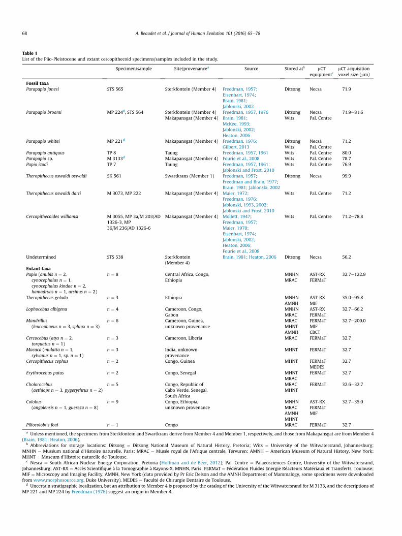
Table 1List of the Plio-Pleistocene and extant cercopithecoid specimens/samples included in the study.
Specimen/sample Site/provenancea Source Stored atb mCTequipmentc
mCT acquisitionvoxel size (mm)
Fossil taxaParapapio jonesi STS 565 Sterkfontein (Member 4) Freedman, 1957;
Eisenhart, 1974;Brain, 1981;Jablonski, 2002
Ditsong Necsa 71.9
Parapapio broomi MP 224d, STS 564 Sterkfontein (Member 4)Makapansgat (Member 4)
Freedman, 1957, 1976Brain, 1981;McKee, 1993;Jablonski, 2002;Heaton, 2006
DitsongWits
NecsaPal. Centre
71.9e81.6
Parapapio whitei MP 221d Makapansgat (Member 4) Freedman, 1976;Gilbert, 2013
DitsongWits
NecsaPal. Centre
71.2
Parapapio antiquus TP 8 Taung Freedman, 1957, 1961 Wits Pal. Centre 80.0Parapapio sp. M 3133d Makapansgat (Member 4) Fourie et al., 2008 Wits Pal. Centre 78.7Papio izodi TP 7 Taung Freedman, 1957, 1961;
Jablonski and Frost, 2010Wits Pal. Centre 76.9
Theropithecus oswaldi oswaldi SK 561 Swartkrans (Member 1) Freedman, 1957;Freedman and Brain, 1977;Brain, 1981; Jablonski, 2002
Ditsong Necsa 99.9
Theropithecus oswaldi darti M 3073, MP 222 Makapansgat (Member 4) Maier, 1972;Freedman, 1976;Jablonski, 1993, 2002;Jablonski and Frost, 2010
Wits Pal. Centre 71.2
Cercopithecoides williamsi M 3055, MP 3a/M 203/AD1326-3, MP36/M 236/AD 1326-6
Makapansgat (Member 4) Mollett, 1947;Freedman, 1957;Maier, 1970;Eisenhart, 1974;Jablonski, 2002;Heaton, 2006;Fourie et al., 2008
Wits Pal. Centre 71.2e78.8
Undetermined STS 538 Sterkfontein(Member 4)
Brain, 1981; Heaton, 2006 Ditsong Necsa 56.2
Extant taxaPapio (anubis n ¼ 2,
cynocephalus n ¼ 1,cynocephalus kindae n ¼ 2,hamadryas n ¼ 1, ursinus n ¼ 2)
n ¼ 8 Central Africa, Congo,Ethiopia
MNHNMRAC
AST-RXFERMaT
32.7e122.9
Theropithecus gelada n ¼ 3 Ethiopia MNHNAMNH
AST-RXMIF
35.0e95.8
Lophocebus albigena n ¼ 4 Cameroon, Congo,Gabon
MNHNMRAC
AST-RXFERMaT
32.7e66.2
Mandrillus(leucophaeus n ¼ 3, sphinx n ¼ 3)
n ¼ 6 Cameroon, Guinea,unknown provenance
MRACMHNTAMNH
FERMaTMIFCBCT
32.7e200.0
Cercocebus (atys n ¼ 2,torquatus n ¼ 1)
n ¼ 3 Cameroon, Liberia MRAC FERMaT 32.7
Macaca (mulatta n ¼ 1,sylvanus n ¼ 1, sp. n ¼ 1)
n ¼ 3 India, unknownprovenance
MHNT FERMaT 32.7
Cercopithecus cephus n ¼ 2 Congo, Guinea MHNT FERMaTMEDES
32.7
Erythrocebus patas n ¼ 2 Congo, Senegal MHNTMRAC
FERMaT 32.7
Cholorocebus(aethiops n ¼ 3, pygerythrus n ¼ 2)
n ¼ 5 Congo, Republic ofCabo Verde, Senegal,South Africa
MRACMHNT
FERMaT 32.6e32.7
Colobus(angolensis n ¼ 1, guereza n ¼ 8)
n ¼ 9 Congo, Ethiopia,unknown provenance
MNHNMRACAMNHMHNT
AST-RXFERMaTMIF
32.7e35.0
Piliocolobus foai n ¼ 1 Congo MRAC FERMaT 32.7
a Unless mentioned, the specimens from Sterkfontein and Swartkrans derive fromMember 4 and Member 1, respectively, and those fromMakapansgat are fromMember 4(Brain, 1981; Heaton, 2006).
b Abbreviations for storage locations: Ditsong ¼ Ditsong National Museum of Natural History, Pretoria; Wits ¼ University of the Witwatersrand, Johannesburg;MNHN ¼ Mus�eum national d'Histoire naturelle, Paris; MRAC ¼ Mus�ee royal de l'Afrique centrale, Tervuren; AMNH ¼ American Museum of Natural History, New York;MHNT ¼ Museum d'Histoire naturelle de Toulouse.
c Nesca ¼ South African Nuclear Energy Corporation, Pretoria (Hoffman and de Beer, 2012); Pal. Centre ¼ Palaeosciences Centre, University of the Witwatersrand,Johannesburg; AST-RX ¼ Acc�es Scientifique �a la Tomographie �a Rayons-X, MNHN, Paris; FERMaT ¼ F�ed�eration Fluides Energie R�eacteurs Mat�eriaux et Transferts, Toulouse;MIF ¼ Microscopy and Imaging Facility, AMNH, New York (data provided by Pr Eric Delson and the AMNH Department of Mammalogy, some specimens were downloadedfrom www.morphosource.org, Duke University), MEDES ¼ Facult�e de Chirurgie Dentaire de Toulouse.
d Uncertain stratigraphic localization, but an attribution to Member 4 is proposed by the catalog of the University of the Witwatersrand for M 3133, and the descriptions ofMP 221 and MP 224 by Freedman (1976) suggest an origin in Member 4.
A. Beaudet et al. / Journal of Human Evolution 101 (2016) 65e7868

Figure 1. Successive processing steps in the deformation-based shape comparisons. First, the surfaces are rigidly aligned in position, orientation, and scale with respect to areference surface (A). From an initial set of aligned surfaces and an ellipsoidal template (B), the deformation fields, the global mean shape (GMS), and the registered surfaces arecomputed (C) and the taxon mean shapes are generated (D). The GMS is subsequently deformed to the fossil specimens (E). The distances from the GMS to the specimens arerendered by color maps and vectors (F). In C and E, the arrows represent the deformations from the GMS to individuals. N indicates the maximum number of specimens included inthe sample.
A. Beaudet et al. / Journal of Human Evolution 101 (2016) 65e78 69
Based on the deformation framework, we generated (i) a GMSassociated with a set of deformations from the GMS to eachendocranium (hereinafter called “GMS-to-individuals”) computedfrom our extant sample, (ii) a TMS for each extant genus investi-gated in our study and represented by more than two individuals,and (iii) deformations from the GMS to fossil endocasts. In additionto GMS-to-individuals deformations, the GMSwas also deformed tothe TMS (hereinafter called “GMS-to-TMS”).
The magnitude of the displacements recorded during thedeformation process (i.e., GMS-to-individuals and GMS-to-TMS)were rendered by color maps from dark blue (in the web version)(lowest displacement values) to red (highest displacement values)on the endocast surfaces (i.e., TMS and individual endocasts;Fig. 1F). In combination with the cartographies, the directions andmagnitudes of the deformations from the GMS to the TMS/in-dividuals are represented by vectors (Fig. 1F).
2.6. Semi-automatic sulci detection
Some cortical details, including the sulcal imprints, wereevident inmost of the fossil specimens (SOM Figs. S1 and S2). Sulcalpattern is usually identified and described by visual inspection ofthe endocranial surface. Based on previous studies (Subsol, 1995,1998), we here use an automatic method for the identification ofneocortical relief in endocasts that includes the algorithm pre-sented by Yoshizawa et al. (2007, 2008) for the detection of topo-graphical variations (i.e., crest lines) in 3D meshes. The sulci areconsidered to be variation points of the surface on a triangle meshand detected via a geometry-based method using curvature linescomputed on the surface. More precisely, the crest lines are definedas salient subsets of the extrema of the principal curvatures onsurfaces. The detected structures were corrected manually by
removing the non-anatomical features (e.g., cracks due to tapho-nomic damage), using “The Primate Brain Bank” (www.primatebrainbank.org) and published studies of cercopithecoidendocasts as references (e.g., Connolly, 1950; Falk, 1978, 1981). This“cleaning” step was performed using a program created withMATLAB R2013a v8.1 (Mathworks) by one of us (J.D.).
2.7. Multivariate analyses
The deformation fields integrating local orientation and theamplitude of the deformations from the GMS to each specimen (i.e.,GMS-to-individuals) were statistically analyzed by computing abetween-group principal component analysis (bgPCA; Durrleman,2010; Mitteroecker and Bookstein, 2011). Based on the covariancematrix of the predefined extant group means, the fossil specimenswere subsequently projected into the shape space. Shape changesand morphological trends observed in the specimens/clustersplotted were depicted by color maps and vectors.
A hierarchical clustering on principal components (HCPC) tree,integrating all the bgPCA components, was generated using theFactoMineR package (Le et al., 2008) for R v.3.2.1 (R DevelopmentCore Team, 2015).
3. Results
3.1. Endocranial volumes
The endocranial volumes measured for both extant and fossilcercopithecoid specimens are presented in Table 2 and plottedagainst body mass in Figure 2. Of these species, Chlorocebus,Erythrocebus, and Cercopithecus have the smallest ECVs relative tobody mass. Taking their “medium” body masses into account, the
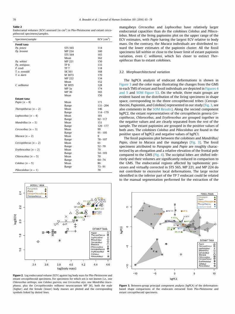
Table 2Endocranial volumes (ECV) assessed (in cm3) in Plio-Pleistocene and extant cerco-pithecoid specimens/samples.
Specimen/sample ECV (cm3)
Fossil taxaPp. jonesi STS 565 114Pp. broomi MP 224 160
STS 564 165Mean 162
Pp. whitei MP 221 150Pp. antiquus TP 8 133P. izodi TP 7 118T. o. oswaldi SK 561 168T. o. darti M 3073 170
MP 222 134Mean 152
C. williamsi M 3055 128MP 3a 174MP 36 165Mean 156
Extant taxaPapio (n ¼ 8) Mean 171
Range 131e204Theropithecus (n ¼ 2) Mean 146
Range 114e179Lophocebus (n ¼ 4) Mean 101
Range 92e117Mandrillus (n ¼ 5) Mean 151
Range 129e177Cercocebus (n ¼ 3) Mean 99
Range 91e105Macaca (n ¼ 2) Mean 9
Range 78e107Cercopithecus (n ¼ 2) Mean 61
Range 52e70Erythrocebus (n ¼ 2) Mean 79
Range 54e103Chlorocebus (n ¼ 5) Mean 70
Range 64e74Colobus (n ¼ 5) Mean 86
Range 73e91Piliocolobus (n ¼ 1) 70
Figure 2. Log endocranial volume (ECV) against log body mass for Plio-Pleistocene andextant cercopithecoid specimens. For specimens for which sex is not known (i.e., oneChlorocebus aethiops, one Colobus guereza, one Cercocebus atys, one Mandrillus leuco-phaeus, plus the Cercopithecoides williamsi neurocranium MP 36), both the male(higher) and the female (lower) body masses are plotted and the correspondingsymbols linked by dotted lines.
A. Beaudet et al. / Journal of Human Evolution 101 (2016) 65e7870
mangabeys Cercocebus and Lophocebus have relatively largerendocranial capacities than do the colobines Colobus and Pilioco-lobus. Most of the living papionins plot on the upper range of theECV estimates, with Papio having the largest ECV relative to bodymass. On the contrary, the Macaca individuals are distributed to-ward the lower estimates of the papionin cluster. All the fossilspecimens fall within or close to the lower limit of extant papioninvariation, even C. williamsi, which lies closer to extinct Ther-opithecus than to extant colobines.
3.2. Morphoarchitectural variation
The bgPCA analysis of endocast deformations is shown inFigure 3 and the color maps illustrating the changes from the GMSto each TMS of extant and fossil individuals are depicted in Figures 4and 5 and SOM Figure S3. On the whole, three main groups areevident based on the distribution of the living specimens in shapespace, corresponding to the three cercopithecoid tribes (Cercopi-thecini, Papionini, and Colobini) represented in our study (Fig. 3, seealso comments in the SOM Results). Along the second componentbgPC2, the extant representatives of the cercopithecin genera Cer-copithecus, Chlorocebus, and Erythrocebus are grouped together inthe negative values and are clearly separated from the rest of thesample. The extant papionins are grouped in the positive values ofboth axes. The colobines Colobus and Piliocolobus are found in thepositive space of bgPC2 and negative values of bgPC1.
The fossil papionins plot between the colobines and Mandrillus/Papio, close to Macaca and the mangabeys (Fig. 3). The fossilspecimens attributed to Parapapio and Papio are roughly charac-terized by an elongation and a relative elevation of the frontal polecompared to the GMS (Fig. 4). The occipital lobes are shifted infe-riorly and their volumes are significantly reduced in comparison tothe GMS. The endocranial regions affected by taphonomic pro-cesses and virtually corrected in STS 565, MP 221, and MP 224 donot contribute to excessive local deformations. The large vectoridentified in the inferior part of the TP 7 endocast could be relatedto the manual segmentation performed for the extraction of the
Figure 3. Between-group principal component analysis (bgPCA) of the deformation-based shape comparisons of the endocasts extracted from Plio-Pleistocene andextant cercopithecoid specimens.
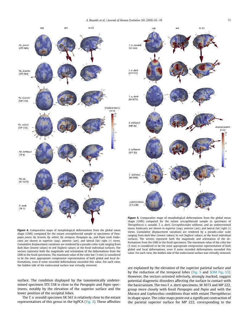
Figure 4. Comparative maps of morphological deformations from the global meanshape (GMS) computed for the extant cercopithecoid sample to specimens of Para-papio jonesi, Pp. broomi, Pp. whitei, Pp. antiquus, Parapapio sp., and Papio izodi. Endo-casts are shown in superior (sup), anterior (ant), and lateral (lat) right (r) views.Cumulative displacement variations are rendered by a pseudo-color scale ranging fromdark blue (lowest values) to red (highest values) at the fossil individual surfaces. Thevectors represent both the magnitude and orientation of the deformations from theGMS to the fossil specimens. The maximum value of the color bar (3 mm) is consideredto be the most appropriate compromise representation of both global and local de-formations, even if some recorded deformations exceeded this value. For each view,the hidden side of the endocranial surface was virtually removed.
Figure 5. Comparative maps of morphological deformations from the global meanshape (GMS) computed for the extant cercopithecoid sample to specimens ofTheropithecus o. oswaldi, T. o. darti, Cercopithecoides williamsi, and an undeterminedtaxon. Endocasts are shown in superior (sup), anterior (ant), and lateral (lat) right (r)views. Cumulative displacement variations are rendered by a pseudo-color scaleranging from dark blue (lowest values) to red (highest values) at the fossil individualsurfaces. The vectors represent both the magnitude and orientation of the de-formations from the GMS to the fossil specimens. The maximum value of the color bar(3 mm) is considered to be the most appropriate compromise representation of bothglobal and local deformations, even if some recorded deformations exceeded thisvalue. For each view, the hidden side of the endocranial surface was virtually removed.
A. Beaudet et al. / Journal of Human Evolution 101 (2016) 65e78 71
surface. The condition displayed by the taxonomically undeter-mined specimen STS 538 is close to the Parapapio and Papio spec-imens, notably by the elevation of the superior surface and thelower position of the occipital lobes.
The T. o. oswaldi specimen SK 561 is relatively close to the extantrepresentatives of this genus in the bgPCA (Fig. 3). These affinities
are explained by the elevation of the superior parietal surface andby the reduction of the temporal lobes (Fig. 5 and SOM Fig. S3).However, the vectors oriented inferiorly, strongly marked, suggestpotential diagenetic disorders affecting the surface in contact withthe basicranium. The two T. o. darti specimens, M 3073 and MP 222,group more closely with fossil Parapapio and Papio and with theMacaca and Lophocebus conditions than with extant Theropithecusin shape space. The color maps point out a significant contraction ofthe parietal superior surface for MP 222, corresponding to the
A. Beaudet et al. / Journal of Human Evolution 101 (2016) 65e7872
reconstructed region and responsible for the relative elevation ofthe occipital and frontal lobes. The M 3073 specimen is close to theGMS and differs from the reference mainly by a bi-temporalenlargement.
The deformation pattern displayed by the fossil specimen M3055, attributed to C. williamsi and included in extant colobinevariability in the bgPCA, is shared with extant colobines (Fig. 5 andSOM Fig. S3). On the contrary, the two other C. williamsi specimens,MP 36 and MP 3a, are closer to the fossil papionins than to theextant colobines. MP 3a shares temporal lobe reduction with livingcolobines as illustrated by the vectors oriented ventro-dorsally(Fig. 5 and SOM Fig. S3), although it also has substantial contrac-tion of the left occipital lobe.
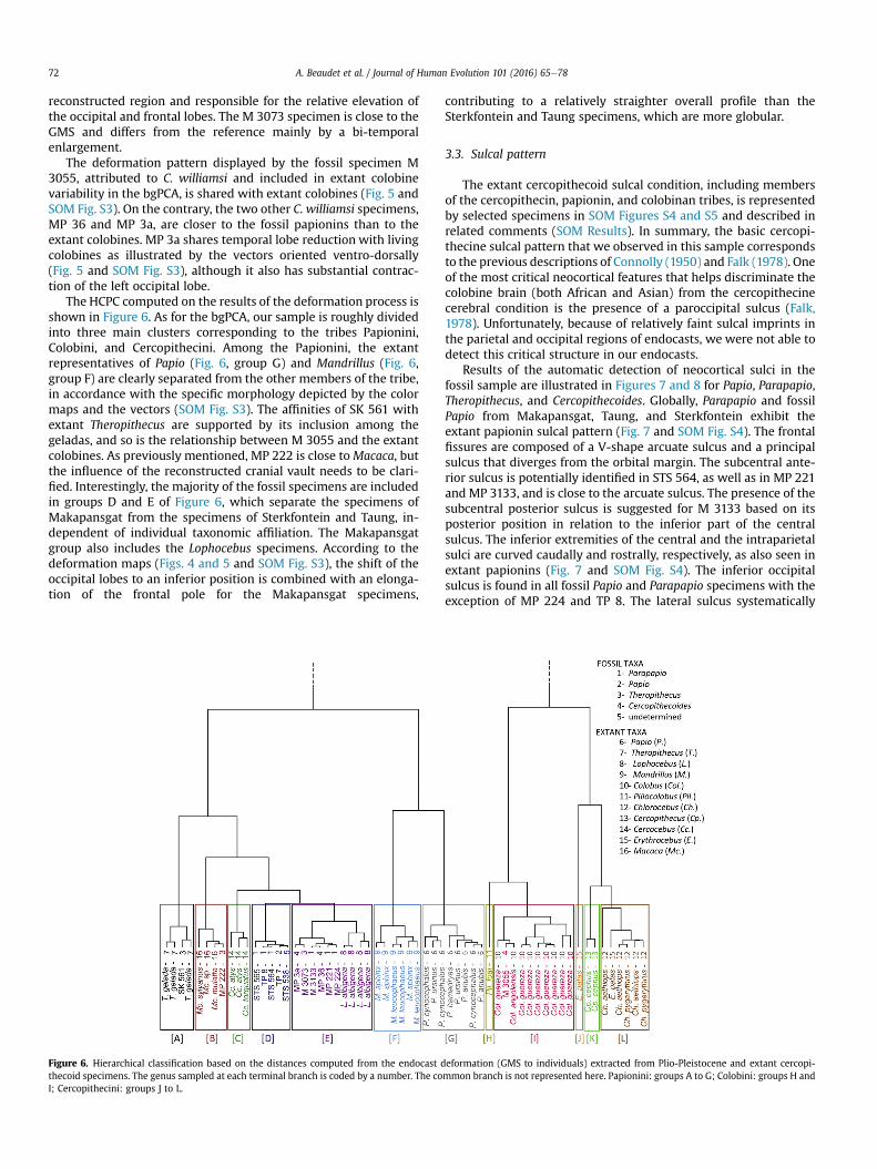
The HCPC computed on the results of the deformation process isshown in Figure 6. As for the bgPCA, our sample is roughly dividedinto three main clusters corresponding to the tribes Papionini,Colobini, and Cercopithecini. Among the Papionini, the extantrepresentatives of Papio (Fig. 6, group G) and Mandrillus (Fig. 6,group F) are clearly separated from the other members of the tribe,in accordance with the specific morphology depicted by the colormaps and the vectors (SOM Fig. S3). The affinities of SK 561 withextant Theropithecus are supported by its inclusion among thegeladas, and so is the relationship between M 3055 and the extantcolobines. As previously mentioned, MP 222 is close toMacaca, butthe influence of the reconstructed cranial vault needs to be clari-fied. Interestingly, the majority of the fossil specimens are includedin groups D and E of Figure 6, which separate the specimens ofMakapansgat from the specimens of Sterkfontein and Taung, in-dependent of individual taxonomic affiliation. The Makapansgatgroup also includes the Lophocebus specimens. According to thedeformation maps (Figs. 4 and 5 and SOM Fig. S3), the shift of theoccipital lobes to an inferior position is combined with an elonga-tion of the frontal pole for the Makapansgat specimens,
Figure 6. Hierarchical classification based on the distances computed from the endocast dthecoid specimens. The genus sampled at each terminal branch is coded by a number. The coI; Cercopithecini: groups J to L.
contributing to a relatively straighter overall profile than theSterkfontein and Taung specimens, which are more globular.
3.3. Sulcal pattern
The extant cercopithecoid sulcal condition, including membersof the cercopithecin, papionin, and colobinan tribes, is representedby selected specimens in SOM Figures S4 and S5 and described inrelated comments (SOM Results). In summary, the basic cercopi-thecine sulcal pattern that we observed in this sample correspondsto the previous descriptions of Connolly (1950) and Falk (1978). Oneof the most critical neocortical features that helps discriminate thecolobine brain (both African and Asian) from the cercopithecinecerebral condition is the presence of a paroccipital sulcus (Falk,1978). Unfortunately, because of relatively faint sulcal imprints inthe parietal and occipital regions of endocasts, we were not able todetect this critical structure in our endocasts.
Results of the automatic detection of neocortical sulci in thefossil sample are illustrated in Figures 7 and 8 for Papio, Parapapio,Theropithecus, and Cercopithecoides. Globally, Parapapio and fossilPapio from Makapansgat, Taung, and Sterkfontein exhibit theextant papionin sulcal pattern (Fig. 7 and SOM Fig. S4). The frontalfissures are composed of a V-shape arcuate sulcus and a principalsulcus that diverges from the orbital margin. The subcentral ante-rior sulcus is potentially identified in STS 564, as well as in MP 221and MP 3133, and is close to the arcuate sulcus. The presence of thesubcentral posterior sulcus is suggested for M 3133 based on itsposterior position in relation to the inferior part of the centralsulcus. The inferior extremities of the central and the intraparietalsulci are curved caudally and rostrally, respectively, as also seen inextant papionins (Fig. 7 and SOM Fig. S4). The inferior occipitalsulcus is found in all fossil Papio and Parapapio specimens with theexception of MP 224 and TP 8. The lateral sulcus systematically
eformation (GMS to individuals) extracted from Plio-Pleistocene and extant cercopi-mmon branch is not represented here. Papionini: groups A to G; Colobini: groups H and

A. Beaudet et al. / Journal of Human Evolution 101 (2016) 65e78 73
intercepts the superior temporal sulcus except in the left hemi-sphere of MP 221. Finally, the overall fissure pattern observed in thetaxonomically undetermined specimen STS 538 is close to theParapapio condition.
While the principal sulcus position in T. o. darti is close to thepapionin pattern (Fig. 8), the arcuate sulcus is relatively curved,as in colobines (SOM Fig. S5). The subcentral anterior sulcus isidentified on the right hemisphere of M 3073 and is connected tothe arcuate sulcus. The subcentral posterior sulcus on the lefthemisphere of M 3073 is linked with the central sulcus, whereas
Figure 7. Virtual reconstructions of Parapapio endocasts with sulcal impressions in superintraparietalis, l ¼ sulcus lunatus, lc ¼ sulcus calcarinus lateralis, oci ¼ sulcus occipitalissca ¼ sulcus subcentralis anterior, scp ¼ sulcus subcentralis posterior). Only sulci mentionedare not to scale.
the central sulcus curves sharply in a caudal direction in theright hemisphere (‘hook-like’ configuration). The intraparietaland lunate sulci are relatively straight and the inferior occipitalsulcus is identified only on the left hemisphere in the case of M3073.
For C. williamsi, except in the left hemisphere of M 3055, thelateral and temporal superior sulci converge but do not intercept, ina similar fashion to Colobus. The frontal sulcal pattern in C. williamsispecimens MP 3a and MP 36 is similar to that of the extantpapionins, based on the presence of relatively straight grooves and
ior (sup), lateral (lat) right (r), and left (l) views (arc ¼ sulcus arcuatus, ip ¼ sulcusinferior, p ¼ sulcus principalis, s ¼ sulcus lateralis, ts ¼ sulcus temporalis superior,in the text are labeled. Question marks indicate uncertain sulcal identifications. Images

Figure 8. Virtual reconstructions of Theropithecus and Cercopithecoides endocasts along with that of the undetermined specimen STS 538, with sulcal impressions in superior (sup),lateral (lat) right (r), and left (l) views (arc ¼ sulcus arcuatus, ip ¼ sulcus intraparietalis, l ¼ sulcus lunatus, lc ¼ sulcus calcarinus lateralis, oci ¼ sulcus occipitalis inferior, p ¼ sulcusprincipalis, s ¼ sulcus lateralis, ts ¼ sulcus temporalis superior, sca ¼ sulcus subcentralis anterior, and scp ¼ sulcus subcentralis posterior). Only sulci mentioned in the text arelabeled. Question marks indicate uncertain sulcal identifications. Images are not to scale.
A. Beaudet et al. / Journal of Human Evolution 101 (2016) 65e7874
the divergence of the principal sulcus from the orbital margin,whereas the configuration in M 3055 is colobine-like (Fig. 8). Theposition of the subcentral anterior sulcus in the right hemispheresof MP 36 and M 3055 is similar to that described for Colobus (SOMFig. S5), but also to fossil papionins (Fig. 7). In all three specimensinvestigated, the inferior end of the central sulcus is curved, as incontemporaneous fossil papionin specimens. The configuration ofthe central sulcus in MP 36 differs from the two other specimens,having a very short cleft connected inferiorly, which could poten-tially be identified as the sulcus subcentralis posterior, contributingto an overall curved aspect. This sulcal arrangement nears the onedetected in T. o. darti in our study.
4. Discussion
4.1. Endocranial volumes
To the best of our knowledge, this current study reports andcompares for the first time the ECVs of Plio-Pleistocene South Af-rican cercopithecoid taxa, with the exception of extinct Ther-opithecus, for which ECVs were previously estimated by Martin(1993) and Elton et al. (2001). Our estimates of T. oswaldi ECVs(134e170 cm3, including both T. o. oswaldi and T. o. darti) fit withinthe fairly wide range estimated by Martin (1993; 154e200 cm3)from three T. oswaldi crania from eastern Africa (Peninj DAT 600/82,
A. Beaudet et al. / Journal of Human Evolution 101 (2016) 65e78 75
a T. oswaldi female, Kanjera BM 32102, a T. oswaldi male, andKanjera BM 14936, a T. oswaldi female). In contrast, ECVs of T. o.oswaldi (168 cm3 for SK 561) and T. o. darti (134 cm3 for MP 222 and170 cm3 for M 3073) reported in our study are higher than the Eltonet al. (2001) estimates (150 cm3 for T. o. oswaldi SK 561, 122 cm3 forT. o. darti MP 222, and 143 cm3 for M 3073; based on regressionequations assessed specifically on Theropithecus). The differencesbetween our and previous studies might be explained by themethods used, as we estimate ECVs directly by virtually filling theneurocranium and extracting the endocranial volume rather thanpacking the braincase with seeds (Martin, 1993) or using regressionequations derived from the relationship between external cranialdimensions and whole cranial capacity in extant Theropithecus andcercopithecoids (Elton et al., 2001). Besides being noninvasive, theassessment of ECVs based on high-resolution imaging techniquesand virtual reconstruction of endocasts is important for under-standing primate paleoneurology, not only because of the moredirect means of ECV estimation but also because ECV can be morereliably delimited through the virtual exploration of internal bonystructures. In addition, our techniques allow the inclusion ofincomplete skulls (e.g., MP 222).
Our current study is focused primarily on methodologicaldevelopment, and hence presents preliminary results. It will needto be extended considerably in the future by integrating a largersample of extant and fossil cercopithecoids (e.g., a total of 339 and22 extant and fossil cercopithecoid specimens respectively weresampled in Elton et al., [2001]) and considering and testing crucialvariables such as sexual dimorphism and allometry. This notwith-standing, in our study, extant Papio has the largest ECVs relative tobody size among both extant and extinct cercopithecoid taxaconsidered, as is also true for brain weight (Martin, 1993). Macacaspecimens (particularly Macaca mulatta) had some of the lowestECV values for cercopithecoids of ‘medium’ body mass. A similarfinding has previously been reported for Macaca sylvanus (Isleret al., 2008) but, as in the present study, interpretation was hin-dered by the small sample. The ECVs computed in our study alsoconform to previous work showing that extant colobines havesmaller brains than other cercopithecoids (Martin, 1993; Isler et al.,2008). One of the most interesting results in our study concernedECV in the fossil colobine C. williamsi, which grouped with the fossilcercopithecine specimens, and at the upper end of the extant cer-copithecoid range. At ~20 kg (Delson et al., 2000), C. williamsi wasmuch larger than extant African colobines, so it is difficult to makedirect comparisons with its closest modern relatives without a fullallometric analysis. Nonetheless, smaller brains (as seen in extantAfrican colobines) may be related to folivory (Martin, 1993), so thefact that C. williamsi, probably a terrestrial forager (Codron et al.,2005), groups with the other large Plio-Pleistocene cercopithe-coids lends support to arguments that it was adaptively different tomodern African colobines. It would be informative to investigatethe ECVs of terrestrial Plio-Pleistocene eastern African Cercopithe-coides taxa estimated to be smaller (C. alemayehui, C. kerioensis,C. meaveae) or bigger (C. kimeui) than C. williamsi (Jablonski andFrost, 2010).
4.2. Taxonomic and evolutionary implications of endocranialorganization
In general, the sulcal patterns we observed in the present studyconformed to those previously described for cercopithecines(Connolly, 1950; Falk, 1978). In Parapapio, the organization of theparietal sulci, and especially the position of the central and intra-parietal sulci, is consistent across individuals. The taxonomicallyundetermined specimen STS 538 shares features with the speci-mens identified as Parapapio, consistent with previous tentative
assignment based on external cranial morphology (Heaton, 2006).The distinction between the gross endocranial morphology of cer-copithecoid specimens from Makapansgat Member 4, on the onehand, and from the slightly younger Taung and SterkfonteinMember 4 assemblages, on the other hand, needs to be explainedby further investigations of the fossil record, such as testing po-tential temporal sensitivity of endocranial organization (Delson,1984, 1988; McKee, 1993; McKee et al., 1995).
Our visualizations also highlighted interesting variation thatadds to our knowledge of sulcal patterns in Old World monkeys. Inthe T. o. darti specimen M 3073, the subcentral posterior sulcus inthe left hemisphere, linked with the central sulcus, appears similarto those described for T. o. oswaldi L 238-29 from Omo (Falk, 1981).When comparing T. o. dartiM3073with the otherMakapansgat T. o.darti, MP 222 (neither of which were available for study by Falk[1981]), as well as the forms from Hadar described by Falk (1981),it is evident that considerable variation exists within the subspe-cies. This variation might overlap with that observed in previouswork (Falk, 1981) on eastern African T. o. oswaldi.
Falk (1981) suggested that the strongly curved (‘hook-like’)inferior end of the central sulcus resulted from the merging of thecentral and subcentral posterior sulci and is derived for T. o. oswaldicompared toT. o. darti. We suggest that T. o. darti fromMakapansgatmay show a transitional gyrification pattern, which could stretchthe appearance of the derived condition back to c. 3Ma (Brock et al.,1977; McFadden et al., 1979; Delson, 1984), rather than to 1.9e2.0Ma as initially proposed by Falk (1981). Moreover, the ‘hook-like’configuration is not found among extant geladas, where the sub-central posterior sulcus remains perceptible. Accordingly, based onthis potential autapomorphic feature, the divergence of the extantT. gelada lineage from the T. oswaldi lineage could predate thechronological period represented by Makapansgat Member 4, asproposed by Jolly (1972). However, given that the mechanism forthe formation of folding in the primate cortex is not yet fully un-derstood (even if probably related to biomechanical constraints[Van Essen, 1997; Hilgetag and Barbas, 2005; Toro and Burnod,2005; Toro, 2012; Bayly et al., 2014; Tallinen et al., 2016]), phylo-genetic interpretations of sulcal pattern should be consideredtentative. It is thus possible that allometric factors influence gyraland sulcal pattern in Theropithecus. This has implications for theway in which we interpret the evolutionary relevance of sulcalpatterns, particularly since a correlation has been reported be-tween increase in brain size and increase in gyrencephaly inmammal orders (Pillay and Manger, 2007). Moreover, because ofthe mechanisms of gyrogenesis (Welker, 1990), variation in devel-opmental processes may also contribute to sulcal patterns inmature individuals. Accordingly, cerebral ontogenetic changes mayalso play a role in the variation seen in the genus Theropithecus,especially given the developmental differences described for extantgeladas compared to other papionins (Swindler and Beynon, 1993).
Another intriguing finding was the variation we observed inC. williamsi endocranial organization. All three specimens includedin this study that are assigned to C. williamsi shared features withColobus, as revealed by the statistical classification of the well-preserved specimen M 3055 among extant colobines, althoughthe morphoarchitecture of C. williamsi endocasts does not mirrorthe pattern seen in extant colobines: specific colobine-like struc-tures such as the paroccipital sulcus were not identified in ourCercopithecoides sample, even if as pointed out by Tobias (1987:746) “valuable information may flow from the presence of a sulcusor of a convolutional bulge on an endocast, whereas the absence ofan impression may be of dubious morphological significance.” Onespecimen, MP 36, also showed a configuration of the inferior part ofthe central sulcus very similar to the T. o. darti specimen M 3073.MP 36 is mainly represented by the neurocranium and meager
A. Beaudet et al. / Journal of Human Evolution 101 (2016) 65e7876
tooth fragments, and has variously been attributed to Simopithecusdarti (by R. Broom and J.S. Jensen; see Freedman [1957]), Pp. broomi(Freedman, 1957; Eisenhart, 1974), and, most recently, C. williamsi(Fourie et al., 2008). Given the sulcal similarities we observed be-tween MP 36 and M 3073, it is possible that MP 36 is actually T. o.darti, although more research to illuminate species-specific varia-tion in cercopithecoids would be desirable. Additional researchwould also be useful to help understand the evolutionary history ofsulcal patterns in colobines. The extant colobine sulcal pattern asdescribed by Falk (1981) in African and Asian taxa, characterizednotably by the presence of an arched intraparietal sulcus and thedivergence of the principal sulcus from the orbital border, amongother aspects, is present in Mesopithecus pentelici (Radinsky, 1974),a Eurasian Pliocene colobine. Recent work has suggested thatC. williamsi is a definitive member of the African colobine clade(Frost et al., 2015), implying that modern African colobines aremore distantly related toMesopithecus than they are to C. williamsi.Thus, similarities in the sulcal patterns of Mesopithecus and extantAfrican colobines must either have arisen convergently, or Cerco-pithecoides shows sulcal morphology that is derived relative toother colobines. Our knowledge of this would also be enhancedfurther by including C. haasgati (McKee et al., 2011), and indeedother large African colobines, into analyses.
It must also be considered that sulcal pattern may be linked tofunction, and it is possible that functional convergence may explainsome of the similarities between C. williamsi and papionins. Basedon sulcal pattern, Falk (1981) hypothesized that T. oswaldi had arelatively expanded cortical somatic sensory and motor face rep-resentation compared to the geladas, potentially due to thespecialization for mastication of abrasive food. However, makinginterpretations of function from sulcal pattern is problematic: someinconsistencies in correspondence between sulcal and cytoarchi-tectural areas have been identified, meaning that the cerebral areasdelimitated by sulci on the external cortical surface do not sys-tematically coincide with functional areas (Amunts et al., 1999; butsee; Fischl et al., 2008). Hence, very little can be reasonably inferredabout function from our data.
5. Conclusions
Despite the relative abundance of nearly complete cercopithe-coid neurocrania in the paleontological assemblages, endocranialcondition has not yet been fully detailed in the South African Plio-Pleistocene records, mainly because of the difficulties related to theextraction of fossil endocasts. Through non-destructive X-ray ra-diation based high-resolution 3D tomography imaging techniques,we successfully reconstructed virtual endocrania from a represen-tative sample of fossil cercopithecoids and reported ECVs for thefirst time, as well as morphological and structural variations. Inparticular, we contributed more information regarding the varia-tion in sulcal pattern in Theropithecus. As previously suggested inthe eastern African context, the Theropithecus fossil lineages pre-sent specific derived sulcal patterns, suggested to be useful fortaxonomic identification, especially of isolated neurocrania. Theidentification of this landmark feature in the Makapansgat recordsupports the possibility of the divergence between the Ther-opithecus fossil lineages and T. gelada prior to 3 Ma. Our investi-gation of C. williamsi endocasts reveals an ECV and sulcal patternsimilar to the papionin condition. Given that Cercopithecoides, acommitted terrestrialist, differed substantially from the arborealexistence observed in its living counterparts, similarities withpapionins in terms of cortical organization might potentially sup-port the hypothesis of an extinct colobine taxon that was adaptivelydifferent from modern African colobines.
Besides traditional analyses of the outer cranial morphology,high resolution virtual imaging of fossil primate endocasts allowingthe automatic detection of neocortical relief coupled withdeformation-based models and statistical analyses have the po-tential to add a significant amount of taxonomic and phylogeneticinformation. Further research in virtual endocranial paleo-primatology to be developed at a continental scale, including fossilspecimens from the rich eastern African cercopithecoid-bearingsites and notably representatives of the Theropithecus genus(Jablonski, 1993; Frost, 2001, 2007; Frost and Delson, 2002; Frostand Alemseged, 2007; Jablonski and Leakey, 2008; Gilbert et al.,2011; Frost et al., 2014), would better clarify the polarity, in-tensity, and timing of the evolutionary changes among cercopi-thecoid lineages.
Acknowledgments
We are indebted to S. Potze (Pretoria), B. Zipfel (Johannesburg), J.Cuisin (Paris), G. Fleury (Toulouse), andW.Wendelen (Tervuren) forhaving granted access to fossil and comparative material undertheir care. We also thank K. Carlson and T. Jashashvili (Johannes-burg), G. Cl�ement and M. Garcia-Sanz (Paris), L. Bam (Pretoria), andD. Maret and E. Coudrais (Toulouse) for (micro-X-ray) tomographicand CBCT acquisitions, and E. Delson (New York) for providing CTscans. For scientific contribution and/or for discussion and com-ments to the results presented in this study, we are especiallygrateful to E. Delson (New York), D. Ginibriere (Toulouse), J. Heaton(Birmingham), N. Jablonski (University Park), O. Kullmer (Frank-furt), R. Macchiarelli (Poitiers & Paris), M. Nakatsukasa (Kyoto), L.Pan (Toulouse), and C. Zanolli (Toulouse). The present versiongreatly benefited from the comments provided by the Editor, theAssociate Editor, and three anonymous reviewers. The Frenchresearch federation FERMaT (FR3089), the National ResearchFoundation (NRF), and Department of Science and Technology(DST) of South Africa are acknowledged for providing micro-X-raytomography laboratory facilities. This work was granted access tothe HPC resources of CALMIP supercomputing center under theallocation 2015-[P1440] attributed to the AMIS laboratory. Researchsupported by the Center of Research and Higher Education (PRES)of Toulouse, the Midi-Pyr�en�ees Region, and the French Ministry ofForeign Affairs.
Supplementary Online Material
Supplementary online material related to this article can befound at http://dx.doi.org/10.1016/j.jhevol.2016.09.003.
References
Amunts, K., Schleicher, A., Bürgel, U., Mohlberg, H., Uylings, H.B.M., Zilles, K., 1999.Broca's region revisited: cytoarchitecture and intersubject variability. J. Comp.Neurol. 412, 319e341.
Armstrong, E., Falk, D., 1982. Primate Brain Evolution. Methods and Concepts.Plenum Press, New York.
Bayly, P., Taber, L., Kroenke, C., 2014. Mechanical forces in cerebral cortical folding: areview of measurements and models. J. Mech. Behav. Biomed. Mater. 29,568e581.
Beaudet, A., 2015. Caract�erisation des structures cranio-dentaires internes descercopith�ecoïdes et �etude diachronique de leurs variations morphologiquesdans la s�equence Plio-Pl�eistoc�ene sud-africaine. Ph.D. Dissertation, Universit�ede Toulouse.
Beaudet, A., Zanolli, C., Engda Redae, B., Endalamaw, M., Braga, J., Macchiarelli, R.,2015. A new cercopithecoid dentognathic specimen attributed to Theropithecusfrom the late Early Pleistocene (c. 1 Ma) deposits of Simbiro, at Melka Kunture,Ethiopian highlands. Comptes Rendus Palevol. 14, 657e669.
Beaudet, A., Braga, J., de Beer, F., Schillinger, B., Steininger, C., Vodopivec, V.,Zanolli, C., 2016. Neutron microtomography-based virtual extraction andanalysis of a cercopithecoid partial cranium (STS 1039) embedded in a brecciafragment from Sterkfontein Member 4 (South Africa). Am. J. Phys. Anthropol.159, 737e745.
A. Beaudet et al. / Journal of Human Evolution 101 (2016) 65e78 77
Benazzi, S., Bookstein, F.L., Strait, D.S., Weber, G.W., 2011. A new OH5 reconstructionwith an assessment of its uncertainty. J. Hum. Evol. 61, 75e88.
Birchette, M.G., 1981. Postcranial remains of Cercopithecoides. Am. J. Phys. Anthro-pol. 54, 201.
Brain, C.K., 1981. The Hunters of the Hunted? An introduction to African CaveTaphonomy. University of Chicago Press, Chicago.
Brock, A., McFadden, P.L., Partridge, T.C., 1977. Preliminary paleomagnetic resultsfrom Makapansgat and Swartkrans. Nature 266, 249e250.
Bruner, E., 2004. Geometric morphometrics and paleoneurology: brain shapeevolution in the genus Homo. J. Hum. Evol. 47, 279e303.
Bruner, E., Manzi, G., Arsuaga, J.L., 2003. Encephalization and allometric trajectoriesin the genus Homo: evidence from the Neandertal and modern lineages. Proc.Natl. Acad. Sci. USA 100, 15335e15340.
Bruner, E., Mantini, S., Ripani, M., 2009. Landmark-based analysis of the morpho-logical relationship between endocranial shape and traces of the middlemeningeal vessels. Anat. Rec. 292, 518e527.
Bruner, E., Martin-Loeches, M., Colom, R., 2010. Human midsagittal brain shapevariation: patterns, allometry and integration. J. Anat. 216, 589e599.
Codron, D., Luyt, J., Lee-Thorp, J., Sponheimer, M., deRuiter, D., Codron, J., 2005.Utilization of savanna-based resources by baboons during the Plio-Pleistocene.S. Afr. J. Sci. 10, 245e248.
Connolly, C.J., 1950. External Morphology of the Primate Brain. C.C. Thomas,Springfield.
Delson, E., 1975. Evolutionary history of the Cercopithecidae. In: Szalay, F.S. (Ed.),Approaches to Primate Paleobiology. Contributions to Primatology 4, Karger,Basel, pp. 167e217.
Delson, E., 1984. Cercopithecoid biochronology of the African Plio-Pleistocene:correlation among eastern and southern hominid-bearing localities. Cour.Forsch. Inst. Senckenberg 69, 199e281.
Delson, E., 1988. Chronology of South African australopiths site units. In: Grine, F.E.(Ed.), Evolutionary History of the ‘Robust’ Australopithecines. Aldine de Gruyter,New York, pp. 317e324.
Delson, E., Terranova, C.J., Jungers, W.L., Sargis, E.J., Jablonski, N.G., Dechow, P.C.,2000. Body mass in Cercopithecidae (Primates, Mammalia): estimation andscaling in extinct and extant taxa. Anthropol. Pap. Am. Mus. Nat. Hist. 83, 1e159.
Dumoncel, J., Durrleman, S., Braga, J., Jessel, J.-P., Subsol, G., 2014. Landmark-free 3Dmethod for comparison of fossil hominins and hominids based on endocraniumand EDJ shapes. Am. J. Phys. Anthropol. 153 (Suppl. 56), 110.
Dumoncel, J., Subsol, G., Durrleman, S., Jessel, J.-P., Beaudet, A., Braga, J., 2016. Howto Build an Average Model When Samples Are Variably Incomplete? Applicationto Fossil Data. The IEEE Conference on Computer Vision and Pattern Recognition(CVPR) Workshops, pp. 101e108.
Durrleman, S., 2010. Statistical Models of Currents for Measuring the Variability ofAnatomical Curves, Surfaces and their Evolution. Ph.D. Dissertation, Universit�eNice-Sophia Antipolis.
Durrleman, S., Pennec, X., Trouv�e, A., Ayache, N., Braga, J., 2012a. Comparison of theendocranial ontogenies between chimpanzees and bonobos via temporalregression and spatiotemporal registration. J. Hum. Evol. 62, 74e88.
Durrleman, S., Prastawa, M., Korenberg, J.R., Joshi, S., Trouv�e, A., Gerig, G., 2012b.Topology preserving atlas construction from shape data without correspon-dence using sparse parameters. In: Ayache, N., Delingette, H., Golland, P.,Mori, K. (Eds.), MICCAI 2012, Part III. LNCS, vol. 7512. Springer, Heidelberg,pp. 223e230.
Durrleman, S., Prastawa, M., Charon, N., Korenberg, J.R., Joshi, S., Gerig, G., Trouv�e, A.,2014. Morphometry of anatomical shape complexes with dense deformationsand sparseparameters. NeuroImage 101, 35e49.
Eisenhart, W.L., 1974. The fossil cercopithecoids of Makapansgat and Sterkfontein.A.B. Thesis, Harvard College.
Elton, S., Bishop, L.C., Wood, B., 2001. Comparative context of Plio-Pleistocenehominin brain evolution. J. Hum. Evol. 41, 1e27.
Falk, D., 1978. Brain evolution in Old World Monkeys. Am. J. Phys. Anthropol. 48,315e320.
Falk, D., 1981. Sulcal patterns of fossil Theropithecus baboons: phylogenetic andfunctional implications. Int. J. Primatol. 2, 57e69.
Falk, D., 2014. Interpreting sulci on hominin endocasts: old hypotheses and newfindings. Front. Hum. Neurosci. 8, 134.
Fischl, B., Rajendran, N., Busa, E., Augustinack, J., Hinds, O., Yeo, B.T., Mohlberg, H.,Amunts, K., Zilles, K., 2008. Cortical folding patterns and predicting cytoarchi-tecture. Cereb. Cortex 18, 1973e1980.
Fourie, N.H., Lee-Thorp, J.A., Ackermann, R.R., 2008. Biogeochemical and cranio-metric investigation of dietary ecology, niche separation, and taxonomy of Plio-Pleistocene cercopithecoids from the Makapansgat Limeworks. Am. J. Phys.Anthropol. 135, 121e135.
Freedman, L., 1957. The fossil Cercopithecoidea of South Africa. Ann. Transv. Mus.23, 121e262.
Freedman, L., 1961. New cercopithecoid fossils, including a new species, from Taung,Cape Province, South Africa. Ann. S. Afr. Mus. 46, 1e14.
Freedman, L., 1976. South African fossil Cercopithecoidea: a re-assessmentincluding a description of new material from Makapansgat, Sterkfontein andTaung. J. Hum. Evol. 5, 297e315.
Freedman, L., Brain, C.K., 1977. A re-examination of the cercopithecoid fossils fromSwartkrans (Mammalia: Cercopithecidae). Ann. Transv. Mus 30, 211e218.
Frost, S.R., 2001. Fossil Cercopithecidae of the Afar Depression, Ethiopia: speciessystematics and comparison to the Turkana Basin. Ph.D. Dissertation, The CityUniversity of New York.
Frost, S.R., 2007. Fossil Cercopithecidae from the Middle Pleistocene DawaitoliFormation, Middle Awash Valley, Afar Region, Ethiopia. Am. J. Phys. Anthropol.134, 460e471.
Frost, S.R., Alemseged, Z., 2007. Middle Pleistocene fossil Cercopithecidae fromAsbole, Afar Region, Ethiopia. J. Hum. Evol. 53, 227e259.
Frost, S.R., Delson, E., 2002. Fossil Cercopithecidae from the Hadar Formation andsurrounding areas of the Afar Depression, Ethiopia. J. Hum. Evol. 43, 687e748.
Frost, S.R., Jablonski, N.G., Haile-Selassie, Y., 2014. Early Pliocene CercopithecidaefromWoranso-Mille (Central Afar, Ethiopia) and the origins of the Theropithecusoswaldi lineage. J. Hum. Evol. 76, 39e53.
Frost, S.R., Gilbert, C.C., Pugh, K.D., Guthrie, E.H., Delson, E., 2015. The hand of Cerco-pithecoideswilliamsi (Mammalia, Primates): earliest evidence for thumb reductionamong colobine monkeys. PLoS One 10 (5), e0125030. http://dx.doi.org/10.1371/journal.pone.0125030.
Fukunishi, K., Sawada, K., Kashima, M., Sakata-Haga, H., Fukuzaki, K., Fukui, Y., 2006.Development of cerebral sulci and gyri in fetuses of cynomolgus monkeys(Macaca fascicularis). Anat. Embryol. (Berl.) 211, 757e764.
Gilbert, C.C., 2007. Craniomandibular morphology supporting the diphyletic originof mangabeys and a new genus of the Cercocebus/Mandrillus clade, Procerco-cebus. J. Hum. Evol. 53, 69e102.
Gilbert, C.C., 2013. Cladistic analysis of extant and fossil African papionins usingcraniodental data. J. Hum. Evol. 64, 399e433.
Gilbert, C.C., Goble, E.D., Kingston, J.D., Hill, A., 2011. Partial skeleton of Ther-opithecus brumpti (Primates, Cercopithecidae) from the Chemeron Formation ofthe Tugen Hills, Kenya. J. Hum. Evol. 61, 347e362.
Glaune
s, J.A., Joshi, S., 2006. Template estimation from unlabeled point set data andsurfaces for computational anatomy. In: Pennec, X., Joshi, S. (Eds.). Proceedingsof the International Workshop on the Mathematical Foundations of Computa-tional Anatomy, Copenhagen, pp. 29e39.
Gonzales, L.A., Benefit, B.R., McCrossin, M.L., Spoor, F., 2015. Cerebral complexitypreceded enlarged brain size and reduced olfactory bulbs in Old World mon-keys. Nat. Commun. 6, 7580.
Gunz, P., 2015. Computed tools for paleoneurology. In: Bruner, E. (Ed.), Humanpaleoneurology. Springer, Switzerland, pp. 39e55.
Gunz, P., Mitteroecker, P., Neubauer, S., Weber, G.W., Bookstein, F.L., 2009. Principlesfor the virtual reconstruction of hominin crania. J. Hum. Evol. 57, 48e62.
Heaton, J.L., 2006. Taxonomy of the Sterkfontein fossil Cercopithecinae: thePapionini of Members 2 and 4 (Gauteng, South Africa). Ph.D. Dissertation,Indiana University.
Hilgetag, C.C., Barbas, H., 2005. Developmental mechanics of the primate cerebralcortex. Anat. Embryol. 210, 411e417.
Hoffman, J.W., de Beer, F.C., 2012. Characteristics of the micro-focus X-ray tomog-raphy facility (MIXRAD) at Necsa in South Africa. In: 18th World Conference ofNondestructive Testing, Durban, South Africa.
Holloway, R.L., 1978. The relevance of endocasts for studying primate brain evolu-tion. In: Noback, C.R. (Ed.), Sensory Systems of Primates. Plenum Press, NewYork, pp. 181e200.
Holloway, R.L., Broadfield, D.C., Yuan, M.S., 2004. The Human Fossil Record: BrainEndocasts, the Paleoneurological Evidence. Wiley-Liss, New York.
Isler, K., Kirk, E.C., Miller, J.M.A., Albrecht, G.A., Gelvin, B.R., Martin, R.D., 2008.Endocranial volumes of primate species: scaling analyses using a comprehen-sive and reliable data set. J. Hum. Evol. 55, 967e978.
Jablonski, N.G., 1993. Theropithecus: The Rise and Fall of a Primate Genus. Cam-bridge University Press, Cambridge.
Jablonski, N.G., 2002. Fossil Old World monkeys: the late Neogene radiation. In:Hartwig, W.C. (Ed.), The Primate Fossil Record. Cambridge University Press,Cambridge, pp. 255e299.
Jablonski, N.G., Frost, S., 2010. Cercopithecoidea. In: Werdelin, L., Sanders, W.J.(Eds.), Cenozoic Mammals of Africa. University of California Press, Berkeley,pp. 393e428.
Jablonski, N.G., Leakey, M.G., 2008. Koobi Fora Research Project. In: The FossilMonkeys, Vol. 6. California Academy of Sciences, San Francisco.
Jolly, C.J., 1972. The classification and natural history of Theropithecus (Simopithecus)(Andrews,1916), baboons of the African Plio-Pleistocene. Bull. Br. Mus. Nat. Hist.Geol. 22, 1e122.
Kashima, M., Sawada, K., Fukunishi, K., Sakata-Haga, H., Tokado, H., Fukui, Y., 2008.Development of cerebral sulci and gyri in fetuses of cynomolgus monkeys(Macaca fascicularis). II. Gross observation of the medial surface. Brain Struct.Funct. 212, 513e520.
Kobayashi, Y., Matsui, T., Haizuka, Y., Ogihara, N., Hirai, N., Matsumura, G., 2014.Cerebral sulci and gyri observed on macaque endocasts. In: Akazawa, T.,Ogihara, N., Tanabe, H.C., Terashima, H. (Eds.), Dynamics of learning in Nean-derthals and modern humans, Volume 2. Springer, Japan, pp. 131e137.
Le, S., Josse, J., Husson, F., 2008. FactoMineR: an R package for multivariate analysis.J. Stat. Softw. 25, 1e18.
Leakey, M.G., 1982. Extinct large colobines from the Plio-Pleistocene of Africa. Am. J.Phys. Anthropol. 58, 153e172.
Leakey, M.G., 1993. Evolution of Theropithecus in the Turkana Basin. In:Jablonski, N.G. (Ed.), Theropithecus: The Rise and Fall of a Primate Genus.Cambridge University Press, Cambridge, pp. 85e123.
Maier, W., 1970. New fossil Cercopithecoidea from the lower Pleistocene cavedeposits of the Makapansgat Limeworks, South Africa. Palaeont. Afr. 13,69e107.
Maier, W., 1972. The first complete skull of Simopithecus darti from Makapansgat,South Africa, and its systematic position. J. Hum. Evol. 1, 395e405.

A. Beaudet et al. / Journal of Human Evolution 101 (2016) 65e7878
Malkova, L., Heuer, E., Saunders, R.C., 2006. Longitudinal magnetic resonance im-aging study of rhesus monkey brain development. Eur. J. Neurosci. 24,3204e3212.
Martin, R.D., 1993. Allometric aspects of skull morphology in Theropithecus. In:Jablonski, N. (Ed.), Theropithecus: The Rise and Fall of a Primate Genus. Cam-bridge University Press, Cambridge, pp. 273e298.
McFadden, P.L., Brock, A., Partridge, T.C., 1979. Palaeomagnetism and the age of theMakapansgat hominid site. Earth Planet. Sci. Lett. 44, 373e382.
McKee, J.K., 1993. Taxonomic and evolutionary affinities of Papio izodi fossils fromTaung and Sterkfontein. Palaeont. Afr. 30, 43e49.
McKee, J.K., Keyser, A.W., 1994. Craniodental remains of Papio angusticeps from theHaasgat cave site, South Africa. Int. J. Primatol. 15, 823e841.
McKee, J.K., Thackeray, J.F., Berger, L.R., 1995. Faunal assemblage seriation ofSouthern African Pliocene and Pleistocene fossil deposits. Am. J. Phys. Anthro-pol. 96, 235e250.
McKee, J.K., von Mayer, A., Kuykendall, K., 2011. New species of Cercopithecoidesfrom Haasgat, North West Province, South Africa. J. Hum. Evol. 60, 83e93.
Mitteroecker, P., Bookstein, F.L., 2011. Linear discrimination, ordination, and thevisualization of selection gradients in modern morphometrics. Evol. Biol. 38,100e114.
Mollett, O.D., 1947. Fossil mammals from the Makapan Valley, Potgietersrust. I.Primates. S. Afr. J. Sci. 43, 295e303.
Neubauer, S., 2014. Endocasts: possibilities and limitations for the interpretation ofhuman brain evolution. Brain Behav. Evol. 84, 117e134.
Neubauer, S., Gunz, P., Hublin, J.-J., 2009. The pattern of endocranial ontogeneticshape changes in humans. J. Anat. 215, 240e255.
Neubauer, S., Gunz, P., Hublin, J.-J., 2010. Endocranial shape changes during growthin chimpanzees and humans: a morphometric analysis of unique and sharedaspects. J. Hum. Evol. 59, 555e566.
Pillay, P., Manger, P.R., 2007. Order-specific quantitative patterns of cortical gyr-ification. Eur. J. Neurosci. 25, 2705e2712.
R. Development Core Team, 2015. R: a language and environment for statisticalcomputing. http://www.R-project.org.
Radinsky, L., 1974. The fossil evidence of anthropoid brain evolution. Am. J. Phys.Anthropol. 41, 15e28.
Sawada, K., Sun, X.Z., Fukunishi, K., Kashima, M., Sakata-Haga, H., Tokado, H., Aoki, I.,Fukui, Y., 2009. Developments of sulcal pattern and subcortical structures of theforebrain in cynomolgus monkey fetuses: 7-tesla magnetic resonance imagingprovides high reproducibility of gross structural changes. Brain Struct. Funct.213, 469e480.
Scott, J.E., 2011. Folivory, frugivory, and postcanine size in the Cercopithecoidearevisited. Am. J. Phys. Anthropol. 146, 20e27.
Smith, B.H., 1991. Dental development and the evolution of life history in Homi-nidae. Am. J. Phys. Anthropol. 86, 157e174.
Smith, R.J., Jungers, W.L., 1997. Body mass in comparative primatology. J. Hum. Evol.32, 523e559.
Specht, M., Lebrun, R., Zollikofer, C.P.E., 2007. Visualizing shape transformationbetween chimpanzee and human braincases. Vis. Comput. 23, 743e751.
Subsol, G., 1995. Construction automatique d'atlas anatomiques morphom�etriques �apartir d'images m�edicales tridimensionnelles. Ph.D. Dissertation, �Ecole Centralede Paris.
Subsol, G., 1998. Crest lines for curve based warping. In: Toga, A. (Ed.), BrainWarping. Academic Press, San Diego, pp. 241e262.
Subsol, G., Gesqui�ere, G., Braga, J., Thackeray, F., 2010. 3D automatic methods tosegment ‘virtual’ endocasts: state of the art and future directions. Am. J. Phys.Anthropol. 141 (Suppl. 50), 226e227.
Swindler, D.R., Beynon, A.D., 1993. The development and microstructure of thedentition of Theropithecus. In: Jablonski, N. (Ed.), Theropithecus: The Rise andFall of a Primate Genus. Cambridge University Press, Cambridge, pp. 351e381.
Szalay, F.S., Delson, E., 1979. Evolutionary History of the Primates. Academic Press,New York.
Tallinen, T., Chung, J.Y., Rousseau, F., Girard, N., Lef�evre, J., Mahadevan, L., 2016. Onthe growth and form of cortical convolutions. Nat. Phys. http://dx.doi.org/10.1038/nphys3632.
Toro, R., 2012. On the possible shapes of the brain. Evol. Biol. 39, 600e612.Toro, R., Burnod, Y., 2005. A morphogenetic model for the development of cortical
convolutions. Cereb. Cortex 15, 1900e1913.Van Essen, D.C., 1997. A tension-based theory of morphogenesis and compact
wiring in the central nervous system. Nature 385, 313e318.Weber, G.W., Bookstein, F.L., 2011. Virtual Anthropology: a guide to a new inter-
disciplinary field. Springer, London.Welker,W.,1990.Whydoes cerebral cortexfissureand fold?Areviewof determinants
of gyri and sulci. In: Jones, E.G., Peters, A. (Eds.), Comparative structure andevolution of cerebral cortex, Part II, vol. 8B. Plenum, New York, pp. 3e136.
Yoshizawa, S., Belyaev, A., Yokota, H., Seidel, H.-P., 2007. Fast and faithful geometricalgorithm for detecting crest lines on meshes. Proceedings of the 15th PacificConference on Computer Graphics and Applications, pp. 231e237.
Yoshizawa, S., Belyaev, A., Yokota, H., Seidel, H.P., 2008. Fast, robust, and faithfulmethods for detecting crest lines on meshes. Computer Aided Geom. D. 25,545e560.
Zollikofer, C.P.E., 2002. A computational approach to paleoanthropology. Evol.Anthropol. 11, 64e67.
Zollikofer, C.P.E., Ponce De Le�on, M.S., Martin, R.D., 1998. Computer-assistedpaleoanthropology. Evol. Anthropol. 6, 41e54.

![A quantitative comparison of the brain and the inner surface of …subsol/WWW/SMEF.0618.Dumoncel.2.pdf · shape data [Durrleman et al 2015, Beaudet et al 2016, Beaudet et al 2017]](https://img.pdfslide.net/doc/110x75/5e7085eb9ab1e952e3755d1d/a-quantitative-comparison-of-the-brain-and-the-inner-surface-of-subsolwwwsmef0618dumoncel2pdf.jpg)


![Untitled-1 [cdn-s3.sappi.com] · Botha, Archie McKeIIar, Sven Karth, Frikkie Rousseau, Okkie Buys and Ben Buys. 6 Early players included Gabriel Nhlebela, Michael Zulu, Bheki Mhlongo,](https://img.pdfslide.net/doc/110x75/5bb9966a09d3f28f6c8c5dc5/untitled-1-cdn-s3sappicom-botha-archie-mckeiiar-sven-karth-frikkie-rousseau.jpg)