Embed Size (px)
Citation preview

Journal of Magnetism and Magnetic Materials xxx (2017) xxx–xxx
Contents lists available at ScienceDirect
Journal of Magnetism and Magnetic Materials
journal homepage: www.elsevier .com/locate / jmmm
Research articles
Superparamagnetic iron oxide-reduced graphene oxide nanohybrid-avehicle for targeted drug delivery and hyperthermia treatment of cancer
http://dx.doi.org/10.1016/j.jmmm.2017.05.0840304-8853/� 2017 Published by Elsevier B.V.
⇑ Corresponding author.E-mail address: [email protected] (D. Bahadur).
1 These authors contributed equally to this work.
Please cite this article in press as: J. Gupta et al., Superparamagnetic iron oxide-reduced graphene oxide nanohybrid-a vehicle for targeted drug dand hyperthermia treatment of cancer, Journal of Magnetism and Magnetic Materials (2017), http://dx.doi.org/10.1016/j.jmmm.2017.05.084
Jagriti Gupta 1, Anand Prakash 1, Manish K. Jaiswal 1, Atanuu Agarrwal, D. Bahadur ⇑Department of Metallurgical Engineering and Materials Science, Indian Institute of Technology Bombay, Mumbai 400076, India
a r t i c l e i n f o
Article history:Received 23 January 2017Received in revised form 8 May 2017Accepted 27 May 2017Available online xxxx
Keywords:DOXNanohybridRGOHeLaHyperthermiaFe3O4
a b s t r a c t
In this work, an efficient superparamagnetic iron oxide-reduced graphene oxide (Fe3O4-RGO) nanohybridhas been synthesized following one-step co-precipitation method. The phase identification, microstruc-ture and magnetic behavior of nanohybrid were characterized by X-ray diffraction, transmission electronmicroscopy (TEM), raman spectroscopy and vibrating sample magnetometer (VSM), respectively. TEMmicrograph confirms the presence of well-segregated Fe3O4 nanoparticles in RGO layers. The layeredRGO minimizes the agglomeration in Fe3O4 nanoparticles with slight reduction in magnetic behavior.Doxorubicin (DOX) has been used as a model drug to investigate the loading efficiency of nanohybridand chemo-thermo therapeutic effect on human cervical cancer (HeLa cells). The DOX loaded nanohybrid(DOX-Fe3O4-RGO) shows maximum inhibition of human cervical cancer cell lines during magnetic fieldassisted hyperthermia treatment. The synergistic effect of nanohybrid demonstrated the potential forcancer cell proliferation prevention up to 90% when treated at the concentration of 2 mg mL�1 for onemillion cells and exposed to AC field of 335 Oe at a fixed frequency of 265 kHz for 35 min.
� 2017 Published by Elsevier B.V.
1. Introduction
Graphene, a monolayer of hexagonally arranged sp2 carbonatoms, is a fascinating material because of unusual electrical, opti-cal, mechanical and thermal properties [1–3]. These propertiesrender graphene suitable for various technological applicationssuch ultrafast graphene based MOSFETs, transparent electrodesfor solar cells, sensors [4], and electrochemical energy storagedevices [3–6]. Recently graphene derivatives (RGO-reducedgraphene oxide, CMG-chemically modified graphene, FG-functionalized graphene and GNR-graphene nanoribbons) havebecome front-runner in graphene based technology because of bio-compatibility in practical concentration range [7,8], large-scaleproduction, cost effective, and ability to integrate with other nano-materials. Most of derivatives contain a wide range of residual oxy-gen functional groups both on the basal planes and at the edgeswith a large number of surface defects. The functional groupsand surface defects are very reactive and can be chemically modi-fied to produce graphene based nanohybrids for anti-bacterialactivities, detection of cancer and in vitro photothermal anticanceractivities [9–11]. Carbon nanotubes (CNTs), a family of graphene,
show good carrier properties by serving as a transporter for biomolecules to the target site, including drugs, vaccines, nucleicacids, vitamins and sugars [12–17]. However, the potential utilityof the derivatives of graphene have not been much explored inother biomedical areas such as targeted drug delivery, thermotherapy of cancer (Hyperthermia) or chemotherapy in spite of hav-ing superior properties than CNTs or other carbon allotropes. Yanget al. exploited the properties of oxygen functional groups of gra-phene derivatives and exhibited the loading of anticancer drugshaving aromatic structures [18]. On the other hand, superparamag-netic magnetic nanoparticles (MNPs), particularly Fe3O4 emergedas an excellent candidate for biomedical applications due to theirbetter chemical stability, biocompatibility, simple processing andeasy functionalization with other nanomaterials [19–21]. Theseunique properties make magnetic nanoparticles versatile for vari-ous biomedical applications especially drug delivery, magnetichyperthermia, magnetic separation, magnetic resonance imaging(MRI) and diagnosis. Patil et al. reported the core shell structureof iron oxide with chitosan for hyperthermia application [22]. Inthe recent time, magnetic hyperthermia has become an importanttool for cancer therapeutics due to its special features such mag-netic selectivity, non-invasive, and harmless to normal cells. Inhyperthermia, cancerous cells are heated above physiological tem-perature [23,24]. It was reported that hyperthermia temperature(42–46 �C) could damage cellular components leading to apoptosis
elivery

2 J. Gupta et al. / Journal of Magnetism and Magnetic Materials xxx (2017) xxx–xxx
[25]. At this temperature, cancerous cells are more susceptible toradiation or chemotherapy. The magnetic core is responsible fordirecting the drugs to a specific site by the action of external mag-netic field. Most of the advanced targeted drug delivery systemscontain Fe3O4 as a core with variety of biodegradable shells includ-ing polymers and proteins [26,27]. The shells might have the func-tion of transporting and releasing drug during their biodegradationprocesses. Recently, Fe3O4-RGO nanocomposite has been shown toexhibit pH responsive properties when combined with anti-cancerchemotherapeutic drug doxorubicin (DOX) [28]. Several systemscontaining Fe3O4 as a core in biodegradable thermo sensitive poly-meric micelles, magnetic liposomes and magnetic lipid nanoparti-cles loading doxorubicin for intracellular delivery have been welldocumented [25,29–32]. However, the magnetic composites withlayered RGO have not been explored much for their efficacy in car-rying/delivering the therapeutics as well as for their cellularresponse. Here, we report a biocompatible, superparamagneticand crystalline Fe3O4-RGO nanohybrid with large surface area thatoffers easy and several binding sites to conjugate the drug mole-cules. Further, in vitro assessment for cancer cell killing efficacyof this nanohybrid through thermo-chemotherapy using humancervical cancer (HeLa) cell lines is reported.
2. Experimental
2.1. Reagents and chemicals
Graphite powder (product No. 496596, 99.99%), Ferric chloridehexahydrate (FeCl3�6H2O- 23648-9) and Ferrous chloride tetrahy-drate (FeCl2�4H2O- 22029-9), doxorubicin (DOX) were purchasedfrom Sigma-Aldrich, USA and are main precursors for the synthesisof Fe3O4-RGO nanohybrids. Other reagents such as N2H4 (80%),H2SO4 (98%), HCl (35%), H2O2 (30%) and KMnO4 were purchasedfrom Thomas Baker, India. All reagents were used without furtherpurification.
2.2. Synthesis of Fe3O4-RGO nanohybrid
The modified Hummer’s method has been used for the synthe-sis of graphite oxide, which involves oxidation and subsequentreduction of graphite powder reported elsewhere [33]. In a typicalprocedure, an excellent dark brown dispersion of graphene oxidewas achieved by ultra-sonication of GO (0.5 mg mL�1). Iron salts,FeCl3�6H2O (5.838 g) and FeCl2�4H2O (3.003 g) in 2:1 M ratios weredispersed into 80 mL suspension of graphene oxide. The mixturewas purged with N2 to eliminate dissolved O2 and stirred for30 min. The mixture was refluxed at 80 �C for another 30 min fol-lowed by quick injection of ammonium hydroxide (NH4OH) undervigorous stirring. Hydrazine hydrate was (1 mL, 80% w/w) addedinto the reaction mixture and further allowed to reflux for the next4 h at 90 �C. After completion of reaction, the obtained black mag-netic precipitate was washed several times with water/ethanol anddried in air.
2.3. DOX loading and release
The layered structure and availability of functional groups inlarge numbers in RGO base Fe3O4-RGO nanohybrid is an ideal car-rier of drug for therapeutic applications. To estimate the loadingcapacity, DOX, an anticancer agent was used as a model drug.The loading of DOX into hybrid was optimized by incubating differ-ent concentrations in aqueous media. In brief, 1 mL solution ofnanohybrid (0.3, 0.4 and 0.5 mg mL�1), was mixed with differentamounts of DOX (20, 30 and 50 mg) and incubated at room temper-ature (RT) in a water bath shaker overnight. Nanohybrid loaded
Please cite this article in press as: J. Gupta et al., Superparamagnetic iron oxideand hyperthermia treatment of cancer, Journal of Magnetism and Magnetic M
with DOX was centrifuged and washed carefully by phosphate buf-fered saline (PBS) to remove the physically adsorbed drug mole-cules on the surface of nanohybrid. Further to estimate theloading capacity of nanohybrid, the fluorescence intensity ofsupernatant which contains unloaded part was subjected to fluo-rometer (Perkin Elmer 1420) and excited at the wavelength of490 nm. The intensity of the recorded spectra yielded the exactamount of DOX using standard DOX calibration curve. The encap-sulation efficiencies were calculated using following relation:
Loading efficiency ð%Þ ¼ IDOX � ðIS þ IWÞ=IDOXÞ � 100where, IDOX is the fluorescence intensity of total feed DOX, IS the flu-orescence intensity of supernatant and IW the fluorescence intensityof washed or surface bound DOX.
2.4. Biocompatibility study
SulphorhodamineB (SRB) assay was performed to evaluate cyto-compatibility of the Fe3O4 and Fe3O4-RGO nanohybrid using HeLacell line. The cells were seeded into 96-well plates at the densityof 1 � 104 cells per well and allowed to grow for 24 h. Differentconcentrations of Fe3O4-RGO (from 0.315 to 2 mg mL�1) andFe3O4-RGO + DOX (from 0.0158 to 100 mg mL�1) in the cell culturemediumwere incubated with the cells along with their DOX equiv-alent (0.0125 to 8 mg mL�1) for next 24 h. The cells were washedthrice with sterilized PBS to remove all the cell debris thereafterand processed for SRB assay to determine their viability using stan-dard protocol [34]. In brief, cells were fixed with 10% tri chloroaci-tic acid (TCA) at 4 �C for 1 h and stained with 0.057% (w/v) SRBdissolved in 1% acetic acid. Cell bound dye was extracted with10 mM Tris buffer saline (pH � 10.5) and then absorbance wasmeasured at wavelength 560 nm using microplate reader procuredfrom Perkin-Elmer Lambda-950 UV/Visible Spectrophotometer.The cell viabilities were calculated using the following equation:
Cell viability ð%Þ ¼ Absorbance of treated cellsAbsorbance of controlled cells
2.5. In vitro magnetic hyperthermia
The heating ability of Fe3O4-RGO was measured from the timedependent calorimetric measurements using 2 mg mL�1 concen-tration of nanohybrid sample in PBS. The AC magnetic field (ACMF)of 335 Oe at a fixed frequency of 265 kHz was used to evaluate thespecific absorption rate (SAR). The SAR was calculated using thefollowing equation:
SAR ¼ C � ðDT=DtÞ � ð1=mFe3O4-RGOÞwhere ‘C’ is specific heat capacity of the sample. The specific heatcapacity of samples was calculated as a mass weighted mean valueof magnetic nanoparticles (Fe3O4) and water. It was presumed thatthe heat-capacity of both samples is negligible due to very low con-centration, and therefore, we have taken the heat capacity of water(4.18 Jg�1 K�1) as heat capacity of samples. It has been calculated bytaking the concentration of nanohybrid (SAR per gram ofnanohybrid). To clarify, the SAR is calculated value using the wholeconcentration of Fe3O4, and Fe3O4-RGO nanohybrid used during thetime-temperature profile (hyperthermia measurement). Thetemperature vs. time plot depicts the rise in temperature due toapplication of external AC magnetic field. For calculation of SAR,the initial linear region of (DT/Dt) is taken into account which inthis case was 7 min.
For the investigation of in vitro magnetic hyperthermia efficacy,HeLa cells were harvested with the density of 1 � 106 in six wellplates. After 24 h, cells were treated with nanohybrid (Fe3O4-RGO) and DOX loaded nanohybrid (DOX-Fe3O4-RGO) along with
-reduced graphene oxide nanohybrid-a vehicle for targeted drug deliveryaterials (2017), http://dx.doi.org/10.1016/j.jmmm.2017.05.084

J. Gupta et al. / Journal of Magnetism and Magnetic Materials xxx (2017) xxx–xxx 3
equivalent of DOX as positive were kept under culture conditionsfor 24 h. Subsequently the cells were washed thrice with PBS toremove residues and non-internalized materials. The cells werefurther trypsinized and collected in 2 mL centrifuge tube for lowrpm centrifugation. The cell pellet was dispersed in 1 mL culturemedia in a tube and exposed to ACMF (335 Oe, 265 kHz) for10 min using induction coil heating system. After ACMF exposurecells were cultured again for 24 h to assess the viabilities usingstandard SRB assay [34]. Cells treated with different conditionswere labelled as (i) Control (ii) Fe3O4-RGO under ACMF (Fe3O4-RGO + ACMF), (iii) DOX (iv) DOX-Fe3O4-RGO, and (v) DOX-Fe3O4-RGO under ACMF (DOX-Fe3O4-RGO + ACMF).
Fig. 1. XRD patterns of (a) Fe3O4 (b) Fe3O4-RGO nanohybrid.
2.6. Materials characterizations
The phase identification and size estimation of the Fe3O4-RGOnanohybrid were analyzed by X-ray diffraction (XRD, Philips pow-der diffractometer PW3040/60 with Cu Ka radiation, k = 1.54 Ǻ),field emission gun-transmission electron microscopy (FEG-TEM,JEOL JEM-2100) and dynamic light scattering. The removal offunctional groups from graphite oxide was confirmed by Ramanscattering measurements performed on Lab RAM HR 800 micro-Raman spectroscopy using the 514.5 nm line of an Argon (Ar+)laser with 10 mW power. The magnetic properties of the sampleswere measured by vibrating sample magnetometer (VSM, LakeShore model -7410).
3. Results and discussion
3.1. Structural characterizations
Fig. 1 shows the XRD patterns of nanocrystalline Fe3O4
nanoparticles (NPs) and Fe3O4-RGO nanohybrid that confirm theformation of magnetite phase with inverse-spinel structure. Nocharacteristic peaks of GO or graphite were seen, suggesting thatthe restacking of GO was effectively inhibited [35]. Again, the mag-netite phase of Fe3O4 in nanohybrid is confirmed by the XPS anal-ysis which exhibits the characteristic doublets of Fe2p XPSspectrum, at 711.2 and 724.8 eV, corresponding to the Fe(2p)3/2and Fe(2p)1/2respectively (Fig. S1). Further, a broad hump inFe3O4-RGO nanohybrid is observed between 20� to 30�, suggestingturbostratic arrangement of the RGO stacked sheets[36]. TEM anal-ysis was performed on the as-prepared nanohybrid and comparedwith pristine Fe3O4 to determine the role of RGO on the coveragedensity and size of Fe3O4 NPs. Because of RGO, the NPs are wellsegregated from one another and that eliminates the well-knownproblem of agglomeration in nanostructures when dispersed ininorganic medium for biological applications without affecting itssize and regular shape. Fig. 2 shows (a) hydrodynamic diameterof Fe3O4 and Fe3O4-RGO, (b and c) TEM micrographs of Fe3O4 andFe3O4-RGO, respectively. Inset of Fig. 1(b and c) shows the particlesize distribution of Fe3O4 and Fe3O4-RGO nanohybrid. From theDLS results, as expected, it was observed that hydrodynamic diam-eter of Fe3O4-RGO is higher than what is seen in TEM image of bothFe3O4 and Fe3O4-RGO. TEM micrographs clearly show that Fe3O4
NPs are spherical in size range of 8–10 nm and these are well dis-persed on the RGO. Moreover, the absence of NPs outside the RGOmatrixes in composite confirms in situ nucleation of NPs on RGO.Further, Fe3O4 nanoparticles were formed over the GO sheets viain-situ process. The process involves the nanoparticle surface co-ordination with reduced GO via hydroxyl (-OH) groups. The slightincrease in size distribution can be attributed to the varying degreeof co-ordination among particles with RGO sheets leading to subtlenon-uniformity in nanohybrid size. Moreover, the obtainedincrease in the size of the nanohybrid in comparison to
Please cite this article in press as: J. Gupta et al., Superparamagnetic iron oxideand hyperthermia treatment of cancer, Journal of Magnetism and Magnetic M
nanoparticle is mainly due to increased thickness of hydrationlayer in presence of RGO sheets.
The synthesis of nanohybrid involves various chemical process-ing starting from oxidation and subsequent reduction of graphiteduring synthesis of Fe3O4-RGO nanohybrid. These substantialstructural changes could easily be observed in Raman spectrashown in Fig. 2d. The existing literature suggests that the transfor-mation of graphite into RGO involves generation and removal offunctional groups, which in turn broaden G-band with a shift athigher frequencies. The D band on the other hand grows as com-pared to G band [37,38]. The Raman spectrum of the pristine gra-phite displays only one prominent G peak at 1581 cm�1,corresponding to the first-order scattering of the E2g mode [39].Raman spectrum of nanohybrid (Fig. 2d) exhibits two prominentbands namely D (1348 cm�1) and G (1600 cm�1) bands with ID/IGratio �1.07. The Raman spectrum of the GO also contains both Gand D bands with ID/IG � 0.93. However, peak shifts in G and Dbands during structural evolution from graphite to RGO are consis-tent with those reported by Stankovich and co-workers [37]. Thesefeatures indicate deoxygenation of GO during synthesis of nanohy-brid and suggests minimization in size of the sp2 graphitic domains[35,39]. Fig. 3 shows the room temperature magnetization (M vs.H) plots of Fe3O4 and Fe3O4-RGO. The M-H curves for both passthrough the origin and do not exhibit any hysteresis at 300 K.These ‘‘S” like behavior of nanohybrid confirms its superparamag-netic nature which is a prerequisite for biological applications as itprohibits the aggregation once the external AC magnetic field(ACMF) is switched off. Further, the maximum magnetization(Ms) of nanohybrid is 48.9 emu g�1 and is lower than the pristineFe3O4 (59.8 emu g�1) which can be attributed to the diamagneticcontribution of RGO in the composite.
3.2. Biocompatibilities and hyperthermia treatments
In the drug loading experiments, three successive amounts ofDOX, 20, 30 and 50 mg were incubated with the three differentamounts of Fe3O4-RGO nanohybrids; 0.2, 0.3 and 0.5 mg in 1 mLultrapure water to optimize the highest loading efficiency as men-tioned in the experimental section. The loading of DOX on nanohy-brid is shown in Fig. 4. The obtained results show that 0.5 mg ofnanohybrid incubated with 50 mg of DOX has got the highest load-ing and hence chosen to study the release behavior and furtherin vitro investigation. This nanohybrid shows excellent loadingcapacity due to p-p stacking in RGO and hydrogen bonding
-reduced graphene oxide nanohybrid-a vehicle for targeted drug deliveryaterials (2017), http://dx.doi.org/10.1016/j.jmmm.2017.05.084

Fig. 2. (a) Hydrodynamic diameter (nm) of Fe3O4 and Fe3O4-RGO, (b) TEM microstructures of Fe3O4 (c) and Fe3O4-RGO (d) Raman spectra of Fe3O4-RGO and GO.
Fig. 3. Magnetization curves (M-H) of Fe3O4 and Fe3O4-RGO nanohybrid.Fig. 4. Drug loading efficiency of nanohybrid concentration.
4 J. Gupta et al. / Journal of Magnetism and Magnetic Materials xxx (2017) xxx–xxx
interaction between deoxygenated epoxide or hydroxyl groups ofRGO with hydroxyl group present in DOX. Fe3O4-RGO nanohybridis agglomerated immediately in acidic conditions (pH 2–3),because of hydrogen-bonding among residual carboxylic func-tional groups of RGO. In contrast to this, it forms a good dispersionin basic conditions (pH 8–9) because of repulsive forces betweennegatively charged RGO. These features of Fe3O4-RGO nanohybridcould work as a potential candidate for targeted chemo-thermotherapy [40,41].
Please cite this article in press as: J. Gupta et al., Superparamagnetic iron oxideand hyperthermia treatment of cancer, Journal of Magnetism and Magnetic M
We have demonstrated cytotoxicity of Fe3O4, Fe3O4-RGO, andDOX-Fe3O4-RGO towards HeLa cancer cell line (Fig. 5). Effects ofDOX-Fe3O4-RGO on the mortality of HeLa cells were assessed usingSRB assay. The cell viability results suggest that the nanohybrid isquite compatible and do not have any toxic effect on HeLa cells inthe considerable range. Quantitatively more than 95% of cancercells are found to be viable even after 24 h of incubation with upto 2 mg mL�1 of Fe3O4 and Fe3O4-RGO nanohybrid (Fig. 5a). Onthe other hand, DOX loaded Fe3O4-RGO (DOX-Fe3O4-RGO)
-reduced graphene oxide nanohybrid-a vehicle for targeted drug deliveryaterials (2017), http://dx.doi.org/10.1016/j.jmmm.2017.05.084
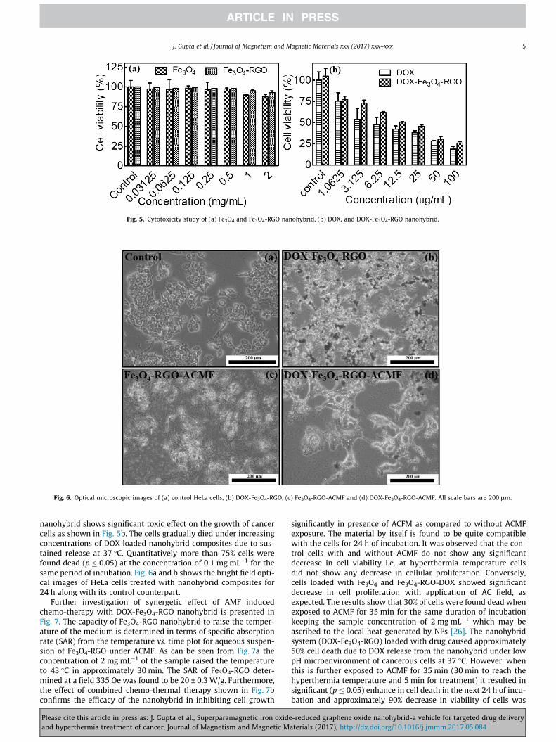
Fig. 5. Cytotoxicity study of (a) Fe3O4 and Fe3O4-RGO nanohybrid, (b) DOX, and DOX-Fe3O4-RGO nanohybrid.
Fig. 6. Optical microscopic images of (a) control HeLa cells, (b) DOX-Fe3O4-RGO, (c) Fe3O4-RGO-ACMF and (d) DOX-Fe3O4-RGO-ACMF. All scale bars are 200 lm.
J. Gupta et al. / Journal of Magnetism and Magnetic Materials xxx (2017) xxx–xxx 5
nanohybrid shows significant toxic effect on the growth of cancercells as shown in Fig. 5b. The cells gradually died under increasingconcentrations of DOX loaded nanohybrid composites due to sus-tained release at 37 �C. Quantitatively more than 75% cells werefound dead (p � 0.05) at the concentration of 0.1 mg mL�1 for thesame period of incubation. Fig. 6a and b shows the bright field opti-cal images of HeLa cells treated with nanohybrid composites for24 h along with its control counterpart.
Further investigation of synergetic effect of AMF inducedchemo-therapy with DOX-Fe3O4-RGO nanohybrid is presented inFig. 7. The capacity of Fe3O4-RGO nanohybrid to raise the temper-ature of the medium is determined in terms of specific absorptionrate (SAR) from the temperature vs. time plot for aqueous suspen-sion of Fe3O4-RGO under ACMF. As can be seen from Fig. 7a theconcentration of 2 mg mL�1 of the sample raised the temperatureto 43 �C in approximately 30 min. The SAR of Fe3O4-RGO deter-mined at a field 335 Oe was found to be 20 ± 0.3 W/g. Furthermore,the effect of combined chemo-thermal therapy shown in Fig. 7bconfirms the efficacy of the nanohybrid in inhibiting cell growth
Please cite this article in press as: J. Gupta et al., Superparamagnetic iron oxideand hyperthermia treatment of cancer, Journal of Magnetism and Magnetic M
significantly in presence of ACFM as compared to without ACMFexposure. The material by itself is found to be quite compatiblewith the cells for 24 h of incubation. It was observed that the con-trol cells with and without ACMF do not show any significantdecrease in cell viability i.e. at hyperthermia temperature cellsdid not show any decrease in cellular proliferation. Conversely,cells loaded with Fe3O4 and Fe3O4-RGO-DOX showed significantdecrease in cell proliferation with application of AC field, asexpected. The results show that 30% of cells were found dead whenexposed to ACMF for 35 min for the same duration of incubationkeeping the sample concentration of 2 mg mL�1 which may beascribed to the local heat generated by NPs [26]. The nanohybridsystem (DOX-Fe3O4-RGO) loaded with drug caused approximately50% cell death due to DOX release from the nanohybrid under lowpH microenvironment of cancerous cells at 37 �C. However, whenthis is further exposed to ACMF for 35 min (30 min to reach thehyperthermia temperature and 5 min for treatment) it resulted insignificant (p � 0.05) enhance in cell death in the next 24 h of incu-bation and approximately 90% decrease in viability of cells was
-reduced graphene oxide nanohybrid-a vehicle for targeted drug deliveryaterials (2017), http://dx.doi.org/10.1016/j.jmmm.2017.05.084

Fig. 7. Temperature vs. time plots of aqueous suspension of Fe3O4 and Fe3O4-RGO nanohybrid (2 mg/ml) at field of 335 Oe (b) Cell viabilities of HeLa cells after treatment withFe3O4-RGO and DOX-Fe3O4-RGO with and without application of ACMF.
6 J. Gupta et al. / Journal of Magnetism and Magnetic Materials xxx (2017) xxx–xxx
observed indicating the synergistic effect of both local heat as wellas released chemo induced by externally applied AC magnetic field.This synergetic effect of hyperthermia under ACMF can be attribu-ted to the cleavage of the weak electrostatic interaction of DOXwith the Fe3O4-RGO nanohybrid primarily due to the mechanicalforce generated locally by ACMF. This eventually facilitates thedrug diffusion and thereby promotes the drug release profile ofthe DOX loaded nanohybrid. Also, magnetic hyperthermiaenhances the chemotherapeutic effect with the disruption of thecytoplasmic regions due to accumulation of significant amount ofdrugs in the nucleus and hence helps to enhance maximum celldeath simultaneously and synergistically. The effect of chemo-thermo therapy of nanohybrids under ACMF is clearly seen in thebright field optical images of cells along with their control counter-part in Fig 6c and d.
It is worthwhile to discuss some of the recently published workwhere researchers have demonstrated the efficacy of the magneticsystems in treating the cancer cells via DOX delivery assisted byACMF. There was an attempt to develop temperature-sensitivehydrogels based drug carrier which is effective in killing the cellsvia change in physical shrinkage of the materials [26,42]. Thoughthe system serves the purpose of delivering the therapeutics viatemperature change, it however fails to show any significant influ-ence due to acidic environment of tumor cells. The system pre-sented here has a unique feature of having sensitivity towardslow pH due to presence of graphene. As stated earlier the interac-tion of polar groups of RGO with that of hydroxyl group of DOXweaken at low pH (cancerous pH-4.3) and hence facilitates theenhanced release of drug in acidic environment as well as AC mag-netic field and therefore enhance the therapeutic effect of hybrid tokill the cancer cells.
4. Conclusion
Superparamagnetic Fe3O4-RGO nanohybrid has been synthe-sized by one step co-precipitation approach using iron salts andGO. The loading of anticancer drug (DOX) has been optimized byaltering the concentrations of nanohybrid and DOX. The acidicmicroenvironment of cancer cells (pH � 4.3) supports drug mole-cules to release due to the sensitivity of graphene in acidic environ-ment as compared to physiological condition (pH � 7.4) whichreduce the side effects of chemotherapeutic drug. Further, mag-netic particles over the RGO sheet make its more promising for
Please cite this article in press as: J. Gupta et al., Superparamagnetic iron oxideand hyperthermia treatment of cancer, Journal of Magnetism and Magnetic M
thermal therapy in the presence of AC magnetic field i.e. hyperther-mia treatment of cancer. Therefore, the synergic effects of thenanohybrid enhance the therapeutic efficacy and shows approxi-mately 90% killing efficiency when exposed to AC field (335 Oe,265 kHz) for 35 min.
Acknowledgements
Financial support from Department of Science and Technology(DST), Government of India is greatly acknowledged. The authorsare grateful to the Centre for Research in Nanotechnology &Science (CRNTS) for TEM and Raman facilities.
Appendix A. Supplementary data
Supplementary data associated with this article can be found, inthe online version, at http://dx.doi.org/10.1016/j.jmmm.2017.05.084.
References
[1] A.K. Geim, K.S. Novoselov, Nat. Mater. 6 (2007) 183.[2] X. Li, X. Wang, L. Zhang, S. Lee, H. Dai, Science 319 (2008) 1229.[3] S. Stankovich, D.A. Dikin, G.H.B. Dommett, K.M. Kohlhaas, E.J. Zimney, E.A.
Stach, R.D. Piner, S.T. Nguyen, R.S. Ruoff, Nature 442 (2006) 282.[4] F. Schedin, A.K. Geim, S.V. Morozov, E.W. Hill, P. Blake, M.I. Katsnelson, K.S.
Novoselov, Nat. Mater. 6 (2007) 652.[5] E. Yoo, J. Kim, E. Hosono, H.-S. Zhou, T. Kudo, I. Honma, Nano Lett. 8 (2008)
2277.[6] M.D. Stoller, S. Park, Y. Zhu, J. An, R.S. Ruoff, Nano Lett. 8 (2008) 3498.[7] Z. Liu, J.T. Robinson, X. Sun, H. Dai, J. Am. Chem. Soc. 130 (2008) 10876.[8] X. Sun, Z. Liu, K. Welsher, J.T. Robinson, A. Goodwin, S. Zaric, H. Dai, Nano Res. 1
(2008) 203.[9] D. Zhang, X. Liu, X. Wang, J. Inorg. Biochem. 105 (2011) 1181.[10] L. Feng, Y. Chen, J. Ren, X. Qu, Biomaterials 32 (2011) 2930.[11] Z.M. Markovic, L.M. Harhaji-Trajkovic, B.M. Todorovic-Markovic, D.P. Kepic, K.
M. Arsikin, S.P. Jovanovic, A.C. Pantovic, M.D. Dramicanin, V.S. Trajkovic,Biomaterials 32 (2011) 1121.
[12] Z. Liu, X. Sun, N. Nakayama-Ratchford, H. Dai, ACS Nano 1 (2007) 50.[13] Z. Liu, K. Chen, C. Davis, S. Sherlock, Q. Cao, X. Chen, H. Dai, Cancer Res. 68
(2008) 6652.[14] A. Bianco, M. Prato, Adv. Mater. 15 (2003) 1765.[15] H. Gao, Y. Kong, D. Cui, C.S. Ozkan, Nano Lett. 3 (2003) 471.[16] Y.H. Xie, A.K. Soh, Mater. Lett. 59 (2005) 971.[17] J. Xie, Q. Xue, Q. Zheng, H. Chen, Mater. Lett. 63 (2009) 319.[18] X. Yang, X. Zhang, Z. Liu, Y. Ma, Y. Huang, Y. Chen, J. Phys. Chem. C 112 (2008)
17554.[19] Z. Liu, J. Ding, J. Xue, New J. Chem. 33 (2009) 88.[20] A. Petri-Fink, M. Chastellain, L. Juillerat-Jeanneret, A. Ferrari, H. Hofmann,
Biomaterials 26 (2005) 2685.
-reduced graphene oxide nanohybrid-a vehicle for targeted drug deliveryaterials (2017), http://dx.doi.org/10.1016/j.jmmm.2017.05.084

J. Gupta et al. / Journal of Magnetism and Magnetic Materials xxx (2017) xxx–xxx 7
[21] J. Gupta, P. Bhargava, D. Bahadur, J. Appl. Physi. 115 (2014) 17B516.[22] R.M. Patil, P.B. Shete, N.D. Thorat, S.V. Otari, K.C. Barick, A. Prasad, R.S.
Ningthoujam, B.M. Tiwale, S.H. Pawar, J. Magn. Magn. Mater. 355 (2014) 22.[23] R.E. Rosensweig, J. Magn. Magn. Mater. 252 (2002) 370.[24] B.B. Lahiri, T. Muthukumaran, J. Philip, J. Magn. Magn. Mater. 407 (2016) 101.[25] J. Gupta, J. Mohapatra, P. Bhargava, D. Bahadur, Dalton Trans. 45 (2016)
2454.[26] M.K. Jaiswal, M. De, S.S. Chou, S. Vasavada, R. Bleher, P.V. Prasad, D. Bahadur, V.
P. Dravid, A.C.S. Appl, Mater. Interfaces 6 (2014) 6237.[27] A. Shanavas, S. Sasidharan, D. Bahadur, R. Srivastava, J. Colloid Interface Sci.
486 (2017) 112.[28] X. Ma, H. Tao, K. Yang, L. Feng, L. Cheng, X. Shi, Y. Li, L. Guo, Z. Liu, Nano Res. 5
(2012) 199.[29] M. Talelli, C.J.F. Rijcken, T. Lammers, P.R. Seevinck, G. Storm, C.F. van Nostrum,
W.E. Hennink, Langmuir 25 (2009) 2060.[30] J.H. Maeng, D.-H. Lee, K.H. Jung, Y.-H. Bae, I.-S. Park, S. Jeong, Y.-S. Jeon, C.-K.
Shim, W. Kim, J. Kim, J. Lee, Y.-M. Lee, J.-H. Kim, W.-H. Kim, S.-S. Hong,Biomaterials 31 (2010) 4995.
[31] V.P. Torchilin, Nat. Rev. Drug Discovery 4 (2005) 145.
Please cite this article in press as: J. Gupta et al., Superparamagnetic iron oxideand hyperthermia treatment of cancer, Journal of Magnetism and Magnetic M
[32] X.-Y. Ying, Y.-Z. Du, L.-H. Hong, H. Yuan, F.-Q. Hu, J. Magn. Magn. Mater. 323(2011) 1088.
[33] H.A. Becerril, J. Mao, Z. Liu, R.M. Stoltenberg, Z. Bao, Y. Chen, ACS Nano 2 (2008)463.
[34] M.K. Jaiswal, R. Banerjee, P. Pradhan, D. Bahadur, Colloids Surf., B 81 (2010)185.
[35] D. Cai, M. Song, J. Mater. Chem. 17 (2007) 3678.[36] W. Shi, J. Zhu, D.H. Sim, Y.Y. Tay, Z. Lu, X. Zhang, Y. Sharma, M. Srinivasan, H.
Zhang, H.H. Hng, Q. Yan, J. Mater. Chem. 21 (2011) 3422.[37] S. Stankovich, D.A. Dikin, R.D. Piner, K.A. Kohlhaas, A. Kleinhammes, Y. Jia, Y.
Wu, S.T. Nguyen, R.S. Ruoff, Carbon 45 (2007) 1558.[38] K.N. Kudin, B. Ozbas, H.C. Schniepp, R.K. Prudhomme, I.A. Aksay, R. Car, Nano
Lett. 8 (2008) 36.[39] F. Tuinstra, J.L. Koenig, J. Chem. Phys. 53 (1970) 1126.[40] C.-J. Hsieh, Y.-C. Chen, P.-Y. Hsieh, S.-R. Liu, S.-P. Wu, Y.-Z. Hsieh, H.-Y. Hsu, A.C.
S. Appl, Mater. Interfaces 7 (2015) 11467.[41] A.K. Swain, L. Pradhan, D. Bahadur, A.C.S. Appl, Mater. Interfaces 7 (2015) 8013.[42] M.K. Jaiswal, A. Pradhan, R. Banerjee, D. Bahadur, J. Nanosci. Nanotechnol. 14
(2014) 4082.
-reduced graphene oxide nanohybrid-a vehicle for targeted drug deliveryaterials (2017), http://dx.doi.org/10.1016/j.jmmm.2017.05.084

本文献由“学霸图书馆-文献云下载”收集自网络,仅供学习交流使用。
学霸图书馆(www.xuebalib.com)是一个“整合众多图书馆数据库资源,
提供一站式文献检索和下载服务”的24 小时在线不限IP
图书馆。
图书馆致力于便利、促进学习与科研,提供最强文献下载服务。
图书馆导航:
图书馆首页 文献云下载 图书馆入口 外文数据库大全 疑难文献辅助工具