Embed Size (px)
Citation preview

7444 | J. Mater. Chem. B, 2017, 5, 7444--7460 This journal is©The Royal Society of Chemistry 2017
Cite this: J.Mater. Chem. B, 2017,
5, 7444
Visualizing molecular distributions forbiomaterials applications with mass spectrometryimaging: a review
Martin R. L. Paine, †ab Pieter C. Kooijman, †ac Gregory L. Fisher, d
Ron M. A. Heeren, a Facundo M. Fernandez bef and Shane R. Ellis *a
Mass spectrometry imaging (MSI) is a rapidly emerging field that is continually finding applications in
new and exciting areas. The ability of MSI to measure the spatial distribution of molecules at or near the
surface of complex substrates makes it an ideal candidate for many applications, including those in the
sphere of materials chemistry. Continual development and optimization of both ionization sources and
analyzer technologies have resulted in a wide array of MSI tools available, both commercially available and
custom-built, with each configuration possessing inherent strengths and limitations. Despite the unique
potential of MSI over other chemical imaging methods, their potential and application to (bio)materials
science remains in our view a largely underexplored avenue. This review will discuss these techniques
enabling high parallel molecular detection, focusing on those with reported uses in (bio)materials
chemistry applications and highlighted with select applications. Different technologies are presented in
three main sections; secondary ion mass spectrometry (SIMS) imaging, matrix-assisted laser desorption
ionization (MALDI) MSI, and emerging MSI technologies with potential for biomaterial analysis. The first
two sections (SIMS and MALDI) discuss well-established methods that are continually evolving both in
technological advancements and in experimental versatility. In the third section, relatively new and
versatile technologies capable of performing measurements under ambient conditions will be introduced,
with reported applications in materials chemistry or potential applications discussed. The aim of this
review is to provide a concise resource for those interested in utilizing MSI for applications such as
biomimetic materials, biological/synthetic material interfaces, polymer formulation and bulk property
characterization, as well as the spatial and chemical distributions of nanoparticles, or any other molecular
imaging application requiring broad chemical speciation.
Introduction
Interactions at the surface of biomaterials have a major impacton their in vivo performance and physiological response of thehost. Therefore, detailed characterization of surface properties,both physical and chemical, is required to engineer new materials
and increase their biocompatibility.1,2 Many analytical techniqueshave been routinely employed to characterize the surface prop-erties of biomaterials including but not limited to; X-ray photo-electron spectroscopy (XPS),3 atomic force microscopy (AFM),4
Auger electron spectroscopy (AES),5 contact angle methods,6
vibrational spectroscopy (e.g., Raman and Fourier transforminfrared spectroscopy),7 near edge X-ray absorption fine structure(NEXAFS)8 and energy dispersive X-ray spectroscopy (EDX).9 Aninformative comparison between these and MS-based approachesis provided in a review by Senoner and Unger.10 Of the afore-mentioned techniques, detailed elemental and topographicalinformation of the surface can be obtained, but this informa-tion usually relates to one or a few chemical species, lackingspecific chemical information at the molecular level. With theexception of vibrational spectroscopy, complementary imagingtechniques are typically required when broadband molecularinformation from both the substrate and the biological materialthat interacts with the substrate is desired.
a M4I, The Maastricht MultiModal Molecular Imaging Institute,
Maastricht University, Maastricht 6229 ER, The Netherlands.
E-mail: [email protected] School of Chemistry and Biochemistry, Georgia Institute of Technology,
Atlanta, GA 30332, USAc TI-COAST, Amsterdam 1098 XH, The Netherlandsd Physical Electronics, Inc., Chanhassen, Minnesota 55317, USAe Integrated Cancer Research Center, Georgia Institute of Technology,
Atlanta, GA 30332, USAf Institute of Bioengineering and Biosciences, Georgia Institute of Technology,
Atlanta, GA 30332, USA
† Authors contributed equally.
Received 21st April 2017,Accepted 11th August 2017
DOI: 10.1039/c7tb01100h
rsc.li/materials-b
Journal ofMaterials Chemistry B
REVIEW

This journal is©The Royal Society of Chemistry 2017 J. Mater. Chem. B, 2017, 5, 7444--7460 | 7445
Mass spectrometry imaging (MSI) enables visualization of abroad range of chemical species in a single experiment with highmolecular specificity and the ability to structurally characterizedetected molecules. As many materials chemistry applicationshave traditionally focused on the analysis of inorganic orcovalently bound materials with high (o1 mm) spatial resolu-tion, secondary ion mass spectrometry (SIMS) has been amainstay for chemical imaging of materials in many disciplinesas it is particularly well suited for such analyses.10–13 However,as research into biomaterials and their associated biomolecularinteractions has emerged, so too has the demand for new MSImethods enabling detection and characterization of many classesof labile biomolecules. With the development of alternativeionization methods and rapid improvements in MSI instrumen-tation, both in terms of mass analyzer and sampling/ionizationtechnologies, massive gains in mass-resolving power, speed andsensitivity have been achieved. These dramatic improvementshave positioned MSI as a unique resource within a growingnumber of analytical facilities. In particular, the use of MSI forthe investigation of biological tissues has flourished, providinga wealth of information on the spatial distribution of pharma-ceuticals, metabolites, lipids, peptides, and proteins frompractically every type of organic substrate.14–16 The increasedusage of MSI for biomolecular investigations has resulted insuccessful methodologies for a broad family of compoundclasses, making the translation to inorganic or non-biologicalsubstrates a logical step. Thus, emerging fields such as bio-material development, where changes in surface compositionand structure of synthetic materials placed inside the body (e.g.,biomimetics or biomedical devices) affect a biological responseare primed for interrogation by MSI. As a surface samplingtechnique, MSI is well equipped to probe biomaterials thatencompass this interface between synthetic substrates andbiological tissue.17,18 Characterizing the biomaterial surfaceproperties (i.e., chemical composition, structure, orientation)and understanding the biological effect these properties haveby measuring biomolecules on these surfaces is paramount tothe development of biomaterial technology.
The ability to perform MSI for a particular applicationdepends largely on the type of desorption/ionization sourceemployed of which there are three major categories; secondaryion mass spectrometry (SIMS), matrix-assisted laser desorption/ionization (MALDI), and various ambient mass spectrometricmethods. Each technique is capable of producing ions in bothpositive- and negative-ion mode (broadly referring to detection ofbasic and acidic compounds, respectively), however their methodof operation varies significantly leading to different popula-tions of ions detected. SIMS represents the most energeticdesorption technique, capable of ablating covalently or ioni-cally bound material and penetrating into the depth of thesubstrate, making it well suited for elemental and inorganicanalyses. MALDI is a softer desorption technique, capable ofdesorbing and ionizing loosely bound inorganic material and awide range of biomolecules (proteins, peptides, lipids, and meta-bolites). Ambient techniques generally represent the softestdesorption techniques and are best suited to delicate substrates
(particularly those not vacuum stable) and labile organic mole-cules in the 50–2000 Da range. Therefore, alternative MSItechniques such as MALDI and ambient methods may be moreeffective in biomaterial applications where broader chemicaldetection is required or when probing delicate substrates.
In this review, we highlight the broad array of MSI techniquescurrently available for molecular detection by showcasing theirreported use in biomaterial applications, as well as broadermaterials science applications to illustrate the potential of thesetechniques for future research. The discussion includes concisedescriptions of the processes underpinning each technique,current developments in instrumentation technology, and keyapplications that exemplify the benefit of MSI for biomaterialsurface analysis. Finally, a perspective on the role of newMSI approaches at the intersection of biomaterial analysis isprovided.
Secondary ion mass spectrometry(SIMS)
SIMS was the first MS technique employed for imaging andarguably the one most familiar to materials scientists.11,19,20
SIMS utilizes the release of charged (ionized) material from asubstrate upon impact of an electrostatically focused high-energyprimary ion beam (traditionally from monoatomic sources suchas Au+, Ga+ and In+). As the high-energy (10–40 keV) primary ionsimpact the surface, a collisional cascade is initiated in the top fewmonolayers of the sample, leading to the ejection of secondaryparticles consisting of sputtered neutral molecules, ions, mole-cular fragments (of both neutrals and ions), and electrons.21,22
The secondary ions generated, typically protonated/deprotonatedions, cationized adducts or radicals, are then detected based ontheir mass-to-charge ratio (m/z), typically using time-of-flight(ToF) mass spectrometry. For SIMS analyses, samples are typicallymounted in a high vacuum sample chamber, must be as flat aspossible and be mounted onto conductive substrates to minimizethe effect of surface charging which can significantly hamperboth spatial and mass resolution as well as sensitivity. Forinsulating samples these are often prepared as thin sections(B1–20 mm) and mounted onto conductive substrates such assilicon, steel or indium-tin oxide coated (ITO) slides. It should benoted that charge compensation can enable analysis of insulatingsamples by actively neutralizing surface charge and such capabilitiesare available on commercial systems.
The primary advantage of SIMS over other MSI techniques isthe ability to restrict the analyzed area down to B100 nm, withfeatures resolved by several beam diameters, providing by farthe highest spatial resolution of any MSI approach. As many(bio)materials applications require sub-micron spatial resolu-tions, SIMS has a distinct advantage over other approachesdescribed below. Combined with the energetic desorption/ionization process, SIMS is particularly well-suited for analysis ofinorganic materials and materials bound to surfaces via chemicalbonds. However, compared to softer desorption/ionizationtechniques like MALDI and DESI, SIMS imparts significantly
Review Journal of Materials Chemistry B

7446 | J. Mater. Chem. B, 2017, 5, 7444--7460 This journal is©The Royal Society of Chemistry 2017
more internal energy to desorbed molecules, resulting in sub-stantial molecular fragmentation. In some cases this featurecan be highly advantageous, e.g., for analysis of elements,covalently-bound surface materials, and for detection ofchemical tags covalently bound to molecules of interest. Thehigh sensitivity of SIMS for elemental imaging is in part due tooperation in the so-called ‘‘dynamic-SIMS’’ mode. In dynamicSIMS high ion doses (41013 ions cm�2) are used to produceions from a large fraction of the entire surface. Due to the highion doses detection of only small molecular fragments andelements originating from the surface is possible due to accumu-lation of surface damage. The high spatial resolution and sensi-tivity offered by dynamic SIMS has recently been exploited in aunique approach termed nano-SIMS. In this approach a cesiumor oxygen primary ion beam is used which can be focused downto B50 nm.23 The unique MS design allows up to seven m/zchannels to be continuously monitored whereby m/z informationis obtained via angular separation in a magnetic sector analyzer.Detectable ions are either elements or diatomic fragments suchas CH�.23–26 Examples of materials chemistry applicationsinclude the analysis of 13C-enriched Resveratrol coated Fe3O4
coated nanoparticles whereby Resveratrol was detected via theenriched 13C signal in areas where it was localized27 and thesimultaneous localization of isotope-labelled lipids and selectedproteins through the use of fluorinated gold immunolabels.28
Nano-SIMS has also been widely used to study dynamic chemicalchanges in biological systems by virtue of its high sensitivityand precise measurements of ion abundance.29,30 For instance,via incorporation and monitoring of stable isotope enrichments(in these cases 15N enriched thymidine), it has been possibleto pinpoint stem cell generation in mice hippocampi31 andrelatively quantify the different origins of new cardiomyocytes.32
Similarly, laser ablation inductively coupled plasma (LA-ICP) massspectrometry provides a highly complementary approach toelement imaging with SIMS. Several authors report the use ofLA-ICP-MS to study the biological uptake of nanoparticles33,34
and the in vivo degradation of metallic implants.35–37 AlthoughLA-ICP-MS cannot compete with the spatial resolution offered bySIMS, it offers excellent quantitative abilities enabling absolutesurface concentrations of elements to be determined with highprecision (typically 5–15% RSD).38–40
For applications that require information on moleculardistributions, such as untargeted (bio)molecular investigations,softer interrogation of the surface is needed. In these cases,a technique known as ‘‘static SIMS’’ can be utilized. In staticSIMS, the ion dose is maintained below 1013 ions per cm2 (the‘‘static-limit’’) ensuring less than 1% of the surface is impactedby a primary ion. Thus, surface damage is minimized and theprobability of detecting larger m/z species having m/z valuesup to B2000 is greatly enhanced. Although detection of intactlabile molecules with molecular weights above 500 Da can stillbe a challenge, characteristic molecular fragments are oftendetectable under such conditions using traditional ion beams.For example, lipid analyses mostly result in the detection oflipid fragment ions (i.e., the phosphatidylcholine headgroupion at m/z 184 in positive-ion mode or free fatty acids in
negative-ion mode) rather than the intact lipid itself, compli-cating interpretation of results.11,25,41
Driven by the increasing need to detect larger molecules,primarily for biomolecular analysis, recent SIMS developmentshave heavily focused on the development of softer ionizationsources that greatly minimize fragmentation and surface damage.42
In particular larger, polyatomic sources such as C60+,43–47 SF5
+,48–50
Aun+,46,51–53 gas cluster beams such as Arn
+ 54–56 and [CO2]n+ 57
and recently water cluster beams58 have revolutionized SIMSand brought it into the realm of true molecular analysis. Withthese sources it is possible to generate intact molecular infor-mation at ion doses above the static limit due to the drasticallyreduced surface damage of these large primary ions.44 In thesecluster ion sources a single primary ion can have a mass ofup to 100 000 Da which greatly reduces fragmentation and sub-surface damage, thereby enabling both softer ionization andsputtering (see below).59 It must be noted that large clusterbeams do not generally offer the r100 nm42 spatial resolutionof atomic or small cluster sources (e.g., Bi3
+). The current state-of-the-art of cluster sources can achieve focused spot sizesdown to several micrometers for gas cluster ion beams and insome cases as low as 300 nm for C60
+ ion beams.60,61 It should benoted that despite the tremendous advances made in moleculardetection with SIMS over recent years many applications stillcenter on the detection of elements or elemental/small molecularmarkers of larger molecules.
A second key advantage of SIMS over other MSI techniques isthe exploitation of the gradual removal of material by the primaryion beam to enable 3D analysis (i.e., depth profiling).62,63 In suchan approach a cluster ion beam is often used to sputter awayseveral monolayers or more of material while minimizing surfacedamage and modification, after which the freshly exposed sur-face is imaged using SIMS. This process can be repeated usingsequential sputter/analysis cycles and enable 3D chemical recon-struction of heterogeneous materials. Crucially, the achievabledepth resolution can be as low as several nanometres,64,65
providing almost a monolayer by monolayer representation ofthe material. In the sections below, we highlight a diverse arrayof applications of SIMS-MSI in materials chemistry with specificemphasis on biomaterials and their interactions. For a morecomprehensive examination of biological tissue imaging appli-cations of SIMS-MSI the reader is referred to a series of reviewspublished on this topic.25,41,66–75
Synthetic polymers
SIMS-MSI has found widespread use for studying polymericmaterials in a diverse array of applications.76–78 For example,using a 5 keV Ar2000
+ beam for sputtering and a Bi3+ beam for
mass analysis, the composition of spin-cast polymer multilayerscontacting alternating layers of polystyrene (PS) and polyvinyl-pyrrolidone (PVP) on silicon substrates has been studied.79
Polymer signals were identified by the characteristic C7H7+ and
C6H10NO+ signals for PS and PVP, respectively and incorporatedPS layers as thin as 45 nm could be resolved. A similar approachhas also been employed using C60
+ for sputtering instead.80
3D-SIMS has also provided valuable insight into surface
Journal of Materials Chemistry B Review
This journal is©The Royal Society of Chemistry 2017 J. Mater. Chem. B, 2017, 5, 7444--7460 | 7447
topographies of poly(bisphenol A-co-decane ether) films. SIMS,for the first time, revealed the presence of hollow interior struc-tures on the surface of these polymers when prepared witheither chloroform or THF solvents. These hollow droplets werefound to have a thickness of several hundred nanometers andwere sandwiched between the two polymer layers.81 As anotherexample it was recently shown how polyelectrolyte multilayercomposite films could be synthesized and exposed to a miner-alization process. The quality of polymeric nucleating agentswas assessed in terms of calcium carbonate mineral infiltrationefficiency by SIMS depth profiling with a combination of a Bi+
beam for analysis and a cluster Arn+ beam for sputtering.82
With respect to biomaterials Jung et al. have deployed 3D-SIMSto study the spatial distributions of the biopolymers celluloseand lignin in tension wood as a model for biomass using a Bi3
+
beam for analysis and an O2+ beam for sputtering.83 Finally, in
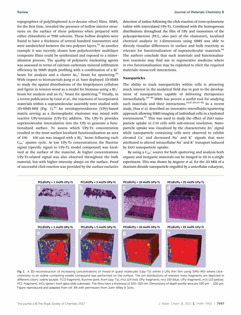
a recent publication by Goor et al., the reactions of incorporatedmaterials within a supramolecular assembly were studied with3D-SIMS-MSI (Fig. 1).84 An ureidopyrimidinone (UPy)-basedmatrix serving as a thermoplastic elastomer was mixed withreactive UPy-tetrazine (UPy-Tz) additive. The UPy-Tz providessupramolecular intercalation into the UPy to generate a func-tionalized surface. To assess which UPy-Tz concentrationresulted in the most surface-localized functionalization an areaof 100 � 100 mm was imaged with a Bi3
+ beam following eachC60
+ sputter cycle. At low UPy-Tz concentrations the fluorinesignal (specific signal to UPy-Tz model compound) was local-ized at the surface of the material. At higher concentrationsUPy-Tz-related signal was also observed throughout the bulkmaterial, but with higher intensity always on the surface. Proofof successful click-reaction was provided by the surface-exclusive
detection of iodine following the click-reaction of trans-cycloocteneiodine with intercalated UPy-Tz. Combined with the homogenousdistributions throughout the film of UPy and monomers of thepolycaprolactone (PCL, also part of the elastomer), localizedchemical analysis in 3-dimensions using SIMS was able todirectly visualize differences in surface and bulk reactivity asrelevant for functionalization of supramolecular materials.84
The authors conclude that such materials and functionaliza-tion reactions may find use in regenerative medicine wherein vivo functionalization may be exploited to elicit the requiredmaterials–tissue/cell interactions.
Nanoparticles
The ability to track nanoparticles within cells is attractingmuch interest in the analytical field due in part to the develop-ment of nanoparticles capable of delivering therapeuticsintracellularly.85–90 SIMS has proven a useful tool for studyingsuch materials and their interactions.10,87,89,91–96 In a recentstudy, Hua et al. described an innovative microfluidic/sputteringapproach allowing SIMS imaging of individual cells in a hydratedenvironment.97 This was used to study the effect of ZnO nano-particle uptake in C10 cells with sub-micron resolution. Nano-particle uptake was visualized by the characteristic Zn+ signalwhile nanoparticle containing cells were observed to exhibitelevated Ca+ and decreased Na+ and K+ signals that wereattributed to altered intracellular Na+ and K+ transport inducedby ZnO nanoparticle uptake.
By using a C60+ source for both sputtering and analysis both
organic and inorganic materials can be imaged in 3D in a singleexperiment. This was shown by Angerer et al. for the 3D MSI of atitanium dioxide nanoparticle engulfed by a unicellular eukaryote,
Fig. 1 A 3D-reconstruction of increasing concentrations of mixed-in guest molecules (Upy-Tz) within a UPy thin film using SIMS-MSI where click-chemistry to an iodine containing model compound was performed on the surface. The ion distributions of relevant mass fragments are depicted indifferent colors: iodine (purple, TCO fragment), fluorine (pink, from Upy-Tz), m/z 124 (red, UPy-fragment), m/z 150 (blue, UPy-fragment), m/z 113 (yellow,PCL-fragment), InO2 (green, from glass slide substrate). The films have a thickness of 100–150 nm. Dimensions of depth profile area are 100 mm � 100 mm.Figure reproduced and adapted from ref. 84 with permission from John Wiley & Sons.
Review Journal of Materials Chemistry B

7448 | J. Mater. Chem. B, 2017, 5, 7444--7460 This journal is©The Royal Society of Chemistry 2017
Tetrahymena pyriformis, where visualization of the incorpora-tion of nanoparticles inside food vacuoles of the eukaryote wasobserved.88 Another powerful example of 3D ToF-SIMS on poly-mer nanoparticles is given by Rafati et al.98 The surface hetero-geneity and relative localization of the poly(lactic-co-glycolic) acid(PLGA) backbone, polyvinyl acrylate (PVA) surfactant, and proteinlysosome (model therapeutic) were visualized to gain understand-ing in the effect of polymer microsphere fabrication parametersand revealed that the lysosome was primarily distributed aroundmicrosphere surface pores.
Multimodal approaches with SIMS, such as with fluorescenceor transmission electron microscopy (TEM), have proven espe-cially useful to track nanoparticles in biological matrices.28,99
Several studies have reported cytotoxicity assessment of nano-particles with ToF-SIMS, such as Fe3O4 nanoparticles100 or poly-meric nanoparticles combined with fluorescence microscopy.101
Recently, the delivery of cytotoxic drugs was evaluated by poly-meric oxaliplatin nanoparticles through monitoring of the time-dependent spread of both the nanoparticle carrier and thePt(II)-based anticancer drug in vivo. The combined fluorescencemicroscopy, TEM and SIMS approach discovered the oxaliplatinNPs are taken up in intracellular vesicles. The consecutive break-down of the carrier material stimulates release of the cytotoxicdrug, confirming the targeted delivery mechanism.102
Tissue engineering/cell culture
Tissue engineering is a young, thriving field of study clearly in needof imaging techniques with the ability to differentiate biologicalresponses based on changes in local molecular composition. Forexample, SIMS has been deployed to study the homogeneity ofcell populations based on their molecular phenotype103 and shedlight on the biological pathway changes related to culturingconditions.104 SIMS can also be applied to evaluate the potentialof tissue engineering stem cell lines. For instance, the osteogenicdifferentiation capabilities of human embryonic stem cell-derivedmesodermal progenitors (hES-MPs) were studied and comparedto human mesenchymal stem cells (hMSCs), one of the mostdocumented cell types for tissue engineering purposes.105 Using3D-SIMS-MSI depth-profiling and 3D-mapping, distinct bio-mineralization patterns were shown, with hES-MPs yieldinghigher hydroxyapatite signal than hMSCs after six weeks butlower after 3 weeks of osteogenic stimulation.106
As ToF-SIMS offers exciting opportunities to correlate surfacechemistry to biological response it is well suited to guide substratedevelopment for proliferation and differentiation control.107–109 Suchan approach has been utilized to assess the cell proliferation effect ofhydrogel substrate additives,110,111 naturally derived extracellularmatrices,112–114 and surface geometries.115 Current state-of-the-artsubstrates tend to have complex compositions and geometries,which require more specific and sensitive methods to verify. Bongoet al. showed their PEDOT(TOS):gelatin composite films were able tosupport cell growth while retaining the beneficial electrical conduc-tivity and mechanical properties of the original polymer substrate.116
The authors used a nano-SIMS instrument to reveal the distributionof carbon, nitrogen and sulphur and demonstrate the homogeneousgelatin incorporation in the film.
Implants
Successful implant integration in the surrounding tissue is ofvital importance to patient health, but much remains unknownabout the mechanics of implant–tissue interaction. In an effortto shed light onto these effects the interaction area betweenbone and a titanium implant has been imaged with ToF-SIMS,revealing distinctive molecular and elemental species for bone,implant, and interaction area.117,118 Gonzalez et al. studied abioactive coated Ti-NB-Hf alloy implant material using Bi3
+
beam and showed this material had a significantly increasedosteoblast adhesion.119 In another study, the distribution ofrapamycin – an immunosuppressant in a coronary stent coating(poly(lactic-co-glycolic acid)) – was studied to determine the effectof the coating application method on drug elution behaviour.120
3D-SIMS-MSI analysis showed a high degree of heterogeneity inthe rapamycin concentration throughout the sample, with themost homogeneous areas providing the most gradual elution. Itwas therefore concluded that the coating application method hasa major effect on the early drug elution behavior and thereforedeserves thorough optimization.
Supramolecular materials such as biodegradable hydrogelsare considered promising drug delivery carrier candidates.Ureidopyrimidinone (UPy) cross-linked poly(ethylene glycol):polycaprolactone (PEG:PCL) hydrogels implanted under therenal capsule of rats have been studied with Au+ SIMS, compar-ing relative distributions of endogenous compounds (lipids andcholesterol) and the implanted polymer (PEG). Interestingly,it was observed that the PEG-related signal was co-localized inthe tissue with cholesterol sulfate, suggesting the occurrence ofcellular infiltration in the polymer.121 This study demonstrates thevast potential of SIMS to study foreign-body response mechanismsand can help monitor material–tissue interactions, for example indrug-delivery applications.
Advances for chemical identification using SIMS
The use of axial ToF analyzers for SIMS requires the primary ionbeam to be pulsed on the order of several nanoseconds to obtainreasonable mass resolution. However, this introduces bothspeed and sensitivity constraints due to the limited duty cycleresulting from the fact that only one ion packet may be injectedinto the analyzer per ToF event. For example, a 2 ns pulsed beamoperating at 10 kHz and a maximum flight time of 100 ms meansonly 0.002% percent of the time is spent generating ions.To overcome these limitations and enable continuous ion gene-ration several groups have developed SIMS instrumentationemploying orthogonal ToF analysers.122–124 Through decouplingof the mass analysis and ion generation these systems can takeparticular advantage of cluster ion beams. Their ability to gene-rate intact molecular ions at higher ion doses with reducedsurface damage makes them ideal to operate in continuous (DC)mode. That is, the beam is effectively generating ions 100% of thetime resulting in significantly increased experimental throughput.69
It should be noted that orthogonal ToF analyzers have less efficienttransmission than axial ToF analyzers, sacrificing part of thetheoretical gain in sensitivity and time.
Journal of Materials Chemistry B Review

This journal is©The Royal Society of Chemistry 2017 J. Mater. Chem. B, 2017, 5, 7444--7460 | 7449
Recent advances in SIMS have also enabled confident struc-tural identification of detected molecules using tandem massspectrometry (MS/MS).122,124,125 Although such capabilities havelong been available for MALDI and ESI-based instruments theywere lacking for SIMS. Such approaches are critical for identifyingunknown surface modifications and unresolved isobaric ions. Todate, MS/MS technologies have been employed mostly for bio-molecular characterization from tissues and cells.122,126 However,structural elucidation/confirmation of detected molecules is alsoof high importance for materials characterization. A drawback ofconventional tandem MSI approaches is the majority of generatedions are discarded, with only the fragments of the selected pre-cursor detected. To overcome this disadvantage a parallel ToFtandem MS system based on an axial ToF design was recentlydeveloped.125,127 The addition of a collision cell and second ToFanalyzer to this design enables simultaneous recording of both MSand MS/MS spectra, thus providing both fragment ion (MS/MS)detection of a selected monoisotopic m/z range and broadband(MS) detection of the remaining ions. The increased chemicalspecificity enabled by MS/MS imaging was demonstrated for heattreated polyethylene terephthalate (PET).128 Fig. 2 shows the totalion and m/z 149 images of which the latter is a known SIMSfragment of PET. The m/z 149 image, along with other images ofions in the full-MS spectrum, reveal the presence of polymercrystals, but these m/z signals also arise from the surroundingsubstrate resulting in ionic signal observed across the fullsampling area. However, when imaging in MS/MS mode usingthe ethylene terephthalate trimer ion (m/z 577) as the precursor,the same PET-characteristic fragments are also produced uponcollision-induced dissociation. Importantly, these are nowdetected in the absence of isobaric interferences as theMS/MS detector is only detecting the m/z 149 signal that originatesfrom the polymer-related precursor. With the use of tandem MSimaging the background interference is eliminated and the poly-mer crystals are observed with higher contrast at a measured lateralresolution of o200 nm while the composition of the crystals wasunequivocally found to be ethylene terephthalate trimers.
With the increased ion yields for larger molecules whenusing cluster sources the intrinsically moderate mass resolu-tion of a ToF analyzer begins to become a limitation. It is widelyknown that in the analysis of complex mixtures, multiple ionsare produced with the same nominal mass but differentelemental composition and thus exact mass. Although highresolving power instrumentation is widely available and com-patible with MALDI and various ambient ionization methods,this has not been the case for SIMS. In an effort to resolve themolecular complexity of biological surfaces using SIMS Smithet al. have orthogonally coupled a C60-SIMS source with aFourier transform ion cyclotron resonance mass spectrometer(FT-ICR MS).129 FT-ICR provides at least an order of magnitudeincrease in mass resolving power compared to ToF systems andis able to provide more specific chemical information from acomplex sample. For example, by applying SIMS-FTICR-MSI tothe analysis of a mouse brain nine different chemical featureswere detected within a 0.4 Da mass range. The high massaccuracy also allows elemental formula for many detected speciesto be rapidly assigned.130 The main drawback of this approach,however, is the sacrifice in required speed and/or spatial resolu-tion resulting from the longer dwell times needed per pixelto accumulate enough ions for adequate signal-to-noise(B0.4 s per pixel). With current developments in the couplingof SIMS with FT-based mass spectrometers, such as the 3DnanoSIMS project,131 it is expected that such approaches willsoon find powerful usage for analysis of localized chemicalcomposition in the materials sciences.
Matrix-assisted laser desorption/ionization (MALDI) and direct laserdesorption/ionization
The discovery of matrix-assisted laser desorption/ionization (MALDI)in the late 1980s was pivotal in the birth of macromolecular MSI(i.e., intact molecular detection).132–134 For the first time,
Fig. 2 Parallel SIMS-MS and tandem SIMS-MS/MS imaging of heat treated polyethylene terephthalate crystals. (A) Total-ion current (sum of all signals)image in full-MS mode. (B) Distribution of m/z 149 in full-MS (MS1) mode. (C) Total-ion current (sum of all signals) image in MS/MS mode following selectionand collision-induced dissociation (CID) of m/z 577 corresponding to the ethylene terephthalate trimer ion. (D) Distribution of m/z 149 in MS/MS moderevealing significantly less background and higher contrast images of the polymer crystals. (E) Corresponding MS/MS spectrum revealing structure-specificfragments arising from the polyethylene terephthalate ion after (CID). Figure adapted from ref. 128 with permission from Cambridge University Press.
Review Journal of Materials Chemistry B

7450 | J. Mater. Chem. B, 2017, 5, 7444--7460 This journal is©The Royal Society of Chemistry 2017
MALDI enabled the direct detection of many different molecularclasses from solid substrates, including large fragile biomoleculessuch as intact proteins, with minimal fragmentation. MALDI isused almost exclusively for organic molecules, however analysisof inorganic materials is also possible,135,136 while direct laserdesorption (i.e., without the matrix) can also be employed forinorganic materials.137 In MALDI, the sample is first mixedwith an organic matrix having strong absorption at the typicalwavelength used for desorption (337 or 335 nm). After mixing,the analyte molecules are co-crystallized with the matrix toform co-crystals. Typically, the sample is then loaded into avacuum stage varying anywhere from 1 mbar to 1 � 10�7 mbarfor analysis depending on the instrument design. In efforts tosimplify sample analysis MALDI analysis at atmospheric pres-sure is also possible138,139 but less commonly used, in part dueto the lower sensitivity resulting from the difficulties in trans-ferring ions from atmospheric pressure into the intermediatevacuum region of the mass spectrometer. MALDI typicallyrequires flat (roughness in low micrometer scale), thin (typically4–20 mm) samples to ensure equal irradiation conditions acrossthe sample and to help minimize charge build up. Surfacecharging is only a major issue for axial-ToF analyzer (the mostwidespread analyzer for MSI) and its negative effect is amelio-rated by mounting the sample on a conductive substrate (e.g.,ITO coated glass slides) onto which the high ion accelerationvoltage is applied. Orthogonal mass analyzers (i.e., those withionization and mass analysis regions decoupled) can acceptinsulating substrates without reducing MS performance. Uponirradiation by a pulsed UV laser, desorption is initiated with themajority of the energy being absorbed by matrix molecules.Analyte ionization occurs through a series of gas-phase reac-tions initiated by photoionised matrix molecules and ultimatelycharge transfer to the analyte with protonated/deprotonated oralkali-adducted (i.e., [M + Na]+) ions generally observed. UnlikeSIMS, MALDI is not strictly a surface analysis technique due tothe matrix solution extracting molecules from within a volumeof the sample surface and its relatively large sampling depth(Bseveral micrometers per sampling position when multiplelaser shots are accumulated). When corrected for samplingvolume, it has been reported that under favorable conditionsSIMS can be up to several orders of magnitude more sensitivethan MALDI.140,141 However, unlike SIMS, the MALDI process issoft and minimal fragmentation is observed resulting in signifi-cantly more interpretable chemical information from complexsurfaces.
As MALDI enables localized analyses, the extension toimaging was first taken in the mid-90s142 and has since beenthe key driver in the development of MSI, particularly forbiomolecular imaging in tissues.70,143–145 MALDI is currentlythe most common method for MSI due to its high sensitivity,significant commercial development, and ability to detect themost diverse array of molecular classes of any MSI technique.For imaging applications, the MALDI matrix is typically appliedusing either a pneumatically-assisted spray, sublimation, orcontrolled droplet deposition (i.e., piezoelectric printing). Forall applications it is essential to ensure a homogenous coverage
of matrix with minimal analyte delocalization on the surface.The application of the matrix presents the greatest source oferror in the reproducibility of MALDI measurements. MALDIspatial resolution is determined by the size of the matrix crystalsand the laser beam diameter on the surface and is currentlyat-best 5–10 mm in commercial instruments and as low as 1 mmin prototype systems.146–148 It should be noted resolutionsr10 mm typically requires matrix application via sublimationwhich can compromise analyte extraction, and thus sensitivity,relative to spray-based approaches. To-date almost all MALDI-MSI applications have focused on biological tissue imaging andfor these the reader is referred to relevant reviews.70,143,144,149
The extension of MALDI-MSI to materials chemistry applica-tions remains, in our view, a dramatically underexploited field.Below we outline recent examples where MALDI-MSI has pro-vided valuable insight into the localized chemical changesoccurring during the processing and preparation of variousmaterials.
MALDI has found widespread use for the chemical analysisof polymeric substrates.118,119 These capabilities have recentlybegun to be extended to visualizing localized chemical changesoccurring on polymeric materials designed for a variety of appli-cations. For example, Crecelius et al. have applied MALDI-MSIto visualize chemical changes within polystyrene (PS) filmsexposed to ultraviolet (254 nm) light.150 Polymer films wereprepared by mixing PS solutions (PS, Mn,SEC = 4760 g mol�1,Mw,SEC = 5000 g mol�1) with toluene, matrix (trans-2-[3-(4-tert-butylphenyl)-2-methyl-2-propenylidene]malononitrile) and AgTFAdissolved in THF and spin coating this mixture onto indium-tin-oxide coated (ITO) slides. Shaped masks were then placed ontothe films prior to UV irradiation for different times. Comparisonof areas exposed to UV light revealed the gradual loss of polymersignals with exposure time which were attributed to polymercross-linking upon photo-generation of backbone radicals. Theability to monitor surface modification upon light irradiationopens up the possibility of studying the chemical changes occur-ring on photoresists and performance coatings.151 Here a negativephotoresist (Novolac) containing benzophenone photo-activator(10% w/w) was used and a wiring diagram imprinted onto thesurface with UV light. The substrate was imaged with a resolutionof 100 mm, and polymer signals characteristic of the Novolac resinmonitored. Of significance was a decrease of the undodecamerand tridecamer polymer signals in areas exposed to UV light. Thisloss in signal correlated with areas of lithographic imprinting andagain was attributed to photo-induced polymer crosslinking, thusdemonstrating the capability of MSI to study the chemicalchanges induced by lithographic structuring such as that usedin PCB board manufacture.
Polymer MSI has also been applied to study heterogeneousbiotic and abiotic degradation of polycaprolactone diol in water.152
After incubation in biotic (artificial stream) and denitrifying (liquorfrom a waste water treatment facility) solutions the polymermaterials were sectioned and studied with MALDI-MSI. Whereasthe chemical composition following aerobic (biotic) conditionswas only slightly altered, the sample exposed to denitrifyingconditions revealed evidence for significant polymer degradation
Journal of Materials Chemistry B Review

This journal is©The Royal Society of Chemistry 2017 J. Mater. Chem. B, 2017, 5, 7444--7460 | 7451
(loss of signal intensity) and oxidation. Degradation was observedto occur more heterogeneously throughout the polymer materialwhen exposed to denitrifying conditions, thus further demon-strating the power of MSI to study localized degradation pro-cesses of polymers and other substrates.
Ultrahigh molecular weight polyethylene (PE-UHMW) is awidely used material for replacing the acetabular cup of hip andknee joints. Despite this popularity, it suffers from relatively shortlifetime with replacement often needed every 5–10 years.153,154
Oxidation of the polymeric material has been recognized as amajor contributor to this short life time. An interaction of thePE-UHMW with the surrounding biological environment con-sisting of synovial fluid is an important contributor to polymermodification. In this light, Frohlich et al. have recently appliedMALDI-MSI to study adsorption of lipids from the synovial fluidonto the polymer surface and its correlation with surfacemodification.155 Following incubation of PE-UHMW in synovialfluid, the polymeric material was analyzed with both MSI(spatial resolution of 10–150 mm) and SEM. After incubation,polymer surfaces were roughened and adsorption of biologicalmaterials could be observed with SEM. MSI allowed the locali-zation and structural identification of adsorbed lipids such asPC, PE and cholesterol directly from the substrates. Chemicalspecificity of MSI was increased by performing MS/MS imagingwhereby characteristic fragment masses are detected and visual-ized. Localization of lipids was found to correlate with thepresence of roughened substrate features. Given the strength ofMSI to detect many molecular species simultaneously, detectionof the polymer substrate itself was also possible. Interestingly,oxidized PE-UHMW was detected by m/z spacing of 74 indicativeof PE-UHMW hydroperoxide, and was also found to be localizedin areas of high lipid adsorption. Combined, application ofMALDI-MSI to joint replacement materials provided the firstevidence of preferential adsorption of lipids from synovial fluid
onto roughened and oxidized surface areas. Oxidation can berationalized by presence of reactive oxygen species in synovialfluid which can have detrimental effects on implant lifetime.Given the lubricating nature lipids play in both native andartificial joints, MSI provides a promising tool to help developnew joint materials that promote positive interactions with thesurrounding biological environment. The same authors havealso reported on the use of MALDI-MSI for localization ofproteins adsorbed onto PE-UHMW incubated with synovialfluid.155 On flat samples homogenous protein distributionswere observed, while preferential adoption onto roughed orfolded areas was observed. In line with the lipid results, thissuggests that in vivo damaged regions are more susceptible toprotein adsorption which may alter implant properties. In relatedapplications concerning the localized interactions of biomoleculeswith biodegradable materials MALDI-MSI has also been deployedto study lipid and protein adsorption onto thermoplastic poly-urethane grafts for vascular replacements.156,157 For example,in one study by Frohlich et al. the diffusion of cholesterol intothe synthetic vessels wall was observed and tentatively attributedto the favorable thermoplastic polyurethane pore size facilitatingsmall molecule diffusion.157
Insight into the self-assembly behavior of mixed peptidefibers has also been recently obtained with MALDI-MSI.158
Self-assembling peptides (peptide 1 = IKHLSVN, peptide 2 =IKFLSVN and peptide 3 = IKYLSVN) were mixed into various twocomponent systems and deposited onto ITO slides where theyaged over time. After matrix application peptide fiber distribu-tions were imaged using high resolution FTICR-MSI. Mixture A(peptide 1, Fig. 3, cyan) and peptide 2 (Fig. 3, magenta) wasfound to initially form long fibers with a relatively homogenousdistribution of the two peptides (days 1–2). This state was hypo-thesized to be only kinetically stable. During days 3–4 the fibrilsbecame smaller and this was correlated with fiber rearrangement
Fig. 3 (left) Time-series MALDI-MSI images of a two component peptide mixture (IKHLSVN in cyan and IKFLSVN in magenta) from day 1–7. An overlay ofthese two images is provided in the third row. (right) Time-series MALDI-MSI images of a two component peptide mixture (IKFLSVN in magenta andIKYLSVN in yellow) after 1, 4 and 7 days. An overlay of these two images is provided in the third row. Both MALDI-MSI datasets show the self-assemblyprocess of the fiber mixtures. TEM images of the corresponding mixtures are provided below the respective MSI images. Reproduced from ref. 158 withpermission from The Royal Society of Chemistry.
Review Journal of Materials Chemistry B

7452 | J. Mater. Chem. B, 2017, 5, 7444--7460 This journal is©The Royal Society of Chemistry 2017
and segregation of the individual peptides. This process wasobserved until day 7 upon which a thermodynamic steady stateis reached and only segregated, individual fibers are observed.In contrast, peptide mixture 2 (peptide 2 and 3) initially formeda thermodynamically stable system whose morphology didnot change significantly over time. MALDI-MSI revealed thatboth peptides were homogeneously distributed. TEM analysisrevealed intermixed flat ribbon and twisted structures whichwere assumed to each correspond to single component fiber.Differences between the mixtures were attributed to the differenthydrophobicity and non-covalent interactions. Unlike opticalimaging techniques that require labelling which can interferewith the self-assembly process, direct MSI analysis permitsanalysis of the unmodified peptides and study of their self-assembly behavior into organized kinetically and thermo-dynamically favored structures.
Carbon nanomaterials are attracting much interest for adiverse array of applications, including biomedical applicationswhere they are loaded into the body. In such applications it iscritical to understand how they are metabolized and in whatorgans they accumulate. Recently, laser desorption/ionization(LDI) has been applied to directly visualize the sub-organ accu-mulations of carbon nanotubes (CNTs), single-layer graphene(GO) and carbon nanodots (CD) after injection into mice.159 Inthis approach direct LDI of dosed tissues (i.e., without theapplication of a MALDI matrix) was used to directly detect carbonnanomaterial by virtue of their low mass carbon cluster signals(e.g., Cn
�, n = 1, 2, 3, 4, 5, etc.). Carbon nanomaterials could berapidly detected in various tissues such as kidney, spleen, lung,liver, brain and heart tissue. For example, Fig. 4 shows resultsobtained from mice spleen revealing preferential accumulationof CNTs in the marginal zone of the spleen with subsequentlower concentrations observed in the red and white pulp,
respectively. Quantitative MSI using dosed tissue homogenatesfor signal calibration was also performed and after calibrationthe maximum signal could be calculated to be B8 pg/20 mmpixel for CNTs in the spleen. Calculated detection limits were0.02, 0.04 and 0.10 mg ml�1 for CNTs, GO and CDs, respectively.Quantitative results revealed the largest uptake of CNTs andGOs in the lung, while CDs preferentially accumulated in thespleen. The extension of this method demonstrated the selec-tive accumulation of drug-loaded CNTs into a tumor, thusproviding a powerful tool for targeted drug delivery applica-tions. In another application LDI-imaging of inkjet-printedpatterns of functionalized gold nanoparticles has also beendemonstrated.160
The above examples highlight a diverse array of materials-chemistry-focused MALDI-MSI applications. However, whencompared to applications to biological tissues, these relativenumbers are quite low. Nonetheless, we believe that the uniqueability of MALDI-MSI to detect most molecular classes, com-bined with the extensive ability of high performance commer-cial instruments offering both high spatial resolution (10 mm)and high mass resolution, should make it a powerful approachfor many researchers and industries where understanding thelocalized chemical composition, its changes to external factors,and interaction with the surrounding environment is key formaterial design and performance. It can be expected suchapproaches will increase in popularity in the near future.
Emerging MSI methods with potentialfor biomaterial applications
Over recent years much work in the MSI field has focused onmaking a broader range of samples accessible. An important
Fig. 4 Mapping of sub-organ distributions carbon nanotubes (CNTs) in mice spleen using direct laser desorption/ionization (LDI). (a) Optical image ofthe spleen tissue. (b) Ion distribution of CNT-specific m/z 72.0 throughout the spleen. (c) Expanded region of (b) revealing accumulation of CNTs in thered pulp of the spleen. Representative LDI mass spectra acquired from red and white pulp regions (d and e, respectively). Scale bars are 2 mm.Reproduced from ref. 159 with permission from Springer Nature.
Journal of Materials Chemistry B Review

This journal is©The Royal Society of Chemistry 2017 J. Mater. Chem. B, 2017, 5, 7444--7460 | 7453
requirement for the methods discussed above is the require-ment that the samples be placed in vacuum. As a result, manysamples (such as those containing water) cannot be analyzed inconditions that closely mimic their natural state. These require-ments have spawned the field of ambient MSI methods that,despite generally offering lower spatial resolution (typicallyB50–300 mm), enables MSI analysis of samples in the openenvironment without any sample pre-treatment (e.g., matrixapplication). The following section will briefly discuss differentMSI techniques that, although have very few reported applica-tions to biomaterial analysis to-date, have the potential to be veryuseful, and possess some unique qualities that are not shared bySIMS or MALDI. These techniques may be complementary toSIMS and MALDI analyses or employed for certain applicationswhere SIMS or MALDI are not applicable, such as non-vacuumcompatible samples.161–169 These ambient MSI techniques canbe highly adaptable, allowing optimization for different sampleshapes and sizes with minimal effort.
One of the most popular ambient MSI techniques is desorptionelectrospray ionization (DESI) due to its simplicity and relative easeof operation.161 For DESI, charged solvent droplets impact thesurface creating a very thin fluid layer where analyte extractionand subsequent desorption occurs.161,170 The spatial resolutionachievable with DESI imaging is generally B150 mm, howeverwith optimized hardware and experimental parameters resolu-tions as low as 35 mm have been reported.171–173 DESI-MS imagingoffers lower spatial resolution than MALDI or SIMS due to thespray based desorption/ionization but is particularly useful forsamples that are bulky, irregularly shaped, or require enhancedextraction of analyte from the substrate.174 DESI-MSI has beenapplied mostly to biological tissue analysis,175 although variousother substrates such as TLC plates, rocks containing hetero-geneous mineral deposits, bulk polymers and polymer coatings,and organic materials have also been studied.176,177 For example,in the analysis of thermoset polymer-based coatings the perfor-mance of formulated antioxidants could be measured in situwithout any sample pretreatment (Fig. 5).177 Another exampleshowcasing the broad applicability of DESI-MS is the surfaceanalysis of biocompatible polymers ex vivo, providing insightinto their interactions with living tissue. Suder and co-workersemployed DESI-MS to investigate the plaque deposits on apolyethylene terephthalate vascular graft that was removedfrom a patient after 2 years serving to replace part of thefemoral artery.178 Imaging cross sections of the plaque depositsrevealed changing lipid profiles associated with atheroscleroticplaque formation and saturation of the polymer surface byendogenous lipids. These results demonstrate that DESI-MS isan ideal analytical approach for biomedical applications whereartificial and biological materials interface due to its selectivityand relatively soft method for probing the sample.
Alternative solvent extraction-based techniques have beendeveloped that differ from DESI by decoupling the extractionand ionization steps.179–183 These techniques have yet to beexploited for any true biomaterials applications but havepotential use in the field including; liquid microjunction solidsampling probes (LMJ-SSP) in their various forms, nano-DESI,
and liquid extraction surface analysis (LESA). The first twoinvolve a continuous flow of solvent in contact with the surface,desorbing analytes at the point-of-contact followed by aspira-tion through an electrospray emitter. In comparison, LESA usesdiscrete amounts of solvent contained within a pipette tip tocreate a temporary liquid microjunction with the surface.184–186
LESA is well equipped for applications where sample carryoverinterferes with continuous imaging experiments but does limitthe spatial resolution to 40.5–1 mm.187 These liquid micro-junction techniques would be excellent candidates for theanalysis of delicate gels and nanofabricated materials that arenot amenable to lasers, heat, or pneumatically-assisted sprays.For example, hydrogel scaffolds containing cell cultures ororganoid assemblies, or where the surface integrity needs to behighly maintained for further analyses after MSI. For example,
Fig. 5 (a) A photograph of a double draw-down panel consisting of twopolyester-based paint formulations, a green pigmented coating containingno HALS (left) and a brown pigmented coating (right) containing TIN123(2 wt% of resin solids). (b) A representative extracted ion chromatogram(XIC) of the m/z 737.5 ion corresponding to [M + H]+ ion of TIN123acquired across the panel left-to-right using DESI-MS. (c) A photographof coil coated metal samples cut from panels that were exposed to 0(far left), 300, 600, 900, and 1500 h (far right) of Q-Sun artificial weatheringand affixed to a microscope slide, and the resulting false color images ofthe extracted ion intensities (ion counts) at (b) m/z 737.5 and (c) m/z 609.5by 2D DESI-MSI. (d) Extracted ion chromatograms integrated over 50 mm2
for each exposure interval (0, 300, 600, 900 and 1500 h) for the ions atm/z 737.5 (open circle, J) and m/z 609.5 (open triangle, n). Reproducedand adapted from ref. 177 with permission from The Royal Society ofChemistry.
Review Journal of Materials Chemistry B

7454 | J. Mater. Chem. B, 2017, 5, 7444--7460 This journal is©The Royal Society of Chemistry 2017
LESA-MS has been applied to study the deposition of tear lipidson worn contact lenses demonstrating the ability of suchtechniques to study the molecular processes associated withbiofouling of polymeric surfaces.188
Laser desorption based techniques
Laser desorption/ablation based techniques are becomingincreasingly popular for ambient MSI due to their high spatialresolution (down to 10 mm sampling spot size) and ability to becoupled to solvent sprays and plasma plumes for enhancedionization. Three major techniques have been reported forMSI that combine laser ablation (LA) with an electrosprayionization source; laser ablation electrospray ionization (LAESI),189
electrospray-assisted laser desorption ionization (ELDI),190 andlaser electrospray mass spectrometry (LEMS).191 For thesetechniques the experimental setup is the same, differing onlyin the type of laser employed to ablate material that is thenentrained within the electrospray plume flowing on-axis towardsthe MS inlet.192,193
Another technique with high potential for biomaterial appli-cations is an LMJ-SSP coupled with transmission laser ablation,being employed for the detection of insoluble surface compo-nents such as small oligomers of polyaniline and elementalanalyses from thin metallic films.194 This ambient imaging
technique represents an ideal method for surface analyseswhere changing distributions of soluble analytes correlate withinsoluble, heterogeneous polymer or metal substrates as thistechnique, theoretically, could be able to characterize bothsample and substrate in a single acquisition.
Thermal desorption based techniques
A recent innovation by Van Berkel and co-workers has led to thedevelopment of a thermal desorption (TD) imaging techniquethat provides the highest spatial resolution for ambient MSIcurrently reported.195,196 The technique combines atomic forcemicroscopy (AFM) with MS where a hybrid sampling probeprovides co-registered topographical, band excitation nano-mechanical,197 and chemical imaging of a surface.198,199 Byheating the AFM tip to 350 1C, thermal desorption coupled withESI or atmospheric pressure chemical ionization (APCI) can beapplied to surfaces with a spatial resolution corresponding to2.0 mm � 2.5 mm pixel sizes.198,199 The technique has beenapplied to printed inks, bacterial colonies on agar plates, andphase-separated polystyrene/poly(2-vinylpyridine) polymer blendthin films. For the polymeric samples, chemical compositions ofvalley and plateau regions within the surface were identified byco-registering topographical measurements and band excita-tion images with mass spectral chemical images (Fig. 6a–d).200
Fig. 6 A schematic illustration of the combined AFM-MS experimental setup with an enlarged view showing the details of the inline APCI and ionmolecule chemistry and an enlarged view of the AFM nano-TA probe positioned B0.3 mm away from the sampling capillary. Co-registered AFM (a) pre-pyrolysis topography image, (b) BE elastic modulus image, (c) post-pyrolysis topography image, and (d) mass spectrometry chemical image for m/z 106,obtained from an B500 nm thin film of phase-separated polystyrene/poly(2-vinylpyridine) blend. The color scale for the topography goes from dark to light,which is proportional to an increase in relative surface height. Highlighted ovals in panels (b–d) indicate areas where the AFM topography, elastic modulus,and mass spectrometry images differ in terms of the presence of P2VP. Reproduced from ref. 200 with permission from American Chemical Society.
Journal of Materials Chemistry B Review
This journal is©The Royal Society of Chemistry 2017 J. Mater. Chem. B, 2017, 5, 7444--7460 | 7455
Fig. 6 also shows a schematic representation of the AFM-MSsetup designed by Van Berkel and co-workers and is an excellentexample of the relative ease that ambient MS affords for ad hocinstrumentation. The AFM-MS technique also demonstrates thecapability of simultaneous multimodal imaging data acquisition,providing added layers of information to each voxel in the datasetthrough co-registration of multiple imaging modalities.
Conclusions
The growing use of MSI for (bio)materials chemistry analyses isevidenced by the number of excellent applications detailed inthis review. However, there is scope for MSI to be an evengreater resource in the field of biomaterials research. Via thisreview we have not set out to provide a comprehensive review ofall reported applications of MSI in the materials sciences, butrather have endeavored to convey the unique opportunitiesalternative techniques offer to materials chemistry investigators.By highlighting the broad chemical sensitivity, specificity, andstructural elucidation capabilities afforded by mass spectrometryand its complementarity with other surface imaging techniqueswe hope to illustrate the usefulness of MSI approaches availableto the materials science community. The ability to analyze manydifferent molecules in a single experiment is particularly impor-tant for understanding the biological response to syntheticmaterials in vivo as it is always a complex array of metabolites,lipids, and proteins that are associated with phenotypic changes.Ambient MSI techniques in particular are poised to see anincrease in usage as there is no requirement for samples to beunder vacuum, allowing the surface analysis of delicate sub-strates such as hydrogels and soft biomimetics – two substratesthat are the foci of many emerging biomedical applications. Thesampling probes used in many ambient MSI techniques are alsovery gentle, especially solvent-based methods such as nano-DESI,allowing the interrogation of delicate surfaces while leavingthem physically unperturbed. Smart drug delivery systems com-prise another emerging field of research, for example, nano- andmicro-particles as drug carriers, in which SIMS imaging isuniquely capable to investigate, being able to monitor spatialdistributions of inorganic particles within biological tissue aswell as changes to select biomolecules within a single experi-ment, all at sub-micro spatial resolution. Further development ofSIMS instrumentation and sample preparation including alter-native primary-ion beams more amenable to detecting labilemolecules intact, increased MS/MS capabilities, and develop-ment of metal- and matrix-enhanced SIMS201,202 for increasedsensitivity and cryogenic preparation/analysis will undoubtedlyresult in greater use of the technique, not only for biomaterialsbut for many other imaging applications. As for MALDI-MSI, dueto its widespread popularity and strong commercial develop-ment for biological imaging applications we believe it is also wellsuited for biomaterials applications. Commercially availableMALDI-MSI instrumentation is now able to provide a combi-nation of both high spatial and mass resolution and majoradvancements are coming from increases in speed of acquisition
and enhanced ionization methods. One notable advancement isthe development of MALDI post-ionization that enables up toa two order of magnitude increase in sensitivity for certainmolecules.203 In addition, transmission geometry MALDI pro-vides an avenue to increase spatial resolution to B1 mm forUV transparent subtrates.204 Such resolution can enable, forexample, studying molecular interactions between cells andmaterials at the subcellular level. Perhaps the largest disadvantageof MSI at the moment is the complications in acquiring quanti-tative MSI data, i.e., absolute analyte quantities per unit area orvolume. Such quantitative information is essential for manybiomaterials applications yet is currently difficult to acquire withMSI. With careful experiment design, however, quantitative MSIdata can be acquired and much progress has been made for thisincreasing in-demand capability over recent years through thedevelopment of various normalization strategies.39
In summary we strongly believe the MSI techniques outlinedrepresent a diverse and useful set of tools for characterizing themolecular composition and responses of (bio)materials andthey should see a growing usage in the biomaterials chemistryfield in the near future.
Conflicts of interest
Gregory L. Fisher is an employee of Physical Electronics, Inc, amanufacturer and supplier of SIMS instruments.
Acknowledgements
This work was performed in the M4I research program financiallysupported by the Dutch Province of Limburg as part of the ‘‘LINK’’program. This research received funding from the NetherlandsOrganization for Scientific Research (NWO) in the framework ofthe technology area COAST of the fund New Chemical Innovations.FMF acknowledges support from the American Society for MassSpectrometry through a postdoctoral fellowship to MRLP, andfrom the National Science Foundation and the NASA AstrobiologyProgram, under the Center for Chemical Evolution (CHE-1504217).
References
1 A. M. Belu, D. J. Graham and D. G. Castner, Biomaterials,2003, 24, 3635–3653.
2 B. Ratner, A. Chilkoti and D. Castner, Clin. Mater., 1992,11, 25–36.
3 M. Lopez, J. Jimenez and A. Gutierrez, Vacuum, 2011, 85,1076–1079.
4 K. D. Jandt, Surf. Sci., 2001, 491, 303–332.5 J. Ong and L. Lucas, Biomaterials, 1998, 19, 455–464.6 K. L. Menzies and L. Jones, Optom. Vis. Sci., 2010, 87,
387–399.7 P. Taddei, A. Tinti and G. Fini, J. Raman Spectrosc., 2001,
32, 619–629.8 B. O. Leung, J. L. Brash and A. P. Hitchcock, Materials,
2010, 3, 3911–3938.
Review Journal of Materials Chemistry B

7456 | J. Mater. Chem. B, 2017, 5, 7444--7460 This journal is©The Royal Society of Chemistry 2017
9 K. Tsuji, K. Nakano, Y. Takahashi, K. Hayashi and C.-U. Ro,Anal. Chem., 2012, 84, 636–668.
10 M. Senoner and W. E. S. Unger, J. Anal. At. Spectrom., 2012,27, 1050–1068.
11 A. Adriaens, L. Van Vaeck and F. Adams, Mass Spectrom.Rev., 1999, 18, 48–81.
12 A. C. Crecelius, J. Vitz and U. S. Schubert, Anal. Chim. Acta,2014, 808, 10–17.
13 A. Benninghoven, Angew. Chem., Int. Ed. Engl., 1994, 33,1023–1043.
14 E. R. Amstalden van Hove, D. F. Smith and R. M. A. Heeren,J. Chromatogr. A, 2010, 1217, 3946–3954.
15 A. Bodzon-Kulakowska, A. Drabik, J. Mystkowska,M. Chlabicz, M. Gacko, J. R. Dabrowski, P. Mielczarek,J. Silberring and P. Suder, J. Biomed. Mater. Res., Part B,2016, 104, 192–196.
16 M. Stoeckli, P. Chaurand, D. E. Hallahan and R. M. Caprioli,Nat. Med., 2001, 7, 493–496.
17 N. Winograd, J. Biomol. Tech.: JBT, 2012, 23, S8.18 Y. Cui, C. Bhardwaj, S. Milasinovic, R. P. Carlson,
R. J. Gordon and L. Hanley, ACS Appl. Mater. Interfaces,2013, 5, 9269–9275.
19 R. F. K. Herzog and F. P. Viehbock, Phys. Rev., 1949, 76,855–856.
20 R. M. A. Heeren, Int. J. Mass Spectrom., 2015, 377, 672–680.21 J. C. Vickerman, Analyst, 1994, 119, 513–523.22 S. J. Pachuta and R. G. Cooks, Chem. Rev., 1987, 87,
647–669.23 C. Lechene, F. Hillion, G. McMahon, D. Benson, A. M.
Kleinfeld, J. P. Kampf, D. Distel, Y. Luyten, J. Bonventre,D. Hentschel, K. M. Park, S. Ito, M. Schwartz, G. Benichouand G. Slodzian, J. Biol., 2006, 5, 20.
24 G. Slodzian, B. Daigne, F. Girard, F. Boust and F. Hillion,Biol. Cell., 1992, 74, 43–50.
25 M. L. Kraft and H. A. Klitzing, Biochim. Biophys. Acta, 2014,1841, 1108–1119.
26 J. L. Guerquin-Kern, T. D. Wu, C. Quintana and A. Croisy,Biochim. Biophys. Acta, Gen. Subj., 2005, 1724, 228–238.
27 I. Lozic, R. V. Hartz, C. A. Bartlett, J. A. Shaw, M. Archer,P. S. Naidu, N. M. Smith, S. A. Dunlop, K. S. Iyer,M. R. Kilburn and M. Fitzgerald, Biomaterials, 2016, 74,200–216.
28 R. L. Wilson, J. F. Frisz, W. P. Hanafin, K. J. Carpenter,I. D. Hutcheon, P. K. Weber and M. L. Kraft, BioconjugateChem., 2012, 23, 450–460.
29 M. L. Steinhauser and C. P. Lechene, Semin. Cell Dev. Biol.,2013, 24, 661–667.
30 M. L. Steinhauser, A. P. Bailey, S. E. Senyo, C. Guillermier,T. S. Perlstein, A. P. Gould, R. T. Lee and C. P. Lechene,Nature, 2012, 481, 516–519.
31 G. Enikolopov, C. Guillermier, M. Wang, L. Trakimas,M. L. Steinhauser and C. Lechene, Surf. Interface Anal.,2014, 46, 140–143.
32 S. E. Senyo, M. L. Steinhauser, C. L. Pizzimenti, V. K. Yang,L. Cai, M. Wang, T.-D. Wu, J.-L. Guerquin-Kern, C. P. Lecheneand R. T. Lee, Nature, 2013, 493, 433–436.
33 C. Scharlach, L. Muller, S. Wagner, Y. Kobayashi, H. Kratz,M. Ebert, N. Jakubowski and E. Schellenberger, J. Biomed.Nanotechnol., 2016, 12, 1001–1010.
34 S. G. Elci, B. Yan, S. T. Kim, K. Saha, Y. Jiang, G. A. Klemmer,D. F. Moyano, G. Y. Tonga, V. M. Rotello and R. W. Vachet,Analyst, 2016, 141, 2418–2425.
35 F. Amerstorfer, S. Fischerauer, L. Fischer, J. Eichler,J. Draxler, A. Zitek, M. Meischel, E. Martinelli, T. Krausand S. Hann, Acta Biomater., 2016, 42, 440–450.
36 J. Draxler, E. Martinelli, A. M. Weinberg, A. Zitek,J. Irrgeher, M. Meischel, S. E. Stanzl-Tschegg, B. Minglerand T. Prohaska, Acta Biomater., 2017, 51, 526–536.
37 M. Meischel, D. Hormann, J. Draxler, E. K. Tschegg,J. Eichler, T. Prohaska and S. E. Stanzl-Tschegg, J. Mech.Behav. Biomed. Mater., 2017, 71, 307–313.
38 M. V. Zoriy and J. S. Becker, Int. J. Mass spectrom., 2007,264, 175–180.
39 S. R. Ellis, A. L. Bruinen and R. M. A. Heeren, Anal. Bioanal.Chem., 2014, 406, 1275–1289.
40 D. Pozebon, G. Scheffler and V. Dressler, J. Anal. At.Spectrom., 2017, 32, 890–919.
41 M. K. Passarelli and N. Winograd, Biochim. Biophys. Acta,2011, 1811, 976–990.
42 N. Winograd, Anal. Chem., 2015, 87, 328–333.43 D. Weibel, N. Lockyer and J. Vickerman, Appl. Surf. Sci.,
2004, 231, 146–152.44 N. Winograd, Anal. Chem., 2005, 77, 142–149.45 D. Weibel, S. Wong, N. Lockyer, P. Blenkinsopp, R. Hill and
J. C. Vickerman, Anal. Chem., 2003, 75, 1754–1764.46 J. Cheng, J. Kozole, R. Hengstebeck and N. Winograd,
J. Am. Soc. Mass Spectrom., 2007, 18, 406–412.47 E. A. Jones, N. P. Lockyer and J. C. Vickerman, Int. J. Mass
spectrom., 2007, 260, 146–157.48 G. Gillen, M. Walker, P. Thompson and J. Bennett, J. Vac.
Sci. Technol., B: Microelectron. Nanometer Struct.–Process.,Meas., Phenom., 2000, 18, 503–508.
49 E. R. Fuoco, G. Gillen, M. B. J. Wijesundara, W. E. Wallaceand L. Hanley, J. Phys. Chem. B, 2001, 105, 3950–3956.
50 A. D. Appelhans and J. E. Delmore, Anal. Chem., 1989, 61,1087–1093.
51 N. Davies, D. E. Weibel, P. Blenkinsopp, N. Lockyer, R. Hilland J. C. Vickerman, Appl. Surf. Sci., 2003, 203–204,223–227.
52 D. Touboul, F. Kollmer, E. Niehuis, A. Brunelle andO. Laprevote, J. Am. Soc. Mass Spectrom., 2005, 16,1608–1618.
53 Z. Li, S. V. Verkhoturov and E. A. Schweikert, Anal. Chem.,2006, 78, 7410–7416.
54 S. Rabbani, A. M. Barber, J. S. Fletcher, N. P. Lockyer andJ. C. Vickerman, Anal. Chem., 2011, 83, 3793–3800.
55 S. Ninomiya, Y. Nakata, Y. Honda, K. Ichiki, T. Seki, T. Aokiand J. Matsuo, Appl. Surf. Sci., 2008, 255, 1588–1590.
56 N. Toyoda, J. Matsuo, T. Aoki, I. Yamada and D. B. Fenner,Appl. Surf. Sci., 2003, 203, 214–218.
57 H. Tian, L. Sparvero, A. A. Amoscato, V. Kagan, H. Bayirand N. Winograd, Indianapolis, 2017.
Journal of Materials Chemistry B Review

This journal is©The Royal Society of Chemistry 2017 J. Mater. Chem. B, 2017, 5, 7444--7460 | 7457
58 S. Sheraz nee Rabbani, A. Barber, J. S. Fletcher,N. P. Lockyer and J. C. Vickerman, Anal. Chem., 2013, 85,5654–5658.
59 Z. Postawa, B. Czerwinski, M. Szewczyk, E. J. Smiley,N. Winograd and B. J. Garrison, Anal. Chem., 2003, 75,4402–4407.
60 H. Tian, L. Sparvero, A. A. Amoscato, V. Kagan, H. Bayirand N. Winograd, ASMS 2017 Indianapolis, 2017.
61 H. Tian, D. A. Six, T. Krucker, J. A. Leeds and N. Winograd,Anal. Chem., 2017, 89, 5050–5057.
62 J. S. Fletcher, N. P. Lockyer and J. C. Vickerman, MassSpectrom. Rev., 2011, 30, 142–174.
63 A. Wucher, G. L. Fisher and C. M. Mahoney, ClusterSecondary Ion Mass Spectrometry, John Wiley & Sons, Inc.,2013, ch. 6, pp. 207–246, DOI: 10.1002/9781118589335.
64 E. Niehuis, R. Moellers, D. Rading and P. Bruener, Surf.Interface Anal., 2014, 46, 70–73.
65 A. Wucher, J. Cheng, L. Zheng and N. Winograd, Anal.Bioanal. Chem., 2009, 393, 1835–1842.
66 C. Bich, D. Touboul and A. Brunelle, Mass Spectrom. Rev.,2014, 33, 442–451.
67 C. Bich, D. Touboul and A. Brunelle, Biointerphases, 2015,10, 7.
68 N. Winograd and B. J. Garrison, Annu. Rev. Phys. Chem.,2010, 61, 305–322.
69 J. Fletcher and J. Vickerman, Anal. Bioanal. Chem., 2010,396, 85–104.
70 K. Chughtai and R. M. A. Heeren, Chem. Rev., 2010, 110,3237–3277.
71 D. Touboul, O. Laprevote and A. Brunelle, Curr. Opin.Chem. Biol., 2011, 15, 725–732.
72 E. J. Lanni, S. S. Rubakhin and J. V. Sweedler, J. Proteomics,2012, 75, 5036–5051.
73 N. P. Lockyer, Methods Mol. Biol., 2014, 1117, 707–732.74 J. Yang and I. Gilmore, Mater. Sci. Technol., 2015, 31,
131–136.75 M. K. Passarelli and A. G. Ewing, Curr. Opin. Chem. Biol.,
2013, 17, 854–859.76 C. M. Mahoney, Mass Spectrom. Rev., 2010, 29, 247–293.77 H. W. Werner, Surf. Sci., 1975, 47, 301–323.78 H. W. Werner and H. van der Wel, Anal. Methods Instrum.,
1995, 2, 111–121.79 J. Bailey, R. Havelund, A. G. Shard, I. S. Gilmore, M. R.
Alexander, J. S. Sharp and D. J. Scurr, ACS Appl. Mater.Interfaces, 2015, 7, 2654–2659.
80 T. Mouhib, A. Delcorte, C. Poleunis and P. Bertrand, Surf.Interface Anal., 2011, 43, 175–178.
81 X. Ren, L.-T. Weng, C.-M. Chan and K.-M. Ng, Anal. Chem.,2012, 84, 8497–8504.
82 I. F. Patel, M. V. Kiryukhin, N. L. Yakovlev, H. S. Gupta andG. B. Sukhorukov, J. Mater. Chem. B, 2015, 3, 4821–4830.
83 S. Jung, M. Foston, U. C. Kalluri, G. A. Tuskan and A. J.Ragauskas, Angew. Chem., Int. Ed., 2012, 51, 12005–12008.
84 O. J. G. M. Goor, H. M. Keizer, A. L. Bruinen, M. G. J.Schmitz, R. M. Versteegen, H. M. Janssen, R. M. A. Heerenand P. Y. W. Dankers, Adv. Mater., 2017, 29, 1604652.
85 S. Wang, J. Lv, J. Ma and S. Zhang, Nanotoxicology, 2016,10, 1129–1135.
86 F. Kollmer, W. Paul, M. Krehl and E. Niehuis, Surf. InterfaceAnal., 2013, 45, 312–314.
87 B. Hagenhoff, D. Breitenstein, E. Tallarek, R. Mollers,E. Niehuis, M. Sperber, B. Goricnik and J. Wegener, Surf.Interface Anal., 2013, 45, 315–319.
88 T. B. Angerer and J. S. Fletcher, Surf. Interface Anal., 2014,46, 198–203.
89 J. Kokesch-Himmelreich, B. Woltmann, B. Torger,M. Rohnke, S. Arnhold, U. Hempel, M. Muller andJ. Janek, Anal. Bioanal. Chem., 2015, 407, 4555–4565.
90 F. P. Y. Barre, R. M. A. Heeren and N. Ogrinc Potocnik,Curr. Pharm. Des., 2017, 23, 1–11.
91 D. R. Baer, D. J. Gaspar, P. Nachimuthu, S. D. Techane andD. G. Castner, Anal. Bioanal. Chem., 2010, 396, 983–1002.
92 H. E. Townley, J. Kim and P. J. Dobson, Nanoscale, 2012, 4,5043–5050.
93 I. R. Fernando, D. P. Ferris, M. Frasconi, D. Malin,E. Strekalova, M. D. Yilmaz, M. W. Ambrogio, M. M.Algaradah, M. P. Hong, X. Chen, M. S. Nassar, Y. Y. Botros,V. L. Cryns and J. F. Stoddart, Nanoscale, 2015, 7, 7178–7183.
94 J. W. Moreau, P. K. Weber, M. C. Martin, B. Gilbert,I. D. Hutcheon and J. F. Banfield, Science, 2007, 316,1600–1603.
95 J.-P. Piret, D. Jacques, J.-N. Audinot, J. Mejia, E. Boilan,F. Noel, M. Fransolet, C. Demazy, S. Lucas, C. Saout andO. Toussaint, Nanoscale, 2012, 4, 7168–7184.
96 Y. P. Kim, H. K. Shon, S. K. Shin and T. G. Lee, MassSpectrom. Rev., 2015, 34, 237–247.
97 X. Hua, C. Szymanski, Z. Wang, Y. Zhou, X. Ma, J. Yu,J. Evans, G. Orr, S. Liu, Z. Zhu and X.-Y. Yu, Integr. Biol.,2016, 8, 635–644.
98 A. Rafati, A. Boussahel, K. M. Shakesheff, A. G. Shard,C. J. Roberts, X. Chen, D. J. Scurr, S. Rigby-Singleton,P. Whiteside, M. R. Alexander and M. C. Davies,J. Controlled Release, 2012, 162, 321–329.
99 C.-Y. Lee, G. M. Harbers, D. W. Grainger, L. J. Gamble andD. G. Castner, J. Am. Chem. Soc., 2007, 129, 9429–9438.
100 H. K. Shon, J. Park, I. Choi, H. M. Park, D. W. Moon andT. G. Lee, J. Nanosci. Nanotechnol., 2011, 11, 638–641.
101 D. J. Graham, J. T. Wilson, J. J. Lai, P. S. Stayton andD. G. Castner, Biointerphases, 2016, 11, 02A304.
102 M. T. Proetto, C. R. Anderton, D. Hu, C. J. Szymanski,Z. Zhu, J. P. Patterson, J. K. Kammeyer, L. G. Nilewski,A. M. Rush, N. C. Bell, J. E. Evans, G. Orr, S. B. Howell andN. C. Gianneschi, ACS Nano, 2016, 10, 4046–4054.
103 C. A. Barnes, J. Brison, M. Robinson, D. J. Graham,D. G. Castner and B. D. Ratner, Anal. Chem., 2012, 84,893–900.
104 N. Georgi, B. Cillero-Pastor, G. B. Eijkel, P. C. Periyasamy,A. Kiss, C. van Blitterswijk, J. N. Post, R. M. Heeren andM. Karperien, Anal. Chem., 2015, 87, 3981–3988.
105 G. M. de Peppo, P. Sjovall, M. Lennerås, R. Strehl,J. Hyllner, P. Thomsen and C. Karlsson, Tissue Eng., Part A,2010, 16, 3413–3426.
Review Journal of Materials Chemistry B

7458 | J. Mater. Chem. B, 2017, 5, 7444--7460 This journal is©The Royal Society of Chemistry 2017
106 G. M. de Peppo, M. Sladkova, P. Sjovall, A. Palmquist,K. Oudina, J. Hyllner, P. Thomsen, H. Petite andC. Karlsson, Tissue Eng., Part A, 2013, 19, 175–187.
107 A. Chilkoti, A. E. Schmierer, V. Perezluna and B. D. Ratner,Anal. Chem., 1995, 67, 2883–2891.
108 Y. Mei, K. Saha, S. R. Bogatyrev, J. Yang, A. L. Hook,Z. I. Kalcioglu, S. W. Cho, M. Mitalipova, N. Pyzocha,F. Rojas, K. J. Van Vliet, M. C. Davies, M. R. Alexander,R. Langer, R. Jaenisch and D. G. Anderson, Nat. Mater.,2010, 9, 768–778.
109 K. Saha, Y. Mei, C. M. Reisterer, N. K. Pyzocha, J. Yang,J. Muffat, M. C. Davies, M. R. Alexander, R. Langer, D. G.Anderson and R. Jaenisch, Proc. Natl. Acad. Sci. U. S. A.,2011, 108, 18714–18719.
110 D. Pasqui, P. Torricelli, M. De Cagna, M. Fini and R. Barbucci,J. Biomed. Mater. Res., Part A, 2014, 102, 1568–1579.
111 T. A. Ulrich, T. G. Lee, H. K. Shon, D. W. Moon andS. Kumar, Biomaterials, 2011, 32, 5633–5642.
112 J. A. DeQuach, V. Mezzano, A. Miglani, S. Lange,G. M. Keller, F. Sheikh and K. L. Christman, PLoS One,2010, 5, e13039.
113 B. N. Brown, C. A. Barnes, R. T. Kasick, R. Michel,T. W. Gilbert, D. Beer-Stolz, D. G. Castner, B. D. Ratnerand S. F. Badylak, Biomaterials, 2010, 31, 428–437.
114 C. A. Barnes, J. Brison, R. Michel, B. N. Brown,D. G. Castner, S. F. Badylak and B. D. Ratner, Biomaterials,2011, 32, 137–143.
115 J. Park, S. Bauer, A. Pittrof, M. S. Killian, P. Schmuki andK. von der Mark, Small, 2012, 8, 98–107.
116 M. Bongo, O. Winther-Jensen, S. Himmelberger, X. Strakosas,M. Ramuz, A. Hama, E. Stavrinidou, G. G. Malliaras,A. Salleo, B. Winther-Jensen and R. M. Owens, J. Mater.Chem. B, 2013, 1, 3860–3867.
117 A. R. Lodding, P. M. Fischer, H. Odelius, J. G. Noren,L. Sennerby, C. B. Johansson, J. M. Chabala and R. Levi-Setti,Anal. Chim. Acta, 1990, 241, 299–314.
118 C. Eriksson, P. Malmberg and H. Nygren, Rapid Commun.Mass Spectrom., 2008, 22, 943–949.
119 M. Gonzalez, E. Salvagni, J. C. Rodriguez-Cabello,E. Ruperez, F. J. Gil, J. Pena and J. M. Manero, J. Biomed.Mater. Res., Part A, 2013, 101, 819–826.
120 G. L. Fisher, A. M. Belu, C. M. Mahoney, K. Wormuth andN. Sanada, Anal. Chem., 2009, 81, 9930–9940.
121 L. A. Klerk, P. Y. Dankers, E. R. Popa, A. W. Bosman,M. E. Sanders, K. A. Reedquist and R. M. Heeren, Anal.Chem., 2010, 82, 4337–4343.
122 J. S. Fletcher, S. Rabbani, A. Henderson, P. Blenkinsopp,S. P. Thompson, N. P. Lockyer and J. C. Vickerman, Anal.Chem., 2008, 80, 9058–9064.
123 R. Hill, P. Blenkinsopp, S. Thompson, J. Vickerman andJ. S. Fletcher, Surf. Interface Anal., 2011, 43, 506–509.
124 A. Carado, M. K. Passarelli, J. Kozole, J. E. Wingate, N. Winogradand A. V. Loboda, Anal. Chem., 2008, 80, 7921–7929.
125 G. L. Fisher, A. L. Bruinen, N. Ogrinc Potocnik, J. S.Hammond, S. R. Bryan, P. E. Larson and R. M. A. Heeren,Anal. Chem., 2016, 88, 6433–6440.
126 A. Carado, J. Kozole, M. Passarelli, N. Winograd, A. Loboda,J. Bunch, J. Wingate, J. Hankin and R. Murphy, Appl. Surf.Sci., 2008, 255, 1572–1575.
127 G. L. Fisher, J. S. Hammond, P. E. Larson, S. R. Bryan andR. M. Heeren, J. Vac. Sci. Technol., B, 2016, 34, 03H126.
128 G. L. Fisher, J. S. Hammond, S. R. Bryan, P. E. Larson andR. M. A. Heeren, Microsc. Microanal., 2017, 1–6, DOI:10.1017/S1431927617000654.
129 D. F. Smith, A. Kiss, F. E. Leach, E. W. Robinson, L. Pasa-Tolic and R. M. A. Heeren, Anal. Bioanal. Chem., 2013, 405,6069–6076.
130 J. D. DeBord, D. F. Smith, C. R. Anderton, R. M. A. Heeren,L. Pasa-Tolic, R. H. Gomer and F. A. Fernandez-Lima, PLoSOne, 2014, 9, e99319.
131 A. Pirkl, R. Mollers, E. Niehuis, M. K. Passarelli,A. Makarov, S. Horning and I. S. Gilmore, Front. Bioeng.Biotechnol., 10th World Biomaterials Congress, Montreal,Canada, 2016, DOI: 10.3389/conf.FBIOE.2016.01.00785.
132 M. Karas, D. Bachmann and F. Hillenkamp, Anal. Chem.,1985, 57, 2935–2939.
133 M. Karas, D. Bachmann, U. Bahr and F. Hillenkamp,Int. J. Mass Spectrom. Ion Processes, 1987, 78, 53–68.
134 K. Tanaka, H. Waki, Y. Ido, S. Akita, Y. Yoshida, T. Yoshida andT. Matsuo, Rapid Commun. Mass Spectrom., 1988, 2, 151–153.
135 A. Dass, A. Stevenson, G. R. Dubay, J. B. Tracy andR. W. Murray, J. Am. Chem. Soc., 2008, 130, 5940–5946.
136 N. C. Dopke, P. M. Treichel and M. M. Vestling, Inorg.Chem., 1998, 37, 1272–1277.
137 C. Y. Shi and C. H. Deng, Analyst, 2016, 141, 2816–2826.138 V. V. Laiko, M. A. Baldwin and A. L. Burlingame, Anal.
Chem., 2000, 72, 652–657.139 A. Rompp and B. Spengler, Histochem. Cell Biol., 2013, 139,
759–783.140 R. M. A. Heeren, L. A. McDonnell, E. Amstalden, S. L.
Luxembourg, A. F. M. Altelaar and S. R. Piersma, Appl. Surf.Sci., 2006, 252, 6827–6835.
141 L. MacAleese, M. C. Duursma, L. A. Klerk, G. Fisher andR. M. A. Heeren, J. Proteomics, 2011, 74, 993–1001.
142 R. M. Caprioli, T. B. Farmer and J. Gile, Anal. Chem., 1997,69, 4751–4760.
143 K. Schwamborn and R. M. Caprioli, Nat. Rev. Cancer, 2010,10, 639–646.
144 K. A. Zemski Berry, J. A. Hankin, R. M. Barkley, J. M. Spraggins,R. M. Caprioli and R. C. Murphy, Chem. Rev., 2011, 111,6491–6512.
145 L. A. McDonnell and R. M. A. Heeren, Mass Spectrom. Rev.,2007, 26, 606–643.
146 N. Ogrinc Potocnik, T. Porta, M. Becker, R. M. A. Heerenand S. R. Ellis, Rapid Commun. Mass Spectrom., 2015, 29,2195–2203.
147 A. Zavalin, J. Yang, K. Hayden, M. Vestal and R. M. Caprioli,Anal. Bioanal. Chem., 2015, 407, 2337–2342.
148 M. Kompauer, S. Heiles and B. Spengler, Nat. Methods,2017, 14, 90–96.
149 R. D. Addie, B. Balluff, J. V. M. G. Bovee, H. Morreau andL. A. McDonnell, Anal. Chem., 2015, 87, 6426–6433.
Journal of Materials Chemistry B Review

This journal is©The Royal Society of Chemistry 2017 J. Mater. Chem. B, 2017, 5, 7444--7460 | 7459
150 A. C. Crecelius, T. Alexandrov and U. S. Schubert, RapidCommun. Mass Spectrom., 2011, 25, 2809–2814.
151 A. C. Crecelius, R. Steinacker, A. Meier, T. Alexandrov,J. Vitz and U. S. Schubert, Anal. Chem., 2012, 84,6921–6925.
152 D. Rivas, A. Ginebreda, S. Perez, C. Quero and D. Barcelo,Sci. Total Environ., 2016, 566–567, 27–33.
153 M. Kurth, P. Eyerer, R. Ascherl, K. Dittel and U. Holz,J. Biomater. Appl., 1988, 3, 33–51.
154 P. Eyerer, M. Kurth, H. A. McKellup and T. Mittlmeier,J. Biomed. Mater. Res., 1987, 21, 275–291.
155 S. M. Frohlich, V. Dorrer, V.-M. Archodoulaki, G. Allmaierand M. Marchetti-Deschmann, EuPa Open Proteomics,2014, 4, 70–80.
156 H. Bergmeister, N. Seyidova, C. Schreiber, M. Strobl,C. Grasl, I. Walter, B. Messner, S. Baudis, S. Frohlich,M. Marchetti-Deschmann, M. Griesser, M. di Franco,M. Krssak, R. Liska and H. Schima, Acta Biomater., 2015,11, 104–113.
157 S. M. Frohlich, M. Eilenberg, A. Svirkova, C. Grasl, R. Liska,H. Bergmeister and M. Marchetti-Deschmann, Analyst,2015, 140, 6089–6099.
158 K. Medini, B. West, D. E. Williams, M. A. Brimble andJ. A. Gerrard, Chem. Commun., 2017, 53, 1715–1718.
159 S. Chen, C. Xiong, H. Liu, Q. Wan, J. Hou, Q. He, A. Badu-Tawiah and Z. Nie, Nat. Nanotechnol., 2015, 10, 176–182.
160 B. Creran, B. Yan, D. F. Moyano, M. M. Gilbert,R. W. Vachet and V. M. Rotello, Chem. Commun., 2012,48, 4543–4545.
161 R. G. Cooks, Z. Ouyang, Z. Takats and J. M. Wiseman,Science, 2006, 311, 1566–1570.
162 D. J. Weston, Analyst, 2010, 135, 661–668.163 G. A. Harris, A. S. Galhena and F. M. Fernandez, Anal.
Chem., 2011, 83, 4508–4538.164 C. Wu, A. L. Dill, L. S. Eberlin, R. G. Cooks and D. R. Ifa,
Mass Spectrom. Rev., 2013, 32, 218–243.165 M. R. L. Paine, P. J. Barker and S. J. Blanksby, Anal. Chim.
Acta, 2014, 808, 70–82.166 M. E. Monge, G. A. Harris, P. Dwivedi and F. M. Fernandez,
Chem. Rev., 2013, 113, 2269–2308.167 P. Nemes and A. Vertes, TrAC, Trends Anal. Chem., 2012, 34,
22–34.168 M.-Z. Huang, C.-H. Yuan, S.-C. Cheng, Y.-T. Cho and
J. Shiea, Annu. Rev. Anal. Chem., 2010, 3, 43–65.169 J. Laskin and I. Lanekoff, Anal. Chem., 2016, 88, 52–73.170 A. Venter, P. E. Sojka and R. G. Cooks, Anal. Chem., 2006,
78, 8549–8555.171 V. Kertesz, G. J. Van Berkel, M. Vavrek, K. A. Koeplinger,
B. B. Schneider and T. R. Covey, Anal. Chem., 2008, 80,5168–5177.
172 V. Kertesz and G. J. Van Berkel, Anal. Chem., 2008, 80,1027–1032.
173 V. Kertesz and G. J. Van Berkel, Rapid Commun. MassSpectrom., 2008, 22, 2639–2644.
174 S. M. Reiter, W. Buchberger and C. W. Klampfl, Anal.Bioanal. Chem., 2010, 400, 2317–2322.
175 C. Wu, A. L. Dill, L. S. Eberlin, R. G. Cooks and D. R. Ifa,Mass Spectrom. Rev., 2012, 32, 218–243.
176 R. V. Bennett, H. J. Cleaves, J. M. Davis, D. A. Sokolov,T. M. Orlando, J. L. Bada and F. M. Fernandez, Anal. Chem.,2013, 85, 1276–1279.
177 M. R. L. Paine, P. J. Barker and S. J. Blanksby, Analyst, 2011,136, 904–912.
178 A. Bodzon-Kulakowska, A. Drabik, J. Mystkowska,M. Chlabicz, M. Gacko, J. R. Dabrowski, P. Mielczarek,J. Silberring and P. Suder, J. Biomed. Mater. Res., Part B,2016, 104, 192–196.
179 G. J. Van Berkel, A. D. Sanchez and J. M. E. Quirke, Anal.Chem., 2002, 74, 6216–6223.
180 G. J. Van Berkel and V. Kertesz, Anal. Chem., 2006, 78,4938–4944.
181 G. J. Van Berkel, V. Kertesz, K. A. Koeplinger, M. Vavrekand A.-N. T. Kong, J. Mass Spectrom., 2008, 43, 500–508.
182 M. S. ElNaggar, B. Prideaux, V. Dartois and J. M. Wiseman,Curr. Metabolomics, 2014, 2, 122–131.
183 C.-C. Hsu, M. S. ElNaggar, Y. Peng, J. Fang, L. M. Sanchez,S. J. Mascuch, K. A. Møller, E. K. Alazzeh, J. Pikula, R. A.Quinn, Y. Zeng, B. E. Wolfe, R. J. Dutton, L. Gerwick,L. Zhang, X. Liu, M. Månsson and P. C. Dorrestein, Anal.Chem., 2013, 85, 7014–7018.
184 D. Eikel and J. Henion, Rapid Commun. Mass Spectrom.,2011, 25, 2345–2354.
185 M. R. L. Paine, P. J. Barker, S. A. Maclauglin, T. W. Mitchelland S. J. Blanksby, Rapid Commun. Mass Spectrom., 2012,26, 412–418.
186 D. Eikel, M. Vavrek, S. Smith, C. Bason, S. Yeh,W. A. Korfmacher and J. D. Henion, Rapid Commun. MassSpectrom., 2011, 25, 3587–3596.
187 L. Tomlinson, J. Fuchser, A. Futterer, M. Baumert,D. G. Hassall, A. West and P. S. Marshall, Rapid Commun.Mass Spectrom., 2014, 28, 995–1003.
188 S. H. J. Brown, L. H. Huxtable, M. D. P. Willcox,S. J. Blanksby and T. W. Mitchell, Analyst, 2013, 138,1316–1320.
189 P. Nemes and A. Vertes, Anal. Chem., 2007, 79, 8098–8106.190 J. Shiea, M.-Z. Huang, H.-J. Hsu, C.-Y. Lee, C.-H. Yuan,
I. Beech and J. Sunner, Rapid Commun. Mass Spectrom.,2005, 19, 3701–3704.
191 E. J. Judge, J. J. Brady, D. Dalton and R. J. Levis, Anal.Chem., 2010, 82, 3231–3238.
192 P. Nemes, A. A. Barton and A. Vertes, Anal. Chem., 2009, 81,6668–6675.
193 S.-Y. Lin, M.-Z. Huang, H.-C. Chang and J. Shiea, Anal.Chem., 2007, 79, 8789–8795.
194 O. S. Ovchinnikova, M. Lorenz, V. Kertesz and G. J. VanBerkel, Anal. Chem., 2013, 85, 10211–10217.
195 O. S. Ovchinnikova and G. J. Van Berkel, Rapid Commun.Mass Spectrom., 2010, 24, 1721–1729.
196 O. S. Ovchinnikova, V. Kertesz and G. J. Van Berkel, Anal.Chem., 2011, 83, 598–603.
197 S. Jesse, R. K. Vasudevan, L. Collins, E. Strelcov,M. B. Okatan, A. Belianinov, A. P. Baddorf, R. Proksch
Review Journal of Materials Chemistry B

7460 | J. Mater. Chem. B, 2017, 5, 7444--7460 This journal is©The Royal Society of Chemistry 2017
and S. V. Kalinin, Annu. Rev. Phys. Chem., 2014, 65,519–536.
198 O. S. Ovchinnikova, M. P. Nikiforov, J. A. Bradshaw, S. Jesseand G. J. Van Berkel, ACS Nano, 2011, 5, 5526–5531.
199 O. S. Ovchinnikova, K. Kjoller, G. B. Hurst, D. A. Pelletierand G. J. Van Berkel, Anal. Chem., 2014, 86, 1083–1090.
200 O. S. Ovchinnikova, T. Tai, V. Bocharova, M. B. Okatan,A. Belianinov, V. Kertesz, S. Jesse and G. J. Van Berkel, ACSNano, 2015, 9, 4260–4269.
201 A. F. M. Altelaar, I. Klinkert, K. Jalink, R. P. J. de Lange,R. A. H. Adan, R. M. A. Heeren and S. R. Piersma, Anal.Chem., 2006, 78, 734–742.
202 F. N. Svara, A. Kiss, T. W. Jaskolla, M. Karas and R. M. A.Heeren, Anal. Chem., 2011, 83, 8308–8313.
203 J. Soltwisch, H. Kettling, S. Vens-Cappell, M. Wiegelmann,J. Muthing and K. Dreisewerd, Science, 2015, 348, 211.
204 A. Zavalin, E. M. Todd, P. D. Rawhouser, J. Yang, J. L. Norrisand R. M. Caprioli, J. Mass Spectrom., 2012, 47, i.
Journal of Materials Chemistry B Review