Embed Size (px)
Citation preview
Research Article Open Access
Volume 1 • Issue 1 • 1000106J Mol Histol Med Physiol, an open access journal
Open AccessReview Article
Dametto, J Mol Histol Med Physiol 2016, 1:1
*Corresponding author: Ericson Dametto, University of Alberta, Edmonton, AB,Canada, Tel: 780-492-3111; E-mail: [email protected]
Received June 10, 2016; Accepted June 16, 2016; Published June 24, 2016
Citation: Dametto E (2016) Histopathology of the Human Brain inNeurocysticercosis. J Mol Histol Med Physiol 1: 106
Copyright: © 2016 Dametto E. This is an open-access article distributed underthe terms of the Creative Commons Attribution License, which permits unrestricted use, distribution, and reproduction in any medium, provided the original author and source are credited.
Histopathology of the Human Brain in NeurocysticercosisEricson Dametto*
University of Alberta, Edmonton, AB CANADA
Keywords: Neurocysticercosis; lymphocytes; plasma cells;fibroblasts; astrocytes; granulomas; fibrosis; gliosis, Taenia solium.
BackgroundTaeniasis is the infection caused by the TS in the intestinal tube,
where the worm develops into the adult form; whereas, cysticercosis is the infection due to immature stages of TS in extra intestinal tissues.
Metacestodes encompasses developmental stages before the adult form [1]. Metacestodes can penetrate the intestinal mucosa and via bloodstream reach several organs: CNS, eyes, muscle, and skin. Within host’s tissues, they grow into fluid-filled bladder worms or cysticerci. Human infection happens by ingesting eggs or metacestodes that can contaminate food or water.
Means of prevention include sanitation, hygiene and inspection of food and water origin.
NCC is a pleomorphic disease because of the diversity of psychiatric and neurologic features, which vary according to the number, size, stage of cysts and parasite-host immunological interaction. They encompass epilepsy, headache, hydrocephalus, intracranial hypertension, motor and sensory deficits, depression, cognitive impairment, and other manifestations. Therapeutics is guided to antiparasitic drugs (e.g. Albendazole, Praziquantel), anti-inflammatories, treatment of associated conditions (e.g. seizures, intracranial hypertension), and some cases require surgery to extirpate the parasite [2].
Diagnosis is based in Epidemiology, clinical presentation and brain imaging. Immunologic assays (Enzyme Linked Immunosorbent Assay, Western blot) are not specific to infection in the CNS [3].
Biopsy of lesions are reserved for times when surgery is necessary (e.g. ocular, spinal cord, 4th ventricle locations); in subcutaneous lesions; or exceptionally, in the brain to conduct differential diagnosis (e.g., suspicion of tumors, abscess, mycosis and tuberculosis).
In the brain, histological alterations are centered on the metacestodes or to the neuronal surrounding tissue. Pathological features related to metacestodes stages emphasize four morphological changes [4].
1) The vesicular stage – in which inflammatory reactions inadjacent tissues are considered absent or imperceptible. The embryo is protected by a thin and translucent membrane that attains the fluid inside the cyst. 2) The vesicular colloidal stage – in which inflammatory reactions start. Vesicular fluid becomes turbid and surrounding tissue edematous. 3) The granular nodular stage – parasite is dead, the capsule and fluid begins to degenerate. 4) The nodular calcified stage – the capsule and the parasite are retracted and calcified. In the literature, consequences of the parasite in the nervous tissue are summarized in pathologic processes such as inflammation, gliosis, fibrosis, necrosis, and interstitial deposits [5].
This work details that, in the cerebral tissue of the host, the inflammatory and immune responses promote four phase of defensive reaction against the parasite.
Phase I
Edema and inflammatory infiltrate surrounds blood vessels in the vicinity of the parasite, the site becomes rich with defensive cells responsive to antigens and bioactive molecules.
Phase IIGliosis presents near to the metacestode. Cell proliferation occurs in
microglia derived from mesodermal tissue that can become phagocytic, in neuroglia cells as astrocytes, and in oligodendrocytes that form the myelin sheath. Glial and neuronal cells together support surrounding inflammation.
AbstractBackground: Neurocysticercosis (NCC) is a common parasitic disease of the Central Nervous System (CNS)
caused by larval stages of Taenia solium (TS). It is an important cause of epilepsy, as well as sensory and motor deficits. NCC’s pathology relates to immunological and inflammatory interactions between host and parasite.
Methods: In human brain, the larval stage of TS and surrounding nervous tissue were evaluated by immunohistochemistry using anti-CD3, anti-CD20, anti-CD68, Masson’s trichrome, and hematoxylin eosin. Photography registered histological details.
Results: The microscopy of NCC’s lesions presents fibrosis, gliosis, perivascular infiltrate, edema, vascular changes, granulomatosis, and calcification. The cyst’s microscopy allows identifying capsule with microvilli and osmotic canaliculli, as well as parasite head with filaments and muscular structures. Immunohistochemistry demonstrates cells responsible for antigen-antibody reactions and wound-repair.
Conclusion: Abnormalities in the nervous tissue and parasite characteristics permit diagnosis and explain pathologic mechanisms within NCC’s lesion, particularly chronic inflammation. The protection of neurons recruits chemical mediators, immunological cells (lymphocytes, plasma cells, macrophages) and wound-repair cells (fibroblasts, giant cells, epithelioid cells and glial cells).
Journal of Molecular Histology & Medical PhysiologyMol
ecul
ar H
istology & Medical Physiology
Citation: Dametto E (2016) Histopathology of the Human Brain in Neurocysticercosis. J Mol Histol Med Physiol 1: 106
Page 2 of 7
Volume 1 • Issue 1 • 1000106J Mol Histol Med Physiol, an open access journal
Phase III
Inflammation elicits cells from connective tissue to develop fibrosis (which are rich in collagen and fibroblasts) and recruits immunological cells to form granulomas (which are composed by giant cells and epithelioid cells) as protective barriers to neural tissue.
Phase IV
Granuloma and fibrosis remains, furthermore necrosis and interstitial deposits are formed in the lesion (necrotic material can be calcified). Vascular changes can develop.
Cells involved in the defensive processes against the metacestodes can be folded in mesenchymal immunological subgroup (lymphocytes T, lymphocytes B, plasma cells, macrophages and eosinophils); mesenchymal epidermal-like subgroup (fibroblast, giant and epithelioid cells); and mesenchymal neuronal subgroup (microglia).
MethodologyThe brain tissue of a patient was processed to produce histologic
sections of the Cysticerco cellulosae and of the surrounding brain tissue. Samples were preserved at formaldehyde 12%, embedded in paraffin, and cut into microtome with a thickness of 5 micron. Serial brain sections were stained with hematoxylin and eosin. Masson’s trichrome staining was used to study fibrosis. Cellular infiltrates (lymphocytes, plasmocytes) and cellular proliferation in the tissue surrounding the cyst (macrophages, epithelioid and giant cells) were differentiated by immunohistochemistry directed to CD3, CD20, CD68 and nervous tissue was marked by glial fibrillary acid protein. Using a microscope at diverse magnifications, the pathologic alterations, and parasite were documented by photography.
ResultsNervous tissue interfaces parasite and perivascular spaces. Debris
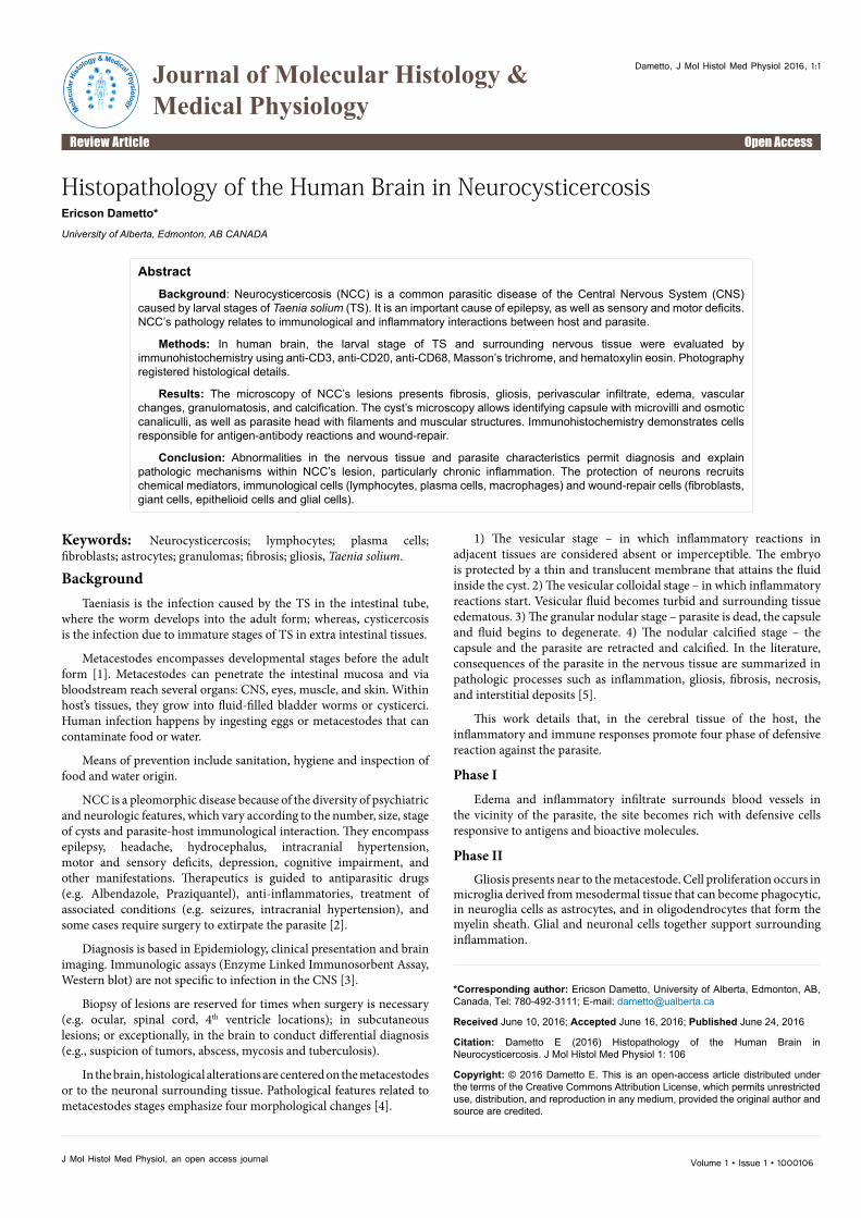
of the parasite and foreign particles that do not pass the tight junctions between the endothelial cells at the level of blood-brain-barrier can be phagocytized in the perivascular spaces. Fibroblasts and plasma cells proliferate in perivascular space (Figure 1). The beginning of defensive processes, as edema and antibody formation, is identified in the perivascular space. In the interface between parasite and nervous tissue predominates final processes, as granulomatosis, fibrosis, and calcification.
Edema
Edema is a major sign of inflammations. Vasogenic edema results from increasing permeability of vessels wall; while, in cytotoxic edema the sodium and potassium pump of the cell membrane is impaired, leading to cellular retention of water.
In neurocysticercosis, vasoactive substances released by host’s cells are an origin of edema; another possible cause of edema is substances from the cyst, which can have vasoactive or cytotoxic properties.
Gliosis
Gliosis stands for changes of glial cells in response to damage of CNS, when the number of astrocytes increases abnormally due to death of neurons it are called astrogliosis. Astrocytes are the main component of the gliosis in the brain tissue near to neurocysticercosis lesions. Astrogliosis is determined by the expression of glial fibrillary acid protein (GFAP) [6]. GFAP has been identified in astrocytes and their cytoplasmic processes that encompass capillaries. Phagocytosis has
been attributed to the astrocytes function; they clean debris, absorbing and digesting it. Microglia cells are present within the lesions; they have also function of phagocytosis. The astrocytes form a matrix with their membrane that fills the surrounding damaged region near to the parasite. Another feature in gliosis is the heterogeneity of cell morphology: astrocytes vary in shape and size (Figures 2 and 3).
Perivascular infiltrate
Perivascular infiltrate is a set of cells between vessels and tissue. The cells found in the specimens of NCC’s lesion and located in perivascular spaces are T-cells CD3, B-cells CD20, plasma cells expressing light chains Kappa and Lambda, macrophages CD68.
Lymphocyte T CD3
T lymphocytes are a class of white blood cells originating from thymus, and they have in their membrane the cluster of differentiation (CD) 3 in almost all stages of development. CD3 is required for T-cell activation and consists of a protein complex that contains four distinct chains. These chains associate with the T-cell receptor to generate an activation signal in T lymphocytes [7]. CD3 can be used to distinguish T-cells from B-cells and myeloid neoplasms (lymphomas and leukaemias) [8]. Antigens released by the metacestode activate T-cells CD3. T-cells secrete cytokines as answer to antigens. Cytokines grow more T-cells, attract macrophages, neutrophils and support T-cells to mature and differentiate in T cell helpers or cytotoxic cells. Lymphocytes T CD3 and Lymphocytes B CD20 occur in the perivascular infiltrate near the parasite (Figure 4).
Lymphocyte B CD20
Lymphocytes B are white blood cells, which in mammals mature in the bone marrow. B cells bind to a specific antigen using their B cell receptors on their membrane. The antigen either can be free or introduced by macrophages or dendritic cells. B cells differentiate into a plasma cell that secretes large amounts of antibodies or memory B cells for persistent protection [9]. CD20 is a component of the cell surface that regulates calcium transport across the plasma membrane [10,11]. In B cells, the engagement of CD20 molecules initiates a signal transduction cascade via tyrosine kinases involved in cell adhesion, proliferation, and survival [12-15]. CD20 is a membrane phosphoprotein present on B cells, it is expressed in lymphocytes precursor and mature B cells, but it is not expressed on plasma cells (Figure 5A) [16]. Human CD20 deficiency results in decreased antibody levels against polysaccharides after vaccination, therefore some authors suggest that CD20 enables lymphocyte B activation independent of T-cells. Antigens that activate B cells with the help of T-cells are known as T cell- dependent antigens and include foreign proteins [13]. Antibody production via
Figure 1: A and D Perivascular space in NCC (H&E, X100). B Fibroblasts and Macrophages close to the vessels (H&E X400). C Plasma cell with Russell bodies close to NCC lesion (H&E, X400). D Perivascular infiltrate.
Citation: Dametto E (2016) Histopathology of the Human Brain in Neurocysticercosis. J Mol Histol Med Physiol 1: 106
Page 3 of 7
Volume 1 • Issue 1 • 1000106J Mol Histol Med Physiol, an open access journal
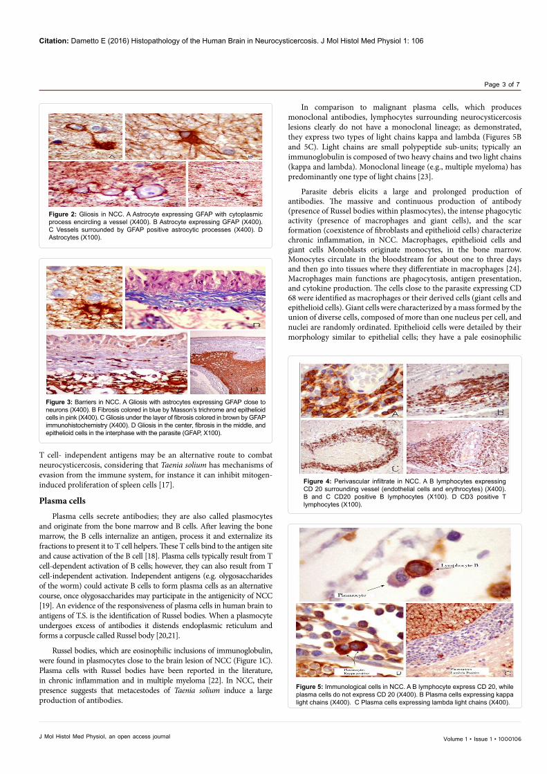
Figure 2: Gliosis in NCC. A Astrocyte expressing GFAP with cytoplasmic process encircling a vessel (X400). B Astrocyte expressing GFAP (X400). C Vessels surrounded by GFAP positive astrocytic processes (X400). D Astrocytes (X100).
Figure 3: Barriers in NCC. A Gliosis with astrocytes expressing GFAP close to neurons (X400). B Fibrosis colored in blue by Masson’s trichrome and epithelioid cells in pink (X400). C Gliosis under the layer of fibrosis colored in brown by GFAP immunohistochemistry (X400). D Gliosis in the center, fibrosis in the middle, and epithelioid cells in the interphase with the parasite (GFAP, X100).
T cell- independent antigens may be an alternative route to combat neurocysticercosis, considering that Taenia solium has mechanisms of evasion from the immune system, for instance it can inhibit mitogen-induced proliferation of spleen cells [17].
Plasma cells
Plasma cells secrete antibodies; they are also called plasmocytes and originate from the bone marrow and B cells. After leaving the bone marrow, the B cells internalize an antigen, process it and externalize its fractions to present it to T cell helpers. These T cells bind to the antigen site and cause activation of the B cell [18]. Plasma cells typically result from T cell-dependent activation of B cells; however, they can also result from T cell-independent activation. Independent antigens (e.g. olygosaccharides of the worm) could activate B cells to form plasma cells as an alternative course, once olygosaccharides may participate in the antigenicity of NCC [19]. An evidence of the responsiveness of plasma cells in human brain to antigens of T.S. is the identification of Russel bodies. When a plasmocyte undergoes excess of antibodies it distends endoplasmic reticulum and forms a corpuscle called Russel body [20,21].
Russel bodies, which are eosinophilic inclusions of immunoglobulin, were found in plasmocytes close to the brain lesion of NCC (Figure 1C). Plasma cells with Russel bodies have been reported in the literature, in chronic inflammation and in multiple myeloma [22]. In NCC, their presence suggests that metacestodes of Taenia solium induce a large production of antibodies.
In comparison to malignant plasma cells, which produces monoclonal antibodies, lymphocytes surrounding neurocysticercosis lesions clearly do not have a monoclonal lineage; as demonstrated, they express two types of light chains kappa and lambda (Figures 5B and 5C). Light chains are small polypeptide sub-units; typically an immunoglobulin is composed of two heavy chains and two light chains (kappa and lambda). Monoclonal lineage (e.g., multiple myeloma) has predominantly one type of light chains [23].
Parasite debris elicits a large and prolonged production of antibodies. The massive and continuous production of antibody (presence of Russel bodies within plasmocytes), the intense phagocytic activity (presence of macrophages and giant cells), and the scar formation (coexistence of fibroblasts and epithelioid cells) characterize chronic inflammation, in NCC. Macrophages, epithelioid cells and giant cells Monoblasts originate monocytes, in the bone marrow. Monocytes circulate in the bloodstream for about one to three days and then go into tissues where they differentiate in macrophages [24]. Macrophages main functions are phagocytosis, antigen presentation, and cytokine production. The cells close to the parasite expressing CD 68 were identified as macrophages or their derived cells (giant cells and epithelioid cells). Giant cells were characterized by a mass formed by the union of diverse cells, composed of more than one nucleus per cell, and nuclei are randomly ordinated. Epithelioid cells were detailed by their morphology similar to epithelial cells; they have a pale eosinophilic
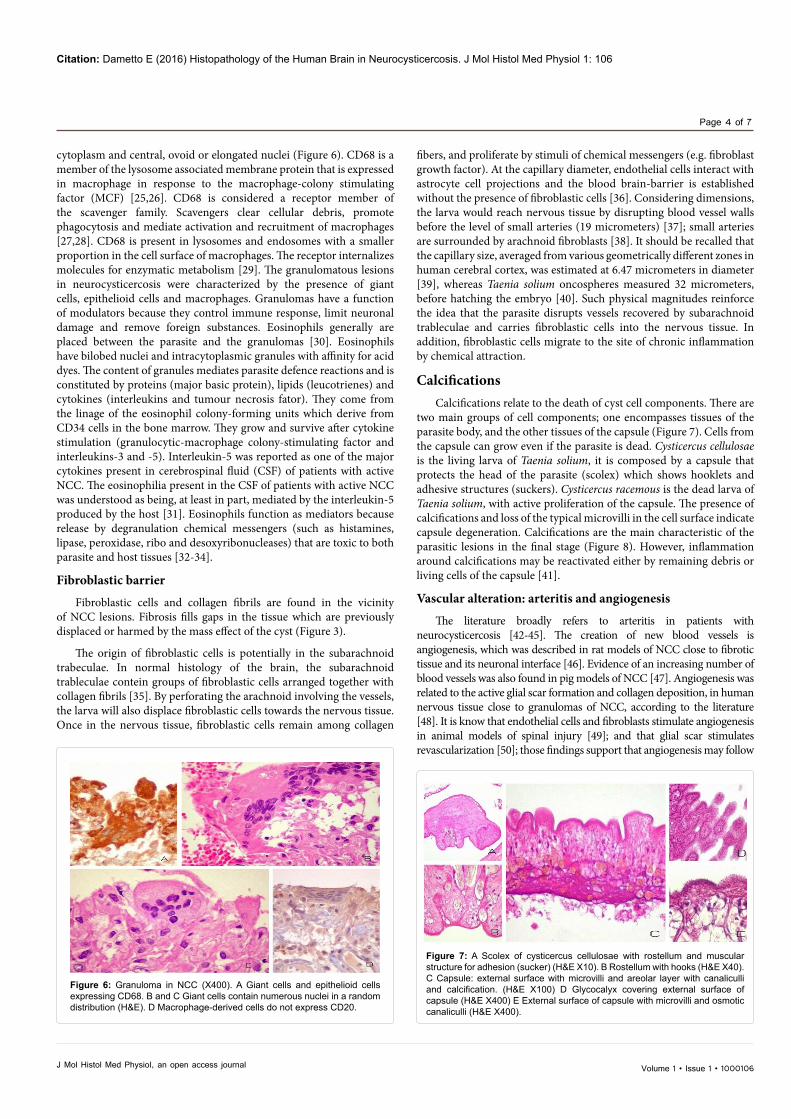
Figure 4: Perivascular infiltrate in NCC. A B lymphocytes expressing CD 20 surrounding vessel (endothelial cells and erythrocytes) (X400). B and C CD20 positive B lymphocytes (X100). D CD3 positive T lymphocytes (X100).
Figure 5: Immunological cells in NCC. A B lymphocyte express CD 20, while plasma cells do not express CD 20 (X400). B Plasma cells expressing kappa light chains (X400). C Plasma cells expressing lambda light chains (X400).
Citation: Dametto E (2016) Histopathology of the Human Brain in Neurocysticercosis. J Mol Histol Med Physiol 1: 106
Page 4 of 7
Volume 1 • Issue 1 • 1000106J Mol Histol Med Physiol, an open access journal
cytoplasm and central, ovoid or elongated nuclei (Figure 6). CD68 is a member of the lysosome associated membrane protein that is expressed in macrophage in response to the macrophage-colony stimulating factor (MCF) [25,26]. CD68 is considered a receptor member of the scavenger family. Scavengers clear cellular debris, promote phagocytosis and mediate activation and recruitment of macrophages [27,28]. CD68 is present in lysosomes and endosomes with a smaller proportion in the cell surface of macrophages. The receptor internalizes molecules for enzymatic metabolism [29]. The granulomatous lesions in neurocysticercosis were characterized by the presence of giant cells, epithelioid cells and macrophages. Granulomas have a function of modulators because they control immune response, limit neuronal damage and remove foreign substances. Eosinophils generally are placed between the parasite and the granulomas [30]. Eosinophils have bilobed nuclei and intracytoplasmic granules with affinity for acid dyes. The content of granules mediates parasite defence reactions and is constituted by proteins (major basic protein), lipids (leucotrienes) and cytokines (interleukins and tumour necrosis fator). They come from the linage of the eosinophil colony-forming units which derive from CD34 cells in the bone marrow. They grow and survive after cytokine stimulation (granulocytic-macrophage colony-stimulating factor and interleukins-3 and -5). Interleukin-5 was reported as one of the major cytokines present in cerebrospinal fluid (CSF) of patients with active NCC. The eosinophilia present in the CSF of patients with active NCC was understood as being, at least in part, mediated by the interleukin-5 produced by the host [31]. Eosinophils function as mediators because release by degranulation chemical messengers (such as histamines, lipase, peroxidase, ribo and desoxyribonucleases) that are toxic to both parasite and host tissues [32-34].
Fibroblastic barrier
Fibroblastic cells and collagen fibrils are found in the vicinity of NCC lesions. Fibrosis fills gaps in the tissue which are previously displaced or harmed by the mass effect of the cyst (Figure 3).
The origin of fibroblastic cells is potentially in the subarachnoid trabeculae. In normal histology of the brain, the subarachnoid trableculae contein groups of fibroblastic cells arranged together with collagen fibrils [35]. By perforating the arachnoid involving the vessels, the larva will also displace fibroblastic cells towards the nervous tissue. Once in the nervous tissue, fibroblastic cells remain among collagen
fibers, and proliferate by stimuli of chemical messengers (e.g. fibroblast growth factor). At the capillary diameter, endothelial cells interact with astrocyte cell projections and the blood brain-barrier is established without the presence of fibroblastic cells [36]. Considering dimensions, the larva would reach nervous tissue by disrupting blood vessel walls before the level of small arteries (19 micrometers) [37]; small arteries are surrounded by arachnoid fibroblasts [38]. It should be recalled that the capillary size, averaged from various geometrically different zones in human cerebral cortex, was estimated at 6.47 micrometers in diameter [39], whereas Taenia solium oncospheres measured 32 micrometers, before hatching the embryo [40]. Such physical magnitudes reinforce the idea that the parasite disrupts vessels recovered by subarachnoid trableculae and carries fibroblastic cells into the nervous tissue. In addition, fibroblastic cells migrate to the site of chronic inflammation by chemical attraction.
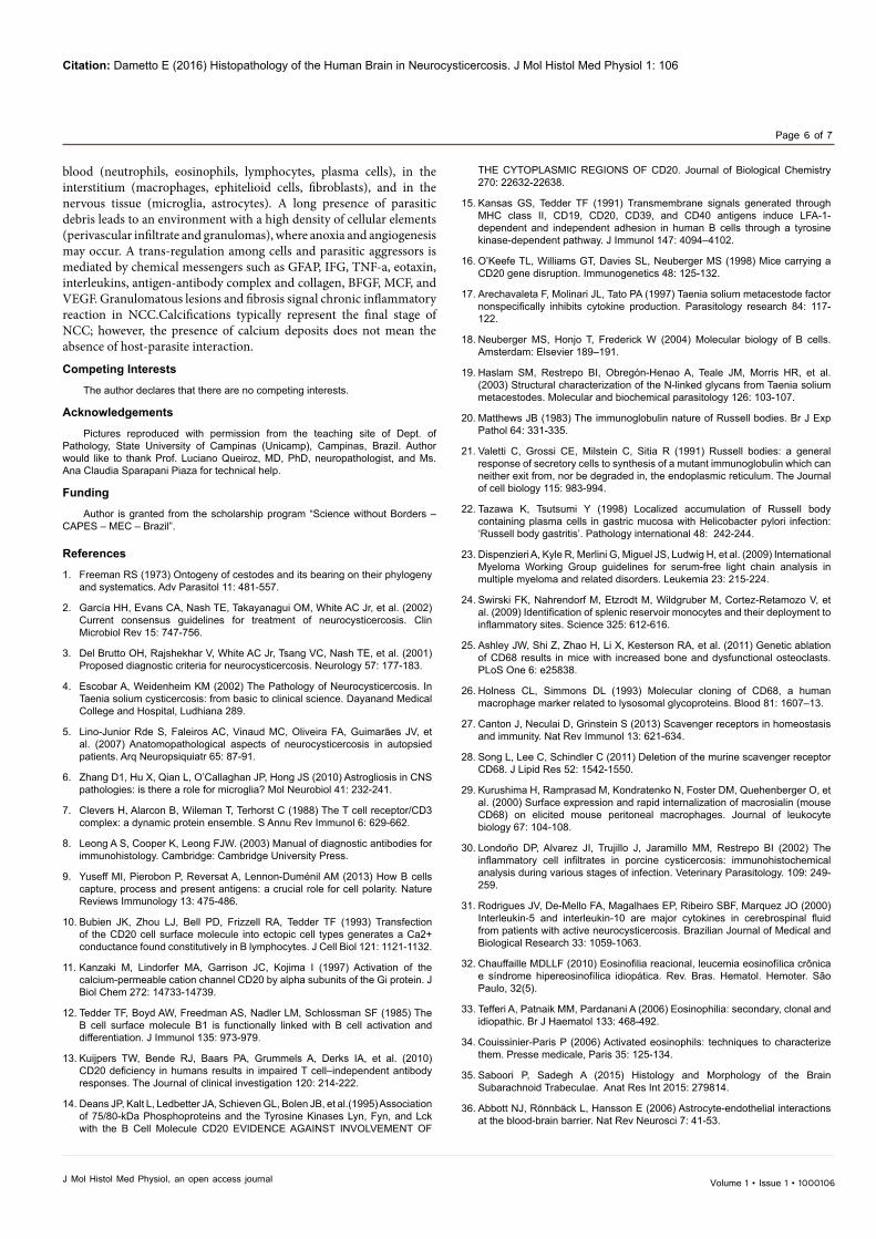
CalcificationsCalcifications relate to the death of cyst cell components. There are
two main groups of cell components; one encompasses tissues of the parasite body, and the other tissues of the capsule (Figure 7). Cells from the capsule can grow even if the parasite is dead. Cysticercus cellulosae is the living larva of Taenia solium, it is composed by a capsule that protects the head of the parasite (scolex) which shows hooklets and adhesive structures (suckers). Cysticercus racemous is the dead larva of Taenia solium, with active proliferation of the capsule. The presence of calcifications and loss of the typical microvilli in the cell surface indicate capsule degeneration. Calcifications are the main characteristic of the parasitic lesions in the final stage (Figure 8). However, inflammation around calcifications may be reactivated either by remaining debris or living cells of the capsule [41].
Vascular alteration: arteritis and angiogenesis
The literature broadly refers to arteritis in patients with neurocysticercosis [42-45]. The creation of new blood vessels is angiogenesis, which was described in rat models of NCC close to fibrotic tissue and its neuronal interface [46]. Evidence of an increasing number of blood vessels was also found in pig models of NCC [47]. Angiogenesis was related to the active glial scar formation and collagen deposition, in human nervous tissue close to granulomas of NCC, according to the literature [48]. It is know that endothelial cells and fibroblasts stimulate angiogenesis in animal models of spinal injury [49]; and that glial scar stimulates revascularization [50]; those findings support that angiogenesis may follow
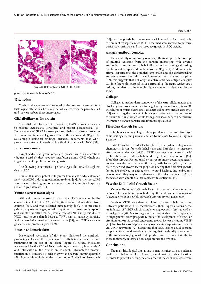
Figure 6: Granuloma in NCC (X400). A Giant cells and epithelioid cells expressing CD68. B and C Giant cells contain numerous nuclei in a random distribution (H&E). D Macrophage-derived cells do not express CD20.
Figure 7: A Scolex of cysticercus cellulosae with rostellum and muscular structure for adhesion (sucker) (H&E X10). B Rostellum with hooks (H&E X40). C Capsule: external surface with microvilli and areolar layer with canaliculli and calcification. (H&E X100) D Glycocalyx covering external surface of capsule (H&E X400) E External surface of capsule with microvilli and osmotic canaliculli (H&E X400).
Citation: Dametto E (2016) Histopathology of the Human Brain in Neurocysticercosis. J Mol Histol Med Physiol 1: 106
Page 5 of 7
Volume 1 • Issue 1 • 1000106J Mol Histol Med Physiol, an open access journal
gliosis and fibrosis in human NCC.
DiscussionThe bioactive messengers produced by the host are determinants of
histological alterations; however, the substances from the parasite elicit and may exacerbate those messengers.
Glial fibrillary acidic protein
The glial fibrillary acidic protein (GFAP) allows astrocytes to produce cytoskeletal structures and project pseudopodia [51]. Enhancement of GFAP in astrocytes and their cytoplasmic processes were observed in areas of gliosis close to the metacestode (Figure 2). Sustaining histological findings, literature documents that GFAP protein was detected in cerebrospinal fluid of patients with NCC [52].
Interferon gamma
Lymphocytes and granulomas are present in NCC alterations (Figures 4 and 6); they produce interferon gamma (IFG) which can trigger astrocytes proliferation and gliosis.
The following experiments together suggest that IFG elicits gliosis due to NCC.
Human IFG was a potent mitogen for human astrocytes cultivated in vitro, and IFG induced gliosis in mouse brain [53]. Furthermore, IFG was present in NCC granulomas prepared in mice, in high frequency (11 of 12 granulomas) [54].
Tumor necrosis factor alpha
Although tumor necrosis factor alpha (TNF-a) occurs in the cerebrospinal fluid of NCC patients, its amount did not differ from controls [55], and was detected infrequently [56]. It is produced primarily by macrophages, as well as by fibroblasts, neurons, lymphoid and endothelial cells [57]. A possible role of TNF-a in gliosis due to NCC must be considered; because, TNF-a can stimulate cytotoxicity and increase inflammation in nervous tissue [58]; and TNF-a activates glial cells and promotes gliosis [59].
Eotaxin and interleukins
Histological specimens of this study illustrated the antibody-producing cells and their precursor B cells being attracted to and maturating in the site of the lesion (Figure 5). Several mediators are elevated in the CSF of NCC patients, e.g. eotaxin, interleukin-5 and interleukin-6; the first is an eosinophil chemotactic protein; interleukin-5 stimulates B cells to grow and secrete immunoglobulin [59]. Interleukine-6 induces the maturation of B cells into plasma cells
[60]; reactive gliosis is a consequence of interleukin-6 expression in the brain of transgenic mice [61]. Those mediators interact to perform perivascular infiltrate and may produce gliosis in NCC lesions.
Antigen-antibody complex
The variability of immunoglobulin synthesis supports the concept of multiple antigens from the parasite interacting with diverse antibodies from the host, this is indicated in the histological finding by plasmocytes kappa and lambda positive (Figure 5). Additionally, in animal experiments, the complex light chain and the corresponding antigen increased intracellular calcium on murine dorsal root ganglion [62]; this suggests that not only the entire antibody-antigen complex can interfere with neuronal tissue surrounding the neurocysticercosis lesions, but also that the complex light chain and antigen can do the same.
Collagen
Collagen is an abundant component of the extracellular matrix that blocks cysticercosis invasion into neighboring brain tissue (Figure 3). In cultures of murine astrocytes, collagen did not proliferate astrocytes [63], supporting the concept of fibrosis as a protective barrier in favor of the neuronal tissue, which would form gliosis secondary to a permissive interaction between parasite and immunological cells.
Fibroblast Growth Factors
Fibroblasts among collagen fibers proliferate in a protective layer of fibrosis against the parasite, and are found close to vessels (Figures 1 and 3).
Basic Fibroblast Growth Factor (BFGF) is a potent mitogen and chemotactic factor for endothelial cells and fibroblasts, it increases after neuronal damage [64,65]. BFGF also regulates neuronal cells’ proliferation and differentiation during brain maturation [66]. Fibroblast Growth Factors (acid or basic) are more potent angiogenic factors than the vascular endothelial growth factor (VEGF) or the platelet-derived growth factor [67]. Considering that fibroblast growth factors are involved in angiogenesis, wound healing, and embryonic development, they may repair damages of the infection, once BFGF is associated with endothelial cells adjacent to cysticerci [48].
Vascular Endothelial Growth Factor
Vascular Endothelial Growth Factor is a protein whose function is to create new blood vessels during the embryonic development (vasculogenesis) or new blood vessels after injury (angiogenesis).
Levels of VEGF were detected higher than controls in sera from untreated patients with neurocysticercosis [68]. Hypoxia is considered an inductor of VEGF which stimulates angiogenesis [69], as well as axonal growth [70]. Macrophages and neutrophils have been implicated in angiogenesis. Macrophages may induce the development of a vascular circuit in tumors via several angiogenic growth factors including VEGF [71]. Neutrophils would promote angiogenesis in dysplasias and tumors via VEGF activation [72]. Suggesting that NCC lesions could demand supplementary blood vessels, considering that the density of cells near to the granulomas (Figure 6) could produce an environment similar to those in tumors, in terms of cell agglomerate and hypoxia.
ConclusionsThe main histological alterations in neurocysticercosis are edema,
perivascular infiltrate, gliosis, fibrosis, granulomatosis and calcification. In order to protect neurons, defenses recruit mesenchymal cells from
Figure 8: Calcifications in NCC (H&E, X400).
Citation: Dametto E (2016) Histopathology of the Human Brain in Neurocysticercosis. J Mol Histol Med Physiol 1: 106
Page 6 of 7
Volume 1 • Issue 1 • 1000106J Mol Histol Med Physiol, an open access journal
blood (neutrophils, eosinophils, lymphocytes, plasma cells), in the interstitium (macrophages, ephitelioid cells, fibroblasts), and in the nervous tissue (microglia, astrocytes). A long presence of parasitic debris leads to an environment with a high density of cellular elements (perivascular infiltrate and granulomas), where anoxia and angiogenesis may occur. A trans-regulation among cells and parasitic aggressors is mediated by chemical messengers such as GFAP, IFG, TNF-a, eotaxin, interleukins, antigen-antibody complex and collagen, BFGF, MCF, and VEGF. Granulomatous lesions and fibrosis signal chronic inflammatory reaction in NCC.Calcifications typically represent the final stage of NCC; however, the presence of calcium deposits does not mean the absence of host-parasite interaction.
Competing Interests
The author declares that there are no competing interests.
Acknowledgements
Pictures reproduced with permission from the teaching site of Dept. of Pathology, State University of Campinas (Unicamp), Campinas, Brazil. Author would like to thank Prof. Luciano Queiroz, MD, PhD, neuropathologist, and Ms. Ana Claudia Sparapani Piaza for technical help.
Funding
Author is granted from the scholarship program “Science without Borders – CAPES – MEC – Brazil”.
References
1. Freeman RS (1973) Ontogeny of cestodes and its bearing on their phylogeny and systematics. Adv Parasitol 11: 481-557.
2. García HH, Evans CA, Nash TE, Takayanagui OM, White AC Jr, et al. (2002) Current consensus guidelines for treatment of neurocysticercosis. Clin Microbiol Rev 15: 747-756.
3. Del Brutto OH, Rajshekhar V, White AC Jr, Tsang VC, Nash TE, et al. (2001) Proposed diagnostic criteria for neurocysticercosis. Neurology 57: 177-183.
4. Escobar A, Weidenheim KM (2002) The Pathology of Neurocysticercosis. In Taenia solium cysticercosis: from basic to clinical science. Dayanand Medical College and Hospital, Ludhiana 289.
5. Lino-Junior Rde S, Faleiros AC, Vinaud MC, Oliveira FA, Guimarães JV, et al. (2007) Anatomopathological aspects of neurocysticercosis in autopsied patients. Arq Neuropsiquiatr 65: 87-91.
6. Zhang D1, Hu X, Qian L, O’Callaghan JP, Hong JS (2010) Astrogliosis in CNS pathologies: is there a role for microglia? Mol Neurobiol 41: 232-241.
7. Clevers H, Alarcon B, Wileman T, Terhorst C (1988) The T cell receptor/CD3 complex: a dynamic protein ensemble. S Annu Rev Immunol 6: 629-662.
8. Leong A S, Cooper K, Leong FJW. (2003) Manual of diagnostic antibodies for immunohistology. Cambridge: Cambridge University Press.
9. Yuseff MI, Pierobon P, Reversat A, Lennon-Duménil AM (2013) How B cells capture, process and present antigens: a crucial role for cell polarity. Nature Reviews Immunology 13: 475-486.
10. Bubien JK, Zhou LJ, Bell PD, Frizzell RA, Tedder TF (1993) Transfection of the CD20 cell surface molecule into ectopic cell types generates a Ca2+ conductance found constitutively in B lymphocytes. J Cell Biol 121: 1121-1132.
11. Kanzaki M, Lindorfer MA, Garrison JC, Kojima I (1997) Activation of the calcium-permeable cation channel CD20 by alpha subunits of the Gi protein. J Biol Chem 272: 14733-14739.
12. Tedder TF, Boyd AW, Freedman AS, Nadler LM, Schlossman SF (1985) The B cell surface molecule B1 is functionally linked with B cell activation and differentiation. J Immunol 135: 973-979.
13. Kuijpers TW, Bende RJ, Baars PA, Grummels A, Derks IA, et al. (2010) CD20 deficiency in humans results in impaired T cell–independent antibody responses. The Journal of clinical investigation 120: 214-222.
14. Deans JP, Kalt L, Ledbetter JA, Schieven GL, Bolen JB, et al.(1995) Association of 75/80-kDa Phosphoproteins and the Tyrosine Kinases Lyn, Fyn, and Lck with the B Cell Molecule CD20 EVIDENCE AGAINST INVOLVEMENT OF
THE CYTOPLASMIC REGIONS OF CD20. Journal of Biological Chemistry 270: 22632-22638.
15. Kansas GS, Tedder TF (1991) Transmembrane signals generated through MHC class II, CD19, CD20, CD39, and CD40 antigens induce LFA-1-dependent and independent adhesion in human B cells through a tyrosine kinase-dependent pathway. J Immunol 147: 4094–4102.
16. O’Keefe TL, Williams GT, Davies SL, Neuberger MS (1998) Mice carrying a CD20 gene disruption. Immunogenetics 48: 125-132.
17. Arechavaleta F, Molinari JL, Tato PA (1997) Taenia solium metacestode factor nonspecifically inhibits cytokine production. Parasitology research 84: 117-122.
18. Neuberger MS, Honjo T, Frederick W (2004) Molecular biology of B cells. Amsterdam: Elsevier 189–191.
19. Haslam SM, Restrepo BI, Obregón-Henao A, Teale JM, Morris HR, et al. (2003) Structural characterization of the N-linked glycans from Taenia solium metacestodes. Molecular and biochemical parasitology 126: 103-107.
20. Matthews JB (1983) The immunoglobulin nature of Russell bodies. Br J Exp Pathol 64: 331-335.
21. Valetti C, Grossi CE, Milstein C, Sitia R (1991) Russell bodies: a general response of secretory cells to synthesis of a mutant immunoglobulin which can neither exit from, nor be degraded in, the endoplasmic reticulum. The Journal of cell biology 115: 983-994.
22. Tazawa K, Tsutsumi Y (1998) Localized accumulation of Russell body containing plasma cells in gastric mucosa with Helicobacter pylori infection: ‘Russell body gastritis’. Pathology international 48: 242-244.
23. Dispenzieri A, Kyle R, Merlini G, Miguel JS, Ludwig H, et al. (2009) International Myeloma Working Group guidelines for serum-free light chain analysis in multiple myeloma and related disorders. Leukemia 23: 215-224.
24. Swirski FK, Nahrendorf M, Etzrodt M, Wildgruber M, Cortez-Retamozo V, et al. (2009) Identification of splenic reservoir monocytes and their deployment to inflammatory sites. Science 325: 612-616.
25. Ashley JW, Shi Z, Zhao H, Li X, Kesterson RA, et al. (2011) Genetic ablation of CD68 results in mice with increased bone and dysfunctional osteoclasts. PLoS One 6: e25838.
26. Holness CL, Simmons DL (1993) Molecular cloning of CD68, a human macrophage marker related to lysosomal glycoproteins. Blood 81: 1607–13.
27. Canton J, Neculai D, Grinstein S (2013) Scavenger receptors in homeostasis and immunity. Nat Rev Immunol 13: 621-634.
28. Song L, Lee C, Schindler C (2011) Deletion of the murine scavenger receptor CD68. J Lipid Res 52: 1542-1550.
29. Kurushima H, Ramprasad M, Kondratenko N, Foster DM, Quehenberger O, et al. (2000) Surface expression and rapid internalization of macrosialin (mouse CD68) on elicited mouse peritoneal macrophages. Journal of leukocyte biology 67: 104-108.
30. Londoño DP, Alvarez JI, Trujillo J, Jaramillo MM, Restrepo BI (2002) The inflammatory cell infiltrates in porcine cysticercosis: immunohistochemical analysis during various stages of infection. Veterinary Parasitology. 109: 249-259.
31. Rodrigues JV, De-Mello FA, Magalhaes EP, Ribeiro SBF, Marquez JO (2000) Interleukin-5 and interleukin-10 are major cytokines in cerebrospinal fluid from patients with active neurocysticercosis. Brazilian Journal of Medical and Biological Research 33: 1059-1063.
32. Chauffaille MDLLF (2010) Eosinofilia reacional, leucemia eosinofílica crônica e síndrome hipereosinofílica idiopática. Rev. Bras. Hematol. Hemoter. São Paulo, 32(5).
33. Tefferi A, Patnaik MM, Pardanani A (2006) Eosinophilia: secondary, clonal and idiopathic. Br J Haematol 133: 468-492.
34. Couissinier-Paris P (2006) Activated eosinophils: techniques to characterize them. Presse medicale, Paris 35: 125-134.
35. Saboori P, Sadegh A (2015) Histology and Morphology of the Brain Subarachnoid Trabeculae. Anat Res Int 2015: 279814.
36. Abbott NJ, Rönnbäck L, Hansson E (2006) Astrocyte-endothelial interactions at the blood-brain barrier. Nat Rev Neurosci 7: 41-53.

Citation: Dametto E (2016) Histopathology of the Human Brain in Neurocysticercosis. J Mol Histol Med Physiol 1: 106
Page 7 of 7
Volume 1 • Issue 1 • 1000106J Mol Histol Med Physiol, an open access journal
37. WIEDEMAN MP (1963) Dimensions of blood vessels from distributing artery to collecting vein. Circ Res 12: 375-378.
38. Patestas M, Gartner LP (2013) A textbook of neuroanatomy. John Wiley &Sons 125.
39. Lauwers F, Cassot F, Lauwers-Cances V, Puwanarajah P, Duvernoy H(2008) Morphometry of the human cerebral cortex microcirculation: generalcharacteristics and space-related profiles. Neuroimage 39: 936-948.
40. Jimenez JA, Rodriguez S, Moyano LM, Castillo Y, García HH (2010)Differentiating Taenia eggs found in human stools: does Ziehl-Neelsen staining help? Tropical Medicine & International Health 15: 1077-1081.
41. Ooi WW, Wijemanne S, Thomas CB, Quezado M, Brown CR, et al. (2011)a calcified Taenia solium granuloma associated with recurrent perilesional edema causing refractory seizures: histopathological features. The Americanjournal of tropical medicine and hygiene 85: 460-463.
42. Barinagarrementeria F, Del Brutto OH (1989) Lacunar syndrome due toneurocysticercosis. Arch Neurol 46: 415-417.
43. Barinagarrementeria F, Cantú C (1992) Neurocysticercosis as a cause ofstroke. Stroke 23: 1180-1181.
44. Jha S, Kumar V (2000) Neurocysticercosis presenting as stroke. Neurol India48: 391-394.
45. Tellez-Zenteno JF, Negrete-Pulido O, Cantú C, Márquez C, Vega-Boada F, etal. (2003) Hemorrhagic stroke associated to neurocysticercosis. Neurologia18: 272-275.
46. Verastegui MR, Mejia A, Clark T, Gavidia CM, Mamani J, et al. (2015) Novelrat model for neurocysticercosis using Taenia solium. Am J Pathol 185: 2259-2268.
47. Sikasunge CS, Johansen MV, Phiri IK, Willingham AL, Leifsson PS. (2009)The immune response in Taenia solium neurocysticercosis in pigs isassociated with astrogliosis, axonal degeneration and altered blood–brainbarrier permeability. Veterinary parasitology 160: 242-250.
48. Alvarez JI, Colegial CH, Castano CA, Trujillo J, Teale JM, et al. (2002)The human nervous tissue in proximity to granulomatous lesions inducedby Taenia solium metacestodes displays an active response. Journal ofneuroimmunology 127: 139-144.
49. Jaeger CB, Blight AR (1997) Spinal cord compression injury in guinea pigs:structural changes of endothelium and its perivascular cell associations afterblood–brain barrier breakdown and repair. Experimental neurology 144: 381-399.
50. Stichel CC, Müller HW (1998) The CNS lesion scar: new vistas on an oldregeneration barrier. Cell Tissue Res 294: 1-9.
51. Lepekhin EA, Eliasson C, Berthold CH, Berezin V, Bock E, et al. (2001)Intermediate filaments regulate astrocyte motility. J Neurochem 79: 617-625.
52. Quintanar JL, Franco LM, Salinas E (2003) Detection of glial fibrillary acidic protein and neurofilaments in the cerebrospinal fluid of patients with neurocysticercosis. Parasitology research 90: 261-263.
53. Yong VW, Moumdjian R, Yong FP, Ruijs TC, Freedman MS, et al. (1991)Gamma-interferon promotes proliferation of adult human astrocytes in vitroand reactive gliosis in the adult mouse brain in vivo. Proceedings of theNational Academy of Sciences 88: 7016-7020.
54. Robinson P, Atmar RL, Lewis DE, White AC Jr (1997) Granuloma cytokines in murine cysticercosis. Infect Immun 65: 2925-2931.
55. Ostrosky-Zeichner L, García-Mendoza E, Ríos C, Sotelo J (1995) Humoraland cellular immune response within the subarachnoid space of patients withneurocysticercosis. Archives of medical research 27: 513-517.
56. Evans CA, Garcia HH, Hartnell A, Gilman RH, Jose PJ, et al. (1998) Elevated concentrations of eotaxin and interleukin-5 in human neurocysticercosis.Infect Immun 66: 4522-4525.
57. Wajant H, Pfizenmaier K, Scheurich P (2003) Tumor necrosis factor signaling. See comment in P Cell Death Differ 10: 45-65.
58. Olmos G, Lladó J (2014) Tumor necrosis factor alpha: a link betweenneuroinflammation and excitotoxicity. Mediators Inflamm 2014: 861231.
59. Feuerstein GZ, Liu T, Barone FC (1993) Cytokines, inflammation, and brain injury: role of tumor necrosis factor-alpha. Cerebrovascular and brainmetabolism reviews 6(4), 341-360.
60. Erta M, Quintana A, Hidalgo J (2012) Interleukin-6, a major cytokine in thecentral nervous system. Int J Biol Sci 8: 1254-1266.
61. Chiang CS, Stalder A, Samimi A, Campbell IL (1994) Reactive gliosis as aconsequence of interleukin-6 expression in the brain: studies in transgenicmice. Dev Neurosci 16: 212-221.
62. Rijnierse A, Kroese AB, Redegeld FA, Blokhuis BR, van der Heijden MW, etal,(2009) Immunoglobulin-free light chains mediate antigen-specific responses of murine dorsal root ganglion neurons. Journal of neuroimmunology 208: 80-86.
63. Nagano N, Aoyagi M, Hirakawa K (1993) Extracellular matrix modulates theproliferation of rat astrocytes in serum-free culture. Glia 8: 71-76.
64. Mocchetti I, Rabin SJ, Colangelo AM, Whittemore SR, Wrathall JR (1996)Increased basic fibroblast growth factor expression following contusive spinal cord injury. Experimental neurology 141: 154-164.
65. Akimoto S, Ishikawa O, Iijima C, Miyachi Y (1998) Expression of basicfibroblast growth factor and its receptor by fibroblast, macrophages and mast cells in hypertrophic scar. European journal of dermatology: EJD 9: 357-362.
66. Gremo F, Presta M (2000) Role of fibroblast growth factor-2 in human brain: a focus on development. See comment in PubMed Commons below Int J DevNeurosci 18: 271-279.
67. Cao R1, Bråkenhielm E, Pawliuk R, Wariaro D, Post MJ, et al. (2003)Angiogenic synergism, vascular stability and improvement of hind-limbischemia by a combination of PDGF-BB and FGF-2. See comment in PubMed Commons below Nat Med 9: 604-613.
68. Tuero I, Palma S, Cabeza F, Saleemi S, Rodriguez S, et al. (2015) AComparative Study of Peripheral Immune Responses to Taenia solium inIndividuals with Parenchymal and Subarachnoid Neurocysticercosis. PLoSNegl Trop Dis 9: e0004143.
69. Shweiki D, Itin A, Soffer D, Keshet E (1992) vascular endothelial growth factor induced by hypoxia may mediate hypoxia-initiated angiogenesis. Nature(Lond.) 359: 843-845.
70. Sondell M, Lundborg G, Kanje M (1999) Vascular endothelial growth factorhas neurotropic activity and stimulates axonal outgrowth, enhancing cellsurvival and Schwann cell proliferation in the peripheral nervous system. JNeurosci 19: 5731-5740.
71. Lin EY, Pollard JW (2007) Tumor-associated macrophages press theangiogenic switch in breast cancer. See comment in PubMed Commons below Cancer Res 67: 5064-5066.
72. Nozawa H, Chiu C, Hanahan D (2006) Infiltrating neutrophils mediate the initial angiogenic switch in a mouse model of multistage carcinogenesis.Proceedings of the National Academy of Sciences 103: 12493-12498.


![Lifecycle of Taenia solium - COnnecting REpositories · 2016. 8. 6. · Intestinal pathology: Taeniasis[9] encephalitis Dementia Chemotaxis Stroke/transient Cerebral pathology: Neurocysticercosis](https://img.pdfslide.net/doc/110x75/5fea96ea10512f47ce5ae921/lifecycle-of-taenia-solium-connecting-repositories-2016-8-6-intestinal-pathology.jpg)


![A case report on subarachnoid and intraventricular ......Neurocysticercosis, caused by larvae of the tapeworm taenia solium, is the most common form of parasitic brain disease globally.[1,4]](https://img.pdfslide.net/doc/110x75/5f08db9c7e708231d4240fcb/a-case-report-on-subarachnoid-and-intraventricular-neurocysticercosis-caused.jpg)