Embed Size (px)
Citation preview

Eph
CRa
b
c
d
e
f
g
h
a
ARRA
KSANSBIB
1
tcira
U
0d
Journal of Neuroscience Methods 188 (2010) 258–269
Contents lists available at ScienceDirect
Journal of Neuroscience Methods
journa l homepage: www.e lsev ier .com/ locate / jneumeth
stablishing a model spinal cord injury in the African green monkey for thereclinical evaluation of biodegradable polymer scaffolds seeded withuman neural stem cells
hristopher D. Pritcharda,c,∗, Jonathan R. Slotkinb, Dou Yuc, Haining Daid, Matthew S. Lawrencee,oderick T. Bronsonf, Francis M. Reynoldsg, Yang D. Tengc, Eric J. Woodardh, Robert S. Langera
Department of Chemical Engineering, Massachusetts Institute of Technology, Cambridge, MA, USAWashington Brain & Spine Institute, Washington, DC, USADepartment of Neurosurgery, Brigham and Women’s Hospital, Boston, MA, USADepartment of Neuroscience, Georgetown University Medical Center, Washington, DC, USARxGen, Inc., Hamden, CT, USADepartment of Pathology, Harvard Medical School, Boston, MA, USAInVivo Therapeutics Corporation, Cambridge, MA, USADepartment of Neurosurgery, New England Baptist Hospital, Boston, MA, USA
r t i c l e i n f o
rticle history:eceived 7 December 2009eceived in revised form 19 February 2010ccepted 22 February 2010
eywords:pinal cord injuryfrican green monkeyon-human primatetem cellsiomaterials
njury modelehavioral scoring
a b s t r a c t
Given the involvement of post-mitotic neurons, long axonal tracts and incompletely elucidated injuryand repair pathways, spinal cord injury (SCI) presents a particular challenge for the creation of preclinicalmodels to robustly evaluate longitudinal changes in neuromotor function in the setting in the presenceand absence of intervention. While rodent models exhibit high degrees of spontaneous recovery fromSCI injury, animal care concerns preclude complete cord transections in non-human primates and otherlarger vertebrate models. To overcome such limitations a segmental thoracic (T9–T10) spinal cord hemi-section was created and characterized in the African green monkey. Physiological tolerance of the modelpermitted behavioral analyses for a prolonged period post-injury, extending to predefined study termi-nation points at which histological and immunohistochemical analyses were performed. Four monkeyswere evaluated (one receiving no implant at the lesion site, one receiving a poly(lactide-co-glycolide)(PLGA) scaffold, and two receiving PLGA scaffolds seeded with human neural stem cells (hNSC)). All sub-
jects exhibited Brown-Séquard syndrome 2 days post-injury consisting of ipsilateral hindlimb paralysisand contralateral hindlimb hypesthesia with preservation of bowel and bladder function. A 20-pointobservational behavioral scoring system allowed quantitative characterization of the levels of functionalrecovery. Histological endpoints including silver degenerative staining and Iba1 immunohistochemistry,for microglial and macrophage activation, were determined to reliably define lesion extent and correlatewith neurobehavioral data, and justify invasive telemetered electromyographic and kinematic studies toeffic
more definitively address. Introduction
Spinal cord injury (SCI) results from penetrating or compressiveraumatic injury to the spine or from compressive lesions asso-
iated with neoplastic growth or vertebral dislocation. Neuronalnjury and recovery is critically guided and impacted by the sur-ounding cells and extracellular environment within the spinal cordnd adjacent tissues, reducing the utility of in vitro assays, and∗ Corresponding author at: Room E25-342, 45 Carleton St., Cambridge, MA 02142,SA. Tel.: +1 212 729 8988; fax: +1 617 258 8827.
E-mail address: [email protected] (C.D. Pritchard).
165-0270/$ – see front matter © 2010 Elsevier B.V. All rights reserved.oi:10.1016/j.jneumeth.2010.02.019
acy and mechanism.© 2010 Elsevier B.V. All rights reserved.
necessitating the study of injury mechanisms and spinal cord phys-iology in in vivo vertebrate models (Feringa et al., 1975; Hall andSpringer, 2004; Jones et al., 2005; Liverman et al., 2005; Thuretet al., 2006; Baptiste and Fehlings, 2007; Rossignol et al., 2007). Arecent review of data derived from the extensive literature relatedto the modeling of SCI to better understand mechanisms of injuryand repair has highlighted the greater relevance and utility of non-human primate models relative to rodents and other vertebrate
species in the preclinical investigation of therapeutic interven-tions (Courtine et al., 2007). Rodents may over-predict the efficacyof interventions given high rates of spontaneous recovery frominduced spinal cord injury, even following profound lesions. Thespinal cord anatomy and physiology of old world monkeys are
roscience Methods 188 (2010) 258–269 259
mamfm(a
b(o2lmdbaas
csTprcedhlbetllt(lfvomi
ameNmP
2
2
fbeTwimAR
C.D. Pritchard et al. / Journal of Neu
ore similar to humans, particularly with respect to the positionnd function of corticospinal tracts (Courtine et al., 2007). This per-its a more critical evaluation of results from preclinical studies,
acilitating translation to humans. Here we report the develop-ent of a surgical model of acute SCI in the African green monkey
Chlorocebus sabaeus) for the evaluation of biomaterial implants astranslational interval between rodent and clinical investigations.
Poly(lactide-co-glycolide) (PLGA) biocompatible andiodegradable porous scaffolds seeded with neural stem cellsNSC) have demonstrated potential as a strategy for the treatmentf central nervous system lesions (Flax et al., 1998; Park et al.,002). A PLGA scaffold seeded with murine NSC (mNSC) promoted
ong-term functional improvements in an adult rat hemisectionodel of SCI as compared to controls (Teng et al., 2002). 70
ays post-injury, treated animals exhibited coordinated, weight-earing hindlimb stepping. Histological and immunocytochemicalnalysis suggested the recovery may have been associated withreduction in tissue loss, possibly resulting from modulation of
econdary injury mechanisms and reduced astrogliosis.To establish an SCI model in which this possibility might be criti-
ally evaluated, a lateral hemisection at level T9–T10 in the thoracicpine was created in the monkey, with removal of the ipsilateral9–T10 segment. This approach bears some similarity to previouslyublished models, where ipsilateral tracts were transected withoutemoval of a full segment or only particular tracts (lateral corti-ospinal efferents, dorsal funiculus afferents) were targeted (Crowet al., 1997; Babu et al., 2000; Edgerton et al., 2004). The lesion wasesigned to result in Brown-Séquard syndrome, characterized inumans at comparable cord levels by paralysis of the ispilateral leg,
oss of ipsilateral muscle tone in the lower abdomen (innervatedy T9–L1), loss of vibration and position sensation in the ipsilat-ral hindlimb, loss of thermal and mechanical pain sensation inhe contralateral leg, lumbar and sacral dermatomes and ipsilateralower thoracic dermatomes, and spastic paresis in the ipsilateraleg resulting in increased knee and ankle jerk reflexes with reten-ion of monosynaptic reflexes involving the lower motor neuronsBrown-Séquard, 1851). Importantly, a unilateral restriction of theesion was implemented with the additional aim of sparing bladderunction and anal muscle tone by allowing compensatory inner-ation by retained contralateral corticospinal efferents. Evaluationf loss and recovery of these multiple lower extremity functionsay permit objective measures of the efficacy of the therapeutic
ntervention with minimal post-operative complications.In addition to establishing methodologies for the generation of
discrete unilateral cord lesion designed to minimize special ani-al care requirements, behavioral and histological endpoints were
valuated for their utility in differentiating treatment outcomes.euromotor and histopathological endpoints were assessed acrossultiple time points following introduction of a PLGA scaffold,
LGA scaffold seeded with hNSC, or no implant.
. Materials and methods
.1. Hemisection model
Four juvenile male African green monkeys ranging in weightrom 2.3 to 2.7 kg were employed in the study. Subjects wereetween 1.8 and 2.0 years old. Baseline clinical and neurologicalxams confirmed good health and suitability for study enrollment.reatment allocation was performed arbitrarily with respect to
eight. All experimental and surgical procedures were carried outn accordance with the Guide for the Care and Use of Laboratory Ani-als (National Academy Press, revised 1996) and the Institutionalnimal Care and Use Committee (IACUC) of the St. Kitts Biomedicalesearch Foundation, where the study was conducted.
Fig. 1. Photograph through surgical microscope of scaffold implanted into T9–T10hemisection lesion. Arrows indicates scaffold position. Scale bar = 10 mm.
To perform the hemisection lesion, monkeys were sedatedwith ketamine/xylazine i.m., and kept on a 0.9% saline drip.Isoflourane anesthesia was applied with depth of anesthesia moni-tored throughout the study. Deep dermal or subcutaneous bleedingwas controlled using electrocautery following skin incision. Thelaminectomy was performed by an en-bloc, lateral lamina cutmethod. Following bone removal, the dural opening was performedwith a #15 surgical blade. Dural edges were tacked to the surround-ing ligaments or musculature with 4.0 non-absorbable sutures. Themidline of the spinal cord was carefully assessed with the visual-ization of anatomic landmarks. At all times, spinal cord veins andsurface vessels were spared unless it was absolutely necessary thatthey be controlled. The segmental hemisection lesion was micro-scopically performed with the use of microneurosurgical dissectioninstruments and micro-suction aspiration. A transverse incisionextending to the midline was made at the caudal point of the inter-section of the T9 dorsal root with the cord and at the rostral pointof the intersection of the T10 dorsal root with the cord, followedby a midline incision extending between those levels. Hemi-cordparenchyma resection to these defined boundaries ensured com-plete transection to the anatomic midline in a lesion extending10 mm in length, without damage to the contralateral hemi-cord. Atthis point in the procedure, if designated, a scaffold was implanted(Fig. 1). The scaffold was sized to fill the hemisection cavity, withoutexerting pressure on the surrounding host tissue during or follow-ing insertion. When this process was complete, the dural tack-upsutures were removed, and the dura was closed with a 4.0 non-absorbable single running suture. Fibrin tissue sealant was appliedto the dural suture line. The fascia was then closed with interrupted3.0 absorbable sutures. After this layer was closed, the subcuta-neous layers were re-approximated with 3.0 absorbable sutures.Then, the deep dermal layer was brought together with interrupted3.0 absorbable sutures. After successful extubation and recoveryfrom anesthesia, a neurologic examination was performed. Vitalsigns were monitored in a manner consistent with standard humanpost-anesthesia care. Following recovery from anesthesia the mon-keys were returned to their cage with a mattress placed on the floorto minimize pressure sore risk. Monkeys were observed twice dailyto assess skin integrity and exclude the possibility of autophagy,which can be observed in the setting of limb denervation. All
monkeys additionally received a pre and postoperative course ofimmunosuppressants, consisting of cyclosporine (0.6 mg/kg), pred-nisolone (0.3 mg/kg) and azathioprine (0.5 mg/kg) i.m. BID starting3 days before implantation and continuing until sacrifice to prevent
2 roscience Methods 188 (2010) 258–269
hpv
2
fdh8wntctimcct
ivsRtsbas
2
abeoasfim
2Mibdwslecosw
Table 1Ambulation chamber video observational neuromotor score.
Scale Description
0 No voluntary function1 Slight one or two joints movement2 Active one or two joints, slight movement others3 Active movement of all three joints, no weight bearing4 Slight weight bearing, consistent dorsal stepping (no plantar
stepping)5 Slight weight bearing, occasional plantar stepping6 Frequent plantar stepping, occasional weight bearing, hops with
partial weight support7 Frequent plantar stepping and weight bearing, occasional FL-HL
coordination,8 Consistent plantar stepping and partial weight supported steps,
occasional FL-HL coordination9 Frequent partial weight supported steps, occasional FL-HL
coordination10 Occasional partial weight supported steps, frequent foot drop
and/or drag, run with partial weight support11 Occasional partial weight supported steps, frequent FL-HL
coordination12 Slight partial weight supported steps, frequent FL-HL coordination,
stands up HL with partial weight support13 Slight partial weight supported steps, consistent FL-HL
coordination, frequent foot drop and/or drag14 Full weight supported steps and consistent FL-HL coordination,
occasional foot drop and/or drag15 Occasional foot drop and/or drag, stand up HL with full weight
support16 Slight foot drop and/or drag, no toe clearance17 No foot drop and/or drag, no toe clearance
TS
60 C.D. Pritchard et al. / Journal of Neu
NSC rejection. Though this immunosuppressant regimen had beenreviously established to be well tolerated, additional attention andigilance was maintained to prevent opportunistic infections.
.2. Neuromotor video recordings
Video recordings of quadrupedal locomotion were generatedor qualitative and quantitative assessment of gait and postureeficiencies associated with motor impairment of the ipsilateralindlimb. To document locomotion, the monkeys were placed in an× 2.5 × 2 foot ambulation chamber. One long side of the chamberas made of PlexiglasTM to allow visualization of the monkey as itavigated the enclosure. Movements were recorded by video con-inuously for a 4-min period with the entire width of the enclosureaptured in the camera’s field of view under optimized illumina-ion. At the end of the 4-min video segment a food reward wasntroduced to the chamber through an aperture in the ceiling to pro-
ote and document upright standing, after which another 4 min oflose-up video of the monkey was obtained. Video sessions wereonducted prior to surgery, postoperative days 2, 3, 4, 6 and 10 andhen weekly for 6 weeks and prior to sacrifice.
Video data were reviewed and rated by a blinded reviewer notnvolved in the in vivo execution of the study. Ratings for eachideo session were based on review of the combined 4-min videoegments collected during the wide field and close-up recordings.atings generated an overall observational neuromotor score forhe ipsilateral and contralateral hindlimbs (Table 1). The ratingcale incorporates the components derived from the spectrum ofehaviors and motions observed in healthy African green monkeysnd was based on previously established methods for observationalcoring (Basso et al., 1996; Babu et al., 2000).
.3. Histology and immunohistochemistry
On the study day indicated in Table 2, the monkeys were euth-nized with intravenous sodium pentobarbital followed by wholeody perfusion fixation with 4% paraformaldehyde. Animals werexamined carefully for external abnormalities including hair lossr decubitus ulcers at bony prominences, palpable masses, andbnormalities in the abdominal, thoracic, and cranial cavities. Theciatic nerves, spinal cord and brain were dissected en masse andxed overnight in 4% paraformaldehyde, following which speci-ens were transferred to phosphate buffered saline.The fixed tissue was treated overnight with 20% glycerol and
% dimethylsulfoxide and embedded in a gelatin matrix usingultiBrainTM (NeuroScience Associates, Knoxville, TN). After cur-
ng in weak formaldehyde solution, the block was rapidly frozeny immersion in 2-methyl butane chilled to −70 ◦C with crushedry ice and mounted on an A860 sliding microtome. The blockas sectioned to generate alternating 40 �m sagittal and coronal
ections spanning the lesion site as well as cervical, thoracic andumbar regions rostral and caudal to the lesion. For each stain,
very 24th section at 720 �m intervals was mounted and pro-essed, yielding approximately 20 individual sections mountedn 10 slides. For ionized calcium-binding adapter molecule (Iba1)taining, sections were stained free-floating. Sections were treatedith hydrogen peroxide and blocking serum, and immunostainedable 2tudy animal summary.
Monkey Weight (kg) Sex Treatment group
Y464 2.80 M Scaffold aloneX992 2.74 M Scaffold + hNSCY156 2.34 M ControlY430 2.79 M Scaffold + hNSC
18 Occasional toe clearance19 Frequent toe clearance20 Normal
with a 1:15,000 dilution of primary polyclonal rabbit anti-Iba1antibody (#01973, WAKO, http://wakousa.com), a goat anti-rabbitsecondary antibody, and an avidin-biotin-HRP complex (Vectas-tain ABC kit, Vector, Burlingame, CA). Incubation times were24 h (4 ◦C) for the primary antibody, 30 min (room temperature)for the secondary antibody, and 1 h (room temperature) for theavidin–biotin–HRP complex. Sections were subsequently treatedwith diaminobenzidine tetrahydrochloride (DAB) and mounted ongelatinized (subbed) glass slides. Amino cupric silver staining wasperformed as described previously (De Olmos et al., 1994).
Adobe Photoshop Creative Suite 4 Extended was used for cordimage compilation and analysis. For each cord, the original 8–10microscopic images of the sections containing the lesions were con-solidated using Photomerge in the reposition layout, shown in Fig. 9.Guidelines for cord lengths and widths were formulated using thestraight line tool, and their pixel measurements were taken usingRuler tool. For lesion areas, original cord border was approximatedwith the outline of the spared portion as a reference with the helpof the magnetic lasso under 10% contrast settings and the brush.Area measurements with square pixel units were taken using themagnetic lasso and record measurements with area data points.
Due to the curvature of both X992 and Y156, the cord images
were straightened out to obtain more realistic data for quantifi-cation of lesion dimensions. While keeping cord width constantbetween the original and modified images, warp was used tostraighten out the image and prepare it for measurements.Surgery date Sacrifice date Sacrifice day
4/19/08 7/11/08 834/19/08 5/29/08 404/20/08 8/8/08 1114/20/08 7/11/08 82

C.D. Pritchard et al. / Journal of Neuroscie
Table 3Quantification of lesion dimensions. All cord widths were normalized to 0.60 cm.All units in cm unless otherwise noted.
Monkey Treatment group Sacrificeday
Lesion area(cm2)
Maximumlesion width
X992 Scaffold + hNSC 40 0.24 0.36Y430 Scaffold + hNSC 82 0.26 0.28
uCeaT
2
wtiblsttptwwdsr(ctrltpfblc−
2
iegpwefsf(l(fi
(45 rpm).Human neural stem cells (hNSC, clone HFB2050) were main-
tained in FalconTM culture flasks in modified NeurobasalTM cellculture medium (with B27 supplements, antibiotics, heparin, andl-glutamine) in a humid incubator (37 ◦C, 5% CO2) (Flax et al., 1998).
Fig. 2. Scanning electron microscope image of scaffold architecture.
Y464 Scaffold alone 83 0.26 0.24Y156 Control 111 0.31 0.29
All data was converted from pixel units to metric unitssing measurements recorded from the original cord samples.ord widths were then normalized to 6 mm for all samples, tonable comparison and remove histological artifact. Linear andrea dimensions were adjusted accordingly and summarized inable 3.
.4. Poly(lactide-co-glycolide) implants
Poly(lactide-co-glycolide) (PLGA) polymer porous scaffoldsere prepared in a similar manner to previously published pro-
ocols (Mikos et al., 1992; Lavik et al., 2002; Teng et al., 2002). Themplant was sized to precisely fit the spinal cord cavity, createdy the segmental hemisection surgical resection. The size of the
esion was determined prior to live surgery by measurement of thepinal cord of a cadaver African green monkey, of similar weighto the enrolled subjects. The ratio of lactic to glycolic acid units inhe PLGA was chosen in order for the scaffold to degrade over aeriod of 4–8 weeks in vivo (Sung et al., 2004). PLGA with a lac-ide/glycolide ratio of 50:50 and intrinsic viscosity 0.55–0.75 dL/gas obtained from Lactel (Durect, Pelham, AL). Thin polymer foamsere fabricated by solvent casting/particulate leaching. PLGA wasissolved in chloroform (Sigma, St. Louis, MO) to obtain a 5% w/vtock solution. The stock solution was mixed with sodium chlo-ide (particle size 180–450 �m by sieving; Sigma) in a 1:1.67 ratio3 mL/5 g) and poured into a 5.5 cm diameter TeflonTM mold. Afterhloroform was evaporated in a fume hood for 48 h, the salt par-icles were leached out by immersion in distilled water for 48 h atoom temperature. The water was changed every 4–8 h during theeaching period. Subsequently, PLGA foam disks were dried on blot-ing paper and lyophilized overnight to remove residual water. Therocess yielded highly porous foam with a thickness of 1 mm. Theoams were cut into segments (5 cm × 2 cm) with a surgical razorlade (VWR, West Chester, PA) and stored in 50 mL polypropy-
ene Falcon test tubes (BD Biosciences, San Jose, CA) in a secondaryontainer over anhydrous calcium sulfate (Drierite, Xenia, OH) at20 ◦C until use.
.5. Cell seeding on poly(lactide-co-glyolide) scaffolds
Scaffolds were trimmed to 5 mm × 12 mm × 1 mm, prior to seed-ng. As PLGA is hydrophobic, the scaffolds were immersed in 70%thanol for 30 min on an orbital shaker at room temperature (Daig-er, Vernon Hills, IL), for the purpose of removing air inside theores, sterilizing the accessible surface for cell attachment, and pre-etting the scaffolds prior to immersion in aqueous media (Mikos
t al., 1992). The excess ethanol was then removed and the scaf-olds were washed 3 times for 10 min in sterile phosphate bufferedaline (PBS, pH 7.4, Invitrogen, Carlsbad, CA) solutions and trans-
TM
erred individually to sterile 2 mL Eppendorf centrifuge tubesDaigger) containing 1.5 mL sterile PBS solutions with extracel-ular matrix (ECM) adhesion molecules (10 �g/mL): poly-d-lysineSigma, St. Louis, MO), laminin (BD Biosciences, San Jose, CA), andbronectin (Sigma, St. Louis, MO). Scaffolds were left overnightnce Methods 188 (2010) 258–269 261
on a sterilized orbital shaker platform to ensure sufficient adsorp-tion of the adhesion proteins on the accessible scaffold surface, aswell as full hydration inside the scaffold pores. The following day,supernatant was removed, and the scaffolds were transferred to a6 well culture plate (FalconTM, BD Biosciences, San Jose, CA), fol-lowed by two 5 min rinses in fresh NeurobasalTM medium (withB27-A supplement, Invitrogen, Carlsbad, CA) on an orbital shaker
Fig. 3. Composite scaffold design.

262 C.D. Pritchard et al. / Journal of Neuroscience Methods 188 (2010) 258–269
F ainingo tion;f gure l
P(tctthga1fgbCIiseeibwm
woa(f(fta(ps
oCoicth
areas where too many cells cluster. The images were then printedinverted with high resolution so that each nucleus appears asa black dot. The total numbers of nuclei stained in each cross-section were counted, and sections from the ends and the center
ig. 4. Seeding of hNSC on PLGA scaffolds. (A): Microscope image of toluidine blue stf multiple microscope images of the different areas of the same scaffold cross-secor DAPI). Scale bar = 500 �m. (For interpretation of the references to color in this fi
rior to seeding, cells were lifted from the flasks using AccutaseTM
Innovative Cell Technologies, San Diego, CA), and concentratedo 5 × 10⁵ per mL to 1 × 10⁶ per mL via a series of centrifugation,ell counting, and re-suspension in fresh medium. The first timehe scaffolds were seeded with hNSC is considered an acclima-ion seeding. For the acclimation seeding, 2 mL of 5 × 10⁵ per mLNSC were used in each well in a 6 well plate. The plates wereently rotated in a sterilized humid chamber on an orbital shakert 37 ◦C for 30 min (40 rpm). For subsequent cell seedings, 2 mL of× 106 per mL hNSC were used. Following the rotations, 3 mL of
resh modified NeurobasalTM medium were added to each well, androwth factors (20 ng/mL, basic fibroblast growth factor, bFGF, Cal-iochem, San Diego, CA; 10 ng/mL, leukemia inhibitory factor, LIF,hemicon, Temecula, CA; 20 ng/mL, epidermal growth factor, EGF,
nvitrogen, Carlsbad, CA), and the plates were maintained in thencubator. The plates were observed under sterilized light micro-cope twice daily, and 2.5 mL of the 5 mL total culture medium inach well was replaced with fresh medium and new growth factorsvery 3 days. The seedings were repeated at total of 4 times (includ-ng the acclimation seeding) with an average interval of 1 week inetween each seeding. Films of PLGA approximately 200 �m thickere sterilized in 70% ethanol and maintained in serum containingedium for 1 week.At the end of the scaffold seeding period, light-field microscopy
as carried out on scaffolds in the culture plates, and imagesf attached hNSC inside the scaffold pores were captured usingn AxioCamTM MRc on an AxioVertTM S100 inverted microscopeCarl Zeiss Microimaging, Thornwood, NY). Representative scaf-olds were then fixed in 4% paraformaldehyde at 4 ◦C overnighton the same day as the surgeries were carried out using scaffoldsrom the same seeding wells), cryoprotected in 30% sucrose solu-ion, embedded in OCTTM compound (Sakura Finetek, Torrance, CA),nd cut into 20 �m thick serial sections for immunocytochemicalICC) analysis. The scaffolds were oriented in the embedding com-ound in such a way that the sections are across the width (5 mmide) of the scaffold.
Microslides with scaffold sections were coverslipped with Flu-rescent Mounting Medium with DAPI (Vector Labs, Burlingame,A), and fluorescent images were captured via an AxioCamTM MRm
n an AxioVertTM S100 inverted microscope (Carl Zeiss Microimag-ng) with a DAPI-specific color filter. Montage images of the entireross-section were created from multiple shots at 10× magnifica-ion, which show clear identification of the nucleus of each attachedNSC. Occasionally, higher magnification images were captured inof a cross-section of a PLGA scaffold seeded with hNSCs (purple dots). (B): Montageinverted greyscale image of DAPI fluorescence (black dots are hNSC nuclei stainedegend, the reader is referred to the web version of the article.)
Fig. 5. Temporal profile of functional recovery post-injury. (A): Video neuromotorscores in ipsilateral (left) hindlimb. (B): Video neuromotor scores in contralateral(right) hindlimb. Designations are as follows: Scaffold + hNSC: mean and standarddeviation for Y430 and X992; n = 2 except for day 1 and day 44 where n = 1 due tosacrifice of X992 at 40 days post-injury. Scaffold alone: Y464. Control (no treatment):Y156.

roscie
obtlcndwin
3
3
swaiwalttc
ttan15
FAaa
C.D. Pritchard et al. / Journal of Neu
f the scaffolds were used for this practice. The average cell num-ers (n) in each cross-section (20 �m) were used to estimate theotal number (N) of hNSC seeded in each scaffold (N = n × (scaffoldength/20 �m)). The cell nucleus counting follows the principle ofonservative counting, by which only the clearly distinguishableuclei were counted. Given the fact that some cells are washed offuring the staining process, and some nuclei were not visible if theyere behind others, the total N estimates derived from this count-
ng method represent a somewhat deflated version of the true cellumbers.
. Results
.1. Implants
The implant was designed as a porous PLGA scaffold to supporteeded hNSC and permit in-growth of endogenous cells in vivo, asell as facilitating nutrient and waste transport. The pores were
pproximately 300 �m in diameter (Fig. 2). To facilitate cell seed-ng, cells were seeded on scaffold layers, 1 mm in thickness, which
ere subsequently sutured together with an outer layer to yieldthree-dimensional hemi-cylindrical structure (Fig. 3). The outer
ayer of the implant was designed to emulate the spinal cord dura,o inhibit infiltration of meningeal cells into the scaffold in casehe dura was not completely resealed post-surgery. The outer layeronsisted of a solid PLGA sheet, approximately 200 �m in thickness.
The seeding protocol led to scaffolds with hNSC attached tohe outer surface, as well as the inner walls of the pores inside
he scaffolds (Fig. 4). Based on 6 sections from 2 scaffolds, theverage number of hNSC attached in each 20 �m scaffold section= 4922, which translates to approximately 2.5 million hNSC in a0 mm × 5 mm × 1 mm scaffold. Therefore, there were an estimatedmillion hNSC on assembled scaffolds.ig. 6. Hematoxylin and eosin staining of lesion margins. (A and B): Rostral (A) and caudrrows indicate preserved polymer matrix. (C and D): Rostral (C) and caudal (D) lesion mppreciated polymer matrix, indicating substantial degradation and clearance of the scafdjacent to the lesion margin is visible in both subjects (A and D). Magnification = 40×.
nce Methods 188 (2010) 258–269 263
3.2. Neuromotor outcomes
Monkeys were enrolled in the study according to the treat-ment schedule indicated in Table 2. The spinal cord hemisectionprocedure and placement of the scaffold or scaffold seeded withhNSC (scaffold + hNSC) was well tolerated in all subjects. Through-out the duration of the study the monkeys remained in good health.The T9–T10 hemisection resulted in Brown-Séquard syndromewith paralysis of the ispilateral leg, slight reduction in ipsilateralmuscle tone in the lower abdomen, loss of pain sensation in thecontralateral leg, and spastic paresis in the ipsilateral leg withincreased knee and ankle jerk reflexes. There was a preservationof bladder and bowel tone, including in the acute setting, and noevidence of joint contractures in the paretic limb. Induced neu-romotor deficits were observed in the immediate post-operativesetting. One monkey (Y430) developed a sore on the dorsal sur-face of the ipsilateral (left) foot 2 weeks post-injury, as a resultof loss of dorsiflexion. The wound improved with routine woundcare.
Video recordings of quadrupedal locomotion and objectretrieval in the ambulation chamber permitted an assessment ofgait and posture deficits and improvements over time. Applica-tion by a trained observer of the specified video scoring procedure(Table 1) to recorded video segments provided a reproducible mea-sure of leg, foot and toe positioning and movements, posturalchanges and overall neuromotor changes. Changes in the videobased neuromotor score were defined for the in the ipsilateral (left)and contralateral (right) side of each monkey (Fig. 5).
3.3. Histopathology and immunohistochemistry
Hematoxylin and eosin (H&E) stained cross-sections in the tho-racic region rostral and caudal to the injury center were examined,
al (B) lesion margins of scaffold + hNSC treated animal 40 days post-injury (X992).argins of scaffold + hNSC treated animal 82 days post-injury (Y430). There was no
fold within 82 days in vivo. Glial cell proliferation and mononuclear cell infiltration
2 roscie
ssdfiaeca
F((d
64 C.D. Pritchard et al. / Journal of Neu
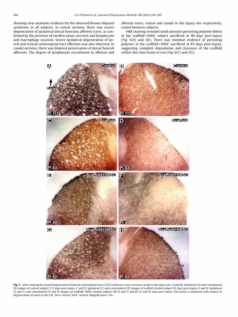
howing clear anatomic evidence for the observed Brown-Séquardyndrome in all subjects. In rostral sections, there was severeegeneration of ipsilateral dorsal funiculus afferent tracts, as con-
rmed by the presence of swollen axons, necrosis and lymphocytend macrophage invasion. Severe ipsilateral degeneration of lat-ral and ventral corticospinal tract efferents was also observed. Inaudal sections, there was bilateral preservation of dorsal funiculifferents. The degree of lymphocyte recruitment to afferent andig. 7. Silver staining for axonal degeneration of lateral corticospinal tracts (CST) in thoracB) images of control subject 111 days post-injury. C and D: ipsilateral (C) and contralateE and G) and contralateral (F and H) images of scaffold + hNSC treated subjects 40 (E aegeneration of axons in the CST. Dors = dorsal. Vent = ventral. Magnification = 10×.
nce Methods 188 (2010) 258–269
efferent tracts, rostral and caudal to the injury site respectively,varied between subjects.
H&E staining revealed small amounts persisting polymer debris
in the scaffold + hNSC subject sacrificed at 40 days post-injury(Fig. 6(A) and (B)). There was minimal evidence of persistingpolymer in the scaffold + hNSC sacrificed at 82 days post-injury,suggesting complete degradation and clearance of the scaffoldwithin this time frame in vivo (Fig. 6(C) and (D)).ic cross-sections caudal to the injury site. (A and B): Ipsilateral (A) and contralateralral (D) images of scaffold treated subject 83 days post-injury. E and H: Ipsilateralnd F) and 82 (G and H) days post-injury. The lesion is unilateral with respect to
roscie
lajbo
Fci(wM
C.D. Pritchard et al. / Journal of Neu
H&E staining revealed evidence of fibrous matter around the
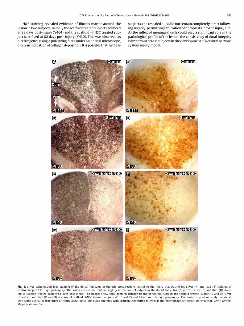
esion in two subjects, namely the scaffold treated subject sacrificedt 83 days post-injury (Y464) and the scaffold + hNSC treated sub-ect sacrificed at 82 days post-injury (Y430). This was observed asirefringence using a polarizing filter under an optical microscope,ften an indication of collagen deposition. It is possible that, in theseig. 8. Silver staining and Iba1 staining of the dorsal funiculus in thoracic cross-secontrol subject 111 days post-injury. The lesion crosses the midline slightly in the cng of scaffold treated subject 83 days post-injury. The images show total bilateralE and G) and Iba1 (F and H) staining of scaffold + hNSC treated subjects 40 (E andith some axonal degeneration of contralateral dorsal funiculus afferents with spatialagnification = 10×.
nce Methods 188 (2010) 258–269 265
subjects, the resealed dura did not remain completely intact follow-
ing surgery, permitting infiltration of fibroblasts into the injury site.As the influx of meningeal cells could play a significant role in thepathological profile of the lesion, the consistency of dural integrityis important across subjects in the development of a central nervoussystem injury model.tions rostral to the injury site. (A and B): Silver (A) and Iba1 (B) staining ofontrol subject in the dorsal funiculus. (C and D): silver (C) and Iba1 (D) stain-damage to the dorsal funiculus in the scaffold treated subject. E and H: silverF) and 82 (G and H) days post-injury. The lesion is predominately unilateral,
ly correlating microglial and macrophage activation. Dors = dorsal. Vent = ventral.

266 C.D. Pritchard et al. / Journal of Neuroscie
Fig. 9. Silver staining for axonal degeneration in thoracic sagittal sections throughthe hemisection lesions. Sections shown are closest to the middle of the cord inthe rostral–caudal direction. (A): Control subject 111 days post-injury. B: scaffoldtreated subject 83 days post-injury. (C and D): Scaffold + hNSC treated subjects 40(C) and 82 (D) days post-injury. The lesion crosses the midline, in reference to thecentral canal where present, into the contralateral gray matter. In these sections thecontralateral lateral funiculi appear to be preserved from the surgical lesion in allsIo
dflavbaaph
ddw(j(llo
human neural stem cells in the promotion of SCI recovery and
ubjects. However, some degenerative staining is visible in contralateral efferents.psi = ipsilateral lesioned side. Contra = contralateral unlesioned side. The cords areriented rostral to caudal from left to right. Scale bar = 1 mm.
H&E staining also revealed lines of hemocynanin deposition,emarking severed blood vessels within the spinal cord. Bloodowing under pressure from severed blood vessels, in particularrterial vessels, is a possible source of mechanical insult to ner-ous system tissue surrounding the lesion. In addition, damagedlood vessels result in local ischemia. The presence of multinucle-ted giant cells around the lesion site was observed in all subjects,nd it is unclear to what extent, if at all, these were specificallyresent in response to foreign polymer materials and allogeneicNSC.
Silver degenerative staining of cross-sections rostral and cau-al to the lesion provided an indication of the extent of axonalisruption. The lesions remained unilateral in the lateral regions,ith severe degeneration of ipsilateral lateral corticospinal tract
CST) efferents (Fig. 7). Some degree of variability between sub-ects exists at the midline in dorsal funiculus afferents (Fig. 8 (A),
C), (E) and (G)). In all animals, longitudinal sections through theesion site showed clear evidence that, at the time of analysis, theesion had crossed the cord midline, resulting in loss of grey mattern the contralateral side (Fig. 9), further studied by quantificationnce Methods 188 (2010) 258–269
of lesion dimensions (Table 3). Axonal damage was observed bothipsilaterally and contralaterally in ventral–medial tracts (Fig. 10).The small sample size, however, was not intended to distinguishthe degree to which these differences were due to variations in thesurgical lesion and individual responses to trauma compared tosecondary injury and wound healing processes that may hypothet-ically be influenced by implantation of scaffolds and hNSC (Teng etal., 2002).
Iba1 immunohistochemistry provided an indication of reactivemicroglial and macrophage abundance and distribution, providinga further measure of the degree to which the impact of the hemisec-tion remained unilateral weeks post-injury. The lesions remainedunilateral in the lateral regions, with substantial Iba1 immunore-activity around ipsilateral lateral CST efferents (Fig. 11). A similardegree of variability as observed by silver staining was observedusing Iba1 immunohistochemistry in the dorsal funiculus rostralto the injury site (Fig. 8(B), (D), (F) and (H)).
4. Discussion
Creation of a hemisection in the thoracic spine at the T9–T10level in the African green monkey resulted in paralysis of the ipsi-lateral hindlimb, with concomitant sparing of bladder and rectalfunction and retention of sufficient overall motor function andmobility and allowed the continuation of feeding, grooming andother healthy activities without supplementary veterinary care.Behavioral evaluations confirmed improvement in post-operativeparalysis over time, but improvement was gradual with sufficientremaining deficit to allow definition of recovery rate and distinctionwithin subjects between phases of recovery and between subjectsexhibiting different degrees of impairment. As such, this Africangreen model of SCI may represent a means by which the safetyand efficacy of therapeutic SCI interventions in a clinically relevantspecies could be assessed humanely.
This study was primarily designed to evaluate the feasibility ofimplementing a lateral hemisection model of SCI in a non-humanprimate, and to identify the specific clinically relevant behavioraland pathological endpoints that could be reproducibly obtained.Coupled kinematic and electromyographic measures in previousprimate studies have demonstrated the feasibility of obtainingthese critical endpoints (Courtine et al., 2005). The associatedtelemetry and surgical interventions, however, are, in themselves,a significant research investment and variable impacting animalwellbeing, with the established need for prolonged postoperativecare. The more critical question addressed in this study was theconsequence and tolerance of a T9–T10 hemisection lesion, and acharacterization of the overall neuromotor phenotype and its evo-lution over time such that more definitive efficacy and mechanismstudies could be rationally and humanely designed. Therefore, thesizes of the treatment groups in the present study were limited andnot designed to provide statistical evaluation of the therapeutic effi-cacy of the implanted PLGA scaffold or PLGA scaffold seeded withhNSC at the lesion site. All subjects exhibited a return toward base-line neuromotor scores in the ipsilateral hindlimb, which exhibitedcomplete post-operative paralysis following the T9–T10 lesion.This indicates a degree of recovery and neural plasticity consis-tent with reported findings in other primate SCI models (Edgertonet al., 2004). This observation encourages additional application ofthe model for more definitive preclinical studies in a larger studycohort to evaluate the therapeutic efficacy of PLGA scaffolds and
repair.Video capture of neuromotor behavior in the ambulation cham-
ber provided a means of documenting and scoring neuromotordeficits and improvement over time. An observation based scoring

C.D. Pritchard et al. / Journal of Neuroscience Methods 188 (2010) 258–269 267
Fig. 10. Silver staining for axonal degeneration of ventral funiculus tracts in thoracic cross-sections caudal to the injury site. (A): Control subject 111 days post-injury. B:scaffold treated subject 83 days post-injury. C and D: scaffold + hNSC treated subjects 40 (C) and 82 (D) days post-injury. The lesion is bilateral with respect to degenerationo 0×.
snstpiii2
ailaSittwpatst
sacssrvi
f axons in the ventral–medial tracts. Dors = dorsal. Vent = ventral. Magnification = 1
ystem, however, may be inadequate to give precise information ofeuromotor performance, due to the potential for the contralateralide to compensate for any loss of weight bearing ability and pos-ure resulting from a unilateral interruption of motor and sensoryathways to the injured hindlimb. The application of kinemat-
cs, electromyography and evoked potentials would contribute tomproved measures of neurologic function and quantification ofpsilateral and contralateral deficit and recovery (Courtine et al.,009).
Histological analysis confirmed creation of a thoracic lesion inll study monkeys. Hematoxylin and eosin staining provided a def-nition of the extent of the lesion and degree of injury associatedymphocyte invasion, axonal swelling and macrophage infiltration,s well as a circumscribed degree of hemorrhage and gross necrosis.ilver staining revealed clear degenerative changes consistent withnterruption of ipsilateral efferent lateral corticospinal tracts at thehoracic level caudal to the lesion and sensory afferents rostral tohe lesion. It was noted that the ventral–medial region of the cord,hich contains both important sensory and motor tracts, may bearticularly sensitive to different degrees of bilateral surgical injurymong subjects. Iba1 immunohistochemistry exhibited a distribu-ion that paralleled the degenerative findings highlighted by theilver staining. As such, Iba1 constituted an additional measure ofhe degree of injury and repair.
Histological evaluations confirmed the creation of a lesion withevere degeneration of ipsilateral motor and sensory axons. In nonimals were the degenerative changes entirely unilateral. Whileontralateral corticospinal tracts were extensively preserved in all
ubjects, in addition to the majority of the dorsal horn in mostubjects, contralateral damage was observed in the ventral–medialegion in all subjects. Contralateral damage is most due to surgicalariability, which will be improved in future, more sophisticated,nvestigations.The persistence of the PLGA scaffold for at least 40 days, anddegradation and clearance from the spinal cord within 82 days post-implantation was observed. Two important changes were made tothe neural stem cells seeded PLGA scaffold used in this study, incomparison with a similar rodent study (Teng et al., 2002). Firstly,human neural stem cells (hNSC) were used instead of murine neuralstem cells. Secondly, the PLGA scaffold was redesigned in responseto observations made in the rodent study (Teng et al., 2002). The rel-ative volume of the inner scaffold was increased to accommodatemore hNSC. It was postulated that a major mechanism of actionof the implanted cells may be due to trophic support rather thanneuronal replacement (Yu et al., 2009). Therefore, it was hypothe-sized that a larger number of hNSC could increase any therapeuticeffect. It was also proposed that the ‘outer scaffold’ in the rodentstudy, consisting of longitudinally oriented channels intended topermit axon in-growth, may have also served to prevent meningealinflammatory cell infiltration. Unpublished in vitro data showedthat the channels were impermeable to fibroblasts. To maximizethe size of the inner scaffold, while providing a physical barrierto meningeal cell infiltration, the outer scaffold was constructedas a 200 �m thin, flexible sheet of PLGA. The observed toler-ance of the current implant will guide implant design for furtherstudies.
The ability to model spinal cord injury by lateral, segmen-tal hemisection in African green monkeys with limited impacton overall wellbeing presents a useful model for the assess-ment of spinal cord injury repair and response to intervention.Greater sensitivity and specificity to treatment effects could
be achieved through expanded samples sizes to address lesionvariability and application of coupled kinematic and electromyo-graphic analyses, as well as further histological markers to identifyspecific mechanisms involved in secondary injury, repair andregeneration.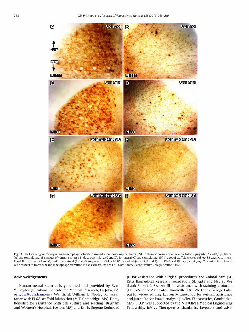
268 C.D. Pritchard et al. / Journal of Neuroscience Methods 188 (2010) 258–269
Fig. 11. Iba1 staining for microglial and macrophage activation around lateral corticospinal tracts (CST) in thoracic cross-sections caudal to the injury site. (A and B): Ipsilateral( psilateE treatew ors = d
A
YetBa
A) and contralateral (B) images of control subject 111 days post-injury. (C and D): Iand H: ipsilateral (E and G) and contralateral (F and H) images of scaffold + hNSCith respect to microglial and macrophage activation in the cord around the CST. D
cknowledgements
Human neural stem cells generated and provided by Evan
. Snyder (Burnham Institute for Medical Research, La Jolla, CA,[email protected]). We thank William L. Neeley for assis-ance with PLGA scaffold fabrication (MIT, Cambridge, MA), Darcyenedict for assistance with cell culture and seeding (Brighamnd Women’s Hospital, Boston, MA) and Dr. D. Eugene Redmondral (C) and contralateral (D) images of scaffold treated subject 83 days post-injury.d subjects 40 (E and F) and 82 (G and H) days post-injury. The lesion is unilateralorsal. Vent = ventral. Magnification = 10×.
Jr. for assistance with surgical procedures and animal care (St.Kitts Biomedical Research Foundation, St. Kitts and Nevis). Wethank Robert C. Switzer III for assistance with staining protocols
(NeuroScience Associates, Knoxville, TN). We thank George Cala-pai for video editing, Lauren Mitarotondo for writing assistanceand Janice Ye for image analysis (InVivo Therapeutics, Cambridge,MA). C.D.P. was supported by the MIT/CIMIT Medical EngineeringFellowship. InVivo Therapeutics thanks its investors and advi-
roscie
sC
A
t
R
B
B
B
B
C
C
C
C
D
C.D. Pritchard et al. / Journal of Neu
ors for support. This study was funded by InVivo Therapeuticsorporation.
ppendix A. Supplementary data
Supplementary data associated with this article can be found, inhe online version, at doi:10.1016/j.jneumeth.2010.02.019.
eferences
abu RS, Muthusamy R, Namasivayam A. Behavioural assessment of functionalrecovery after spinal cord hemisection in the bonnet monkey. Journal of Neuro-logical Sciences 2000;178:136–52.
aptiste DC, Fehlings MG. Update on the treatment of spinal cord injury. Progress inBrain Research 2007;161:217–33.
asso DM, Beattie MS, Bresnahan JC. Graded histological and locomotor outcomesafter spinal cord contusion using the NYU weight-drop device versus transac-tion. Experimental Neurology 1996;139:244–56.
rown-Séquard CÉ. De la transmission croisée des impressions sensitives par lamoelle épinière. Comptes rendus de la Société de biologie 1851;2:33–44.
ourtine G, Roy RR, Hodgson J, McKay H, Raven J, Zhong H, Yang H, Tuszynski MH,Edgerton VR. Kinematic and EMG determinants in quadrupedal locomotion ofnon-human primate (rhesus). Journal of Neurophysiology 2005;93:3127–45.
ourtine G, Bartlett Bunge M, Fawcett JW, et al. Can experiments in nonhuman pri-mates expedite the translation of treatments for spinal cord injury in humans?Nature Medicine 2007;13(5):561–6.
ourtine G, Gerasimenko Y, van den Brand R, Yew A, Musienko P, Zhong H, Song B, AoY, Ichiyama RM, Lavrov I, Roy RR, Sofroniew MV, Edgerton VR. Transformation of
nonfunctional spinal circuits into functional states after the loss of brain input.Nature Neuroscience 2009;12:1333–42.rowe MJ, Bresnahan JC, Shuman SL, Masters JN, Beattie MS. Apoptosis and delayeddegeneration after spinal cord injury in rats and monkeys. Nature Medicine1997;3(1):73–6.
e Olmos JS et al. Neurotoxicology and Teratology 1994;16:545–61.
nce Methods 188 (2010) 258–269 269
Edgerton VR, Tillakaratne NJK, Bigbee AJ, de Leon RD, Roy RR. Plasticity of the spinalneural circuitry after injury. Annual Review of Neuroscience 2004;27:145–67.
Feringa ER, Johnson RD, Wendt JS. Spinal cord regeneration in rats after immuno-suppressive treatment. Theoretic considerations and histologic results. Archivesof Neurology 1975;32(10):676–83.
Flax JD, Aurora S, Yang C, et al. Engraftable human neural stem cells respondto developmental cues, replace neurons, and express foreign genes. NatureBiotechnology 1998;16:1033–9.
Hall ED, Springer JE. Neuroprotection and acute spinal cord injury: a reappraisal.NeuroRx 2004;1:80–100.
Jones TB, McDaniel EE, Popovich PG. Inflammatory-mediated injury and repairin the traumatically injured spinal cord. Current Pharmaceutical Design2005;11:1223–36.
Lavik E, Teng YD, Snyder E, Langer R. Seeding neural stem cells on scaffolds of PGA,PLA, and their copolymers. Methods in Molecular Biology 2002;198:89–97.
Liverman CT, Altevogt BM, et al. Spinal cord injury: progress, promises and priorities.Washington, DC: National Academies Press; 2005.
Mikos AG, Sarakinos G, Leite SM, Vacanti JP, Langer R. Laminated three-dimensional biodegradable foams for use in tissue engineering. Biomaterials1992;14(5):323–30.
Park KI, Teng YD, Snyder EY. The injured brain interacts reciprocally with neural stemcells supported by scaffolds to reconstitute lost tissue. Nature Biotechnology2002;20:1111–7.
Rossignol S, Schwab M, Schwartz M, Fehlings MG. Spinal cord injury: time to move?The Journal of Neuroscience 2007;27(44):11782–92.
Sung HJ, Meredith C, Johnson C, Galis ZS. The effect of scaffold degrada-tion rate on three-dimensional cell growth and angiogenesis. Biomaterials2004;25:5735–42.
Teng YD, Lavik EB, Qu X, Park KI, Ourednik J, Zurakowski D, Langer R, Snyder EY.Functional recovery following traumatic spinal cord injury mediated by a uniquepolymer scaffold seeded with neural stem cells. Proceedings of the National
Academy of Sciences of the United States of America 2002;99(5):3024–9.Thuret S, Moon LDF, Gage FH. Therapeutic interventions after spinal cord injury.Nature Neuroscience 2006;7:628–43.
Yu D, Neeley WL, Prichard CD, Slotkin JR, Woodard EJ, Langer R, Teng YT. Blockade ofperoxynitrite-induced neural stem cell death in the acutely injured spinal cordby drug-releasing polymer. Stem Cells 2009;27:1212–22.