Embed Size (px)
Citation preview
www.manuscriptscientific.com
Journal of Oral Health and Dentistry
Research
Case Report
Manuscript Scientific Services
Journal of Oral Health and Dentistry Research (JOHDR) 1
Return of Sensibility After 7 Years in a Traumatized Tooth Treated by The
Regenerative Process: A Case Report
Jason Kwok and Mike Sabeti*
*Advanced Specialty Program in Endodontics UCSF School of Dentistry, Parnassus Ave San Francisco, USA
Corresponding author: Mike Sabeti, Advanced Specialty Program in Endodontics UCSF School of Dentistry, Parnassus Ave San Francisco, USA, Tel:
2812355967; E-mail: [email protected]
Abstract Background: In this case report we describe a case of successful regenerative treatment in which sensibility was recovered seven years after the
regenerative process.
Case description: An immature maxillary right central incisor (tooth #11) with a necrotic pulp and a periodical radiolucency in a seven-year-old patient was
treated using a regenerative protocol. Bleeding was inducted into the canal system and mineral trioxide aggregate was place over the blood clot followed by
a permanent composite restoration. Radiographic examination showed resolution of the periapical pathosis, continued development of the most apical
portion of the root and finally closure of the root apex.
Practical Implication: This case exemplified that re-establishment of a sensory component of the tissue formed in the root canal after a regenerative
procedure is possible years after the treatment is completed. In addition, the regenerative procedure does produce continued development of root dentin and
prevention or healing of endodontically related apical periodontitis.
Keywords: Regeneration, Revascularization, Trauma
INTRODUCTION
Treatment of the immature permanent dentition after
traumatic incidents represents a unique challenge in
endodontics. Open apices and the potential for fracture of
thin dentinal root walls are just a few of the challenges faced
by clinicians managing immature teeth with necrotic pulps.
Traditionally, calcium hydroxide (Ca(OH)2) was used to
induce hard tissue formation and an apical barrier.
Apexification using Ca(OH)2 does result in a barrier across
the open apex but not with the continued development of
root dentin. In addition, a sequela of apexification is the
higher likelihood of fracture of the remaining thin root
dentin walls [1].
After the development of mineral trioxide aggregate (MTA)
in the early 1990s, the use of this bio ceramic material
supplanted the use of Ca(OH)2 as a material of choice in
open apex teeth. It reduced appointment times and produced
true closure of open apices [2]. The barrier technique,
however, did not allow continued development of an
immature tooth. Hence, the tooth was subject to a higher
likelihood of fracture [3].
The continued development of an immature tooth has long
been thought to be beneficial for the longevity and prognosis
of a tooth [4]. The concept of pulpal regenerative was first
introduced in the 1960s by Nygaard-Ostby [5,6]. Through
histological evaluation, he discovered hard tissue deposition
on the thin root walls of the canal. The hard tissue deposition
occurred by induction and migration of stem cells into the
canal space. With proper stimulation, modifying growth
factors and appropriate scaffolding, the stem cells
differentiated and formed a pulp-dentin-like complex and
revascularized the tooth [7-10].
Successful regenerative results in prevention or healing of
apical periodontitis and the continued development of strong
root dentin [11-13]. There are numerous case reports on
regeneration, but reports with a return of sensibility are
limited [11,14-19]. Moreover, there are very limited reports
to date [20] that have demonstrated sensory function
returning to the tooth many years after the regenerative
process has been completed.
The purpose of this case report is to highlight a highly
unusual case in which sensory function was recovered years
after the regenerative process was completed in an immature
tooth which had a necrotic pulp and long-term development
of the most apical portion of the root occurred.
Received: March 31, 2021; Revised: April 11, 2021; Accepted: April 14,
2021
Citation: Kwok J & Sabeti M. (2021) Return of Sensibility After 7 Years in
a Traumatized Tooth Treated by The Regenerative Process: A Case Report.
J Oral Health Dent Res, 1(1): 1-6.
Copyright: ©2021 Kwok J & Sabeti M. This is an open-access article
distributed under the terms of the Creative Commons Attribution License,
which permits unrestricted use, distribution, and reproduction in any
medium, provided the original author and source are credited.
Manuscript Scientific Services
Journal of Oral Health and Dentistry Research (JOHDR) 2
J Oral Health Dent Res, 1(1): 2021 Kwok J & Sabeti M
CASE REPORT
A seven-year-old Filipino female presented with her
grandmother to the Dental Clinic at the University of
California San Francisco School of Dentistry in April 2010
with intermittent pain associated with the right maxillary
central incisor (tooth #11). The tooth had a history of a
complicated crown fracture. The gingival around the tooth
was inflamed. Sensibility testing at the initial visit resulted
in a no response to cold and positive responses to both
percussion and palpation. Electronic pulp testing resulted in
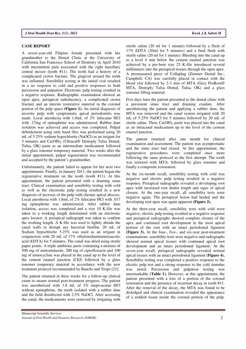
a negative response. Radiographic examination showed an
open apex, periapical radiolucency, a complicated crown
fracture and an interim restorative material in the coronal
portion of the pulp space (Figure 1). An initial diagnosis of
necrotic pulp with symptomatic apical periodontitis was
made. Local anesthesia with 1.8mL of 2% lidocaine HCl
with .17mg of epinephrine was administered. Rubber dam
isolation was achieved and access was completed. Pulpal
debridement using only hand files was performed using 20
mL of 5.25% sodium hypochlorite (NaOCl) as an irrigant for
5 minutes and Ca(OH)2 (Ultracal® Dentsply Tulsa Dental,
Tulsa, OK) paste as an intermediate medicament followed
by a glass ionomer temporary material. Two weeks after the
initial appointment, pulpal regeneration was recommended
and accepted by the patient’s grandmother.
Unfortunately, the patient failed to appear for her next two
appointments. Finally, in January 2011, the patient began the
regenerative treatment on the tooth (tooth #11). At this
appointment, the patient presented with a draining sinus
tract. Clinical examination and sensibility testing with cold
as well as the electronic pulp testing resulted in a new
diagnosis of necrosis of the pulp with chronic apical abscess.
Local anesthesia with 1.8mL of 2% lidocaine HCl with .017
mg epinephrine was administered. After rubber dam
isolation, access was completed and a size 10 K-file was
taken to a working length determined with an electronic
apex locator. A periapical radiograph was taken to confirm
the working length. A K-file was used to lightly abrade the
canal walls to disrupt any bacterial biofilm. 20 mL of
Sodium hypochlorite 5.25% was used as an irrigant in
conjunction with 20 mL of 17% ethylenediaminetetraacetic
acid (EDTA) for 5 minutes. The canal was dried using sterile
paper points. A triple antibiotic paste containing a mixture of
500 mg of metronidazole, 200 mg of ciprofloxacin and 100
mg of minocycline was placed in the canal up to the level of
the cement enamel junction (CEJ) followed by a glass
ionomer temporary material in accordance with the new
treatment protocol recommended by Banchs and Trope [21].
The patient returned in three weeks for a follow-up clinical
exam to ensure normal post-treatment progress. The patient
was anesthetized with 1.8 mL of 3% mepivacaine HCl
without epinephrine, the tooth isolated with a rubber dam
and the field disinfected with 2.5% NaOCl. After accessing
the canal, the medicaments were removed by irrigating with
sterile saline (20 ml for 1 minute) followed by a flush of
17% EDTA (20ml for 5 minutes) and a final flush with
sterile saline (20 ml for 1 minute). Bleeding into the canal up
to a level 4 mm below the cement enamel junction was
achieved by a pre-bent size 25 K-file introduced several
millimeters into the periapical tissues through the open apex.
A premeasured piece of Collaplug (Zimmer Dental Inc.,
Campbell, CA) was carefully placed in contact with the
blood clot followed by 2-3 mm of MTA (Grey ProRoot®
MTA, Dentsply Tulsa Dental, Tulsa, OK) and a glass
ionomer filling material.
Five days later the patient presented to the dental clinic with
a persistent sinus tract and draining exudate. After
anesthetizing the patient and applying a rubber dam, the
MTA was removed and the canal system irrigated with 20
mL of 5.25% NaOCl for 5 minutes followed by 20 mL of
sterile saline. Then, Ca(OH)2 paste was placed into the canal
as an intracanal medicament up to the level of the cement
enamel junction.
The patient returned after one month for clinical
examination and assessment. The patient was asymptomatic
and the sinus tract had closed. At this appointment, the
regenerative procedures were completed once more
following the same protocol as the first attempt. The tooth
was restored with MTA, followed by glass ionomer and
finally a composite restoration.
At the six-month recall, sensibility testing with cold was
negative and electric pulp testing resulted in a negative
response. Periapical radiographs revealed a developing root
apex with increased root dentin length and signs of apical
closure. At the one-year recall, all sensibility tests were
negative again. The periapical lesion had healed and the
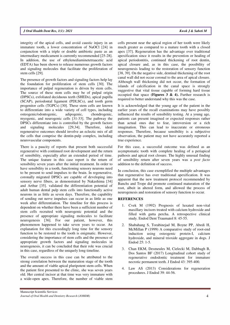
developing root apex was again apparent (Figure 2).
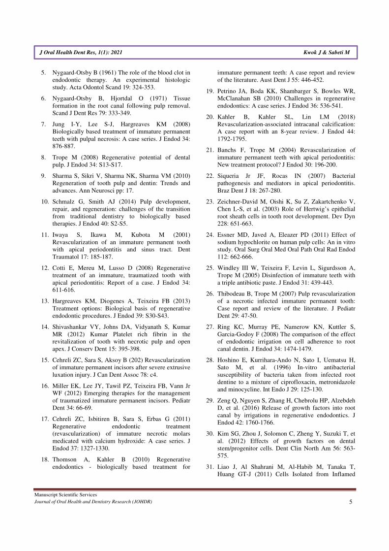
At the three-year recall, sensibility tests with cold were
negative, electric pulp testing resulted in a negative response
and periapical radiographs showed complete closure of the
apex and continued root development in the most apical
portion of the root with an intact periodontal ligament
(Figure 3). At the four-, five-, and six-year post-treatment
examinations, sensibility tests were negative and radiographs
showed normal apical tissues with continued apical root
development and an intact periodontal ligament. At the
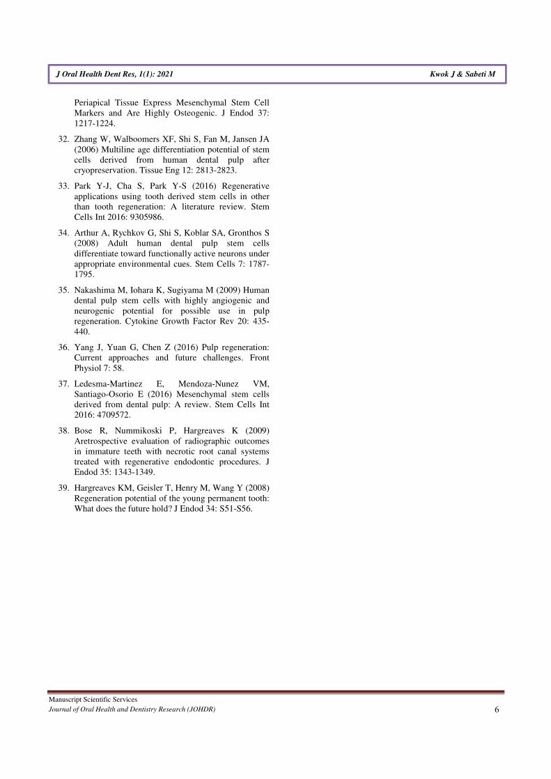
seven-year recall, periapical radiographs revealed normal
apical tissues with an intact periodontal ligament (Figure 4).
Sensibility testing was completed a positive response to the
electric pulp test and a strong response to the cold stimulus
was noted. Percussion and palpation testing was
unremarkable (Table 1). However, at this appointment, the
patient presented with a loss of a portion of the coronal
restoration and the presence of recurrent decay in tooth #11.
After the removal of the decay, the MTA was found to be
dislodged and clinical examination revealed the appearance
of a reddish tissue inside the coronal portion of the pulp.
Manuscript Scientific Services
Journal of Oral Health and Dentistry Research (JOHDR) 3
J Oral Health Dent Res, 1(1): 2021 Kwok J & Sabeti M
Subsequently, MTA was then placed followed by glass
ionomer and composite restoration material.
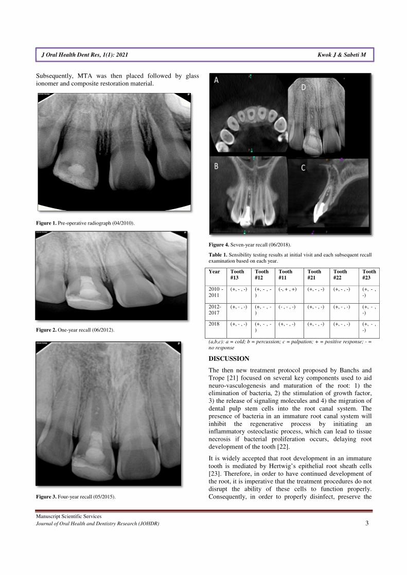
Figure 1. Pre-operative radiograph (04/2010).
Figure 2. One-year recall (06/2012).
Figure 3. Four-year recall (05/2015).
Figure 4. Seven-year recall (06/2018).
Table 1. Sensibility testing results at initial visit and each subsequent recall
examination based on each year.
Year Tooth
#13
Tooth
#12
Tooth
#11
Tooth
#21
Tooth
#22
Tooth
#23
2010 -
2011
(+, - , -) (+, - , -
)
(-, + , +) (+, - , -) (+, - , -) (+, - ,
-)
2012-
2017
(+, - , -) (+, - , -
)
(- , - , -) (+, - , -) (+, - , -) (+, - ,
-)
2018 (+, - , -) (+, - , -
)
(+, - , -) (+, - , -) (+, - , -) (+, - ,
-)
(a,b,c): a = cold; b = percussion; c = palpation; + = positive response; - =
no response
DISCUSSION
The then new treatment protocol proposed by Banchs and
Trope [21] focused on several key components used to aid
neuro-vasculogenesis and maturation of the root: 1) the
elimination of bacteria, 2) the stimulation of growth factor,
3) the release of signaling molecules and 4) the migration of
dental pulp stem cells into the root canal system. The
presence of bacteria in an immature root canal system will
inhibit the regenerative process by initiating an
inflammatory osteoclastic process, which can lead to tissue
necrosis if bacterial proliferation occurs, delaying root
development of the tooth [22].
It is widely accepted that root development in an immature
tooth is mediated by Hertwig’s epithelial root sheath cells
[23]. Therefore, in order to have continued development of
the root, it is imperative that the treatment procedures do not
disrupt the ability of these cells to function properly.
Consequently, in order to properly disinfect, preserve the
Manuscript Scientific Services
Journal of Oral Health and Dentistry Research (JOHDR) 4
J Oral Health Dent Res, 1(1): 2021 Kwok J & Sabeti M
integrity of the apical cells, and avoid caustic injury in an
immature tooth, a lower concentration of NaOCl [24] in
conjunction with a triple or double antibiotic paste as an
intermediary medicament is currently recommended [25-28].
In addition, the use of ethylenediaminetetraacetic acid
(EDTA) has been shown to release numerous growth factors
and signaling molecules that facilitate the proliferation of
stem cells [29].
The presence of growth factors and signaling factors help lay
the foundation for proliferation of stem cells [30]. The
importance of pulpal regeneration is driven by stem cells.
The source of these stem cells may be of pulpal origin
(DPSCs), exfoliated deciduous teeth (SHEDs), apical papilla
(SCAP), periodontal ligament (PDLSCs), and tooth germ
progenitor cells (TGPCs) [30]. These stem cells are known
to differentiate into a wide variety of cell types, including
osteogenic/odontogenic, adipogenic, chondrogenic,
myogenic, and neurogenic cells [31-33]. The pathway the
DPSCs differentiate into is controlled by the growth factors
and signaling molecules [29,34]. Therefore, ideal
regenerative outcomes should involve an eclectic mix of all
the cells that comprise the dentin-pulp complex, including
neurovascular components.
There is a paucity of reports that present both successful
regenerative with continued root development and the return
of sensibility, especially after an extended period of time.
The unique feature in this case report is the return of
sensibility seven years after the initial treatment. In order to
have sensibility in a tooth, functioning sensory neurons need
to be present to send impulses to the brain. In regenerative,
coronally migrated DPSCs are capable of developing into
sensory nerve fibers, as demonstrated by Nakashima [34]
and Arthur [35]. validated the differentiation potential of
adult human dental pulp stem cells into functionally active
neurons in as little as seven days. Therefore, the capability
of sending out nerve impulses can occur in as little as one
week after differentiation. The timeline for this process is
dependent on whether there have been a sufficient number of
stem cells recruited with neurogenic potential and the
presence of appropriate signaling molecules to facilitate
neurogenesis [36]. For our patient, however, this
phenomenon happened to take seven years to occur. An
explanation for this exceedingly long time for the sensory
function to be restored to the tooth is enigmatic. However,
considering the importance of stem cells and the presence of
appropriate growth factors and signaling molecules in
neurogenesis, it can be concluded that their role was crucial
in this case, regardless of the uniquely long timeline.
The overall success in this case can be attributed to the
strong correlation between the maturation stage of the tooth
and the amount of viable apical pluripotent stem cells. When
the patient first presented to the clinic, she was seven years
old. Her central incisor at that time was very immature with
a wide-open apex. Therefore, the number of viable stem
cells present near the apical region of her tooth were likely
much greater as compared to a mature tooth with a closed
apex [37]. Regeneration has the advantage over traditional
apexification since it results in the prevention or healing of
apical periodontitis, continued thickening of root dentin,
apical closure and, as in this case, the possibility of
neurogenesis leading to the restoration of sensory function
[38, 39]. On the negative side, dentinal thickening of the root
canal wall did not occur coronal to the area of apical closure.
Although wall thickening did not occur, the formation of
islands of calcification in the canal space is strongly
suggestive that vital tissue capable of forming hard tissue
occupied that space (Figures 3 & 4). Further research is
required to better understand why this was the case.
It is acknowledged that the young age of the patient in the
earlier years of the recall examinations may have possibly
influenced the results of sensibility testing. At a young age,
patients can present imagined or expected responses rather
than actual ones due to fear, introversion or a rich
imagination. This can lead to inaccurate or erroneous
responses. Therefore, because sensibility is a subjective
observation, the patient may not have accurately reported a
true experience.
For this case, a successful outcome was defined as an
asymptomatic tooth with complete healing of a periapical
pathosis and apical root closure. The highly unusual finding
of sensibility return after seven years was a post facto
addition to the definition of success.
In conclusion, this case exemplified the multiple advantages
that regenerative has over traditional apexification. It was
apparent that the new treatment protocol recommended by
Banchs and Trope did promote continued maturation of the
root, albeit in altered form, and allowed the process of
neurogenesis and restoration of sensory function to occur.
REFERENCES
1. Cvek M (1992) Prognosis of luxated non-vital
maxillary incisors treated with calcium hydroxide and
filled with gutta percha. A retrospective clinical
study. Endod Dent Traumatol 8: 45-55.
2. Shabahang S, Torabinejad M, Boyne PP, Abedi H,
McMillan P (1999) A comparative study of root-end
induction using osteogenic protein-I, calcium
hydroxide, and mineral trioxide aggregate in dogs. J
Endod 25: 1-5.
3. Chan EKM, Desmeules M, Cielecki M, Dabbagh B,
Dos Santos BF (2017) Longitudinal cohort study of
regenerative endodontic treatment for immature
necrotic permanent teeth. J Endod 43: 395-400.
4. Law AS (2013) Considerations for regeneration
procedures. J Endod 39: 44-56.
Manuscript Scientific Services
Journal of Oral Health and Dentistry Research (JOHDR) 5
J Oral Health Dent Res, 1(1): 2021 Kwok J & Sabeti M
5. Nygaard-Otsby B (1961) The role of the blood clot in
endodontic therapy. An experimental histologic
study. Acta Odontol Scand 19: 324-353.
6. Nygaard-Otsby B, Hjortdal O (1971) Tissue
formation in the root canal following pulp removal.
Scand J Dent Res 79: 333-349.
7. Jung I-Y, Lee S-J, Hargreaves KM (2008)
Biologically based treatment of immature permanent
teeth with pulpal necrosis: A case series. J Endod 34:
876-887.
8. Trope M (2008) Regenerative potential of dental
pulp. J Endod 34: S13-S17.
9. Sharma S, Sikri V, Sharma NK, Sharma VM (2010)
Regeneration of tooth pulp and dentin: Trends and
advances. Ann Neurosci pp: 17.
10. Schmalz G, Smith AJ (2014) Pulp development,
repair, and regeneration: challenges of the transition
from traditional dentistry to biologically based
therapies. J Endod 40: S2-S5.
11. Iwaya S, Ikawa M, Kubota M (2001)
Revascularization of an immature permanent tooth
with apical periodontitis and sinus tract. Dent
Traumatol 17: 185-187.
12. Cotti E, Mereu M, Lusso D (2008) Regenerative
treatment of an immature, traumatized tooth with
apical periodontitis: Report of a case. J Endod 34:
611-616.
13. Hargreaves KM, Diogenes A, Teixeira FB (2013)
Treatment options: Biological basis of regenerative
endodontic procedures. J Endod 39: S30-S43.
14. Shivashankar VY, Johns DA, Vidyanath S, Kumar
MR (2012) Kumar Platelet rich fibrin in the
revitalization of tooth with necrotic pulp and open
apex. J Conserv Dent 15: 395-398.
15. Cehreli ZC, Sara S, Aksoy B (202) Revascularization
of immature permanent incisors after severe extrusive
luxation injury. J Can Dent Assoc 78: c4.
16. Miller EK, Lee JY, Tawil PZ, Teixeira FB, Vann Jr
WF (2012) Emerging therapies for the management
of traumatized immature permanent incisors. Pediatr
Dent 34: 66-69.
17. Cehreli ZC, Isbitiren B, Sara S, Erbas G (2011)
Regenerative endodontic treatment
(revascularization) of immature necrotic molars
medicated with calcium hydroxide: A case series. J
Endod 37: 1327-1330.
18. Thomson A, Kahler B (2010) Regenerative
endodontics - biologically�based treatment for
immature permanent teeth: A case report and review
of the literature. Aust Dent J 55: 446-452.
19. Petrino JA, Boda KK, Shambarger S, Bowles WR,
McClanahan SB (2010) Challenges in regenerative
endodontics: A case series. J Endod 36: 536-541.
20. Kahler B, Kahler SL, Lin LM (2018)
Revascularization-associated intracanal calcification:
A case report with an 8-year review. J Endod 44:
1792-1795.
21. Banchs F, Trope M (2004) Revascularization of
immature permanent teeth with apical periodontitis:
New treatment protocol? J Endod 30: 196-200.
22. Siqueria Jr JF, Rocas IN (2007) Bacterial
pathogenesis and mediators in apical periodontitis.
Braz Dent J 18: 267-280.
23. Zeichner-David M, Oishi K, Su Z, Zakartchenko V,
Chen L-S, et al. (2003) Role of Hertwig’s epithelial
root sheath cells in tooth root development. Dev Dyn
228: 651-663.
24. Essner MD, Javed A, Eleazer PD (2011) Effect of
sodium hypochlorite on human pulp cells: An in vitro
study. Oral Surg Oral Med Oral Path Oral Rad Endod
112: 662-666.
25. Windley III W, Teixeira F, Levin L, Sigurdsson A,
Trope M (2005) Disinfection of immature teeth with
a triple antibiotic paste. J Endod 31: 439-443.
26. Thibodeau B, Trope M (2007) Pulp revascularization
of a necrotic infected immature permanent tooth:
Case report and review of the literature. J Pediatr
Dent 29: 47-50.
27. Ring KC, Murray PE, Namerow KN, Kuttler S,
Garcia-Godoy F (2008) The comparison of the effect
of endodontic irrigation on cell adherence to root
canal dentin. J Endod 34: 1474-1479.
28. Hoshino E, Kurrihara-Ando N, Sato I, Uematsu H,
Sato M, et al. (1996) In-vitro antibacterial
susceptibility of bacteria taken from infected root
dentine to a mixture of ciprofloxacin, metronidazole
and minocycline. Int Endo J 29: 125-130.
29. Zeng Q, Nguyen S, Zhang H, Chebrolu HP, Alzebdeh
D, et al. (2016) Release of growth factors into root
canal by irrigations in regenerative endodontics. J
Endod 42: 1760-1766.
30. Kim SG, Zhou J, Solomon C, Zheng Y, Suzuki T, et
al. (2012) Effects of growth factors on dental
stem/progenitor cells. Dent Clin North Am 56: 563-
575.
31. Liao J, Al Shahrani M, Al-Habib M, Tanaka T,
Huang GT-J (2011) Cells Isolated from Inflamed
Manuscript Scientific Services
Journal of Oral Health and Dentistry Research (JOHDR) 6
J Oral Health Dent Res, 1(1): 2021 Kwok J & Sabeti M
Periapical Tissue Express Mesenchymal Stem Cell
Markers and Are Highly Osteogenic. J Endod 37:
1217-1224.
32. Zhang W, Walboomers XF, Shi S, Fan M, Jansen JA
(2006) Multiline age differentiation potential of stem
cells derived from human dental pulp after
cryopreservation. Tissue Eng 12: 2813-2823.
33. Park Y-J, Cha S, Park Y-S (2016) Regenerative
applications using tooth derived stem cells in other
than tooth regeneration: A literature review. Stem
Cells Int 2016: 9305986.
34. Arthur A, Rychkov G, Shi S, Koblar SA, Gronthos S
(2008) Adult human dental pulp stem cells
differentiate toward functionally active neurons under
appropriate environmental cues. Stem Cells 7: 1787-
1795.
35. Nakashima M, Iohara K, Sugiyama M (2009) Human
dental pulp stem cells with highly angiogenic and
neurogenic potential for possible use in pulp
regeneration. Cytokine Growth Factor Rev 20: 435-
440.
36. Yang J, Yuan G, Chen Z (2016) Pulp regeneration:
Current approaches and future challenges. Front
Physiol 7: 58.
37. Ledesma-Martinez E, Mendoza-Nunez VM,
Santiago-Osorio E (2016) Mesenchymal stem cells
derived from dental pulp: A review. Stem Cells Int
2016: 4709572.
38. Bose R, Nummikoski P, Hargreaves K (2009)
Aretrospective evaluation of radiographic outcomes
in immature teeth with necrotic root canal systems
treated with regenerative endodontic procedures. J
Endod 35: 1343-1349.
39. Hargreaves KM, Geisler T, Henry M, Wang Y (2008)
Regeneration potential of the young permanent tooth:
What does the future hold? J Endod 34: S51-S56.