Embed Size (px)
Citation preview

ISSN 0973 – 9874 J.Pharm.Chem CODEN: JPCOCM
Journal of Pharmacy and Chemistry(An International Research Journal of Pharmaceutical and Chemical Sciences)
Indexed in Chemical Abstract
www.stfindia.com
Editor-in-chief (Hon.)
Prof K. N. JAYAVEERADirector,
Oil Technological Research InstituteJawaharlal Nehru Technological University
Anantapur
Editor (Hon.)
Prof. G. KRISHNA MOHANCenter for Pharmaceutical Sciences
Jawaharlal Nehru Technological UniversityHyderabad
Executive Editor
Dr. K. BALAJI
EDITORIAL ADVISORY BOARD
Prof. V. Malla Reddy India
Prof. S.C. Mandal India
Prof. S. Srihari India
Prof. S. Venkantaraman India
Prof. D.R Krishna India
Prof. S. Kavimani India
Prof. A. Ramachandraiah India
Prof. K.V.S.R.G. Prasad India
Prof. M. Khalilullah India
Prof. K. Mukkanti India
Prof. A. Naidu India
Prof. P.K. Dubey India
Prof. G.R.K. Naidu India
Dr. B.M. Rao India
Prof. Gopal Mugeraya India
Dr. K.B. Chandrasekhar India
Prof. T. Muragesan USA
Prof. Jonathan R Dimmock Canada
Prof. Helton Max M. Santos Protuguese
Prof. Damaris Silveira Brazil
Prof. Mustafa Iraz Turkey
Prof. Abdel Nasser B. Singab Egypt
Prof. Ali Asghar Hemmati Iran
Prof. Mohd. Mehedi Masud Bangladesh
Prof. R.N. Yadav India
Dr. Krishna K Banaudha USA
Prof. V.K. Dixit India
Prof. T. Satyanarayana India
Prof. A. Varadarajulu India
Prof. K. Lakshman India

January - March 2010 2 Journal of Pharmacy and Chemistry • Vol.4 • Issue.1
ISSN 0973 – 9874 J.Pharm.Chem CODEN: JPCOCM
Journal of Pharmacy and Chemistry(An International Research Journal of Pharmaceutical and Chemical Sciences)
Volume 4 • Issue 1 • January – March 2010
�
VIEWS
The views and opinions expressed in this journal are those of the contributors; Science-Tech Foundation does notnecessarily concur with the same. All correspondence should be addressed to the Editor-In-Chief (Hon.),Journal of Pharmacy and Chemistry (Science-Tech Foundation), Plot No 22, Vidyut Nagar, Anantapur - 515 001,Andhra Pradesh, India. Phone:+91-8554 274677, Mobile: +91-94414 89324•e-mail: [email protected] Subscription: Rs. 800/- • www.stfindia.com
Contents
Antiurolithiatic and Antioxidant Effects of Cassia occidentalis L. onExperimentally Induced Calcium Oxalate Urolithiasis in Rats ..........................................................................................3
SAILAJA B, BHARATHI K AND PRASAD KVSRG
Spectral, electrochemical and molecular modeling studies of Schiff bases of somehydrazinylthiazocoumarins and 2-acetyl pyridine ........................................................................................................... 10
VIJAYBHASKAR P, KRISHNA V AND RAMACHANDRAIAH A
Formulation, Characterization And Evaluation Of Granisetron Buccal Patches .......................................................17
SWATHI M, JITHAN AUKUNURU AND MADHUSUDAN RAO Y
Gastroretentive Delivery Systems: A Short Review ......................................................................................................... 22
RAGHUNADHA GUPTA C, PURUSHOTHAMAN M, DWARAKANADHA REDDY P AND VIJAYA RATNA J
Preparation and characterization of gliclazide microcapsules ....................................................................................... 29
DHANALEKSHMI UNNIKRISHNAN NAIR, NARRA KISHORE AND NEELAKANTA REDDY PULLY
Anti-inflammatory and antipyretic activities of Smilax perfoliata. Lour. ..................................................................... 34
VENKATESHWAR REDDY A, RAMA RAO T, NAIDU A, SUDHAKAR Y, HABIBUDDIN M AND YOGANANDA REDDY K
INSTRUCTION TO AUTHORS......................................................................................................................................... 38

January - March 2010 3 Journal of Pharmacy and Chemistry • Vol.4 • Issue.1
*Address for correspondence:[email protected]
Antiurolithiatic and Antioxidant Effects of Cassia occident occident occident occident occidentalisalisalisalisalis L.on Experimentally Induced Calcium Oxalate Urolithiasis in Rats
SAILAJA B, BHARATHI K AND PRASAD KVSRG*Institute of Pharmaceutical Technology, Sri Padmavati Mahila Visvavidyalayam (Women’s University),
TIRUPATI - 517502, Andhra Pradesh, India.
ABSTRACT
In the indigenous system of medicine in India, the seeds of Cassia occidentalis L. are claimed tobe useful in the treatment of urinary stones. Present study evaluates the effect of ethanolic extractof C. occidentalis seeds on experimentally induced calcium oxalate stones in male albino rats,employing 0.75% v/v ethylene glycol and 2% w/v ammonium chloride in drinking water for 15 days.Calculogenic rats exhibited enhanced levels of calcium and oxalate deposition in the kidney, urinaryexcretion of calcium, oxalate, creatinine and an increase in the kidney weights on formation of therenal calculi. Rats treated with the ethanolic extract of C. occidentalis (0.5g and 1g/kg, oral), showeda marked reduction in the elevated urolithic parameters, lipid peroxidation and enhanced levels ofreduced glutathione and catalase. Results of the present study revealed that C. occidentalis seeds areeffective in treating calcium oxalate stones.
Key words: Cassia occidentalis, Calcium oxalate renal stones, Ethylene glycol/Ammonium chloride,Antiurolithiatic activity, Oxidative stress, Antioxidant activity.
IntroductionUrolithiasis or formation of stones in the urinary
system is a problem even in today’s developed world, dueto a number of extrinsic and intrinsic factors that influencekidney stone formation. Though it’s not a life threateningdisease, most of the times, the discomfort is intolerable. Inhumans, calcium oxalate (CaOx) is the most commoncomponent (70-80%) of kidney stones [1]. Studies showthat oxalate (Ox) induced peroxidative injury is alsoinvolved in the process of CaOx crystal deposition [2, 3].
Surgical removal and Extracorporeal Shock WaveLithotripsy are the most common ways to treat urinarystones. However, stone recurrence is very common withthese methods. Urolithiaisis can also be treated withdrugs, but many of these drugs produce a number ofmetabolic adverse effects that limit their long term use.Hence, a safe and effective alternative treatment employingnatural resources may be beneficial. Many plants havebeen used to treat urinary stones in the traditional systemsof medicine throughout the world for centuries. Cassiaoccidentalis L. (family: Caesalpinaceae), a common weedof waste lands, popularly known as stinking weed is oneamong them. The seeds are widely used in the Indianindigenous system of medicine to treat urinary stones [4].
Earlier researchers reported the antimicrobial activity ofthe plant [5]. Literature survey on C. occidentalis revealedthat systematic pharmacological studies have not beencarried out to verify its use in treating urinary stones.Therefore, the objectives of the present study were toevaluate the antiurolithiatic and antioxidant activities ofthe ethanolic extract of the seeds of C. occidentalis againstethylene glycol (EG) and ammonium chloride (AC) inducedCaOx urolithiasis in rats.
Materials and MethodsPlant material
C. occidentalis plant was authenticated by Dr. K.Madhavachetty, Department of Botany, Sri VenkateswaraUniversity, Tirupati. Dried pods were collected fromNalanda nagar, Tirupati during January-February of 2008.A voucher specimen was preserved at Institute ofPharmaceutical Technology, Sri Padmavati MahilaVisvavidyalayam, Tirupati.
Extraction
Coarsely powdered seeds of C. occidentalis (500 g)were extracted with 2L of ethanol (95%) by refluxing overa water bath for 4 h at 70°C. The extract was filtered andthe procedure was repeated three times. The filtrates werepooled and converted to a semisolid consistency in vacuum(yield 10.5% w/w). The semisolid extract thus obtained
January - March 2010 4 Journal of Pharmacy and Chemistry • Vol.4 • Issue.1
was stored in a refrigerator (80 C) until further use. Fromthe extract a 10 %w/v aqueous suspension was constitutedbefore dosing of the rats. The extract was subjected tophytochemical testing.
Animals
Adult male albino rats of Wistar strain, weighing 150-200g were used in the present study. The rats were housedin polypropylene cages under hygienic conditions andmaintained on standard pellet diet (Gold Mohur, Bangalore)and water ad libitum. The animals were acclimatized andmaintained at 25±2°C in a well ventilated room under 12hlight /12h dark cycle throughout the study. All experimentalprocedures were performed in accordance to animal ethicalnorms and all experimental protocols were approved byInstitutional Animal Ethical Committee.
Acute toxicity and gross behavioral changes study
Five groups of rats consisting of six per group werefasted overnight with free access to drinking water. Group1 acted as normal and received distilled water (10 ml/kg,oral). Group 2 to 5 animals received 0.5, 1.0, 2.0 and 4.0g/kg of ethanolic extract of C. occidentalis (ECO)respectively, orally. After administering the extract, theanimals were observed continuously for 2 h and thenintermittently at one hour interval up to 4 h and at the endof 48 h, the number of deaths was recorded to calculateLD
50. During acute toxicity studies, the animals were also
observed for gross behavioral changes [6, 7].
Assessment of antiurolithiatic activity
Induction of urolithiasis
Current study employed drinking water containing0.75% (v/v) ethylene glycol (EG) and 2% (w/v) ammonium
chloride (AC) ad libitum for 15 days to induce CaOxurolithiasis in rats [8].
Experimental design
Rats were divided into seven groups comprising ofsix per group and were subjected to different treatments, asshown in Table-1.
Determination of urinary parameters
After hydrating with 5 ml distilled water orally, therats were placed in separate metabolic cages. From normal,preventive control and preventive treated groups, 24 hurine samples were collected from overnight fasted rats onday 15, whereas the samples from curative control andcurative treated groups were collected on day 30. Thesupernatant obtained upon centrifugation of the urinesamples at 2,500 rpm at 30 ±2°C for 5 min was used todetermine pH and quantitative estimation of calcium, oxalate[9, 10] and creatinine [11].
Kidney homogenate analysis
At the end of the experimental periods, the ratswere sacrificed by decapitation. Kidneys were perfusedwith ice-cold normal saline. One kidney from eachanimal was carefully separated, washed in ice-cold 0.15 MKCl and weighed. Then the kidney was homogenizedin 10N HCl to get 10% w/v of homogenate. The homogenatewas centrifuged at 2,500 rpm at 30 ± 2°C for 3 min andthe supernatant was used to estimate calcium [9] andoxalate [10].
Determination of oxidative stress (OS)
Other kidney from each animal was carefully incisedand washed with chilled normal saline and weighed. Thenthe kidney was homogenized with ice cold phosphate
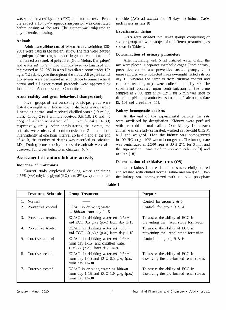
Table 1
Treatment Schedule Group Treatment Purpose
1. Normal —— Control for group 2 & 5
2. Preventive control EG/AC in drinking water Control for group 3 & 4ad libitum from day 1-15
3. Preventive treated EG/AC in drinking water ad libitum To assess the ability of ECO inand ECO 0.5 g/kg (p.o.) from day 1-15 preventing the renal stone formation
4. Preventive treated EG/AC in drinking water ad libitum To assess the ability of ECO inand ECO 1.0 g/kg (p.o.) from day 1-15 preventing the renal stone formation
5. Curative control EG/AC in drinking water ad libitum Control for group 5 & 6from day 1-15 and distilled water10ml/kg (p.o) from day 16-30
6. Curative treated EG/AC in drinking water ad libitum To assess the ability of ECO infrom day 1-15 and ECO 0.5 g/kg (p.o.) dissolving the pre-formed renal stonesfrom day 16-30
7. Curative treated EG/AC in drinking water ad libitum To assess the ability of ECO infrom day 1-15 and ECO 1.0 g/kg (p.o.) dissolving the pre-formed renal stonesfrom day 16-30

January - March 2010 5 Journal of Pharmacy and Chemistry • Vol.4 • Issue.1
buffer (pH 7.4) to get 10% w/v homogenate. Nucleardebris was separated by centrifuging the homogenate at800 rpm at 4°C for 5 min. The resultant supernatant wasfurther centrifuged at 10,000rpm at 4°C for 20min to getpost mitochondrial supernatant, which was used to estimatelipid peroxidation as malondialdehyde (MDA) [12] andantioxidants, reduced glutathione (GSH) [13] and catalase[14].
In vitro antioxidant studies
DPPH free radical scavenging
The antioxidant activity of the extract was determinedby 1,1-diphenyl-2-picrylhydrazyl (DPPH) free radical invitro scavenging [15]. The extract in ethanol (3 ml) atdifferent concentrations (100, 200, 400, 800 and 1000 µg/ml) was incubated with 1ml of the ethanolic solution ofDPPH (100µM) at 30±2°C for 20 min and the absorbancewas read at 517 nm. For each concentration, the assay wascarried out in triplicate. Percentage scavenging of theDPPH by the extract was expressed by taking the differencein absorbance between the control and the test. Ascorbicacid was used as the standard. For ascorbic acid and ECO,IC
50 values (concentration required to scavenge 50% of the
free radicals) were also determined. The results wereexpressed as means of triplicates.
Nitric oxide free radical scavenging
Nitric oxide free radical scavenging was determinedby studying the inhibition of the generation of nitric oxidefrom sodium nitroprusside. 3ml of each 100, 200, 400, 800and 1000 µg/ml of ECO was dissolved in ethanol andincubated with 1ml of sodium nitroprusside (10mM) inphosphate buffer (pH 7.7) at 25ºC for 120 min. Afterincubation, 0.5 ml of the reaction mixture was diluted with0.5 ml of Griess reagent (2% o-phosphoric acid, 1%sulphanilamide and 0.1% N-napthylethylenediamine). Theabsorbance of the pink chromophore, formed duringdiazotization of nitrite with sulphanilamide and subsequentcoupling with N-napthylethylenediamine was measured at546 nm against the corresponding blank solution. For eachconcentration, the assay
was carried out in triplicate. The degree of freeradical scavenging in the presence and absence of differentconcentrations of the extract was measured. The differencein absorbance between the control and the test was takenand expressed as percentage free radical scavenging of theNO by the extract [16]. Ascorbic acid was used as standard.IC
50 values (concentration required to scavenge 50% of the
free radicals) for ascorbic acid and ECO were alsodetermined. The results were expressed as means oftriplicates.
Statistical analysis
The results were presented as Mean ± SEM. One wayanalysis of variance (ANOVA) followed by Scheffe’s testfor multiple comparisons were used for the measurement
of inter-group variation. Statistical significance wasconsidered at P<0.05.
ResultsPreliminary phytochemical evaluation
The preliminary phytochemical screening of ECOshowed the presence of flavonoids, tannins, saponins,steroids, anthraquinones and carbohydrates.
Acute toxicity and gross behavioral changes studies
Ethanolic extract of ECO was found to be safe, as noanimal died on administration of the extract up to 4 g/kg, orally and there were no significant gross behavioralchanges except diuretic and laxative effects.
Antiurolithiatic activityKidney weight
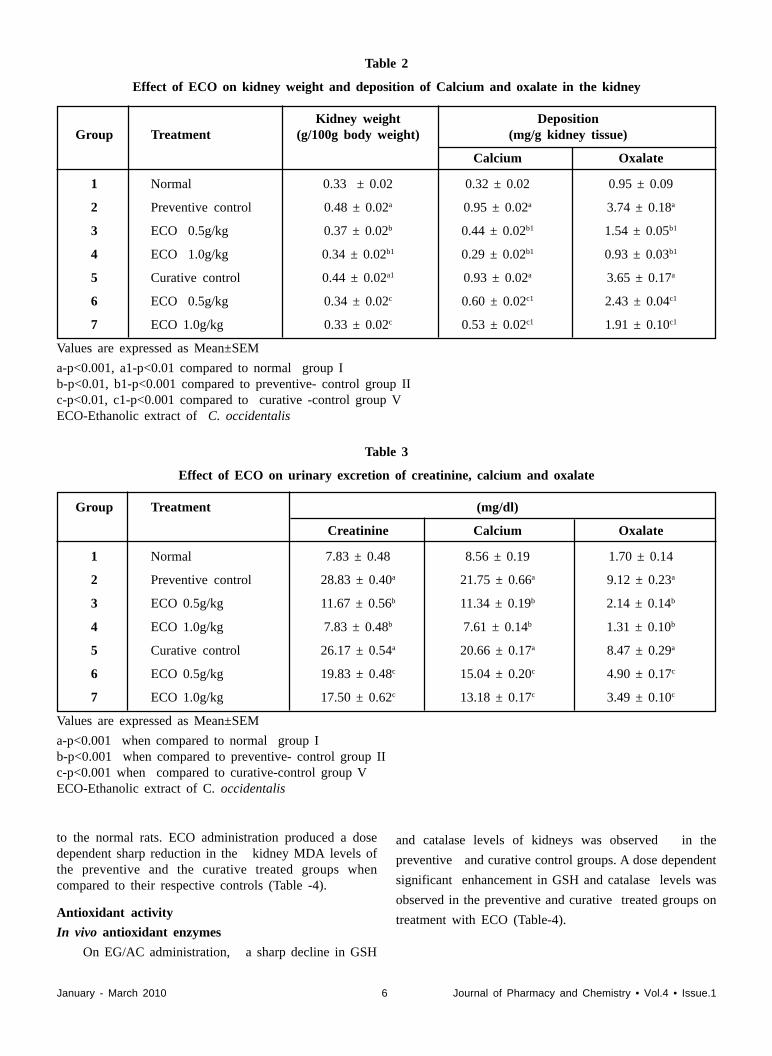
In both the preventive and curative control groups,on administration of EG/AC in drinking water, asignificant increase in kidney weight was observed whencompared to the normal group. On treatment with ECOat 0.5 and 1.0 g/kg orally, a significant reduction inkidney weight was observed in both the preventive andcurative treated groups, when compared to their respectivecontrols (Table-2).
Urinary pH
In normal rats, the pH was 7.0 to 7.5. EG/AC indrinking water ad libitum reduced urinary pH to 5.0 – 6.0in the preventive and curative control groups. In thesegroups, treatment with ECO restored the pH to normal (6.5– 7.0).
Deposition of calcium and oxalate in the kidney
In the control group 2 and 5 animals, administrationof EG/AC in drinking water resulted in a significantenhancement in kidney calcium and oxalate deposition.On ECO treatment, a dose dependent reduction inkidney calcium and oxalate deposition was observed inthe preventive and curative treated groups (group 3,4,6 &7) (Table- 2).
Urinary excretion of calcium, oxalate and creatinine
A significant enhancement in the urinary excretion ofcalcium, oxalate and creatinine was observed in thepreventive and curative control groups on CaOx renalstone formation. On treatment with ECO, a dose dependentsignificant reduction in urinary calcium, oxalate andcreatinine excretion was observed in the preventive andcurative treated groups (Table-3).
Oxidative stressIn vivo lipid peroxidation
On administration of EG/AC in drinking water, theMDA content of the kidneys was increased in thepreventive and curative control groups, when compared
January - March 2010 6 Journal of Pharmacy and Chemistry • Vol.4 • Issue.1
Table 2
Effect of ECO on kidney weight and deposition of Calcium and oxalate in the kidney
Kidney weight DepositionGroup Treatment (g/100g body weight) (mg/g kidney tissue)
Calcium Oxalate
1 Normal 0.33 ± 0.02 0.32 ± 0.02 0.95 ± 0.09
2 Preventive control 0.48 ± 0.02a 0.95 ± 0.02a 3.74 ± 0.18a
3 ECO 0.5g/kg 0.37 ± 0.02b 0.44 ± 0.02b1 1.54 ± 0.05b1
4 ECO 1.0g/kg 0.34 ± 0.02b1 0.29 ± 0.02b1 0.93 ± 0.03b1
5 Curative control 0.44 ± 0.02a1 0.93 ± 0.02a 3.65 ± 0.17a
6 ECO 0.5g/kg 0.34 ± 0.02c 0.60 ± 0.02c1 2.43 ± 0.04c1
7 ECO 1.0g/kg 0.33 ± 0.02c 0.53 ± 0.02c1 1.91 ± 0.10c1
Values are expressed as Mean±SEM
a-p<0.001, a1-p<0.01 compared to normal group Ib-p<0.01, b1-p<0.001 compared to preventive- control group IIc-p<0.01, c1-p<0.001 compared to curative -control group VECO-Ethanolic extract of C. occidentalis
Table 3
Effect of ECO on urinary excretion of creatinine, calcium and oxalate
Group Treatment (mg/dl)
Creatinine Calcium Oxalate
1 Normal 7.83 ± 0.48 8.56 ± 0.19 1.70 ± 0.14
2 Preventive control 28.83 ± 0.40a 21.75 ± 0.66a 9.12 ± 0.23a
3 ECO 0.5g/kg 11.67 ± 0.56b 11.34 ± 0.19b 2.14 ± 0.14b
4 ECO 1.0g/kg 7.83 ± 0.48b 7.61 ± 0.14b 1.31 ± 0.10b
5 Curative control 26.17 ± 0.54a 20.66 ± 0.17a 8.47 ± 0.29a
6 ECO 0.5g/kg 19.83 ± 0.48c 15.04 ± 0.20c 4.90 ± 0.17c
7 ECO 1.0g/kg 17.50 ± 0.62c 13.18 ± 0.17c 3.49 ± 0.10c
Values are expressed as Mean±SEM
a-p<0.001 when compared to normal group Ib-p<0.001 when compared to preventive- control group IIc-p<0.001 when compared to curative-control group VECO-Ethanolic extract of C. occidentalis
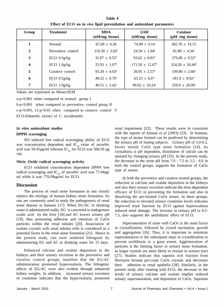
to the normal rats. ECO administration produced a dosedependent sharp reduction in the kidney MDA levels ofthe preventive and the curative treated groups whencompared to their respective controls (Table -4).
Antioxidant activity
In vivo antioxidant enzymes
On EG/AC administration, a sharp decline in GSH
and catalase levels of kidneys was observed in the
preventive and curative control groups. A dose dependent
significant enhancement in GSH and catalase levels was
observed in the preventive and curative treated groups on
treatment with ECO (Table-4).
January - March 2010 7 Journal of Pharmacy and Chemistry • Vol.4 • Issue.1
In vitro antioxidant studies
DPPH scavenging
NO induced free radical scavenging ability of ECOwas concentration dependent and IC
50 value of ascorbic
acid was 58.45µg/ml whereas IC50
for ECO was 566.50 µg/ml.
Nitric Oxide radical scavenging activity
ECO exhibited concentration dependent DPPH freeradical scavenging and IC
50 of ascorbic acid was 77.04µg/
ml while it was 776.09µg/ml for ECO.
DiscussionThe process of renal stone formation in rats closely
mimics the etiology of human kidney stone formation. Sorats are commonly used to study the pathogenesis of renalstone disease in humans [17]. When EG/AC in drinkingwater is administered orally, EG is converted to endogenousoxalic acid by the liver [18] and AC lowers urinary pH[19], thus promoting adhesion and retention of CaOxparticles within the renal tubules [20]. Association ofoxalate crystals with renal tubular cells is considered as apotential factor in the renal stone formation [21]. Hence inthe present study, rats were rendered lithogenic byadministering EG and AC in drinking water for 15 days.
Enhanced calcium and oxalate deposition in thekidneys and their urinary excretion in the preventive andcurative control groups, manifest that the EG/ACadministration promoted hyperoxaluria. The lithogeniceffects of EG/AC were also evident through enhancedkidney weights. In addition, increased urinary excretionof creatinine indicates that the hyperoxaluria, promoted
renal impairment [22]. These results were in consistentwith the reports of Atmani et al (2003) [23]. In humans,the type of stones formed can be predicted by determiningthe urinary pH of fasting subjects. Urinary pH of 5.0-6.5,favors mostly CaOx type stone formation [24]. Ascrystalluria is pH dependent, dissolution of calculi can beattained by changing urinary pH [25]. In the present study,the decrease in the urine pH from 7.0 - 7.5 to 5.5 - 6.0 inboth the control groups, supports the formation of CaOxtype of stones.
In both the preventive and curative treated groups, thereduction in calcium and oxalate deposition in the kidneysand also their urinary excretion indicate the dose dependentefficacy of ECO in preventing the formation and also indissolving the pre-formed CaOx stones. In these groups,the reduction in elevated urinary creatinine levels indicatesimproved renal function by ECO against hyperoxaluriainduced renal damage. The increase in urinary pH to 6.5-7.5, also supports the antilithiatic effect of ECO.
Supersaturation of urine with CaOx is the main factorin crystallization, followed by crystal nucleation, growthand aggregation [26]. Thus, it is important to minimizesupersaturation or the subsequent steps in crystallization toprevent urolithiasis to a great extent. Agglomeration ofparticles is the limiting factor in urinary stone formation,as larger crystals are more likely to remain in urinary tract[27]. Studies indicate that saponin rich fraction fromHerniaria hirsuta pre-coats CaOx crystals and decreasestheir adhesion to renal epithelia [28]. Similarly, in thepresent study after treating with ECO, the decrease in thelevels of urinary calcium and oxalate implies reducedurinary supersaturation, and deposition of CaOx. Saponins
Table 4
Effect of ECO on in vivo lipid peroxidation and antioxidant parameters
Group Treatment MDA GSH Catalase(nM/mg tissue) (nM/mg tissue) (µM /mg tissue)
1 Normal 67.00 ± 4.30 74.99 ± 4.14 365.70 ± 14.15
2 Preventive control 119.30 ± 3.02a 24.50 ± 1.64a 81.80 ± 4.34a
3 ECO 0.5g/kg 31.87 ± 0.55b 93.62 ± 8.81b 276.80 ± 9.52b
4 ECO 1.0g/kg 25.93 ± 1.07b 171.56 ± 12.67b 324.20 ± 10.44b
5 Curative control 93.20 ± 4.03a 26.95 ± 2.57a 109.80 ± 2.66a
6 ECO 0.5g/kg 49.52 ± 0.70c 63.23 ± 6.0c1 181.9 ± 8.92c1
7 ECO 1.0g/kg 40.51 ± 2.42c 99.02 ± 10.24c 259.9 ± 20.09c
Values are expressed as Mean±SEM
a-p<0.001 when compared to normal group I
b-p<0.001 when compared to preventive- control group II
c-p<0.001, c1-p<0.01 when compared to curative -control V
ECO-Ethanolic extract of C. occidentalis

January - March 2010 8 Journal of Pharmacy and Chemistry • Vol.4 • Issue.1
may be responsible for these effects, as the preliminaryphytochemical tests indicated the presence of saponins,anthraquinones, steroids and flavonoids in C. occidentalis.
In addition, herbal remedies with antimicrobial activityprotect the anti-adherent layer of renal mucosa, a protectivebarrier against urinary stone deposition [29]. Anti-microbialactivity of anthraquinones from C. occidentalis has beenreported [5] which also might be responsible for theantiurolithic activity of the extract in the present study, asanthraquinones, flavonoids and steroids were isolated byearlier investigators from the plant [30,31].
The lithogenic effects of EG are mainly attributed toits oxidative damage [20]. Polyunsaturated fatty acids ofrenal membrane are prone to ROS mediated LPO onexposure to Ox/CaOx [32, 33]. The ROS uses upantioxidants considerably thus constitutes OS. In addition,low levels of renal cellular glutathione favor LPO andsubsequent retention of calcium and oxalate in the kidneys[34]. Enhanced LPO and reduced levels of GSH andcatalase in the preventive and curative control groupsindicate that hyperoxaluria, promoted extensive generationof ROS. These ROS may have been responsible for cellulardamage and subsequent enhancement in LPO andaccumulation and retention of Ox and deposition of CaOxcrystals. These results were in agreement with earlierreports of Toblli et al (2002) [34].
Studies in rats indicate that treatment with herbalextracts and antioxidants reduce EG induced CaOxcrystallization in the kidney and subsequent OS [35-37]. Inaddition, Curhan et al (1998) reported that dailyconsumption of tea reduces kidney stone formation inhumans as its flavonoids act by directly quenching ROSand by chelating metal ions like iron and copper [38-40].For this reason, the present study was also focused onevaluation of the antioxidant effect of ECO in urolithicrats.
In the preventive and curative treated groups, thedose dependent decline in kidney MDA content reflects,suppression of LPO and alleviation of OS, providingconvincing evidence for the effect of the extract indecreasing hyperoxaluria induced LPO. The dosedependent increase in the levels of antioxidant enzymesGSH and catalase reflects the potential of the extract inresisting hyperoxaluria induced OS. In vitro antioxidantstudies also revealed that the extract is a rich source ofantioxidants. Hence, flavonoids present in ECO mighthave prevented OS through scavenging ROS and bychelating metal ions.
Thus, the extract may have exhibited antiurolithiaticactivity through inhibiting crystal aggregation by pre-coating of CaOx crystals with saponins, protecting theantiadherent layer of renal mucosa by anthraquinones andscavenging of ROS by flavonoids. From the present study
results, C. occidentalis seems to be a promising plant intreating CaOx urinary stones. However, further chemicaland pharmacological studies on the isolated components ofC. occidentalis seeds may be helpful in ascertaining theantiurolithiatic and antioxidant effects of the plant.
References[1] Siener R and Hesse A. World J Urol 2005; 23:304.
[2] Hacket RL, Shevock PN and Khan SR. J Urol 1990;144:1535.
[3] Selvam R and Sridevi D. Ind Biochem Biophys 1996;33:62.
[4] Rajiv SK and Shweta S. Ethnobiology, Surabhi Publications,Rasta Sanghiji, SMS Highway, Jaipur; India, 1st ed, 200:279.
[5] Jain SC, Sharma RA and Mittal C. Phytotherapy Research1998; 12:200.
[6] Crossland J. Lewis Pharmacology, New York, ChurchillLivingstone, 1980:137.
[7] Sheth UK, Dandkar NK and Usha GK. Selected topics inexperimental pharmacology, 1st ed. 1972:124.
[8] Khan SR and Glenton P. J Urol 1995; 153:811.
[9] Lorentz K. Clin Chim Acta 1982; 126:327.
[10] Hodgkinson A and Williams A. Clin Chim Acta 1972;36:127.
[11] Browers LD. Clin Chem 1980; 26:551.
[12] Nichans WG and Sannelson D. Eur J Biochem 1968;6:126.
[13] Jollow D, Mitchel L, Zampaglione N and Gilete J.Pharmacol 1974; 11:151.
[14] Aebi H. Catalase in vitro, SP Colowick, NO Kaplane (eds).Meth Enzymol 1984; 105:121.
[15] Blois MS. Nature 1958; 181:1199.
[16] Marcocci L, Maguire JJ, Droy-Lefaix MT and Parker L.Biochem Biophys Res Commun 1994; 201:748.
[17] Khan SR. World J Urol 1997; 15:236.
[18] Khan SR and Hackett RL. Scan Electr Microsc 1985;11:759.
[19] De Bruijn WC, et al; scanning microscopy. 1994; 8:541-50.
[20] Touhami M, Laroubi A, Elhabazi K, Louna F, Zrara I,Grases F and Chait A. BMC Urol 2007; 7:18.
[21] Atmani F, Slimani Y, Mimouni M, Aziz M, Hacht B andZiyyat A. J Ethnopharmacol 2004; 95:87.
[22] Gowenlock AH. Varley’s Practical Clinical Biochemistry,6th ed. CBS publishers, New Delhi, 1988:773.
[23] Atmani F, Slimani Y, Mimouni M and Hacht B. BJU Int2003; 92:137.
[24] King Jr JS. J Urol 1967; 97:583.
[25] Vermeulen CW and Goetz R. J Urol 1954; 72:93.
[26] Finlayson B. Kidney Int 1978; 13:344.

January - March 2010 9 Journal of Pharmacy and Chemistry • Vol.4 • Issue.1
[27] Kok DJ and Khan SR. Kidney Int 1994; 46:847.
[28] Atmani F, Farell G and Lieske JC. J Urol 2004; 172:1510.
[29] Denyer SP. Guide to microbiological control inpharmaceuticals, Ellis Harwood, London, 1990:11.
[30] Rastogi RP and Mehrotra BN. Compendium of IndianMedicinal Plants, A CDRI series, PIB, New Delhi, 1990:Vol (1) 151.
[31] Hatano T, Mizuta S, Ito H and Yoshida T. Phytochemistry1999; 52:1379.
[32] Karadi RV, Gadge NB, Alagawadi KR and Savadi RV. JEthnopharmacol 2006; 105:306.
[33] Devinder S, Rajnendrapal K, Vikas C and Kanwaljit C. JMed Food 2006; 9:443.
[34] Toblli JE, Ferder L, Stella I, De Cavanagh MVE, AngerosaM and Inserra F. J Urol 2002; 168:1550.
[35] Thamilselvan S, Hackett RL and Khan SR. J Urol 2000;164:224.
[36] Thamilselvan S, Khan SR and Menon M. Urol Res 2003;31:3.
[37] Selvam R. Urol Res 2002; 30:35.
[38] Curhan G, Willet W, Speizer F and Stampfer M. Am InternMed 1998; 128:534.
[39] Vinson J, Dabbagh Y, Serry M and Jang J. J Agri FoodChem 1995; 43:2800.
[40] Miller N, Castelluccio C, Tijburg L and Rice-Evans C.FEBS Letters, 1996; 392:40.
�

January - March 2010 10 Journal of Pharmacy and Chemistry • Vol.4 • Issue.1
*Address for [email protected]
Spectral, electrochemical and molecular modeling studies ofSchiff bases of some hydrazinylthiazocoumarins and 2-acetyl
pyridine
VIJAYBHASKAR P, KRISHNA V AND RAMACHANDRAIAH A*Department of Chemistry, National Institute of Technology Warangal,
Warangal-506004, Andhra Pradesh, India.
ABSTRACT
The spectral and voltammetric behavior of a series of 3-{2-[N-(1-pyridin-2-yl-ethylidene)hydrazino]thaizol-4-yl}coumarins (1a-1d) have been studied. Plots of absorbance vs pH were used forevaluation of their pK
b values. The electrochemical reductions of (1a-1d) have been studied over a
wide pH range at mercury electrode. All the compounds exhibit a characteristic two-electron irreversiblereduction wave corresponding to the electrochemical reduction of the azomethine group. Relevantelectrochemical data such as diffusion coefficient, charge transfer co-efficient (±
na) and forward rate
constant (k0h), etc have also been evaluated. Photometric and voltammetric methods of assaying 1a-
1d have been developed. Molecular modeling was done on the compounds to correlate the stability andstructural aspects of them to the observed data.
Key words: Thiazolylcoumarins, Cyclic voltammetry, Molecular modeling, Pyridine-2-hydrazones,Acid-base equilibria
IntroductionHydrazinylthiazocoumarins
are extended derivatives ofchromone-2-one (2) nucleus andfind numerous therapeuticapplications [1-6]. A variety ofSchiff bases have been generatedby adducting the hydrazine of thisseries with suitable carbonylcompounds. These Schiff baseshave evinced considerable research interest amongelectrochemists for their versatile electrochemical behaviorwith mechanism that varies with the solvent, pH, nature ofelectrode material and other electrochemical experimentalconditions [7-9]. Review of the literature, reveals that reportson electrochemical and spectral investigations of compoundspossessing all the three, viz, thiazocoumarine, hydrazoneand pyridyl moieties are scanty. Hence, we considered itimportant to study the spectral and electrochemicalcharacteristics of a few representative such molecules (1a-1d) which are the Schiff bases of hydrazinylthiazocoumarinsand 2-acetyl pyridine.
ExperimentalAll the chemicals used were of AnalaR grade. Stock
solutions of (1a-1d) (1x10⁻³ M) were prepared in aqueousalcohol media. Buffers of different pH (ionic strength = 0.05M) were prepared according to literature procedure [10]. AnATI Orion Model 902 Ion Meter was used for pH-metry.The UV-visible spectra of the solutions were recorded on anAnalyticJena Specord Ratio Recording Spectrophotometer.The electrochemical measurements such as cyclicvoltammetry, differntial pulse polorography etc., wererecorded on a Metrohm 663 VA Stand whereas coulometrywas on a BAS Model CV-27 Voltammograph. Molecularmodeling was carried out on ChemOffice Pro 10.0 platform.
The electrochemical recordings were run on a 25 mLsample solution, containing 5 mL of stock solution of thesample and 20 mL of the desired buffer, transferred into theelectrochemical cell. Before each run, nitrogen or argon gaswas purged for about 4 minutes to dispel the dissolvedoxygen. Buffer (20 mL) and 5 mL of water were mixed forblank run.
Results and DiscussionsSpectral Studies and Acid-Base Equilibria: The
electronic spectra of 1a in various aqueous buffers are shown
January - March 2010 11 Journal of Pharmacy and Chemistry • Vol.4 • Issue.1
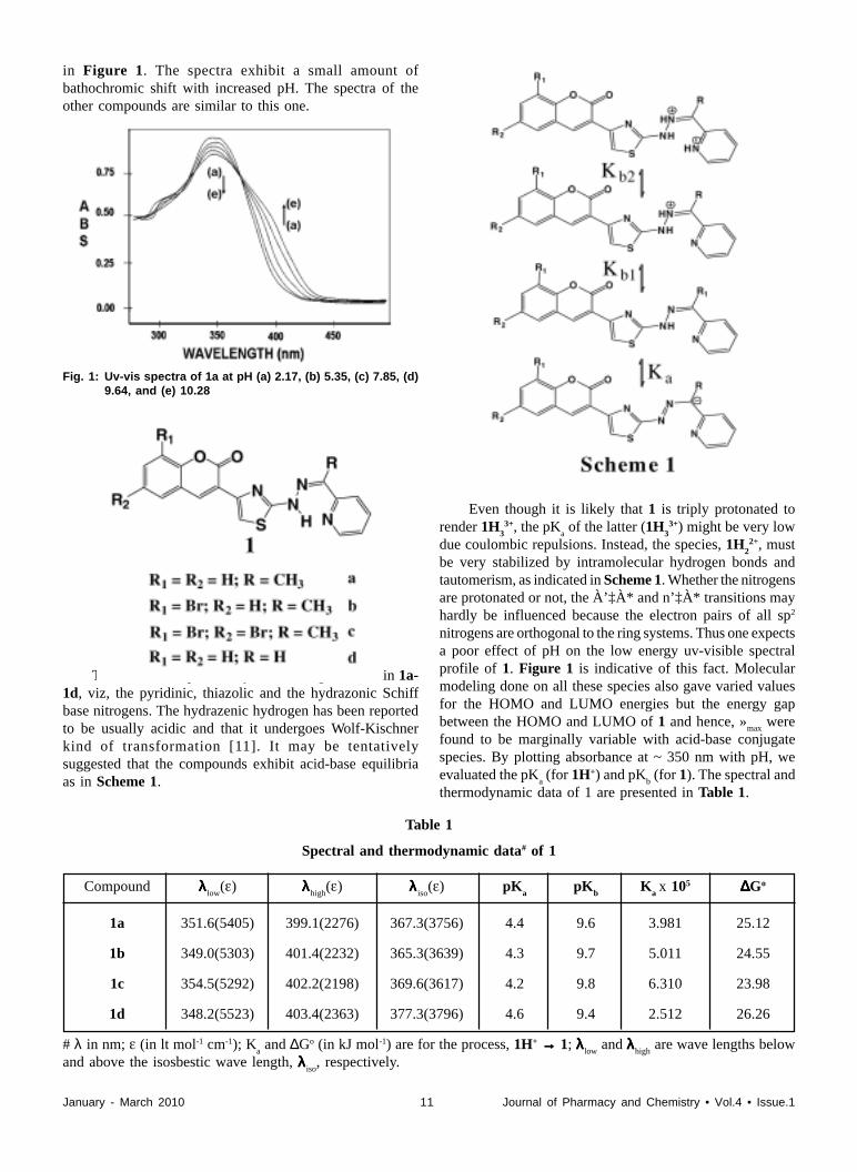
in Figure 1. The spectra exhibit a small amount ofbathochromic shift with increased pH. The spectra of theother compounds are similar to this one.
Fig. 1: Uv-vis spectra of 1a at pH (a) 2.17, (b) 5.35, (c) 7.85, (d)9.64, and (e) 10.28
There are three potentially basic nitrogen atoms in 1a-1d, viz, the pyridinic, thiazolic and the hydrazonic Schiffbase nitrogens. The hydrazenic hydrogen has been reportedto be usually acidic and that it undergoes Wolf-Kischnerkind of transformation [11]. It may be tentativelysuggested that the compounds exhibit acid-base equilibriaas in Scheme 1.
Even though it is likely that 1 is triply protonated torender 1H
33+, the pK
a of the latter (1H
33+) might be very low
due coulombic repulsions. Instead, the species, 1H2
2+, mustbe very stabilized by intramolecular hydrogen bonds andtautomerism, as indicated in Scheme 1. Whether the nitrogensare protonated or not, the À’‡À* and n’‡À* transitions mayhardly be influenced because the electron pairs of all sp2
nitrogens are orthogonal to the ring systems. Thus one expectsa poor effect of pH on the low energy uv-visible spectralprofile of 1. Figure 1 is indicative of this fact. Molecularmodeling done on all these species also gave varied valuesfor the HOMO and LUMO energies but the energy gapbetween the HOMO and LUMO of 1 and hence, »
max were
found to be marginally variable with acid-base conjugatespecies. By plotting absorbance at ~ 350 nm with pH, weevaluated the pK
a (for 1H+) and pK
b (for 1). The spectral and
thermodynamic data of 1 are presented in Table 1.
Table 1
Spectral and thermodynamic data# of 1
Compound λλλλλ low(ε) λλλλλhigh
(ε) λλλλλ iso(ε) pK
apK
bK
a x 105 ∆∆∆∆∆Go
1a 351.6(5405) 399.1(2276) 367.3(3756) 4.4 9.6 3.981 25.12
1b 349.0(5303) 401.4(2232) 365.3(3639) 4.3 9.7 5.011 24.55
1c 354.5(5292) 402.2(2198) 369.6(3617) 4.2 9.8 6.310 23.98
1d 348.2(5523) 403.4(2363) 377.3(3796) 4.6 9.4 2.512 26.26
# λ in nm; ε (in lt mol-1 cm-1); Ka and ∆Go (in kJ mol-1) are for the process, 1H+ →→→→→ 1; λλλλλ low
and λλλλλhigh are wave lengths below
and above the isosbestic wave length, λλλλλ iso, respectively.

January - March 2010 12 Journal of Pharmacy and Chemistry • Vol.4 • Issue.1
In many cases, the 2-electron reduction of the azomethinegroup of the Schiff bases intrinsically happens in aconsecutive pair of single electron transfer processes as
-CH-NH-k1-C=N- + e- + H+ (2a)
-CH-NH- + e- + H + -CH2-NH-k2 (2b)
giving two successive reduction current peaks [14]. If k2
>>> k1, one obtains a single but two electron reduction peak
with k. The second consecutive reaction has a paralleldimerization process [15] as
2 (-CH-NH-) -CH-NH-
-CH-NH-
kd
(3)
Usually, the kd value is far slower (due to steric
restrictions) than k2 and the chemical dimerization step, (3),
would influence the cyclic voltammetric profile when onlythe k
2 is slow. If the on-setting of the potential responsible
for the second consecutive reduction, (2b), is delayed, thereis a finite probability of the formation of the dimer whichwould undergo an easier electrochemical reduction as
2 (-CH2-NH-)-CH-NH-
-CH-NH-+ 2e- + 2H+
(4)
The presence of only a single but two electron-twoproton reduction indicates that the k
2 is very fast and k
d is
very slow. The effect of scan rate on the cyclic voltammetricresponse of 1 in low pH buffers was studied. A shoulder wasobserved at lower cathodic potential due to (4) at slow scanrates because of the finite life for the product of (2a) toengage in (3). At faster scan rates the potential needed for(2b) is too quickly reached to allow any scope for (3). Thatthe E
p values of 1a-1c are considerably more cathodic than
those of 1d and others with more open –C=N– moietysupports the fact that the methyl substitution on theazomethine group hinders a facile electron transfer at the -C=N- site.
The linearity of the plots of peak current, ip, vs ν1/2, the
square root of the scan rate, suggests that the electrochemicalreduction in the entire pH range, 2 - 12, is diffusion-controlled. The cathodic shift of peak potential, E
p, with
increased scan rate also supports the irreversible nature ofthe electrode reaction. From plots of E
p vs log ν, at different
pH values, the transfer coefficients, αna
, were evaluated. Theplots of E
p vs pH of 1a-1d are linear with the slopes
compatible to a 2-proton participation. With the knowledgeof n and α
na, we could evaluate the diffusion coefficient, D,
Electrochemical studies: It has been shown above that thecompounds do not exhibit appreciable pH dependence intheir electronic spectral profiles due to poor effect of protontransfer on the energy gaps of the HOMO and LUMOorbitals. However, the electron transfer characteristics areexpected to very much species-dependent.
The cyclic voltammograms of 1a, as a representative of1, in several buffers, are presented in Figure 2. All thecompounds offer irreversible reduction peaks in aqueoussolution in the pH range 2-11. It is also obvious that the peakpotentials drift cathodically with increased pH indicatingthe involvement of H+ ions in the electrochemical reductionmechanism. Further, at very low pH, the reduction peak isslightly split whereas at elevated pH one observes a singlepeak.
Even though, 1a-1d appear to have severalelectrochemically active sites, many of them are hard to bereduced due to aromatic stability. However, the azomethinesite sandwiched between an aromatic pyridine and aminothiazole rings must be readily reducible. The electrochemicalreduction behaviour of Schiff bases is well documented[12]. Curve fitting of Randles-Sevscik equation besidescoulometric analyses establishes the number of electronsinvolved as 2.
Since the compounds undergo neither hydrolysis likemany Schiff bases are famous for [13] nor any other chemicalreaction in these buffers, the 2-electron transfer is attributedto the reduction of the -C=N- moiety of 1 or protonated 1i.e., 1H 2+ or 1H+ as
-CH-NH--C=N- + 2e- + 2H+ k (1)
Fig. 2: Cyclic voltammograms of 1a (2.06 x 10⁻⁴ M) in buffers(ionic strength = 0.05M) of pH (a) 2.56, (b) 5.93, (c)7.80, (d) 9.62 and (e) 10.48

January - March 2010 13 Journal of Pharmacy and Chemistry • Vol.4 • Issue.1
for the electroactive species, of 1a-1d, from the Randles-Sevcik equation,
ip = 2.69 x 10⁻⁵ n(αn
a )½AD½½½c (5)
applicable for an irreversible but diffusion controlled electrontransfer cyclic voltammetirc peaks, where i
p is
the peak
current, A is the area of the electrode, ½ is the scan rate and
c is the concentration of the electro active species.
Compounds, 1b and 1c have additional reducible stepsin their C–Br bonds. The electrochemistry of organo halogencompounds is well documented [16]. The –C–X– bond(where X is a halogen substituent) is known to offer anirreversible electron transfer due to the process,
XCHC X +e-, H+
(6)
in aqueous media. However, in the present case of 1, whereinthe C-X is on a stable aromatic ring, such reduction has beenfound to be very near to the hydrogen evolution and hencecould not be investigated further. The electrochemical dataof 1 are presented in Table 2.
Metal coordination: Compounds, 1a-1d, are expected to bea set of facile chelating agents given the presence of severalnitrogen atoms at appropriate locations of the molecularskeleton. In Figure 3 are shown the ethanolic electronicspectra of (i) 1a, (ii) CoCl
2.6H
2O, and (iii) the mixture of
CoCl2.6H
2O and 1a in a 1:2 molar ratio. Neither (i) nor (ii)
nor the additive of spectrum of (i) and (ii) matches with (iii),indicating a metal-ligand interaction between Co2+ and 1. AJob’s plot also gave the M:L ratio as 1:2.
Fig. 3: Overlapped electronic spectra of (i) 1a, (3.74 x 10⁻⁵ M),(ii) CoCl2.6H2O (1.87 x 10⁻⁵ M) and (iii) solutioncontaining 1a (3.74 x 10⁻⁵ M) and CoCl2.6H2O (1.87 x 10-
5 M)
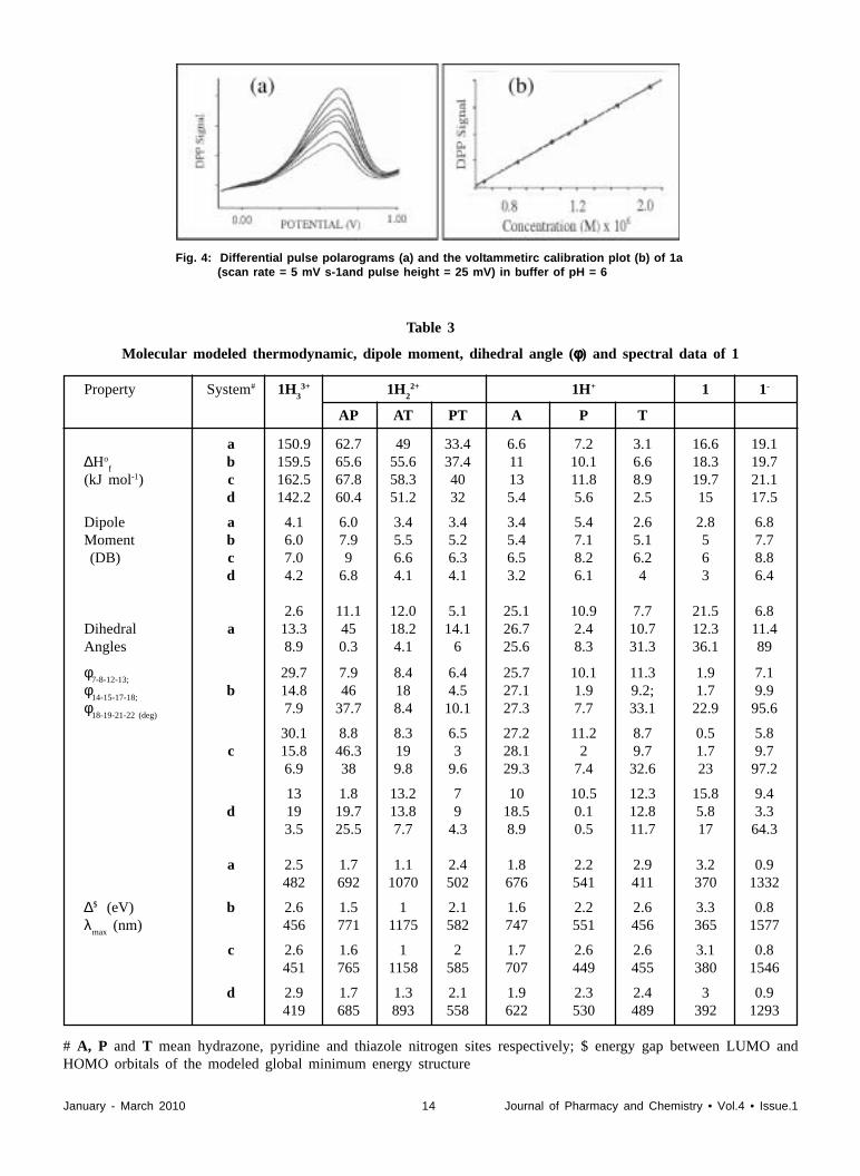
Assaying of 1: Based on the electrochemical response of 1,an excellent electrochemical assaying of 1, in differentialpulse polarography (DPP), has been developed. The DPPcurves of 1 at pH=8.00 along with the calibration curve(inset) are shown in Figure 4.
Table 2
Cyclic voltammetric data of 1
Compound pH -Ep (V)# αααααna
koh x109 D
o x 106
vs (cm2 /s) (cm2 /s)Ag|||||AgCl
2.77 0.645 0.43 5.12 8.884.85 0.774 0.44 4.31 8.23
1a 6.57 0.813 0.45 3.44 8.158.66 0.936 0.47 2.2 8.82
10.23 1.143 0.58 2.01 9.13
2.95 0.734 0.39 5.47 5.464.75 0.863 0.45 4.41 5.03
1b 6.52 0.904 0.47 4.07 5.718.79 1.025 0.49 3.34 6.02
10.93 1.257 0.53 2.74 5.53
2.55 0.721 0.42 6.91 2.454.65 0.833 0.44 6.09 2.16
1c 6.37 0.879 0.45 5.41 2.338.89 1.038 0.48 4.97 1.3811.13 1.271 0.56 4.3 1.27
2.38 0.614 0.43 7.42 8.994.21 0.746 0.44 6.57 9.05
1d 6.19 0.807 0.45 5.49 9.548.76 0.918 0.51 4.63 9.3411.01 1.122 0.55 3.8 9.21
January - March 2010 14 Journal of Pharmacy and Chemistry • Vol.4 • Issue.1
Table 3
Molecular modeled thermodynamic, dipole moment, dihedral angle (φφφφφ) and spectral data of 1
Property System# 1H3
3+ 1H2
2+ 1H+ 1 1-
AP AT PT A P T
a 150.9 62.7 49 33.4 6.6 7.2 3.1 16.6 19.1∆Ho
fb 159.5 65.6 55.6 37.4 11 10.1 6.6 18.3 19.7
(kJ mol-1) c 162.5 67.8 58.3 40 13 11.8 8.9 19.7 21.1d 142.2 60.4 51.2 32 5.4 5.6 2.5 15 17.5
Dipole a 4.1 6.0 3.4 3.4 3.4 5.4 2.6 2.8 6.8Moment b 6.0 7.9 5.5 5.2 5.4 7.1 5.1 5 7.7 (DB) c 7.0 9 6.6 6.3 6.5 8.2 6.2 6 8.8
d 4.2 6.8 4.1 4.1 3.2 6.1 4 3 6.4
2.6 11.1 12.0 5.1 25.1 10.9 7.7 21.5 6.8Dihedral a 13.3 45 18.2 14.1 26.7 2.4 10.7 12.3 11.4Angles 8.9 0.3 4.1 6 25.6 8.3 31.3 36.1 89
φ7-8-12-13;
29.7 7.9 8.4 6.4 25.7 10.1 11.3 1.9 7.1φ
14-15-17-18;b 14.8 46 18 4.5 27.1 1.9 9.2; 1.7 9.9
φ18-19-21-22 (deg)
7.9 37.7 8.4 10.1 27.3 7.7 33.1 22.9 95.6
30.1 8.8 8.3 6.5 27.2 11.2 8.7 0.5 5.8c 15.8 46.3 19 3 28.1 2 9.7 1.7 9.7
6.9 38 9.8 9.6 29.3 7.4 32.6 23 97.2
13 1.8 13.2 7 10 10.5 12.3 15.8 9.4d 19 19.7 13.8 9 18.5 0.1 12.8 5.8 3.3
3.5 25.5 7.7 4.3 8.9 0.5 11.7 17 64.3
a 2.5 1.7 1.1 2.4 1.8 2.2 2.9 3.2 0.9482 692 1070 502 676 541 411 370 1332
∆$ (eV) b 2.6 1.5 1 2.1 1.6 2.2 2.6 3.3 0.8
λmax
(nm) 456 771 1175 582 747 551 456 365 1577
c 2.6 1.6 1 2 1.7 2.6 2.6 3.1 0.8451 765 1158 585 707 449 455 380 1546
d 2.9 1.7 1.3 2.1 1.9 2.3 2.4 3 0.9419 685 893 558 622 530 489 392 1293
# A, P and T mean hydrazone, pyridine and thiazole nitrogen sites respectively; $ energy gap between LUMO andHOMO orbitals of the modeled global minimum energy structure
Fig. 4: Differential pulse polarograms (a) and the voltammetirc calibration plot (b) of 1a(scan rate = 5 mV s-1and pulse height = 25 mV) in buffer of pH = 6

January - March 2010 15 Journal of Pharmacy and Chemistry • Vol.4 • Issue.1
Molecular Modeling of 1: Molecular modeling studies werecarried out on 1a-1d using CambridgeSoft ChemOffice Ultrasoftware. The calculated thermodynamic values of variousacid-base conjugate species in several of their tautomericforms are collected in Table 3 while their standard heats offormation are shown pictorially in Figure 5. The globalenergy-minimized structures of the most thermodynamicallystable species of 1a are shown in Figure 6 along with thenumbering scheme for the atoms. It is observed that coumarinand thiazole are slightly away from coplanarity with lessthan 20o dihedral angle between them whereas the hydrazonemoiety makes as much as 35o dihedral angle with the pyridinering system in 1. However, the dihedral angle between thecoumarin and thiazole rings falls considerably in 1H+
(especially in 1H(III) with little change between thehydrazone and pyridine π-systems. This preference toestablish an acute angle between adjacent ring-planes farless than electrostatically preferred 90o is attributed to thepossibility of Huckel’s aromaticity for the whole molecularπ-system that has 26 π electrons including the lone pairs of
electrons on the sp3 nitrogen of the hydrazine moiety, oxygenand sulfur (with n=6 in 4n+2 rule) in 1. Electron pair orbitalson the hydrazonium and pyridine nitrogens are orthogonalto the molecular π-system and hence do not contribute to thearomaticity. The fact that 1H is more stable than any otheracid-base conjugates of 1 supports this conclusion becausethe additional hydrogen bonds possible in 1H favour greaterplanarity between the successive rings. Conformationalenergy trends of 1a over these planes for its most stableacid-base conjugates, viz., 1 and 1H+, can be visualizedfrom their double-dihedral plots over the congruent atoms,C(15)-N(17)-N(18), shown in Figure 7.
The quantum mechanical HOMO-LUMO energycalculations have been used for computing the expectedgas-phase electronic transitions for various conjugates andtautomers of 1. These values are collected in Table 3 for 1and 1H+ along with the experimental spectral data. Thetrends of the theoretical and experimental absorption maximaare in good agreement.
Fig. 5: Histographic representation of the standard heats offormation of various acid-base conjugates of 1 (1Hn isn-many protonated 1 and 1- is deprotonated 1; I, II, andIII are azo, pyridine and thiazole nitrogen sitesrespectively
Fig. 6: Stereographic ball and stick model of 1a with the numbering scheme of atoms
Fig. 7: Double dihedral drive torsional energy diagrams overC(15)-N(17)-N(18) link of 1a and 1aH(III) (1a protonatedon thiazole nitrogen
January - March 2010 16 Journal of Pharmacy and Chemistry • Vol.4 • Issue.1
�
References[1] Wawzonek O, Heterocyclic Compounds, John Wiley and
Sons, New York, 1975; 173.
[2] Dean FM, Naturally Occurring Oxygen Ring Compounds,2nd ed. Butter Worths, London, 1963;75.
[3] Livingtone R, Rod’s Chemistry of Carbon Compounds, 4thed. Elsevier, Amsterdam, 1977; 66.
[4] Stauton J, Comprehensive Organic Chemistry, 4th ed. Oxford,1979; 629.
[5] Kartritzky AR, Rees CW, Comprehensive Hetero CyclicChemistry, Pergaman Press, Oxford, 1984; 3.
[6] Hantzsch , Weber HJ. Ber 1887; 20:311.
[7] Hantzsch. Ann 1888; 1:249.
[8] Dadson RM, King LC. J Am Chem Soc 1950; 92:2242.
[9] King LC, Hlavacek RJ. J Am Chem Soc 1950; 92:722.
[10] Ju Luirie, Handbook of Analytical Chemistry, Mir PublishersMoscow, 1978.
[11] Hutchins RO, Hutchins MK, Comp. Org Syn 1991; 8:327-343.
[12] Andrieux CP, Saveant JM. Electroanal Chem 1971 ; 33:453.
[13] Unver H, Polat K, Ucar M, Zengin DM. Spectrosc. Lett2003; 36:287.
[14] Zuman P, The Elucidation of Organic Electrode Processes,Academic Press, New York, 1969.
[15] Hammam E, Tawfik A, Ghoneim, MM. J Pharm BiomedAnal 2004; 36:149.
[16] Bard AJ, Faulkner LR, Electrochemical Methods andApplications, John Willey, New York, 1980.

January - March 2010 17 Journal of Pharmacy and Chemistry • Vol.4 • Issue.1
*Address for correspondence:[email protected]
IntroductionAbsorption of therapeutic agents from the oral mucosa
overcomes premature drug degradation within the gastrointestinal tract, as well as active drug loss due to first passmetabolism that may be associated with other routes ofadministration. Buccal delivery of drugs became anattractive alternate to the oral route of drug administrationby providing rich blood supply that drains drug directlyinto the jugular vein and chance to administer drugs topatients who cannot be dosed orally[1-3]. Variousmucoadhesive formulations were suggested for buccaldelivery that includes buccal patches[4], adhesive tablets[5]and adhesive gels[6]. Buccal patches overcome some ofthe drawbacks of other dosage forms. They have uniquecharacteristics including flexibility, relatively rapid onsetof drug delivery, sustained drug release and rapid declinein the serum drug concentration when the patch is removed.The patch is confined to the buccal area over which it isattached and therefore the absorption profile may have lessinter and intra-individual variability [7, 8].
Granisetron is a selective 5-HT3
receptor antagonistused in treatment of chemotherapy-induced, radiation-induced and post-operative nausea and vomiting. The oralroute of administration of GRN is also impractical forpatients who are vomiting or who have impaired gastricemptying [9, 10]. Both parenteral and oral formulationshave also been used, but buccal route may obviate the needfor repeated injections, repeated oral dosing and also beuseful in patients who cannot tolerate oral dosage forms. Itis less invasive than IV or subcutaneous administration.Physicochemical (low molecular weight 312.4g/mol, lowdose 1-2mg , Log P 2.6) and pharmacokinetic (t
1/2 4-6hr,
absolute bioavailability about 60%) parameters made GRNto be suitable for buccal delivery[11-13].
In this investigation we developed GRN buccal patcheswith a dissolvable matrix using HPMC E 15, with aninsoluble backing membrane. The developed patches wereevaluated for in vitro release, ex vivo permeation throughporcine buccal membrane and mechanical properties.
Materials and MethodsGranisetron was a gift from by Natco pharma,
Hyderabad, A.P, India. Hydroxypropyl methylcellulose E
Formulation, Characterization And Evaluation Of GranisetronBuccal Patches
SWATHI M1, JITHAN AUKUNURU1,* AND MADHUSUDAN RAO Y2
1Vaagdevi College of Pharmacy, Warangal, India-506001.
2University College of Pharmaceutical Sciences, Kakatiya University, Warangal, India-506009.
ABSTRACT
The aim of this investigation was to develop and evaluate mucoadhesive buccal patches ofGranisetron(GRN). Permeation of GRN was determined in vitro using porcine buccal membrane.Buccal patches were developed by solvent-casting technique using Hydroxypropyl-methylcellulose(HPMC) as mucoadhesive polymer. The patches were evaluated for in vitro release,moisture absorption,mechanical properties and ex vivo permeation studies. The optimized formulation,based on in vitro release, ex vivo permeation studies and moisture absorption studies, was subjectedfor in vitro residence time using porcine buccal membrane. In vitro flux of GRN was calculated to be3.19±0.27µg.hr–1
.cm–2. In vitro drug release and moisture absorbed was governed by HPMC content.
Increasing concentration of HPMC delayed the drug release. All formulations followed Zero orderrelease kinetics where as the release pattern was non-Fickian. The mechanical properties, tensilestrength (9.58±3.45kgmm–2 for formulation F3) and elongation at break reveal that the formulationswere found to be strong but not brittle. Formulations showed a significant permeation through porcinebuccal membrane and convenient residence time. The results indicate that suitable bioadhesive buccalpatches of GRN with desired permeability and suitable mechanical properties could be prepared usingHPMC.
Keywords:Buccal,Granisetron,Mucoadhesive, Mechanical properties.
January - March 2010 18 Journal of Pharmacy and Chemistry • Vol.4 • Issue.1
15(HPMC E 15) was procured from Loba Chemie Pvt.Ltd., India. Phenol red was obtained from Hi MediaLaboratories Pvt. Ltd. Mumbai, India. All reagents usedwere of analytical grade.
Tissue Preparation (Isolation)
Porcine buccal tissue was taken from local slaughter-house. It was collected within 10 minutes after slaughter ofthe pig and tissue was kept in Kreb’s buffer solution. Itwas bought immediately to the laboratory and was mountedwithin 2 hours of isolation of buccal tissue. The tissue wasrinsed thoroughly using phosphate buffer saline to removeany adherent material. The epithelium was separated fromthe underlying connective tissue using surgical procedure.Sufficient care was taken to prevent any damage to thebuccal epithelium [14].
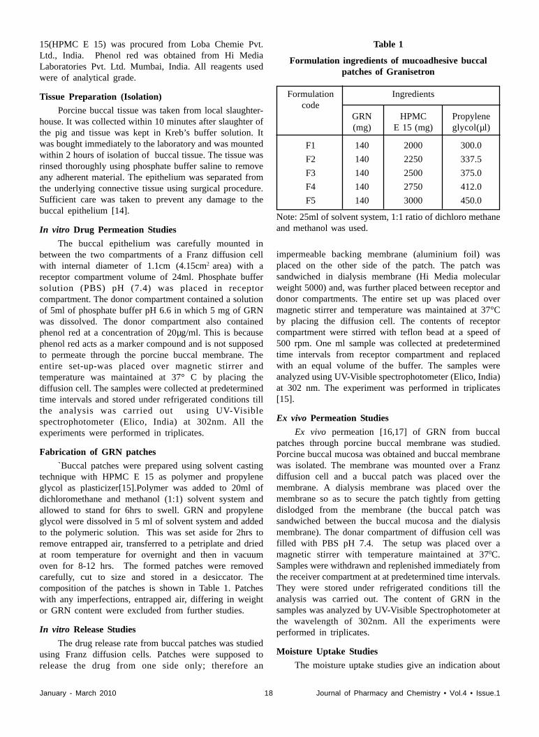
In vitro Drug Permeation Studies
The buccal epithelium was carefully mounted inbetween the two compartments of a Franz diffusion cellwith internal diameter of 1.1cm (4.15cm2 area) with areceptor compartment volume of 24ml. Phosphate buffersolution (PBS) pH (7.4) was placed in receptorcompartment. The donor compartment contained a solutionof 5ml of phosphate buffer pH 6.6 in which 5 mg of GRNwas dissolved. The donor compartment also containedphenol red at a concentration of 20µg/ml. This is becausephenol red acts as a marker compound and is not supposedto permeate through the porcine buccal membrane. Theentire set-up-was placed over magnetic stirrer andtemperature was maintained at 37° C by placing thediffusion cell. The samples were collected at predeterminedtime intervals and stored under refrigerated conditions tillthe analysis was carried out using UV-Visiblespectrophotometer (Elico, India) at 302nm. All theexperiments were performed in triplicates.
Fabrication of GRN patches
`Buccal patches were prepared using solvent castingtechnique with HPMC E 15 as polymer and propyleneglycol as plasticizer[15].Polymer was added to 20ml ofdichloromethane and methanol (1:1) solvent system andallowed to stand for 6hrs to swell. GRN and propyleneglycol were dissolved in 5 ml of solvent system and addedto the polymeric solution. This was set aside for 2hrs toremove entrapped air, transferred to a petriplate and driedat room temperature for overnight and then in vacuumoven for 8-12 hrs. The formed patches were removedcarefully, cut to size and stored in a desiccator. Thecomposition of the patches is shown in Table 1. Patcheswith any imperfections, entrapped air, differing in weightor GRN content were excluded from further studies.
In vitro Release Studies
The drug release rate from buccal patches was studiedusing Franz diffusion cells. Patches were supposed torelease the drug from one side only; therefore an
impermeable backing membrane (aluminium foil) wasplaced on the other side of the patch. The patch wassandwiched in dialysis membrane (Hi Media molecularweight 5000) and, was further placed between receptor anddonor compartments. The entire set up was placed overmagnetic stirrer and temperature was maintained at 37°Cby placing the diffusion cell. The contents of receptorcompartment were stirred with teflon bead at a speed of500 rpm. One ml sample was collected at predeterminedtime intervals from receptor compartment and replacedwith an equal volume of the buffer. The samples wereanalyzed using UV-Visible spectrophotometer (Elico, India)at 302 nm. The experiment was performed in triplicates[15].
Ex vivo Permeation Studies
Ex vivo permeation [16,17] of GRN from buccalpatches through porcine buccal membrane was studied.Porcine buccal mucosa was obtained and buccal membranewas isolated. The membrane was mounted over a Franzdiffusion cell and a buccal patch was placed over themembrane. A dialysis membrane was placed over themembrane so as to secure the patch tightly from gettingdislodged from the membrane (the buccal patch wassandwiched between the buccal mucosa and the dialysismembrane). The donar compartment of diffusion cell wasfilled with PBS pH 7.4. The setup was placed over amagnetic stirrer with temperature maintained at 370C.Samples were withdrawn and replenished immediately fromthe receiver compartment at at predetermined time intervals.They were stored under refrigerated conditions till theanalysis was carried out. The content of GRN in thesamples was analyzed by UV-Visible Spectrophotometer atthe wavelength of 302nm. All the experiments wereperformed in triplicates.
Moisture Uptake Studies
The moisture uptake studies give an indication about
Table 1
Formulation ingredients of mucoadhesive buccalpatches of Granisetron
Formulation Ingredientscode
GRN HPMC Propylene(mg) E 15 (mg) glycol(µl)
F1 140 2000 300.0
F2 140 2250 337.5
F3 140 2500 375.0
F4 140 2750 412.0
F5 140 3000 450.0
Note: 25ml of solvent system, 1:1 ratio of dichloro methaneand methanol was used.

January - March 2010 19 Journal of Pharmacy and Chemistry • Vol.4 • Issue.1
the relative moisture absorption capacities of polymers andan idea whether the formulations maintain their integrityafter absorption of moisture. The study was carried out asper procedure reported earlier [18] . Briefly, agar (5% w/v) was dissolved in hot water, transferred into petriplatesand allowed to solidify. Six patches from each formulationseries were placed in vaccum oven overnight prior to thestudy to remove moisture if any and laminated on one sidewith water impermeable backing membrane. They werethen incubated at 37°C for one hour over the agar surface.The initial and final weights were recorded and percentagemoisture absorption was calculated by using the formula.
Final weight – Initial weight%Moisture absorption = ––––––––––––––––––––– X 100 Initial weight
Measurement of Mechanical Properties
Mechanical properties of the patches were evaluatedusing a microprocessor based advanced force gauzeequipped with a motorized test stand (Ultra Test, Mecmesin,West Sussex, UK), equipped with a 25 kg load cell. Filmstrip with the dimensions 60 x 10 mm and without anyvisual defects were cut and positioned between two clampsseparated by a distance of 3 cm. Clamps were designed tosecure the patch without crushing it during the test, thelower clamp was held stationary and the strips were pulledapart by the upper clamp moving at a rate of 2 mm/secuntil the strip broke. The force and elongation of the filmat the point when the strip broke was recorded. The tensilestrength and elongation at break values were calculatedusing the formula[19].
Force at break (kg)Tensile strength (kg. mm–2) = ––––––––––––––––––––––
Initial cross sectional area of the sample (mm2)
Elongation at break (%mm–2) =
Increase in length (mm) 100–––––––––––––––––––––– X –––––––––––––––––– Original length(mm) Cross sectional area (mm2)
Measurement of in vitro Residence Time
The in vitro residence time was determined using USPdisintegration apparatus. The disintegration medium was800 ml of PBS (pH 6.6) maintained at 37±20C. Thesegments of porcine buccal mucosa, each of 3 cm length,were glued to the surface of a glass slab, which was thenvertically attached to the apparatus. Three mucoadhesivefilms of each formulation were hydrated on one surfaceusing PBS (pH 6.6) and the hydrated surface was broughtinto contact with the mucosal membrane. The glass slabwas vertically fixed to the apparatus and allowed to moveup and down. The film was completely immersed in thebuffer solution at the lowest point and was out at thehighest point. The time required for complete erosion or
detachment of the film from the mucosal surface wasrecorded[20].
Drug-Polymer Interaction Study
To study the possible interaction between Granisetronand Polymeric materials of the patches, infrared (IR)spectroscopy was carried out on pure substances and theirphysical mixture. The IR spectra were recorded using IR-Spectrophotometer (Perkin Elmer FT-IR, Perkin ElmerInst.USA) by KBr pellet method.
Results and DiscussionsDrug Permeation Studies through the Porcine BuccalMembrane
Porcine buccal mucosa has been the most frequentlychosen model for in vitro permeation studies because of itssimilarity to human tissue and is available in large quantitiesfrom slaughter houses. Cumulative amount of GRNpermeated through the porcine buccal epithelium is shownin Figure.1. The isolated membrane was intact as nodetectable level of phenol red, which was used as a non-absorbable marker compound, was found in the receivercompartment. The thickness of the isolated membrane,measured with a digital micrometer (Mitutoyo, Japan),ranged from 1040 to 1880 microns. Cumulative amount ofGRN permeated in 6 hr was about 71.52 ± 4.12 % and fluxwas calculated to be 3.19±0.27µg. hr–1
.cm–2
.
Fig. 1: Ex v ivoEx v ivoEx v ivoEx v ivoEx v ivo permeation of GRN (5.0 mg) throughporcine buccal mucosa, values represented as Mean±S.D (n = 3)
In vitro Drug Release StudiesThe drug release profiles of GRN patches were shown
in Figure. 2. The drug release was governed by theamount of matrix forming polymer. An increase in polymerconcentration causes an increase in the viscosity of the gelas well as formation of a gel layer with a longer diffusionalpath. This could cause a decrease in the effective diffusioncoefficient of the drug and therefore a reduction in thedrug release rate. Formulation F1 showed maximum drugrelease (83.08± 3.36%) where as formulation F5 showedlowest release of (58.43±2.67%), among the series. Data ofthe in vitro release was fit into different equations andkinetic models to explain the release kinetics of GRN from

January - March 2010 20 Journal of Pharmacy and Chemistry • Vol.4 • Issue.1
buccal patches. Zero order model seemed to be the mostappropriate model describing release kinetics from allpatches (0.986, 0.987, 0.988,0.995, 0.976 for formulationF1 to formulation F5).On other hand ‘·’ values indictedthat amount of released drug was by non Fickiandiffusion[21,22].Increasing the concentration of the polymerin the formulations showed a sustained effect on GRNrelease. This is because, as the proportion of these polymersin the matrix increased, there was an increase in theamount of water uptake and proportionally greater swellingleading to a thicker gel layer. Zero-order release fromswellable hydrophilic matrices occurs as a result of constantdiffusional pathlengths. When the thickness of the gelledlayer and thus the diffusional pathlengths remain constant,zero-order release can be expected. In this investigationsimilar behavior was predicted and obtained.
Fig. 2: Release profiles of GRN from mucoadhesive buccalpatches, values represented as Mean ±S.D (n=3)
Ex vivo Permeation Studies
Figure 3 shows the graphical representation ofcumulative percentage drug permeated from the buccalpatches.The results of drug permeation from buccal patchesof Granisetron through the porcine buccal mucosa revealthat drug was released from the formulation and permeatedthrough the porcine buccal membrane,hence could possiblypermeate through the human buccal membrane. The resultsindicated that the drug permeation was more in F3 amongthe last three formulations and about 72.25% of Granisetroncould permeate through the buccal membrane in 4 hrs.
Moisture Uptake Studies
Moisture absorption studies evaluate the integrity of
the formulation upon exposure to moisture and the resultswere shown in Table 2. The percentage moisture observedranged from about 59.6±11.99% to 156.34±9.27 % w/w fordifferent formulations. Formulations F1 and F2 weredeformed during the study. The results reveal that,percentage of moisture absorption was increased withincrease in polymer content of formulations(Table 2). Whenthe patches were placed without backing membranecomplete swelling followed by erosion was observedindicating that the drug release mechanism involves swellingof the polymer initially, followed by drug release from theswollen matrix by diffusion.
Mechanical Properties of Patches
Ideal buccal film, apart from good bio adhesivestrength, should be flexible, elastic and strong enough towithstand breakage due to stress caused during its residencein the mouth. The tensile strength (TS) and elongation atbreak (E/B) shows the strength and elasticity of the film.A soft and weak polymer is characterized by a low TS andE/B; a hard and brittle polymer is defined by a moderateTS, and low E/B; a soft and tough polymer is characterizedby a moderate TS and a high E/B; whereas a hard andtough polymer is characterized by high TS and E/B. It issuggested that an ideal buccal film should have a relativelyhigh TS and E/B. The results of the mechanical propertiesi.e. TS and E/B were presented in Table 2. TS increasedwith the increase in polymeric content but E/B values
Table 2
Moisture absorption, Mechanical properties of GRN buccal patches, values represented as mean±S.D (n=3)
Formulations Moisture absorbed (%w/w) TensileStrength (Kg.mm2) Elongation at break (mm-2)
F1 Deformed 3.87±1.28 117±8.24
F2 Deformed 8.74±1.74 94±7.58
F3 59.67±11.99 9.58±3.45 75±6.42
F4 63.72±1.94 11.58±2.42 70±5.24
F5 156.34±9.27 13.24±2.74 53.6±3.72
Fig. 3. Ex vivoEx vivoEx vivoEx vivoEx vivo permeation studies of selected mucoadhesivebuccal patches of GRN, values represented asMean±S.D (n=3)
January - March 2010 21 Journal of Pharmacy and Chemistry • Vol.4 • Issue.1
�
decreased with the increase in polymer content. MaximumTS was exhibited by F5 patch (13.24±2.74kg.mm–2) andminimum was exhibited by F1(3.87±1.28 kg.mm–2).Maximum E/B was seen with F1(117±8.24% mm–2) andthe least was observed with F5 (53.6±3.72% mm–2).
In vitro residence time
In vitro residence time was determined for theformulations F3, F4, F5.The formulations F1, F2 wereeroded in moisture absorption study. So they were notsuitable as buccal patches. The in vitro residence time ofthe formulations was in order of F4> F3> F5.
Drug - Polymer Interaction Study
The IR spectral analysis of Granisetron alone showedthat the principal peaks were observed at wave numbers of1648.15, 1559.88, 1228.95 and 3235.58. In the IR spectraof the physical mixture of Granisetron and HPMC E151647.90, 1560.15, 1242.12 and 3448.42 were observed forthe Granisetron. However, some additional peaks wereobserved with physical mixtures, which could be due tothe presence of polymer. These results suggest that there isno interaction between the drug and polymer used in thepresent study. It is already well known that the commonpolymers such as HPMC popular in controlled/sustainedrelease matrix type formulations because of theircompatibility with a number of drugs.
Selection of optimized formulation
Based on in vitro release and moisture absorptionstudies formulation F3 was selected as the best formulation.Formulation F1 showed maximum drug release 83.08 ±3.36 %, where as formulation F2 showed 79.37±4.46%drug release. Formulations F1 and F2 were deformedduring Moisture absorption studies, these formulations couldnot be expected to maintain the integrity after administration.The results indicated that the drug permeation was more inF3 among the last three formulations therefore, formulationF3 was selected as best formulation and subjected forfurther investigation.
AcknowledgementsThe authors would like to thank Natco Pharma,
Hyderabad, A.P, India for providing gift sample ofGranisetron.the authors also like to thank principal andmanagement of Vaagdevi College of Pharmacy for providingnecessary facility useful in conduction of this work.
References[1]. Edith Mathiowitz, Chckering III Donald E, Claus-Michael
Lehr. Bioadhesive Drug Delivery Systems, Fundamentals,Novel Approaches, and Development, Design and The
Pharmaceutical Sciences, New York: Marcel Dekker 1999;98: 541-562.
[2]. Hans E. Junginger, Janet A. Hoogstraate, J. Coos Verhoef.J Cont Rel 1999; 62: 149-159.
[3]. Rathbone M J, Drummond B, Trucker l. Adv Drug Del Rev1994; 13: 1-22.
[4. Luana Periolia, Valeria Ambrogia, Fausta Angelicia, MaurizioRiccia, Stefano Giovagnolia, Marinella Capuccellab, CarloRossia. J Cont Rel 2004; 99: 73– 82.
[5]. Choi HG, Jung JH, Yong CS, Rhee CD, Lee MK, Han J H,Park K M, Kim C K. J Cont Rel 2000; 68: 405-412.
[6]. Jones DS, Brown AF, Woolfson AD, Djokic J, Adams V. JPharm Sci 1999; 88: 592-598.
[7]. Kashappa Goud, Pramod Kumar T M. AAPS Pharm SciTech 2004; 5(3): 1-9.
[8]. Amir H Shojaei. J Pharm Pharmaceut Sci 1998; 1(1): 15-30.
[9]. Matti Aapro. The Oncologist 2004; 9: 673-686.
[10]. Pouran Layegh, Mohammad Javad Mojahedi, ParisaEmamgholi Tabar Malekshah,Fakhrozaman Pezeshkpour.Indian Journal of Dermatology, Venereology and Leprology2007; 73(4): 231-234.
[11]. Alfonso Gurpide, Belen Sadaba, Salvador Martin-Algarra,Jose R, Azanaza. The Oncologist 2007; 12: 1151-1155.
[12]. Plosker GL, Goa KL. Drugs 1991; 42: 805-824.
[13]. Upward JW, Arnold BDC, Link C, Pierce DM, Allen A,Tasker TCG. Eur J Cancer 1990; 26: 12-15.
[14]. Chandra Sekhar Kolli, Ramesh Gannu, Vamshi VishnuYamsani, Kishan V, MadhsudanRao Yamsani. InternationalJournal of Pharmaceutical Sciences and Nanotechnology2008; 1(1): 64-70.
[15]. Pramod Kumar TM, Kashappa Goud Desai, ShivakumarHG. J Pharm Educ 2002; 36(3): 147-151.
[16]. Mona Semalty, Ajay Semalty, Ganesh Kumar, Vijay Jugal.International Journal of Pharmaceutical Sciences andNanotechnology 2008; 1(2): 184-189.
[17]. Giacomo Di Colo, Ylenia Zambito, Chira Zaino. J PharmSci 2008; 97(5): 1652-1677.
[18]. Vamshi Vishnu Y, Chandrasekhar K, Ramesh G ,MadhusudanRao Y. Curr Drug Del 2007; 4: 27-39.
[19]. Peh KK, Wong CF. J Pharm Sci 1999; 2: 53-61.
[20]. Semalty A, Mona Bhojwani, Bhatt GK, Guptha GD,Shrivatav AK. Indian J Pharm Sci 2005; 67(5): 548-552.
[21]. Patel Vishnu M, Prajapati Bhupendra G, Patel MadhabahaiM. Acta Pharm 2007; 57: 61-72.
[22]. Peppas NA. Pharm Acta Helv 1985; 60: 110-111.

January - March 2010 22 Journal of Pharmacy and Chemistry • Vol.4 • Issue.1
Gastroretentive Delivery Systems: A Short Review
RAGHUNADHA GUPTA. C, PURUSHOTHAMAN M, DWRAKANATH REDDY PAND VIJAYA RATNA J*
AU College of Pharmaceutical Sciences, Andhra University,Visakhapatnam 530003, Andhra Pradesh, India.
ABSTRACT
Recent scientific and patent literature indicates increased interest among academics and industrialresearch groups regarding the novel dosage forms that can be retained in the stomach for a prolongedand predictable period of time. One of the most feasible approaches for achieving a prolonged andpredictable dug delivery profiles in the gastrointestinal tract is to control the gastric residence time,using gastroretentive dosage forms that will provide us with new and important therapeutic options.From the formulation and technological point of view, the preparation of a floating drug deliverysystem is a considerably easy and logical approach. In this paper, the gastric physiology and thereported intragastric delivery systems have been presented briefly.
Key words: Gastro retentive delivery systems, effervescent floating systems, non effervescentfloating systems, single unit and multiple units.
*Author for [email protected]
IntroductionUnder certain circumstances prolonging the gastric
retention of a delivery system for achieving greatertherapeutic benefit of the drug substance is desirable. Forexample, drugs that are absorbed in the proximal part ofthe gastrointestinal tract [1], and drugs that are less solublein or are degraded by the alkaline pH may benefit fromprolonged gastric retention [2, 3]. In addition, for local andsustained drug delivery to the stomach and proximal smallintestine to treat certain conditions, prolonged gastricretention of the therapeutic moiety may offer numerousadvantages including improved bioavailability andtherapeutic efficacy, and possible reduction of dose size [4-6]. It has been suggested that prolonged local availabilityof antibacterial agents may augment their effectiveness intreating Helicobacter pylori (H. Pylori) related peptic ulcers[7]. H. pylori live deep within the gastric mucus layer andprolonged local application of drug product is needed forsufficient drug to diffuse to the bacteria. Menon et al. [8]have compared the absolute bioavailability of furosemidein dogs from commercial conventional products and afloating dosage form. Higher bioavailability from floatingdosage form when compared to the non-floating commercialproducts of furosemide has been attributed to the fact thatthe upper gastrointestinal tract is the primary site ofabsorption for the drug.
It was suggested that compounding narrow absorptionwindow drugs in a unique pharmaceutical dosage formwith gastroretentive properties would enable an extendedabsorption phase of these drugs. After oral administration,such a dosage form would be retained in the stomach andwould release the drug there in a controlled and prolongedmanner, so that the drug would be supplied continuously toits absorption sites in the upper gastrointestinal tract. Thismode of administration would best achieve the knownpharmacokinetic and pharmacodynamic advantages ofcontrolled release dosage forms for these drugs [9].
Gastro retentive delivery systems (GRDS), however,are not suitable for drugs that may cause gastric lesions,e.g., non-steroidal anti-inflammatory agents. Also, the drugsubstances that are unstable in the strong acidic environmentof the stomach are not suitable candidates to be incorporatedinto such systems. In addition, these systems do not offersignificant advantages over the conventional dosage formsfor drugs, which are absorbed throughout the gastrointestinaltract [10].
The need for gastroretentive dosage forms (GRDFs)has led to extensive efforts in both academia and industrytowards the development of such drug delivery systems.These efforts resulted in GRDFs that were designed inlarge part based on the following approaches: (a) lowdensity form of the dosage form that causes buoyancyabove gastric fluid; (b) high density dosage form that isretained in the bottom of the stomach; (c) bioadhesion to
January - March 2010 23 Journal of Pharmacy and Chemistry • Vol.4 • Issue.1
the stomach mucosa; (d) slowed motility of thegastrointestinal tract by concomitant administration of drugsor pharmaceutical excipients; (e) expansion by swelling orunfolding to a large size which limits emptying of thedosage form through the pyloric sphincter. However, it isrecognized that there are many physiological constraintswhich may limit development of such delivery systems.Following is a brief description of the physiologicalconsiderations pertaining to designing GRDS.
Basic Gastrointestinal Tract PhysiologyIt is well recognized that the stomach is used as a
depot for controlled release dosage forms. The stomach issituated in the left upper part of the abdominal cavityimmediately under the diaphragm [11]. The stomach iscomposed of the following parts: fundus, above the openingof the esophagus into the stomach; body, the central part;and antrum. The proximal portion of the stomach comprisingfundus and body regions, serves as a reservoir for ingestedmaterials, secretes digestive juices and propels chyme, amilky mixture of food with gastric juices, to the distal partof the stomach i.e., antrum. Antrum serves as the majorsite for mixing and for pumping which leads to gastricemptying [12]
. Its size varies from 25 to 50 ml according
to the amount of distention. Following a meal it is 1500ml,after the food emptied, a ‘collapsed’ state is obtained witha resting volume of only 25–50 ml. The pylorus is ananatomical sphincter situated between the most terminalantrum and the duodenum [13]
. In the fasting state pH of
the stomach is 2. But after ingestion of food pH graduallyrises to a level of 6.5 and then gradually declines to the pHof fasting state over a period of a few hours.
The intrinsic properties of the drug molecule and thetarget environment for delivery are the major determiningfactors in the bioavailability of the drug. Factors such aspH, enzymes, nature and volume of secretions, residencetime, and effective absorbing surface area of the site ofdelivery play an important role in drug liberation andabsorption. A brief survey of the relevant physiologicalfeatures that pose challenges to the development of aneffective gastroretentive delivery system is presented below.
Gastric pHThe gastric pH is not constant; it is rather influenced
by various factors like diet, disease, presence of gases,fatty acids, and other fermentation products. In addition,the gastric pH exhibits intra-as well as inter-subjectvariation. This variation in pH may significantly influencethe performance of orally administered drugs. It has beenreported that the mean value of gastric pH in fasted healthysubjects is 1.1±0.15 [14 – 16]. On the contrary, the meangastric pH in fed state in healthy males has been reportedto be 3.6±0.4, [17] and the pH returns to basal levelin about 2 to 4 hours. However, in the fasted state,basal gastric secretion in women is slightly lower than inmen.
Gastric pH may be influenced by age, pathologicalconditions and drugs. About 20% of the elderly peopleexhibit either diminished (hypochlorohydria) or no gastricacid secretion (achlorohydia) leading to basal pH valueover 5.0[18]. Pathological conditions such as perniciousanemia and AIDS may significantly reduce gastric acidsecretion leading to elevated gastric pH. [19, 20] In addition,drugs like H
2 receptor antagonists and proton pump
inhibitors significantly reduce gastric acid secretion.
Gastric pH is an important consideration in selectinga drug substance, excipients, and drug carrier(s) fordesigning intragastric delivery systems.
Gastric EmptyingThe process of gastric emptying occurs both during
fasting and fed states: however, the pattern of motilitydiffers markedly in the two states. In the fasted state, it ischaracterized by an interdigestive series of electrical eventsthat cycle both through the stomach and small intestineevery 2-3 hrs [21]. This activity is called the interdigestivemyoelectric cycle or migrating myoelectric complex(MMC), which is often divided into four consecutivephases as described by Wilson and Washington [22].
Phase I is a quiescent period lasting from 30 to 60minwith rare contractions.
Phase II is a period of similar duration consisting ofintermittent action potentials and contractions that graduallyincrease in intensity and frequency as the phase progressesand it lasts for about 20 to 40 min.
Phase III is a short period of intense, large regularcontractions lasting from 10 to 20 min. It is this phase,which gives the cycle the term, “housekeeper wave”, sinceit serves to sweep undigested materials out of the stomachand down the small intestine. As phase III of one cyclereaches the end of the small intestine, phase III of the nextcycle begins in the duodenum.
Phase IV is a brief transitional phase that occurs betweenphase III and phase I of two consecutive cycles. The motoractivity in the fed state is induced 5-10 min after ingestionof a meal and persists as long as food remains in thestomach. It consists of regular and frequent contractions.These contractions are not as severe as those in the thirdphase of fasted motility pattern.
Some of the important aspects of the physiology ofthe gastric emptying process are as follows [23]
• The rate of movement of the dosage form from stomachto intestine is affected by the multiple chemical factors andthe physical size of the medication.
• The chemical components of the gastric fluid willinteract with the intestinal receptors. These receptors willcontrol the rate of gastric emptying by neuronal or hormonalmeans.

January - March 2010 24 Journal of Pharmacy and Chemistry • Vol.4 • Issue.1
• Emptying of the dosage form is also influenced bywhether it is taken on an empty stomach, in an interdigestivestate (with or soon after a meal), or in a digestive state.
• Small particles, regardless of size, density, or texturethat are ingested during the interdigestive state becomecoated by mucus and these coated dosage forms are emptieduniformly from the stomach.
• During the digestive state, the larger particles areretained in the stomach until the meal is essentially emptied.
• Emptying times of the solid dosage forms range from5 min to 5 h depending on the size of the medication andwhether the individual is in the interdigestive or digestivestate when the medication is administered.
Thus, an understanding of the physiology of gastricemptying is important in developing intragastric drugdelivery systems especially in the case of the floatingdrug-delivery systems.
Approaches to Prolong Gastric Residence Time (GRT)of Drug Delivery System:
Various devices such as mucoadhesive, floating,swelling and high-density systems have been developed toincrease GRT of a dosage form. Physiological features ofthe upper gastrointestinal tract pose a considerable challengeto develop such systems. Nevertheless, in vivo studies ofcertain systems have shown promising results.
Mucoadhesive Systems
The idea of bioadhesive began with the clear need tolocalize a drug at a certain site in the GI tract. Thereforea primary objective of using a bioadhesive system orallywould be achieved by obtaining a substantial increase inthe residence time of the drug for local drug effect and topermit once daily dosing [24].
The mucoadhesive systems extend their gastroretentivetime by adhering themselves to the gastric mucousmembrane. Bioadhesion on soft tissues of certain naturalor synthetic polymers has been exploited to control as well
as to prolong the gastric retention of the delivery systems.The adhesion of the polymers with the mucous membranemay be mediated by hydration, by bonding, or by receptormediation. In hydration mediated adhesion, the hydrophilicpolymers become sticky and mucoadhesive upon hydration.Bonding mediated adhesion may involve mechanical orchemical bonding. Chemical bonds may involve covalentor ionic bonds or Van der Waals forces between thepolymer molecules and the mucous membrane. Receptormediated adhesion takes place between certain polymersand specific receptors expressed on gastric cells. Thepolymers could be anionic or cationic or nonionic. A briefclassification of these polymers is mentioned in Table 1.
Table 1
A list of bioadhesive polymers
Anionic Cationic Nonionic
Carboxymethyl Polylysine Polyethylene
cellulose glycol
Chondroitin sulfate Polybrene Polyvinyl
pyrrolidone
Poly acrylic acid Dextran
Pectin
Carageenan
Chitosan
Alginic acid
Robinson et al [26] studied the bioadhesive propertiesof a broad spectrum of polymers; his group also reportedthat albumin beads containing chlorthiazide mixed withpolycarbophil offered sustained release for eight hoursafter being administered orally in the form of capsules torats. Although these materials have shown good bioadhesionin vitro and in vivo in rats, results in the humans weredisappointing because of two problems. One is that thebioadhesive formulation must attach to the surface of themucus layer, which is a continuously eroding surface andthe other is that the delivery system must withstand the GItract motility.
Smart and Kellaway [27] reported prolonged gastricretention of dosage forms coated with Carbomer in mice.In vivo data of granules containing microcrystalline chitosanand furosemide showed higher AUC than that of theconventional dosage form. [28] Also, the granules exhibitedslow release characteristic with a marked lag time. Itappeared that due to its mucoadhesive properties, chitosanretained the drug in the gastric mucosa for longer period oftime. Although the in vivo data exhibited promising resultsfor intragastric delivery, further characterization andevaluation of the system is necessary. Freeze-dried non-covalent polyionic complexes with porous structure basedon polyacrylic acid and chitosan intended for delivering
Fig.1: Schematic representation of interdigestive motilitypattern. (Adapted from Ref. [25].)
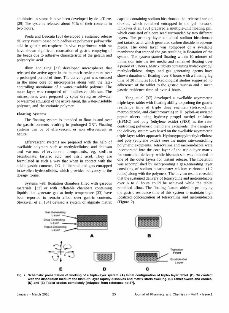
January - March 2010 25 Journal of Pharmacy and Chemistry • Vol.4 • Issue.1
antibiotics to stomach have been developed by de laTorre.[29] The systems released about 70% of their contents intwo hours.
Preda and Leucuta [30] developed a sustained releasedelivery system based on bioadhesive polymers: polyacrylicacid in gelatin microsphere. In vivo experiments with rathave shown significant retardation of gastric emptying ofthe beads due to adhesive characteristic of the gelatin andpolyacrylic acid.
Illum and Ping [31] developed microspheres thatreleased the active agent in the stomach environment overa prolonged period of time. The active agent was encasedin the inner core of microspheres along with the rate-controlling membrane of a water-insoluble polymer. Theouter layer was composed of bioadhesive chitosan. Themicrospheres were prepared by spray drying an oil/wateror water/oil emulsion of the active agent, the water-insolublepolymer, and the cationic polymer.
Floating Systems
The floating system is intended to float in and overthe gastric contents resulting in prolonged GRT. Floatingsystems can be of effervescent or non effervescent innature.
Effervescent systems are prepared with the help ofswellable polymers such as methylcellulose and chitosanand various effervescent compounds, eg, sodiumbicarbonate, tartaric acid, and citric acid. They areformulated in such a way that when in contact with theacidic gastric contents, CO
2 is liberated and gets entrapped
in swollen hydrocolloids, which provides buoyancy to thedosage forms.
Systems with floatation chambers filled with gaseousmaterials, [32] or with inflatable chambers containingliquids that generate gas at body temperature [33] havebeen reported to remain afloat over gastric contents.Stockwell et al. [34] devised a system of alginate matrix
capsule containing sodium bicarbonate that released carbondioxide, which remained entrapped in the gel network.Ichikawa et al. [35] prepared a multiple-unit floating pill,which consisted of a core seed surrounded by two differentlayers. The primary layer contained sodium bicarbonateand tartaric acid, which generated carbon dioxide in aqueousmedia. The outer layer was composed of a swellablemembrane that trapped the gas resulting in floatation of thesystem. The system started floating within 10 minutes ofimmersion into the test media and remained floating overa period of 5 hours. Matrix tablets containing hydroxypropylmethylcellulose, drugs, and gas generating agents haveshown duration of floating over 8 hours with a floating lagtime of 30 minutes [36]. Radiological studies suggested noadherence of the tablet to the gastric mucosa and a meangastric residence time of over 4 hours.
Yang et al [37] developed a swellable asymmetrictriple-layer tablet with floating ability to prolong the gastricresidence time of triple drug regimen (tetracycline,metronidazole, and clarithromycin) in H. pylori–associatedpeptic ulcers using hydroxy propyl methyl cellulose(HPMC) and poly (ethylene oxide) (PEO) as the rate-controlling polymeric membrane excipients. The design ofthe delivery system was based on the swellable asymmetrictriple-layer tablet approach. Hydroxypropylmethylcelluloseand poly (ethylene oxide) were the major rate-controllingpolymeric excipients. Tetracycline and metronidazole wereincorporated into the core layer of the triple-layer matrixfor controlled delivery, while bismuth salt was included inone of the outer layers for instant release. The floatationwas accomplished by incorporating a gas-generating layerconsisting of sodium bicarbonate: calcium carbonate (1:2ratios) along with the polymers. The in vitro results revealedthat the sustained delivery of tetracycline and metronidazoleover 6 to 8 hours could be achieved while the tabletremained afloat. The floating feature aided in prolongingthe gastric residence time of this system to maintain highlocalized concentration of tetracycline and metronidazole(Figure 2).
Fig. 2: Schematic presentation of working of a triple-layer system. (A) Initial configuration of triple- layer tablet. (B) On contactwith the dissolution medium the bismuth layer rapidly dissolves and matrix starts swelling. (C) Tablet swells and erodes.(D) and (E) Tablet erodes completely [Adapted from reference no.37].

January - March 2010 26 Journal of Pharmacy and Chemistry • Vol.4 • Issue.1
Fig. 3: (A) Multiple-unit oral floating drug delivery system. (B) Working principle of effervescent floating drug delivery system[Adapted from reference no.38].
Kawashima et al [40] prepared multiple-unit hollowmicrospheres by emulsion solvent diffusion technique. Drugand acrylic polymer were dissolved in an ethanol-dichloromethane mixture, and poured into an aqueoussolution of PVA with stirring to form emulsion droplets.The rate of drug release in micro balloons was controlledby changing the polymer to drug ratio. Micro balloonswere floatable in vitro for 12 hours when immersed inaqueous media. Radiographical studies proved that microballoons orally administered to humans were dispersed inthe upper part of stomach and retained there for threehours against peristaltic movements.
El-Gibaly [41] developed floating hollowmicrocapsules of chitosan by ionic interaction with anegatively charged surfactant. The system remained buoyantfor over 12 hours in simulated gastric fluids and exhibitedsustained drug release. Streubel et al [42] prepared single-unit floating tablets based on polypropylene foam powderand matrix-forming polymer. Incorporation of highly porousfoam powder in matrix tablets provided density muchlower than the density of the release medium. A 17% w/wfoam powder (based on mass of tablet) was achieved invitro for at least 8 hours. It was concluded that varying theratios of matrix-forming polymers and the foam powdercould alter the drug release patterns effectively.
Thanoo et al [43] developed polycarbonatemicrospheres by solvent evaporation technique.Polycarbonate in dichloromethane was found to give hollowmicrospheres that floated on water and simulated biofluidsas evidenced by scanning electron microscopy (SEM).High drug loading was achieved and drug-loadedmicrospheres were able to float on gastric and intestinalfluids. It was found that increasing the drug to polymerratio increased both their mean particle size and the releaserate of the drug.
Fell et al [44] prepared floating alginate beadsincorporating amoxycillin. The beads were produced bydropwise addition of alginate into calcium chloride solution,followed by removal of gel beads and freeze-drying.
Ichikawa et al [38] developed a new multiple type offloating dosage system composed of effervescent layersand swellable membrane layers coated on sustained releasepills. The inner layer of effervescent agents containingsodium bicarbonate and tartaric acid was divided into 2sublayers to avoid direct contact between the two agents.These sublayers were surrounded by a swellable polymermembrane. When this system was immersed in the bufferat 37ºC, it settled down and the solution permeated into theeffervescent layer through the outer swellable membrane.CO
2 was generated by the neutralization reaction between
the two effervescent agents, producing swollen pills (likeballoons) with a density less than 1.0 g/mL. It was foundthat the system had good floating ability independent ofpH and viscosity of the solution in the G.I.T and the drug(para-amino benzoic acid) was released in a sustainedmanner. This finding is shown in Fig.No.3.
Fassihi and Yang [39] developed a zero-order controlledrelease multilayer tablet composed of at least two barrierlayers and one drug layer. All the layers were made ofswellable, erodible polymers and the tablet was found toswell on contact with aqueous medium. As the tabletdissolved, the barrier layers eroded away to expose moreof the drug. Gas evolving agent was incorporated in eitherof the two barrier layers, which caused the tablet to floatand increased the retention of the tablet in the patient’sstomach.
Non-effervescent floating dosage forms use a gel formingor swellable cellulose type of hydrocolloids,polysaccharides, and matrix-forming polymers likepolycarbonate, polyacrylate, polymethacrylate, andpolystyrene. The formulation method includes a simpleapproach of thoroughly mixing the drug and the gel-forming hydrocolloid. After oral administration this dosageform swells in contact with gastric fluids and attains a bulkdensity of G I fluids. The air entrapped within the swollenmatrix imparts buoyancy to the dosage form. The swollengel-like structure which is formed thus acts as a reservoirand allows sustained release of drug through the gelatinousmass.
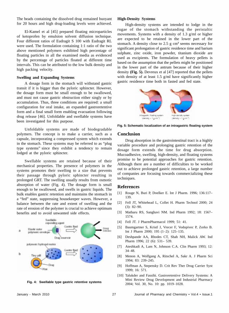
January - March 2010 27 Journal of Pharmacy and Chemistry • Vol.4 • Issue.1
The beads containing the dissolved drug remained buoyantfor 20 hours and high drug-loading levels were achieved.
El-Kamel et al [45] prepared floating microparticlesof ketoprofen by emulsion solvent diffusion technique.Four different ratios of Eudragit S 100 with Eudragit RLwere used. The formulation containing 1:1 ratio of the twoabove mentioned polymers exhibited high percentage offloating particles in all the examined media as evidencedby the percentage of particles floated at different timeintervals. This can be attributed to the low bulk density andhigh packing velocity.
Swelling and Expanding Systems
A dosage form in the stomach will withstand gastrictransit if it is bigger than the pyloric sphincter. However,the dosage form must be small enough to be swallowed,and must not cause gastric obstruction either singly or byaccumulation. Thus, three conditions are required: a smallconfiguration for oral intake, an expanded gastroretentiveform and a final small form enabling evacuation followingdrug release [46]. Unfoldable and swellable systems havebeen investigated for this purpose.
Unfoldable systems are made of biodegradablepolymers. The concept is to make a carrier, such as acapsule, incorporating a compressed system which extendsin the stomach. These systems may be referred to as “plugtype systems” since they exhibit a tendency to remainlodged at the pyloric sphincter.
Swellable systems are retained because of theirmechanical properties. The presence of polymers in thesystems promotes their swelling to a size that preventstheir passage through pyloric sphincter resulting inprolonged GRT. The swelling usually results from osmoticabsorption of water (Fig. 4). The dosage form is smallenough to be swallowed, and swells in gastric liquids. Thebulk enables gastric retention and maintains the stomach ina “fed” state, suppressing housekeeper waves. However, abalance between the rate and extent of swelling and therate of erosion of the polymer is crucial to achieve optimumbenefits and to avoid unwanted side effects.
Fig. 4: Swellable type gastric retentive systems
High-Density Systems
High-density systems are intended to lodge in therugae of the stomach withstanding the peristalticmovements. Systems with a density of 1.3 g/ml or higherare expected to be retained in the lower part of thestomach. A density close to 2.5 g cm3 seems necessary forsignificant prolongation of gastric residence time and bariumsulphate, zinc oxide, iron powder, titanium dioxide areused as excipients. The formulation of heavy pellets isbased on the assumption that the pellets might be positionedin the lower part of the antrum because of their higherdensity (Fig. 5). Devreux et al [47] reported that the pelletswith density of at least 1.5 g/ml have significantly highergastric residence time both in fasted and fed state.
Fig. 5: Schematic localization of an intragastric floating system
ConclusionDrug absorption in the gastrointestinal tract is a highly
variable procedure and prolonging gastric retention of thedosage form extends the time for drug absorption.Mucoadhesive, swelling, high-density, and floating systemspromise to be potential approaches for gastric retention.Although there are a number of difficulties to be workedout to achieve prolonged gastric retention, a large numberof companies are focusing towards commercializing thesetechniques.
References[1] Rouge N, Buri P, Doelker E. Int J Pharm. 1996; 136:117–
139.
[2] Fell JT, Whitehead L, Collet H. Pharm Technol 2000; 24(3): 82–90.
[3] Matharu RS, Sanghavi NM. Ind Pharm 1992; 18: 1567–1574.
[4] Fell JT. J PharmPharmacol 1999; 51: 41.
[5] Baumgartner S, Kristl J, Vrecer F, Vodopivec P, Zorko B.Int J Pharm 2000; 195 (1–2): 125–135.
[6] Deshpande AA, Rhodes CT, Shah NH, Malick AW. IndPharm 1996; 22 (6): 531– 539.
[7] Ateshkadi A, Lam N, Johnson C.A. Clin Pharm 1993; 12:34–48.
[8] Menon A, Wolfgang A, Ritschel A, Sakr A. J Pharm Sci1994; 83: 239–245.
[9] Hoffman A, Stepensky D. Crit Rev Ther Drug Carrier Syst1999; 16: 571.
[10] Talukder and Fassihi. Gastroretentive Delivery Systems: AMini Review Drug Development and Industrial Pharmacy2004; Vol. 30, No. 10: pp. 1019–1028.
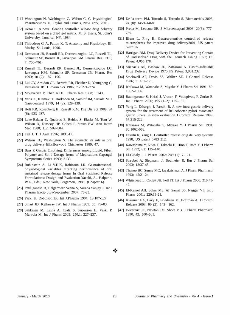
January - March 2010 28 Journal of Pharmacy and Chemistry • Vol.4 • Issue.1
[11] Washington N, Washington C, Wilson C. G. PhysiologicalPharmaceutics. II, Taylor and Francis, New York, 2001.
[12] Desai S. A novel floating controlled release drug deliverysystem based on a dried gel matrix, M. S. thesis, St. John’sUniversity, Jamaica, NY, 1984.
[13] Thibodeau G. A, Patton K. T. Anatomy and Physiology. III,Mosby, St. Louis, 1996.
[14] Dressman JB, Berardi RR, Dermentzoglou LC, Russell TL,Schmaltz SP, Barnett JL, Jarvenpaa KM. Pharm. Res. 1990;7: 756–761.
[15] Russell TL, Berardi RR, Barnett JL, Dermentzoglou LC,Jarvenpaa KM, Schmaltz SP, Dressman JB. Pharm. Res1993; 10 (2): 187– 196.
[16] Lui CY, Amidon GL, Berardi RR, Fleisher D, Youngberg C,Dressman JB. J Pharm Sci 1986; 75: 271–274.
[17] Mojaverian P, Chan KKH. Pharm Res 1988; 5:243.
[18] Varis K, IHamaki T, Harkonen M, Samlof IM, Siruala M. JGastroenterol 1979; 14 (2): 129–139.
[19] Holt P.R, Rosenberg H, Russell R.M. Dig Dis Sci 1989; 34(6): 933–937.
[20] Lake-Bakaar G, Quadros E, Beidas S, Elaskr M, Tom W,Wilson D, Dinscoy HP, Cohen P, Straus EW. Ann InternMed 1988; 112: 502–504.
[21] Fell J. T. J Anat 1996; 189:517.
[22] Wilson CG, Washington N. The stomach: its role in oraldrug delivery EllisHorwood Chichester 1989; 47.
[23] Bass P. Gastric Emptying: Differences among Liquid, Fiber,Polymer and Solid Dosage forms of Medications CapsugelSymposium Series 1993; 2133.
[24] Rubinstein A, Li V.H.K, Robinson J.R. Gastrointestinal-physiological variables affecting performance of oralsustained release dosage forms In Oral Sustained ReleaseFormulations: Design and Evaluation Yacobi, A., Halperin,W.E., Eds.; New York, Pergamon, 1988; (Chapter 6).
[25] Patil ganesh B, Belgamwar Veena S, Surana Sanjay J. Int JPharma Excip July-September 2007; 76-83.
[26] Park. K. Robinson JR. Int J.Pharma 1984; 19:107-127.
[27] Smart JD, Kellaway IW. Int J Pharm 1989; 53: 79–83.
[28] Sakkinen M, Linna A, Ojala S, Jurjenson H, Veski P,Marvola M. Int J Pharm 2003; 250,1: 227–237.
[29] De la torro PM, Torrado S, Torrado S. Biomaterials 2003;24 (8): 1459–1468.
[30] Preda M, Leucuta SE. J Microencapsul 2003; 20(6): 777–789.
[31] Illum L, Ping H. Gastroretentive controlled releasemicrospheres for improved drug delivery2001; US patent6207197.
[32] Harrigan RM. Drug Delivery Device for Preventing Contactof Undissolved Drug with the Stomach Lining 1977; USPatent 4,055,178.
[33] Michaels AS, Bashaw JD, Zaffaroni A. Gastro-InflatableDrug Delivery Device 1975;US Patent 3,901,232.
[34] Stockwell AF, Davis SS, Walker SE. J Control Release1986; 3: 167–175.
[35] Ichikawa M, Watanabe S, Miyake Y. J Pharm Sci 1991; 80:1062–1066.
[36] Baumgartner S, Kristl J, Vrecer, F, Vodopivec, P, Zorko B.Int J Pharm 2000; 195 (1–2): 125–135.
[37] Yang L, Esharghi J, Fassihi R. A new intra gastric deliverysystem for the treatment of helicobacter pylori associatedgastric ulcers: in vitro evaluation J Control. Release 1999;57:215-222.
[38] Ichikawa M, Watanabe S, Miyake Y. J Pharm Sci 1991;80:1062-066.
[39] Fassihi R, Yang L. Controlled release drug delivery systems1998; US patent 5783 212.
[40] Kawashima Y, Niwa T, Takechi H, Hino T, Itoth Y. J PharmSci 1992; 81: 135–140.
[41] El-Gibaly I. J Pharm 2002; 249 (1): 7– 21.
[42] Streubel A, Siepmann J, Bodmeier R. Eur J Pharm Sci2003; 18:37-45.
[43] Thanoo BC, Sunny MC, Jayakrishnan A. J Pharm Pharmacol1993; 45:21-24.
[44] Whitehead L, Collett JH, Fell JT. Int J Pharm 2000; 210:45-49.
[45] El-Kamel AH, Sokar MS, Al Gamal SS, Naggar VF. Int JPharm 2001; 220:13-21.
[46] Klausner EA, Lavy E, Friedman M, Hoffman A. J ControlRelease 2003; 90 (2): 143– 162.
[47] Devereux JE, Newton JM, Short MB. J Pharm Pharmacol1990; 42: 500–501.
�
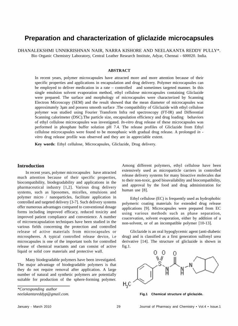
January - March 2010 29 Journal of Pharmacy and Chemistry • Vol.4 • Issue.1
*Corresponding [email protected].
IntroductionIn recent years, polymer microcapsules have attracted
much attention because of their specific properties,biocompatibility, biodegradability and applications in thepharmaceutical industry [1,2]. Various drug deliverysystems, such as liposomes, micelles, emulsions andpolymer micro / nanoparticles, facilitate application incontrolled and targeted delivery [3-7]. Such delivery systemsoffer numerous advantages compared to conventional dosageforms including improved efficacy, reduced toxicity andimproved patient compliance and convenience. A numberof microencapsulation techniques have been studied in thevarious fields concerning the protection and controlledrelease of active materials from microcapsules ormicrospheres. A typical controlled release device, i.emicrocapsules is one of the important tools for controlledrelease of chemical reactants and can consist of activeliquid or solid core materials and protective wall.
Many biodegradable polymers have been investigated.The major advantage of biodegradable polymers is thatthey do not require removal after application. A largenumber of natural and synthetic polymers are potentiallysuitable for production of the sphere-forming polymer.
Among different polymers, ethyl cellulose have beenextensively used as microparticle carriers in controlledrelease delivery systems for many bioactive molecules dueto their non-toxic, good bioavailability and biocompatibility,and approval by the food and drug administration forhuman use [8].
Ethyl cellulose (EC) is frequently used as hydrophobicpolymeric coating materials for extended drug releaseapplications [9]. Microcapsules were prepared from ECusing various methods such as phase separation,coacervation, solvent evaporation, either by addition of anon-solvent, or of an incompatible polymer [10-13].
Gliclazide is an oral hypoglycemic agent (anti-diabeticdrug) and is classified as a first generation sulfonyl ureaderivative [14]. The structure of gliclazide is shown infig.1.
Fig.1 Chemical structure of gliclazide.
Preparation and characterization of gliclazide microcapsules
DHANALEKSHMI UNNIKRISHNAN NAIR, NARRA KISHORE AND NEELAKANTA REDDY PULLY*.Bio Organic Chemistry Laboratory, Central Leather Research Institute, Adyar, Chennai - 600020. India.
ABSTRACT
In recent years, polymer microcapsules have attracted more and more attention because of theirspecific properties and applications in encapsulation and drug delivery. Polymer microcapsules canbe employed to deliver medication in a rate – controlled and sometimes targeted manner. In thissingle emulsion solvent evaporation method, ethyl cellulose microcapsules containing Gliclazidewere prepared. The surface and morphology of microcapsules were characterized by ScanningElectron Microscopy (SEM) and the result showed that the mean diameter of microcapsules wasapproximately 3µm and possess smooth surface .The compatibility of Gliclazide with ethyl cellulosepolymer was studied using Fourier Transform Infra red spectroscopy (FT-IR) and DifferentialScanning calorimeter (DSC).The particle size, encapsulation efficiency and drug loading behaviorsof ethyl cellulose microcapsules was investigated. In-vitro drug release of these microcapsules wasperformed in phosphate buffer solution pH 7.4. The release profiles of Gliclazide from Ethylcellulose microcapsules were found to be monophasic with gradual drug release. A prolonged in -vitro drug release profile was observed and they are in appreciable extent.
Key words: Ethyl cellulose, Microcapsules, Gliclazide, Drug delivery.
January - March 2010 30 Journal of Pharmacy and Chemistry • Vol.4 • Issue.1
In general, rapid (GI)absorption is required for oralhypoglycemic drugs,in order to prevent a sudden increasein blood glucose level after food intake in patients withdiabetes mellitus.Hence there is always a need for thedevelopment of sustained release patient compliantformulation of gliclazide,as it is short acting sulfonylurea[15].The use of extended release products offerspotential advantages like sustained blood levels, attenuationof adverse effects and improved patient compliance. Henceto develop gliclazide formulations in controlled releaseform is important and useful.
In this research, the drug-loaded microspheres wereprepared by new technique offered a short time processand consuming less quantity of solvent for their preparationand easy to proceed. In-vitro drug release profile wasperformed in a phosphate buffer solution of pH 7.4. Thisformulation of controlled delivery of gliclazide has notbeen reported previously.
Materials and methods Ethyl cellulose (EC) polymer was purchased fromAldrich (Germany). Acetone, used as solvent was purchasedfrom SISCO research laboratories pvt.ltd (India). LiquidParaffin a lubricant was obtained from sd fine-chem ltd(Mumbai). Gliclazide was purchased from Sigma(Germany).The other reagents and solvents were analyticalgrade and were used as received
Preparation of microcapsules
The single emulsion solvent evaporation techniquewas applied to the fabrication of hydrophobic drug loadedmicrocapsules. Ethyl cellulose was dissolved in 10 mlacetone and specified amount of gliclazide was added andthis mixture was sonicated for 5 minutes using Ultrasonicator .The above solution was added into 100ml ofliquid paraffin and allowed to homogenize at 1000 rpm for30 minutes using Ultra turrax homogenizer. Then theobtained microcapsules were separated by vacuum filtration.The microcapsules were washed with petroleum ether (60-80) and dried at room temperature for 4 hrs.
Characterization of MicrocapsulesMorphology of microcapsules
The morphology of the microcapsules were observedby a Scanning Electron Microscope (Philips XL 30ESEM).An appropriate sample of microcapsules wasmounted on metal stubs ,using double – sided adhesivetape. Samples were platinum coated and observed formorphology, at acceleration of 20 KV.
Particle size analysis of microcapsules
The particle size and distribution of the microcapsuleswere measured with a Photon correlation spectroscopy(PCS: size mode V161/NBIO2 SZ2).Microcapsules werefirst suspended in distilled water and subjected to sonicationand vortexing before analysis.
Encapsulation efficiency and drug loading
Microcapsules(100 mg) were crushed in a glass mortarand pestle , and the powdered microcapsules weresuspended in 20 ml of phosphate buffer (pH 7.4).After 24hours ,the solution was filtered and the filtrate was analyzedfor drug content using UV visible spectrophotometer(PerkinElmer) at 227nm .The drug loading and encapsulationefficiency was calculated using the equation Eqs.(1) and(2).
Drug Loading =
Amount of drug in microcapsules–––––––––––––––––––––––––––––– x 100 --- (1)
Microcapsules sample weight
Drug encapsulation efficiency =
Amount of drug in microcapsule ––––––––––––––––––––––––––––– x 100 --- (2) Microcapsules sample weight
FT-IR analysis
Infrared spectra for the drug, ethyl cellulose andprepared microcapsules were obtained after prepartion ofKBr pellet, using a Perkin Elmer FTIR spectrophotometercombined to PC with Spectrum 2000 analysis software.
Differential scanning Calorimetry
The thermo gram of gliclazide, ethyl cellulose andgliclazide loaded ethyl cellulose microcapsules wereobtained using Differential Scanning Calorimetry (DSC)using DSC Q 200 V 23 .10 Build 79 apparatus .Theoperating temperature used was between 30 °C and300°C.The temperature gradient was 10 °C per minute.
In- vitro release studies
The In-vitro release studies were performed by takinggliclazide loaded ethyl cellulose microcapsules (equivalentto 10mg of gliclazide ) were suspended in 50ml of phosphatebuffer pH 7.4 in a mechanical shaking bath (100 cycles /min),with temperature adjusted to37o C. At selected timeintervals, 2ml of the sample was removed and replacedwith same amount of fresh buffer. The sample was thenfiltered through 0.2 µ membrane and analysed by UV-spectrophotometry at λ
max of 227nm.
Results and DiscussionSize and morphological characterization ofmicrocapsules
Fig 2. Shows the morphology of gliclazide(1 part)loaded ethyl cellulose (4 parts)microcapsules. Themicrocapsules obtained under these conditions werespherical, smooth, free flowing and individuallyhomogeneously distributed without evidence of collapsedspheres and diameter was about 2µm. Smooth surfacereveals complete removal of acetone and liquid paraffinfrom microcapsules. The particle size and distribution of

January - March 2010 31 Journal of Pharmacy and Chemistry • Vol.4 • Issue.1
the microcapsules were measured with a laser diffractionparticle size analyzer and found that all microcapsuleformulations have particle size smaller than 24µm diameterwith narrow size distribution, and the mean particle size ofmicrocapsules ranged from 1 to 3 µm.
Fig. 2: Scanning electron micrographs of Gliclazide loadedmicrocapsules
Fourier Transform Infra Red spectroscopy analysis
Fourier transform infra red spectroscopy was used toanalyze the interaction between gliclazide and ethyl
cellulose (Fig No.3) .The spectra of gliclazide showedpeaks of – NH stretching (3272 cm-1),= CH stretching(2945 cm-1),O=C (1708 cm-1),C=C aromatic (1595 cm-
1),C-H deformation (1435 cm-1),SO 2 NH (1348 cm-1).Similar
peaks were seen in gliclazide – loaded ethyl cellulosemicrocapsules .This observation further confirmed by DSCstudies.
Differential scanning Calorimetry
DSC observation confirmed that there was nointeraction between gliclazide and ethyl cellulose polymer.Gliclazide showed endotherm at 100 °C (Fig No.4) ethylcellulose showed exotherm at 225°C (Fig No.5) andgliclazide loaded ethyl cellulose microcapsules showedboth exotherm and endotherm which was identical to theplain drug and ethyl cellulose polymer thermogram (FigNo.6).Hence the compatability of gliclazide in ethylcellulose microcapsules was evaluated. The DSC studyapparently revealed that there was no drug decompositionnor drug- polymer interactions occurred in the freshlyprepared microcapsules.
Effect of processing parameters on ethyl cellulosemicrocapsules preparation
The effect of polymer: drug ratio on encapsulationefficiency and drug loading is shown in Table.1.Gliclazideand ethyl cellulose microcapsules in three different ratioswere prepared. From these 4:1 ratio was selected as bestratio because of high drug loading and encapsulation
Fig. 3: FT IR Spectra of pure drug localized (GL), polymer Ethyl cellulose (EC) and microcapsule formulation (EC+GL mixture)

January - March 2010 32 Journal of Pharmacy and Chemistry • Vol.4 • Issue.1
of polymer with respect to the amount of drug [16]. Basedon above concept 4:1 ratio show high entrapment efficiencyand drug loading. The mean particle diameter of 4:1 ratiowas approximately 2.23µm.
In -vitro release of gliclazide
In vitro release behavior of gliclazide from the ECmicrocapsules were studied in Phosphate Buffer Saline pH7.4. The polymer: drug ratios on release profiles from threebatches were investigated and shown in Figure 7. Thegliclazide release profiles from the microcapsules werefound to occur in a monophasic manner, with gradualrelease of gliclazide from microcapsules. Gliclazidemicrocapsules in the ratio of 4:1 released 88 % of theloaded gliclazide in 7 days. The increase in drug contentinfluences the absolute release profiles such that both thecumulative amount of drug released at any time and theinduction period increases. This suggests that the level ofdrug loading was a main factor that controlled the rate ofdrug release, the microcapsules with a higher loadingreleased gliclazide faster [17-,19].The increase in drugcontent increased the amount of drug close to the surfacewhich is responsible for an increased initial burst [19-21].This initial burst helps to attain initial therapeuticplasma concentration of drug. Here we found that in thethree different formulations (F1, F2, and F3) the releaserates vary depending upon the amount of the drug loadedin gliclazide microcapsules (Fig 7), hence these resultssuggested that drug loading amount should be taken into
Fig. 4: Thermogram of Gliclazide core material
Fig. 5: Thermogram of Ethyl Cellulose polymer
Fig. 6: Thermogram of gliclazide loaded ethyl cellulosemicrocapsules
Table.1
Properties of the ethyl cellulose-loaded microcapsule prepared by Single emulsion solventdiffusion method (±S.D.,n =3)
Formulation Polymer Ratio Drug Loading Encapsulation Mean Diameter,Codes and Drug (%) Efficienc (%) (µµµµµm)
F1 2:01 13.78(±0.68) 59.87(±1.47) 2.39(±0.12)
F2 3:01 11.98(±0.60) 64.92(±1.69) 2.59(±0.11)
F3 4:01 18.55(±0.54) 71.93(±2.55) 2.23(±0.08)
efficiency when compared to other formulation. The drugloading and encapsulation efficiency were mainly affectedby the polymer drug ratios. Encapsulation efficiencyincreases with increase polymer drug ratio. This improvedencapsulation efficiency may be due to greater proportion

January - March 2010 33 Journal of Pharmacy and Chemistry • Vol.4 • Issue.1
[7] Simoes S, Moreira JN, Fonseca C, Duzgunes N. Adv DrugDeliver Rev 2004; 56: 947–965.
[8] N Wakiyama, K Juni, M.Nakano. Chem Pharm Bull 1982;30: 3719–3727.
[9] Pearnchob N & Bodmeier. Int J Pharm 2003; 268: 1–11.
[10] Babtsov V, Shapiro Y & Kvitnitsky, E. US Patent Office,Pat. 2005 No. 6 932 984.
[11] Shen R & Walter G. US Patent Office, Pat. 2005. No. 6939 560.
[12] Zandi M, Pourjavadi A, Hashemi SA & Arabi, H.PolymerInternational 1998 ;47: 413–418.
[13] Zhang Z-Y, Ping Q-N & Xiao B. J Controll Rel 2000; 66:107–113.
[14] Goodmanan & Gillmans,The Pharmacological Basis ofTherapeutics.11edition,Mc Graw Will Publishing 2005 ,1021
[15] Padma V Devarajan and Ganesh Chandra S.Sonavane. Drugdevelopment and Industrial Pharmacy 2007; 33:101-111.
[16] Dongming Peng, Kelong,Yenfei liu,Suquin Liu. Int J Pharm2006;11:82-83.
[17] Sato T, Kanke M,Schroedar G, Deluca. Pharm Res 1998;5:21-30
[18] Preeti V. Kulkarni, Jathi Keshavayya and VenkatatraoH.Kulkarni. Poly adv technol 2007;18:814-821.
[19] Avinash Budhian, Steven J.Siegel, Karen I.Winey. Int JPharm 2007; 87-92.
[20] T. Brandau. International. J Pharm.2002; 242: 179-84.
[21] Uhrich K.E., et.al, 1999; 99:3181-3198.
[22] Zengqian Shi,Yongfeng Zhou,Deyue Yangfeng Zhou,DeyueYan. Polymer 2006; 8073-8074.
Fig. 7: Release of gliclazide from microcapsules prepared using different ratios of EC: gliclazide in pH 7.4 PBS, 2:1 for F1,3:1for F2 and 4:1 for F3 (±S.D ., n=3).
consideration when trying to achieve a desired releaseprofile [22]
ConclusionThe gliclazide loaded EC microcapsules were prepared
and evaluated. The microcapsules had a spherical, smoothmorphology and a diameter of approximately 2µm. Morethan 70% encapsulation efficiency and a controlled releaserate were obtained by varying the process parameters. Thegliclazide loaded EC microcapsules had a long releaseperiod of about 7 days. The present results suggest that theobtained microcapsules could be useful as a prolongeddrug delivery system for diabetic treatment. Accordingly,the next step of this work will be to study the therapeuticeffect of these microcapsules in vivo.
References[1] Yu Xiang, Tang Xuezhi, Pan Caiyuan. Polymer 2005; 46:
11149-11156.
[2] He Xiaodong, Ge Xuewu W, Wang Mozhen, ZhangZhicheng. Polymer 2005; 46: 7598-604.
[3] Kataoka K, Harada A, Nagasaki Y. Advanced Drug Delivery2001; 47: 113–131.
[4] Soppimath KS, Aminabhavi TM., Kulkarni AR, RudzinskiWE. J Controlled Release 2001; 70: 1–20.
[5] Agnihotri SA, Mallikarjuna NN, Aminabhavi TM. JControlled Release 2004; 100:5–28.
[6] Constantinidesa PP, Tustianb A, Kessler DR. Adv DrugDeliver Rev 2004; 56:1243– 1255
�
January - March 2010 34 Journal of Pharmacy and Chemistry • Vol.4 • Issue.1
*Address for correspondence:[email protected]
Anti-inflammatory and antipyretic activities ofSmilax perfoliatSmilax perfoliatSmilax perfoliatSmilax perfoliatSmilax perfoliata.a.a.a.a. Lour.
*VENKATESHWAR REDDY A*, RAMA RAO T1, NAIDU A2, SUDHAKAR Y3,HABIBUDDIN M4 AND YOGANANDA REDDY K5
*Anwarul-Uloom College of Pharmacy, New Mallepally, Hyderabad - 500 001, Andhra Pradesh, India.1 Mohammadia Collge of Pharmacy, Warangal Road, Khammam, Andhra Pradesh, India.
2 Professor of Chemistry, College of Engineering, JNTU, Hyderabad, Andhra Pradesh, India.
3 Govt. Polytechnic, Kadapa, Andhra Pradesh, India.
4Alliance Institute of Advanced Pharmaceutical Sciences, Aditya Trade Center,
Ameerpet, Hyderabad, Andhra Pradesh, India.5International Science-Tech Research Institute, Anantapur 515001, Andhra Pradesh, India.
ABSTRACT
The ethanolic extracts of Smilax perfoliata. Lour (EESP). (200, 400 and 600 mg/kg orally) weretested in carrageenan-induced rat paw oedema and brewer’s yeast-induced pyrexia in rats to assesstheir anti-inflammatory and antipyretic activities respectively. The paw volumes and pyrexia in ratswere reduced significantly (P < 0.05) compared to that of control. These results indicate that theextract possess anti-inflammatory and antipyretic properties.
Key words: Smilax perfoliata, Anti-inflammatory and Antipyretic activity, EESP.
IntroductionSmilax perfoliata (Smilacaceae) is used in folklore
medicine for treating various disorders like venereal diseases,urinary diseases, leprosy, hemiplegia, psychosis,parkinsonism, muscle wasting and rheumatism [1]. Theliterature survey showed that there is no specific evaluationof this plant for its role in the above mentioned disorders. Inthe current research, an effort was made to study theantipyretic and anti-inflammatory potential of ethanolicextract of Smilax perfoliata by following standardpharmacological screening methods.
Materials and MethodsPlant material
The shrubs of Smilax perfoliata Lour (Smilacaceae)were collected from the hill regions of Tirupathi, ChittorDistrict of Andhra Pradesh, India. The plant was authenticatedand certified by taxonomist Dr.K.Madhava Chetty, Asst.Prof.,Department of Botany, S.V. University and Tirupathi. A.P.The entire plant was collected in the month of August,washed with water several times, shade dried, pulverized bya mechanical grinder and subjected to extraction process.
Preparation of extract
5000 grams of the powdered drug was accurately weighedand extracted using analytical grade solvents starting withHighly non polar petroleum ether (60 – 80°C) to successivelyincreasing the polarity viz. chloroform, acetone, ethyl acetateand ethanol (95%) following soxhlation method. All theextracts were concentrated by using vacuum dryer until asemisolid extract is obtained, dried at less than 50oC andkept in desiccators for further studies.
Preliminary phytochemical investigation
A preliminary phytochemical investigation [2] wascarried out for all the extracts obtained from the Smilaxperfoliata using analytical grade chemicals, solvents andreagents. The respective yields and the preliminaryphytochemical investigation results were given in Table 1.
Animals
Albino rats (Wistar) weighing 150-200g and albinomice weighing 20-25g of either sex were used in this study.They were procured from Sri Venkateshwara Enterprises,Bangalore. The animals were acclimatized for one weekunder laboratory conditions. They were housed inpolypropylene cages and maintained at 27°C ± 2°C under 12hrs dark / light cycle. They were fed with standard rat feed(Gold Mohr Lipton India Ltd.) and water ad libitum wasprovided. The litter in the cages was renewed thrice a week
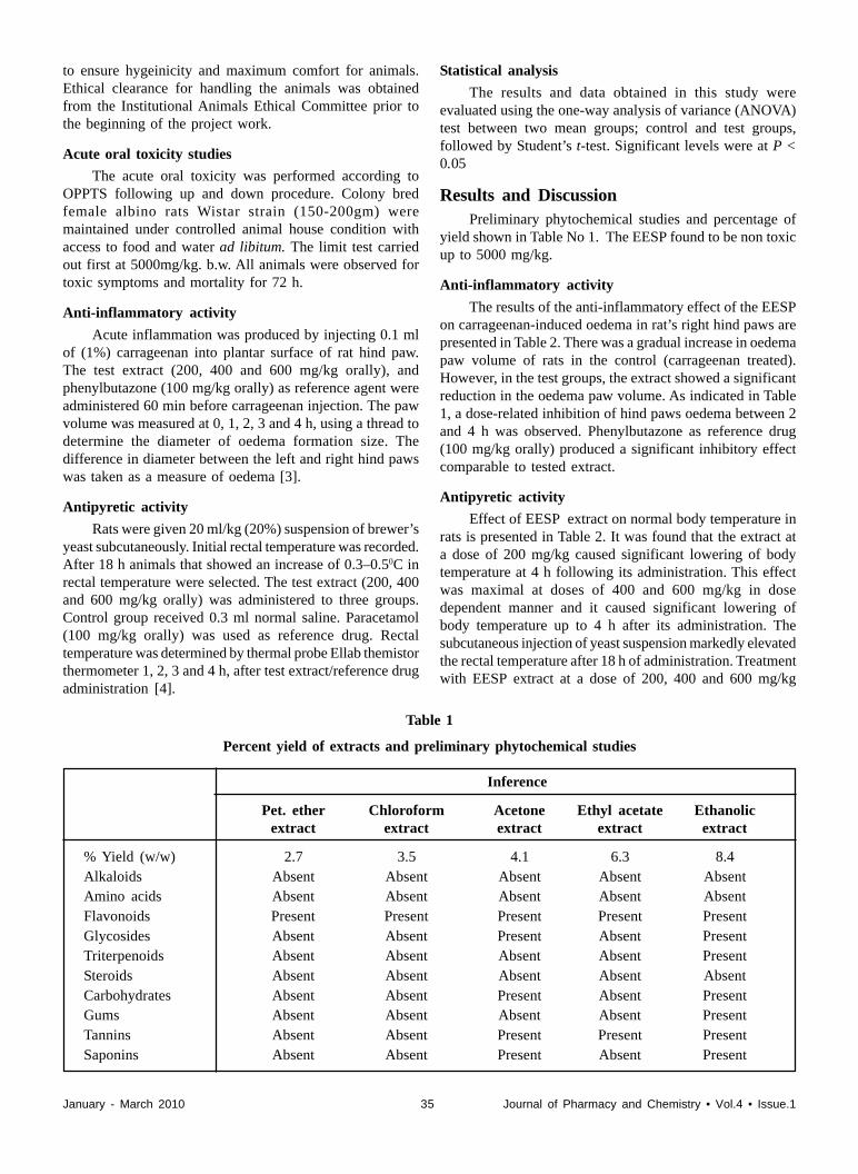
January - March 2010 35 Journal of Pharmacy and Chemistry • Vol.4 • Issue.1
to ensure hygeinicity and maximum comfort for animals.Ethical clearance for handling the animals was obtainedfrom the Institutional Animals Ethical Committee prior tothe beginning of the project work.
Acute oral toxicity studies
The acute oral toxicity was performed according toOPPTS following up and down procedure. Colony bredfemale albino rats Wistar strain (150-200gm) weremaintained under controlled animal house condition withaccess to food and water ad libitum. The limit test carriedout first at 5000mg/kg. b.w. All animals were observed fortoxic symptoms and mortality for 72 h.
Anti-inflammatory activity
Acute inflammation was produced by injecting 0.1 mlof (1%) carrageenan into plantar surface of rat hind paw.The test extract (200, 400 and 600 mg/kg orally), andphenylbutazone (100 mg/kg orally) as reference agent wereadministered 60 min before carrageenan injection. The pawvolume was measured at 0, 1, 2, 3 and 4 h, using a thread todetermine the diameter of oedema formation size. Thedifference in diameter between the left and right hind pawswas taken as a measure of oedema [3].
Antipyretic activity
Rats were given 20 ml/kg (20%) suspension of brewer’syeast subcutaneously. Initial rectal temperature was recorded.After 18 h animals that showed an increase of 0.3–0.50C inrectal temperature were selected. The test extract (200, 400and 600 mg/kg orally) was administered to three groups.Control group received 0.3 ml normal saline. Paracetamol(100 mg/kg orally) was used as reference drug. Rectaltemperature was determined by thermal probe Ellab themistorthermometer 1, 2, 3 and 4 h, after test extract/reference drugadministration [4].
Statistical analysis
The results and data obtained in this study wereevaluated using the one-way analysis of variance (ANOVA)test between two mean groups; control and test groups,followed by Student’s t-test. Significant levels were at P <0.05
Results and DiscussionPreliminary phytochemical studies and percentage of
yield shown in Table No 1. The EESP found to be non toxicup to 5000 mg/kg.
Anti-inflammatory activity
The results of the anti-inflammatory effect of the EESPon carrageenan-induced oedema in rat’s right hind paws arepresented in Table 2. There was a gradual increase in oedemapaw volume of rats in the control (carrageenan treated).However, in the test groups, the extract showed a significantreduction in the oedema paw volume. As indicated in Table1, a dose-related inhibition of hind paws oedema between 2and 4 h was observed. Phenylbutazone as reference drug(100 mg/kg orally) produced a significant inhibitory effectcomparable to tested extract.
Antipyretic activity
Effect of EESP extract on normal body temperature inrats is presented in Table 2. It was found that the extract ata dose of 200 mg/kg caused significant lowering of bodytemperature at 4 h following its administration. This effectwas maximal at doses of 400 and 600 mg/kg in dosedependent manner and it caused significant lowering ofbody temperature up to 4 h after its administration. Thesubcutaneous injection of yeast suspension markedly elevatedthe rectal temperature after 18 h of administration. Treatmentwith EESP extract at a dose of 200, 400 and 600 mg/kg
Table 1
Percent yield of extracts and preliminary phytochemical studies
Inference
Pet. ether Chloroform Acetone Ethyl acetate Ethanolicextract extract extract extract extract
% Yield (w/w) 2.7 3.5 4.1 6.3 8.4Alkaloids Absent Absent Absent Absent AbsentAmino acids Absent Absent Absent Absent AbsentFlavonoids Present Present Present Present PresentGlycosides Absent Absent Present Absent PresentTriterpenoids Absent Absent Absent Absent PresentSteroids Absent Absent Absent Absent AbsentCarbohydrates Absent Absent Present Absent PresentGums Absent Absent Absent Absent PresentTannins Absent Absent Present Present PresentSaponins Absent Absent Present Absent Present

January - March 2010 36 Journal of Pharmacy and Chemistry • Vol.4 • Issue.1
decreased the rectal temperature of the rats in dose dependentmanner (Table 3).
The antipyretic effect started as early as 1 h and theeffect was maintained for 4 h, after its administration. Boththe standard drug paracetamol 100 mg/kg and tested EESPextract significantly reduced the yeast-elevated rectaltemperature compared to control group.
ConclusionBased on the results of the present study it can be
concluded that the ethanolic extract of Smilax perfoliat has
potential dose-dependent anti-inflammatory and antipyreticactivity, it may due to active principles present in this extract.The activity is in dose dependent manner.
References1] K Madhava Chetty, K Tulasi Rao. Flowering plants of
Chittoor District AP., India 2008, Students Offset Printers,Tirupathi, pp:359
[2] Kokate CK. Practical Pharmacognosy ,2nd edn., VallabhPrakashan, Delhi, 125 (1988).
[3] Winter CA, Risley EA, Nuss GW. Proc Soc Exp Biol Med1962;111:544-547.
[4] Smith PK and Hambourger WE. J Pharmacol Exp Ther1935;54:346-349.
Table 2
Effect of EESP on normal body temperature
Treatment Dose(mg/kg) Rectal temperature (Cº ) before and after treatment
0 1 2 3 4
Control (saline) 5ml/kg 37.2±0.2 37.3±0.1 37.2±0.1 37.3±0.1 37.1±0.1 EESP 200 37.1±0.1 36.6±0.1* 36.6±0.1* 36.6±0.2* 36.4±0.1*
400 37.2±0.3 36.4±0.1* 36.4±0.1* 36.2±0.1* 36.2±0.1*600 37.4±0.1 36.9±0.1* 36.2±0.1* 36.2±0.1* 36.0±0.1*
Values are Mean ± S.E.M. (n = 6), *P < 0.05 compared with control valuesEESP : Ethanolic extract of Smilax perfoliata.
Table 1Anti-inflammatory activity of EESP and phenylbutazone (100 mg/kg) on carrageenan-
induced rat paw oedema in the right hind-limb paw of rats
Treatment Dose Time (h) Average
(mg/kg) 0 1 2 3 4 oedema formation
Control (CT) - 0.47±0.09 0.71±0.01 0.88±0.31 0.72±0.14 0.69
EESP 200 - 0.38±0.22 0.34±0.11* 0.28±0.11* 0.22±0.01* 0.30*
400 - 0.32±0.16 0.31±0.09* 0.24±0.14* 0.20±0.09* 0.26*
600 - 0.25±0.09 0.22±0.12* 0.20±0.13* 0.18±0.08* 0.21*
phenylbutazone 100 - 0.20±0.03 0.18±0.08* 0.18±0.12* 0.17±0.01* 0.18*
Values are mean ± S.E.M. (n = 6), *P < 0.05 of the difference between the left and the right hind pawsEESP : Ethanolic extract of Smilax perfoliata.
Table 3
Antipyretic activity of EESP and paracetamol (100 mg/kg) on Brewer’s yeast-induced pyrexia in rats
Rectal temperature (Cº) before and after treatment
Treatment (Dose Before treatment After treatment
mg/kg) 0h 18h 1h 2h 3h 4h
Control (saline) 5 ml/kg 37.6±0.2 39.2±0.1 39.2±0.1 39.2±0.1 39.2±0.1 39.2±0.1EESP 200 37.5±0.1 39.9±0.1 38.6±0.1* 38.6±0.2* 38.2±0.1* 38.0±0.1*400 37.4±0.3 39.8±0.1 38.5±0.1* 38.4±0.1* 38.0±0.2* 37.6±0.1*600 37.4±0.1 39.9±0.1 38.6±0.2* 38.2±0.1* 37.9±0.1* 37.4±0.1*
Paracetamol 100 37.8±0.2 39.5±0.1 38.3±0.1* 38.3±0.1* 37.9 ±0.1* 37.3±0.1*
Values are mean ± S.E.M. (n = 6), *P < 0.05EESP : Ethanolic extract of Smilax perfoliata.

January - March 2010 37 Journal of Pharmacy and Chemistry • Vol.4 • Issue.1
Science – Tech Foundation
Journal of Pharmacy and Chemistry(An International Research Journal of Pharmaceutical and Chemical Sciences)
(Indexing in CAS)
Plot No. 22, Vidyut Nagar, Anantapur – 515 001(A.P), INDIA
MEMBERSHIP FORMDear Editor,
I/We wish to be Annual Member / Life Member of Journal of Pharmacy and Chemistry (AnInternational Research Journal of Pharmaceutical and Chemical Sciences) and agree to abide by your rules andregulations
1. Name in full: _________________________________________________ Male Female
(In block letters)
2. Nationality: _______________________________ 3. Date of birth:___________________________
3. Corresponding Address: _____________________________________________________________
____________________________________________________________________________________
____________________________________________________________________________________
5. Institutional / Business Address: _____________________________________________________
6. Phone : (O): ______________ (R): _______________ E-mail :______________________________
7. Academic Qualifications : ___________________________________________________________
8. Profession and Experience : __________________________________________________________
9. Present Position / Post : _____________________________________________________________
10. Pharmaceutical / Chemical interest: _____________________________________________________
Membership Fees Life Annual
Individual Rs. 8000/- US $ 800 Rs. 800/- US $ 80
Institutional Rs. 15,000/- US $ 1500 Rs. 1500/- US $ 150
Rs. / $ ________________________________ remitted by Bank / Cash
DD No. ________________ Date _____________ Banker’s Name___________________________
DD’s are drawn in favour of Science-Tech Foundation, Payable at Anantapur.
Place: ____________________
Date: _____________________(Signature of the applicant)
Note: (i) All authors should be member of this Journal, (ii) Saperate Photostat copy may be filled and send.
ISSN 0973–9874

January - March 2010 38 Journal of Pharmacy and Chemistry • Vol.4 • Issue.1
GENERAL REQUIREMENTS: Journal of Pharmacy andChemistry (ISSN 0973-9874) is a quarterly Journal, Indexingin CAS(Coden:JPCOCM) which publishes original researchwork that contributes significantly to further the scientificknowledge in Pharmaceutical Sciences (PharmaceuticalTechnology, Pharmaceutics, Biopharmaceutics,Pharmacokinetics, Pharmaceutical Chemistry, ComputationalChemistry and Molecular Drug Design, Pharmacognosy andPhytochemistry, Pharmacology, Pharmaceutical Analysis,Pharmacy Practice, Clinical and Hospital Pharmacy, CellBiology, Genomics and Proteomics, Pharmacogenomics, StemCell Research, Vaccines & Cera, Bioinformatics andBiotechnology of Pharmaceutical Interest) and in ChemicalSciences (Inorganic, Soil, Forensic, Analytical, Nano,Environmental, Polymer, Physical, Agricultural, Medicinal,Biochemistry, Organic, Computational, Food, Pesticides etc).Manuscripts are accepted for consideration by Journal ofPharmacy and Chemistry on the condition that they representoriginal material, have not been published previously, are notbeing considered for publication elsewhere, and have beenapproved by each author. Review articles, research papers,short communication and letters to the editor may be submittedfor publication.
SUBMISSION OF MANUSCRIPTS: Typewrittenmanuscripts prepared using MS Word should be submitted intriplicate and RW-CD to Prof. Dr. K.N Jayaveera, Editor-in-Chief of Journal of Pharmacy and Chemistry , Plot No 22,Vidyut Nagar, Ananthapur- 515 001, Andhra Pradesh, India.e-mail: [email protected]
All components of the manuscript must appear within a singleelectronic file: references, figure legends and tables mustappear in the body of the manuscript.
TYPING INSTRUCTION: The following detailedinstructions are necessary to allow direct reproduction of themanuscript for rapid publishing. If instructions are not followed,the manuscript will be returned for retyping. The followingtypefaces, in 12 points size, are preferred: Times Roman.
GENERAL FORMAT: The typing area must be exactly 6 5/8” (168 mm) wide by 9 7/8” (250 mm) long. Justify marginsleft and right (block format). The entire typing area of eachpage must be filled, leaving no wasted space. Text should bedouble-spaced, special care should be taken to insure thatsymbols, superscripts and subscripts are legible and do notoverlap onto lines above or below. Make sure text lines areequidistant.
TITLE: On the first page of the manuscript, start title 1” (25mm) down from top text margin. Type title in all capitalletters, centred on the width of the typing area and single-spaced if more than one line is required. The title should bebrief, descriptive and have all words spelled out. Double-space, then type the author(s) name(s), single-spaced if morethan one line is required. Double-space, than type author(s)
INSTRUCTION TO AUTHORSaddress(es), single-spaced, capitalizing first letters of mainwords. Quadruple-space before Abstract.
ABSTRACT: Centre, type and underline abstract heading,capitalizing the first letter. A double-space should separate theheading from the abstract text. Indent abstract textapproximately 1/2” (13 mm) from both left and right margins.The abstract should be intelligible to the reader withoutreference to the body of the paper and be suitable forreproduction by abstracting services. Introduction to the text(without a heading) should being four spaces below theabstract using full margins.
KEY WORDS: Three or more key words must be providedby authors for indexing of their article. Key words will belisted directly below the Abstract. Abbreviated forms ofchemical compounds are not acceptable. Spell out entirely,using the official nomenclature. Example: L-dihydroxyphenylalanine (L-DOPA)
MAJOR HEADINGS: Papers must include the majorheadings: Introduction, Methods, Results, Discussion,Acknowledgments and References. Capitalize first letter,underline, and centre headings on width of typing area.
TABLES/FIGURES: Incorporate tables and/or figures (B &W) with their legends into the main body of text.
REFERENCES: Referneces should be referred to a number[1] in the text and be listed according to this numbering at theend of the paper. Only papers and books that have beenpublished or in press may be cited; unpublished manuscriptsor manuscripts submitted to a journal but which have not beenaccepted may not be cited.
The references shoulld comprise the following informationand in the given order and with given punctuation as given inthe example below: Author name (s), Initials (s), PublicationTitle, Page Number, Year of Publication.
Standard Journal Article:
[1] Bhattacharyya D, Pandit S, Mukherjee R, Das N, SurTK. Indian J Physiol Pharmacol 2003; 47:435.
[2] Skottova N, Krecman V. Physiol Res 1998; 47:1.
Book:
[1] Ghosh MN. Fundamentals of Experimental Pharmacology,2nd ed. Calcutta Scientific Book Agency, 1984:154.
Proofs will be sent to the corresponding author. These shouldbe returned as quickly as possible.
The facts and view in the article will be of the authors andthey will be totally responsible for authenticity, validityand originality etc. the authors should give an undertakingwhile submitting the paper that the manuscripts submittedto journal have not been published and have not beensimultaneously submitted or published elsewhere andmanuscripts are their original work.
Annual Subscription: Rs.800/-.
www.stfindia.com
This Quarterly Journal is Printed, Published, Owned and Edited by Dr. K.N. Jayaveera, Published from Science-Tech Foundation,Plot No.22, Door No. 12/3/925, Vidyut Nagar, Anantapur - 515 001. Phone:+91-8554 274677, Mobile: +91-94414 89324 • www.stfindia.comand Printed at Vipla Computer Services (Designers & Multi Colour Offset Printers) 1-8-725A/1/A, Balaji Bhagyanagar Apts., Nallakunta,Hyderabad - 500 044. Ph. 040-27676910, 27677078, 27654003.