Embed Size (px)
Citation preview

Contents lists available at ScienceDirect
Journal of Photochemistry & Photobiology, B: Biology
journal homepage: www.elsevier.com/locate/jphotobiol
Influence of laser therapy on the dynamic formation of extracellular matrixin standard second degree burns treated with bacterial cellulose membrane
Patricia Keler Freitas Machado Vasconcellosa, Manuela Pimentel Nóiaa,Isabele Cardoso Vieira De Castroa,b, Jean Nunes dos Santosa, Antonio Luiz B. Pinheirob,Aparecida Maria Cordeiro Marquesb, Eduardo Antonio Gonçalves Ramosc,Clarissa Gurgel Rochaa,c,⁎
a Laboratory of Oral Surgical Pathology, School of Dentistry, Federal University of Bahia, Av. Araújo Pinho, 62, Canela -, Salvador, Bahia 40110-150, Brazilb Center of Biophotonics, School of Dentistry, UFBA, Federal University of Bahia, Av. Araújo Pinho, 62, Canela, Salvador, Bahia 40110-150, Brazilc Laboratory of Pathology and Molecular Biology, Oswaldo Cruz Foundation, Fiocruz, Rua Waldemar Falcão, 121, Candeal, Salvador, Bahia 40296-710, Brazil
A R T I C L E I N F O
Keywords:Second-degree burnsLaserBacterial cellulose membraneTissue repair
A B S T R A C T
The present study aims to assess the influence of Aluminum-Gallium-Indium-Phosphide laser (AlGaInP laser,λ=660 nm), whether or not in association with the application of a membrane of bacterial cellulose (Nexfill™),during recovery from induced second-degree burns at the dorsum of Wistar rats. (Rattus norvegicus, Wistar).Forty-eight animals have been distributed into four groups: Control (burns remained untreated), Group I (laser-treated), Group II (treated with Nexfill), and Group III (laser + Nexfill™). In addition to a morphological ana-lysis, immunohistochemical analysis has been performed for type I collagen, type III collagen, fibronectin, andlaminin. The Fisher's Test was used to assess differences among groups (p < 0,05). A larger amount of collagentype III was observed in Control, Group II and Group III when compared with Group I (p < 0,05). Group I andGroup III have shown a greater collagen deposition when compared with Group II (p < 0,05), but the amount ofcollagen was similar in Group I, Group III, and Control. Group III has shown larger fibronectin amounts incomparison with Group II (p < 0,05). As regards laminin, Group I has shown a predominant discontinuitypattern on the basal lamina in comparison with Control, Group II, and Group III (p < 0,05). It is concluded thatin this current study the laser when used alone (Group I) hasn't influenced collagen deposition neither has itacted on fiber pattern (fibril and/or reticular). Moreover, laser application hasn't accelerated the repair ofwounds caused by inflicted second-degree burns.
1. Introduction
Burns are one of the commonest types of trauma facing humanbeings [1] and, regardless of a worldwide decline in the death ratearising from burns, associated non-fatal sequelae frequently tend tolead to permanent impairments [2]. In such injuries, the clinicaltreatment and repair process are dependent on the extent and depth ofthe damage. In many cases, burns can immediately be treated by meansof autologous graft – which promptly leads to a permanent and sa-tisfactory wound closure. However, in many situations this type oftreatment appears to be impossible or unlikely to succeed, for instancein cases of infected or severely extensive wounds [3,4].
Therefore, temporary wound dressings are deemed necessary inorder to maintain the function of the wound, reduce infection, relievethe pain and metabolic stress, in addition to providing blood supply and
protection against trauma [3,5,6]. In light of this fact, research intowound dressings has advanced the production of a wide range of syn-thetic and biological dressings for wound care and management [7].Options readily available include the bacterial cellulose biomembrane,a biosynthetic polymer that provides optimum conditions for epidermalregeneration owing to its nanomorphological characteristics, protectionagainst infection and ability to hold water; in addition, it enables thetransfer of medicines into the wound [8–10].
The influence of laser on wound healing has recently motivated anumber of experimental studies - some of which drawing attention tobiostimulation and healing properties [11–15]. Laser therapy triggerscellular processes and a response from the vascular system which ap-pear to have a direct impact on tissue repair [16]. As a result, researchon the effects of photobiomodulators on burns has become increasinglycommon [1,17–19] and fairly recently scholarly papers have addressed
https://doi.org/10.1016/j.jphotobiol.2018.03.009Received 13 November 2017; Received in revised form 5 March 2018; Accepted 9 March 2018
⁎ Corresponding author at: Laboratory of Oral Surgical Pathology, School of Dentistry, Federal University of Bahia, Av. Araújo Pinho, 62, Canela, Salvador, Bahia 40110-150, Brazil.E-mail address: [email protected] (C.G. Rocha).
Journal of Photochemistry & Photobiology, B: Biology 182 (2018) 1–8
Available online 16 March 20181011-1344/ © 2018 Elsevier B.V. All rights reserved.
T

an associated use of laser and occlusive dressings [20,21]. Nonetheless,substantial evidence is still lacking as regards the bacterial biocellulosemembrane in association with laser use in the healing of second-degreeburns.
Given the above considerations and also taking into account theperceived advantages of bacterial cellulose membrane and laser use, itis arguably valid to undertake research as to assess possible effects of acombined use of both therapies for burn treatment, ensuring faster,more efficient, and less painful healing with better functional andcosmetic results.
2. Materials and Methods
2.1. Sample Selection
A total of forty-eight male, albino, young, healthy rats (Rattus nor-vegicus, Wistar) aged two months and weighing 150 g–200 g were ran-domly selected from the Laboratory Vivarium at Gonçalo MonizResearch Center (FIOCRUZ, Bahia, Brazil). The animals were housedindividually at 22 °C under a light/dark cycle established as 12 h lighton, with ready access to Labina rat feed and water ad libitum. The ratswere distributed into four groups of 12 animals as follows: Control,Group I (laser radiation), Group II (Nexfill™ bacterial cellulose mem-brane), and Group III (laser radiation associated with the application ofNexfill™ membrane).
2.2. Ethical Considerations
In order for this research to be conducted, prior approval was re-ceived from the Committee for Experimentation and Animal Use(CEUA) at Gonçalo Moniz Research Center (CPqGM-FIOCRUZ – BA),under the Protocol 008/2007. Adherence to ethical principles was en-sured for the performance of experimental use of animals as well astheir vivisection for academic-scientific purposes.
2.3. Induction of Standard Second-Degree Burn Injury
The animals were anesthetized with 75mg/kg Ketamine and 10mg/kg Xylazine intraperitoneal injection. Next, an area measuring 2× 2 cmwas depilated at the dorsum of the animal so that antisepsis was per-formed with polyvinylpyrrolidone-iodine solution. Infliction of burnwounds has been performed in accordance with Meyer and Silva'smodified technique [22]. To this end, a brass bar with a cube-shaped tipwas pre-heated in boiling water for 1min and subsequently placed onthe skin of the animal for 0.5 s, and timed with a digital timer. Once thesurgery procedure completed, an oral dose of Sodium Dipyrone wasadministered to each and every rat according to their weight. Theepidermis on the wound area was removed with a scalpel blade no. 15as to expose the subcutaneous tissue. This was a necessary for the ap-plication of the Nexfill™ cellulose membrane on the superficial dermis.
2.4. Laser Radiation Protocol
The animals allocated in Group I and Group III received radiationfrom a red laser AlGaInP (λ=660 nm, 40mW, ø=4mm2, t=125 s)with a SAEF (Spatial Average Energy Fluence) at 20 J/cm2 split intofour points of 5 J/cm2 each [19,23]. Controls were sham-irradiated.Irradiation was performed immediately after the infliction of burns andevery 48 h until one day before the death of the animals, which oc-curred 24 h, 3 days, 7 days, and 14 days later in a CO2 chamber. Asummary of the laser parameters used on the study is depicted onTable 1.
2.5. Morphological and Immunohistochemical Analysis
Following death, the tissue fragments were removed from the
wound area, placed in formalin at 10% and routinely processed forstaining with hematoxylin-eosin, picrosirius for fibrosis and orceinanalysis of elastic fibers. Histological sections were morphologicallydiagnosed in a candid fashion by an experienced pathologist by lightmicroscopy (Zeiss Axioskop) and the treated groups were compared tothe control group (no treatment). The inflammatory infiltrate wasclassified as absent, predominantly polymorphonuclear, predominantlymononuclear, or mixed. Inflammatory infiltration and edema (if pre-sent), the presence of adipocytes in the dermis, and production of col-lagen and elastic fibers were semi-quantitatively marked as absent (0),mild (+), moderate (++) and strong (+++).
The deposition of the extracellular matrix was assessed by im-munohistochemistry using a polymer system (AdvanceTM, DakoCorporation) and antibodies directed against the following proteins:collagen types I and III, fibronectin, and laminin (Table 2).
In order to undertake immunostaining animals dead at 24 h of theexperiment were excluded. Collagen I and III were analyzed in order tocalculate the percentage of wound covering, by taking into considera-tion only the papillary dermis (score 0: 0–10% coverage; score 1:11–30%; score 2: 31–60% and score 3:> 60%) and the fiber pattern(reticular, fibril, or mixed). The fibers were regarded as reticular whenthey appeared to be only partially stained, and fibril when stained in itsentirety.
For fibronectin protein, the analysis was performed by using thefollowing criteria: presence of protein underlying the epithelial base-ment membrane and distribution in the papillary dermis in the focaland dispersed patterns; percentage of wound covering (score 1: up to30%, score 2: 31 to 60%, and score 3:> 60% of coverage) and patternof the fibers (reticular, fibril, or mixed). For this protein, the animalswere also considered for three days.
Analysis of laminin was made according to its location in the sub-epithelial basement membrane or vascular basement membrane.Criteria for both stainings: percentage for staining (≤50% and >50%), continuity (continuous and discontinuous), and thickness (thin,thick, or mixed).
2.6. Statistical Analysis
Data were compiled into an Excel™ (Microsoft) Spreadsheet andthen transferred to the Graph Pad Prism Version 5.0, Software Inc. (LaJolla, California, USA). For comparison and assessment of statisticaldifferences among the experimental groups, the Fisher's non-parametrictest was applied considering the value of p < 0.05.
3. Results
In the morphological analysis, acute, dense inflammatory infiltratewas present in the Control Group, in addition to an edema within 24 h.As early as seven days, the specimens showed complete wound healing,with no inflammatory infiltrate and it has been noted an increase incollagen fibers within seven to 14 days. Group I has shown within 24 hdense, acute inflammatory infiltrate, so remaining until the seventh day
Table 1Summary of the laser parameters used on the study.
Parameters Laser
Wavelength (nm) 660SAEF (J/cm2) (per session) 20Energy density (J/cm2) (per point) 5 J/cm2
Power output (mW) 40Illuminated area (cm2) 4Mode CWSpot (cm2) 0.04Intensity (mW/cm2) 1.000Exposure time (per session) 125 s
P.K.F.M. Vasconcellos et al. Journal of Photochemistry & Photobiology, B: Biology 182 (2018) 1–8
2

of the experiment. At 14 days, this group still had chronic or mixedinflammatory infiltrate and edema in 75% of specimens.
During the experimental time of 24 h, Group II has shown acuteinflammatory infiltrate which evolved to mixed, and a mild edema onthe seventh day with increased expression of collagen fibers. There wasa decrease in elastic fibers in 75% of the specimens in this group on the14th day, with strong staining of collagen fibers and absence of in-flammatory infiltrate. Group III had moderate acute inflammatory
infiltrate at 24 h. There was still edema after three days, disappearingcompletely on the seventh day. Between seven to 14 days it has beennoted an increase in collagen fibers and a decrease in elastic fibers.
In order to make an assessment of immunostaining of collagen I andIII, fibronectin and laminin (Figs. 1-4), the groups were statisticallycompared regardless the experimental time and there was a higheramount of type III collagen in the Control, Group II, and Group III whencompared to Group I (p < 0.05). As far as type I collagen is concerned,
Table 2Clone, manufacturer, dilution, antigen retrieval, and incubation time of antibodies.
Antibody Clone Manufacturer Dilution Antigen retrieval Incubation time
Fibronectin Polyclonal Abcam 1:1000 NO RETRIEVAL 20′Laminin Polyclonal Dako 1:100 Trypsin 1% 30′ at 37 °C 1 hCollagen I 5D8 Abbiotec 1:400 Trypsin 1% 30′ at 37 °C 1 hCollagen III Polyclonal Abbiotec 1:300 Trypsin 1% 30′ at 37 °C 1 h
Fig. 1. Immunodetection of collagen type III. All groups presented evolution in the deposition of collagen type III. A, B, C (control group): high deposition of collagen type III. D, E, F(Group I): a low deposition of collagen type III in all experimental periods is evidenced; G, H, I (Group II) and J, K, L (Group III): high deposition of collagen type III and a better collagenfiber parallelism is observed on the 14th day.
P.K.F.M. Vasconcellos et al. Journal of Photochemistry & Photobiology, B: Biology 182 (2018) 1–8
3

Groups I and III had a higher deposition of this protein as compared toGroup II and this difference was statistically significant (p < 0.05).However, there was no statistically significant difference in collagen Ideposition among Group I, Group III, and Control. Furthermore, abetter filling by fibronectin has been described in Group III whencompared with Group II (p < 0.05) (Fig. 5).
As regards the laminin pattern on the subepithelial basementmembrane, Group I has shown a pattern of discontinuity and this resultwas significant when this group was compared with Control, Group II,and Group III (p < 0.05). The laminin expression in the basal mem-brane of vessels was similar in all groups during all trials of this ex-periment: > 50%, continuous and thick (Table 3).
4. Discussion
It has been investigated in this study the extent to which a therapyemploying Aluminum-Gallium-Indium-Phosphide laser of λ=660 nm,
whether or not in association with Nexfill™ bacterial cellulose mem-brane, could lead to better healing of second-degree burns using ananimal model for research. For this analysis, morphological analysisand immunohistochemical technique were performed in order to assessthe formation of the extracellular matrix by means of the deposition ofmatrix proteins which play key roles in the skin repair process: types Iand III collagens, fibronectin, and laminin.
Acquisition of a burn model for this study is similar to the protocolused by Bayat et al. [17] to cause a second-degree burn. However, thereare reports in the literature which reproduced third-degree burns ontheir experiments [1,19,24] whereas there is research which makes noreference to the depth of injury inflicted [18,21,25]. This variety ofmethodologies used has contributed to some difficulty in finding com-parable and results among scholarly sources reproducible results amongscholarly sources.
Traditionally, the assessment of collagen type I and type III providesa clear sign of improvement in the healing process, given that in the
Fig. 2. Immunodetection of collagen I. A, B, C (Control group): Normal collagen I deposition; D, E, F (Group I): collagen I deposition similar to the control group; G, H, I (Group II): Lowcollagen I deposition; J, K, L (Group III): Normal collagen I deposition. In all groups, the fibers underwent modification from the mixed pattern (reticular and fibrillar) to a pure fibrillarpattern. A greater collagen fiber parallelism in the 14th day in all the experimental groups is observed.
P.K.F.M. Vasconcellos et al. Journal of Photochemistry & Photobiology, B: Biology 182 (2018) 1–8
4

early stages of repair, collagen synthesis type III predominates and isthen gradually replaced by collagen fibers type I, thicker, stronger, andmore prevalent in the normal tissue [26]. Considering these char-acteristics, laser photobiomodulation have been considered a poten-tially useful strategy in the treatment of skin lesions as it stimulates thesynthesis of collagen type I, thereby enhancing the tissue repair processand promoting connective-tissue stability [27–29].
In the present study, the Groups that solely received laser therapy orin association with the application of Nexfill™ biocellulose membranewere those with the lowest deposition of type III collagen on the se-venth day of the experiment. As the repair process has evolved, therewas a tendency of increased collagen deposition III, with an increase inscores for all groups. However, Control, Group II, and Group III had ascore of 3 in two or three animals, whereas Group I (laser) hasn't shownany animal with that score - and this result was statistically significant(p < 0.05) compared with the other groups. This result is in ac-cordance with the morphological analysis demonstrating a presence ofinflammatory infiltrate as intense on the seventh day of the experiment.
As far as type I collagen is concerned all groups behaved similarly,as there was progress regarding deposition of type I collagen as therepair process evolved. While on the third day of the experiment a fewanimals showed score 1, on the 14th day all animals in Control, Group Iand Group III had a score 3 for collagen I, against only one animal withthis score in Group II. When compared to others, regardless of the ex-perimental time Group II showed collagen deposition I significantlylower (p < 0.05). Nevertheless, Groups I and III showed no differencesin the deposition of collagen I, when compared to Control. The patternof the fibers has suffered mixed pattern modification (reticular and fi-bril) to a purely fibril pattern in all groups, demonstrating filling thewound with thicker collagen fibers.
These results appear to suggest that there was no positive influenceof therapies applied in the deposition of collagen types I and III.Assessment of results for collagen III also indicates a negative inter-ference of the laser on the seventh day of the experiment. These find-ings are in line with other authors in that no histological differenceswere found in the wound healing process when the laser was utilized
Fig. 3. Immunodetection of fibronectin. A, B, C (control group); D, E, F (Group I); G, H, I (Group II): Less deposition of fibronectin when compared to the other experimental groups. Areticular pattern is evidenced; J, K, L (Group III): The highest amount of fibronectin deposition is observed when compared to group II.
P.K.F.M. Vasconcellos et al. Journal of Photochemistry & Photobiology, B: Biology 182 (2018) 1–8
5
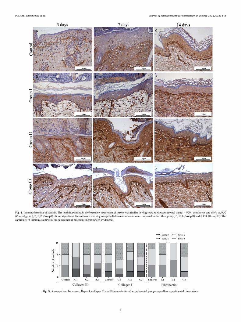
Fig. 4. Immunodetection of laminin. The laminin staining in the basement membrane of vessels was similar in all groups at all experimental times:> 50%, continuous and thick. A, B, C(Control group); D, E, F (Group I): shows significant discontinuous marking subepithelial basement membrane compared to the other groups; G, H, I (Group II) and J, K, L (Group III): Thecontinuity of laminin staining in the subepithelial basement membrane is evidenced.
Control G1 G2 G3 Control G1 G2 G3 Control G1 G2 G30
4
8
12
Score 0
Score 1
Score 2
Score 3
Num
ber
of a
nim
als
Fig. 5. A comparison between collagen I, collagen III and Fibronectin for all experimental groups regardless experimental time-points.
P.K.F.M. Vasconcellos et al. Journal of Photochemistry & Photobiology, B: Biology 182 (2018) 1–8
6

therapeutically [17,18,25,30,31].When considering laser dosimetry, AlGhamdi et al. [33] proposed
an energy density ranging between 0.5 and 4 J/cm2 as ideal for in-creasing the proliferation rate of cultured cells. According to Pinheiroet al. [34], when laser light is irradiated on monolayers of culturedcells, maintenance of polarity and consistency may determine the bio-logical results, the same occurring when irradiating an ulcer. Thus,lower doses of irradiation should be applied to mucous membranes,ulcers, or wounds, unlikely intact skin due to stratification. In an ex-tensive review of literature on photobiomodulation, Tuner and Hode[35] described an energy density between 1 and 4 J/cm2 as the mostappropriate in wound repair, at the risk of the laser exert an inhibitoryeffect on the repair process.
The aforemetioned irradiation protocols was used in researcheswhich reported positive results from laser use in second-degree andthird-degree burn repair [19,32] or cutaneous wounds in malnourishedanimals [23]. On the other hand, unlike observed in the literature[19,23,32] the same total energy density (20 J/cm2) in the current trialhad no positive effects on the repair of second-degree burns. In addi-tion, the divergences of our outcomes might be due to the laser spot size(4 mm2) used in this investigation that was not arranged to cover theentire wound area (400mm2).
Overall, during experimental trials for all groups there was a higherscore for the deposition of collagen I as compared to III. The replace-ment of collagen type III by type I and a change in fiber pattern whichbecome thicker (fibril) on the last day of the experiment are indicativeof organization, maturation, and tensile strength in the healing area.This has occurred in all groups of assessed in a similar manner[26,29,36].
Assessment of laminin staining, performed as per Henriques et al.[37] has demonstrated that the animals receiving laser therapy in iso-lation (Group I) showed the worst results in the analysis with regard tothe pattern of continuity in comparison with Control, Group II, andGroup III, and difference was statistically significant (p < 0.05).Morphological analysis showed a persistent inflammation in the laser-irradiated Group with a persistent edema, dense in the papillary dermis.It is known that the inflammatory process directly affects the ECMdynamics and the extension of the inflammation phase is the mainfactor causing delayed healing or scar formation [26]. It is likely thatthis pattern of discontinuity observed in the laser-treated group mightbe attributed to the presence of inflammatory infiltrate in the analyzedarea, thereby resulting in the release of proteolytic enzymes responsiblefor the degradation of tissue components at the wound site [38,39].
Groups receiving application of the cellulose membrane in isolationor in combination with laser have significantly shown better resultsthan the laser group (p < 0.05) in relation to the continuity of lamininstaining in the subepithelial basement membrane. The assessment ofthese results in isolation suggests that the use of cellulose biomembraneappears to favor the deposition of this protein. This result may be re-lated to its nanostructural configuration, which prevents contamination
of the wound area, restricts fluid loss in a unique configuration thatmimics the properties of the ECM by binding to water and behaving as ahydrogel that integrates with tissue allowing cell migration through thestructure [9,10]. Regarding the laminin expression in vascular base-ment membrane, all groups have shown similar results.
It has been noted during the immunohistochemical analysis theexpression of fibronectin in the epithelial tissue in several regions. Anumber of works in the literature has implied that keratinocytes arecells that secrete and deposit fibronectin in the pericellular matrix[40–42], thus suggesting the important role of this glycoprotein also inintercellular adhesion of the epithelial tissue.
Considering the important role fibronectin plays for the repairprocess, therapies that may trigger an increase in deposition can behelpful allies in this process, especially when dealing with wounds thatpose some difficulty or show delay in healing. However, the presentstudy has failed to verify a bio-stimulant effect of laser on fibronectindeposition in the wound area. Groups I, II, and III showed the fi-bronectin expression predominantly discontinuous near the basementmembrane in comparison with the Control group, with no statisticalsignificance, nonetheless.
5. Conclusion
In concluding, in this study we haven't found any beneficial effectwhen the laser was utilized in isolation (Group I) in the process ofcollagen deposition III and I; similarly, no favorable effect was found incollage patterns (fibril and/or reticular), acceleration of repair in in-duced burn injury in the dorsum of rats.
There was a trend towards a greater deposition of type III collagenin the wounds treated with a biocelulose membrane in isolation (GroupII) or with laser therapy (Group III), as early as seven days afterwardswhen these groups were compared with their laser-treated counterparts(Group I). The continuity of the laminin staining in the subepiteliallayer of the membrane in Groups II and III suggests that the cellulosemembrane either in isolation or in combination with the laser has fa-vored re-epithelialization in second-degree burns.
However, further research into the use of the cellulose biomem-brane in burn repair is clearly needed in order to assure the beneficialeffects for a prompt re-epithelialization. The application of the bio-cellulose membrane in isolation has ultimately reduced the woundcovering by fibronectin.
Acknowledgments
The authors greatfully acknowledge financial support by theConselho Nacional de Pesquisa (CNPq, 308595/2016-5), Brazil.
References
[1] S.C. Nunez, C.M. França, D.F.T. Silva, G.E.C. Nogueira, R.A. Prates, M.S. Ribeiro,
Table 3Percentage (%) for staining, continuity and thickness for subepithelial basement membrane and basal membrane of vessels.
Subepithelial basement membrane basal membrane of vessels
Groups Control n (%) Group I n (%) Group II n (%) Group III n (%) Control n (%) Group II n (%) Group II n (%) Group III n (%)
% for staining≤50% – 1 (8,3) – – – – – –>50% 12 (100) 11 (91,7) 12 (100) 12 (100) 12 (100) 12 (100) 12 (100) 12 (100)
ContinuityContinuous 9 (75) 4 (33,3) 4 (33,3) 12 (100) 12 (100) 12 (100) 12 (100) 12 (100)Discontinuous 3 (25) 8 (66,7) 8 (66,7) – – – – –
ThicknessThin 2 (17) 1 (8,3) – – – – – –Thick – – 4 (33,3) 7 (58,3) 9 (75) 12 (100) 12 (100) 12 (100)Mixed 10 (83) 11 (91,7) 8 (66,7) 5 (41,7) 3 (25) – – –
P.K.F.M. Vasconcellos et al. Journal of Photochemistry & Photobiology, B: Biology 182 (2018) 1–8
7

The influence of red laser irradiation timeline on burn healing in rats, Lasers Med.Sci. 28 (2012) 633–641.
[2] M.D. Peck, Epidemiology of burns throughout the world. Part I: distribution andrisk factors, Burns 37 (2011) 1087–1100.
[3] J.R. Saffle, Closure of the excised burn wound: temporary skin substitutes, Clin.Plast. Surg. 36 (2009) 627–641.
[4] M.C. Ferreira, A.O. Paggiaro, C. Isaac, N. Teixeira Neto, G.B. Dos Santos, Substitutoscutâneos: conceitos atuais e proposta de classificação, Rev. Bras Cir Plást 26 (2011)696–702.
[5] M. Held, J. Rothenberger, A.S. Engelke, D.S. Tolzmann, B.J. Esfahani, H.E. Schaller,A. Rahmanian-Schwarz, Evaluation of commonly used temporary skin dressings anda newly developed collagen matrix for treatment of superficial wounds, Adv. SkinWound Care 28 (2015) 551–554.
[6] R.R.J. Guirro, E.C.O. Guirro, C.C. Martins, F.R. Nunes, Analysis of low-level laserradiation transmission in occlusive dressings, Photomed. Laser Surg. 28 (2010)459–463.
[7] A.G. Haddad, A.G. Giatsidis, D.P. Orgill, E.G. Halvorson, Skin substitutes andBioscaffolds: Temporary and Permanent Coverage, Clin. Plast. Surg. 44 (3) (2017Jul) 627–634.
[8] G.F. Picheth, C.L. Pirich, M.R. Sierakowski, M.A. Woehl, C.N. Sakakibara, C.F. deSouza, A.A. Martin, R. da Silva, R.A. de Freitas, Bacterial cellulose in biomedicalapplications: a review, Int. J. Biol. Macromol. 3 (104) (2017) 97–106.
[9] Pecoraro, et al., Bacterial cellulose from Glucanacetobacter xylinus: preparation,properties and applications, Monomers, Polymers and Composites from RenewableResources, First ed., Elsevier, Oxford, 2008.
[10] N. Petersen, P. Gatenholm, Bacterial cellulose-based materials and medical devices:current state and perspectives, Appl. Microbiol. Biothecnol. 91 (2011) 1277–1286.
[11] A.J. Hussein, A. A, M.A. Falih, A.N. Hassan, Effects ofa low level laser onthe ac-celeration of wound healing in rabbits, N. Am. J. Med. Sci. 3 (2011) 193–197.
[12] A.M. Rocha Junior, R.G. Oliveira, R.E. Farias, L.C.F. Andrade, F.M. Aarestrup,Modulation of fibroblast proliferation and inflammatory response by low-intensitylaser therapy in tissue repair process, An. Bras. Dermatol. 81 (2006) 150–156.
[13] T.N. Demidova-Rice, E.V. Salomatina, A.N. Yaroslavsky, I.M. Herman,M.R. Hamblin, Low-level light stimulates excisional wound healing in mice, LasersSurg. Med. 39 (2007) 706–715.
[14] I.C.V. Decastro, S.C.P. Oliveira-Sampaio, Monteiro JSDC, Ferreira MFL, J.N. Santos,Pinheiro ALB, Evaluation of the effect of laser radiation on fibroblast proliferationin repair of skin wounds of rats with iron deficiency anemia, Proc. SPIE 7887(2011) 1–6.
[15] S. Martu, C. Amalinei, M. Tatarciuc, M. Rotaru, O. Potarnichie, L. Liliac, et al.,Healing process and laser therapy in the superficial periodontum: a histologicalstudy, Romanian J. Morphol. Embryol. 53 (2012) 111–116.
[16] F. Heu, C. Forster, B. Namer, A. Dragu, W. Lang, Effect of low-level laser therapy onblood flow and oxygen- hemoglobin saturation of the foot skin in healthy subjects: apilot study, Laser Ther. 22 (2013) 21–30.
[17] M. Bayat, M.M. Vasheghani, N. Razavi, S. Taheri, M. Rakhshan, Effect of low-levellaser therapy on the healing of second-degree burns in rats: a histology:ical andmicrobiological study, J. Photochem. Photobiol. B Biol. 78 (2005) 171–177.
[18] F.A.H. Al-Watban, M.D. Delgado, Burn healing with a diode laser: 670 nm at dif-ferent doses as compared to a placebo group, Photomed. Laser Surg. 23 (2005)245–250.
[19] G.C.S. Meireles, J.N. Santos, P.O. Chagas, A.P. Moura, A.L.B. Pinheiro, Effectivenessof laser photobiomodulation at 660 or 780 nanometers on the repair of third-degreeburns in diabetic rats, Photomed. Laser Surg. 26 (2008) 47–54.
[20] E.C.O. Guirro, M.I.L. Montebelo, B.A. Bortot, M.A.C.B. Torres, M.L.O. Polacow,Effect of laser (670 nm) on healing of wounds covered with occlusive dressing: ahistologic and biomechanical analysis, Photomed. Laser Surg. 28 (2010) 629–634.
[21] M.D.M. Dantas, et al., Improvement of dermal burn healing by combining sodiumalginate/chitosan-based films and low level laser therapy, J. Photochem. Photobiol.
B 105 (2011) 51–59.[22] T.N. Meyer, A.L.A. Silva, A standard burning model using rats, Acta Cir. Bras. 14
(1999) 1–4.[23] A.L.B. Pinheiro, A.L.B. Vieira, D.S. Almeida, G.C.S. Meireles, C.M. Carvalho,
J.N. Santos, Phototherapy improves healing of cutaneous wounds in nourished andundernourished Wistar rats, Braz. Dent. J. 15 (2004) 21–28.
[24] J.M. Moraes, et al., Anti-inflammatory effect of low-intensity laser on the healing ofthird-degree burn wounds in rats, Lasers Med. Sci. 28 (2013) 1169–1176.
[25] A. Schlager, P. Kronberger, F. Petschke, H. Ulmer, Low-power laser light in thehealing of burns: a comparision between two different wavelengths (635 nm and690 nm) and a placebo group, Lasers Surg. Med. 27 (2000) 39–42.
[26] E.A. Gantwerker, D.B. Hom, Skin: histology and physiology of wound healing, Clin.Plast. Surg. 39 (2012) 85–97.
[27] A.R.A.P. Medrado, L.S. Pugliese, S.R.A. Reis, Z.A. Andrade, Influence of low levellaser therapy on wound healing and its biological action upon myofibroblasts,Lasers Surg. Med. 32 (2003) 239–244.
[28] L.S. Pugliese, A.L. Medrado, S.R.A. Reis, Z.A. Andrade, The influence of low-levellaser therapy on biomodulation of collagen and elastic fibers, Pesqui Odontol. Bras.17 (2003) 307–313.
[29] H. Liu, Y. Dang, Z. Wang, X. Chai, Q. Ren, Laser induced collagen remodeling: acomparative study in vivo on mouse model, Lasers Surg. Med. 40 (2008) 13–19.
[30] G. Anneroth, G. Hall, H. Rydén, L. Zettergvist, The effect of low-energy infra-redlaser radiation on wound healing in rats, Br. J. Oral Maxillofac. Surg. 26 (1988)12–17.
[31] G. Hall, G. Anneroth, T. Schennings, L. Zettergvist, H. Ridén, Effect of low levelenergy laser irradiation on wound healing. An experimental study in rats, Swed.Dent. J. 18 (1994) 29–34.
[32] E.T. Trajano, L.A. da Trajano, M.A. Dos Santos, N.G. Venter Silva, L.C. de Porto,A. de Fonseca, A. Monte-Alto-Costa, Low-level red laser improves healing of second-degree burn when applied during proliferative phase, Lasers Med. Sci. 30 (2015)297–304.
[33] K.M. AlGhamdi, A. Kumar, N.A. Moussa, Low-level laser therapy: a useful techniquefor enhancing the proliferation of various cultured cells, Lasers Med. Sci. 27 (2012)237–249.
[34] A.L.B. Pinheiro, A. Brugnera Junior, F.A.A. Zanin, Aplicação do laser na odonto-logia, First ed., GEN, Santos, São Paulo, 2010.
[35] J. Tuner, L. Hode, It's all in the parameters: a clinical analysis of some well-knownnegative studies on low-level laser therapy, J. Clin. Laser Med. Surg. 16 (1998)245–248.
[36] R.D. Novaes, et al., The energy density of laser light differentially modulates theskin morphological reorganization in a murine model of healing by secondary in-tention, Int. J. Exp. Pathol. 95 (2013) (2013) 138–146.
[37] A.C.G. Henriques, M.G. Vasconcellos, H.C. Galvão, L.B. De Souza, R.A. Freitas,Comparative analysis of the immunohistochemical expression of collagen IV, MMP-9 and TIMP-2 in odontogenic cysts and tumors, Oral Surg. Oral Med. Oral Pathol.Oral Radiol. Endod. 112 (2011) 468–475.
[38] L.B. Quinderé, C.F.W. Nonaka, L.B. Souza, L.P. Pinto, Expressão imuno-histoquímica de colágeno IV, tenascina-C e fibronectina em lesões centrais eperiféricas de células gigantes, Rev. Inst. Ciênc Saúde. 26 (2008) 226–231.
[39] C.A.S. Gurgel, et al., Immunolocalisation of laminin-1 in keratocystic odontogenictumors, Acta Histochem. 112 (2010) 624–629.
[40] M. Kubo, D.A. Norris, S.E. Howell, S.R. Ryan, R.A. Clark, Human keratinocytessynthesize, secrete, and deposit fibronectin in the pericellular matrix, J. Invest.Dermatol. Jun 82 (6) (1984) 580.
[41] A. Linnala, E. Balza, L. Zardi, I. Virtanen, Human amnion epithelial cells assembletenascins and three fibronectin isoforms in the extracellular matrix, FEBS Lett. 8(317) (1993) 74–78.
[42] M. Kubo, et al., Immunoelectron microscopic localization of fibronectin in culturedhuman keratinocytes, Arch. Dermatol. Res. 286 (1994) 448–454.
P.K.F.M. Vasconcellos et al. Journal of Photochemistry & Photobiology, B: Biology 182 (2018) 1–8
8