Embed Size (px)
Citation preview
Jugular Fossa LesionsKenan I. Arnautovic, M.D., and Ossama AI-Mefty, M.D.
Learning Objectives: After reading this article, the participant should be able to:1. Describe the basic anatomy of the jugular fossa and foramen.2. Describe the differential diagnosis ofjugular fossa lesions.3. Recall basic preoperative diagnostics and operative approaches to jugular fossa lesions.
lar foramen. It lodges the jugular bulb, which continues intothe jugular vein inferiorly (Fig. 1).
In the neurosurgical literature, and even in extensiveanatomic studies, both the jugular foramen and the jugular fossa often are referred to by the term "jugular foramen."This use of the term may be the result either of simple erroror the user's wish to provide a broader anatomic description of the area, and this confusion may be the underlyingreason for the current lack of agreement regarding the internal anatomic organization of "the jugular foramen." Regardless of the reason for this mix-up, the jugular foramen andthe jugular fossa are two distinct anatomic formations,although they are intimately related.
The treatment of jugular fossa lesions has been revolutionized by the development of modern diagnostic modalities, refinement of microsurgical techniques, publicationof microsurgical anatomic studies, development of skullbase approaches, advances in neuroanesthesia and intraoperative neurophysiologic monitoring, and careful multidisciplinary perioperative planning. These lesions noware treated with radical resection, and the rates of permanent surgical morbidities or mortalities are low.
Anatomic ConsiderationsThe jugular foramen is a skull opening, or gap, that con
nects the posterior cranial fossa and the jugular fossa. It isformed by the jugular incisuras in the temporal and occipital bones. It lies in an oblique position, from the lateral aspectposteriorly toward the medial aspect anteriorly. Classically,it is described as subdivided by a fibrous or bony bridge (theintrajugular septum) into two parts, which serve as a passage for Cranial Nerve IX and the inferior petrosal sinus (parsnervina or nervosa) and for Cranial Nerves Xand XI and thejugular vein. More recent anatomic studies have disputedthis theory of the anatomic organization of the jugular foramen/ and there still is no single accepted view of its anatomy.
The jugular fossa, located at the inferior aspect (inferiorsurface) of the petrous part of the temporal bone, is a deepdepression, the size of which varies from skull to skull. Itcommunicates with the posterior cranial fossa via the jugu-
Differential DiagnosisThe most cornmon differential diagnoses of lesions in the
jugular fossa include, in descending order of frequency, glomusjugulare tumors/ neurinoma of the lower cranial nerves (Cranial Nerves IX-XI), and meningiomas (Figs. 2-4). The broaderdifferential diagnosis of jugular fossa tumors may include chordoma/ chondrosarcoma, primary cholesteatoma, plasmacytoma/ epidermoid tumors, choroid plexus papilloma,chondroma, temporal bone carcinoma, salivary gland tumors,aneurysm, metastases, and cerebellar hemangioblastoma.
The preoperative radiologic diagnosis and differentialdiagnosis are important when jugular fossa lesions are thesubject, because preoperative management and operativeplanning may differ considerably depending on the type
==========---- ------.------- of lesion in question, e.g., planning preoperative emboliza-tion for glomus jugulare tumors, or determining how muchof the tumor involves bone if it is a meningioma.
=------ - - Category: Tumor, Anatomy
KeyWords: Jugular Fossa, Jugular Foramen, Glomus Jugulare, Neurinoma, Meningioma
B
Figure 1. A, Right-side, dry anatomic specimen photograph delineating the jugular foramen (white arrows) and the jugular fossa (blackarrows). Outside-inside view from the anterior perspective. B, Rightside dry anatomic specimen photograph delineating the jugular foramen (black arrows) and the jugular fossa (white arrows).Inside-outside view from the posterior perspective. Note the difference between the two.
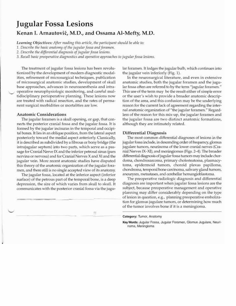
Glomus jugulare tumors have a "salt-and-pepper" appearance on noncontrast-enhanced Tl-weighted Nffil scans, whichrepresent a flow void network from their rich vascularity
-<md-eDhance_nonhomo~neousJyafter the in'ection of con-
Figure 2. Glomus jugulare tumor. A, Axial T1-weighted noncontrastimage showing the salt-and-pepper tumor appearance in the rightjugUlar fossa (*). B, Sagittal T1-weighted postcontrast MRI scan showing the nonhomogeneous enhancement of the tumor (*).
trast material (Fig. 2). However, they have a propensity toerode and destroy the bone, particularly the jugular spine
and the carotid crest (caroticojugular spine)-the bone thatseparates the petrous carotid artery from the jugular bulb.Meningiomas often invade the bone, including the jugularspine and particularly the jugular tubercle, producing "hyperostosis" and bone thickening, but not bone erosion (Fig. 4B).
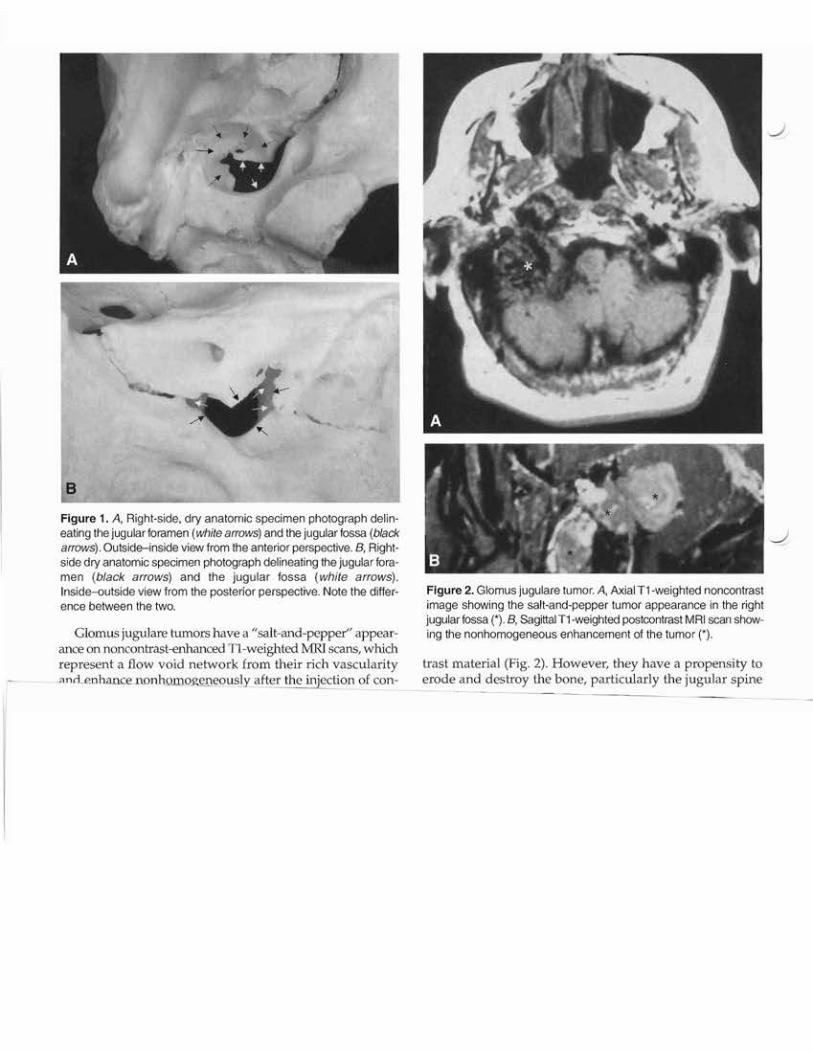
Jugular fossa neurinomas originate from the lower cranial nerves. They enlarge the jugular fossa with smooth,distinct sclerotic margins seen on CT scans with bone window ("bone scalloping") (Fig. 3D). They may contain cysts,which appear as hypointensities on Tl-weighted MRI scans.Frank bony invasion is very rare. Although they may mimicthe appearance of meningiomas on both Tl- and T2weighted MRI scans, the enhancement seen in meningiomasusually is considerably greater. Neurinomas often assume
3
Figure 3. Jugular fossa neurinoma. A, AxialT1-weighted noncontrast MRI scan. Note theisointense tumor in the right jugular fossa (*).B, The same tumor on the postcontrast axialT1-weighted MRI scan. Note the hyperintense,homogeneous enhancement of the tumor. C,Sagittal postcontrastT1-weighted MRI scan ofthe same patient. Note the tumor (*) displacingthe internal carotid artery (arrows). D, Axial CTscan (bone windows). Note the scalloping ofthe right jugular fossa (arrows). E, Magneticresonance venogram of the same patient. Notethe occlusion of the venous flow in the rightjugular fossa.
a "dumbbell" shape, which can be bestrecognized on coronal or sagittal MRIprojections (Fig. 3C).
Glomus Jugulare TumorsGlomus tumors or paraganglioma orig
inate from paraganglia tissue, whichbelongs to the extra-adrenal chromaffintissue system. This tissue is distributedsymmetrically in close relation to the arterial vasculature and cranial nerves of theontogenic gill arches. Different types ofparaganglia include jugulotympanic,intercarotid, subclavian or supraaortic,orbital, coronary, laryngeal, pulmonary,and aorticopulmonary. In the ear, thereare an average of three paraganglia oneach side, with a decrease in number after60 years of age. They accompany, withequal frequency, Jacobson's (the tympanicbranch of the IXth nerve) or Arnold's (theauricular branch of the Xth nerve) nerves.
More than 50% are located in the jugular fossa; the rest arelocated in the middle ear. They appear encapsulated, lobulated, and tan-gray to purple-red in color. Their vascular supply is derived from the postauricular, occipital, internalmaxillary, and internal carotid arteries. Although every glomus tumor may secrete catecholarnines and other neuropeptides, only 1% to 3% of these tumors present with clinicallydetectable symptoms, because the serum norepinephrine mustbe elevated to at least four to five times the normal level beforesymptoms (e.g., headaches, excessive perspiration, tachycardia, pallor, and nausea) are produced. Thus, evaluation ofserum and urine levels of catecholamines is part of the routine preoperative evaluation. In these cases, preoperativeadministration of alpha- and beta-blockers is important.
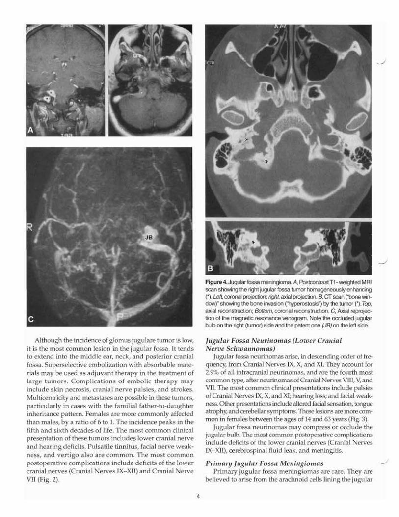
Although the incidence of glomus jugulare tumor is low,it is the most common lesion in the jugular fossa. It tendsto extend into the middle ear, neck, and posterior cranialfossa. Superselective embolization with absorbable materials may be used as adjuvant therapy in the treatment oflarge tumors. Complications of embolic therapy mayinclude skin necrosis, cranial nerve palsies, and strokes.Multicentricity and metastases are possible in these tumors,particularly in cases with the familial father-to-daughterinheritance pattern. Females are more commonly affectedthan males, by a ratio of 6 to 1. The incidence peaks in thefifth and sixth decades of life. The most common clinicalpresentation of these tumors includes lower cranial nerveand hearing deficits. Pulsatile tinnitus, facial nerve weakness, and vertigo also are common. The most commonpostoperative complications include deficits of the lowercranial nerves (Cranial Nerves IX-XII) and Cranial NerveVII (Fig. 2).
4
Figure 4. Jugular fossa meningioma. A, PostcontrastT1- weighted MRIscan showing the right jugular fossa tumor homogeneously enhancing(*). Left, coronal projection; right, axial projection. B, CT scan ("bone window)" showing the bone invasion ("hyperostosis") by the tumor (*). Top,axial reconstruction; Bottom, coronal reconstruction. C, Axial reprojection of the magnetic resonance venogram. Note tl1e occluded jugularbulb on the right (tumor) side and the patent one (JB) on the left side.
Jugular Fossa Neurinomas (Lower CranialNerve Schwannomas)
Jugular fossa neurinomas arise, in descending order of frequency, from Cranial Nerves IX, X, and XI. They account for2.9% of all intracranial neurinomas, and are the fourth mostcommon type, after neurinomas of Cranial Nerves VIII, V, andVII. The most common clinical presentations include palsiesof Cranial Nerves IX, X, and XI; hearing loss; and facial weakness. Other presentations include altered facial sensation, tongueatrophy, and cerebellar symptoms. These lesions are more common in females between the ages of 14 and 63 years (Fig. 3).
Jugular fossa neurinomas may compress or occlude thejugular bulb. The most common postoperative complicationsinclude deficits of the lower cranial nerves (Cranial NervesIX-XII), cerebrospinal fluid leak, and meningitis.
Primary Jugular Fossa MeningiomasPrimary jugular fossa meningiomas are rare. They are
believed to arise from the arachnoid cells lining the jugular
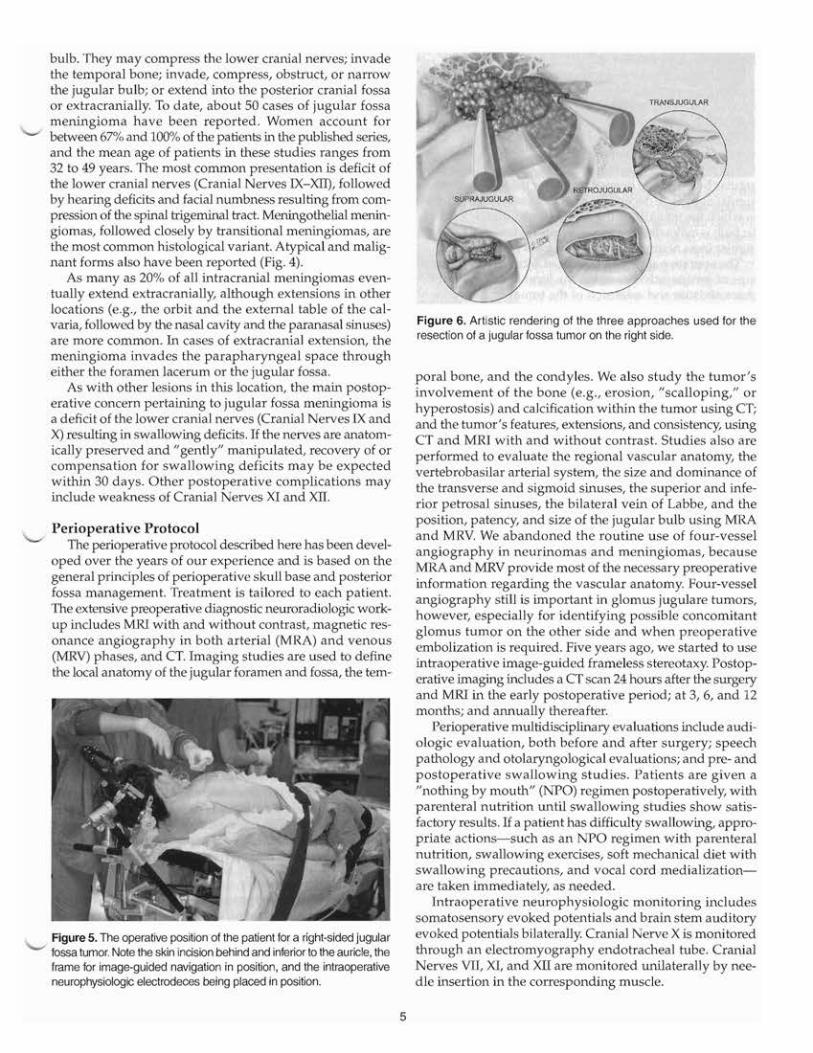
bulb. They may compress the lower cranial nerves; invadethe temporal bone; invade, compress, obstruct, or narrowthe jugular bulb; or extend into the posterior cranial fossaor extracranially. To date, about 50 cases of jugular fossameningioma have been reported. Women account forbetween 67% and 100% of the patients in the published series,and the mean age of patients in these studies ranges from32 to 49 years. The most common presentation is deficit ofthe lower cranial nerves (Cranial Nerves IX-XII), followedby hearing deficits and facial numbness resulting from compression of the spinal trigeminal tract. Meningothelial meningiomas, followed closely by transitional meningiomas, arethe most common histological variant. Atypical and malignant forms also have been reported (Fig. 4).
As many as 20% of all intracranial meningiomas eventually extend extracranially, although extensions in otherlocations (e.g., the orbit and the external table of the calvaria, followed by the nasal cavity and the paranasal sinuses)are more common. In cases of extracranial extension, themeningioma invades the parapharyngeal space througheither the foramen lacerum or the jugular fossa.
As with other lesions in this location, the main postoperative concern pertaining to jugular fossa meningioma isa deficit of the lower cranial nerves (Cranial Nerves IX andX) resulting in swallowing deficits. If the nerves are anatomically preserved and "gently" manipulated, recovery of orcompensation for swallowing deficits may be expectedwithin 30 days. Other postoperative complications mayinclude weakness of Cranial Nerves XI and XII.
Perioperative ProtocolThe perioperative protocol described here has been devel
oped over the years of our experience and is based on thegeneral principles of perioperative skull base and posteriorfossa management. Treatment is tailored to each patient.The extensive preoperative diagnostic neuroradiologic workup includes MRI with and without contrast, magnetic resonance angiography in both arterial (MRA) and venous(MRV) phases, and CT. Imaging studies are used to definethe local anatomy of the jugular foramen and fossa, the tem-
Figure 5. The operative position of the patient for a right-sided jugularfossa tumor. Note the skin incision behind and inferior to the auricle, theframe for image-guided navigation in position, and the intraoperativeneurophysiologic electrodeces being placed in position.
5
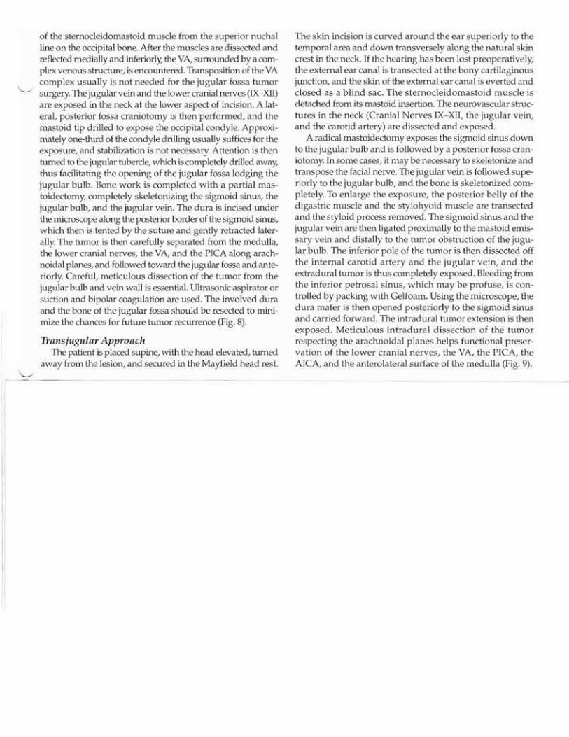
Figure 6. Artistic rendering of the three approaches used for theresection of a jugular fossa tumor on the right side.
poral bone, and the condyles. We also study the tumor'sinvolvement of the bone (e.g., erosion, "scalloping," orhyperostosis) and calcification within the tumor using CT;and the tumor's features, extensions, and consistency, usingCT and MRI with and without contrast. Studies also areperformed to evaluate the regional vascular anatomy, thevertebrobasilar arterial system, the size and dominance ofthe transverse and sigmoid sinuses, the superior and inferior petrosal sinuses, the bilateral vein of Labbe, and theposition, patency, and size of the jugular bulb using MRAand MRV We abandoned the routine use of four-vesselangiography in neurinomas and meningiomas, becauseMRA and MRV provide most of the necessary preoperativeinformation regarding the vascular anatomy. Four-vesselangiography still is important in glomus jugulare tumors,however, especially for identifying possible concomitantglomus tumor on the other side and when preoperativeembolization is required. Five years ago, we started to useintraoperative image-guided frameless stereotaxy. Postoperative imaging includes a CT scan 24 hours after the surgelYand MRI in the early postoperative period; at 3,6, and 12months; and annually thereafter.
Perioperative multidisciplinalY evaluations include audiologic evaluation, both before and after surgery; speechpathology and otolalYngological evaluations; and pre- andpostoperative swallowing studies. Patients are given a"nothing by mouth" (NPO) regimen postoperatively, withparenteral nutrition until swallowing studies show satisfactory results. If a patient has difficulty swallowing, appropriate actions-such as an NPO regimen with parenteralnutrition, swallowing exercises, soft mechanical diet withswallowing precautions, and vocal cord medializationare taken immediately, as needed.
Intraoperative neurophysiologic monitoring includessomatosensory evoked potentials and brain stem auditoryevoked potentials bilaterally. Cranial Nerve X is monitoredthrough an electromyography endotracheal tube. CranialNerves VII, XI, and XII are monitored unilaterally by needle insertion in the corresponding muscle.
Operative ApproachThe approach to a jugular fossa tumor should be tailored
to each patient. Because these tumors involve the jugularfossa and beyond, the skull base approach is required fortheir removal. However, the patency and dominance of theinvolved jugular bulb dictate the surgical approach. Threeapproaches are possible: the transjugular approach, commonly practiced in resection of glomus tumors, in which thejugular bulb itself is opened; or either the supra- or retrojugular approach, depending on the extension of the tumor,in which the integrity of the venous flow through the jugular bulb is maintained, as is commonly done in patients withjugular fossa neurinoma or jugular fossa meningioma.
The operative approach is tailored according to the findings of preoperative imaging, the local anatomy, and thecharacteristics and extension of the tumor in each patient(Figs. 3-9). Three different routes are used:
1. The suprajugular approach. This presigmoid, infralabyrinthine route is chosen if the jugular bulb is patentand the tumor extends primarily anteriorly (Fig. 7).
2. The retrojugular approach. This transcondylar, transtubercle route is used if the jugular bulb is patent and thetumor was extending predominantly behind it (Fig. 8).
3. The transjugular approach. This is an infratemporal route,used in cases in which the jugular bulb is totally occludedby a tumor (Fig. 9).
Suprajugular ApproachIn the suprajugular approach, the patient is placed supine
with a shoulder roll under the shoulder and the head elevated,turned away from the lesion, and fixed in a Mayfield headholder.The skin incision circles behind the ear, starting at the temporalarea anteriorly and superiorly and extending down into theupper neck horizontally along the skin crease. The skin flap isthen retracted anteriorly and inferiorly. The sternocleidomastoidmuscle is retracted inferiorly and posteriorly. A mastoidectomyis then performed, followed by complete skeletonization of thesigmoid sinus, the jugular bulb, and the jugular vein. The jugular fossa is accessed in the presigmoid, in.fralabyrinthine space.
Figure 7. The suprajugular approach. Note the infralabyrinthine position of the tumor, the sigmoid sinus (8S); the jugular bulb (JB), whichis patent, the jugular vein (J\I); the superior petrosal sinus (8PS); andthe labyrinth (right side).
6
IX X
Figure 8. The retrojugular approach. Note the retrojugular positionof the tumor covered by the arachnoid membrane, the sigmoid sinus(8S), Cranial Nerves VII and VIII, and the lower cranial nerves (IX-XI)stretched over the tumor (right side).
IX
/X
/
JV
sps-"."
Figure 9. The transjugular approach. Note the tumor occluding thejugular bulb, which is open, the sigmoid sinus (88) and the jugularvein (J\I), both of which are ligated; the superior petrosal sinus (8PS);the internal carotid artery (leA); and the lower cranial nerves extracranially (IX-XI).
The dura superior to the patent jugular bulb and inferior to thelabyrinth is opened. The cerebrospinal fluid is then released fromthe cerebellomeduUary cistern and tumor is dissected away fromthe lower cranial nerves (Cranial Nerves IX - XI), the posteroinferior (PICA) and anteroinferior (ArCA) cerebellar arteries, and the vertebral artery (VA) respecting the arachnoidalplanes. Debulking of the tumor is done by suction and bipolarcoagulation or by an ultrasonic aspirator and is completed withmicrosurgical radical resection of the tumor (Fig. 7).
Retrojugular ApproachIn the retrojugular approach, the patient is placed supine
and rotated about 40 degrees, keeping the head and neck elevated 30 degrees in neutral position and fixing it in the Mayfield headholder. The skin incision is curved around the eartwo fingerbreadths behind the mastoid, extending down transversely along the horizontal neck crease. The muscles of theneck are dissected in three layers, beginning with detachment
of the sternocleidomastoid muscle from the superior nuchalline on the occipital bone. After the muscles are dissected andreflected medially and inferiorly, the VA, surrow1ded by a complex venous structure, is encountered. Transposition of the VAcomplex usually is not needed for the jugular fossa tumorsurgery. The jugular vein and the lower cranial nerves (lX-XO.)are exposed in the neck at the lower aspect of incision. A laterat posterior fossa craniotomy is then performed, and themastoid tip drilled to expose the occipital condyle. Approximately one-third of the condyle drilling usually suffices for theexposure, and stabilization is not necessary. Attention is thenturned to the jugular hlbercle, which is completely drilled away,thus facilitating the opening of the jugular fossa lodging thejugular bulb. Bone work is completed with a partial mastoidectomy, completely skeletonizing the sigmoid sinus, thejugular bulb, and the jugular vein. The dura is incised underthe microscope along the posterior border of the sigmoid sinus,which then is tented by the suture and gently retracted laterally. The tumor is then carefully separated from the medulla,the lower cranial nerves, the VA, and the PICA along arachnoidal planes, and followed toward the jugular fossa and anteriorly. Carefut meticulous dissection of the tumor from thejugular bulb and vein wall is essential. Ultrasonic aspirator orsuction and bipolar coagulation are used. The involved duraand the bone of the jugular fossa should be resected to minimize the chances for future tumor recurrence (Fig. 8).
Transjugular ApproachThe patient is placed supine, with the head elevated, turned
away from the lesion, and secured in the Mayfield head rest.
The skin incision is curved around the ear superiorly to thetemporal area and down transversely along the natural skincrest in the neck. If the hearing has been lost preoperatively,the external ear canal is transected at fue bony cartilaginousjunction, and the skin of the external ear canal is everted andclosed as a blind sac. The sternocleidomastoid muscle isdetached from its mastoid insertion. The neurovascular structures in the neck (Cranial Nerves IX-XII, the jugular vein,and the carotid artery) are dissected and exposed.
A radical mastoidectomy exposes the sigmoid sinus downto the jugular bulb and is followed by a posterior fossa craniotomy. In some cases, it may be necessary to skeletonize andtranspose the facial nerve. The jugular vein is followed superiorly to the jugular bulb, and the bone is skeletonized completely. To enlarge the exposure, the posterior belly of thedigastric muscle and the stylohyoid muscle are transectedand the styloid process removed. The sigmoid sinus and thejugular vein are then ligated proximally to the mastoid emissary vein and distally to the tumor obstruction of the jugular bulb. The inferior pole of the tumor is then dissected offthe internal carotid artery and the jugular vein, and theextradural tumor is thus completely exposed. Bleeding fromthe inferior petrosal sinus, which may be profuse, is controlled by packing with Gelfoam. Using the microscope, thedura mater is then opened posteriorly to the sigmoid sinusand carried forward. The intradural tumor extension is thenexposed. Meticulous intradural dissection of the tumorrespecting the arachnoidal planes helps functional preservation of the lower cranial nerves, the VA, the PICA, theAICA, and the anterolateral surface of the medulla (Fig. 9).
Readings
AI-Mefty 0: Meningiomas of the posterior cranial base. In Eisenberg MB,Al-Mefty 0 (eds): Operative Atlas of Meningiomas. Philadelphia: Lippincott-Raven, 1997, pp 209-348
AI-Mefty 0, Fox JL, Rifai A, et al: A combined infTatemporal and posteriorfossa approach for the removal of giant glomus tumors and chondrosarcomas. Surg NeuroI28:423, 1987
Anand VK, Leonetti JP, Al-Mefty 0: Neurovascular considerations in surgeryof glomus tumors with intracranial extensions. Lanjngoscope 103:722, 1993
Amautovic Kl, Al-Mefty 0: Primary jugular fossa meningiomas. JNeurosurg97:12,2002
Ayeni SA, Ohata K, Tanaka K: The microsmgical anatomy of the jugular foramen. JNeurosurg 83: 903, 1995
Borba LAB, Al-Mefty 0: Paragangliomas of the skull base. Neurosurgenj Quarterly 5:256,1995
Fisch U, Pillsbury HC: InfTatemporal fossa approach to lesions in the temporal bone and base of the skull. Arch Otolaryngoll05:99, 1979
George B, Dematons C, Cophignon J: Lateral approach to the anterior portion of the foramen magnum. Applica tion to surgical removal of 14 benigntumors. Technical note. Surg NeuroI29:494, 1988
Glasscock ME: The history of glomus tumors: a personal perspective. Laryngoscope 103:3,1993
Hakuba A, Hashi K, Fujitani K, et al: Jugular foramen neurinomas. Surg Neurolll: 83, 1979
Katsuta T, Rhoton AL Jr, Matsushima T: The jugular foramen: microsurgicalanatomy and operative approaches. Neurosurgenj 41:149,1997
Kaye AH, Hahn JF, Kinney SE, et a1: Jugular foramen schwannomas. JNeurosurg 60:1045, 1984
Molony TB, Brackman DE, Lo WWM: Meningiomas of the jugular foramen. --./Otolaryngol Head Neck Surg 106:128, 1992
Osbom AG: Brain tumors and tumor-line processes. In Osbom AG (00): Diagnostic Neuroradiology. 51. Louis: Mosby, 1994, pp 507-510
Rhoton AL, Buza R: Microsurgical anatomy of the jugular foramen. JNeurosurg 42:541, 1975
Samii M, Babu R, Tatagiba M, et al: Surgical treatment of jugular foramenschwannomas. JNeurosurg 82:924,1995
Sekhar LN, Bucur SD, Schessel DA: Operative approaches to jugular foramen lesions. InSekhar LN, de Oliveira E (eds): Cranial Microneurosurgery:Approaches and Techniques. New York: Thieme, pp 482-511
Sen C, Hague K, Kacchara R, et al: Jugular foramen: microscopic anatomicfeatures and indications for neural preservation with reference to glomus tumors involving the temporal bone. Neurosurgery 48:838, 2001
Tekkok IH, Ozkan OE, TW'all E, et al: Jugular foramen meningioma. Reportof a case and review of literature. JNeurosurg Sci 41:283, 1997
Vrionis FD, Robertson JH, Gardner G, et al: Temporal bone meningiomas.Skull Base Surg 9:127, 1999