Embed Size (px)
Citation preview

AN ABSTRACT OF THE DISSERTATION OF
Franklin A. Hays for the degree of Doctor of Philosophy in Biochemistry and
Biophysics presented on April 14, 2005.
Title: Sequence Dependent Conformational Variations in DNA Holliday Junctions
Abstract approved:
Pui Shing Ho
Four-stranded DNA junctions (also known as Holliday junctions) are structural
intermediates involved in a growing number of biological processes including DNA
repair, genetic recombination, and viral integration. Although previous studies have
focused on understanding the conformational variability and sequence-dependent
formation of Holliday junctions in solution there have been relatively few insights into
junction structure at the atomic level. Recent crystallographic studies have
demonstrated that the more compact stacked-X junction form has an antiparallel
alignment of DNA strands and standard Watson-Crick base pairs across the central
crossover region. Junction formation within this crystallographic system was seen to
be dependent on a common trinucleotide sequence motif ("ACC-triplet" at the 6th, 7th
and 8thpositions of the decanucleotide sequence d(CCnnnN6N7N8GG)) containing a
series of stabilizing direct and solvent-mediated hydrogen bonding interactions. This
thesis addresses questions concerning the nucleotide sequence-dependent formation
Redacted for privacy

and conformational variability of DNA Holliday junctions as determined by single
crystal x-ray diffraction.
We have used the modified bases 2,6-diaminopurine and inosine to
demonstrate that minor groove interactions adjacent to the trinucleotide junction core
are not major contributors to overall conformation. In addition, incorporation of
guanine into the sixth position of this core does not have a significant effect on
junction geometry. Meanwhile, incorporation of 5-bromouracil into the eighth
position perturbs the geometry in terms of the interduplex angle as well as the defined
conformational variables, Jroii and Jslide. These novel junction structures demonstrate
that the nucleotide sequence within the central core generates a position specific
relationship between molecular interactions at the junction crossover and overall
structural geometry.
A systematic crystallographic screen of the trinucleotide core region is
presented here as an unbiased, comprehensive, search for sequences that stabilize
junctions. As the result of this screen, we can extend the core sequence motif to
'N6Y7C8' where N6 is an adenine, guanine, or cytosine nucleotide and Y7 is either a
cytosine or thymine (if N6 = adenine) nucleotide. Using these novel junction
structures, we demonstrate that base sequence within the central core has a significant
effect on the overall geometry of the junction. Thus, this central region of the
structure may serve as a linchpin for determining the local and global conformation
and overall variability of the four-stranded DNA Holliday junction. These
observations raise some interesting questions regarding the importance of this core
region in biological processes such as genetic recombination.
© Copyright by Franklin A. Hays
April 14, 2005
All Rights Reserved

Sequence Dependent Conformational Variations in DNA Holliday Junctions
by
Franklin A. Hays
A DISSERTATION
submitted to
Oregon State University
in partial fulfillment ofthe requirements for the
degree of
Doctor of Philosophy
Presented April 14, 2005Commencement June 2005

Doctor of Philosophy dissertation of Franklin A. Hays presented on April 14, 2005
APPROVED:
Maj4r Professor, Representing the Department of Biochemistry and Biophysics
of the DepàrtmeIif Biochemistry and Biophysics
Dean of
I understand that my dissertation will become part of the permanent collection ofOregon State University libraries. My signature below authorizes release ofmydissertation to any reader upon request.
Franklin A. Havg' Author
Redacted for privacy
Redacted for privacy
Redacted for privacy
Redacted for privacy

ACKNOWLEDGMENTS
I thank my graduate advisor and mentor, Dr. P. Shing Ho, for his patient and
dedicated support throughout my training in his laboratory. Shing, your commitment
to training students within your lab and teaching them how to engage the process of
scientific discovery and description is impressive. I thrived in the environment you
established and I appreciate your efforts. I would also like to thank all current and
former members of the Ho lab for establishing the foundation on which a lot of my
work is based. I would like to thank Jeffrey Vargason for supporting me during the
steep learning curve of the first two years of graduate school. You taught me that any
hurdle can be overcome through focused effort. Thank you Amy Teegarden, Zebulon
Jones, Michael Harms, Dustin Raup and Emily Cavaliere for your help and assistance
with the crystallographic screen project ("just one more setup!"). I would like to thank
Andrea Voth and Patricia Khuu for joining the lab and pursuing some very interesting
scientific prospects. Both of you will do great!
To the Karplus lab, thank you for constantly providing support on all aspects
of crystallographic data collection, refinement and analysis. Dr. Andrew Karplus,
thank you for improving the quality of my oral presentations and teaching x-ray
crystallography. Though all of my structures are still double helical I would like to
thank Zachary Wood, I admire your zest and love for science Zac! In Rick Faber I
found a great friend and somebody that I enjoyed many conversations with on all
aspects of Oregon life and science. Thank you for taking care of the diffractometer
Rick and constantly putting up with my "need it now" pushiness! I owe a significant
thank you to Ganapathy N. Sarma for always having a colorful quote. And finally, I
would like to thank the remaining members of my doctoral committee for their
support: Drs. Joe Beckman, Mike Schimerlik and Mary Slabaugh.
I would like to thank my undergraduate advisor - Dr. H. Olin Spivey. Thank
you Dr. Spivey for always putting up with my lab antics and providing an atmosphere
to pursue what I enjoy.
Thank you to my family and friends for their support through many years of
schooling. I would like to especially thank my parents, Ken and Mary Hays, for
providing space to explore and freedom to make my own decisions. As for the
Thibodeaux's (Jackie, Morris, David, Micayla and Mariah) - you guys are always a
source of enjoyment and I look forward to what the future holds. Thanks to my little
sister Tonya for being around in my absence and to my brother David for turning into
an amazing friend. You are doing great David (including Autunm and Aleeca)!
To my extended family, thank you for always loving and supporting me.
Sharon and Allen - thank you so much for all that you have done during my "never-
ending" pursuit of higher education. Geoff and Jo- thanks for providing the jump
drive on which this thesis took shape! Thank you Tina for pushing me to excel -
nothing goes unnoticed. Thank you Robert Kaster for introducing me to Miles Davis
and Charles Mingus - they both proved to be invaluable during graduate school.
And finally, I want to thank my wife and best friend Susan. You are an
amazing wife and friend for which I will always be indebted. You, above all else,
have experienced every step along the path to this degree and I sincerely thank you for
every ounce of support. Thank you for saying yes.

CONTRIBUTION OF AUTHORS
P. Shing Ho was involved in the design, analysis, and writing of each
manuscript. Jeffrey Watson assisted in some aspects of manuscript preparation for
Chapter 2. Jeffrey M. Vargason designed and determined initial crystallization
conditions for the d(CCAGTACTGG) sequence in Chapter 3 and played a consultative
role in the subsequent structure solution and refinement. Zebulon J. R. Jones provided
some assistance with refinement for the d(CCDGTACTGG) structure in Chapter 4.
Amy Teegarden, Zebulon J. R. Jones, Michael Harms and Dustin Raup were involved
in DNA purification, crystallization, and structure refinement for data presented in
Chapter 5. Emily Cavaliere helped with some aspects of crystallization and structure
refinement for material in Chapter 5. Dustin Raup was involved in the initial stages of
crystallization for the d(CCGATATCGG) sequence in Chapter 6.

TABLE OF CONTENTS
1. Introduction. 12. CAUTION! DNA Crossing: The crystal structures of Holliday Junctions ............... 7
2.1 Summary ............................................................................................................... 8
2.2 B.C.Before Crystal Structures .........................................................................10
2.3 Single Crystal Structures of DNA Holliday Junctions ....................................... 11
2.3 Sequence and ions affect the geometry of DNA junctions ................................. 14
2.4 Conclusions and Persi,ectives .............................................................................20
2.5 Acknowledgement ..............................................................................................23
3. Effect of Sequence on the Conformational Geometry of DNA Holliday Junctions 24
3.1 Summary ............................................................................................................. 25
3.2 Introduction......................................................................................................... 26
3.3 Materials and Methods ........................................................................................32
3.3.1 Crystallization and structure ofd(CCGGCGCCGG)................................. 323.3.2 Crystallization and structure determination ofd(CCAGTACTGG) andd(CCAGTACbr5UGG) ......................................................................................... 33
3.4 Results ................................................................................................................. 36
3.4.1 Structure of d(CCGGCGCCGG) as a Holliday Junction........................... 373.4.2 Structure ofd(CCAGTI4CTGG) as B-DNA duplexes.................................. 473.4.3 Structure ofd(CCAGTACbr5UGG) as a Holliday Junction ....................... 47
3.5 Discussion ........................................................................................................... 53
3.6 Acknowledgements .............................................................................................62
4. Influence of Minor Groove Substituents on the Structure of DNA HollidayJunctions ................................................................................................................... 63

TABLE OF CONTENTS (Continued)
4.1 Summary .64
4.2 Introduction .65
4.3 Materials and Methods........................................................................................71
4.3.1 Crystallization and structure solution ofd(CCIGTACm5CGG)................. 714.3.2 Crystallization and structure solution ofd(CCDGTACTGG...................... 72
4.4 Results .................................................................................................................75
4.4.1 Structure ofd(CCIGTACm5CGG) as a Holliday Junction ......................... 764.4.2 B-DNA Structure ofd(CCDGTACTGG) ..................................................... 88
4.5 Conclusions ......................................................................................................... 93
4.6 Acknowledgements .............................................................................................95
5. How sequence defines structure? A crystallographic map of DNA structure andconformation ............................................................................................................. 96
5.1 Summary..............................................................................................................97
5.2 Introduction .........................................................................................................97
5.3 Materials and Methods ...................................................................................... 101
5.4.1 Structural Classes ..................................................................................... 1035.4.2 Crystallization Conditions ........................................................................ 1045.4.3 Crystal Lattices ......................................................................................... 1065.4.4 Crystallographic map of DNA structural space ....................................... 107
5.5 Discussion ......................................................................................................... 110
5.6 Acknowledgements ...........................................................................................118
6. Cation Effects on the Geometry and Formation of the DNA Junction ind(CCGATATCGG) ................................................................................................ 119
6.1 Summary ........................................................................................................... 120

TABLE OF CONTENTS (Continued)
Page
6.2 Introduction. 1206.3 Results ............................................................................................................... 125
6.3.1 Structure ofd(CCGATATCGG) as resolved B-DNA duplexes ................. 1276.3.2 Overall Structures ofd(CCGA TA TCGG) as DNA Holliday junctions..... 1306.3.3 Comparison ofJunction Geometry........................................................... 1316.3.4 Comparison of Junction Crossover Regions ............................................. 135
6.4 Discussion ......................................................................................................... 139
6.5 Materials and Methods...................................................................................... 149
6.5.1 Crystallization and structure determination of B-DNA formd(CCGATATCGG) ............................................................................................. 1506.5.2 Crystallization and structure determination ofd(CCGA TA TCGG) low saltjunction ............................................................................................................... 1516.5.3 Crystallization and structure determination ofd(CCGA TA TCGG) high salt
junction ............................................................................................................... 152
6.6 Acknowledgements ........................................................................................... 154
7. Conclusion and Discussion..................................................................................... 155
8. Bibliography ........................................................................................................... 162
9.APPENDIX ............................................................................................................ 176

LIST OF FIGURES
Figure
2.1 DNA Holliday junctions ........................................................................................... 9
2.2 Core interactions in the stacked-X junction............................................................ 13
3.1 Open-X and stacked-X forms of the Holliday junction .........................................27
3.2 Crystal structure of d(CCGGCGCCGG) as a DNA Holliday junction .................38
3.3 Superposition of d(CCGGCGCCGG) and d(CCGGTACCGG) junctions ............43
3.4 Atomic interactions within the trinucleotide core of the d(CCGGCGCCGG)junction viewed perpendicular to the junction dyad axis ....................................... 45
3.5 Crystal structure of d(CCAGTACbr5UGG) as a DNA Holliday Junction ............ 48
3.6 Atomic interactions within the trinucleotide core of the d(CCAGTACbr5UGG)junction viewed perpendicular to the junction dyad axis ....................................... 51
3.7 Superposition of the d(CCAGTACbr5UGG) junction (blue) on thed(CCGGTACCGG) junction (red) .........................................................................58
4.1 Three general forms of DNA Holliday junctions: (a) parallel stacked-X, (b)open-X, and (c) antiparallel stacked-X ..................................................................66
4.2 Standard and modified nucleotide base pairs located in the R3-Y8 base pair step .70
4.3 DNA Holliday junction crystal structure of d(CCIGTACm5CGG) .......................77
4.4 Stabilizing interactions within the stacked-X core region ..................................... 80
4.5 Minor groove solvent interactions adjacent to R3Y8 base step in DNA Holliday
junctions................................................................................................................. 83
4.6 Structure of d(CCDGTACTGG) ............................................................................89
5.1 Structures from the crystallographic screen of the IR sequenced(CCnnnN6N7N8GG), where all 64 combinations of the N6N7N8 trinucleotide aresampled................................................................................................................. 105

LIST OF FIGURES (Continued)
Figure ige5.2 Map of DNA structure space sampled by crystals of
d(CCnnnN6N7N8GG) ............................................................................................ 108
5.3 Correlating sequence effects to atomic interactions injunctions .......................... 111
6.1 Conformational variability of DNA Holliday junctions ....................................... 122
6.2 Structure of the d(CCGATATCGG) sequence as a resolved B-DNA duplex in theP212121crystal form............................................................................................. 128
6.3 Structures of the sequence d(CCGATATCGG) as four-stranded Hollidayjunctions in the C2 crystal form ...........................................................................132
6.4 Stabilizing interactions within the central core of ATC-LS and ATC-HS .......... 136
6.5 Schematic of conformational changes associated with decreasing saltconcentration for the d(CCGATATCGG) sequence ............................................ 146

LIST OF TABLES
Table
2.1 Structures of DNA Holliday junctions .16
3.1 Data collection and refinement statistics ................................................................. 34
3.2 Comparison of global conformational geometry values for d(CCGGCGCCGG)and d(CCAGTACbr5UGG) with previous junction structures ofd(CCGGTACCGG) and d(CCGGTACm5CGG) .................................................... 40
3.3 Minor groove widths measured as the phosphorus P to P distance for thephosphodiester linkage between dinucleotides ...................................................... 41
3.4 Comparison of helical twist ................................................................................... 42
4.1 Data collection and refinement statistics ................................................................74
4.2 Comparison of conformational parameters for DNA Holliday junction structuresof the d(CCGGTACm5CGG), d(CCIGTACm5CGG) and d(CCAGTACbr5UGG)sequences, and B-DNA structures of d(CCAGTACTGG) in the monoclinic andhexagonal crystal forms, d(CCDGTACTGG) and d(CCGGCGCCGG) ............... 86
5.1 Conformations observed in the single-crystals of d(CCnnnN6N7N8GG).............. 100
5.2 Helical parameters for structures from the crystallographic screen of thesequence motif d(CCnnnN6N7N8GG) ................................................................... 115
6.1 Data collection and refinement statistics for three conformations of thesequence d(CCGATATCGG) ............................................................................... 126
6.2 Global conformation of DNA Holliday junctions.................................................143
7.1 Comparison of global conformational geometry for all currently known inverted-repeat stacked-X DNA Holliday junctions ........................................................... 157

LIST OF APPENDIX TABLES
Table
Supplemental Table 1. 177
SupplementalTable 2 .................................................................................................. 184
SupplementalTable 3 .................................................................................................. 189
DEDICATION
I dedicate this thesis to my wife Susan. I am eternally grateful for her love, friendship,
advice and steadfast support - qualities that I realize were instrumental to the
completion of this work.

Sequence Dependent Conformational Variations in DNA Holliday Junctions
Chapter 1
Introduction
Four-stranded DNA junctions were first proposed by Robin Holliday in 1964
to explain abnormal segregation of alleles in fungi and yeast (1, 2). These junctions,
now commonly referred to as Holliday junctions, have since been identified as
structural intermediates in a number of biological processes including homologous
recombination, viral integration and DNA repair. At some point, in most of these
cellular processes, the junction structure is recognized by proteins which may perform
any number of roles, from stabilizing a specific junction conformation to resolving the
intermediate into B-DNA duplexes. Holliday junctions, composed of four DNA
strands with duplex arms projecting from a central core, are known to be highly
variable in global conformation. Understanding this conformational variability in both
the presence, and absence, of bound protein is instrumental to better understanding
how junctions participate in biological systems. This thesis aims to better characterize
how nucleotide sequence within the core region of DNA-only junction structures
affects their confonnation.
It has become very clear that four-stranded Holliday junctions play an integral
part in many recombination specific pathways. Following DNA replication, which

2generates a duplicate copy of each allele, a single strand nick is introduced into each
DNA molecule allowing a reciprocal exchange of strands between adjacent duplexes.
In prokaryotic cells this process is facilitated by the RecA protein while eukaryotic
systems have a RecA homolog called Rad5 1. Following ligation of the exchanged
strands, a four-stranded Holliday junction is formed that is capable of migrating along
the linked DNA arms in a process called branch migration. Branch migration is
typically facilitated by a host of cellular recombination specific proteins (3-6), though
it can also occur spontaneously (7). Following junction migration, the heteroduplex
structure is resolved back into duplex DNA by the action of junction specific enzymes
(integrases and nucleases) which cleave specific junction strands. This process of
branch migration between adjoined DNA duplexes followed by junction resolution is a
hallmark of the Holliday model of genetic recombination (2). Overall the general
features of this model have remained intact, though it has undergone some revisions in
detail (8-11).
Relatively recent advancements in our understanding of homologous
recombination, especially at the atomic level, have provided significant insights into
this fundamental cellular process. For instance, crystal structures of all three
components of the prokaryotic RuvABC "resolvasome" have now been determined
either in complex with DNA (RuvA), in complex with each other (RuvA and RuvB) or
the protein by itself in the absence of DNA (RuvA, RuvB and RuvC) (3, 12-15).
RuvA is a large tetrameric protein that binds to and stabilizes the junction structure
which facilitates ATP-dependent branch migration by the RuvB helicase. The
junction resolvase RuvC completes the recombination process in this system by

3cleaving the junction structure, producing two duplex DNA's (15, 16). Thus, we
currently have a significant amount of detailed information on the process of
homologous recombination in prokaryotic systems, especially in regards to the
RuvABC resolvasome (5), yet relatively liftie is currently known about the same
processes in eukaryotic organisms (6). Analogous branch migration and junction
resolution activities have been observed in mammalian extracts (17-19), although, to
date, very few proteins have actually been identified (6, 20). Relatively recent studies
have demonstrated that the eukaryotic helicases WRN, BLM and RecQ5l3 are capable
of catalyzing branch migration in vitro, though it still isn't clear as to what their
functionality may actually be (21-23). Indeed, in the case of BLM, the actual function
of the protein may be to suppress crossover formation (24). Thus, it seems fairly clear
that four-stranded junctions are important intermediates in homologous recombination,
yet there is still a lot to be learned about their function and structure. Specifically, we
now know that four-stranded DNA junctions serve as substrates for a wide range of
cellular proteins, such as RuvA or WRN, and understanding the intrinsic structural
properties of these junctions could provide valuable insights into their recognition and
function.
A wealth of solution studies have demonstrated that Holliday junctions exist in
two general conformations: open-X and stacked-X (25-28). In low salt solutions, the
junction adopts the extended open-X form to minimize the strong repulsion between
negatively charged phosphates at the cross-over strands that exchange between DNA
double-helices (Figure 2. ib). With increasing cation concentrations the four duplex
arms of the junction structure coaxially stack on one another to form semi-continuous

B-DNA duplexes (Figure 2.la and 2.lc). This latter junction conformation,
generally referred to as the stacked-X form, has two continuous (noncrossover) and
two non-continuous (crossover) strands which can be oriented in either a parallel or
antiparallel fashion. Chapter 2 of this thesis provides a more detailed review of the
initial characterization and formation of these two junction conformations. The open-
X form is the primary conformation that undergoes branch migration between two
joined duplex DNA's and has been observed in crystal structures with RuvA (3) and
the site specific recombinase Cre (4, 29, 30). Stacked-X Holliday junctions on the
other hand have been proposed as the predominant form recognized by junction
resolvases such as Hjc (3], 32) or T4 endonuclease VII (33, 34), although no co-
crystal structure has yet been determined of a resolvase-junction complex. RuvC,
which cleaves junctions in a sequence specific manner (35), seems to be an exception
as it has been shown to induce an unfolded, symmetric, form of the junction upon
binding (36, 37). Thus, there is a large disparity in the conformation of four-stranded
junctions in biological systems and precisely how junction recognition occurs is still
largely unknown.
This conformational variability of the junction structure has been the focus of a
large number of studies using an assortment of techniques (such as gel electrophoresis
(26, 38, 39), NMR (40), molecular modeling (4]) and dynamics (42), fluorescence
resonance energy transfer (28, 43) and chemical probing (44), to name a few) with a
common objective to understand how sequence and solvent affect formation and
conformation of DNA Holliday junctions (45). These studies produced a general
consensus on the arrangement of DNA strands in the open-X conformation but also

5generated a significant amount of controversy regarding the stacked-X conformation
(41). The stacked-X junction model that evolved from these studies had standard
Watson-Crick base pairs within the junction core and antiparallel arrangement of DNA
strands (28, 41), instead of parallel as proposed by the original Holliday model. This
controversy surrounding the stacked-X conformation led to a significant amount of
effort to try and obtain atomic structures in order to shed light on properties of the
junction crossover and arrangement of DNA strands. Surprisingly, it wasn't until very
recently that crystal structures of the stacked-X junction were determined using the
DNA sequences d(CCGGTACCGG) and d(CCGGGACCGG) (46, 47). Chapter 2 of
this thesis provides a more detailed overview of the initial crystallographic results, but
generally the structures corresponded to the stacked-X model proposed by von
Kitzing, et al. (41) with both sharing a common ACC trinucleotide motif in the central
crossover region of the junction (underlined above). Furthermore, a series of
stabilizing interactions were identified within the central core of both structures and a
subsequent study demonstrated that disruption of these core interactions produced a
change in global junction conformation (48). Thus, as the result of these novel
junction structures a system is in place to study the effects of sequence within the
central crossover of the stacked-X junction on global conformation.
One of the first questions we addressed in the studies described in the current
thesis was to determine if a central A6C7C8 sequence was truly required in the general
inverted repeat sequence d(CCnnnN6N7N8GG), where N6N7N8 can be any of the four
common nucleotides and 'nnn' are specified accordingly to maintain the inverted
repeat motif of the sequences, for junction formation within the present system. In

Chapter 3 we describe the initial expansion of this core sequence motif to include a
guanine nucleotide in position N6 (GCC core) and a 5-bromouracil but not thymine
nucleotide in position N8 (ACbr5U core). Additionally, in Chapter 4 we demonstrate
that the major groove interactions within the N3N8 base step are key to junction
formation within this crystallographic system. In order to determine the sequence
dependence on junction formation in a more systematic fashion we pursued a
crystallographic screen of all 64 potential sequence possibilities within the junction
core. These results are described in more detail within Chapter 5 along with three
supplementary tables in Appendix 1 which outline the current status of the screen.
One surprising feature of this crystallographic screen was the identification of four
amphimorphic sequences which crystallize in two different structural classes. In
Chapter 6 we describe in greater detail one of these amphimorphic sequences,
d(CCGATATCGG), which crystallized as both B-form DNA and four-stranded
Holliday junction in a salt dependent manner. This result is very interesting and novel
in that it allows us to characterize the affects of decreasing calcium chloride
concentration on the atomic structure of the four-stranded junction which leads,
ultimately, to resolved B-DNA duplexes of the same DNA sequence. The results
presented in this thesis provide a detailed characterization, at the atomic level, of how
sequence variation within the central core of the junction structure affects its global
conformation. These results are discussed and summarized in broader terms within
Chapter 7.
7Chapter 2
CAUTION! DNA Crossing: The crystal structures of Holliday Junctions
Franklin A. Hays, Jeffrey Watson and P. Shing Ho
Published in Journal of Biological Chemistry,
American Society for Biochemistry and Molecular Biology, Bethesda, MD, USA
2003, 278 (50), 49663-49666

2.1 Summary
A four-stranded DNA junction (Figure 2.1 a) was first proposed by Robin
Holliday in 1964 as a structural intermediate in a mechanistic model to account for
how genetic information is exchanged in fungi (1). This mechanism for genetic
exchange is now generally known as homologous recombination and the four-stranded
intermediate as the Holliday junction. The general mechanism for recombination has
undergone a number of revisions in detail, but the Holliday junction remains a key
component in the process and in a growing number of analogous cellular mechanisms
(reviewed in (49, 50)), including site-specific recombination (51), resolution of stalled
replication forks (52, 53), DNA repair (5, 54), and phage integration (55).
Consequently, the structural and dynamic properties of the Holliday junction have
been the focus of intense study since the mid-1960s. As is often the case in science, a
multitude of single crystal structures of Holliday junctions in various forms have been
solved over a relatively short period of time, but only after decades of disappointment.
The first structures of junctions in complexes with recombination and DNA repair
proteins were reported in 1997 and 1998 (30, 56), while junctions as RNAIDNA
complexes (57, 58) and in DNA only constructs (46, 47) emerged just as the twentieth
century came to a close. In this review, we will focus on the structures of DNA-only
junctions and their geometries, as defined by sequence and ion dependent interactions.

III'
Jill iiii
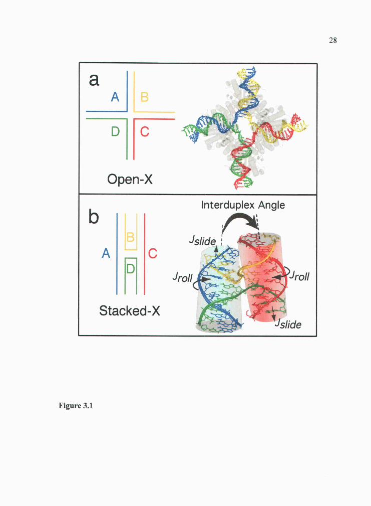
Interduplex Angle
Figure 2.1. DNA Holliday junctions. Schematics of four-stranded DNA junctionsin the parallel stacked-X (a), open-X (b), and antiparallel stacked-X (c) configurations,and definitions for the geometric parameters that vary in the stacked-X form (d). Theparallel stacked-X is the topology of the junction initially proposed by Holliday (1),while the open-X form is seen in protein complexes and the antiparallel stacked-X isseen in the DNA and RNAIDNA junctions. The geometry of the stacked-X structure,as seen in single-crystal structures, varies according to the angle between the stackedduplex arms (interduplex angle), and the sliding (J11) and rolling (Jroii) of these armsalong their respective helical axes.

10
2.2 B.C.Before Crystal Structures
Before any of the crystal structures of junctions were available, the general
architecture and dynamic properties of the Holliday junction were already being
defined through biochemical and biophysical studies on immobilized junctions
assembled from asymmetric sequences (reviewed in (50)). From comparative gel and
solution studies, the junction was known to undergo ion dependent conformational
transitions. In low salt solutions, the junction adopts an extended "open-X" form to
minimize the strong repulsion between negatively charged phosphates at the cross-
over strands that exchange between DNA double-helices (Figure 2.lb). The junction
collapses to a stacked-X form when the phosphates are screened by cations,
particularly by magnesium (Mg2) and other divalent ions. This more compact
junction pairs the arms into coaxially stacked, semicontinuous duplexes, with each
stacked duplex having an outside non-exchanging strand and an inside cross-over
strand. The crossed-over strands can be oriented either in parallel or antiparallel
directions.
Although the parallel configuration of the stacked-X junction (Figure 2.la) has
been suggested to be formed by extrusion of cruciform structures in negatively
supercoiled closed circular DNAs (59, 60) and in synthetic DNA arrays (61), it is the
antiparallel form that is seen by gel (26, 38, 62) and, subsequently, fluorescence
resonance energy transfer studies (27, 28, 63), and by atomic force microscopy in
interlinked sheets of DNA (64, 65). The basic model that emerged for the antiparallel

11stacked-X junction (26, 41) has the arms paired and stacked to form near continuous
B-type DNA duplexes, interrupted only by the U-turn of the junction cross-over. The
two opposing stacked duplexes are related by a right-handed twist to give an
interduplex angle of +60° across the junction. One of the problems with the
antiparallel configuration, however, is that it does not allow migration of the junction
along homologous DNA sequences. A resolution to this problem is that the stacked-X
junction is not static, but can undergo dynamic conformational rearrangements: a
conformational isomerization can occur in which the stacked duplex arms switch
partners in a sequence dependent manner (40, 43, 66). This isomerization transition
has recently been observed in single molecules (25), and has been proposed to involve
the open-X structure (which does support junction migration) as a transient
intermediate conformation (46). Additionally, stacked-X junction isomerization can
affect the genetic outcome of homologous recombination through production of either
patched or spliced gene products. The single crystal structures of DNA junctions and
junctions in complex with proteins are now providing conformational details of the
two junction forms at the atomic level, and, particularly with the stacked-X form, an
understanding of how sequence and ions affect these structural details.
2.3 Single Crystal Structures of DNA Holliday Junctions
The first single-crystal structure of a DNA junction was seen in the Crc
recombination complex (30). There are now structures of at least three unique

12complexes with recombination proteins (56, 67, 68) and, in all cases, the junctions
adopt the extended open-X form, with the DNA geometries determined primarily by
the topology of the binding surfaces of the proteins (reviewed in (49)). The question of
how DNA sequence and other factors affect the inherent structure of the Holliday
junction remains and is being answered by single-crystal structures in the absence of
proteins.
The first DNA Holliday junction crystal structures came not by design, but by
happenstance while attacking other problems. The sequence d(CCGGACCGG) was
the first structure of a DNA Holliday junction, but the initial intent was to study the
effects of tandem GA-mismatches on B-DNA (46). Shortly afterwards, the structure of
a junction with all standard Watson-Crick base pairs (47) was solved as a control
study for the psoralen cross-linked structure of d(CCGGTACCGG) (69). In these and
all subsequent DNA-only studies, the junctions adopt the antiparallel stacked-X
conformation (Figure 2.2) with general structural features that are remarkably similar
to the model proposed in 1988 from solution and gel studies (26, 41). There were,
however, some surprising observations that could not be predicted from earlier studies
on asymmetric junctions, first being that these decanucleotide sequences could be
crystallized as junctions at all. The single-crystal structures of many 10 base-pair
sequences have been reported since 1987 (reviewed in (70)) as B-DNA, A-DNA and
left-handed Z-DNA. In fact, early models of junctions were constructed using
structures of decanucleotides in crystal lattices that resemble the general geometry of

13
Figure 2.2. Core interactions in the stacked-X junction. The structure ofd(CCGGTACCGG) as a junction (ACC-2Na structure) (47) is shown along with thecore interactions at the junction cross-over that account for the formation andgeometry of this DNA conformation. The locations of the core interactions (see Table2.1) in the overall structure are indicated by boxes, and the regions in the boxesenlarged in each inset panel. The C8-C7 and G3-A6 interactions are shown in the topinset panel while the C7-A6 interaction is shown in the middle panel. Blue dotshighlight hydrogen bonding interactions that are important within these interactions,while red dots are used to show hydrogen bonds in the standard Watson-Crick basepairs. The bottom inset panel shows the environment surrounding the sodium ion(Nat) that sits in a cavity formed by the phosphates of two A6 and the bases of the A6and C7 nucleotides.

14the stacked-X junction (71, 72). What became apparent when the actual Holliday
junction structures were solved was that a common A6C7C8 trinucleotide sequence and
an associated set of unique intramolecular interactions define a common core in the
two junctions (47, 73). Since these two early breakthroughs, we are learning that both
sequence and ions at and near this ACC-core affect these interactions, and
consequently the geometry of the stacked-X junction, in both subtle and profound
ways.
2.3 Sequence and ions affect the geometry of DNA junctions
The mismatched and inverted repeat junctions are very similar in structure (73),
but show minor structural differences that could be attributed to an unusual base
interaction at the GA mismatched base pairs (46). The first indication that the
standard geometry of Holliday junctions could be perturbed in a systematic way was
seen in the structure of the d(CCGGTACm5CGG) (48), where m5C is a 5-
methylcytosine. This methylated structure demonstrated that, in addition to the
interduplex angle, the stacked duplexes can be rotated and shifted relative to each
other across the junction cross-over (we label these Jr011 and respectively, 2.1 d),
and that these two geometric parameters were dependent on the interactions at the
junction core. The Jsljde parameter measures the extent to which the stacked helices
translate about their respective helix axes relative to the center of the junction cross-
over, while Jroli rotates these duplexes along the helix axes (a large Jroii opens the

15major groove surfaces of the junction cross-over, with a value of Jroll = 1 8O
indicating that the major and minor grooves of the junction are equally accessible).
There are now 12 junction structures (as of the date of publication) deposited in
the Nucleic Acid Database (74) representing 6 unique sequence types. All of the
structures are in the antiparallel stacked-X form, and vary according to their
interduplex angles, Jr011 and Jslide (Table 2.1). What have we learned from these
structures? Our first lesson is that the sequence requirements at the junction core are
not exclusive to ACC, but can be expanded to Pu6C7Py8, where Pu6 can be either the
purine bases adenine or guanine, and Pys can be cytosine, 5-methylcytosine, or 5-
bromouracil (br5U), but not thymine. This may be expanded further as more sequences
become crystallized as junctions. The terminal base-pairs i Gio) are not critical for
formation of junctions, and can be replaced by T1A10 base pairs, as long as the
trinucleotide core sequence remains intact (75).
As the sequence is changed and/or base substituent groups are added at the
trinucleotide core, it is becoming increasingly clear that the core intramolecular
interactions not only dictate the formation of the junction, but also its geometry as
defined by the interduplex angle and, more significantly, J01i and In this
discussion, we will start by defining a standard set of interactions that are associated
with the reference "unperturbed" d(CCGGTACCGG) junction (ACC-4Na, see Table
2.1 for description of this nomenclature) (47) (Figure 2.2). In this case, 0 A and
Jroii 1 60. The key interactions in this reference structure involve base sub stituents in
the major grooves of the stacked duplex arms and the phosphate oxygens of the

Table 2.1. Structures of DNA Holliday junctions. Each published structure and available single crystal structures of DNA onlyHolliday junctions are listed with the abbreviation used in the review, a description (the sequence, number of DNA strands in theasymmetric unit (asu), important cation clusters in structure and reference), the geometric parameters (interduplex angle (IDA), .Jrlj
and .LIjde), and core interactions.
Structure1 Description of Structure Geometry2 Core Interactions3
Sequence asu Cation Ref4 IDA 1, Jjjde C8-C7 G3-A6 C7-A6 Na-Cavity
ACC-4Na CCGGTACCGG 4 Na (46) 41.4° 159.6° 0 A Direct 1 H20 Direct Na
ACC-2Na CCGGTACCGG 2 Na (78) 42.7 163.5° 2.0 A Direct 1H20 Direct Absent
ACC-2Cal CCGGTACCGG 2 Ca UD0024 39.6° 158.5° 0 A Direct 1H20 Absent H20
ACC-2Ca2 CCGGTACCGG 2 Ca UD0025 39.4° 158.8° 0 A Direct 1H20 1H20 H20
tACC-4Ca TCGGTACCGA 4 Ca (74) 39.6° 159.9° 0 A Direct 1H20 Direct 1-120
tACC-2Ca TCGGTACCGA 2 Ca UD0023 39.6° 168° 3.6A Direct Absent Absent 1120
tACC-2Srl TCGGTACCGA 2 Sr (74) 43.0° 169.0° 2.4 A Direct 21120 11120 Absent
tACC-2Sr2 TCGGTACCGA 2 Sr UD0026 43.3° 169.5° 2.4 A Direct 2H20 1H20 ll20gACC-2Na CCGGACCGG 2 Na (45) 40.6° 174.5° 0 A Direct 11120 Absent 1120
ACmC-2Ca CCGGTACm5CGG 2 Ca (47) 41.3° 170.4° 3.4A 1H20 21120 1H20 Absent
GCC-2Na CCGGCGCCGG 2 Na (75) 40.0° 159.8° 0 A Direct 1H20 Direct Absent
ACbU-2Ca CCAGTACbr5UGG 2 Ca (75) 38.8° 164.6° 2.4 A Absent Absent 11120 Na

Table 2.1 (Continuted)
'Junctions are designated by the core sequence at N6N7N8 positions, number of DNA stands in the asu, the primary cation type locatedin the structure, and, for multiple identical entries, order in which they were listed.
2ThC geometric parameters for the junctions (interduplex angle or IDA, froll, and JcIIde) were calculated as previously described (38).
3The core interactions are C8-C7 (from the N4 nitrogen of the C,, nucleotide base to phosphate oxygen of C7 of the cross-over strands),G3-A6 (from the G3 nucleotide of an outside strand to phosphate oxygen of A6), and C7-A(, (from the N4 nitrogen of the C7 nucleotidebase to phosphate oxygen of A6 of the cross-over strands). These can be direct hydrogen bonds, or meditated by one or more solventmolecules. In addition, the cavity of the junction sandwiched by the phosphate oxygens of the N6 nucleotides of the crossover strands,may contain a sodium ion.
4References are given for published structures, while those that are unpublished are identified by the Nucleic Acid Database accessioncodes.

18junction cross-over, and include 1) a direct hydrogen bond between the N4 nitrogen
of the C8 base and the C7 nucleotide phosphate oxygen (C8-C7 interaction), 2) a
solvent mediated interaction from the complementary G3 nucleotide and the A6
nucleotide phosphate oxygen (G3-A6 interaction) and 3) a direct hydrogen bond from
the N4 nitrogen of the C7 base to the A6 phosphate oxygen (C7-A6 interaction) (2.2,
insets). The first interaction helps explain the requirement for a cytosine at the C8
position, while the last interaction, for the first time, provides a rationale for the C7
cytosine of the original ACC-core trinucleotide. The involvement of the A6 phosphate
in two of these three interactions may explain the importance of having a purine base
at the Pu6 position of the core.
As we change this reference sequence, we see losses in the core interactions that
affect both the structure and stability of the junction. For the most part (with one
exception) structures that maintain all three key interactions effectively adopt the
unperturbed reference structure of the ACC-4Na junction. The GA mismatched
(gCC-2Na) junction (46) has lost the C7-A6 interaction and, consequently, we see a
significant Jr0!1 to open the major groove surfaces. Similarly, the methylated
d(CCGGTACm5CGG) junction (48) (ACmC-2Ca) places additional intervening
solvent into the C8-C7, C7-A6, and G3-A6 interactions, and these perturbations are
associated with large values for both Jslide and Ji.
In the case of the br5U-containing junction of ACbU-2Ca, the first two core
interactions (C8-C7 and G3-A6) are entirely missing, and the C7-A6 interaction becomes
solvent mediated (76). Why then is this not the most perturbed structure? In this
structure, a new water-mediated interaction is introduced between the N7 nitrogen of

the A6 nucleotide along one crossing strand and the phosphate oxygen of the A6
nucleotide of the opposite crossing strand (A6-A6 interaction). This compensating A6-
A6 interaction does not, however, diminish the importance of the three core
interactions for the formation of junctions. Indeed, the ACbU-2Ca sequence, under our
conditions, has been crystallized as both a junction (76) and as resolved B-DNA
duplexes (in a different crystal system), suggesting that the thermodynamic difference
between the two forms in this particular sequence is very small and is associated with
the loss of these core interactions. The sequence d(CCGGCGCCGG) (GCC-2Na) has
also been crystallized in both conformations (76-78), but the crystallization conditions
for the junction versus B-DNA were very different and may account for the
conformational differences. The GCC-2Na junction maintains the three core
interactions and, consequently, is nearly identical to the reference ACC-4Na structure,
even though the adenine A6 of the core trinucleotide has been replaced by a guanine
base.
Cation interactions are expected to be very important in the structure and
formation of the junction, particularly divalent cations. In the crystal structures, a
sodium ion was originally identified in the central cavity of the ACC-4Na structure
(47), and a similar solvent has been observed in several other structures (Table 2.1);
however, its presence or absence in itself does not appear to be a major determinant of
the junction structure or stability. In the case of the ACbU-2Ca junction (76), its
presence may be an added factor that contributes to the stability of the junction in the
absence of 2/3 of the core interactions, while its absence may help account for the
perturbations to Jsljde and Jr011 in the closely related ACC-2Na junction (79).

20The other significant cation interactions involve aqueous calcium (48) and
strontium clusters (75) that sit in the grooves of the stacked duplexes in several
junctions. Again, the effects of these cation complexes on the structure of the junction
depend on how they perturb the core interactions. For example, five strontium-water
complexes were located in the minor grooves of each stacked duplex of the tACC-
2Srl (75) and tACC-2Sr2 junctions. In both cases, additional waters intervene in the
core G3-A6 and C7-A6 interactions, resulting in nearly identical perturbations to Jr011
and Js1ide
2.4 Conclusions and Perspectives
We are now starting to understand how sequence, ions, and drugs (this latter
effect, though not discussed here, is covered previously in (69, 80)) affect the detailed
conformation of the Holliday junction in single-crystals. As with any set of
crystallographic structures, however, we need to address the question of whether the
conclusions that we draw from these studies correspond to how DNA behaves in
solution and in the cell. In most respects, the crystal structures do indeed conform to
the general features of Holliday junctions as defined from previous studies, with one
exception. In all cases, the interduplex angles of the crystal structures are about 200
more shallow than the 6O0 observed from earlier gel mobility, FRET and AFM
studies (26-28, 38, 62-64). One reason for this discrepancy may be that the
crystallographic studies all use symmetric sequences where the junction is fixed in
place by specific intermolecular interactions. In contrast, the non-crystallographic

21studies on junctions were nearly all applied to immobilized junctions that are
assembled from four unique asymmetric sequences and are thus locked in place by
sequence design rather than by any defined set of molecular interactions, other than
standard Watson-Crick base pairing. Consequently, we would expect these sequence
immobilized constructs to be missing the core and even solvent mediated interactions
between the stacked arms and the phosphates at the junction cross-over seen in the
crystal structures and therefore would show significant deviation from the reference
ACC-4Na junction. This is supported by AFM studies which show that indeed the
interduplex angle of asymmetrically locked junctions are while the angle in
similar constructs that incorporate the symmetric ACC core trinucleotide is 43 , as
seen in the crystal structures (65). In addition, these same ACC-containing constructs
were shown by hydroxyl radical footprinting to cross-over at the nucleotide positions
seen in the crystal structures (65). Thus, we can assert that the sequence dependent
interactions at the junction core, as identified in the single crystal structures, are
relevant in other systems and that these interactions do indeed define the
conformational details of the stacked-X junction even in solution.
Are these molecular details important for the participation of Holliday
junctions in recombination-dependent cellular processes? As we know, junctions in
the antiparallel stacked-X form cannot migrate along the DNA sequence, but can do so
if it adopts the open-X form. In solution, stacked-X stability and conformational
switching rates have been shown to be dependent on the population of open-X
junction available for branch migration (7, 81). We see that disrupting one or more of
the core interactions allows the stacked-X junction to roll, slide, and twist. We

22suggest, therefore, that sequences in which this set of core interactions are disrupted
or compromised by intervening solvent will be more conformationally malleable and
thus be more amenable to switching to an open-X form and, consequently, promote
branch migration.
How might these core interactions affect protein binding? As we have already
seen, the junctions in protein complexes all adopt the open-X and not the stacked-X
form. Thus, the barriers to conformational switching that we have discussed above
should also contribute to the energetics of protein bindingany effect that facilitates
formation of the open-X structure should also favor protein binding. In addition, we
see that these core interactions, as they are affected by sequence and ions, specify the
accessibility of the major and minor grooves (as measured by the parameter Jroii).
Many junction-resolving enzymes, such as T4 endonuclease VII (33), are known to
recognize the stacked-X structure from the minor groove face and therefore we see
that accessibility also plays an important role in protein recognition and binding. In
short, it appears that a stacked-X junction that is structurally malleable, which we
propose is determined by a set of core interactions, will also have a lower barrier to
conformational switching and be more accessible, both of which are important for
binding and recognition by junction resolving and DNA repair enzymes. At this point,
however, it is unclear exactly how specific proteins take advantage of the structural
and dynamic features of the stacked-X junction to define their sequence dependent
functions.

23
2.5 Acknowledgement
The work from P. S. Ho's laboratory has been supported by the National Institutes of
Health (R1GM62957A), the National Science Foundation (MCB0090615), and the
Environmental Health Sciences Center at Oregon State University (NIEHS ESOO210).

Chapter 3
Effect of Sequence on the Conformational Geometry of DNA Holliday Junctions
Franklin A. Hays, Jeffrey M. Vargason, and P. Shing Ho
Published in Biochemistry,
American Chemical Society, Washington, D.C., USA
2003, 42, 9586-9597
25
3.1 Summary
Structures of the DNA sequences d(CCGGCGCCGG) and
d(CCAGTACbr5UGG) are presented here as four-way Holliday junctions in their
compact stacked-X forms, with antiparallel alignment of the DNA strands. Thus the
ACC-trinucleotide motif, previously identified as important for stabilizing the
junction, is now extended to PuCPy, where Pu is either an adenine or guanine, and Py
is either a cytosine, 5-methyl cytosine, or 5-bromouracil, but not thymine nucleotide.
We see that both sequence and base substituents affect the geometry of the junction in
terms of the interduplex angle as well as the previously defined conformational
variables, J011 (the rotation of the stacked duplexes about their respective helical axis)
and Jslide (the translational displacement of the stacked duplexes along their respective
helical axis). The structures of the GCC and parent ACC containing junctions fall into
a distinct conformational class that is relatively undistorted in terms of Js/jde and Jr011,
with interduplex angles of 4O-43°. The substituted ACbr5U structure, however, is
more akin to that of the distorted methylated ACm5C containing junction, withjsljde
2.3 A), similar Jr011 (2.5) opening the major groove-side of the junction, but shows a
reduced interduplex angle. In contrast, the analogous d(CCAGTACTGG) sequence
has to date been crystallized only as resolved B-DNA duplexes. This suggests that
there is an electronic effect of substituents at the pyrimidine Py position on the
stability of four-stranded junctions. The single crystal structures presented here,
therefore, show how sequence affects the detailed geometry and, subsequently, the
associated stability and conformational dynamics of the Holliday junction.
3.2 Introduction
The exchange of genetic information across double-helical DNA through
recombination is an important process in DNA biochemistry, and has been implicated
in the mechanisms of DNA repair, replication restart, and viral integration (49). The
critical role of a four-stranded junction as a DNA intermediate in recombination was
proposed nearly forty years ago by Robin Holliday (1). The structure of this four-
stranded complex has been elucidated in detail through a series of single crystal
structures as complexes with proteins (49) and, more recently, as isolated DNA
constructs (80). All of the DNA junctions that have been crystallized to date are
decanucleotides with a common ACC trinucleotide motif (or its methylated variant
ACm5C) at the N6N7N8 positions (represented by the sequence d(CCGGTACCGG),
where the underlined nucleotides are the ACC junction core). Not surprisingly, these
structures have many common structural features. All adopt the compact antiparallel
stacked-X form in which pairs of the duplex arms stack collinearly into nearly
continuous double-helices (broken only at the cross-over point of the junction on the
inside strand of each pair, Figure 3.1). The structure with a methylated analogue of
the AC C-core shows that the substituent group perturbs the stacked-X geometry.
Here, we present the single crystal structures as Holiday junctions of the two

27
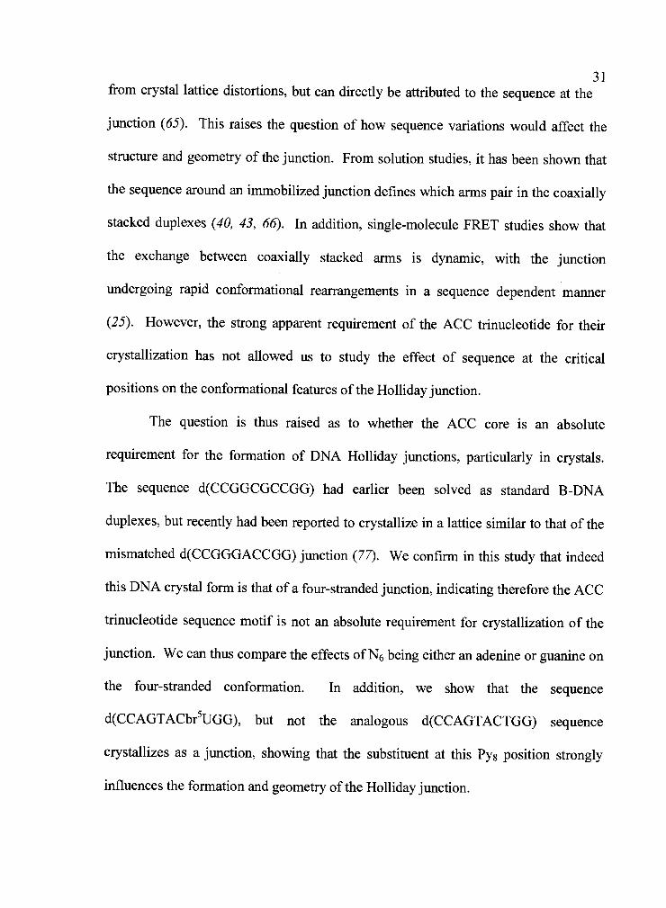
Figure 3.1. Open-X (a) and stacked-X (b) forms of the Holliday junction. Thephosphodeoxyribose backbone of the DNAs are traced with ribbons, and color-codedto distinguish between strands of the junction. a. The open-X form modeled from theDNA in the single-crystal structure of the Cre-loxP complex (67). The protein isshown in grey, with the a-helices rendered as cylinders. b. The compact stacked-Xstructure of the reference d(CCGGTACCGG) single-crystal structure (47). Thecoaxially stacked arms in each column of pseudocontinuous duplexes are shown ascylinders. The conformational parameters of the junction are shown as the twist of thestacked duplexes across the junction (the interduplex angle), the translation of each setof stacked duplexes along their respective helix axes (J1), and the rotation of theduplexes about the axes (J0ii).
DHC
Open-X
Interduplex Angle
sIide
A C
roII roII
Stacked-XsIide
Figure 3.1
28

29sequences d(CCGGCGCCGG) and d(CCAGTACbr5UGG) which do not contain the
ACC junction motif at the core trinucleotide (underlined) positions.
The compact stacked-X form of the Holliday junction was first proposed from
gel mobility assays (38, 62) and fluorescence resonance energy transfer studies in
solution (27, 28, 63) to form under high salt conditions in nonhomologous sequence
constructs that immobilize the four-way junction at specific sites. The general model
from these studies are that high concentrations of cations help to screen the negatively
charged phosphates of the backbone allowing the four arms, which would be splayed
out from the junction in the open-X form (Figure 3.1 a), to pair and coaxially stack to
form two sets of semicontinuous double-helices (Figure 3.lb). The solution studies
also showed that these pairs of stacked duplexes are related across the junction cross-
over by a right-handed interduplex angle of-6O°. This general geometric relationship
was confirmed by atomic force microscopy (64), but the most detailed molecular
details of this conformation have come only from single-crystal X-ray diffraction
studies.
The structure of the Holliday junction has now been determined in a number of
nucleic acid constructs, including RNA-DNA complexes (57, 58) and DNA constructs
with true inverted repeat patterns (47), mismatched base-pairs (46), the cross-linking
drug psoralen (69), and methylated cytosines (48). The DNA structures show that the
geometry of the compact stacked-X junction (Figure 3.lb) was basically that
determined in solution, with the coaxially stacked duplexes related by a right-hand
rotation across the junction cross-over, in all but one case so far, the DNA junction
structures were determined from decanucleotide sequences with a core ACC

30trinucleotide (or its methylated analogue, ACm5C) at the N6N7N8 positions, as in the
sequences d(CCGGGACCGG) (46), d(CCGGTACCGG) (47), and d(TCGGTACCGA)
(75). The one exception to date is the structure of the psoralen cross-linked sequence
d(CCGCT*AGCGG), which was proposed to form a junction because of the
destabilization of the duplex by the drug cross-linked thymine base (T*) (69).
Otherwise, the crystal structures demonstrate the significance of this core ACC
trinucleotide, particularly through the interactions from the cytosine base at the C8
cytosine to the phosphate backbone at the junction cross-overs.
The methylated junction seen in d(CCGGTACm5CGG) first showed that, in
addition to the interduplex angle, the geometry of the junction can also be affected
along and around the helix axes of the collinear duplex arms (48). The methyl
substituent was shown to disrupt the direct hydrogen bonding interactions between the
cytosine C8 base and the phosphate backbone of the cross-over. Furthermore, the
accessibility of the major and minor grooves of the stacked duplex arms were shown
to depend on the rotation of these duplexes along their helix axes (Jroii) and the arms
were seen to slide along their axes (Jslide) to affect the overall symmetry of the
complex. Similar distortions to Jroii were seen to be affected by the types of cation
interactions localized at the junction (75).
The interduplex angle relating the orientation of the stacked duplex arms
across all of these ACC-type junctions are relatively constant, at 40°, which appears
to be more shallow than the 60 seen in solution (50). However, a study of
immobilized junctions that incorporated the ACC trinucleotide motif showed that this
angle, as well as many details concerning the position of the cross-over did not result
31from crystal lattice distortions, but can directly be attributed to the sequence at the
junction (65). This raises the question of how sequence variations would affect the
structure and geometry of the junction. From solution studies, it has been shown that
the sequence around an immobilized junction defines which arms pair in the coaxially
stacked duplexes (40, 43, 66). In addition, single-molecule FRET studies show that
the exchange between coaxially stacked arms is dynamic, with the junction
undergoing rapid conformational rearrangements in a sequence dependent manner
(25). However, the strong apparent requirement of the ACC trinucleotide for their
crystallization has not allowed us to study the effect of sequence at the critical
positions on the conformational features of the Holliday junction.
The question is thus raised as to whether the ACC core is an absolute
requirement for the formation of DNA Holliday junctions, particularly in crystals.
The sequence d(CCGGCGCCGG) had earlier been solved as standard B-DNA
duplexes, but recently had been reported to crystallize in a lattice similar to that of the
mismatched d(CCGGGACCGG) junction (77). We confirm in this study that indeed
this DNA crystal form is that of a four-stranded junction, indicating therefore the ACC
trinucleotide sequence motif is not an absolute requirement for crystallization of the
junction. We can thus compare the effects of N6 being eitheran adenine or guanine on
the four-stranded conformation. In addition, we show that the sequence
d(CCAGTACbr5UGG), but not the analogous d(CCAGTACTGG) sequence
crystallizes as a junction, showing that the substituent at this Pyg position strongly
influences the formation and geometry of the Holliday junction.

323.3 Materials and Methods
All deoxyoligonucleotides were synthesized on an Applied Biosystems DNA
synthesizer in the Center for Gene Research and Biotechnology at Oregon State
University using phosphoramidite chemistry, with the trityl-protecting group left intact
at the 5'-tenninal nucleotide for subsequent purification by reverse-phase HPLC. The
purified DNA was deprotected by treatment with 3% acetic acid for fifteen minutes,
neutralized with ammonium hydroxide, and desalted on a Sigma G-25 Sephadex
colunm. Samples were lyophilized and stored at 80C, then resuspended in deionized
double-distilled water prior to crystallization. All crystals were grown using the
sitting drop vapor diffusion method.
3.3.1 Crystallization and structure of d(CCGGCGCCGG). Crystals were
grown at 25CC from solutions containing 0.5 mM DNA, 25mM sodium cacodylate
buffer (pH 7.0), 7.5mM CaC12, and 0.1mM spermine tetrahydrochloride in the
crystallization drop, and equilibrated against a reservoir solution of 28% (v/v) 2-
methyl-2,4-pentanediol (MPD). These are very different conditions compared to those
that yielded crystals of the B-DNA duplex form of the sequence (82) (B-DNA duplex
crystals were grown at 4C by microdialysis, with solutions containing 2mM DNA,
10mM Tris-HC1, pH 7.0, 150mM MgC1 and equilibrated against the same buffer with
24% MPD).
A thin diamond plate crystal measuring 0.25 mm x 0.25mm x 0.1 mm was
used for data collection and found to be in the monoclinic C2 space group, with unit
cell dimensions a = 66.51 A, b = 24.18 A, c = 37.00 A, and 3 = 110.010. X-ray
33diffraction data were collected to 1.7 A resolution at liquid nitrogen temperatures
using CuKa radiation from a RUH3R generator with an RAXIS-IV image plate
detector. The volume of the unit cell in this space group indicated that there are two
strands of DNA in the asymmetric unit of the crystal lattice. Thus a search model was
constructed using two strands of the d(CCGGTACCGG) Holliday junction (47), one
crossover and one non-crossover, for molecular replacement using EPMR (83) against
2.5A data. A distinct structural solution with two unique DNA strands adjacent to the
crystallographic 2-fold axis yielded a correlation coefficient of 59% and 52%.
Subsequent refinement in CNS (84) using rigid body refinement, followed by
simulated annealing, several rounds of positional and individual B-factor refinement,
and addition of solvent produced final values of = 22.9% and Rfree = 26.0%
(Table 2.1). The coordinates and structure factors have been deposited in the Protein
Data Bank (74) with accession number 1P4Y.
3.3.2 Crystallization and structure determination of d(CCAGTACTGG) and
d(CCAGTACbr5UGG). Crystals of d(CCAGTACTGG) were grown at 20°C from
solutions containing 0.6mM DNA in 5mM Tris-HC1 buffer (pH7.5), 25mM calcium
acetate, and 7% MPD, and equilibrated against 30% MPD. A single crystal measuring
0.2mm x 0.2mm x 0.3 nmi was used for data collection under liquid nitrogen
temperatures on BlO-CARS beamline 14-BMC at the Advanced Photon Source,
Argonne National Labs, with 1 .OA radiation. Crystals of d(CCAGTACbr5UGG) were
grown at room temperature from solutions containing 0.6mM DNA in 5mM Tris-HC1

34
Table 3.1. Data collection and refinement statistics
d(CCGGCGCCGG) d(CCAGTACbr5UGG) d(CCAGTACTGG)
Data Collection
Unit Cell Parameters a = 66.5A, b = 24.2A, a 64.9A, b = 22.6A, a = b 33.4A,
c = 37.OA, 13= 110.00 c 38.IA, 13
= 106.80 c = 87.7A
Space group C2 C2 P6122
Total reflections 50,581 13,013 32,690
Unique reflections 5,137 4,011 2,143
Resolution 50 A - 1.6 A 50 A - 1.8 A 50 A - 2.OA
Completenessa 78.7% (48.0%) 82.0% (34.9%) 94.6% (53.2%)
<I/sigma>a 22.1 (2.7) 23.8 (4.1) 25.3 (3.3)
Rmerge 4.9%(27.8%) 4.1%(18.9%) 4.8%(12.4%)
RefinementResolution 20 A-l.7A 20 A-1.9 A 20 A-2.0 A
(Rfree)C 22.9% (26.0%) 2 1.5% (24.3%) 23.6% (26.8%)
DNA atoms 404 404 202
Solvent atoms 116 89 67
RMSD bond lengthsd 0.007 A 0.005 A 0.0 15 A
RMSD bond angles' 1.24° 0.88° 1.75°
Conformation Junction Junction B-DNA
avalues in parentheses refer to the highest resolution shell
bRmerge = hk1 L 'hId, 1 - <I / 'hid, where I,,,,, is the intensity of a reflection and isthe average of all observations of this reflection and its symmetry equivalents.
CRcryst = hk1 FObS kFcaic I / hkI IFObSI. Rfree = for 10% of reflections that were not used inrefmement (84). All refinements were performed targeting maximum likelihood.
Root mean square deviation of bond lengths and angles from ideal values.

35buffer (pH=7.5), 120mM calcium acetate, and 16% MPD, and equilibrated against
20% MPD. A 1 .9A dataset was collected on this crystal at liquid nitrogen
temperatures using CuKa radiation from the in-house RUH3R generator with an
RAXIS-IV image plate detector. Crystals of the d(CCAGTACTGG) are in the
hexagonal space group P6122 with unit cell dimensions a = 33.38 A, b = 33.38 A, and
c = 87.67 A, while those of d(CCAGTACbr5UGG) are in the monoclinic space group
C2 with unit cell dimensions a = 64.84 A, b = 22.58 A, c = 38.07 A, and 1O6.78.
The structure of d(CCAGTACTGG) was solved by molecular replacement
against a 2.0 A dataset using a previously solved B-DNA duplex model in a similar
hexagonal lattice. Resulting solutions from an EPMR search yielded a correlation
coefficient of 64.2% and 47.4%. Subsequent refinements in CNS using rigid
body refinement, simulated annealing followed by standard positional and individual
B-factor refinement, and addition of solvent resulted in final values of = 23.6%
and Rfree = 26.8%.
The lattice of d(CCAGTACbr5UGG) crystal was similar to previously
crystallized DNA junctions, suggesting that this br5U analogue of the
d(CCAGTACTGG) sequence could in fact be a four-stranded complex. Thus, the
structure of this sequence was solved by molecular replacement using the two unique
strands of the d(CCGGTACm5CGG) junction structure (48) as the initial search model
in an EPMR search. The search resulted in a position and orientation of the model
having a correlation coefficient of 74% and R-Factor of 44.5% for the best solution.
An initial round of rigid body refinement and simulated annealing in CNS yielded
values of R,=42.5% and Rfree 43.5%. Omit maps calculated with the crossover
36phosphates removed and after simulated annealing of the edited model showed
distinct F0-F density consistent with the cross-overs of the junction and
discontinuities in phosphodiester backbones along the stacked DNA duplexes. Thus,
the structure of d(CCAGTACbr5UOG) was refined as a DNA Holliday junction with
two strands, one crossover and one non-crossover, in the asymmetric unit, with the
two additional related strands generated by a crystallographic two-fold axis to form a
complete four-stranded complex. Refinement was carried out in CNS using rigid body
and simulated annealing routines followed by standard positional and individual B-
factor refinement producing a final RS=2 1.5% and Rfree= 24.3% after addition of
solvent. The coordinates and structure factors have been deposited in the Protein Data
Bank (74) with accession number 1P4Z for d(CCAGTACTGG) and 1P54 for
d(CCAGTACbr5UGG).
All data was reduced using the HKL suite of programs (85). Root-mean-
square-deviation (RMSD) values were calculated using the algorithm (86)
implemented in the program ProFit v2.2 (http://www.bioinf.org.uk/software/profit/).
Structural analysis was performed with CURVES 5.2 (87) and X3DNA (88).
3.4 Results
The crystal structures presented here show that d(CCGGCGCCGG) and
d(CCAGTACbr5UGG) both crystallize as four-stranded Holliday junctions in the
compact antiparallel stacked-X form, with the former nearly identical to that of the

37parent d(CCGGTACCGG) junction (47) and the latter most closely related to that
of the methylated sequence d(CCGGTACm5CGG) (48). The sequence
d(CCAGTACTGG), however, was determined to be resolved B-DNA duplexes, now
in two unrelated crystal forms. Thus, this study shows that the A6C7C8 trinucleotide
motif identified as important for stabilizing the junction can accommodate other
nucleotide bases at the 6th and 8th positions. In addition, the two bases have their own
distinct effect on the conformation of the detailed intramolecular interactions and,
consequently, the overall geometries relative to previously determined structures.
Thus, the resulting junction structures will be interpreted through comparisons
between the current and previously reported structures in their respective structural
classes, with the d(CCGGTACCGG) structure serving as the reference for the
unsubstituted and d(CCGGTACm5CGG) for the substituted junctions. These
structures therefore allow us to define each component of the junction geometry,
including the interduplex angle, rolling of the stacked duplex columns relative to each
other (Jrøji) and sliding of one junction relative to the other along their helical axes
(Jslide), and how they can be affected by the sequences at the core of the junction.
3.4.1 Structure of d(CCGGCGCCGG) as a Holliday Junction. The single
crystal structure of the sequence d(CCGGCGCCGG) (which we will call the GCC-
junction), originally solved as a B-DNA duplex (82) under different crystallization
conditions, was recently reported to crystallize in a crystal lattice that is isomorphous
with that of a DNA four-way junction (77). We show here that, under our
crystallization conditions, the sequence indeed is a Holliday junction. The complete

38
5' 3'
a C1G10II
Li5' 3' CG9 3' 1C1G10 G3C8 )
C2G9 G4C7G3C8 C5G6G4C7TG605C5G6-'-07G4G605 C8G3C7G4 G9C2C8G3 G15C1G9C2 3' 5' 5'
IO 5'3'
C c8-
:'<c C7
Figure 3.2. Crystal structure of d(CCGGCGCCGG) as a DNA Holliday junction.a. Sequence topology of the d(CCGGCGCCGG) junction. The four-strandedantiparallel stacked-X Holliday junction is generated by applying the crystallographictwo-fold symmetry to the two unique strands (bold). Strands are numbered from 1 to10 in the 5' to 3' direction, with the inside crossing strands colored red and the outsidenoncrossing strands colored blue. b, The atomic structure of d(CCGGCGCCGG).Chemical bonds in the structure are rendered as sticks and the phosphodeoxyribosebackbone rendered as a solid ribbon (colors and strand designations are as in a, figurerendered with Insightil from MSI/Biosym, Inc.). c, Electron density map. The 2F0-Fmap (contoured at 1 c) shows the discontinuity in electron density between nucleotidesG6 and C7 in the stacked DNA duplexes, but bridging between adjacent stackedduplexes to form the junction cross-overs (panel created with Bobscript (89)).

junction is generated by 2-fold symmetry applied to the two unique strands (one
outside noncrossover and one inside crossover strand) of the asymmetric unit. This
four-stranded complex (Figure 3.2a) has four armstwo longer six base pair anns
(from the C1 to G6 of one outside strand paired with G10 to C5 of one cross-over
strand) and two short four base pair arms (airing the C7 to G10 of the same outside
strand to G4 to Ci of an alternative cross-over strand. Each six base-pair duplex
domain is stacked over a four base-pair duplex to form the near continuous ten base
pair helices of the stacked-X junction (Figure 3.2b), with the cross-over occurring
between nucleotides G6 and C7 of the inside cross-over strands (Figure 3.2c).
In nearly all respects, this GCC-junction is identical to that of the reference
d(CCGGTACCGG) structure (ACC-junction, Figure 3.3, RMSD 0.43 A for all
common DNA atoms). The stacked double-helical arms are related by an interduplex
angle of 40.0° in the GCC-junction as compared to the 41.4° angle in ACC (Table 3.2).
Furthermore, there is no sliding of the duplexes across the junction cross-over in GCC
(Js1ide 0 A) relative to the ACC-junction. The exposure of the major and minor
grooves are also nearly identical between the sequences, with J.0i1 values = 159° in
both junction structures.
One interesting difference between the GCC-j unction and other DNA junctions
to date is that the width of the minor groove (as measured by the phosphorus to
phosphorus distance, Table 3.3) is wider at the CcGdG6Cs and G6CsICrG4
dinucleotide steps (18.5 A and 19.5 A respectively compared to the averages of 17.5 A
(0.26A S.D.) and 18.8 (0.35 A S.D.) for ACN type junctions). In addition, we see that
the G6C5/C7G4 dinucleotide steps are underwound by 3° as compared to the

Table 3.2. Comparison of global conformational geometry values ford(CCGGCGCCGG) (GCC) and d(CCAGTACbr5UGG) (ACbr5U) with previousjunction structures of d(CCGGTACCGG) (ACC, (47)) and d(CCGGTACm5CGG)(ACm5C, (48)).
Junctiona Interduplex Angle j1.b Jip
ACC 41.4° 159.6°
ACm5C 41.3° -1.7A per duplex 170.4°
GCC 40.00 oA 159.8°
ACbr5U 38.8° 1 .2 A per duplex 164.00
a
Trinucleotide core region corresponding to N6-N7-N8 positions of thegeneral sequence d(CCN3N4N5N6N7N8GG)
bRelative to d(CCGGTACCGG) junction, as estimated by the slide seen
for one set of duplexes when the opposing stacked duplexes aresuperimposed with those of the ACC-junction. Values are for theduplexes on both sides of the junction, sliding symmetrically. If one setof stacked duplexes are superimposed, then the opposing stacked double-helices would show a slide of twice the value reported in this table (see,for example, Figure 3.7).
Angle between vectors extended from the center of the junction topoints that bisect the phosphates on the two outside strands thatcomplement the nucleotides at the gap of the junctions (see for exampleFigure 3.4.)
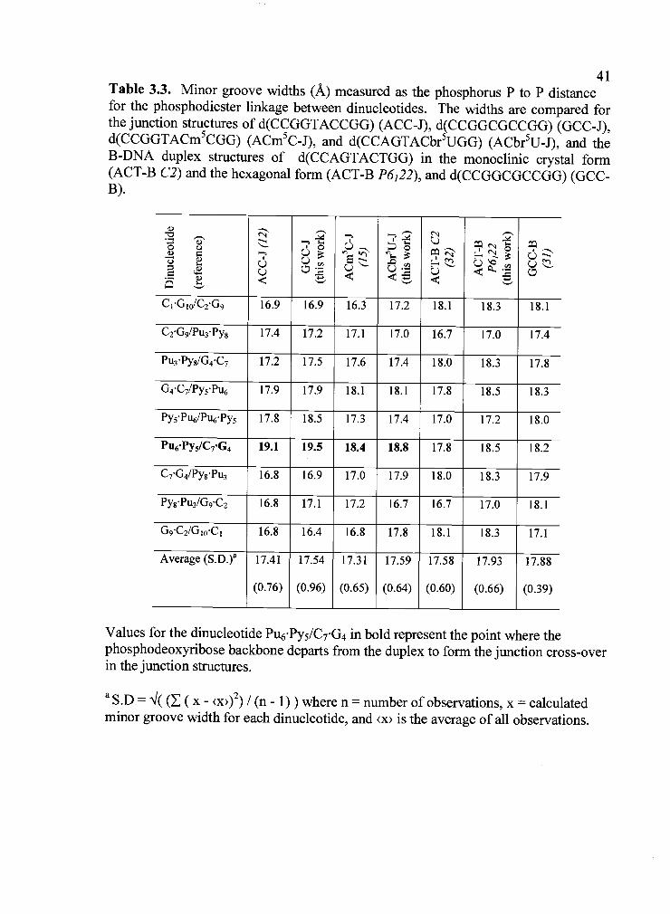
41Table 3.3. Minor groove widths (A) measured as the phosphorus P to P distancefor the phosphodiester linkage between dinucleotides. The widths are compared forthe junction structures of d(CCGGTACCGG) (ACC-J), d(CCGGCGCCGG) (GCC-J),d(CCGGTACm5CGG) (ACm5C-J), and d(CCAGTACbr5UGG) (ACbr5U-J), and theB-DNA duplex structures of d(CCAGTACTGG) in the monoclinic crystal form(ACT-B C2) and the hexagonal form (ACT-B P6122), and d(CCGGCGCCGG) (GCC-B).
Q--i
i
2-.ö LCIC.)
E
0L)
C1G10/C2G9 16.9 16.9 16.3 17.2 18.1 18.3 18.1
C2G9/Pu3Py8 17.4 17.2 17.1 17.0 16.7 17.0 17.4
Pu3Py8/G4C7 17.2 17.5 17.6 17.4 18.0 18.3 17.8
G4C7/PyçPu6 17.9 17.9 18.1 18.1 17.8 18.5 18.3
Py5PuIPuPy5 17.8 18.5 17.3 17.4 17.0 17.2 18.0
Pu6Py5/C7G4 19.1 19.5 18.4 18.8 17.8 18.5 18.2
C7G4/PysPu3 16.8 16.9 17.0 17.9 18.0 18.3 17.9
Py8Pu3/G9C2 16.8 17.1 17.2 16.7 16.7 17.0 18.1
G9C2/G10C1 16.8 16.4 16.8 17.8 18.1 18.3 17.1
Average (S.D.)a 17.41 17.54 17.31 17.59 17.58 17.93 17.88
(0.76) (0.96) (0.65) (0.64) (0.60) (0.66) (0.39)
Values for the dinucleotide Pu6PysIC7G4 in bold represent the point where thephosphodeoxyribose backbone departs from the duplex to form the junction cross-overin the junction structures.
a S.D 'I( ( (x - <x>)2) / (n - 1)) where n = number of observations, x = calculatedminor groove width for each dinucleotide, and x is the average of all observations.

42Table 3.4. Comparison of helical twist. Measured using a global axis in CURVES5.1 (28), for the junction structures of d(CCGGTACCGG) (ACC-J),d(CCGGCGCCGG) (GCC-J), d(CCGGTACm5CGG) (ACm5C-J), andd(CCAGTACbr5UGG) (ACbr5U-J), and the B-DNA duplex structures ofd(CCAGTACTGG) in the monoclinic crystal form (ACT-B C2) and the hexagonalform (ACT-B P6122), and d(CCGGCGCCGG) (GCC-B).
-u' cö-.. o
L)
C1G10/C2G9 38.0° 38.00 37.4° 45.1° 30.1° 26.5° 36.3°
C2G9/Pu3Py8 41.1° 36.7° 38.5° 43.9° 50.5° 51.8° 37.3°
Pu3Py8/G4C7 39.0° 41.8° 38.1° 34.6° 22.1° 26.9° 36.4°
G4C7/PyçPu 34.3° 33.4° 37.0° 28.8° 34.9° 37.2° 35.5°
Py5Pu6/Pu6Py5 32.1° 33.7° 41.5° 37.9° 47.8° 32.7° 30.1°
Pu6Py5/C7G4 33.6° 30.6° 26.5° 32.6° 34.9° 37.2° 36.1°
C7G4/Py8Pu3 40.9° 43.0° 40.0° 32.9° 22.1° 26.9° 42.6°
PygPu3/G9C2 39.9° 37.4° 38.6° 51.7° 50.5° 51.8° 24.8°
G9C2IG10C1 41.3° 42.0° 38.9° 30.9° 30.1° 26.5° 44.0°
Average (S.D.)a 37.80° 37.40° 37.39° 37.6° 35.89° 35.28° 35.90°
(3.55) (4.31) (4.30) (7.69) (11.28) (10.33) (5.81)
Values for the dinucleotide Pu6Pys/C7G4 in bold represent the point where thephosphodeoxyribose backbone departs from the duplex to form the junction cross-overin the junction structures.
aS.D = °'I( ( (x - <x>)2) / (n - 1)) where n = number of observations, x = calculated
helical twist for each dinucleotide, and <x> is the average of all observations.

43
Figure 3.3. Superposition of d(CCGGCGCCGG) and d(CCGGTACCGG)junctions. The structures of the two junctions are superimposed using the commonatoms of the stacked arms on the right of the junction in this figure. In superpositionof the overall structure, RMSD = 0.426 A for all common DNA atoms.

44analogous A6T5IC7G4 dinucleotide step in the junctions (2.4), and significantly
underwound (by 5.5°) compared to the comparable steps in the B-DNA duplex of the
same sequence. In fact, the duplexes in all of the junction structures are underwound
by an average of '-2° (37.54°, S.D. = 4.99°) compared to their B-DNA counterparts
(35.69°, S.D. 9.08°) (Table 3.4), and all of the dinucleotides that span the junction in
these structures are underwound by -8° relative to the identical or analogous
sequences as standard B-DNA (Table 3.4).
Not surprisingly, many of the structural details seen in the GCC-junction are
similar to those in the ACC-junction. For example, the direct hydrogen bonding
interaction between the amino N4-nitrogen at the major groove surface of the cytosine
C8 base to the phosphate oxygen of C7 that was first identified as helping to stabilize
the ACC-junction (Figure 3.4a) is also seen in the current GCC-junction. The
associated solvent mediated hydrogen-bonding interaction that bridges the keto-
oxygen of the complementary G3 nucleotide to the cross-over phosphate oxygen at A6
in the ACC-junction (51 in Figure 3.4a), however, is now a network in the current
GCC-junction (Figure 3.4b), similar to that seen previously in the ACm5C-junction
structure. Thus, the commonality of the direct C8 amino to C7 phosphate hydrogen
bond and the similarities of the junction geometries across these structures suggest a
correlation between this particular interaction at the atomic level and the general
conformational features for this class of structures. However, the solvent that was
assigned as a sodium ion in the center of the ACC-junction is not present in the current
structure, suggesting that this solvent interaction is not crucial to the overall geometry
of this structural class.

45
Figure 3.4. Atomic interactions within the trinucleotide core of thed(CCGGCGCCGG) junction viewed perpendicular to the junction dyad axis.Hydrogen bonds are shown as dotted lines and solvent molecules as spheres. Thenucleotides in the junction cross-over are labeled, along with the major (M) and minor(m) grooves. The 1roii angles are shown as the angle between the vectors extendingfrom the center of the junction to points that bisect the phosphates of the outsidestrands that complement the nucleotides of the junction gap in the inside strands. a,The reference d(CCGGTACCGG) junction. The important interactions had beenidentified as the direct hydrogen bond between the amino N4-nitrogen of C8 to the C7phosphate oxygen 02P, a water mediated hydrogen bond between the keto 06-oxygenof G3 and G6 phosphate oxygen 01 P, and solvent (sodium ion) coordinated to the 02Pphosphate oxygens of the G6 nucleotides (47). b, The d(CCGGCGCCGG) junction.This structure shows the direct hydrogen bond between nucleotide C8 and the crossingphosphate, and a set of interactions mediated by solvent (Si to S3) from the majorgroove of the G3 nucleotide to the opposing crossing phosphates. The solvent arelabeled as follows: Si is a water molecule that is hydrogen bonded to the Pu3nucleotide that complements the Py8 of the junction trinucleotide core; S2 is a waterthat is hydrogen bonded directly to the Py8 position of the junction core; S3 is thesolvent (proposed as the Na+ ion displaced from the center of the ACC-junction) thatsits between the phosphates of the junction cross-over strands.

46
Figure 3.4

473.4.2 Structure of d(CCAGTACTGG) as B-DNA duplexes. Our experience
with the d(CCGGCGCCGG) sequence indicated that, although originally reported to
be a B-DNA duplex (90), we should study the structure of the sequence
d(CCAGTACTGG) in our crystallization solutions. However, even under conditions
that have yielded junctions, this sequence crystallized as double-stranded B-DNA,
although in a different crystal form than was previously reported. The space group of
the current crystals is hexagonal P6122, as compared to the earlier monoclinic C2 form
(90). In both crystal forms, the asymmetric unit is one strand of the decanucleotide,
with the second strand generated by the crystal symmetry. This precludes the
sequence in either crystal form from being a Holliday junction since at least two
unique strands are required to form the crossed four-stranded complex. The current
and previous B-DNA structures of d(CCAGTACTGG) are very similar, RMSD
1 .03A for all nonhydrogen atoms, and similar minor groove widths and helical twists
at each dinucleotide step along the double-helix (Tables 3.3 & 3.4). The noticeable
differences result from differences in the duplex-duplex interactions of the two crystal
lattices. For example, the duplexes in the monoclinic crystal form have the
nonstandard Bli conformations (as defined by a dihedral angle l8O) at the
phosphoribose spanning the C2pA3, G4pT5, and T8pG9 dinucleotide steps along the
DNA chain. In contrast, only the C2pA3 dinucleotide step shows this Bli
conformation in the current hexagonal crystal form.
3.4.3 Structure ofd(CCAGTACbr5UGG) as a Holliday Junction. The sequence
d(CCAGTACbr5UGG) was initially designed as a means to introduce a bromine heavy

48
--,.w-
:.
I ,.
.
:!A
',t'tA
I I.
Figure 3.5. Crystal structure of d(CCAGTACbr5UGG) as a DNA HollidayJunction. a, Sequence topology of the d(CCAGTACbr5UGG) junction. The four-stranded antiparallel stacked-X Holliday junction is generated by applying thecrystallographic two-fold symmetry to the two unique strands (bold). Strands arenumbered from 1 to 10 in the 5' to 3' direction, with the inside crossing strandscolored red and the outside noncrossing strands colored blue. b, The atomic structureof d(CCAGTACbr5UGG). Chemical bonds in the structure are rendered as sticks, thephosphodeoxyribose backbone rendered as a solid ribbon, and bromines rendered asspheres (colors and strand designations are as in a, figure rendered with Insightil fromMSIIBiosym, Inc.). c, Electron density map. The 2F0-F map (contoured at icT)
shows the discontinuity in electron density between nucleotides G6 and C7 in thestacked DNA duplexes, but bridging between adjacent stacked duplexes to form thejunction cross-overs (figure in panel created with Bobscript (89)).
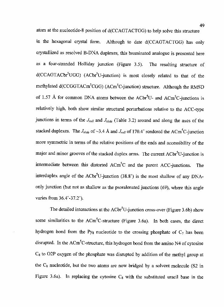
49atom at the nucleotide-8 position of d(CCAGTACTGG) to help solve this structure
in the hexagonal crystal form. Although to date d(CCAGTACTGG) has only
crystallized as resolved B-DNA duplexes, this brominated analogue is presented here
as a four-stranded Holliday junction (Figure 3.5). The resulting structure of
d(CCAGTACbr5UGG) (ACbr5U-junction) is most closely related to that of the
methylated d(CCGGTACm5CGG) (ACm5C-junction) structure. Although the RMSD
of 1.57 A for conmion DNA atoms between the ACbr5U- and ACm5C-junctions is
relatively high, both show similar structural perturbations relative to the ACC-type
junctions in terms of the Jroii and Jsljde (Table 3.2) around and along the axes of the
stacked duplexes. The J1td of-3.4 A and Jr011 of 170.4° rendered the ACm5C-junction
more symmetric in terms of the relative positions of the ends and accessibility of the
major and minor grooves of the stacked duplex arms. The current ACbr5U-junction is
intermediate between this distorted ACm5C and the parent ACC-junctions. The
interduplex angle of the ACbr5U-junction (38.8°) is the most shallow of any DNA-
only junction (but not as shallow as the psoralenated junctions (69), where this angle
varies from 36.4°-37.2°).
The detailed interactions at the ACbr5U-junction cross-over (Figure 3.6b) show
some similarities to the ACm5C-structure (Figure 3 .6a). In both cases, the direct
hydrogen bond from the Py8 nucleotide to the crossing phosphate of C7 has been
disrupted. In the ACm5C-structure, this hydrogen bond from the amino N4 of cytosine
C8 to 02P oxygen of the phosphate was disrupted by addition of the methyl group at
the C8 nucleotide, but the two atoms are now bridged by a solvent molecule (S2 in
Figure 3.6a). In replacing the cytosine C8 with the substituted uracil base in the

50ACbr5U-junction, the hydrogen bond donating amino group of the cytosine has
been replaced by a hydrogen bond accepting keto-oxygen. This precludes the
formation of the direct hydrogen bonding interaction, regardless of the substituent
group added to the C5 carbon of the base. A solvent (S2, Figure 3.6b) remains
hydrogen bonded to this keto-oxygen, but does not span the gap to the phosphate
oxygen of the junction cross-over.
The ACC-, GCC-, and ACm5C-junctions all show a network of one or more
solvent molecules that help bridge the complementary G3 nucleotide base of the C8G3
base pair to the crossing phosphate of A6. The analogous A3 nucleotide of the br5U8-
A3 base pair shows no such network of bridging solvent molecules. We had
previously suggested that one of the bridging solvent molecules in the ACm5C
network (S3, Figure 3.6a) was the sodium ion in the center of the ACC-junction, but
displaced by the collapse of the junction core. The loss of this series of solvent
between A3 and the A6 phosphate would suggest that the ion could have buried itself
back into the center of the ACbr5U-junction, and the electron density in the center of
the junction supports that assertion. A solvent molecule was seen to be sandwiched
between, and located very close to (<2.5A), the O1P phosphate oxygen atoms of the
crossing A6 nucleotides (Nat, Figure 3 .6b). It is interesting, however, that the volume
estimated for this binding site corresponds to a sphere with radius 1.25 A, which is
now larger than that of the ACm5C junction (at 0.9 A radius) and even the unmodified
ACC junction (at 1.13 A radius) (91). Finally, a new solvent (S8, Figure 3 .6b) is seen
spanning the O1P oxygens of the C7 cytosines in both crossing strands.
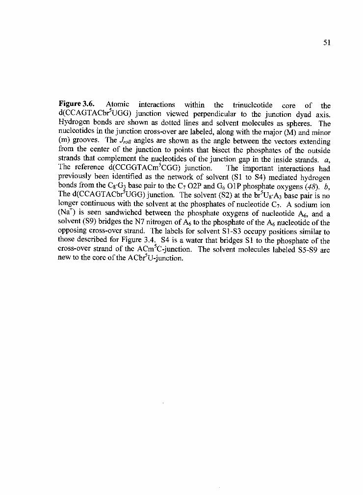
51
Figure 3.6. Atomic interactions within the trinucleotide core of thed(CCAGTACbr5UGG) junction viewed perpendicular to the junction dyad axis.Hydrogen bonds are shown as dotted lines and solvent molecules as spheres. Thenucleotides in the junction cross-over are labeled, along with the major (M) and minor(m) grooves. The Jrol1 angles are shown as the angle between the vectors extendingfrom the center of the junction to points that bisect the phosphates of the outsidestrands that complement the nucleotides of the junction gap in the inside strands. a,The reference d(CCGGTACm5CGG) junction. The important interactions hadpreviously been identified as the network of solvent (Si to S4) mediated hydrogenbonds from the C8G3 base pair to the C7 02P and G6 O1P phosphate oxygens (48). b,The d(CCAGTACbr5UGG) junction. The solvent (S2) at the br5U8A3 base pair is nolonger continuous with the solvent at the phosphates of nucleotide C7. A sodium ion(Nat) is seen sandwiched between the phosphate oxygens of nucleotide A6, and asolvent (S9) bridges the N7 nitrogen of A6 to the phosphate of the A6 nucleotide of theopposing cross-over strand. The labels for solvent S1-S3 occupy positions similar tothose described for Figure 3.4. S4 is a water that bridges Si to the phosphate of thecross-over strand of the ACm5C-junction. The solvent molecules labeled S5-S9 arenew to the core of the ACbr5U-junction.

52
Figure 3.6

53Thus, in the current structure of the ACbr5U-j unction, there are no direct or
solvent mediated hydrogen bonds between the major groove surface of the br5U8A3
base pair and the phosphate oxygens of the junction cross-over, even though many of
the solvent molecules remain hydrogen bonded to the bases at the major groove
surface. There is, however, an interaction at the other end of the Pu6C7Py8
trinucleotide motif, with the N7 nitrogen of the A6 nucleotide base now bridged
through a solvent molecule (S9, Figure 3.6b) to the 02P oxygen of the A6 nucleotide
of the opposite crossing strand. This would seem to compensate for the loss of
interactions between the stacked arms and the core of the junction that appears to be
critical to the stability of the junction in this system.
3.5 Discussion
In this study, we have determined the single crystal structures of the sequences
d(CCGGCGCCGG) and d(CCAGTACbr5UGG) as four-stranded Holliday junctions in
the antiparallel stacked-X conformation, and the structure of d(CCAGTACTGG) as
resolved B-DNA duplexes. The two new junctions are the first such structures from
sequences that do not contain the previously identified ACC-core trinucleotide
sequence at the N6N7N8 positions of the general sequence d(CCN3N4N5N6N7N8GG).
Thus, we have now expanded this central sequence motif for the crystallization of
junctions to Pu6C7Py8, where Pu is either a guanine or adenine purine-containing
nucleotide and Py is a cytosine, 5-methylcytosine, or 5-bromouracil pyrimidine

54nucleotide, but not a thymine nucleotide. Our previous study with the methylated
d(CCGGTACm5CGG) sequence (48) showed that perturbations to the overall
geometry of the Holliday junction can include a slide in the stacked duplexes across
the junction (Jsljde) and a roll of the duplexes along their helix axes (f,.011). With the
current study, we see that sequence and substituent effects define distinct classes of
stacked-X junctions: the AC C-type structure (the original junction structure), and
ACm5C class (which shows significant Jslide and Jr011 relative to ACC). In addition,
these effects are seen to perturb the interduplex angle relating the orientation of the
stacked duplex arms across the junction.
A comparison of the GCC- and ACC-junctions show that the Pu6 position of
the Pu6C7Py8 motif does not dramatically perturb the conformation of the Holliday
junction. The only noticeable effect on the general geometry of the conformation in
replacing the adenine with a guanine base at this position is to make the interduplex
angle shallower (from 41.4° in ACC to 40° in the GCC-structure). It is not surprising
that the two structures are so similar. The nucleotide at the N6 position is the initial
point of departure of the DNA strand that crosses-over to form the junction. However,
the purine base at this position remains paired with the complementary strand of the
stacked duplex arms at this point and is not directly involved in the interactions of the
cross-over.
In contrast to Pu6, the base and the substituent at the Py8 position of the now
expanded Pu6C7Py8 trinucleotide motif has a significant affect on the conformation of
the junction. The sliding (Js/,de) and rolling of the duplexes along and around their
respective helix axes (Jroii) previously defined in the d(CCGGTACm5CGG) structure

55are also observed here with the br5U deoxynucleotide at the Py8 position, but are
less exaggerated. Thus, the Jsljde distortions are primarily induced by steric
interactions between the methyl or bromo-substituent and the deoxyphosphoribose
backbone of the cross-over strands. h the current structure, the bromine is in direct
contact with one of these phosphates. Thus, we can think of the junction as the pivot
point not only to twist the stacked duplexes around (in defining the interduplex angle),
but to push against and thus slide these columns of base pairs relative to each other.
The rolling the stacked duplexes about their respective helical axes (Jroii) has
the effect of opening the major groove surface of the junction. What we see by
comparison of these structures is that the direct interaction between the C8 cytosine
and the cross-over phosphates fixes the orientation of the opposing stacked duplexes
of the ACC- and 0CC-junctions. This is not surprising, since hydrogen bonding
interactions involving nucleotide bases have well defined geometric constraints in
terms of distances and angles between hydrogen bond donors and acceptors. Thus,
such interactions predetermine the relationships between the major groove surface of
the arms and the core phosphates of the junction. The addition of intervening solvent
molecules, as in the ACm5C and ACbr5U junctions, can be thought of as "lubricating"
the interaction surfaces, relieving the constraints imposed by the ACC- and GCC-
interactions and thereby allowing more variations within the geometries of the
junctions, allowing variability in the interduplex angle (as seen in the more shallow
angle of the ACbr5U-junction), as well as perturbations to and Jroll (evident in
both the substituted structures). This does not, however, suggest that the structure of
the junction itself is more flexible. A comparison of all the junction crystal structures

56show that the nucleotides directly at and those paired to nucleotides at the cross-
over have lower average temperature or B-factors than those nucleotides that are
distant from the junction core.
The other distortion seen in the d(CCAGTACbr5UGG) structure, relative to all
other junctions, is the shallower interduplex angle. This angle, which is approximately
400 (ranging from 40° to 41.4°) for all PuCC type junction structures, becomes 38.4°
with the ACbr5U structure. The distortions to Jsljde and Jr011 means that the best
comparison is between the two structures with substituents at the Py8 position
(ACm5C and ACBr5U), which places these two structures in their own class. In this
comparison, we see that the br5U base induces a rotation about the junction relative
to the ACm5C-junction, which results from substitution of an A3br5U8 for the
G3m5C8 base pair. The rotation of the interduplex angle is thus not simply a
substituent effect, but more likely is associated with the Abr5U base pair at this
position.
Why does sequence have such an effect on the structure of the junctions? The
answer to this question may be seen in the interactions at the ACbr5U trinucleotide
that compensate again for the loss of interactions between the major groove of the
Pyg Pu3 base pair to junction phosphates seen in all of the other structures of the
complex. There were no direct or even solvent mediated bridges at this surface.
However, there is a compensating solvent bridge from A6 across to the phosphate of
the opposite crossing strand. We can now imagine this as being a factor that both
stabilizes and defines the interduplex angle of the ACbr5U junction.

57Alternatively or in addition, we had previously proposed that the interduplex
angle is defined by a short phosphate-phosphate interaction that is distant (three base
pairs away) from the actual junction cross-over (69). We again see this very short
oxygen to oxygen distance (3.69 A) and, in the GCC-junction structure, observe two
solvent molecules that are 2.4 A from one set of the phosphate oxygen atoms. If
these two closely interacting solvents are assigned as sodium ions rather than waters,
then the net charge at the phosphate of the N10 nucleotide would be +1, which would
provide a rational for this short interarm phosphate-phosphate distance. This short
phosphate-phosphate distance remains even in the ACm5C (at 3.5 A) and ACbr5U (at
3.6 A) structures. In both junctions, the direct amino nitrogen to the cross-over
phosphate of C7 in the ACC-junction has been disrupted (by the methyl group of
ACm5C and, additionally, by the replacement of the amino nitrogen with a keto
oxygen in ACbr5U). This supports a model that the components of the helical
structure of the duplex arms (twist, rise, tilt, etc.) help to define this angle. For
example, in the current ACPy structures, we see that both the average helical twist
overall and across the unique trinucleotide steps at C2-Pu3-G4 and C7-Pys-G9 are
significantly shallower in the ACbr5U-junction as compared to the ACm5C-junction.
The orientation of the phosphates in close approach would in fact result in a shallower
interduplex angle when the double-helical arms are unwound in this class of shifted
and rolled junctions (Figure 3.7).
In the original ACC-junction, we had observed a solvent molecule (which was
assigned as a sodium cation) imbedded in a central cavity formed by the phosphates of
the crossing strands. We now observe this same solvent molecule in the center of the

58
Figure 3.7. Superposition of the d(CCAGTACbr5UGG) junction (blue) on thed(CCGGTACCGG) junction (red). Atoms are rendered as stick models with thephosphodiester backbone traced by ribbons. One set of stacked duplex arms (yellowcylinder) from each structure was used for superposition. a, View along the junctionswith the nonsuperimposed stacked duplex arms in front. The ACbr5U-junction shows
2.3 A Jsljde along the helix axis. b, View into the junctions with the superimposedstacked arms to the right. The non-superimposed stacked duplexes show the andJr011 effects imposed by the br5UA base pair of the ACbr5U core trinucleotide on theoverall geometry of the Holliday junction.

Figure 3.7
59

60ACbr5U-junction, but not the ACm5C or GCC structures. We can thus conclude
that neither the Jr011 nor Jsljde distortions alone account for the accessibility of the
junction to the intruding ion. Why does the ACbr5U trinucleotide favor formation of
crystals of the junction while ACT does not? The difference is obviously the bromine
versus the methyl group at the C5 carbon of the Pys nucleotide base. Since the two
substituents are nearly identical in size (both approximately 2 A radius) and
hydrophobicity, the difference in behavior likely arises from differences in their
electronic propertiesbromine is electron withdrawing, making it slightly electron
rich, while methyl groups are electron donating to an aromatic ring system and thus
are slightly electron poor. One can suggest then that the electrostatic interactions
between the substituent and the close phosphate group from the junction cross-over
might be the distinguishing factor; however, this would predict that the methyl rather
than bromo-group should stabilize the junction, which is the opposite of what is
observed here. We suggest, therefore, that the electrostatic effect is indirect, affecting
for example the base pair stacking. The helical twist at the CGfbr5UA dinucleotide
steps in the ACbr5U-junction are 8 to lO larger than the comparable CG!TA steps
of the B-DNA structures (Table 3.4). This is true regardless of whether the base pairs
are at the junction core (CrG4Ibr5Us.A3) or not (A3br5U8/G4C7), indicating again that
the effects of nucleotide sequence and substituents on the structure of the stacked
duplex arms of the junction mirror those seen in standard B-DNA duplexes (69). This
difference in the helical twist at the junction core would dramatically affect the
position of the substituent groups relative to the crossing phosphates. In the ACbr5U-
junction, the bromine sits against the phosphate oxygen and above the deoxyribose

61ring of the preceding C7 nucleotide. A rotation of this base pair by _100 in the stack
(as would be expected in a theoretical ACT-junction) would place the analogous
methyl group of an ACT junction core nearly equidistant from the two crossing
phosphates, and in contact with neither. One possible explanation therefore for why
the thymine does not facilitate crystallization of the junction is that the effects of the
methyl group on the electron distribution of the thymine base results in a base-stacking
geometry that does not force the substituent to push against the phosphates of the
junction and, consequently, would not induce a of the stacked helices. The result
may then be that the solvent bridged interaction between adenine A6 (across the gap of
the inside strands of the complex) could not be formed. Other factors, including
solvent effects, may contribute to or may be more important than the electronic effect
on base stacking described here.
Thus, we see from this series of structures, that the overall geometry of the
junctions varies according to the nucleotides within the trinucleotide core sequence
motif. This is consistent with the understanding that the nucleotides immediately
flanking the junction determines how helical arms pair to form the stacked duplexes
(40, 66), and the rates of conformational isomerization to switch these pairing partners
(25). The current structures therefore provide an atomic level view of how these
sequence variations would affect the stability of the four-way stacked-X junction,
which also defines the ability of such junctions to undergo conformation dependent
dynamic processes, including branch migration and protein binding.

623.6 Acknowledgements
We thank Drs. Z. Wood and R. Faber for helpful discussion during data
refinement, and Dr. J. Watson for critical reading of the manuscript. This work was
funded by grants from the National Institutes of Health (R1GM62957A) and the
National Science Foundation (MCBOO9O6 15). The X-ray diffraction facilities were
supported by the Proteins and Nucleic Acids Facility Core of the Environmental
Health Sciences Center at OSU (NIEHS ESOO21O) and a grant from the Murdock
Charitable Trust. Use of the Advanced Photon Source was supported by the U.S.
Department of Energy, Basic Energy Sciences, Office of Science, under Contract No.
W-31-109-Eng-38. Use of BioCARS Sector 14 was supported by the National
Institutes of Health, National Center for Research Resources, under grant number
RR07707.
Chapter 4
Influence of Minor Groove Substituents on the Structure of DNA HollidayJunctions
Franklin A. Hays, Zebulon J.R. Jones and P. Shing Ho
Published in Biochemistry,
American Chemical Society, Washington, D.C., USA
2004, 43 (30), 98 13-9822

4.1 Summary
The inosine (I) containing sequence d(CCIGTACm5CGG) is shown to
crystallize as a four-stranded DNA junction. This structure is nearly identical to the
antiparallel junction formed by the parent d(CCGGTACm5CGG) sequence (48) in
terms of its conformational geometry, and inter- and intramolecular interactions within
the DNA and between the DNA and solvent, even though the 2-amino group in the
minor groove of the important G3m5C8 base pair of the junction core trinucleotide
(italicized) has been removed. In contrast, the analogous 2,6-diaminopurine sequence
d(CCDGTACTGG) crystallizes as resolved duplex DNAs, just like its parent sequence
d(CCAGTACTGG) (76). These results demonstrate that it is not the presence or
absence of the 2-amino group in the minor groove of the R3Y8 base pair that specifies
whether a sequence forms a junction, but the positions of the extracyclic amino and
keto groups in the major groove. Finally, the study shows that the arms of the junction
can accommodate perturbations to the B-DNA conformation of the stacked duplex
arms associated with the loss of the 2-amino substituent, and that two hydrogen
bonding interactions from the C7 and Y8 pyrimidine nucleotides to phosphate oxygens
of the junction cross-over specify the geometry of the Holliday junction.

65
4.2 Introduction
DNA Holliday junctions are ephemeral intermediates that have been
implicated in the mechanisms of a large number of biological processes. A four-
stranded DNA junction was proposed by Holliday over 40 years ago (1) to help
explain the process of gene conversion in yeast and has since been implicated in a
diverse array of related mechanisms including genetic recombination, DNA repair,
and viral integration (5, 17, 24, 55, 92). The involvement of these structural
intermediates over such a broad range of biological processes, typically serving to
protect and expand the coding potential of genomes, has made junctions the focus of
intense study. A recent series of single crystal structures reveal how interactions
between the major groove of the double-helical arms and the phosphates at the
junction cross-over help to define the formation of these complexes. We present here a
systematic study to elucidate the contribution of the minor groove 2-amino substituent
groups on the formation of four-stranded junctions in crystals.
The conformation of DNA Holliday junctions is now understood to be highly
variable. Solution studies have shown that junctions exist in two general
conformationsthe open-X and stacked-X forms (Figure 4.1). The open-X form
predominates under low salt conditions, with the four B-DNA arms arranged in
essentially a square planar configuration to minimize the electrostatic interactions
between the negatively charged phosphates of the phosphoribose backbones. High
concentrations of monovalent and divalent cations help to shield these phosphates,
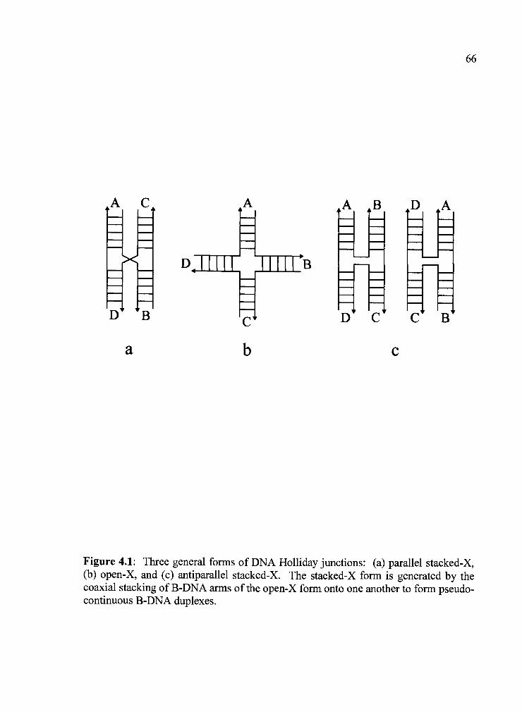
I---'- - ------U-
III- -- -
C
Figure 4.1: Three general forms of DNA Holliday junctions: (a) parallel stacked-X,(b) open-X, and (c) antiparallel stacked-X. The stacked-X form is generated by thecoaxial stacking of B-DNA arms of the open-X form onto one another to form pseudo-continuous B-DNA duplexes.

67allowing the junction to assume the more compact stacked-X structure in which the
arms are coaxially stacked to form two semi-continuous B-DNA duplexes. In the
stacked-X structure, the strands of the B-DNA arms can, in theory, assume either a
parallel or antiparallel relationship (26, 62). To date, only the antiparallel form has
been definitively observed in solution (26, 41). Furthermore, the duplexes of the
antiparallel stacked-X junction can be related in a left or right handed orientation
(Figure 4.1 c). The antiparallel junction can be locked in one specific isomeric form or
can be dynamic and readily switch between forms with the open-X structure serving as
an intermediate (25, 43, 66).
Several single crystal structures of these important biological intermediates
have now been determined at the atomic level (reviewed in (80, 93)). Comparison of
junction forming sequences has identified a trinucleotide core motif as being
instrumental to the formation of these four-stranded structures. The sequence of this
trinucleotide core was originally defined as A6C7C8 (47), but later expanded to
Pu6C7Py8, where Pu is either the adenosine or guanosine purine nucleotides, and Py
can be a cytosine, 5-methylcytosine (m5C) or 5-bromouridine (br5U) pyrimidine-type
nucleotide, but not thymidine (76). These nucleotides are involved in a number of
crucial interactions that link the major groove surfaces of the stacked duplex arms to
the cross-over phosphates. Atomic force microscopy (AFM) and nuclease digestion
studies on synthetic junctions that incorporate the core ACC trinucleotide show the
same overall geometry and cross-over topology as the crystal structures (65).
The conformation of junctions in solution and in the crystals appears to be
related to these sequence dependent intramolecular interactions. A detailed

comparison of the available crystal structures show that the junctions can be
grouped into two classes. Those structures that show a set of direct hydrogen bonding
interactions, between i) the Py8 base and the 02P phosphate oxygen of C7, ii) the
outside G3 nucleotide with the O1P phosphate oxygen of Pu6, and iii) the N4 amino
group of C7 and the O1P phosphate oxygen of Pu6, have a well-defined geometry,
while structures in which one or more of these interactions are missing or mediated by
solvent show greater variability in conformation (93). Thus, it has become
increasingly clear that the nucleotides within, and adjacent to the cross-over region of
DNA Holliday junctions play key roles in defining the global geometry and formation
of the stacked-X conformation.
Up to this point, it has not been clear what effect interactions in the minor
groove may have on the formation and geometry of the junction. For example, a
hexaaquo-calcium (II) ion has been observed in the minor groove as a common feature
of the junction arms in several of the higher resolution structures, which may play an
electrostatic role in stabilizing the complex. The currently available structures,
however, do not directly tell us how important the substituent interactions are in this
narrow groove. The sequence d(CCGGTACm5CGG) forms a junction (48), but
d(CCAGTACTGG) does not (76). In this comparison, we see that the extracyclic
amino and keto substituents at the major groove surface are switched between the G3-
m5Cs and A3-T6 base pairs, but the difference in conformation may arise instead from
the lack of a 2-amino group in the minor groove of the A3-T6 base pairs of the latter
sequence. To distinguish between the effects of minor groove and major groove
substituents, we have studied the single crystal structures of the sequences
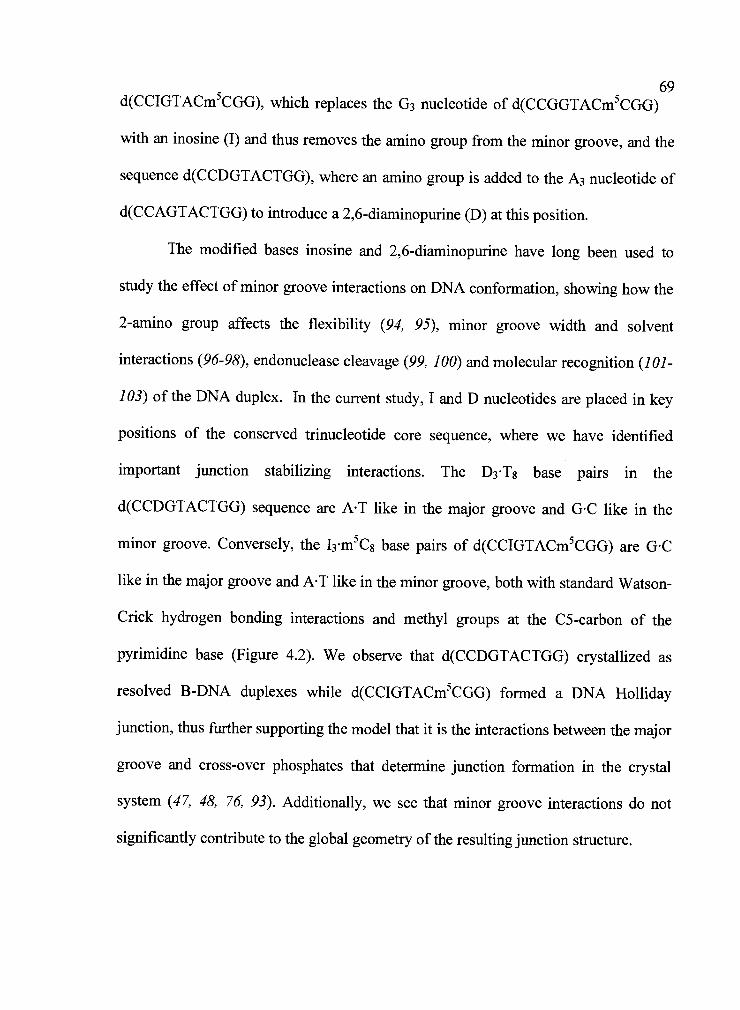
d(CCIGTACm5CGG), which replaces the G3 nucleotide of d(CCGGTACm5CGG)
with an inosine (I) and thus removes the amino group from the minor groove, and the
sequence d(CCDGTACTGG), where an amino group is added to the A3 nucleotide of
d(CCAGTACTGG) to introduce a 2,6-diaminopurine (D) at this position.
The modified bases inosine and 2,6-diaminopurine have long been used to
study the effect of minor groove interactions on DNA conformation, showing how the
2-amino group affects the flexibility (94, 95), minor groove width and solvent
interactions (96-98), endonuclease cleavage (99, 100) and molecular recognition (101-
103) of the DNA duplex. In the current study, I and D nucleotides are placed in key
positions of the conserved trinucleotide core sequence, where we have identified
important junction stabilizing interactions. The D3T8 base pairs in the
d(CCDGTACTGG) sequence are AT like in the major groove and GC like in the
minor groove. Conversely, the I3m5C8 base pairs of d(CCIGTACm5CGG) are GC
like in the major groove and AT like in the minor groove, both with standard Watson-
Crick hydrogen bonding interactions and methyl groups at the C5-carbon of the
pyrimidine base (Figure 4.2). We observe that d(CCDGTACTGG) crystallized as
resolved B-DNA duplexes while d(CCIGTACm5CGG) formed a DNA Holliday
junction, thus further supporting the model that it is the interactions between the major
groove and cross-over phosphates that determine junction formation in the crystal
system (47, 48, 76, 93). Additionally, we see that minor groove interactions do not
significantly contribute to the global geometry of the resulting junction structure.
04 N6 II_/Ia?. S
T)-(A .Jm02
C5M N4.....
\:I::ILc:
S. * *5.
02
[iI M
m
70
C5M04 N6
* ,
5
55*02 N2
Figure 4.2: Standard and modified nucleotide base pairs located in the R3-Y8 basepair step. Exocyclic substituents are labeled with the location of the major (M) andminor (m) groove faces denoted adjacent to each base pair. Fm5C base pair is similarto a &m5C base pair from the major groove face (light yellow) and an AT base pairfrom the minor groove face (light blue). A DT base pair is similar to an AT base pairfrom the major groove (light green) and a &m5C base pair from the minor groove(light red). All base pairs have standard Watson-Crick hydrogen bonding interactionsas indicated by green circles between each base. 5-methyl cytosine bases are used forconsistency with the parent d(CCGGTACm5CGG) junction structure and thymidinenucleotides in the Y8 position.

71
4.3 Materials and Methods
All deoxyoligonucleotides were synthesized using solid-phase
phosphoramidite chemistry with the trityl-protecting group left attached to the 5'-
terminal hydroxyl group to facilitate subsequent purification from failure sequences
via reverse phase HPLC. Preparative reverse phase HPLC was performed using a
Microsorb 300 A pore size C-18 column measuring 250 mm X 21.4 mm ID at a flow
rate of 8 mi/mm in 0.1 mM triethylamine acetate with an acetonitrile gradient. The full
length sequence typically eluted at 1 minutes post-injection in a mobile phase
concentration of 20% acetonitrile. Purified tritylated DNA was deprotected by
treatment with 3% aqueous acetic acid for fifteen minutes with gentle agitation,
neutralized with concentrated ammonium hydroxide, and desalted on a Sigma G-25
Sephadex column yielding purified DNA. Purified DNA was stored at -80° C as a
lyophilized powder and resuspended in deionized double-distilled water prior to use.
All crystals were grown in 10 p.1 sitting drops by the vapor diffusion method and
equilibrated against a 30 ml reservoir volume.
4.3.] Crystallization and structure solution of d(CCIGTACm5CGG). Thin
diamond crystals of d(CCIGTACm5CGG), in the monoclinic C2 space group with
dimensions a = 64.9 A, b 24.9 A, c = 37.1 A, and = 109.2°, were grown from
solutions containing 25 mM Na-Cacodylate (pH 7.0), 0.5 mM DNA, 180 mM CaC12
and 2.4 mM spermine tetra-hydrochioride equilibrated against 20% vol/vol 2-methyl-
2,4-pentanediol (MPD) at 25° C. X-ray diffraction data to 2.0 A resolution was
72collected at liquid nitrogen temperatures using CuKa radiation from a Rigaku
(Tokyo) RU-H3R rotating anode generator operating at 50kV and lOOmA with an
Raxis-IV image plate detector on a single crystal measuring 0.55 X 0.25 X 0.05 mm.
An artificial mother liquor containing 25% MPD was used for cryoprotection. The
space group and unit cell dimensions of d(CCIGTACm5CGG) are very similar to
previous junction crystals of the parent d(CCGGTACm5CGG) sequence, with two
strands in the asymmetric unit (a.s.u.). Thus a search model using the
d(CCGGTACm5CGG) junction (48) was used in EPMR (83) to solve the structure by
molecular replacement, initially to 3.0 A resolution. A distinct solution with a
correlation coefficient of 77.5% and = 36.2% was obtained with two unique
strands in the a.s.u. adjacent to a crystallographic 2-fold symmetry axis. A simulated
annealing omit-map with the C7 crossover phosphate removed showed distinct F0-F
density within the crossover region indicative of a DNA Holliday junction.
Subsequent refinement of this initial junction structure in CNS (84) using rigid body
refinement, simulated annealing, several rounds of positional and individual B-factor
refinement, as well as addition of solvent resulted in a final of 23.6% and Rfree of
25.8% (Table 4.1).
4.3.2 Crystallization and structure solution of d(CCDGTACTGG. Long rod-
like crystals of d(CCDGTACTGG) were grown at room temperature in 5 mM
TRJS(HCI) (pH 7.5), 23 mM calcium acetate, 0.6 mM DNA, and 16% MPD
equilibrated against a reservoir solution of 28% MPD. Crystals were flash frozen
directly out of the drop prior to data collection. Data for a crystal measuring 0.30 X
0.1 X 0.1 mm in size was collected to 1.85 A resolution at liquid nitrogen

73temperatures on BioCARS beamline 14-BMC at the Advanced Photon Source,
Argonne National Labs, with 0.9 A radiation. The crystals were indexed in the
hexagonal space group P6122 with unit cell dimensions a 33.0 A, b 33.0 A, and c
= 88.6 A, and are isomorphous to the crystals of the parent d(CCAGTACTGG)
sequence. The structure of d(CCDGTACTGG) was solved by molecular replacement
using the coordinates of the d(CCAGTACTGG) structure (UD003O). An EPMR
search (against 3.0 A data) using this initial search model of one strand adjacent to a
crystallographic symmetry axis produced a correlation coefficient of 77.1% and
of 37.5%. Initial round of rigid body and rigid parts refinement on this model followed
by simulated annealing produced a starting of 35.3% and Rfree of 38.8%.
Structure refinement was carried out in CNS using standard positional and individual
B-factor refinement followed by assignment of solvent molecules producing a final
of 23 .2% and Rfree of 25.8% (Table 4.1).
All data were processed and reflections merged and scaled using DENZO and
SCALEPACK from the HKL suite of programs (85). Resolution cutoffs were
determined according to <I/c(I)>, completeness, Rmerge, and Rmeas statistics. Rmeas was
determined using the program available at http://loop8.bio1ogie.uni-konstanz.de/-kay/
and root-mean-square-deviation (RMSD) values were calculated using the McLachlan
algorithm as implemented in the program ProFit v2.2
(http://www.bioinf.org.uk/software/profit/) (86). Structural analysis was performed
using CURVES 5.2 (87). The coordinates and structure factors have been deposited in

74
Table 4.1. Data collection and refinement statistics
Data Collection
Unit Cefl
a (A)
b(A)
c(A)
f3 angle (°)
Space group
Total reflections
Unique reflections
Resolution (A)
Completeness(%)a
I/sigmaa
Rmerge(%)'
Refinement
Resolution
Rcryst (Rfree)(%)C
DNA toms
Solvent atoms
Average B-Factors (A2)
DNA atoms (Solvent atoms)
RMSD bond lengths (A)4
RMSD bond angles (0)4
d(CCIGTACmCGG) d(CCDGTACTGG)
64.9
24.9
37.1
109.22
C2
9439
3492
18.2-2.0
93.4 (61.8)
15.8 (2.4)
5.0 (29.1)
18.2-2.2
23.57 (25.82)
404
62
11.1 (11.7)
0.003
0.8
Structural conformation Holliday Junction
avalues in parentheses refer to the highest resolution shell
33.0
33.0
88.6
P6(1)22
34659
2528
17.5-1.85
96.3 (72.2)
29.9 (4.98)
5.6 (24.5)
17.5-1.90
23.21 (25.83)
203
45
10.9 (20.6)
0.067
6.2
B-DNA
bRmergehk1 i I 1hki, - <lhki / E1kI i lw where I is the intensity of a
reflection and <I>w is the average of all observations of this reflection and itssymmetry equivalents.
CRcrysr = Fobs kFcatc / h1d IFobsI. Rfree = for 10% of reflections thatwere not used in refinement (104).
4Root mean square deviation of bond lengths and angles from ideal values.
the Protein Data Bank (105) with accession number iS 1 L for d(CCIGTACm5CGG)
and 1S1K for d(CCDGTACTGG).
4.4 Results
75
Major and minor groove interactions are known to affect the local and global
conformation of nucleic acid structures (106-109). The crystal structures presented
here elucidate the role of nucleotide substituent groups in the minor groove and, by
extension, the major groove in the formation of DNA Holliday junctions. The
sequence d(CCIGTACm5CGG) was seen to crystallize as a DNA Holliday junction
while d(CCDGTACTGG) crystallized as resolved B-DNA duplexes. In terms of base
substituents, DT base pairs are AT like from the major groove and GC like from the
minor groove while Fm5C base pairs are GC like from the major groove and AT like
from the minor groove. Thus, this study demonstrates that specific atomic interactions
which have been identified at the major groove surface of the trinucleotide core region
of DNA Holliday junctions are specific for GC base pairs in the major groove and do
not accommodate AT base pair substituents. Furthermore, the inosine junction
structure provides an opportunity for direct comparison with the parent
d(CCGGTACm5CGG) junction structure (ACm5C junction) to determine whether the
minor groove 2-amino group in the R3-Y8 base step helps to define the geometry of the
junction conformation.
764.4.1 Structure of d(CCIGTACm5CGG) as a Holliday Junction. The single-
crystal structure of the d(CCIGTACm5CGG) sequence (iACm5C junction) is a DNA
Holliday junction in the right-handed antiparallel stacked-X conformation. The full
four-stranded structure consists of two unique strandsone crossover and one
noncrossoverrelated by a crystallographic 2-fold symmetry axis with the crossover
occurring between nucleotides A6 and C7 of each set of pseudocontinuous B-DNA
duplexes. These duplexes are formed by the stacking of a shorter four base pair arm
(nucleotides C1 to G4 of an outside non-crossover strand with C7 to G10 from a
crossover strand) onto a longer six base pair arm (nucleotides T5 to G10 of an outside
strand stacked on nucleotides C1 to A6 of a crossover strand) (Figure 4.3).
The overall conformation of this junction, indeed of any crystal structure of four-
stranded junctions, can be described by the geometric relationships between the set of
stacked B-DNA arms and the center of the junction. We have defined these
relationships and how they can be accurately measured very explicitly elsewhere (48,
110), and will briefly describe them here. The most easily recognized of these
relationships is the interduplex angle (IDA), which is the angle that relates the ends of
the stacked arms to the center of the junction cross-over. This is the angle that was
estimated to be 6O in solution and by AFM for synthetic junctions (26, 38, 62, 64,
111) that are fixed by sequence complementarity of four unique strands. The IDA
when measured in this way, however, is a length dependent parameter; therefore, we
have defined the parameter as a measure of the angle formed by the helical axes
projected onto the resolving plane of the junction. This is length independent and,
thus, is a more general descriptor for the angle relating the stacked arms of this

77
Figure 4.3: DNA Holliday junction crystal structure of d(CCIGTACm5CGG). (a)Nomenclature used for the d(CCIGTACm5CGG) junction with the full four-strandedcomplex generated by the application of a crystallographic two-fold symmetry axis tothe two unique strands (blue and orange). An asterisk adjacent to the eighthnucleotide in each decamer sequence denotes the presence of a 5-methyl cytosinebase. (b) Atomic structure of the d(CCIGTACm5CGG) DNA Holliday junction.Nucleotides are rendered as sticks with the phosphodeoxyribose backbone rendered asa solid ribbon with the 5t and 3' ends labeled accordingly (panel was rendered withInsightil from MSIIBiosym, Inc.). (c) 2F0-F electron density (contoured at 1 c)depicting the junction crossover region with continuous density between nucleotidesA6 and C7 of the junction crossover strands. Nucleotides T5, A6, C7, and m5Cgrendered as sticks. Coloring scheme and strand designations in all panels are the same(panel created with Bobscript (89) and rendered in Raster3D (112)).

78
4 S p
...
I 4
._.,l.ti.
-
I .I
.::-:..-
---_
I;i
I:i., '.- .,
::
Figure 4.3

79structure. In addition, the stacked B-DNA arms can translate along and rotate about
their respective helix axes. These are reflected in the parameters Jslide and Jroji,
respectively. The actual IDA for any crystal structure is thus a function of Jslide,
Jroli, and the length of each arm.
The structure of the iACm5C junction is nearly identical to that of the parent
ACm5C junction in terms of its global geometry and detailed interactions. The global
conformation of the iACm5C junction structure (Js1id = 2.23 A, itwist = 42.5°, Jroll =
143.6° and IDA 61.4°) is nearly identical to that of the parent ACm5C junction (Jslide
= 2.25 A, jjwisi 44.3°, Jr011 = 145.9° and IDA = 60.0°) in terms of these geometric
parameters and as measured by the RMSD between like atoms (0.5 10 A). The detailed
interactions within the iACm5C junction are also very similar to the parent structure,
with minimal deviations. We have previously observed (47, 48, 76) that the global
junction conformation is dependent on a set of key atomic interactions at the
trinucleotide core region (Figure 4.4). These interactions include: i) direct hydrogen
bond between the exocyclic amine of C8 and a phosphate oxygen on the C7 backbone
phosphate (C7-Y8 interaction), ii) a solvent mediated interaction between outside R3 (R
is either an adenine or guanine nucleotide) keto oxygen and a phosphate oxygen of N6
(R3-N6 interaction), and iii) a direct hydrogen bond between the O1P phosphate
oxygen of N6 and the exocyclic amine of C7 (N6-C7 interaction). Consistent with this
observation we see here that the solvent mediated interaction between the 13 and A6
nucleotides (R3-N6 interaction) is indeed present and mediated by a solvent molecule
similar to that seen in the original d(CCGGTACCGG) junction (47). The N6-C7
interaction is also present and mediated by a solvent molecule similar to the parent

E:1&
Figure 4.4: Stabilizing interactions within the stacked-X core region. Structure of theparent ACm5C junction is shown with the location the R6C7Y8 trinucleotide coreregion hydrogen bonding interactions indicated by boxes. The area within these boxesis enlarged, as indicated by the corresponding box colors (red and blue), providingmore detailed information on each stabilizing interaction. For comparison, the coreregion interactions are also shown for the unperturbed junction structure of thesequence d(CCGGTACCGG) (47). Within these inset boxes Watson-Crick hydrogenbonding interactions are indicated by green circles while core region atomicinteractions are shown as orange circles. The C7-Y8 and R3-N6 interactions are shownat the top while the A6-C7 interaction is shown in the bottom box. DNA atoms arerendered as sticks and colored by strand with the respective 3' and 5' termini noted.No direct or solvent mediated interaction is observed between the C7 02P and m5CgN4 atoms (distance between these atoms is 4.4 A as indicated by the black arrow)within the iACm5C-J structure.

81
d(CCIGTACm5CGG) d(CCGGTACCGG) d(CCGGTACm5CGG)
(C7-C8) (C7-m5C8)
13 (13-A6) G3 (G3-A6) G3 (G3-A6)
(A6-C7) (A6-C7) (A6-C7)
IIA J ''- C7L1....
S..
°O. . N4 At oif
Figure 4.4

82ACm5C junction structure (48). It was surprising that no direct or solvent mediated
interaction was observed between the N4 exocyclic amine of C8 and the O1P
phosphate oxygen of C7 (C7-Y8 interaction) in this inosine containing structure. This
C7-Y8 interaction had been observed as a direct or solvent mediated set of hydrogen
bonding interactions in all but one of the previous DNA Holliday junction structures,
suggesting that it is significant. In the case of the one previous exception and the
current structure, the distance between the 02P of C7 and N4 of C8 is similar to that
when solvent mediated interactions have been observed, suggesting that solvent is
present, but was not resolved in the crystal structures.
Ino sine has been shown to affect structural details of the DNA duplex,
including groove widths, solvent interactions, and flexibility. In this case, we observe
similar effects on the B-DNA arms of the iACm5C junction. The width of the minor
groove (as measured by the distance from the 13 to G9 backbone phosphorous atoms)
adjacent to the I3m5C8 base pair is narrowed by 0.6 A relative to the parent ACm5C
junction. This had previously been attributed to the loss of the minor groove 2-amino
group (106, 107, 113, 114). This narrower groove disrupts the solvent structure within
the groove. Most notably, the hexaaquo-calcium (II) ion that sits in the minor groove
at nucleotide positions G3 and C8 in the ACm5C junction is no longer present in the
current structure (Figure 4.5), yet the global conformation of the iACm5C junction is
nearly identical to the ACm5C junction. Thus, we can conclude that the presence of
the minor groove calcium ion in the ACm5C junction does not contribute significantly
to the perturbed conformation. The absence of this calcium ion was rather surprising
given the twenty-fold higher CaCl2 concentration in the crystallization solutions of the

83
Figure 4.5: Minor groove solvent interactions adjacent to R3Y8 base step in DNAHolliday junctions. (a) Minor groove of d(CCIGTACm5CGG) structure between theC2G9 and G4C7 base pairs. 2F0-F electron density (contoured at lc) is shown inblue for minor groove solvent molecules assigned within this region. Solventmolecules are labeled with distances between solvent waters S7-S9 and SlO being:S7-SlO = 2.9 A, S8-SlO = 4.2 A, and S9-SlO = 2.6 A. (b) Minor groove of thed(CCGGTACm5CGG) Holliday junction structure. (c) Minor groove of thed(CCAGTACbr5UGG) Holliday junction structure. View for all panels is looking intothe minor groove with solvent water rendered as green spheres and calcium ionsrendered as purple spheres. Interactions between calcium ions and solvent water aredenoted by solid lines colored green and purple. All nucleotides are rendered as sticksand colored according to atom type (figure created with Bobscript (89) and rendered inRaster3D (112)).

84
Figure 4.5

85iACm5C junction as compared to the ACm5C junction. In fact, no cations were
identified in the structure of the iACm5C junction, although solvent is located in close
proximity to the position where the calcium ion has been identified in previous
junction structures (Figure 4.5). Any attempt to model this solvent as a calcium ion,
even with reduced occupancies, resulted in a significant increase in Rfree.
Additionally, there is no ordered solvent around this location in the electron density
maps that indicate anything other than water molecules in the minor groove.
The loss of the minor groove 2-amino group not only disrupts the solvent
interactions but increases the propeller twist of the I3m5C8 base pair and the G4C7
base pair 3' to the inosine base (Table 4.2). These perturbations are thought to increase
duplex flexibility in the region missing a minor groove 2-amino group (95, 107).
Greater flexibility is often manifested by an increase in propeller twist (115) and
alteration of the backbone as observed by the presence of the BlI backbone
conformation (113, 116). It is interesting to note that both inosine residues within the
iACm5C junction adopt the unusual Bli conformation, as is consistent with these
previous studies. Comparison of the inosine junction with the parent ACm5C junction
shows that both guanine residues in the corresponding positions of the ACm5C
junction also adopt this unusual backbone conformation. Upon closer inspection of the
junction structures it is apparent that the unusual backbone conformation in this
location of the structures results from crystal packing interactions and not a loss of the
2-amino group in the minor groove. Additionally, a Bil conformation observed in the
inosine structure for G19, yet not observed in the ACm5C junction, is also caused by
crystal packing interactions (the 01 P of G19 is 3.3 A from a symmetry related 03' of

86
Table 4.2. Comparison of conformational parameters for DNA Holliday junctionstructures of the d(CCGGTACm5CGG) (ACm5C-J), d(CCIGTACm5CGG) (iACm5C-J) and d(CCAGTACbr5UGG) (ACbr5U-J) sequences, and B-DNA structures ofd(CCAGTACTGG) in the monoclinic (ACT-B C2) and hexagonal (ACT-B P6122)crystal forms, d(CCDGTACTGG) (dACT-B) and d(CCGGCGCCGG) (GCC-B).
-o
-
E .. C.))
0C.)
-S
C.)
R' '-INC.)
N NNI
N N
MinorGroove C2G9/Pu3Py8 17.14 16.80 17.04 16.66 17.06 17.41 17.37
Width (A)"
Pu3Py8/G4C7 17.62 17.00 17.40 18.00 18.20 18.10 17.80dihedra1 171.1 134.0 -105.4 -86.2 -81.5 -100.4 152.4
angle (°)Pu3
(BIT) (B!!) (BI) (BI) (BI) (BI) (BIT)
Pu13167.0 170.2 -94.6
N/A N/A N/A128.5
(Bli) (B!!) (BI) (Bli)
Py8167.9 -173.4 152.9 164.9 -158.4 -102.6
-127.9(BIT) (BI) (Bli) (BIT) (BI) (BI)
G,9-173.0 179.3 -145.7
N/A N/A N/A -114.1(BI) (Bli) (BI)
x-Displacement C2G9 2.11 1.61 0.98 1.01 0.72 -1.19 0.02
(A)
Pu3Py8 2.11 1.69 0.70 0.83 -0.30 -1.13 0.51Tip(°) C2G9 -0.56 -1.19 2.46 2.88 -5.17 -6.95 -0.84
Pu3Py8 -6.37 4.30 -23.46 -18.02 -16.36 -11.57 -0.68BASE PAIR
PropellerTwist (°)
C2G9 -7.6 -8.7 -22.8 -30.4 -22.3 -7.1 -14.4
Pu3Py8 -2.8 -8.7 -10.0 -20.7 -23.1 -18.0 3.8G4C7 -15. 8 -40.0 -16.5 -8.6 -20.5 -16.5 -11.8
Stagger (A) C2G9 0.28 0.70 0.59 0.27 -0.10 0.41 -0.57Pu3Py8 0.15 -0.46 0.35 0.24 0.14 -0.63 -0.15G4C7 -0.05 -1.39 -0.22 0.34 -0.01 -0.02 0.17
Buckle (°) C2G9 -0.8 -7.8 -0.5 -2.3 3.7 -1.3 3.1Pu3Py8 4.6 7.3 -2.9 -3.1 -4.2 4.3 0.0G4C7 0.6 -6.1 7.5 14.9 11.6 -10.2 4.6

87
Table 4.2 (Continued).
DIMERSTEP__________
Helical TwistC2G9/Pu3Py8 37.0 25.0 43.4 50.5 49.0 39.0 36.3
Pu3Py8/G4C7 36.7 42.4 32.6 23.2 26.9 37.7 35.8RoIl(°) C2G9/Pu3Py9 -5.8 5.5 -25.9 -20.9 -11.2 -4.6 0.2
Pu3Py8/G4C8 2.4 -12.4 7.4 17.9 5.5 3. 6 -2.9Slide (A) C2G9/Pu3Py10 0.27 0.51 0.91 1.05 0.98 0.45 0.35
Pu3Py8/G4C9 -0.05 0.35 -0.57 0.19 0.08 -0.42 0.08Shift (A) C2G9/Pu3Py11 0.00 0.08 -0.28 -0.18 -1.01 0.05 0.49
Pu3Py8/G4C10 -1.03 -0.80 -0.66 0.35 0.26 0.81 -0.62Rise (A) C2G9/Pu3Py12 3.47 3.10 3.54 3.56 3.57 3.35 3.59
Pu3Py8/G4C11 3.50 3.70 3.11 3.03 3.26 3.85 3.42TiIt(°) C2G9/Pu3Py13 -0.99 8.25 -5.87 -6.88 -12.69 3.85 -2.51
Pu3Py8/G4C12 -8.03 -0.25 -12.17 -8.15 -11.49 -10.54 -3.28
aReferences are provided for published structures.
bMinor groove interstrand width as measured by the P-P distance across adjacentdinucleotide steps (11 7). Sum of the van der Waals radii (5.8 A) for the twophosphate groups have not been subtracted from the reported values.

['I,]
G20). Thus, there is no clear indication from the iACm5C junction structure that loss
of the minor groove 2-amino groove results in greater flexibility of the B-DNA arms.
Comparison of crystallographic B-factors between these two junctions shows the same
general patterns with no apparent increase in mobility or disorder of the inosine bases
(average B-factor for DNA atoms in the iACm5C structure is 11.1 A2 vs. 13.9 A2 in
the ACm5C junction).
4.4.2 B-DNA Structure of d(CCDGTACTGG). The d(CCDGTACTGG)
sequence (dACT structure) crystallized as resolved B-DNA duplexes with one unique
strand in the a.s.u., and the second strand of the duplex generated by a crystallographic
two-fold symmetry axis (Figure 4.6). Having a single DNA strand in the a.s.u.
precludes this structure from being a DNA Holliday junction, since a junction requires
a minimum of two strands in the a.s.u.one crossover and one non-crossover strand.
This B-DNA duplex contains a DT base pair which has base substituents in the major
groove that are identical to an AT base pair and substituents in the minor groove that
are identical to a GC base pair (Figure 4.2). The crystallization of this DT base pair
containing sequence as resolved B-DNA duplexes demonstrates that the 2-amino
group at the purine R3 base is not sufficient to define a junction in this lattice system
and, therefore, further supports the model that it is the major groove interactions at the
trinucleotide core that are responsible for stabilization of the four-stranded
conformation in crystals
The B-DNA structure of dACT is very similar, but not identical to those of the
parent sequence d(CCAGTACTGG) in the hexagonal (RMSD 1.17 A for like

Figure 4.6: Structure of d(CCDGTACTGG). (a) Structure of d(CCDGTACTGG) asa B-DNA double-helix in the P6122 crystal form. 2F0-F electron density (contouredat 1 c) depicting the unique asymmetric unit strand (rendered as red sticks) which sitsadjacent to a crystallographic symmetry axis. (b) 2F0-F electron density (contouredat 1 ) depicting the closest approach between two duplexes. Arrows indicate wherethe phosphoribose backbone would deviate if this sequence were to form a junction.Since the structure of d(CCDGTACTGG) has only one strand in the a.s.u. we canconclude with certainty that this sequence does not form a DNA Holliday junction, asa minimum of two unique strands are required for junction structures (figure createdwith Bobscript (89) and rendered in Raster3D (112)).

F!]
F
,-'
'I
b :4_1s
(-.
L. -
A6
Figure 4.6

91nonhydrogen atoms, ACT-P6122) (76) and monoclinic (RMSD = 2.01 A for like
nonhydrogen atoms, ACT-C2) space groups (90). This observation that the ACT B-
DNA structure can be crystallized in essentially the same monoclinic C2 space group
as nearly all of the crystals of junctions suggests that the lattice system does not
preclude, and indeed can accommodate either DNA conformation. Thus, by
extension, the effects on the structure of the B-DNA duplex from introducing a 2-
amino group into the minor groove appear to be independent of crystal type. The
dACT structure reported here and ACT-P6122, are in the same crystal lattice (P6 2 2)
with very similar crystal packing interactions. We do, however, observe crystal
packing interactions that cause minor perturbations to the local base geometry (such as
the close approach of the C7 base with a symmetry related phosphate group of A6).
We should note that there are noticeable deviations from ideal geometry in the
dACT structure as noted by the high RMSD values for the bond lengths and angles in
this structure (Table 4.1) which can not be entirely accounted for by crystal packing
interactions (the analogous ACT-P6122 shows overall RMSD values for bond lengths
and angles that are very close to ideal (76)). Divalent cations have previously been
shown to cause significant perturbations to DNA structures (109). Thus, it may be the
presence of two hexaaquo-calcium (II) ions interacting with the C7 and A6 nucleobases
that are responsible for significant distortions to the bases from their ideal geometries.
Introduction of a minor groove 2-amino group has been shown to increase the
minor groove width, increase helical twist, and alter the general base characteristics
such as shift, slide, rise, tilt, roll, and propeller twist of nucleotides within B-DNA
duplexes (106, 107, 113, 114). Indeed, we see a significant increase in the helical twist

92for the DT base pair by -10.8° when compared to the corresponding step in the
ACT structures, and 2° greater compared to the corresponding step in the B-DNA
structure of d(CCGGCGCCGG) (GCC-B) (82). This increase in helical twist does not
extend to the overall twist of the duplex. The average helical twist for the dACT
duplex is 34.5° (SD = 5.1°), which is similar to 35.3° for the ACT-P6122 (SD 8.8°,
(76)) structure and 36.3° for the ACT-C2 (SD = 10.3°, (90)) structure. Thus, the
dACT B-DNA structure is not over wound overall even though the individual DT
base pairs display greater helical twist. Furthermore, there is a 0.75 A increase in the
minor groove width around the DT base pair when compared to the ACT-C2 structure
(90) and 0.35 A increase when compared to the ACT-P6122 structure. This widening
is expected from the addition of the 2-amino group in this location.
The presence of a 2-amino group in the minor groove is also known to disrupt
the spine of hydration which is commonly observed in the minor groove of B-DNA
duplexes (118). Indeed, we see a disruption of solvent in the distal regions of the
minor groove adjacent to the 2-amino group of the 2,6-diaminopurine relative to the
parent d(CCAGTACTGG) structures. Thus, we can see that the introduction of the
2,6-diaminopurine base into the d(CCDGTACTGG) B-DNA structure results in a DT
base pair which is more GC like then AT like in terms of general characteristics of
the minor groove (Table 4.2) yet still forms resolved B-DNA duplexes and not a four
way junction.

934.5 Conclusions
We solved the single crystal structures of the DNA sequences
d(CCJGTACm5CGG) and d(CCDGTACTGG) in order to elucidate the effect of major
and minor groove substituents on the formation of four-stranded Holliday junctions.
The two sequences differ from their parent sequences d(CCGGTACm5CGG) and
d(CCAGTACTGG) by the presence of an Fm5C or DT base pair in the third step, thus
either eliminating or adding an amino substituent in the purine 2-position of the G3C8
base pair or the A3T8 base pair of the respective structures. This renders the Fm5C
base pairs to be GC like in the major groove and AT like in the minor groove, while
DT base pairs are AT like in the major groove and GC like in the minor groove
(Figure 4.2). The sequence d(CCIGTACm5CGG) was seen to crystallize as a DNA
Holliday junction in a right handed antiparallel stacked-X configuration just like its
parent sequence, while the d(CCDGTACTGG) sequence formed resolved B-DNA
duplexes, again like its parent sequence. Thus, we can conclude that the minor groove
substituents at the N6C7Y8 trinucleotide core motif do not play a significant role in the
formation of four-stranded junctions.
There are, however, effects from the 2-amino substituent on the conformation
and solvent structure of the duplex arms and, indirectly, on the major groove
interactions that help us to focus on those structural features that are important and not
important in defining the conformation of the Holliday junction in this crystal system.
It is now clear that the width of the helix in and around the cross-over point does not
affect the formation or geometry of the junction. Narrowing the minor groove by

removing the 2-amino group does not interfere with the junction, while widening it
by the introduction of this group does not induce its formation. The associated solvent
rearrangements, including displacement of cations resulting from the narrowing of the
minor groove has little effect on the geometry of the junction. The overwinding or
underwinding of the helix and buckling or propeller twist of the base pairs also has
little effect. This study, therefore, reinforces our contention that the conformation of
the helical arms are determined primarily by the sequence dependent features of the
arms and is only minimally perturbed by the junction, and that the junction can
accommodate a number of conformational variations in these arms. The compensatory
over and underwinding of the helix (as seen in the increased helical twist around the
inosine bases, but associated underwinding of adjacent base pairs to give an average
helical twist that is similar to the parent junction) suggests that the long range
interactions between arms, as previously proposed (73), may remain an important
component of the junction.
Perhaps the most significant result from this study is that we can now distinguish
two specific interactions at the major groove surface that are the primary determinants
of the junction geometry. Specifically, the hydrogen bonds from the Y8 amino
nitrogen to the cross-over phosphate oxygen at C7 and the amino nitrogen of C7 to the
A6 phosphate oxygen of the opposing duplex arm, whether they are direct or solvent
mediated, are the two defining interactions. The hydrogen bond from R3 to the A6
phosphate oxygen of the adjacent duplex can be mediated by one or two intervening
solvent molecules without significantly affecting the conformation. Additionally, the
monovalent cation at the center of the original d(CCGGTACCGG) junction structure

95was previously shown to not play a significant role in defining the junction
conformation (76). Thus, the conformation and structural variability of the antiparallel
stacked-X Holliday junction appears to hinge only on these two specific interactions,
and it is the perturbation of these interactions by sequence, base substituents, or drugs
and ions that define the gross geometric features of this recombination intermediate.
4.6 Acknowledgements
We thank Ganapathy Sarma for helpful discussions during manuscript preparation.
This work was supported by grants from the National Institutes of Health
(R1GM62957A) and the National Science Foundation (MCB0090615). The X-ray
diffraction facilities are supported by the Proteins and Nucleic Acids Facility Core of
the Environmental Health Sciences Center at OSU (NIEHS ESOO21O) and a grant
from the Murdock Charitable Trust. Access to BioCARS Sector 14 was supported by
the National Institutes of Health, National Center for Research Resources, under Grant
RR07707.

Chapter 5
How sequence defines structure? A crystallographic map of DNA structure andconformation
Franklin A. Hays, Amy Teegarden, Zebulon J.R. Jones, Michael Harms, Dustin Raup,Jeffrey Watson, Emily Cavaliere and P. Shing Ho
In press, Proc. Nati. Acad. Sd. USA,
The National Academy of Sciences, Washington, D.C., USA

97
5.1 Summary
The fundamental question of how sequence defines conformation is explicitly
answered if the structures of all possible sequences of a macromolecule are
determined. We present here a crystallographic screen of all permutations of the
inverted repeat DNA sequence d(CCnnnN6N7N8GG), where N6, N7 and N8 are any of
the four naturally occurring nucleotides. At this point, 63 of the 64 possible
permutations have been crystallized from a defined set of solutions. When combined
with previous work, we have assembled a dataset of 37 single-crystal structures from
29 of the sequences in this motif, representing three structural classes of DNA (B-
DNA, A-DNA, and four-stranded Holliday junctions). This dataset includes a unique
set of amphimorphic sequences, those that crystallize in two different conformations
and serve to bridge the three structural phases. We have thus constructed a map of
DNA structures which can be walked through in single nucleotide steps. Finally, the
resulting dataset allows us to dissect in detail the stabilization of and conformational
variations within structural classes, and identify significant conformational deviations
within a particular structural class that result from sequence rather than crystal or
crystallization effects.
5.2 Introduction
The basic principle that sequence defines the three-dimensional structure of a
macromolecule was first established in 1957 by Anfinsen (119), who showed that

98ribonuclease A can be reversibly denatured and renatured in solution. The exact
relationship between sequence and conformation, however, remains elusivethe
"protein-folding problem", the long coveted "Holy Grail" in protein chemistry, is yet
to be solved. This is not from lack of effortthe question of how sequence determines
structure has been attacked by nearly every conceivable experimental and theoretical
approach. The effect of sequence on the structure-stability relationship in T4 lysozyme
has been extensively studied by crystallographic and thermodynamic analyses (120),
while the propensity of single amino acids to effect formation of isolated a-helices
have been studied using host-guest peptides (121, 122). However, if the structures of
all possible sequence combinations of a macromolecule are determined, then this
problem is solved explicitly. We present here the results from a crystallographic
screen of all possible sequence permutations within a defined inverted repeat (IR)
sequence motif to construct a map of DNA structures that are available to this
sequence motif.
DNA is highly polymorphic, capable of adopting a large variety of structures
in crystals and in solution, including right and left-handed double-helices, triple-
helices, and four-stranded G-quartets and Holliday junctions (123, 124). The current
dataset of DNA structures has grown over the years, but not in a systematic manner;
therefore, it has been difficult, if not impossible, to relate the structures within the
framework of a common lineage of sequence or environment. Our attempt to
crystallize all the possible combinations of a defined DNA sequence motif from a
common set of crystallization solutions initiated with the serendipitous findings that
the sequences d(CCGGGACCGG) (46) and d(CCGGTACCGG) (47) crystallize as
99four-stranded Holliday junctions, the central intermediate in recombination and
recombination dependent cellular processes (6). A common ACC trinucleotide core at
nucleotides N6N7N8 and an associated set of intramolecular interactions were
subsequently identified that fix the junction (where the phosphoribose backbone
crosses-over between B-DNA duplexes) and thus allow its crystallization in these
sequences (93). In order to search for other trinucleotides that stabilize junctions in an
unbiased manner, we designed a crystallographic screen to solve the crystal structures
of all 64 permutations of the sequence d(CCnnnN6N7N8GG), where N6N7N8 can be any
of the four common nucleotides and nnn are specified accordingly to maintain the JR
motif and thus seif-complementarity of the sequences (Table 5.1). The sequences in
this study will be refened to by the unique N6N7N8 trinucleotide motif. Although,
when isolated, there are only 32 unique trinucleotides, once placed in the context of
this motif, each of the 64 possible trinucleotides becomes unique. For example, the
TTT sequence in this motif defines the overall sequence d(CCAAATTTGG)the
AAATTT central core of this sequence is associated with highly curved B-DNA (125,
126). In contrast, the complementary AAA sequence would be found in
d(CCTTTAAAGG), where the TTTAAA core is known to not show significant
curvature of the DNA helix.
This IR sequence motif also has the potential to adopt other DNA structures
the trinucleotide has been suggested to be the minimum motif to distinguish between
the double-helical forms of B- and A-DNA (127, 128). Thus, we expected the current
crystallographic screen to sample at least three different DNA structures (Figure 5.1 a).

100Table 5.1. Conformations observed in the single-crystals of d(CCnnnNV7N8GG).The trinucleotides N6N7N8 are presented as a triplet table, with conformations thathave been determined from this screen (either new or repeats of prior structures) inbold and conformations that were determined by other groups but have not beenrepeated in the screen in lower case. B-DNA structures are labeled as "B", A-DNA as"A", and four-stranded Holliday junction as "J". Sequences that have been crystallizedin the screen, but whose structures have not yet been determined are labeled as "x".
N7N6 G C A T N3
A x x x G
G B/a b/J B B CA B x x AA B x b TA A x x G
c x AU x B C- x x x Ax b x x Tx x x x G
A B J B B/J CB x B x Ab B b x TA a x x G
T x x B Cx x x B Ax b x x T

101The IR motif, however, is limited in that it samples only those structures that are
available to self-complementary sequences with Watson-Crick base pairs, thereby
effectively excluding, for example, G-quartets and I-motifs. In addition, the non-
alternating CC/GG dinucleotides at the two ends effectively exclude left-handed Z-
DNA from this screen. We, therefore, consider this study as the first step towards
developing a set of structures under a common framework that, upon extending this
motif, will eventually allow us to distinguish the effects of sequence on all possible
DNA forms. For this study, we will distinguish between overall structure and the
detailed conformational variations that can occur within a particular structural class.
The advantage of this crystallographic approach is that sequence effects on
structure and conformation can be directly related to specific molecular interactions.
The potential problems, however, are those purported to be inherent in DNA
crystallography, including the potential that lattice interactions greatly influence or
actually induce the conformation observed in the crystal (129-13 1). The results
demonstrate that the sequence motif designed for this study is highly crystallizable and
samples DNA structures and conformational variations within these structural classes
broadly, apparently independent of such overt crystal lattice effects.
5.3 Materials and Methods
Deoxyoligonucleotides were synthesized with the dimethoxytrityl protecting
group left intact at the 5'-terminus to facilitate purification by preparative reverse-
phase HPLC. Sequences were detritylated by treatment with 3% acetic acid and passed
102over a gel filtration colunm to yield purified DNA stocks. The DNAs were stored
at -80°C as lyophilized powders and redissolved in Millipore water prior to use
without any further purification.
All sequences were crystallized by sitting drop vapor diffusion, with setups of
10 tL total sample drops containing 25-100 mM sodium cacodylate buffer at pH 7.0,
0-325 mM CaC12 and 0-3 mM spermine equilibrated against 30 mL of 2-35% aqueous
2-methyl-2,4-pentanediol in the reservoir (Table 3 in Appendix). These solutions are
similar to previous conditions used to crystallize DNA oligomers in this laboratory
(47, 76). Each sequence was subjected to the full range of CaCl2 and spermine
concentrations, with each crystal form refined individually to obtain diffraction quality
single crystals. X-ray diffraction data were collected in-house on a Rigaku
diffractometer with an R-AXIS IV detector, or at the Advanced Photon Source and
Advanced Light Source synchrotrons. Data were reduced using the HKL suite of
programs. Structures were solved by molecular replacement using isomorphous
structures available in the Nucleic Acid Database (NDB) (74) as initial models or by
multiple wavelength anomalous dispersion phasing. Structures have all undergone
initial refinement with addition of solvent, with most having Rfree < 30%. All
structures have been deposited into the Protein Data Bank and the NDB (Supplemental
Table 2 in Appendix), and will be updated as they become fully refined.

103
5.4 Results
In order to study how sequence defines the structure and conformation of
DNA, we attempted to determine the crystal structures of all 64 permutations of the IR
sequence d(CCnnnN6N7N8GG), where N6, N7 and N8 are any of the four naturally
occurring nucleotides. At this point in the study, a remarkable 63 of the possible 64
sequences (the lone exception being CGA) have been crystallized from a common set
of crystallization solutions (Supplemental Table I in Appendix), indicating that this
sequence motif is readily crystallizable and thus ideal for this type of exhaustive
structural screen. We currently have solved the structures of 23 sequences from the
screen and, along with similar structures from previous studies on sequences in this
same motif, have a total of 29 sequences for the study. Crystals that were previously
solved and have been reproduced using the current solutions are included in the 23.
The screen itself yielded 24 new structures from 21 previously unreported sequences.
Although incomplete, the current structures from the screen are sufficient to define a
set of general rules that show how individual nucleotides distinguish between various
DNA structures and affect their detailed conformations.
5.4.1 Structural Classes: The structures resulting from this screen fall into
three classesthe right-handed double-helical forms of B- and A-DNA and four-
stranded Holliday junction (Figure 5.la). Not surprisingly, a majority of these
sequences (17, including 5 from previous reports) crystallize as B-DNA duplexes, 7
form only A-DNA and one sequence (ACC) forms only the junction (Table 5.1). A
unique aspect of the results, however, is that a set of amphimorphic sequences (those

104that crystallize as two different structures) have been identified that link each of
these structural phases. They include ATC, which crystallized as both B-DNA and
junction, and CCC, which crystallized as both A-DNA and junction under the
crystallization conditions of the screen. In addition, GCC, which was previously
reported as B-DNA (82), was crystallized under our conditions as a junction (76) (the
B-form was crystallized with Mg2, while the junction was with Ca2 cations). These
amphimorphic sequences, therefore, sit at the interfaces between the junction and the
two duplex DNA forms. Finally, GGC, which was previously reported as A-DNA
(132), was crystallized as B-DNA in the current study (the difference being the
alcoholic precipitant used to crystallize the A-DNA form) and represents a sequence at
the B-A interface.
5.4.2 Crystallization Conditions: In comparing crystallization solutions, we see
first that double-stranded A- and B-DNAs crystallize across nearly the entire range of
divalent and polyvalent cations (Figure 5. ib). B-DNAs are seen to crystallize at lower
spermine concentrations, but, interestingly, at higher calcium (II) concentrations than
A-DNAs. Still, all sequences were subjected to the entire range of solutions in this
screen and, therefore, there was no bias towards any structural form designed into the
experiment. Four-stranded junctions generally crystallize under lower Ca2 solutions
(below 15 mM) than the B-DNAs. A comparison of the crystallization solutions of the
amphimorphic sequences, however, confirm our expectations that high concentrations
of divalent cations are required to shield the negative electrostatic potential at the
phosphates of the compact stacked-X junction (26) and to prevent migration of the

b 350250
150
E 50
C%J
o 20
cø
o 10
7
8
-0 0B°Bô B
0A00o.j.. 0A A
0 0"\0 AJ
0j--.. 0 )00? 0
0.0 0.5 1.0 1.5 2.0
[Spermine] (mM)
on
105
Figure 5.1. Structures from the crystallographic screen of the JR sequenced(CCnnnN6N7N8GG), where all 64 combinations of the N6N7N8 trinucleotide aresampled. a. The conformations observed in the single crystal structures of thissequence include standard B-DNA (AGC structure shown), the altered A-DNA duplex(GGG structure shown), and the four-stranded Holliday junction (ACC structureshown). The positions of the N6N7N8 trinucleotide are labeled in each of the structures.b. The CaC12 and spermine concentrations yielding crystals of B-DNA (B), A-DNA(A) and junctions (J) are compared, with the concentration of CaC12 plotted on alogarithmic scale. Open circles indicate conditions that yielded crystals, but where theconformation has not been determined. Only one label for each form is denoted incases where crystallization conditions overlap. The conditions for crystallization of theamphimorphic sequences ATC and CCC are encompassed in ovals and labeled by thetrinucleotide sequence.

106junction along the DNA strands (81) of a given sequence. ATC was seen to form
junctions at higher concentrations of Ca2 and B-DNA duplexes at lower cation
concentrations. Interestingly, CCC forms a junction at higher Ca2 concentrations, but
is A-DNA at lower concentrations. Thus, for any particular sequence, the junction is
stabilized by higher concentrations of divalent cations, as expected for B-DNA (26),
but we now also see that this is true also for the A-DNA duplex. We note, however,
that in both these cases, the duplex and junction forms can coexist at the intermediate
Ca2 crystallization solutions.
A comparison of the two forms of GCC shows that Ca2 is more effective than
Mg2 at stabilizing the junction over B-DNA (it should be noted that ACC crystallizes
as a junction with either Ca2 or Mg2 (46, 47). Finally, GGC is seen to be induced by
alcohol to form A-DNA. Although one expects alcohols to favor the A-form (133),
this study directly implicates alcoholic precipitants on the structural class in crystals.
Thus, the solution conditions that favor crystallization of each conformation are
generally consistent with what has been observed for the behavior of DNA in solution.
5.4.3 Crystal Lattices: The crystals for which we have diffraction data (43
sequences) fall into 15 unique space groups (including 7 of the 14 Bravais lattice
types) and 27 associated crystal forms, demonstrating that the structures of this
sequence motif are not restricted by the crystal lattice, but are free to assume a variety
of crystal forms (Supplemental Table 2 in Appendix). In addition, the two
amphimorphic sequences ATC and CCC have crystal forms that are specific for their
respective conformations, but that these crystal forms can coexist under certain
conditions. Furthermore, at least one space group (monoclinic C2) is common for both
107B-DNA and the junction, and a second (orthorhombic F212121) is seen for both A-
DNA and B-DNA. Our supposition, therefore, is that each sequence adopts a crystal
lattice that can accommodate the particular structure(s) formed in the crystallization
solution. Thus, the crystal lattice serves less as a tyrant (129, 130) here than as an
experimental facilitator that leaves no doubt concerning the structure(s) of each
sequence.
5.4.4 Crystallographic map of DNA structural space: The dataset resulting
from this screen defines a phase map that relates DNA structures to sequence and
environment (Figure 5.2). We can walk through this map of DNA structures in single
nucleotide steps starting with the ACC sequence, thus far the only core trinucleotide
that uniquely forms the Holliday junction. A transition of the central C of ACC to T
generates the amphimorphic ATC sequence that sits at a junction/B-DNA interface
that is dependent on the concentration of divalent cations. The transition from ACC to
GCC defines a similar interface, but one that is dependent on the type of divalent
cation. To fully enter the B-phase, additional transitions or transversions to convert
ATC to (G/C/T)TC, or GCC to GCT or GTC are required. Alternatively, the
transversion of ACC to CCC yields an amphimorphic sequence at the junction/A-
DNA interface. The trinucleotide is pulled further into the fully A-DNA phase by
systematic transitions and transversions from CCC that lead towards GGG, the classic
A-DNA trinucleotide. Finally, the interchange between A- and B-DNA duplexes is
seen to occur through the amphimorphic sequence GGC in a solvent dependent
manner, or, more directly, from GGT to AGT, which is consistent with the
understanding that AlT favors B-DNA (134).

108
Figure 5.2. Map of DNA structure space sampled by crystals of d(CCmmN6N7N8GG).The map is divided into three specific structural classes (labeled "B" for B-DNA, "A"for A-DNA and "J" for junctions) and the interfaces between each conformationalphase. The sequences in upper case letters define those that have been uniquely solvedor reproduced in the current study, while those in lowercase are structures fromprevious studies, but not reproduced here. The rectangle around GCC indicates thatthe structure is induced by a change in divalent cations (from Ca2 to Mg2). Similarly,the oval around GGC indicates that the A-form is induced by alcohol. Arrows tracepaths through the conformational map as the N6N7N8 trinucleotide undergoes single-nucleotide transitions or transversions. These are not unique paths, but show one set ofconsistent single-nucleotide steps through the conformational space.

0C
Figure 5.2
ttC4 p. hA Bp.cTc
GTC gtt
GAC'-.'AAC 4
I
AAA aat
I
gcc7
t
AGA agt ACT G CT GCA1 $ $ $
JJ&BAGC TCT-.-CCT
IGGC\cc _______
$ B&AJ&A
GGT
I
$
GGGu TGG.-.tc$ $
GGA CGG1 ACCC.t
ILJ
CDI
ICD
0CD(I)

110
5.5 Discussion
This study started with the goal of identifying trinucleotides in the
d(CCnnnN6N7N8GG) sequence motif that form four-stranded DNA Holliday junctions,
and indeed the screen has done that. The trinucleotides N6N7N8 = ACC, GCC, ATC,
and CCC that are now identified as junction-forming are associated with specific
interactions observed in the four-stranded complex. One unique aspect of the study is
that each phase of the structure map is linked by amphimorphic sequences, which
allows us to delineate the effects of single nucleotides at each position of the
trinucleotide core on the stability of the junction relative to both A- and B-DNA
duplexes. The parent ACC trinucleotide has been shown to stabilize DNA junctions in
the presence of various cations (75), with &A mismatches (46), in drug cross-linked
constructs (69), and with the terminal CG base pairs of decanucleotide motif replaced
by TA base pairs (75). Thus, ACC is defined as the most stabilizing of the junction
forming trinucleotides. A common feature of all of the junction trinucleotides is the
cytosine at the N8 position. The current study shows that not all NNC type
trinucleotides form junctions; thus, the cytosine at N8 appears to be essential, but not
sufficient to define a junction. Its importance can directly be attributed to the
hydrogen bond from the cyto sine N4 amino to the phosphate oxygen at the junction
cross-over (Figure 5.3). We note, however, that this interaction can be partially
replaced by an analogous Br" O halogen bond, as in the structure of the ACbr5U
junction (76, 135).

111
d(CCnnnN6N7N8GG)
Figure 5.3. Correlating sequence effects to atomic interactions in junctions. Theinteractions that are identified as being important for fixing the junction in ACC areshown in the insets. General rules for junction forming sequences are noted in green,red, and blue for the nucleotides N6, N7, and N8, respectively. The inset for thecytosine C8 to phosphate of N7 is rotated relative to the orientation of the overallstructure.

112In addition, all of the current junction-forming trinucleotides have a pyrimidine at
the central N7 position. Comparing ACC to the amphimorphic ATC trinucleotide,
however, indicates that a cytosine is more effective than thymine at stabilizing the
junction. There is a potential interaction between the N4 amino group of cytosine and
a phosphate oxygen on the same strand but at the DNA duplex across the junction.
This interaction, however, is longer (3.2-3.5 A) than would be expected for an
effective hydrogen bond. In addition, we see that the methyl group of the ATC
structure is oriented directly towards and is within 4.1-4.5 A of the oxygen atoms of
this same phosphate. This observation suggests that the stabilizing effect of the
pyrimidine base is primarily electrostatic, with the N4 amino group of cytosine being
more effective than the methyl group of thymine as a counter to the phosphate oxygen.
Thus, the general sequence rule is NCC > NTC in forming the junction for
electrostatic reasons.
With a single exception, N6 of the N6N7N8 trinucleotide is a purine, with A> G
in stabilizing the junction. This order is evident from the observation that ATC is
amphimorphic and capable of forming a junction, but GTC forms B-DNA. The
exception to a purine at N6 is CCC. Again, the results indicate that a cytosine at N6 is
less stabilizing to the junction than either A or G through the argument that CCC is
amphimorphic and CTC is B-DNA. Thus, the series at the N6 nucleotide is A> G> C.
It is interesting that the amphimorphic CCC sequence crystallizes as a duplex
in the A-form, but as a four-stranded junction with arms that adopt the B-DNA
structure. This suggests that, at least for DNA, the four-stranded junction favors B-
type double-helices even if the sequence has a strong propensity for A-DNA. Again,

113this results from the hydrogen bonding interaction of the cytosine at N8 that is
required to stabilize the junction in the JR motif (this interaction would not be
available with deep major groove that one would expect with A-DNA arms).
The conformation map shows that A-DNA is associated with the trinucleotide
motifs GGN, NGG, and CC(C/G). B-DNA is favored as individual C/G base pairs of
these A-DNA triplets are replaced by T/A base pairs (Figure 5.2). When applying
previous trinucleotide rules to distinguish A- from B-form DNA, those derived from
calculations of hydrophobic surfaces (128) correctly predicted 16/24 (67%) of the
sequences crystallized as A- or B-DNA, while those from experimental alcohol
titrations (127) correctly predicted 2 1/28 (75%) sequences (not all trinucleotides are
represented in the respective scales). The relatively poor showings reflect the
fundamental differences between these two former methods for predicting A- and B-
DNA and the design of the current experiment. The hydrophobicity scale was derived
from a structural dataset that was highly variable in how the sequences were
crystallized (relying on the available structures at the time) and which included a large
number of lattice distorted A-DNAs. On the other hand, the alcohol titration scale was
determined spectroscopically as the amount of trifluoroethanol required to induce a B-
to A-DNA transition. The current study, in contrast, relates the two conformations
through sequences that are explicitly determined from nearly identical crystallization
solutions. Thus, structures derived by the crystallographic screen provide a potential
means to derive a set of rules to predict the sequence formation of A- and B-DNA
from a consistent dataset.

114In addition to the structural map, the structures from this crystallographic
screen also allow us to relate sequence to conformational variations that are important
for recognition and function of each structural class (9 such parameters are
summarized in Table 5.2). A comparison of the base pairs and base step parameters of
the structures in this study shows that A-DNAs are conformationally homogenous
compared with B-DNA (Supplemental Table 3 in Appendix). We would not draw this
conclusion from a comprehensive analysis of all A-DNA crystal structures available in
the NDB (74). Although the A-DNA structures detailed here show some sequence
dependent variations in their double-helical conformations, they do not vary
dramatically from the canonical form. We attribute this to the consistency in the
current dataset of A-DNA structures, where solution conditions and sequence end-
effects have been controlled.
The B-DNA duplexes and junction arms show similar sequence-dependent
conformational variation, with the mean and degree of variability of each helical
parameter being very similar between the two structural forms. This supports the
model that the arms of the junction mirror the properties of B-DNA in general (73).
When the helical parameters for the amphimorphic sequence ATC are compared
between the junction and B-DNA structures, the helical arms of the junction are seen
to be more typical, in many respects, of standard B-DNA than the actual B-DNA
structure for this sequence. For example, the B-form of ATC shows significant
buckling within and slide and roll between stacked base pairs from the average B-
DNA duplex of the dataset. In contrast, the arms of the junction more or less fall into
the norms of the B-DNA structures, with the exception that they are slightly

Table 5.2. Flelical parameters for structures from the crystallographic screen of the sequence motif d(CCnnnN6N7N8GG). Therotational and translational parameters that characterize the helical conformations of nucleic acid structures (as defined in (109), andcalculated by the program 3DNA (87)) are compared for the mean values of all base pairs (<Base pairs> (standard deviations)) and asmeans averaged across the structures (<Structure> (standard deviation of mean)) for structures in the screen that are B-DNA, four-stranded junctions, and A-DNA.
Structures
8-DNA
<Base Pairs>
<Structures>
GCA
Junctions
<Base Pairs>
<Structures>
A-DNA
<Base Pairs>
<Structures>
Rotational Parameters (°)
Helical PropellerTilt Roll BuckleTwist Twist
Translational Parameters (A)
Rise Slide X-displacement Z,,
34.7 -12.0 -0.62 1.74 -0.23 3.30 0.66 0.53 -0.67(15.5) (8.1) (10.9) (7.98) (8.57) (0.4) (1.02) (1.56) (0.59)
35.6 -11.6 -0.05 2.18 -0.31 3.31 0.71 0.49 -0.68(1.2) (2.8) (0.52) (1.51) (2.62) (0.06) (0.40) (0.48) (0.24)
38.2' -12.9 -0.43 -0.12' -4.94' 3.39 1.32' 1.43' -0.9
37.5 -11.2 -0.14 1.90 -1.16 3.40 1.63 2.11 -0.97(4.3) (8.1) (4.69) (4.47) (6.09) (0.23) (1.11) (1.89) (1.07)
37.5 -11.2 -0.14 1.90 -1.16 3.40 1.63 2.11 -0.97(0.33) (3.0) (0.65) (0.52) (2.93) (0.03) (0.11) (0.13) (0.14)
30.4 -7.82 0.62 6.88 -0.57 3.30 -1.74 -4.47 2.27(4.2) (8.47) (3.91) (5.69) (7.78) (0.24) (0.31) (1.25) (0.39)
30.4 -7.82 0.62 6.88 -0.57 3.30 -1.74 -4.47 2.27(0.8) (3.57) (0.67) (1.71) (2.42) (0.06) (0.15) (0.25) (0.15)

Table 5.2 (Continued)
Amphimorphic Junction Structures
ATC (B-DNA) 36.8 -17.3 -0.06 -0.08 7.35 3.34 -0.07 -0.16 -0.26
ATC (Junction-LS) 37.8 -13.0 0.47 1.53 0.91 3.34 1.63 1.99 -1.08
ATC (Junction-iIS) 37.2 -16.5 0.20 2.28 0.37 3.38 1.77 2.11 -1.15
CCC (Junction) 38.0 -14.2 -1.28 2.15 -7.19 3.37 1.60 2.60 -0.97
CCC (A-DNA) 30.5 -8.09 -0.29 7.1 0.83 3.24 -1.76 13.66 2.38
'Values that fall at least 1 S.D. outside the mean of the average structural class.

117overwound, show a positive slide, and a displacement of the phosphate group (Zp)
that is more negative than B-DNA duplexes. It is clear from an analysis of the overall
dataset, however, that these features are particular to the cross-over of the DNA
strands of the junctions.
There are, however, specific sequences that fall well outside the standard
conformational variations of the B-DNA double-helix. The overall structure of GCA,
for example, deviates significantly from standard B-DNA, with 5 of the 9 parameters
in Table 5.2 falling at least 1 S .D. from the mean values for the overall structures of
this class. In particular, the duplex is highly overwound (at 9.4 base pairs per turn as
estimated from the helical twist), with the stacked base pairs showing significant roll,
buckling and slide. The experimental design of the study indicates that these variations
are defined by the sequence rather than by the crystal lattice or crystallization
conditions. Interestingly, the sequence TGCGCA is the repeating binding motif for at
least one eukaryotic promoter (136) and, therefore, such conformational perturbations
may play a role in protein recognition.
In summary, a crystallographic dataset of DNA structures is being assembled
from a well-defined sequence motif and a relatively consistent set of crystallization
solutions. This set allows us to correlate sequence and environment with structural
classes and conformational variability within structural classes. Thus, it is clear that
the strategy of broadly sampling structures by crystallographic screening of a specific
sequence motif directly defines the effects of sequence on macroscopic behavior at the
level of detailed molecular interactions. Although currently limited to DNA structures
of self-complementary sequences, the results of the study shows that the basic premise

118is correct: if the structures of all permutations of a molecule can be determined, the
sequence effects on the overall structure and the details of their conformation are
explicitly known.
5.6 Acknowledgements
The authors thank members of the Karplus lab and Dr. J. Vargason for help with
crystallographic problems. This work was supported by grants from the National
Institutes of Health (R1GM62957A) and the National Science Foundation
(MCB 0090615). X-ray diffraction facilities were supported by the Proteins and
Nucleic Acids Facility Core of the Environmental Health Sciences Center at OSU
(NIEHS ESOO21O) and by the Murdock Charitable Trust. Several DNA oligomers
were synthesized in the Center for Gene Research and Biotechnology at Oregon State
University. The BioCARS Sector 14 beamline is supported by the National Institutes
of Health, National Center for Research Resources under grant number RR07707, and
the Advanced Photon Source supported by the U.S. Department of Energy under
Contract No. W-31-109-Eng-38. The Advanced Light Source at Lawrence Berkeley
National Laboratory is supported by the U.S. Department of Energy under Contract
No. DE-ACO3-76SF00098.
119Chapter 6
Cation Effects on the Geometry and Formation of the DNA Junction ind(CCGATATCGG)
Franklin A. Hays, Dustin Raup and P. Shing Ho
Formatted for Submission

120
6.1 Summary
The folding and function of four-stranded DNA junctions is highly dependent
on cation concentration. In this study, we present crystal structures of the inverted
repeat decamer DNA sequence d(CCGATATCGG) crystallized in three different
concentrations of calcium (II) chloride. Under high and intermediate concentrations
of calcium ions this sequence forms two distinct conformations of four-stranded
stacked-X Holliday junction with an antiparallel alignment of DNA strands.
Surprisingly, this sequence also crystallized as resolved B-DNA duplexes under
intermediate and low divalent ion concentrations. Comparison of the folded junction
structures shows a translation and rotation of each duplex arm of the junction with
decreasing concentration of calcium chloride. This displacement of the helical
junction arms generates a more symmetric central core that increases in volume and
contains fewer stabilizing hydrogen bonding interactions. Thus, these structures
provide a quantitative view of the affects of calcium concentration on junction
geometry that lead, at low calcium concentrations, to resolved B-DNA duplexes for
the same decamer DNA sequence.
6.2 Introduction
Four-stranded DNA (Holliday) junctions are now known to be involved in a
growing number of cellular processes that generally serve to protect or expand the

121coding potential of organisms. These processes include re-initiation of stalled
replication forks, DNA repair, viral integration and genetic recombination. A
common feature in these disparate biological functions is the conformational
variability of the junction structure which can adopt different global conformations in
a sequence and solution dependent manner. Understanding this variability is
instrumental to gaining insights into how junctions function in biological systems.
Holliday junctions are generally formed by the reciprocal exchange of DNA
strands between two adjacent B-DNA duplexes. A number of experimental
approaches have demonstrated that the global junction conformation is highly
dependent on cation concentration (26, 27, 66, 137). Under low cation concentrations
the negatively charged phosphodiester DNA backbone produces a square-planar
structure containing four duplex arms oriented towards the corners of a box ("open-X"
conformation, Figure 6.1 b); interestingly, recent molecular dynamics simulations have
suggested the possibility of an additional tetrahedral conformation (138). As shielding
of the negative phosphodiester backbone occurs with increasing cation concentration
the arms collapse into a coaxially stacked compact form commonly referred to as the
stacked-X structure (Figure 6.la and 6.lc). Recent studies using single-molecule
FRET have provided a new view of the junction structure as being very dynamic and
undergoing a range of conformational transitions (25). These transitions are
dependent on nucleotide sequence within the central crossover and cation
concentration surrounding the junction structure. Though these elegant experiments
have provided a wealth of information on junction dynamics they have not provided

---
A
---
F:]
ii:
Aa
U
--I- _I- -
'ii
C
122
Figure 6.1. Conformational variability of DNA Holliday junctions. The four-stranded DNA Holliday junction can undergo a range of transitions in conformation ina salt and sequence dependent manner. Under low salt conditions the junction favorsthe square-planer open-X conformation (b) with the arms oriented towards the fourcorners of a square. At higher salt concentrations, where shielding of the negativephosphodiester backbone can occur, the duplex arms of the open-X structure (A, B, Cand D) coaxially stack to form the stacked-X structure (a and c).

123insights into the atomic details of what occurs during transitions between stacking
conformers - details such as does varying cation concentration influence nucleotide
geometry within the stacked-X junction core? Atomic details of junction structures
are biologically important since the stacked-X form is thought to be recognized by
certain junction resolving enzymes, such as Hjc or T4 endonuclease VII, and the
dynamic nature of the junction may play a role in its recognition by cellular
recombination and DNA repair enzymes. Indeed, branch migration has been shown to
be inhibited by concentrations of divalent cations known to stabilize the stacked-X
junction conformation (81).
Relatively recent advances using crystallographic methods are providing a
great deal of information on the atomic structure of 4H-junctions which have been
observed in both the presence (3, 29) and absence of bound proteins (46, 47, 75, 76).
The latter, protein free forms all crystallize in the more compact stacked-X
conformation with an antiparallel alignment of DNA strands formed by two
continuous and two non-continuous (crossover) strands. Arrangement of the DNA
strands in this manner produces two distinct arms within the pseudocontinuous B-
DNA duplexes of the junction structure (each duplex is composed of a short four base
pair arm sitting on top of a longer six base pair arm) which are joined by the central
crossover region (Figure 6.1). Surprisingly, junction structures have now been solved
using a variety of decamer sequences containing true inverted-repeat patterns (47, 75),
base mismatches (46), cytosine methylation (48), psoralen crosslinking (69), and
halogen bonding interactions (76, 135). All of these structures have a common
general sequence motif in the central crossover region with a series of highly

124conserved inter- and intra-strand hydrogen bonding interactions that stabilize the
junction conformation (93). Previous solution and structural studies have shown that
disruption of these core interactions, which reside within the major groove face of the
junction structure, have a significant effect on junction conformation (43, 93). Most
of these interactions likely serve to shield the large density of negative charge within
the junction crossover region resulting from the close proximity of backbone
phosphate oxygens of the exchanging DNA strands. Substitution of these partially
negative phosphate oxygen atoms with neutral methyl groups results in a loss, or
significant reduction, in the need for cations to fold the stacked-X structure (139).
A surprising feature of the stacked-X junction structures solved by
crystallographic methods is the lack of divalent cations which have been observed
around the central core of the junction, a region previously described as containing a
very high electrostatic potential (140, 141). Brownian dynamics simulations proposed
a site-specific metal ion binding adjacent to the central core of the junction within the
minor groove face (142) which was later observed to contain a sodium ion in the
junction crystal structure of d(TCGGTACCGA) (75). Additionally, several structures
contain a sodium ion within the central cavity of the junction crossover which may
help shield the electrostatic potential within the core (47, 76). Although site-specific
binding has been proposed for specific ions (142, 143) the general consensus seems to
be that group I and group II metal ions interact with the junction center in a rapidly
exchanging and noncooperative manner (63, 137) which affects the energetic barriers
to conformer transitions (25). This lack of site-specific divalent ion coordination sites
within the central crossover region of junction structures may lend credence to the

125proposition of "atmospheric binding" in which a cloud of highly mobile ions serve
to shield the negative charge density within the junction center (144).
The question is then raised as to whether or not cation concentration is having
the same effect on the sequences used for crystallographic studies as those used in
previous solution experiments. In the present study we have solved three structures of
the inverted-repeat decamer DNA sequence d(CCGATATCGG) which crystallizes as
resolved B-DNA duplexes or 4H-junction in a salt dependent maimer. These
structures provide the first observation at atomic resolution of the effects of decreasing
cation concentration on the structure of a four-stranded DNA Holliday junction that
leads ultimately to resolved B-DNA duplexes of the same sequence.
6.3 Results
The crystal structures presented here demonstrate how decreasing calcium
concentration induces a conformational transition between a four-stranded Holliday
junction and resolved B-DNA duplexes for the inverted-repeat sequence
d(CCGATATCGG). Under relatively high salt conditions this sequences crystallizes
as a four-stranded junction and, as salt concentration is decreased, it forms resolved B-
DNA duplexes. Interestingly, we identified a junction under intermediate salt
conditions that was conformationally perturbed relative to the junction crystallized in
high salt solutions. This intermediate structure is very informative since the duplex
arms of the junction have translated along their respective helix axes and twisted about

126
Table 6.1. Data collection and refinement statistics for three conformations of thesequence d(CCGATATCGG) ad B-DNA (ATC-B) and as junctions at low salt (ATC-LS) and high salt (ATC-HS).
ATC-B ATC-LS ATC-HSData__________UnitCell
a(A) 35.2 64.9 65.0b(A) 39.1 24.0 24.6c(A) 33.0 40.3 37.5f3angle(°) - 118.69 112.09Space group P21212 C2 C2
Total reflections 47521 94804 86918Unique reflections 5378 4894 4063Resolution (A) 16.0-1.61 12.70- 1.65 30.1 - 1.84
Completeness(%)a 98.9 (90.3) 90.2 (75.3) 98.6 (96.9)J/(J)a
6.5 (2.3) 14.1 (2.9) 15.0 (1.1)Rmerge(%) 5.1 (33.0) 5.7 (28.3) 6.1 (62.6)
RefinementResolution 16.0 - 1.61 12.70 - 1.65 30.1 - 1.84Rcryst (Rfree)(%)C 26.0 (27.0) 23.1 (27.5) 20.2 (23.2)DNA atoms 404 404 404Solvent atoms 133 119 172Average B-Factors (A2)DNA atoms (Solvent atoms) 7.2 (10.6) 9.1 (15.5) 7.5 (15.3)
RMSDbondlengths(A)" 0.014 0.010 0.010
RMSD bond angles (0)d 2.5 1.8 1.7Structural conformation B-DNA 4H-Junction 4H-Junction[Cad2] in crystallization solution 5 mM 55 mM 110 mMavalues in parentheses refer to the highest resolution shell
bRmerge = ThkL <I>hld I hk1 'hid, where I is the intensity of a reflectionand I> is the average of all observations of this reflection and its symmetryequivalents.
ii Fo's kFcaic I 'hkI F0b. Rfree for 10% of reflections that were notused in refinement (104).
mean square deviation of bond lengths and angles from ideal values.

127the junction core, both of which are reflected by a decrease in the interduplex
angle, when compared to the high salt junction. These perturbations indicate, as
expected, that a decrease in divalent cation concentration perturbs junction
conformation and leads to a more symmetric structure. This tendency to become more
symmetric is reasonable considering the requirement of an open-X junction
intermediate for the formation of resolved B-DNA duplexes from a stacked-X
junction. Using both junction forms of this sequence motif we can quantitate the
cation-dependent conformational transition between the stacked-X and open-X
junction forms that lead, ultimately, to resolved B-DNA duplexes.
6.3.1 Structure of d(CCGATA TCGG) as resolved B-DNA duplexes. In 5mM
CaC12 the sequence d(CCGATATCGG) crystallized as resolved B-DNA duplexes
(ATC-B structure) with one duplex in the crystallographic asymmetric unit (Figure
6.2). To verify that this crystal form was indeed B-DNA and not a four-stranded
junction we created a simulated annealing omit map for the phosphate group of N7, the
branching position for all current crystal structures of unmodified stacked-X junctions,
which showed clear Fo-Fc density for the assigned T7 phosphate position indicating
that the phosphodiester backbone continues along the duplex structure instead of
branching over to a neighboring duplex. The ATC-B structure has a number of
similarities to the B-DNA forming sequence d(CCTTTAAAGG), described previously
by Dickerson and coworkers (BDOO51, (145)), which was used to solve the present
structure via molecular replacement (RMSD for like non-hydrogen atoms between
ATC-B and BDOO51 is 0.41 A). As previously observed for AT rich B-DNA
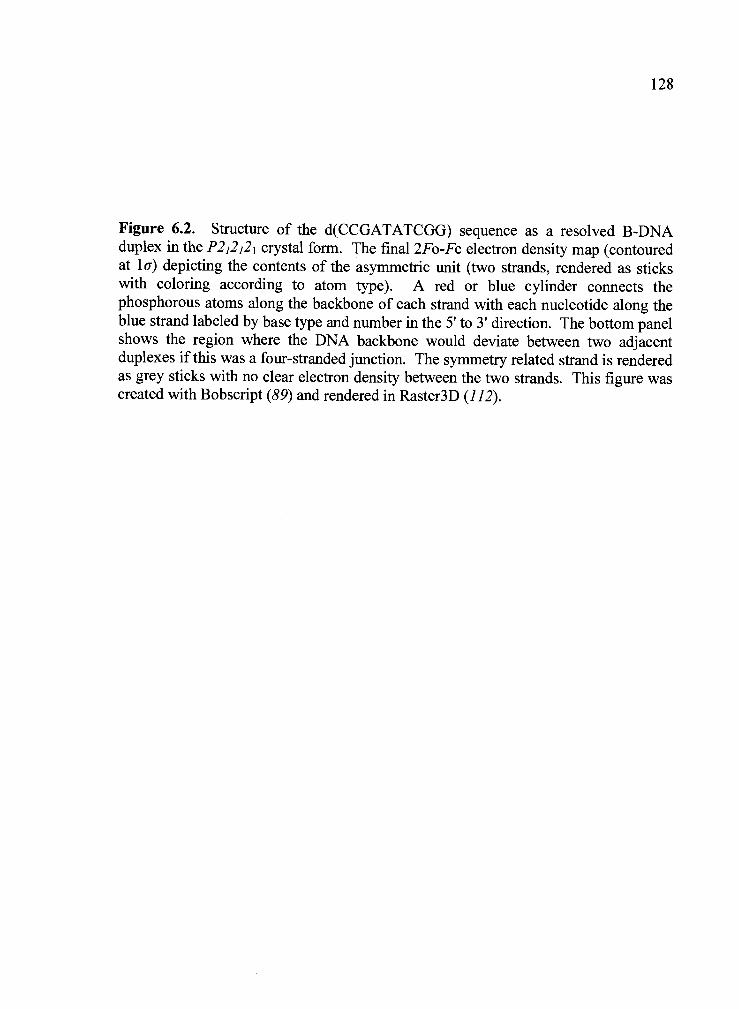
128
Figure 6.2. Structure of the d(CCGATATCGG) sequence as a resolved B-DNAduplex in the P212121 crystal form. The final 2Fo-Fc electron density map (contouredat 1 a) depicting the contents of the asymmetric unit (two strands, rendered as stickswith coloring according to atom type). A red or blue cylinder connects thephosphorous atoms along the backbone of each strand with each nucleotide along theblue strand labeled by base type and number in the 5' to 3' direction. The bottom panelshows the region where the DNA backbone would deviate between two adjacentduplexes if this was a four-stranded junction. The symmetry related strand is renderedas grey sticks with no clear electron density between the two strands. This figure wascreated with Bobscript (89) and rendered in Raster3D (112).

T
T7
Figure 6.2
19
20
129

130sequences (146, 147), ATC-B contains a semi-continuous "spine of hydration"
within the minor groove of the duplex structure, arranged in the classic "zig-zag"
pattern between hydration shells, with solvent waters interacting primarily with the 02
atoms of pyrimidines and N3 atoms of purines. No divalent or monovalent cations
were identified in the solvent model of this structure, which has approximately thirteen
solvent atoms per base pair (133 preferred hydration sites were identified).
The ATC-B structure has a significant widening of the minor groove (average
phosphate-corrected minor groove width is 5.34 A), with the widest point at the T5
nucleotide (7.9 A), that has previously been attributed to deformability of TA base
steps resulting in a bending of the DNA duplex and a concomitant narrowing of the
major groove (148). This widening of the minor groove does not translate to
significant backbone perturbations in the form of Bil phosphate conformations or
uncommon sugar puckers. The phosphate group of A6 is the only BlI (113, 116)
conformation present in the ATC-B structure resulting from crystal packing
interactions between the 02P atoms ofG20 and A6. Likewise, backbone sugar puckers
found in ATC-B (C2'-endo, C1'-exo, 01'-endo and C3'-exo) are typical for standard B-
DNA duplexes with no C3'-endo family puckers present (149). Analysis of individual
base pair and base step geometry within the ATC-B structure shows many of the
expected characteristics for TA rich central regions of DNA structures (increased
helical twist, propeller twist, roll, slide, etc.) and has been discussed in great detail
elsewhere (149-151) for similar structures.
6.3.2 Overall Structures of d(CCGA TA TCGG) as DNA Holliday junctions.
Two conformationally unique DNA Holliday junction structures of the sequence

131d(CCGATATCGG) were determined from solutions containing 55 mM (ATC-LS
structure) and 110 mM (ATC-HS structure) CaC12 (Figure 6.3). The ATC-LS and
ATC-HS structures will be discussed relative to each other since they are very similar
in terms of nomenclature and general arrangement of the DNA strands (Figure 6.3).
These right-handed stacked-X junctions have an antiparallel alignment of the DNA
strands (RMSD for like non-hydrogen atoms between ATC-LS and ATC-HS junctions
is 0.57 A). Each junction is formed by a 2-fold rotation of the two unique strands (one
noncrossover and one crossover) in the crystallographic asymmetric unit to generate a
four-stranded DNA Holliday junction with pseudo-continuous duplex arms on each
side of the central crossover. These adjoined duplexes contain two longer six base
pair arms (from the C1 to A6 nucleotides of the crossover arm paired with G20 to T15 of
a noncrossover arm) on top of shorter four base pair arms (pairing T7 to G10 of a
crossover strand with A14 to C11 of a noncrossover strand) with the crossover
occurring between the A6 and T7 nucleotides of the inside strands (Figure 6.3). The
central crossover region is composed of the nucleotides A6, T7 and C8 of the
noncontinuous DNA strands.
6.3.3 Comparison of Junction Geometry. As evident from above, analyzing
the global conformation of these four-stranded junctions is a fundamental part of
comparing numerous structures and gaining a better understanding of how variability
in conformation may play a role in biological processes. Recent efforts to explicitly
define junction geometry (48, 110) have led to the development of quantitative
parameters which describe the spatial arrangement of the junction duplexes, relative to

132
Figure 6.3. Structures of the sequence d(CCGATATCGG) as four-stranded Hollidayjunctions in the C2 crystal form. The final 2Fo-Fc electron density (contoured at la)depicting the contents of the asymmetric unit (two strands, one crossover and one non-crossover) rendered as sticks with coloring according to atom type (panels createdwith Bobscript (89) and rendered in Raster3D (112)). The phosphodeoxyribosebackbone for each strand is rendered as a continuous ribbon. Nomenclature, backbonecoloring and orientation for each junction structure is the same as the central diagramwith the full four-stranded structure formed by a crystallographic 2-fold symmetryaxis in the center. The ATC-LS junction was crystallized in the presence of 55 mMCaC12 while ATC-HS crystallized in the presence of 110 mM CaC12.

Figure 6.3
3' 5'
G20C1G19C2
' 5'
C18G3 G10C20T17A4 G9C19A16T5 C8G18TI5A6T T7 A17A14T7 .LA6T16G13C8 T5A15C12G9 A4T14C11G10 G3C135' 3' C2G12
C1 G115' 3'

134the central core region, in three-dimensional space. Jslide defines the translational
movement of the duplexes along their helix axes, J1i is rotation of the duplexes
relative to the junction center, and Jt defines the twist of each duplex relative to the
resolving plane (110). Taken together, these parameters affect the angle relating each
duplex end to the junction center, generally referred to as the interduplex angle (IDA).
Comparison of the IDA's between ATC-LS (IDA = 69.00) and ATC-HS (IDA = 69.9°)
indicates a small conformational difference between the two junction forms. Indeed, a
closer inspection of each conformational parameter for ATC-LS (Jslide = 0.39 A, Jtwist =
41.0° and Jr011 = 149.7°) and ATC-HS (Jslide = 1.00 A, = 44.0° and Jr011 148.7°)
demonstrates that the duplex arms have translated towards the six base duplex ends
and twisted towards a more parallel junction structure with decreasing salt
concentration. Additionally, the major groove face of the ATC-HS junction has
become smaller relative to ATC-LS. These perturbations to the overall junction
geometry produce a more symmetric, planar, structure as the salt concentration
decreases and a more compact junction core at higher salt concentrations.
In addition to the above perturbations there are a number of additional
differences in structure when comparing the low and high salt junctions to each other
and the ATC-B structure. The average minor groove width of both junctions (4.54 A
for ATC-LS and 4.26 A for ATC-HS) is considerably narrower then the minor groove
of ATC-B (7.02 A) between the fourth and sixth base steps of the duplex arms.
Though the minor groove in this region is very similar for both junctions there is a
noticeable difference in major groove width for the same region, ATC-HS has an

135average major groove width between the AT7 and A6T15 base steps of 10.63 A
compared to 11.20 A for ATC-LS.
6.3.4 Comparison of Junction Crossover Regions. Previous results have
demonstrated that specific hydrogen bonding interactions within the junction core are
important factors in determining the global conformation and formation of these
stacked-X junctions. These central core interactions include (i) a direct hydrogen
bond between the C7 phosphate oxygen and extracyclic amine of C8 (N7-C8
interaction), (ii) a solvent-mediated interaction between the outside G3 keto oxygen
and A6 phosphate oxygen (G3-A6 interaction), and (iii) a direct hydrogen bond
between the O1P phosphate oxygen of A6 and the N4 extracyclic substituent of N7 (47,
93). Consistent with these observations we observe the direct N7-C8 hydrogen
bonding interaction (2.85 A in ATC-LS vs. 2.61 A in ATC-HS) in both junction
structures (Figure 6.4). Both structures have additional solvent waters within the G3-
A6 and A6-T7 stabilizing interactions, similar to the previously solved ACm5C junction
structure (48). It is interesting to point out that the C5 methyl group of T7 for both
ATC-LS and ATC-HS is pointed directly at the A6 phosphate group which may help
shield negative electrostatic potential within the core (Figure 6.4). A notable
difference between the two junction structures is the presence of additional
compensating interactions within the ATC-HS junction core (such as the N-N6
interaction in Figure 6.4). These interactions are more extensive within the ATC-HS
structure and have been observed in other conformationally perturbed junction
structures such as ACbr5U in Chapter 3 (76).

136
Figure 6.4. Stabilizing interactions within the central core of ATC-LS and ATC-HS.The central T5, A6, T7 and C8 nucleotides are shown for each strand of the centralcrossover for the ATC-LS and ATC-HS junction structures (numbering of nucleotideson the symmetry related strand is incremented by ten). DNA is rendered as ball andsticks with coloring according to atom type (blue = nitrogen, red = oxygen, greycarbon and yellow = phosphorous). The phosphodeoxyribose backbone is rendered asa continuous ribbon connecting phosphorous atoms along the same strand. The N6-N7interaction is shown at the top of each panel with the corresponding N7-C8 interactionshown at the bottom (hydrogen bonding distance in parentheses). The volume of thecentral cavity is noted for each core and was calculated as described in the text. Thisfigure was created with Bobscript (89) and rendered in Raster3D (112).

c1
TI'17
Figure 6.4
N6-N7
Na+
Ce ral cavity(volume= 17.83 A3, SD=1.71 A3)
ATC-LScentral cavity
(volume = 9.52 A3, SD=2.26 )
ATC-HS

138In addition to these hydrogen bonding interactions, which have been
identified in some form in every stacked-X structure solved to date (93), there is a
central cavity located between the backbone phosphate atoms of A6 that contained a
sodium ion in the parent ACC junction structure (47). Not surprisingly, the central
cavity of ATC-LS contains a sodium ion in the same location while the central cavity
of ATC-HS contains a partially occupied water molecule adjacent to the
crystallographic two-fold symmetry axis (the ATC-LS sodium is directly on the
crystallographic two-fold). This displacement may result from a compression, or
pinching, of the central cavity in the ATC-HS structure as indicated by the distance
between A6 phosphate oxygens (distance between phosphate oxygens is equal to 5.9 A
in ATC-HS and 6.7 A in ATC-LS). Indeed, the central core region of the ATC-HS
structure is more compact compared to ATC-LS. The average phosphorous-
phosphorous distance within the junction core is 0.54 A greater in ATC-HS then in
ATC-LS (8.73 A vs. 8.19 A using T5A6T7 phosphorous atoms adjacent to the cavity).
If the average distance between backbone atoms within the junction core decreases
with increasing salt concentration then one would expect a corresponding decrease in
the size of the central cavity that has been previously described (48). Indeed, the
volume of the central cavity, calculated using the program VOIDOO (91) (standard
van der Waals radii for DNA atoms; multiple rounds calculated with different
molecule orientations, probe radius, grid spacing, and grid-shrink factor), within the
ATC-LS structure is almost twice the size of the same cavity within the ATC-HS
structure (9.52 A3 with SD = 2.26 A3 for ATC-HS vs. 17.83 A3 with SD = 1.71 A3 for
ATC-LS). Is the compression of this cavity due to local distortions in the backbone

139atoms of the core or is it related to the global conformational change of the
junction? This cavity, formed by the T5A6T7 nucleotides, resides on the major groove
face of the junction structure. If it was simply local distortions then we could expect
an increase in the major groove width of the ATC-HS structure relative to ATC-LS,
assuming compaction is not related to a global conformational change of the junction
(i.e. a change in the orientation of the duplex arms relative to each other). Instead of
an increase, we observe a 0.68 A decrease in major groove width of ATC-FIS (10.61 A
for ATC-HS vs. 11.29 A for ATC-LS) which indicates that conformational changes
must play some role in compressing the central cavity.
6.4 Discussion
In this study, we determined the salt-dependent single crystal structures of the
sequence d(CCGATATCGG) from two crystal forms yielding both four-stranded
DNA Holliday junctions, in a stacked-X antiparallel conformation, and B-DNA
duplexes. The N6N7N5 trinucleotide core of the general inverted-repeat sequence
d(CCnnnN6N7N8GG) has been previously described and contains a series of inter- and
intra- strand hydrogen bonding interactions that stabilize the stacked-X junction
conformation (47, 93). The current junction structures represent the first from
sequences that do not contain a common cytosine nucleotide in the N7 position of this
core region. Furthermore, previous studies have demonstrated that divalent cations
can have a significant effect on global junction conformation (26, 38, 66, 137) with the
square-planar conformation being favored in low cation concentrations while the

140stacked-X form predominates under conditions that provide shielding of the
negatively charged phosphodiester backbone (Figure 6.1). Subsequent studies on
crystal structures of stacked-X junctions derived from a number of different sequences
has yielded a set of quantitative parameters describing the conformation of the
stacked-X junction (110)). A unique aspect of the present study is that a comparison
of the two d(CCGATATCGG) junction forms (ATC-HS and ATC-LS) revealed a salt-
dependent change in global conformation producing a less compact core and more
symmetric junction structure as the salt concentration is decreased. We propose this
change in conformation is associated with the transformation of the stacked-X junction
towards the open-X intermediate which can undergo spontaneous branch migration
producing, as we also observe for this sequence, resolved B-DNA duplexes. Thus,
these crystal structures provide the first detailed view of junction conformational
changes resulting from decreasing cation concentration which, eventually, leads to the
duplex form of the same sequence.
The current studies were initiated by a systematic crystallographic screen of
the sequence dependence on junction formation where we expanded the core sequence
motif to include the ACC, GCC, CCC and ATC trinucleotides (152). The ATC
junctions are the only currently available structures where the common cyto sine in the
N7 position of the junction core has been replaced by another base, in this case a
thymine nucleotide. Incorporation of a thymine into the central core drives this
sequence motif into an amphimorphic class that forms two DNA conformations,
unlike the parent ACC junction that has only been observed as a DNA Holliday
junction (152). If electrostatic interactions are a significant driving force in junction

if!folding then the presence of a thymine in the N7 position may be less efficient in
stabilizing the electrostatic charge from the A6 phosphate atoms when compared to the
extracyclic N4 amine of a cytosine base. Indeed, in every current ACC junction
structure there is a long range, or solvent mediated, hydrogen bonding interaction
between the N4 substituent of C7 and the A6 phosphate oxygen. In the ATC junctions
this shielding may occur as the result of the C5 methyl groups of T7 on the crossover
strands which are oriented directly towards these A6 phosphate oxygens. Furthermore,
AT base pairs are known to produce more significant local distortions when compared
to GC base pairs by increasing parameters such as propeller twist and helical twist as
well as a concomitant narrowing of the minor groove. This becomes more evident by
the incorporation of a thymine residue into the N8 position of the parent ACC core
(ACT sequence) which has only been observed as resolved B-DNA duplexes (76).
Overall the trinucleotide core interactions within ATC-LS are very similar to the
parent ACC junction structure. The ATC-HS structure has a number of additional
compensating interactions which have been observed previously in the
conformationally perturbed d(CCAGTACbr5UGG) junction structure (76). Previous
studies have also shown the importance of sequence affects in stabilizing the stacked-
X form or dictating a preference for a specific stacking conformer (25, 38, 40, 43).
This interplay between cation neutralization of highly negatively charged regions and
specific intra- and inter-strand hydrogen bonding interactions between base
substituents may have a concerted affect in determining junction conformation. Thus,
the current structures provide a means to not only expand the trinucleotide core
142sequence motif but also allow us to refine our understanding of how interactions
within the central junction core stabilize and influence junction conformation.
The conformation and local geometric characteristics of stacked-X junctions
are known to be perturbed by cation binding. When the junction forming sequence
d(TCGGTACCGA) is crystallized in the presence of high concentrations of strontium
ions the resulting structure is perturbed relative to the parent d(CCGGTACCGG)
structure with an increase in the average Jroll and IDA parameters (Table
6.2). The authors proposed that these conformational changes were the result of a
"strontium spine" in the minor groove of the junction structure (75). This minor
groove binding of strontium ions is consistent with the overall trend of calcium ions,
the only other divalent cation identified in junctions to date, being localized to the
minor groove of the four base pair arm of junction crystal structures (48, 76). A study
of minor groove substituent interactions on junction formation suggested that calcium
binding in the minor groove may not have a significant affect on junction structure
even though it is adjacent to the N13-N8 base pair of the trinucleotide core (153)).
Only one calcium ion was identified in the current ATC junction structures and it is
located in a similar location within the minor groove of the four base pair arm of
ATC-HS. This isn't entirely surprising given that, excluding the strontium junction
(UDOO21), this calcium position is the only location which has been previously
reported to contain a divalent cation in all stacked-X structures solved to-date.
Sodium is the only metal ion which has been observed in the immediate vicinity of the
junction crossover, typically in the cavity of the major groove face (47, 76). This
raises an intriguing question - if divalent cations (especially calcium and magnesium)

143
Table 6.2. Global conformation of DNA Holliday junctions. Comparison of globalconformational geometry for junctions containing the parent A-C7C8 trinucleotide coresequence motif and the present junction structures of d(CCGATATCGG). Thecalculation and definition of each conformational parameter has been previouslydescribed (110).
Junction Jt15t (°) Jslide (A) Jr011 (°) IDA (°)
UD0008 (47) 37.8 0.15 139.9 65.9
UDOO15 (79) 39.5 -0.36 145.3 68.3
UDOO18 (75) 39.3 0.34 142.1 66.6
UD0023 40.6 1.06 146.6 67.4
UD0024 38.5 0.15 142.2 67.2
UD0025 37.7 0.11 142.7 66.7
UDOO21 (75) 44.5 0.58 150.1 70.2
UD0026 45.4 0.46 150.3 70.7
ATC-LS 41.0 0.39 149.7 69.0
ATC-HS 43.9 0.90 148.4 70.0

144facilitate conformational changes between the ATC-LS and ATC-HS junction
structures then why are no calcium ions identified around the central core which is
where previous studies have suggested they have the greatest impact on junction
folding and conformation (50, 143)? In order for identification to occur through
crystallographic methods the solvent ions would need to be fairly well ordered or
highly occupied in specific positions of the atomic structure. Indeed, FRET studies
have demonstrated that ion binding to the center of stacked-X junctions occurs in a
very rapid and noncooperative maimer (63, 137). This rapid exchange could
conceivably preclude cation identification using crystallographic methods. It should
be noted that we have identified solvent waters within each junction structure with
relatively low B-factors which may indicate that they are disordered ions that can not
be definitively assigned with the current data. Taken in context these results are more
indicative of group II ion binding to the junction center occurring in a passive maimer
and not to specific sites where phosphates are in close approach.
One of the two locations where a metal ion has been identified within the
major groove face of the junction crossover is the central cavity created by the N6
phosphate oxygens. This central cavity in the initial d(CCGGTACCGG) junction
structure contained a well defined sodium ion which has since been identified in other
junction structures (47, 76). Interestingly, the ATC-LS structure also has a well
defined sodium ion in this central cavity, which is the only metal ion identified in the
structure, while ATC-HS has two partially occupied preferred hydration sites (154)
adjacent to the crystallographic two-fold within the cavity center. Displacement of
solvent in this central ion binding site within the ATC-HS structure is likely due to the

145change in overall geometry of the structure which produces a more compact
central core (average P-P distance for ATC-HS is 8.19 A vs. 8.73 A in ATC-LS using
T5A6T7 phosphorous atoms adjacent to the cavity). Compaction of this central cavity
is evident from a comparison of the cavity volumes for ATC-HS (9.52 A3 with SD =
2.26 A3) and ATC-LS (17.83 A3 with SD = 1.71 A3). As compression of the central
cavity occurs the solvent atom is likely extruded to either side of the cavity where it
can still shield the phosphate oxygens, a similar solvent extrusion model was proposed
previously with the compressed cavity of d(CCGGTACm5CGG) (48).
The present results shows that the conformation of the d(CCGATATCGG)
junction is perturbed with decreasing salt concentration. The lower [CaC12] generates
a 0.51 A decrease in Jslide, 2.9° decrease in Jtt and a 1.3° increase in which
combine to produce a one degree decrease in the interduplex angle. Thus, a decrease
in salt concentration causes the junction structure to open up and expose a greater
amount of the major groove face generating a more symmetric overall core (Figure
6.5). This opening, or roll, of the duplex arms is combined with a decrease in the twist
of the duplex arms. If a perfectly parallel junction is considered to be fully symmetric
in terms of twist then this decrease is a movement towards the more symmetric form
where the helix axes of each duplex arm would be aligned coincident with each other.
Simply twisting the arms of the stacked-X junction to make them more aligned with
one another is not sufficient to produce a fully symmetric structure - the arms would
have to translate along the helix axis of each duplex, since they are offset in the
compact stacked-X form, to produce a more

146
Figure 6.5. Schematic of conformational changes associated with decreasing saltconcentration for the d(CCGATATCGG) sequence. The calcium chlorideconcentration corresponding to specific DNA conformations within this study is notedalong the top. B-DNA duplexes are shown as cylinders with individual DNA strandsas solid black arrows pointing in a 5' to 3' direction. Translational or rotationaldisplacement of cylinders is denoted by dashed grey arrows pointing in the directionof duplex movement. The conformational variability of four-stranded junctions can bedescribed by the parameters Jslide and J.011 (110). With decreasing [CaC12] theangle between the duplex arms of the junction and the translationaldisplacement of each arm along the duplex helix axis (Jslide) decreases. Whencombined with an increase in Jroii, increasing exposure of the major groove junctionface, the overall junction core becomes more symmetric and less compact as the[CaCl2] decreases. If each of these trajectories is continued to its extreme thehypothetical structures within the grey box would result. We should note thatconformations within the grey box are not observed in the present study, rather theyare simply a continuation of the trajectories for each conformational parameter.Presence of the open-X junction form (denoted in brackets), though not observed inthe current study, is suggested by the presence of resolved B-DNA duplexes at lowerconcentrations of calcium chloride.

11055 [CaCl2](mM) 0
1i5
twist -+
sIide
roII
Figure 6.5
iI
'If1 $
1
MigrationResolution

148symmetric junction center. Indeed, we see a significant decrease (0.51 A) in Jslide
as the salt concentration is decreased which generates a less compact and more open
junction core. As the stacked-X junction becomes more symmetric, or planar, with
decreasing salt concentration the duplex arms will eventually repel away from one
another into the fully extended open-X conformation. With inverted-repeat DNA
sequences we would expect the open-X form to rapidly produce resolved B-DNA
duplexes since branch migration in these sequences is an isoenergetic process (the
resolved B-DNA duplexes will have an identical sequence). Thus, even though the
open-X structure is not observed in the present studies we anticipate that this form
exists as the link between the stacked-X and resolved structures.
Overall these results suggest that the transition between a stacked-X and open-
X junction structure occurs via the rotation and translation of the duplex arms of the
stacked-X junction producing a more symmetric and less compact core. Crossover
stabilization likely occurs through hydrogen bonding networks between solvent water,
extracyclic base substituents, phosphate oxygens, and ordered cation hydration shells.
As these interactions are disrupted, either by specific modifications (48, 76, 135) or
through changes in salt concentration (75), the conformation of the junction structure
becomes perturbed. Thus, the central core may serve as a linchpin for determining, or
stabilizing, the conformation of four-stranded DNA junctions. This has biological
implications since the bacterial recombination protein RuvA is known to interact with
the crossover region of four-stranded junctions through two acidic-pins (Glu55 and
Asp56) which repel the phosphodiester backbone of the crossover strands away from
each other through electrostatic repulsion and help facilitate base-pair rearrangements

149(3, 155). These interactions are essential to the activity of RuvA and may play an
additional role in targeting the RuvC endonuclease to the junction cleavage site within
the RuvABC resolvasome (155). RuvC may have particular importance in the current
discussion since it may recognize the stacked-X conformation of the Holliday junction
(36). Thus, changes in junction conformation could conceivably affect its recognition
by protein machinery in vivo. Arguably there is still a lot to be understood, especially
in eukaryotic organisms, regarding the exact role of four-stranded junctions in
biological systems. What has become clear is that the stacked-X form of the four-
stranded junction is conformationally malleable and significantly affected by sequence
and atomic interactions within the central core region.
6.5 Materials and Methods
Deoxyoligonucleotides of the sequence 5'-CCGATATCGG-3' were
synthesized using f3-cyanoethyl phosphoramidite chemistry and obtained from
Midland Certified Reagent Company (Midland, TX). Upon arrival the tritylated
oligomers were evaporated to a neutral pH following removal of protecting groups by
hydrolysis with concentrated ammonium hydroxide for 24 hours at room temperature.
The 5'-O-dimethoxytrityl (DMT) group was left attached to facilitate separation from
failure sequences and reaction by-products using preparative reverse-phase high
performance liquid chromatography (Microsorb 300 A pore size C-18 column
measuring 250 mm X 21.4 nmi at a flow rate of 8 mL / mm in 0.1 mM triethylamine
acetate with an acetonitrile gradient). The DMT-decamer sequence typically eluted

1501 mm postinjection in a mobile phase concentration of 20% acetonitrile. DMT
was removed by treatment with 3% aqueous acetic acid for 15 minutes with gentle
agitation, neutralized with concentrated ammonium hydroxide, and desalted on a
Sephadex G-25 (Sigma) column. Purified DNA was stored at -80 °C as a lyophilized
powder and resuspended in deionized double-distilled water prior to use, without
strand annealing. Crystallization was carried out at room temperature in 10 pL sitting
drops by the vapor diffusion method and equilibrated against a 30 mL reservoir
volume. Crystal morphologies consistent with junctions (thin diamond plates vs. long
rods for the B-DNA form) for the sequence d(CCGATATCGG) were observed under
a range of salt concentrations (20 mM to 110 mM CaC12), though between 20 mM and
55 mM CaC12 the B-DNA crystal form would also typically be observed in the same
drop. B-DNA only crystals were observed in solutions containing 0 to 15 mM CaC12.
6.5.1 Crystallization and structure determination of B-DNA form
d(CCGATATCGG). The B-DNA form of d(CCGATATCGG) crystallized as long
rods in solutions containing 0.65 mM DNA, 5 mM CaC12, 75 mM NaCacodylate (pH
7.0) and 0.9 mM spermine-4HC1 against a reservoir solution of 30% 2-methyl-2,4-
pentanediol (MPD). X-ray diffraction data to 1.61 A was collected at liquid nitrogen
temperatures using CuKa radiation from a Rigaku (Tokyo) RU-H3R rotating anode
generator operating at 50 kV and 100 mA (A1 = 10, 180 15 minute exposures) with an
Raxis-IV image plate detector on a single crystal measuring 250 mm X 50 mm X 50
mm. The crystal was indexed in the orthorhombic spacegroup P212121 with unit cell
dimensions (a 35.2 A, b = 39.1 A, and c = 33.0 A) similar to the previously solved
structure of d(CCAAATTTGG) (BDOO51, (145)). A search model derived from this

151structure was used for molecular replacement in EPMR (83) which produced a
unique solution with a correlation coefficient of 68.3% and of 39.7%.
Subsequent refinement of this model in Refmac5 (156) produced a final of
25.7% and Rfree of 27.5% (Table 6.1).
6.5.2 Crystallization and structure determination of d(CCGA TA TCGG) low
salt junction. Crystals were grown in solutions containing 0.7 mM DNA, 55 mM
CaC12, 0.8 mM spermine4HCl, and 75 mM NaCacodylate (pH 7.0) against a reservoir
solution containing 30% by volume MPD. Initial small diamond plate crystals
appeared after 30 days with optimal crystals appearing after two additional months. A
single crystal measuring 250 mm X 375 mm X 50 mm was used for data collection.
Initial data collection was performed at liquid nitrogen temperatures using CuKa
radiation from a Rigaku (Tokyo) RU-H3R rotating anode generator operating at 50 kV
and 100 mA (M = 10, 180 15 minute exposures) with an Raxis-IV image plate
detector. This data was later merged with data collected from BioCARS beamline 14-
1DB at the Advanced Photon Source (Argonne National Laboratory, Argonne, IL)
using 1.07 A radiation, M1 = 10, 100 20 second exposures, and a MAR CCD detector
(Table 6.1). The final, merged, dataset extended to usable resolution of 1.65 A. The
crystal was indexed in monoclinic spacegroup C2 with unit cell dimensions (a=64.9 A,
b= 24.0 A, c= 40.3 A, a='=90°, and 3=118.69°) isomorphous to a previously solved
high salt ATC junction. Thus, a search model was derived from this previously solved
structure and used in EPMR for molecular replacement. A distinct solution was
obtained with a correlation coefficient of 44.8% and of 49.6% and two unique
strands adjacent to a crystallographic 2-fold symmetry axis. To verify that this was

152indeed a 4H-junction we generated a simulated annealing omit map for the C-
crossover phosphate which produced distinct F0-F density within the trinucleotide
core region. The initial round of rigid body and rigid parts refinement on this model
followed by simulated annealing produced a starting 30.9% and Rfree = 37.0%.
Subsequent refinement was performed in CNS (84) using standard positional and
individual B-factor refinement along with assignment of solvent atoms. This model
was then completed in Refmac5 (156) using TLS refinement (157, 158), with each
strand assigned as a TLS group, resulting in a final of 23.7% and Rfree of 27.3%.
6.5.3 Crystallization and structure determination of d(CCGA TATCGG) high
salt junction. Crystals of the high salt junction form were grown in solutions
containing 0.7 mM DNA, 110 mM CaCl2, 0.8 mM spermine4HC1, and 75 mM
NaCacodylate (pH 7.0) against a reservoir solution containing 30% by volume MPD.
Small diamond plate crystals grew over a period of approximately 30 days with
diffraction quality crystals appearing after an additional two months. A single crystal
measuring 500 mm X 375 mm X 50 mm was used for data collection. Collection of
synchrotron diffraction data (to 1.85 A) was performed on BioCARS beamline 14-
BMD at the Advanced Photon Source (Argonne National Laboratory, Argonne, IL)
using 0.98 A radiation, LW = 1°, 120 45 second exposures and an ADSC Quantum 4
detector (Table 6.1). The crystal was indexed in monoclinic spacegroup C2 with unit
cell dimensions (a=65.0 A, b= 25.0 A, c= 37.5 A, a=y=90°, and 1 12.09°)
isomorphous to the junction form of d(CCGGGCCCGG) (unpublished data). Thus, a
search model was derived from this previously solved structure and used in EPMR for
molecular replacement. A distinct solution was obtained with a correlation coefficient
153of 75.2% and of 34.0% and two unique strands adjacent to a crystallographic
2-fold symmetry axis. To verify that this was indeed a 4H-junction we generated a
simulated annealing omit map for the C7 crossover phosphate which produced distinct
F0-F density within the trinucleotide core region. The initial round of rigid body and
rigid parts refinement on this model followed by simulated annealing produced a
starting = 31.0% and Rfree = 35.0%. Subsequent refinement was performed in
CNS (84) using standard positional and individual B-factor refinement along with
assignment of solvent atoms. This model was then completed in Refmac5 (156) using
TLS refinement (157, 158), with each strand assigned as a TLS group, resulting in a
final of 20.0% and Rfree of 23.6%.
Prior to data collection, all crystals were transferred to artificial mother liquor
containing 25% MPD and flash cooled to -180°C in liquid nitrogen for cryoprotection.
All data were processed and reflections merged and scaled using DENZO and
SCALEPACK from the HKL suite of programs (85). Resolution cutoffs were
determined based on data completeness, I/cT(I), andRmei-ge. Room-mean-square
deviation (RMSD) values were calculated using the McLachlan algorithm as
implemented in the ProFit version 2.2 program
(http://www.bioinf.org.uklsoftware/profit) (86)). Structural analysis was performed
using CURVES 5.2 (87) and 3DNA (88). All assigned solvent atoms within the
present structures are listed according to their densities in the final 2F0-F map, with
the first solvent atom having the highest density, using a program provided by the
Karplus lab at Oregon State. The coordinates and structure factors have been deposited

in the Protein Data Bank (105) as entries 1PNY for ATC-B, 1PNY for ATC-LS,
and 1PNY for ATC-HS.
6.6 Acknowledgements
154
This work was funded by grants from the National Institutes of Health
(R1GM62957A) and the National Science Foundation (MCB0090615). The X-ray
diffraction facilities are supported by the Proteins and Nucleic Acids Facility Core of
the Environmental Health Sciences Center at OSU (NIEHS ESOO21O) and a grant
from the Murdock Charitable Trust. Access to the Advanced Photon Source was
supported by the U.S. Department of Energy, Basic Energy Sciences, Office of
Science, under Contract W-31-109-Eng-38. Access to BioCARS Sector 14 was
supported by the National Institutes of Health, National Center for Research
Resources, under Grant RR07707.

155Chapter 7
Conclusion and Discussion
Conformational variability, including the ability to undergo branch migration
and subsequent resolution, is a fundamental characteristic of four-stranded DNA
Holliday junctions in biological systems. Although DNA crossovers are known to be
recognized by a growing number of proteins in vivo, it isn't very clear how intrinsic
junction properties influence this recognition and subsequent resolution. The current
studies were initiated to better understand the role that the sequence within the central
crossover region may play in determining junction conformation and formation. As a
result of these studies, four general conclusions about junction structure and function
have emerged.
Stacked-X junction formation and conformational variability occurs primarily
in a sequence-dependent manner within the current system. Both of the initial
stacked-X junction structures were stabilized by the presence of a common ACC
trinucleotide sequence in the N6N7N8 position of the general sequence motif
d(CCnnnN6N7N8GG). Work described in this thesis expands the core sequence motif
to N6N7C8 where N6 is an adenine, guanine, or cytosine nucleotide and N7 is either a
cytosine or thymine (when N6 = adenine) nucleotide (excluding non-standard bases).
Stabilization of these structures as stacked-X junctions is surprising since inverted-

156repeat sequences have been shown to undergo spontaneous branch migration to
resolved B-DNA duplexes (7). Stacked-X junction formation within the current
system occurs through favorable base stacking and major groove hydrogen bonding
interactions that fix the junction crossover at a specific location within the inverted-
repeat sequence. These stabilizing major groove interactions also have a significant
effect on junction conformation, producing translational and rotational displacement
of the helical junction arms upon disruption (Table 7.1). Indeed, previous solution
studies have shown that nucleotide sequence at or near the junction crossover has a
significant effect on the formation and conformer preference of stacked-X junctions
(25, 43, 66). Since junctions are expected to form across a broad range of sequences
in vivo, these data may suggest that specific trinucleotide core sequences can stabilize
the stacked-X junction in biological processes. Many junction resolvases, such as
RuvC or CCE1, have strong sequence specificities for cleavage while remaining
highly specific for four-stranded junctions in terms of recognition. RusA, which is a
X-phage junction resolvase, has a strong preference for junction cleavage at ,1CC sites
within the central crossover region (159, 160). Thus, the sequence-dependent
formation within the current crystallographic system may serve in biological systems
to stabilize specific junction conformers and facilitate subsequent resolution by
junction resolvases to duplex DNA products. In addition, the sequence effects on
global conformation may play a significant role in junction recognition by cellular
proteins, including resolvases and DNA repair proteins.

157
Table 7.1. Comparison of global conformational geometry for all currently knowninverted-repeat stacked-X DNA Holliday junctions. The calculation and definition ofeach conformational parameter has been previously described (110). Each junction isdesignated by its corresponding trinucleotide core sequence (for structures resultingfrom this thesis) or by an NDB accession code (for previously published structures).Perturbations to global junction geometry are generally related to sequence or cationdependent disruption of stabilizing interactions within major groove face of thejunction core.
Junction Jtwist(0)
Jslide (A) Jr011(0) IDA (0)
iACm5C (153) 42.5 2.23 143.6 61.4
ACbr5U (76) 37.1 1.27 130.4 68.5
GCC (76) 37.0 -0.04 143.6 62.0
CCC 43.1 1.03 147.0 67.4
ATC-LS
ATC-HS
41.0
43.9
0.39
0.90
149.7
148.4
69.0
70.0
UD0008 (47) 37.8 0.15 139.9 65.9
UD0015 (48) 44.3 2.25 145.9 60.0
UDOO15 (79) 39.5 -0.36 145.3 68.3
UDOO18 (75) 39.3 0.34 142.1 66.6
UD0023 40.6 1.06 146.6 67.4
UD0024 38.5 0.15 142.2 67.2
UD0025 37.7 0.11 142.7 66.7
UDOO21 (75)) 44.5 0.58 150.1 70.2
UD0026 45.4 0.46 150.3 70.7

158Major groove interactions within the junction core have a sign4ficant
influence on junction formation and conformation. A common theme throughout this
thesis has been the focus on specific stabilizing hydrogen bonding interactions within
the junction core. Chapter 2 discusses the nature of these interactions in detail (93).
With the exception of a direct or solvent mediated interaction between the extracyclic
amine of C8 and phosphate oxygen of N7, all of the interactions can be replaced by
compensating interactions. Indeed, the number of stabilizing interactions within the
major groove of the junction core increases as the conformation becomes more
distorted (see ACm5C, ACbr5U, and ATC-HS structures for examples). In terms of
stabilizing interactions within the core, the key may therefore be the ability of the core
nucleotides to form a range of stabilizing interactions and not just the specific
interactions proposed in the initial d(CCGGTACCGG) paper (47) (excluding the
direct or solvent mediated N7-C8 interaction which appears to be essential for junction
formation within the current system). The results of the minor groove substituent
study in Chapter 4 demonstrate that minor groove interactions within the region of the
central junction core are not a determining factor in junction formation within the
current system (76). Furthermore, perturbations to local displacement (rotational and
translational) parameters of core nucleotides appear to be tolerated as long as they do
not significantly disrupt the ability to form major groove stabilizing interactions. Two
examples of this principle are the ACT sequence in Chapter 3 (76), which forms
resolved B-DNA duplexes, and the methylated junctions in Chapter 4. These
methylated junctions (ACm5C and iACm5C) (48, 153) are almost identical in global
conformation yet the inosine base in the N3N8 base step induces a number of local
159distortions to the helical arm (such as the BIT backbone conformation of the
inosine nucleotides or a narrowing of the minor groove and disruption of the minor
groove solvent network) of the junction. Thus, major groove stabilizing interactions
play a significant role in the formation and stabilization of the stacked-X junction.
Cation binding to the major groove face of the junction core does not occur in
a site-specflc manner. A surprising aspect in the current body of stacked-X DNA
Holliday junction structures is the lack of group (IT) divalent cations assigned at, or
near, the central junction crossover. This crossover region contains a high
electrostatic potential resulting from the close proximity of backbone phosphate atoms
(50, 140, 161). Previous studies have tried to ascertain whether specific cation
binding sites existed within the junction core and to account for the drastic salt-
dependent conformational changes in junction conformation (63, 137, 142, 143) and
preference for stacking conformer (43, 161). The data presented in this thesis suggest
that site-specific binding of divalent cations to the junction core does not occur for the
present constructs. This is especially evident with the ATC junction structures where
a decrease in calcium (II) chloride concentration induced a transition in conformation
that leads to resolved B-DNA duplexes. The only calcium cation identified in either
junction structure is in the minor groove of the four base pair arm, a location shown in
Chapter 4 to not significantly affect junction conformation. These results suggest that
cation binding to the junction core does not occur in a site-specific manner. Previous
fluorescence studies suggested that cation interaction with the junction core occurred
in a rapid and highly mobile manner. Thus, diffuse clouds of ions may shield
phosphate oxygens within the junction core. Cation interactions through these diffuse

160clouds may play a significant role in the conformational variability and transitions
of the folded junction.
The central core, or crossover region, of the junction structure serves as a
focal point in determining overall junction conformation. A key observation resulting
from work presented in this thesis is that perturbations such as alteration of
trinucleotide core sequence (base substituents) or disruption of major groove hydrogen
bonding interactions within the central core of the folded junction structure can have a
significant effect on junction conformation (compare junction structures) and
formation (compare crystallographic screen results). Thus, the central core may have
a key role in the function of four-stranded junctions in biological systems. Not
surprisingly, a number of cellular proteins interact with the junction through contacts
within the central crossover region. RuvA, which targets four-stranded junctions as
part of the RuvABC resolvasome, has two highly conserved amino acids (G1u55 and
Asp56) that interact with the junction core and stabilize the open conformation by
repelling the negative DNA backbone (3, 13, 155). Recent modeling studies of the
junction resolvase T7 endonuclease I with a four-stranded junction has an extended I-
bridge passing across the major groove face of the junction core. Protein-junction
interactions distal to the central core are likely to be involved in these assorted
biological systems as well though the central junction core is highly correlated with
junction conformation in the folded form. Conformational details of the junction arms
within the stacked-X junction (such as helical twist, groove width, propeller twist,
etc.) are generally determined in a sequence-dependent manner and not induced as a
result of junction formation. This indicates that the stacked-X junction structure can

161tolerate disruptions to base geometry distal to the central core without significant
changes in global conformation (as demonstrated in Chapter 4). On the other hand,
perturbations within the central core region can have significant affects on junction
geometry, especially within two nucleotides 3' of the junction crossover (7th and 8th
positions in current studies). The current results both provide indirect evidence for the
importance of the central core region in biological systems and provide direct
evidence for a correlation between base sequence and atomic interactions within the
core region and junction conformation.

162Bibliography
1. Holliday, R. (1964) A mechanism for gene conversion in fungi, Genet. Res. 5,282-304.
2. Holliday, R. (1974) Molecular aspects of genetic exchange and geneconversion, Genetics 78, 273-87.
3. Ariyoshi, M., Nishino, T., Iwasaki, H., Shinagawa, H., and Morikawa, K.(2000) Crystal structure of the holliday junction DNA in complex with a singleRuvA tetramer, Proc. Nati. Acad. Sci. USA 97, 8257-62.
4. Van Duyne, G. D. (2001) A structural view of cre-loxp site-specificrecombination, Annu. Rev. Biophys. Biomol. Struct. 30, 87-104.
5. Dickman, M. J., Ingleston, S. M., Sedelnikova, S. E., Rafferty, J. B., Lloyd, R.0., Grasby, J. A., and Hornby, D. P. (2002) The RuvABC resolvasome, Eur. IBiochem. 269, 5492-501.
6. Liu, Y., and West, S. C. (2004) Happy Hollidays: 40th anniversary of theHolliday junction, Nat. Rev. Mol. Cell Biol. 5, 937-44.
7. Panyutin, I. G., and Hsieh, P. (1994) The kinetics of spontaneous DNA branchmigration, Proc. Natl. Acad. Sci. USA 91, 2021-5.
Meselson, M. S., and Radding, C. M. (1975) A general model for geneticrecombination, Proc. Nati. Acad. Sci. USA 72, 358-61.
9. Szostak, J. W., Off-Weaver, T. L., Rothstein, R. J., and Stahl, F. W. (1983)The double-strand-break repair model for recombination, Cell 33, 25-35.
10. Lin, F. L., Sperle, K., and Sternberg, N. (1984) Model for homologousrecombination during transfer of DNA into mouse L cells: role for DNA endsIn the recombination process, Mol. Cell. Biol. 4, 1020-34.
11. Haber, J. E., Ira, G., Malkova, A., and Sugawara, N. (2004) Repairing adouble-strand chromosome break by homologous recombination: revisitingRobin Holliday's model, Philos. Trans. R. Soc. Lond., B Biol. Sci. 359, 79-86.
12. Yamada, K., Miyata, T., Tsuchiya, D., Oyama, T., Fujiwara, Y., Ohnishi, T.,Iwasaki, H., Shinagawa, H., Ariyoshi, M., Mayanagi, K., and Morikawa, K.(2002) Crystal structure of the RuvA-RuvB complex: a structural basis for theHolliday junction migrating motor machinery, Mol. Cell 10, 671-8 1.

16313. Rafferty, J. B., Sedelnikova, S. E., Hargreaves, D., Artymiuk, P. J., Baker,
P. J., Sharples, G. J., Mahdi, A. A., Lloyd, R. G., and Rice, D. W. (1996)Crystal structure of DNA recombination protein RuvA and a model for itsbinding to the Holliday junction, Science 274, 415-21.
14. Yamada, K., Kunishima, N., Mayanagi, K., Ohnishi, T., Nishino, T., Iwasaki,H., Shinagawa, H., and Morikawa, K. (2001) Crystal structure of the Hollidayjunction migration motor protein RuvB from Thermus thermophilus HB8,Proc. Nat!. Acad. Sci. USA 98, 1442-7.
15. Ariyoshi, M., Vassylyev, D. G., Iwasaki, H., Nakamura, H., Shinagawa, H.,and Morikawa, K. (1994) Atomic structure of the RuvC resolvase: a hollidayjunction-specific endonuclease from E. coli, Cell 78, 1063-72.
16. Sha, R., Liu, F., Iwasaki, H., and Seeman, N. C. (2002) Parallel symmetricimmobile DNA junctions as substrates for E. coli RuvC Holliday junctionresolvase, Biochemistry 41, 10985-93.
17. Liu, Y., Masson, J. Y., Shah, R., O'Regan, P., and West, S. C. (2004) RAD51Cis required for Holliday junction processing in mammalian cells, Science 303,243-6.
18. Constantinou, A., Davies, A. A., and West, S. C. (2001) Branch migration andHolliday junction resolution catalyzed by activities from mammalian cells,Cell 104, 259-68.
19. Constantinou, A., Chen, X. B., McGowan, C. H., and West, S. C. (2002)Holliday junction resolution in human cells: two junction endonucleases withdistinct substrate specificities, EMBO J 21, 5577-85.
20. Ceschini, S., Keeley, A., McAlister, M. S., Oram, M., Phelan, J., Pearl, L. H.,Tsaneva, 1. R., and Barrett, T. E. (2001) Crystal structure of the fission yeastmitochondrial Holliday junction resolvase Ydc2, EMBO 1 20, 6601-11.
21. Garcia, P. L., Liu, Y., Jiricny, J., West, S. C., and Janscak, P. (2004) HumanRECQ5beta, a protein with DNA helicase and strand-annealing activities in asingle polypeptide, EMBO 1 23, 2882-91.
22. Constantinou, A., Tarsounas, M., Karow, J. K., Brosh, R. M., Bohr, V. A.,Hickson, I. D., and West, S. C. (2000) Werner's syndrome protein (WRN)migrates Holliday junctions and co-localizes with RPA upon replication arrest,EMBO Rep. 1,80-4.
23. Karow, J. K., Constantinou, A., Li, J. L., West, S. C., and Hickson, I. D.(2000) The Bloom's syndrome gene product promotes branch migration ofholliday junctions, Proc. Nat!. Acad. Sci. USA 97, 6504-8.

16424. Wu, L., and Hickson, I. D. (2003) The Bloom's syndrome helicase
suppresses crossing over during homologous recombination, Nature 426, 870-4.
25. McKinney, S. A., Declais, A. C., Lilley, D. M., and Ha, T. (2003) Structuraldynamics of individual Holliday junctions, Nat. Struct. Biol. 10, 93-7.
26. Duckett, D. R., Murchie, A. I., Diekmann, S., von Kitzing, E., Kemper, B., andLilley, D. M. (1988) The structure of the Holliday junction, and its resolution,Cell 55, 79-89.
27. Clegg, R. M., Murchie, A. I., and Lilley, D. M. (1994) The solution structureof the four-way DNA junction at low-salt conditions: a fluorescence resonanceenergy transfer analysis, Biophys. 1 66, 99-109.
28. Murchie, A. I., Clegg, R. M., von Kitzing, E., Duckett, D. R., Diekmann, S.,and Lilley, D. M. (1989) Fluorescence energy transfer shows that the four-wayDNA junction is a right-handed cross of antiparallel molecules, Nature 341,763-6.
29. Gopaul, D. N., Guo, F., and Van Duyne, G. D. (1998) Structure of the Hollidayjunction intermediate in Cre-loxP site-specific recombination, EMBO 1 17,4175-87.
30. Guo, F., Gopaul, D. N., and van Duyne, G. D. (1997) Structure of Crerecombinase complexed with DNA in a site-specific recombination synapse,Nature 389, 40-6.
31. Middleton, C. L., Parker, J. L., Richard, D. J., White, M. F., and Bond, C. S.(2004) Substrate recognition and catalysis by the Holliday junction resolvingenzyme Hje, Nucleic Acids Res. 32, 5442-51.
32. Nishino, T., Komori, K., Tsuchiya, D., Ishino, Y., and Morikawa, K. (2001)Crystal structure of the archaeal holliday junction resolvase Hjc andimplications for DNA recognition, Structure (Camb) 9, 197-204.
33. Raaijmakers, H., Vix, 0., Toro, I., Golz, S., Kemper, B., and Suck, D. (1999)X-ray structure of T4 endonuclease VII: a DNA junction resolvase with anovel fold and unusual domain-swapped dimer architecture, EMBO 1 18,1447-58.
34. Bhattacharyya, A., Murchie, A. I., von Kitzing, E., Diekmann, S., Kemper, B.,and Lilley, D. M. (1991) Model for the interaction of DNA junctions andresolving enzymes, I Mol. Biol. 221, 119 1-207.

16535. Fogg, J. M., Schofield, M. J., White, M. F., and Lilley, D. M. (1999)
Sequence and functional-group specificity for cleavage of DNA junctions byRuvC of Escherichia coli, Biochemistry 38, 11349-58.
36. Bennett, R. J., and West, S. C. (1996) Resolution of Holliday junctions ingenetic recombination: RuvC protein nicks DNA at the point of strandexchange, Proc. Nati. Acad. Sci, USA 93, 12217-22.
37. Bennett, R. J., and West, S. C. (1995) Structural analysis of the RuvC-Hollidayjunction complex reveals an unfolded junction, J Mol. Biol. 252, 2 13-26.
38. Cooper, J. P., and Hagerman, P. J. (1987) Gel electrophoretic analysis of thegeometry of a DNA four-way junction, J Mol. Biol. 198, 711-9.
39. Gough, G. W., and Lilley, D. M. (1985) DNA bending induced by cruciformformation, Nature 313, 154-6.
40. Carlstrom, G., and Chazin, W. J. (1996) Sequence dependence and directmeasurement of crossover isomer distribution in model Holliday junctionsusing NMR spectroscopy, Biochemistry 35, 3534-44.
41. von Kitzing, E., Lilley, D. M., and Diekmann, 5. (1990) The stereochemistryof a four-way DNA junction: a theoretical study, Nucleic Acids Res. 18, 2671-83.
42. Yu, J., Ha, T., and Schulten, K. (2004) Conformational model of the Hoilidayjunction transition deduced from molecular dynamics simulations, NucleicAcids Res. 32, 6683-95.
43. Mu, S. M., Fee, R. S., Millar, D. P., and Chazin, W. J. (1997) Crossoverisomer bias is the primary sequence-dependent property of immobilizedHolliday junctions, Proc. Natl. Acad. Sci. USA 94, 9080-4.
44. Churchill, M. E., Tullius, T. D., Kallenbach, N. R., and Seeman, N. C. (1988)A Holliday recombination intermediate is twofold symmetric, Proc. Natl.Acad. Sci. USA 85, 4653-6.
45. Lilley, D. M., and Norman, D. G. (1999) The Holliday junction is finally seenwith crystal clarity, Nat. Struct. Biol. 6, 897-9.
46. Ortiz-Lombardia, M., Gonzalez, A., Eritja, R., Aymami, J., Azorin, F., andCoil, M. (1999) Crystal structure of a DNA Holliday junction, Nat. Struct.Biol. 6, 9 13-7.
47. Eichman, B. F., Vargason, J. M., Mooers, B. H., and Ho, P. S. (2000) TheHoiliday junction in an inverted repeat DNA sequence: sequence effects on thestructure of four-way junctions, Proc. Natl. Acad. Sd. USA 97, 3971-6.

16648. Vargason, J. M., and Ho, P. S. (2002) The effect of cytosine methylation
on the structure and geometry of the Holliday junction: the structure ofd(CCGGTACm5CGG) at 1.5 A resolution, J Biol. Chem. 277,21041-9.
49. Lilley, D. M., and White, M. F. (2001) The junction-resolving enzymes, Nat.Rev. Mol. Cell. Biol. 2, 433-43.
50. Lilley, D. M. (2000) Structures of helical junctions in nucleic acids, Q. Rev.Biophys. 33, 109-59.
51. Nunes-Duby, S. E., Matsumoto, L., and Landy, A. (1987) Site-specificrecombination intermediates trapped with suicide substrates, Cell 50, 779-88.
52. Cox, M. M., Goodman, M. F., Kreuzer, K. N., Sherratt, D. J., Sandier, S. J.,and Marians, K. J. (2000) The importance of repairing stalled replication forks,Nature 404, 37-41.
53. Haber, J. E., and Heyer, W. D. (2001) The fuss about Mus8l, Cell 107, 55 1-4.
54. Declais, A. C., Fogg, J. M., Freeman, A. D., Coste, F., Hadden, J. M., Phillips,S. E., and Lilley, D. M. (2003) The complex between a four-way DNAjunction and T7 endonuclease I, EMBO J 22, 13 98-409.
55. Subramaniam, S., Tewari, A. K., Nunes-Duby, S. E., and Foster, M. P. (2003)Dynamics and DNA substrate recognition by the catalytic domain of lambdaintegrase, J Mol. Biol. 329, 423-3 9.
56. Hargreaves, D., Rice, D. W., Sedelnikova, S. E., Artymiuk, P. J., Lloyd, R. G.,and Rafferty, J. B. (1998) Crystal structure of E.coli RuvA with bound DNAHolliday junction at 6 A resolution, Nat. Struct. Biol. 5, 441-6.
57. Nowakowski, J., Shim, P. J., Stout, C. D., and Joyce, G. F. (2000) Alternativeconformations of a nucleic acid four-way junction, .1 Mol. Biol. 300, 93-102.
58. Nowakowski, J., Shim, P. J., Prasad, G. S., Stout, C. D., and Joyce, G. F.(1999) Crystal structure of an 82-nucleotide RNA-DNA complex formed bythe 10-23 DNA enzyme, Nat. Struct. Biol. 6, 151-6.
59. Kato, M., Hokabe, S., Itakura, S., Minoshima, S., Lyubchenko, Y. L., Gurkov,T. D., Okawara, H., Nagayama, K., and Shimizu, N. (2003) Interarminteraction of DNA cruciform forming at a short inverted repeat sequence,Biophys. J 85, 402-8.
60. Shlyakhtenko, L. S., Potaman, V. N., Sinden, R. R., and Lyubchenko, Y. L.(1998) Structure and dynamics of supercoil-stabilized DNA cruciforms, JMo!. Biol. 280, 61-72.

16761. Sha, R., Liu, F., Millar, D. P., and Seeman, N. C. (2000) Atomic force
microscopy of parallel DNA branched junction arrays, Chem. Biol. 7, 743-5 1.
62. Cooper, J. P., and Hagerman, P. J. (1989) Geometry of a branched DNAstructure in solution, Proc. Natl. Acad. Sci. USA 86, 7336-40.
63. Clegg, R. M., Murchie, A. I., Zechel, A., Carlberg, C., Diekmann, S., andLilley, D. M. (1992) Fluorescence resonance energy transfer analysis of thestructure of the four-way DNA junction, Biochemistry 31, 4846-56.
64. Mao, C., Sun, W., and Seeman, N. C. (1999) Designed two-dimensional DNAHolliday junction arrays visualized by atomic force microscopy, I Am. Chem.Soc. 121, 5437-5443.
65. Sha, R., Liu, F., and Seeman, N. C. (2002) Atomic force microscopicmeasurement of the interdomain angle in symmetric Holliday junctions,Biochemistry 41, 5950-5.
66. Grainger, R. J., Murchie, A. I., and Lilley, D. M. (1998) Exchange betweenstacking conformers in a four-Way DNA junction, Biochemistry 37, 23-32.
67. Gopaul, D. N., Guo, F., and van Duyne, G. D. (1998) Structure of the Hollidayjunction intermediate in Cre-loxP site-specific recombination., EMBO 1 17,4 175-4187.
68. Chen, Y., Narendra, U., lype, L. E., Cox, M. M., and Rice, P. A. (2000)Crystal structure of a Fip recombinase-Holliday junction complex: assembly ofan active oligomer by helix swapping, Mol. Cell 6, 885-897.
69. Eichman, B. F., Mooers, B. H., Alberti, M., Hearst, J. E., and Ho, P. S. (2001)The crystal structures of psoralen cross-linked DNAs: drug-dependentformation of Holliday junctions, I Mol. Biol. 308, 15-26.
70. Dickerson, R. E. (1999) in Oxford Handbook of Nucleic Acid Structure(Neidle, S., Ed.) pp 145-197, Oxford University Press, New York.
71. Wood, A. A., Nunn, C. M., Trent, J. 0., and Neidle, S. (1997) Sequence-dependent crossed helix packing in the crystal structure of a B-DNA decameryields a detailed model for the Holliday junction, I. Mol. Biol. 269, 827-41.
72. Goodsell, D. S., Grzeskowiak, K., and Dickerson, R. E. (1995) Crystalstructure of C-T-C-T-C-G-A-G-A-G. Implications for the structure of theHolliday junction, Biochemistry 34, 1022-9.
73. Eichman, B. F., Ortiz-Lombardia, M., Aymami, J., Coll, M., and Ho, P. S.(2002) The inherent properties of DNA four-way junctions: comparing thecrystal structures of holliday junctions, I. Mol. Biol. 320, 1037-51.

16874. Berman, H. M., Olson, W. K., Beveridge, D. L., Westbrook, J., Gelbin, A.,
Demeny, T., Hsieh, S. H., Srinivasan, A. R., and Schneider, B. (1992) Thenucleic acid database. A comprehensive relational database of three-dimensional structures of nucleic acids, Biophys. 1 63, 751-9.
75. Thorpe, J. H., Gale, B. C., Teixeira, S. C., and Cardin, C. J. (2003)Conformational and hydration effects of site-selective sodium, calcium andstrontium ion binding to the DNA Holliday junction structured(TCGGTACCGA)(4), I Mol. Biol. 327, 97-109.
76. Hays, F. A., Vargason, J. M., and Ho, P. S. (2003) Effect of sequence on theconformation of DNA holliday junctions, Biochemistry 42, 9586-97.
77. Aymami, J., Pous, J., Lisgarten, J. N., and Coil, M. (2002) Crystallization andpreliminary X-ray analysis of the DNA decamers d(CCGGATCCGG) andd(CCGGCGCCGG), Acta Crystallogr. DBiol. Crystallogr. 58, 310-1.
78. Heinemann, U., and Alings, C. (1989) Crystallographic study of one turn ofG/C-rich B-DNA, I Mol. Biol. 210, 369-81.
79. Thorpe, J. H., Teixeira, S. C., Gale, B. C., and Cardin, C. J. (2002) Structuralcharacterization of a new crystal form of the four-way Holliday junctionformed by the DNA sequence d(CCGGTACCGG)2: sequence versus lattice?,Acta Crystaliogr. Sect. D Biol. Crystallogr. 58, 567-569.
80. Ho, P. S., and Eichman, B. F. (2001) The crystal structures of DNA Hollidayjunctions, Curr. Opin. Struct. Biol. 11, 302-8.
81. Panyutin, I. G., Biswas, I., and Hsieh, P. (1995) A pivotal role for the structureof the Holliday junction in DNA branch migration, EMBO 1 14, 18 19-26.
82. Heinemann, U., Alings, C., and Bansal, M. (1992) Double helix conformation,groove dimensions and ligand binding potential of a G/C stretch in B-DNA,EMBOJ 11, 1931-9.
83. Kissinger, C. R., Gehlhaar, D. K., and Fogel, D. B. (1999) Rapid automatedmolecular replacement by evolutionary search, Acta Crystallogr. D Biol.Grystallogr. 55 (Pt 2), 484-91.
84. Brunger, A. T., Adams, P. D., Clore, G. M., DeLano, W. L., Gros, P., Grosse-Kunstleve, R. W., Jiang, J. S., Kuszewski, J., Nilges, M., Pannu, N. S., Read,R. J., Rice, L. M., Simonson, T., and Warren, G. L. (1998) Crystallography &NMR system: A new software suite for macromolecular structuredetermination, Acta Crystallogr. D Biol. Crystallogr. 54 (Pt 5), 905-21.
85. Otwinowski, Z., and Minor, W. (1997) Processing of X-ray Diffraction DataCollected in Oscillation Mode, Methods Enzymol. 276, 3 07-326.

86. McLachlan, A. D. (1982) Rapid Comparison of Protein Structures, ActaCrystallogr. A38, 871-873.
87. Lavery, R., and Skienar, H. (1989) Defining the structure of irregular nucleicacids: conventions and principles, I Biomol. Struct. Dyn. 6, 65 5-67.
88. Lu, X. J., and Olson, W. K. (2003) 3DNA: a software package for the analysis,rebuilding and visualization of three-dimensional nucleic acid structures,Nucleic Acids Res. 31, 5108-21.
89. Esnouf, R. M. (1999) Further additions to MoiScript version 1.4, includingreading and contouring of electron-density maps, Acta Crystallogr. D Biol.Crystallogr. 55 (Pt 4), 93 8-40.
90. Kielkopf, C. L., Ding, S., Kuhn, P., and Rees, D. C. (2000) Conformationalflexibility of B-DNA at 0.74 A resolution: d(CCAGTACTGG)(2), .1. Mol.Biol. 296, 787-801.
91. Kleywegt, G. J., and Jones, T. A. (1994), Acta Crystallogr. D Biol.Crystallogr. D50, 178-185.
92. Rafferty, J. B., Bolt, B. L., Muranova, T. A., Sedelnikova, S. E., Leonard, P.,Pasquo, A., Baker, P. J., Rice, D. W., Sharples, G. J., and Lloyd, R. G. (2003)The structure of Escherichia coli RusA endonuclease reveals a new Hollidayjunction DNA binding fold, Structure (Camb) 11, 1557-67.
93. Hays, F. A., Watson, J., and Ho, P. S. (2003) Caution! DNA crossing: crystalstructures of Holliday junctions, I Biol. Chem. 278, 49663-6.
94. Lankas, F., Cheatham, T. E., 3rd, Spackova, N., Hobza, P., Langowski, J., andSponer, J. (2002) Critical effect of the N2 amino group on structure, dynamics,and elasticity of DNA polypurine tracts, Biophys. 1 82, 2592-609.
95. Cubero, E., Guimil-Garcia, R., Luque, F. J., Eritja, R., and Orozco, M. (2001)The effect of amino groups on the stability of DNA duplexes and triplexesbased on purines derived from inosine, Nucleic Acids Res. 29, 2522-34.
96. Lavigne, M., and Buc, H. (1999) Compression of the DNA minor groove isresponsible for termination of DNA synthesis by HIV- 1 reverse transcriptase,I Mol. Biol. 285, 977-95.
97. Teplukhin, A. V., Poltev, V. I., and Chuprina, V. P. (1991) Dependence of thehydration shell structure in the minor groove of the DNA double helix on thegroove width as revealed by Monte Carlo simulation, Biopolymers 31, 1445-53.

17098. Woods, K. K., Lan, T., McLaughlin, L. W., and Williams, L. D. (2003)
The role of minor groove functional groups in DNA hydration, Nucleic AcidsRes. 31, 1536-40.
99. Brennan, C. A., Van Cleve, M. D., and Gumport, R. I. (1986) The effects ofbase analogue substitutions on the methylation by the EcoRI modificationmethylase of octadeoxyribonucleotides containing modified EcoRl recognitionsequences, .1 Biol. Chem. 261, 7279-86.
100. Brennan, C. A., Van Cleve, M. D., and Gumport, R. I. (1986) The effects ofbase analogue substitutions on the cleavage by the EcoRI restrictionendonuclease of octadeoxyribonucleotides containing modified EcoRIrecognition sequences, J. Biol. Chem. 261, 7270-8.
101. Hamy, F., Asseline, U., Grasby, J., Iwai, S., Pritchard, C., Slim, G., Butler, P.J., Karn, J., and Gait, M. J. (1993) Hydrogen-bonding contacts in the majorgroove are required for human immunodeficiency virus type-i tat proteinrecognition of TAR RNA, I Mol. Biol. 230, 111-23.
102. Pastor, N., MacKerell, A. D., Jr., and Weinstein, H. (1999) TIT for TAT: theproperties of inosine and adenosine in TATA box DNA, 1 Biomol. Struct.Dyn. 16, 787-810.
103. Tuschl, T., Ng, M. M., Pieken, W., Benseler, F., and Eckstein, F. (1993)Importance of exocyclic base functional groups of central core guano sines forhammerhead ribozyme activity, Biochemistry 32, 11658-68.
104. Brunger, A. T. (1992) Free R value: a novel statistical quantity for assessingthe accuracy of crystal structures, Nature 355, 472-475.
105. Berman, H. M., Westbrook, J., Feng, Z., Gilliland, G., Bhat, T. N., Weissig,H., Shindyalov, I. N., and Bourne, P. E. (2000) The Protein Data Bank, NucleicAcids Res. 28, 235-42.
106. Lan, T., and McLaughlin, L. W. (2001) Minor groove functional groups arecritical for the B-form conformation of duplex DNA, Biochemistry 40, 968-76.
107. Sun, Z., Chen, D., Lan, T., and McLaughlin, L. W. (2002) Importance of minorgroove functional groups for the stability of DNA duplexes, Biopolymers 65,2 11-7.
108. Chiu, T. K., and Dickerson, R. B. (2000) 1 A crystal structures of B-DNAreveal sequence-specific binding and groove-specific bending of DNA bymagnesium and calcium, I Mol. Biol. 301, 9 15-45.

171109. Minasov, G., Tereshko, V., and Egli, M. (1999) Atomic-resolution crystal
structures of B-DNA reveal specific influences of divalent metal ions onconformation and packing, I Mo!. Biol. 291, 83-99.
110. Watson, J., Hays, F. A., and Ho, P. S. (2004) Definitions and Analysis of DNAHolliday Junction Geometry, Nucleic Acids Res. 32, 1-11.
111. Clegg, R. M., Murchie, A. I., Zechel, A., and Lilley, D. M. (1993) Observingthe helical geometry of double-stranded DNA in solution by fluorescenceresonance energy transfer, Proc. Nat!. Acad. Sci. USA 90, 2994-8.
112. Merritt, B. A., and Bacon, D. J. (1997) Raster3D: Photorealistic MolecularGraphics, Methods Enzymol. 277, 505-524.
113. Wellenzohn, B., Flader, W., Winger, R. H., Halibrucker, A., Mayer, E., andLiedl, K. R. (2001) Exocyclic groups in the minor groove influence thebackbone conformation of DNA, Nucleic Acids Res. 29, 5036-43.
114. Xuan, J. C., and Weber, I. T. (1992) Crystal structure of a B-DNA dodecamercontaining inosine, d(CGCIAATTCGCG), at 2.4 A resolution and itscomparison with other B-DNA dodecamers, Nucleic Acids Res. 20, 5457-64.
115. el Hassan, M. A., and Calladine, C. R. (1996) Propeller-twisting of base-pairsand the conformational mobility of dinucleotide steps in DNA, I Mol. Biol.259, 95-103.
116. Hartmann, B., Piazzola, D., and Lavery, R. (1993) BI-BlI transitions in B-DNA, Nucleic Acids Res. 21, 56 1-8.
117. El Hassan, M. A., and Calladine, C. R. (1998) Two distinct modes of protein-induced bending in DNA, I Mo!. Biol. 282, 331-43.
118. Howard, F. B., and Miles, H. T. (1984) 2NH2A X T helices in the ribo- anddeoxypolynucleotide series. Structural and energetic consequences of 2NH2Asubstitution, Biochemistry 23, 6723-32.
119. Anfinsen, C. B. (1973) Principles that govern the folding of protein chains,Science 181, 223-3 0.
120. Matthews, B. W. (1996) Structural and genetic analysis of the folding andfunction of T4 lysozyme, FASEB 1 10, 35-41.
121. Ramshaw, J. A., Shah, N. K., and Brodsky, B. (1998) Gly-X-Y tripeptidefrequencies in collagen: a context for host-guest triple-helical peptides, IStruct. Biol. 122, 86-91.

172122. Persikov, A. V., Ramshaw, J. A., Kirkpatrick, A., and Brodsky, B. (2000)
Amino acid propensities for the collagen triple-helix, Biochemistry 39, 14960-7.
123. Lebrun, A., and Lavery, R. (1997) Unusual DNA conformations, Curr. Opin.Struct. Biol. 7, 348-54.
124. Minsky, A. (2004) Information content and complexity in the high-orderorganization of DNA, Annu. Rev. Biophys. Biomol. Struct. 33, 3 17-42.
125. Stefi, R., Wu, H., Ravindranathan, S., Skienar, V., and Feigon, J. (2004) DNAA-tract bending in three dimensions: solving the dA4T4 vs. dT4A4conundrum, Proc. Nat!. Acad. Sci. USA 101, 1177-82.
126. Marini, J. C., Levene, S. D., Crothers, D. M., and Englund, P. T. (1982) BentHelical Structure in Kinetoplast DNA, Proc. Nat!. Acad. Sci. USA 79, 7 664-7668.
127. Tolstorukov, M. Y., Ivanov, V. I., Malenkov, G. G., Jernigan, R. L., andZhurkin, V. B. (2001) Sequence-dependent B<-->A transition in DNAevaluated with dimeric and trimeric scales, Biophys. J 81, 3409-2 1.
128. Basham, B., Schroth, G. P., and Ho, P. 5. (1995) An A-DNA triplet code:thermodynamic rules for predicting A- and B-DNA, Proc. Nat!. Acad. Sci. USA 92, 6464-8.
129. DiGabriele, A. D., Sanderson, M. R., and Steitz, T. A. (1989) Crystal latticepacking is important in determining the bend of a DNA dodecamer containingan adenine tract, Proc. Nat!. Acad. Sd. USA 86, 1816-20.
130. Dickerson, R. E., Goodsell, D. S., and Neidle, S. (1994) "...the tyranny of thelattice..." Proc. Nati. Acad. Sci. USA 91, 3579-83.
131. Ramakrishnan, B., and Sundaralingam, M. (1993) Crystal packing effects onA-DNA helix parameters: a comparative study of the isoforms of the tetragonal& hexagonal family of octamers with differing base sequences, I Biomol.Struct. Dyn. 11, 11-26.
132. Mayer-Jung, C., Moras, D., and Timsit, Y. (1998) Hydration and recognitionof metbylated CpG steps in DNA, EMBO 1 17, 2709-18.
133. Sprecher, C. A., Baase, W. A., and Johnson, W. C., Jr. (1979) Conformationand circular dichroism of DNA, Biopolymers 18, 1009-19.
134. Peticolas, W. L., Wang, Y., and Thomas, G. A. (1988) Some rules forpredicting the base-sequence dependence of DNA conformation, Proc. Nat!.Acad. Sci. USA 85, 2579-83.

173135. Auffinger, P., Hays, F. A., Westhof, E., and Ho, P. S. (2004) Halogen
bonds in biological molecules, Proc. Nat!. Acad. Sc!. USA 101, 16789-94.
136. Okano, R., Mita, T., and Matsui, T. (1992) Characterization of a novelpromoter structure and its transcriptional regulation of the murine laminin B 1gene, Biochim Biophys. Acta. 1132, 49-57.
137. Duckett, D. R., Murchie, A. I., and Lilley, D. M. (1990) The role of metal ionsin the conformation of the four-way DNA junction, EMBO. J 9, 583-90.
138. J00, C., McKinney, S. A., Lilley, D. M., and Ha, T. (2004) Exploring rareconformational species and ionic effects in DNA Holliday junctions usingsingle-molecule spectroscopy, .1. Mo!. Biol. 341, 739-5 1.
139. Liu, J., Declais, A. C., and Lilley, D. M. (2004) Electrostatic interactions andthe folding of the four-way DNA junction: analysis by selective methylphosphonate substitution, I Mo!. Biol. 343, 851-64.
140. Olmsted, M. C., and Hagerman, P. J. (1994) Excess counterion accumulationaround branched nucleic acids, I Mol. Biol. 243, 9 19-29.
141. Fenley, M. 0., Manning, G. S., Marky, N. L., and Olson, W. K. (1998) Excesscounterion binding and ionic stability of kinked and branched DNA, Biophys.Chem. 74, 135-52.
142. van Buuren, B. N., Hermann, T., Wijmenga, S. S., and Westhof, E. (2002)Brownian-dynamics simulations of metal-ion binding to four-way junctions,Nuc!eic Acids Res. 30, 507-14.
143. Mollegaard, N. E., Murchie, A. I., Lilley, D. M., and Nielsen, P. E. (1994)Uranyl photoprobing of a four-way DNA junction: evidence for specific metalionbinding,EMBOJ. 13, 1508-13.
144. Porschke, D. (1976) Thermodynamic and kinetic parameters of ioncondensation to polynucleotides. Outer sphere complex formed by Mg++ions,Biophys. Chem. 4, 383-94.
145. Mack, D. R., Chiu, T. K., and Dickerson, R. E. (2001) Intrinsic bending anddeformability at the T-A step of CCTTTAAAGG: a comparative analysis ofT-A and A-T steps within A-tracts, I Mo!. Bio!. 312, 103 7-49.
146. Shui, X., McFail-Isom, L., Hu, G. G., and Williams, L. D. (1998) The B-DNAdodecamer at high resolution reveals a spine of water on sodium, Biochemistry37, 8341-55.
147. Drew, H. R., and Dickerson, R. E. (1981) Structure of a B-DNA dodecamer.III. Geometry of hydration, I Mol. BioL 151, 535-56.
174148. Goodsell, D. S., Kaczor-Grzeskowiak, M., and Dickerson, R. E. (1994)
The crystal structure of C-C-A-T-T-A-A-T-G-G. Implications for bending ofB-DNA at T-A steps, J Mol. Biol. 239, 79-96.
149. Dickerson, R. E., and Drew, H. R. (1981) Structure of a B-DNA dodecamer. II.Influence of base sequence on helix structure, J Mo!. Biol. 149, 761-86.
150. Olson, W. K., Gorin, A. A., Lu, X. J., Hock, L. M., and Zhurkin, V. B. (1998)DNA sequence-dependent deformability deduced from protein-DNA crystalcomplexes, Proc. Nail. Acad. Sd. USA 95, 11163-8.
151. Lu, X. J., El Hassan, M. A., and Hunter, C. A. (1997) Structure andconformation of helical nucleic acids: analysis program (SCHNAaP), J Mol.Biol. 273, 668-80.
152. Hays, F. A., Teegarden, A., Jones, Z. J. R., Harms, M., Raup, D., Watson, J.,Cavaliere, E., and Ho, P. S. (2005) How sequence defines structure: Acrystallographic map of DNA structure and conformation, Proc. Nati. Acad.Sd. US.A., in press.
153. Hays, F. A., Jones, Z. J., and Ho, P. 5. (2004) Influence of minor groovesubstituents on the structure of DNA Holliday junctions, Biochemistry 43,98 13-22.
154. Faerman, C. H., and Karplus, P. A. (1995) Consensus preferred hydration sitesin six FKBP 1 2-drug complexes, Proteins 23, 1-11.
155. Ingleston, S. M., Sharples, G. J., and Lloyd, R. G. (2000) The acidic pin ofRuvA modulates Holliday junction binding and processing by the RuvABCresolvasome, EMBO .1 19, 6266-74.
156. Murshudov, G. N., Vagin, A. A., and Dodson, E. J. (1997) Refinement ofmacromolecular structures by the maximum-likelihood method, ActaCrysta!!ogr. D Bio!. Crystallogr. 53, 240-55.
157. Winn, M. D., Isupov, M. N., and Murshudov, G. N. (2001) Use of TLSparameters to model anisotropic displacements in macromolecular refinement,Acta Crystallogr. D Biol. Crystallogr. 57, 122-33.
158. Winn, M. D., Murshudov, G. N., and Papiz, M. Z. (2003) MacromolecularTLS refinement in REFMAC at moderate resolutions, Methods Enzymol. 374,300-21.
159. Chan, S. N., Harris, L., Bolt, E. L., Whitby, M. C., and Lloyd, R. G. (1997)Sequence specificity and biochemical characterization of the RusA Hollidayjunction resolvase of Escherichia coli, J Biol. Chem. 272, 14873-82.

175160. Giraud-Panis, M. J., and Lilley, D. M. (1998) Structural recognition and
distortion by the DNA junction-resolving enzyme RusA, J Mol. Biol. 278,117-33.
161. Fenley, M. 0., Olson, W. K., Tobias, I., and Manning, G. S. (1994)Electrostatic effects in short superhelical DNA, Biophys. Chem. 50, 255-71.
162. Tippin, D. B., and Sundaralingam, M. (1997) Nine polymorphic crystalstructures of d(CCGGGCCCGG), d(CCGGGCCm5CGG),d(Cm5CGGGCCm5CGG) and d(CCGGGCC(Br)5CGG) in three differentconformations: effects of spermine binding and methylation on the bendingand condensation of A-DNA, I Mol. Biol. 267, 1171-85.
163. Timsit, Y., and Moras, D. (1994) DNA self-fitting: the double helix directs thegeometry of its supramolecular assembly, EMBO J 13, 2737-46.
164. Kielkopf, C. L., Bremer, R. E., White, S., Szewczyk, J. W., Turner, J. M.,Baird, E. E., Dervan, P. B., and Rees, D. C. (2000) Structural effects of DNAsequence on T.A recognition by hydroxypyrrole/pyrrole pairs in the minorgroove, I Mol. Biol. 295, 557-67.
165. Prive, G. G., Yanagi, K., and Dickerson, R. E. (1991) Structure of the B-DNAdecamer C-C-A-A-C-G-T-T-G-G and comparison with isomorphous decamersC-C-A-A-G-A-T-T-G-G and C-C-A-G-G-C-C-T-G-G, I Mol. Biol. 217, 177-99.
166. Shakked, Z., Guzikevich-Guerstein, G., Frolow, F., Rabinovich, D.,Joachimiak, A., and Sigler, P. B. (1994) Determinants of repressor/operatorrecognition from the structure of the trp operator binding site, Nature 368, 469-73.
167. van Aalten, D. M., Erlanson, D. A., Verdine, G. L., and Joshua-Tor, L. (1999)A structural snapshot of base-pair opening in DNA, Proc. Nati. Acad. Sci. USA 96, 11809-14.
168. Ramakrishnan, B., and Sundaralingam, M. (1993) High resolution crystalstructure of the A-DNA decamer d(CCCGGCCGGG). Novel intermolecularbase-paired G*(G.C) triplets, I. Mol. Biol. 231, 43 1-44.
169. Ramakrishnan, B., Sekharudu, C., Pan, B., and Sundaralingam, M. (2003)Near-atomic resolution crystal structure of an A-DNA decamerd(CCCGATCGGG): cobalt hexammine interaction with A-DNA, ActaCrystallogr. D Biol. Crystallogr. 59, 67-72.

176
APPENDIX

Supplemental Table 1: Conformations and crystallization conditions for sequences of the type d(CCnnnN6N7N8GG), where theN6N7N8 core trinucleotide is varied to be any of the four standard nucleotides and nnn are nucleotides that maintain the self-éomplementarity of the sequences. Sequences are listed according to the unique N6N7N8 trinucleotide sequence, along with theconformation (and reference if not from the current study), composition of the crystallization solution in terms of monovalent (M),divalent (M2), multivalent cations (M) and any precipitating agent in the crystallization solution. Sequences not assigned to astructural class are listed last (core sequences 30-63) and within these sequences a space group is assigned when known.
Core Structure Composition of Ciystallization Solution Reservoir Space Comments
Buffer Salt Spermine Precipitant Precipitant Group(reference)
1. ACC Junction 75 mM 15 mM CaCl2 0.1 mM - 30% MPD C2 Repeat of(47)NaCac
Junction 75 mM NaCac 15 mM CaC12 - 2.5% MPD C2 (47)
Junction 40 mM NaCac 80 mM KCI 12 mM 10% MPD 35% MPD C2 2 strands asu (79)
2. GCC Junction 25 mM 7.5 mM 0.1 mM - 28% MPD C2 Junction (76)NaCac CaCl2
B-DNA 10mM 150 mM - - (see comment) R3 Microdialysis -buffer,TRISHCI MgC12 salt, MPD (82)
3. ATC Junction 75 mM 55 mM CaCl2 0.8 mM - 30% MPD C2 Novel junctionNaCac
Junction 75 mM 110 mM 0.8 mM - 30% MPD C2 Novel junctionNaCac CaCl2
B-DNA 75 mM 5 mM CaCl2 0.9 mM - 30% MPD P212121 Isomorphous to AAANaCac (145)
60 mM 5 mM CaCI1 1.4 mM - 35% MPD P212121
NaCac
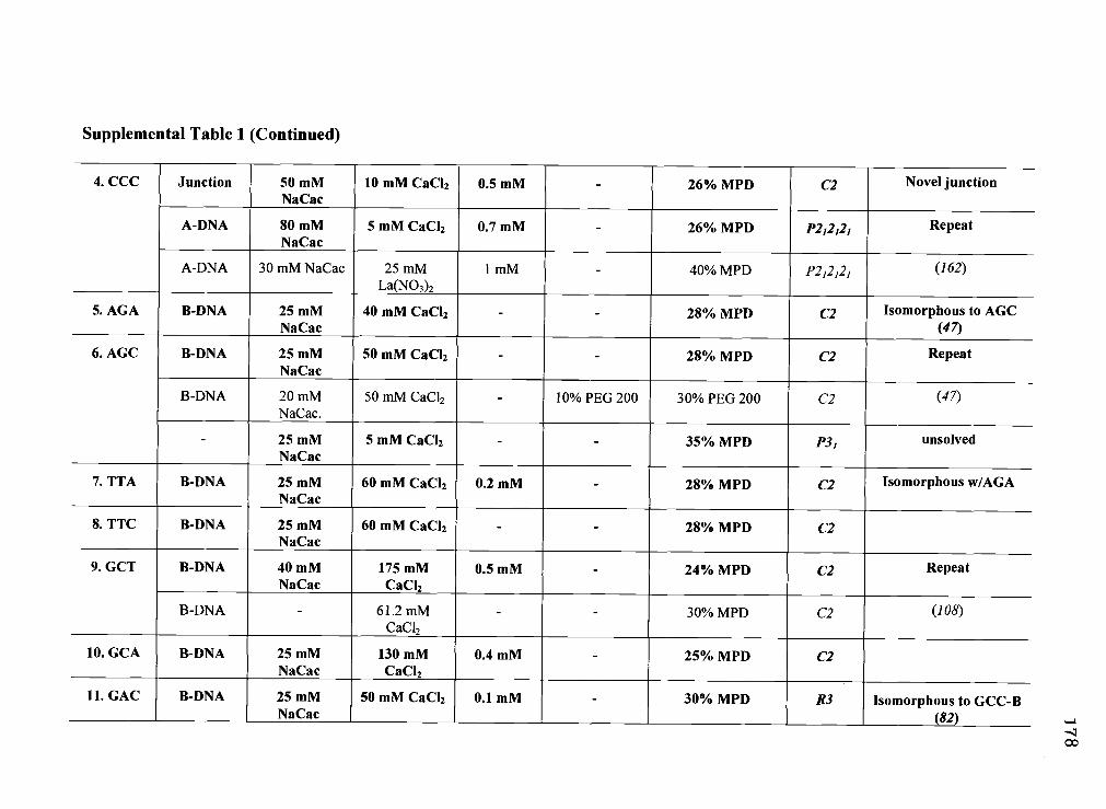
Supplemental Table 1 (Continued)
4. CCC Junction 50mM 10 mM CaCl2 0.5mM - 26% MPD C2 Novel junctionNaCac
A-DNA 80 mM 5 mM CaCl2 0.7 mM - 26% MPD P2,2121 RepeatNaCac
A-DNA 30 mM NaCac 25 mM 1 mM - 40% MPD P212121 (162)La(NO3)2
5. AGA B-DNA 25 mM 40 mM CaCl2 - - 28% MPD C2 Isomorphous to AGCNaCac (4')
6. AGC B-DNA 25 mM 50 mM CaCl2 - - 28% MPD C2 RepeatNaCac
B-DNA 20 mM 50 mM CaCl2 - 10% PEG 200 30% PEG 200 C2 (47)NaCac.
- 25 mM 5 mM CaCJ2 - - 35% MPD p31 unsolvedNaCac
7. TTA B-DNA 25 mM 60 mM CaCl2 0.2 mM - 28% MPD C2 Isomorphous w/AGANaCac
8. TTC B-DNA 25 mM 60 mM CaCl2 - - 28% MPD C2NaCac
9. GCT B-DNA 40 mM 175 mM 0.5 mM - 24% MPD C2 RepeatNaCac CaCl2
B-DNA 61.2mM - - 30% MPD C2 (108)CaCl2
10. GCA B-DNA 25 mM 130 mM 0.4 mM - 25% MPD C2NaCac CaCl2
11. GAC B-DNA 25 mM 50 mM CaCl2 0.1 mM - 30% MPD R3 Isomorphous to GCC-BNaCac (82)
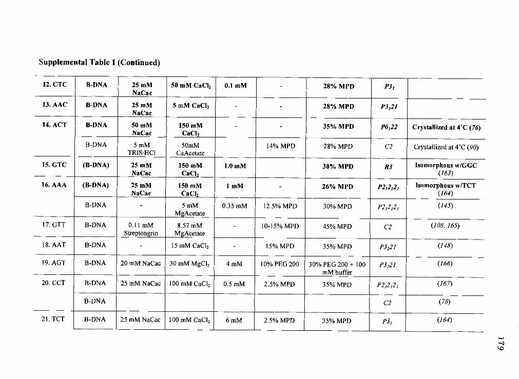
Supplemental Table 1 (Continued)
12. CTC B-DNA 25 mM 50 mM CaCl2 0.1 mM - 28% MPD P31NaCac
13. AAC B-DNA 25 mM 5 mM CaCl2 - - 28% MPD P3221NaCac
14. ACT B-DNA 50 mM 150 mM - - 35% MPD P6122 Crystallized at 4C (76)NaCac CaCl2
B-DNA 5 mM 50mM 14% MPD 28% MPD C2 Crystallized at 4C (90)TRISHCl CaAcetate
15. GTC (B-DNA) 25 mM 150 mM 1.0 mM 30% MPD R3 Isomorphous w/GGCNaCac CaCl2 (163)
16. AAA (B-DNA) 25 mM 150 mM 1 mM - 26% MPD P212121 Isomorphous w/TCTNaCac CaCl2 (164)
B-DNA 5 mM 0.35 mM 12.5% MPD 30% MPD P212121 (145)MgAcetate
17. GTT B-DNA 0.11 mM 8.57mM - 10-15% MPD 45% MPD C2 (108, 165)Streptongrin MgAcetate
18. AAT B-DNA - 15 mM CaCl2 - 15% MPD 35% MPD P3221 (148)
19. AGT B-DNA 20 mM NaCac 30 mM MgCl2 4 mM 10% PEG 200 30% PEG 200 + 100 P3221 (166)mM buffer
20. CCT B-DNA 25 mM NaCac 100 mM CaCl2 0.5 mM 2.5% MPD 35% MPD P212121 (167)
B-DNA C2 (78)
21. TCT B-DNA 25mM NaCac 100mM CaCl2 6mM 2.5% MPD 35% MPD p31 (164)

Supplemental Table 1 (Continued)
B-DNA 26 mM NaCac 101 mM CaC12 7 mM 2.5% MPD 35% MPD P212121 (164)
22. GGC B-DNA 25 mM 60 mM CaCl2 0.1 mM - 28% MPD R3 Repeat of (163)NaCac
B-DNA 50 mM NaCac 20 mM 1 M - 30% MPD R3 pH 6.0 (163)MgAcetate
A-DNA 25 mM NaCac 3.5 mM - 15% MPD 30% MPD, + cryst. P6522 pH 6.0 (132)MgAcetate solution
23. CCG A-DNA 25 mM 25 mM CaCl2 1.3 mM - 20% MPD P212121 RepeatNaCac
A-DNA 40 mM NaCac - 5 mM - 40% MPD P212121 (168)
24. CGG A-DNA 25 mM 60 mM CaCl2 0.4 mM - 28% MPD P222 Isomorphous wIGGANaCac
25. GGA A-DNA 25 mM 50 mM CaCl2 0.1 mM - 28% MPD P212121NaCac
26. GGG A-DNA 25 mM 40 mM CaCl2 2 mM - 28% MPD P212121 Isomorphous w/GGANaCac
27. GGT A-DNA 25 mM 50 mM CaCl2 1.8 mM - 20% MPD P212121 Isomorphous wIGGANaCac
28. TGG A-DNA 25 mM 40 mM CaCl2 0.1 mM - 28% MPD P212121 Isomorphous w/CCGNaCac
29. TCG A-DNA 40 mM NaCac 0.5 mM - 50% MPD P43212 pH 6.0 (169)Co(NH3)6
30. AGG 25 mM 50 mM CaCl2 0.1 mM - 28% MPD P41212 data collectedNaCac
ri
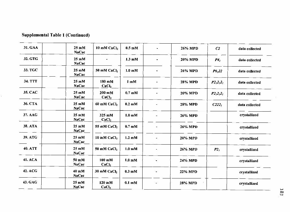
Supplemental Table 1 (Continued)
31. GAA 25 mM 10 mM CaCl2 0.5 mM - 26% MPD C2 data collectedNaCac
32. GTG 25 mM - 1.3 mM - 20% MPD P41 data collectedNaCac
33. TGC 25 mM 50 mM CaCl2 1.0 mM 26% MPD P6222 data collectedNaCac
34. TTT 25 mM 180 mM 1 mM - 28% MPD P212121 data collectedNaCac CaCl2
35. CAC 25 mM 200 mM 0.7 mM - 20% MPD P212121 data collectedNaCac CaCl2
36. CTA 25 mM 60 niM CaCl2 0.2 mM - 28% MPD C2221 data collectedNaCac
37. AAG 25 mM 325 mM 1.0 mM - 26% MPD ctystallizedNaCac CaCl2
38. ATA 25 mM 55 mM CaCl2 0.7 mM - 26% MPD crystallizedNaCac
39. ATG 25 mM 10 mM CaCl2 1.2 mM - 20% MPD crystallizedNaCac
40. ATT 25 mM 50 mM CaCl2 1.0 mM - 26% MPD P21 crystallizedNaCac
41. ACA 50 mM 100 mM 1.0 mM - 24% MPD crystallizedNaCac CaCl2
42. ACG 40 mM 30 mM CaCl2 0.3 mM - 22% MPD crystallizedNaCac
43. GAG 25 mM 120 mM 0.1 mM - 28% MPD crystallizedNaCac CaC12

Supplemental Table 1 (Continued)
44. GAT 25 mM 30 mM CaCl2 1.1 mM - 30% MPD crystallizedNaCac
45. GTA 25 mM 70 mM CaCl2 1.0 mM - 12% MPD crystallizedNaCac
46. GCG 50 mM 10 mM CaCl2 0.8 mM - 28% MPD crystallizedNaCac
47. TAA 30 mM 10 mM CaCl2 0.5 mM - 22% MPD crystallizedNaCac
48. TAG 30 mM 30 mM CaCl2 0.3 mM - 26% MPD crystallizedNaCac
49. TAT 25 mM 150 mM 1.0 mM - 30% MPD P21 crystallizedNaCac CaCl2
50. TAC 25 mM 20 mM CaCl2 0.5 mM - 28% MPD C2 crystallizedNaCac
51. TGA 25 mM 80 mM CaCl2 0.5 mM - 22% MPD crystallizedNaCac
52. TGT 25 mM 80 mM CaCl2 0.2 mM - 24% MPD crystallizedNaCac
53. TTG 50 mM 5 mM CaCl2 0.7 mM - 26% MPD crystallizedNaCac
54. TCA 25 mM 5 mM CaCl2 0.1 mM - 28% MPD crystallizedNaCac
55. TCC 40 mM 12 mM CaCl2 1.0 mM - 10% MPD crystallizedNaCac
56. CAA 25 mM 75 mM CaCl2 1.3 mM - 26% MPD P21 crystallizedNaCac

Supplemental Table 1 (Continued)
57. CAG 25 mM 90 mM CaCl2 0.1 mM - 20% MPD crystallizedNaCac
58. CAT 25 mM 30 mM CaCl2 0.8 mM - 30% MPD crystallizedNaCac
59. CGT 25 mM 60 mM CaCl2 0.6 mM - 20% MPD crystallizedNaCac
60. CGC 25 mM 325 mM 0.1 mM - 26% MPD crystallizedNaCac CaCl2
61. CTG 25 mM - 0.9 mM - 20% MFD P21 crystallizedNaCac
62. CTT 25 mM 50 mM CaCl2 0.1 mM - 28% MPD crystallizedNaCac
63. CCA 60 mM 100 mM 0.7 mM - 24% MPD crystallizedNaCac CaCl

Supplemental Table 2: Crystallographic parameters for sequences of the type d(CCnnnN6N7N8GG), where the N6N7N8 coretrinucleotide is varied to be any of the four standard nucleotides and nnn are nucleotides that maintain the self-complementarity of thesequences. Sequences are listed according to the unique N6N7N8 trinucleotide sequence, along with the Protein Data Bank accessionnumber, structural class (and reference if not from the current study), crystal morphology as specified by the space group, unit cellparameters (only the unique nonorthorhombic angle is given), resolution, overall completeness of the data, of the data, the finalvalues of and Rfree for the refined structure, and any comments concerning the assignment of the conformation. Structuresdetermined in this study or from this laboratory are in bold. Most new structures (those without references) have not been fullyrefined to their lowest and Rfree values. Structures from this study that are isomorphous (within 8% of unit cell parameters) withstructures that have already been determined in this or previous studies may not be fully refined. For sequences without an assignedstructural class we have collected full datasets and are in the process of obtaining a structural solution.
Core PDB Structure Space Unit Cell Parameters Crystallographic data Rcr;t Rfree CommentsAccession Group (%) (%) (reference).
Resolution Completeness R,,erge
1. ACC 1ZEZ Junction C2 a=66.0 A, b24.1 A, 1.95 A 91.8% 7.4 22.9 27.9 Repeat of(1)c=36.9_A,_=_114.8°
1DCW Junction C2 az66.5A,b23.5A, 2.1 A 97.4% 4.5 23.0 31.7 (1)c76.9_A,_fr"l 14.8°
1JUC Junction C2 a=64.9 A, b=25.4 A, 2.35 A 96.4% 6.4 22.6 26.8 2 strands asuc37.4 A, =1 10.57°
(2)
2. GCC 1P4Y Junction C2 a=66.5 A, b=24.2 A, 1.70 A 78.7% 4.9 22.9 26.0 Junction (3)c=37.0_A,_p=iio.0°
1CGC B-DNA R3 a=53.7 A, b53.7 A, 2.2 A - 16.7 - (4)c=44.6_A,_y°°120.0°
3. ATC 1ZF4 Junction C2 a=64.9 A, b24.0 A, 1.65 A 79.3% 5.5 23.1 27.5 Novel junctionc=40.3_A,_1=1 18.7°
1ZF3 Junction C2 a=65.0 A, b24.6 A, 1.85 A 98.6% 6.1 20.2 23.1 Novel junctionc=37.5_A,_fl=112.1°
H

Supplemental Table 2 (Continued)
IZFC B-DNA P212121 a=35.2 A, b=39.1 A, 1.61 A 98.9% 5.1 26.0 27.0 Isomorphousc=33.0 A to AAA (5)
- P212121 a=33.1 A, b=40.2 A, 1.85 A 91.7% 15.5 - - unsolvedc=46.3_A
4. CCC 1ZF2 Junction C2 a=65.0 A, b=24.7 A, 1.95 A 88.9% 10.3 23.1 27.3 Novel junctionc=36.8_A,_=110.6°
1ZF1 A-DNA P21212, a=24.8 A, b=44.4 A, 1.18 A 93.5% 2.8 22.2 24.5 Repeatc=46.3_A
321D A-DNA P212121 a24.9 A, b44.9 A, 2.15 A 77% 3.4 16.2 18.6 (6)e48.1_A
5. AGA 1ZEW B-DNA C2 a=63.7 A, b=24.6 A, 2.25 A 88.6% 5.7 24.8 29.8 Isomorphousc=40.6 A, fl°422.6° to AGC (1)
6. AGC 1ZFM B-DNA C2 a=63.9 A, b=24.3 A, 2.20 A 92.3% 4.8 25.9 34.0 Repeatc=40.4 A, =122.6°
IDCV B-DNA C2 a=64.1 A, b=25.9 A, 2.5 A 98.2% 5.0 20.7 31.7 (1)c=39.9_A,_f3=122.0°
- - P31 a=b=1I1.3 A, c=33.4 A, 2.0 A 96.8% 7.5 - - unsolvedy=122.0°
7. TTA 1ZFH B-DNA C2 a=61.8 A, b=24.3 A, 2.51 A 99.0% 3.8 32.0 34.1 Isomorphousc41.9 A, °°122.7° wIAGA
8. TTC 1ZFF B-DNA C2 a34.3 A, b20.3 A, 0.94 A 81.1% 4.6 42.0 41.2 Resolutionc=33.5 A, =1 12.0° Extended
- - C2 a=62.1 A, b=26.5 A, 2.0 A 94.0% 5.5 - - unsolvedc=67.4_A,_=95.2°
9. GCT IZF5 B-DNA C2 a31.7 A, b=25.7 A, 0.99 A 89.2% 3.3 25.5 24.2 Repeatc=34.0_A,_=116.7°
1EN9 B-DNA C2 a=32.2 A, b=25.4 A, 0.98 A 93.3% 3.1 14.1 15.1 (7)c34.2_A,_J31 17,2°

Supplemental Table 2 (Continued)
10. GCA IZFE B-DNA C2 a=64.0 A, b=23.8 A, 2.35 A 90.9% 5.6 27.5 30.7c=46.1_A,_=126.1°
11. GAC 1ZF7 B-DNA R3 a=53.4 A, b=53.4 A, 1.05 A 93.8% 5.1 27.6 28.8 Isomorphousc=43.9 A, y=120.0' to GCC-B (4)
12. CTC 1ZFG B-DNA P31 a=33.8 A, b=33.8 A, 1.71 A 92.3% 25.2 30.0 31.8
13. AAC 1ZF0 B-DNA P3221 a=33.3 A, b=33.3 A, 1.50 A 67.6% 7.9 23.7 26.0c=93.6_A, y=120.0°
14. ACT 1P4Z B-DNA P6122 a=33.4 A, b=33.4 A, 2.00 A 94.6% 4.8 23.6 26.8 (3)c=87.7_A, y=120.0°
ID8G B-DNA C2 a31.7A,b25.7A, 0.74A 90.0% 5.6 10.5 13.1 (8)c=34.2_A,_1 16.90
15. GTC - (B-DNA) R3 a=52.2 A, b=52.2 A, 3.00 A 79.6% 17.7 Isomorphousc=46.25 A w/GGC (9)
16. AAA - (B-DNA) P212121 a=33.3 A, b=40.0 A, 3.00 A 64.5% 21.2 - - Isomorphousc=44.4 A w/TCT (10)
1IKK B-DNA P212121 a=35.6A,b38.7A, 1.60A 99.2% 8.4 17.9 23.6 (5)c=32.5_A
17.GTT 1EN3 B-DNA C2 a32.OA,b25.4A, 0.98 A 95.0% 4.7 14.1 15.9 (7, 11)c33.6A,113.0°
18. AAT 167D B-DNA P3221 a=33.2 A, b=33.2 A, 2.30 A 91.0% - 20.0 - (12)
19. AGT BDJO61 B-DNA P3221 a24.9 A, b=32.9 A, 1.95 A - 8.7 17.0 - (13)c=95.1_A,7120.0°
20. CCT ICW9 B-DNA P212121 a=34.4 A, b43.2 A, 1.55 A 95.5% 7.8 20.3 23.2 (14)c=72.4A
1BD1 B-DNA C2 a32.2A,b25.5A, 1.60A - - 16.0 - (15)c=34.8_A,_frI 16.70

Supplemental Table 2 (Continued)
21. TCT ICVX B-DNA P31 a=34.2 A, b34.2 A, 2.27 A 96.1% 2.3 23.7 23.8 (10)c=46.1A,-y=120.0
1CVY B-DNA P222 a34.5 A, b=38.6 A, 2.15 A 97.2% 4.1 22.9 22.9 (10)c46.1_A
22. GGC IZFB B-DNA R3 a53.6 A, b=53.6 A, 1.75 A 99.5% 5.7 23.3 28.3 Repeat of(9)c=44.8_A, ,=12O.O0
1QC1 B-DNA R3 a=53.7 A, b53.7 A, 2.5 A 7.8 14.7 (9)c45.0_A,_yi2o.O
382D A-DNA P6522 a41.5 A, b41.5 A, 2.2 A 88.9% 5.6 19.7 (16)c57.5_A,_-y=l2o.o°
23. CCC IZEX A-DNA P212121 a=24.0 A, b45.1 A, 1.65 A 93.8% 4.9 26.2 26.1 Repeatc=44.9_A
160D A-DNA P212121 a=24.9A,b44.6A, 1.7A - - 18.5 (17)c=46.9_A
24. CCG 1ZEY A-DNA P212121 a=24.5 A, b44.3 A, 1.65 A 84.8% 7.8 26.6 32.7 Isomorphousc=47.7A w/GGA
25. GGA 1ZFA A-DNA P212121 a=23.9 A, b=44.6 A, 1.50 A 97.9% 11.2 23.5 28.3c=46.8_A
26. GGG 1ZF9 A-DNA P212121 a23.2 A, b45.3 A, 1.38 A 92.2% 3.7 23.7 25.9 isomorphousc=47.6 A w/GGA
27. GCT 1ZF8 A-DNA P2,2121 a=23.8 A, b=44.9 A, 1.48 A 90.2% 7.3 21.9 26.3 Isomorphousc=49.6 A w/GGA
28. TGG IZF6 A-DNA P212121 a25.2 A, b41.2 A, 1.45 A 93.0% 5.1 25.6 30.6 Isomorphousc=44.4 A w/AHJO6O
29. TCG 1M77 A-DNA P43212 a44.3 A, b44.3 A, 1.25 A 90.0% 5.5 16.3 18.5 (18)c57.5_A,_yl20.0°
30. AGG - - P41212 a=b= 45.0 A, c= 77.1 A 1.80 A 99.5 % 6.2 - - data collected

Supplemental Table 2 (Continued)
31. GAA - - C2 a61.9 A, b26.0 A, 3.10 A 67.0 % 17.8 - - data collectedc=41.1_A,_=124.9°
32. GTG - - P41 a=b=36.8 A, c=87.1 A 3.50 A 92.6 % 7.9 - - data collected
33. TGC - - P6222 a=b=356.3 A, c32.6 A 2.00 A 97.9 % 8.5 - - data collected
34. TTT - - P212121 a=33.0 A, b=39.4 A, 1.40 A 88.1 % 26.3 - - data collectedc=46.8_A
35. CAC - - P212,21 a=50.4 A, b=53.5 A, 2.30 A 54.7 % 9.4 - - data collectedc=56.7_A
36. CTA - - C2221 a=62.0 A, b=68.7 A, 1.60 A 86.0 % 4.2 - - data collectedc=68.6_A

Supplemental Table 3: Conformational parameters for sequences of the type d(CCnnnN6N7N8GG), where the N6N7N8 coretrinucleotide is varied to be any of the four standard nucleotides and 'nnn' are nucleotides that maintain the seif-complementarity of thesequences. Sequences are listed according to the unique N6N7N8 trinucleotide sequence, along with the Protein Data Bank accessionnumber and the corresponding translational or rotational displacement parameters. Values for each parameter were calculated usingthe 3DNA program (88) and then averaged across the entire structure. The overall average and standard deviation for eachdisplacement parameter is listed for the B-DNA, A-DNA and Holliday junction structural classes.
CORE NDBAccession
HelicalTwist (°)
Roll(°)
Tilt(°)
PropellerTwist (°)
Buckle(°)
Rise(A)
X-disp(A)
Slide(A)
Inclinationp (A
B-DNA DuplexesGCA 1ZFE 38.2 -0.12 -0.43 -12.9 -4.94 3.39 1.43 1.32 1.71 -0.90
ICVX 35.0 4.24 0.11 -15.1 -0.55 3.29 0.37 0.76 7.36 -0.77TCT1CVY 35.0 4.63 0.35 -13.4 -1.14 3.31 0.21 0.82 9.19 -0.775DNB 35.3 2.23 0.00 -10.3 0.00 3.31 0.45 0.78 5.10 -0.85CT T1EN33 35.1 2.67 0.00 -10.5 0.00 3.24 0.25 0.73 6.14 -0.86IQCI 34.7 2.84 0.08 -16.9 1.07 3.37 0.45 0.54 4.70 -0.42GGC1ZFB 35.2 3.29 -0.21 -10.4 -3.74 3.33 0.71 0.86 6.31 -0.69
1ENE6 35.6 2.63 0.00 -10.2 0.00 3.27 0.62 1.07 7.45 -1.08GCT 1EN9 35.2 2.28 0.00 -10.2 0.00 3.29 0.65 1.07 6.76 -1.03
IZF5 35.8 1.86 0.00 -10.5 0.00 3.29 0.71 1.01 5.88 -0.86CCC 1CGC 35.7 3.40 1.03 -9.26 0.38 3.39 0.62 0.76 6.25 -0.40GAC IZF7 35.3 1.77 0.23 -12 -0.44 3.35 0.45 0.72 5.04 -0.66CTC IZFG 36.4 5.33 0.62 -10.6 -2.27 3.32 0.39 0.61 7.35 -0.65
1CW9 35.4 1.68 0.00 -8.49 0.00 3.35 0.80 1.02 5.22 -0.81CCT1BD1 35.4 1.68 0.00 -8.49 0.00 3.35 0.80 1.02 5.22 -0.81
ATC 1ZFC 36.8 -0.08 -0.06 -17.3 7.35 3.34 -0.16 -0.07 0.86 -0.26ACT 167D 33.7 1.13 0.08 -7.84 -1.06 3.24 -0.31 -0.08 1.75 -0.33AGC IDCV 36.8 0.26 -0.70 -8.76 -1.50 3.40 0.94 0.72 0.57 -0.35AGA 1ZEW 38.2 -0.58 -0.79 -15.3 -3.00 3.39 1.68 1.30 0.54 -0.82
1D8G 35.5 2.63 0.00 -9.26 0.00 3.29 0.55 1.04 7.66 -1.02A CT1P4Z 35.1 3.14 0.00 -12.1 0.00 3.17 0.09 0.51 5.43 -0.53
AAT BDJO55 33.7 2.96 0.45 -14.2 1.05 3.29 -0.42 0.04 5.03 -0.44

Supplemental Table 3 (Continued)
AAC IZFO 34.5 2.07 -1.78 -9.68 -4.13 3.22 0.43 0.45 3.41 -0.49AAA IIKK 37.3 0.31 -0.10 -15.5 5.37 3.29 0.05 0.09 0.91 -0.42
OVERALLAVERAGE 35.6 2.18 -0.05 -11.6 -0.31 3.31 0.49 0.71 4.83 -0.68
STANDARDDEVIATION 1.16 1.51 0.52 2.81 2.62 0.06 0.48 0.40 2.53 0.24
Holliday JunctionsGCC 1P4Y 37.2 1.77 -0.29 -8.86 -3.27 3.43 2.54 1.59 2.15 -0.72CCC IZF2 38.0 2.15 -1.28 -14.2 -7.19 3.37 2.60 1.60 2.10 -0.97
1ZF4 37.8 1.53 0.47 -13.0 0.91 3.34 2.83 1.63 1.99 -1.08ATC1ZF3 37.2 2.28 0.20 -16.5 0.37 3.38 4.17 1.77 2.11 -1.15IJUC 37.6 0.92 0.72 -7.52 2.19 3.40 1.01 1.40 1.89 -0.86
1DCW 37.3 2.40 -0.23 -9.61 -0.93 3.40 3.64 1.67 2.24 -1.05Acc1DCW 37.6 1.71 0.02 -9.52 0.00 3.40 2.53 1.67 2.29 -0.99IZEZ 37.1 2.41 -0.75 -10.4 -1.39 3.44 3.63 1.67 2.12 -0.93
37.5 1.90 -0.14 -11.2 -1.16 3.40 2.87 1.63 2.11 -0.97
0.3 0.52 0.65 3.04 2.93 0.03 0.97 0.11 0.13 0.14
A-DNA DuplexesTGG 1ZFE 30.9 9.35 1.28 -9.95 -3.61 3.27 17.3 -1.67 -4.48 2.32TCG 1M77 31.4 9.39 0.00 -13.0 0.00 3.35 17.2 -1.48 -4.15 2.17GGT 1ZF8 29.4 5.76 -0.12 -4.03 3.24 3.27 11.4 -1.77 -4.49 2.45GGG 1ZF9 28.8 6.43 1.37 -9.8 -3.99 3.23 13.2 -1.88 -4.95 2.32GGC 382D 30.9 9.23 0.00 -11.7 0.00 3.24 17.1 -1.53 -4.38 2.35GGA 1ZFA 30.4 4.94 1.22 -2.63 2.56 3.38 9.51 -1.91 -4.48 2.01CGG 1ZEY 30.4 4.87 1.48 -1.87 1.10 3.39 9.31 -1.90 -4.40 1.98
160D 30.2 6.25 0.77 -8.1 -1.42 3.39 12.1 -1.79 -4.57 2.29CCG1ZEX 30.1 6.79 0.89 -8.27 -2.58 3.30 12.9 -1.81 -4.68 2,39
CCC 321D 31.3 5.57 0.22 -8.6 -2.37 3.30 10.2 -1.63 -3.94 2.30
C

Supplemental Table 3 (Continued)
IZF1 30.5 7.10 -0.29 -8.09 0.83 3.24 13.7 -1.76 -4.66 2.38
OVERALL 30.4 6.88 0.62 -7.82 O.57 3.30 13.1 -1.74 -4.47 2.27
STANDARD 0.77 1.71 0.67 3.57 2.42 0.06 3.01 0.15 0.27 0.15