Embed Size (px)
Citation preview
Genome-wide antagonism between 5-hydroxymethylcytosine and DNA methylation in the adult mouse brain
Junjie U. GUO1,2,3,*, Keith E. SZULWACH4,*, Yijing SU1,3, Yujing LI4, Bing YAO4, Zihui XU4, Joo Heon SHIN5, Bing XIE5, Yuan GAO1,5, Guo-li MING1,2,3, Peng JIN4, and Hongjun SONG1,2,3
1Institute for Cell Engineering, Johns Hopkins University School of Medicine, Baltimore, MD 21205, USA
2The Solomon H. Snyder Department of Neuroscience, Johns Hopkins University School of Medicine, Baltimore, MD 21205, USA
3Department of Neurology, Johns Hopkins University School of Medicine, Baltimore, MD 21205, USA
4Department of Human Genetics, Emory University School of Medicine, 615 Michael Street, Atlanta, GA 30322, USA
5Lieber Institute for Brain Development, Johns Hopkins University School of Medicine, Baltimore, MD 21205, USA
Abstract
Mounting evidence points to critical roles for DNA modifications, including 5-methylcytosine
(5mC) and its oxidized forms, in the development, plasticity and disorders of the mammalian
nervous system. The novel DNA base 5-hydroxymethylcytosine (5hmC) is known to be capable of
initiating passive or active DNA demethylation, but whether and how extensively 5hmC functions
in shaping the post-mitotic neuronal DNA methylome is unclear. Here we report the genome-wide
distribution of 5hmC in dentate granule neurons from adult mouse hippocampus in vivo. 5hmC in
the neuronal genome is highly enriched in gene bodies, especially in exons, and correlates with
gene expression. Direct genome-wide comparison of 5hmC distribution between embryonic stem
cells and neurons reveals extensive differences, reflecting the functional disparity between these
two cell types. Importantly, integrative analysis of 5hmC, overall DNA methylation and gene
expression profiles of dentate granule neurons in vivo reveals the genome-wide antagonism
Correspondence: aHongjun SONG, bPeng JIN, [email protected], [email protected].*These authors contributed equally to this work.
Junjie Guo, Keith E. Szulwach, Yijing Su, Yujing Li, Bing Yao, Zihui Xu, Joo Heon Shin, Bing Xie, Yuan Gao, Guo-li Ming, Peng Jin and Hongjun Song declare that they have no conflict of interest.
Compliance with ethics guidelinesAll institutional and national guidelines for the care and use of laboratory animals were followed.
Author contributionsH.S. and P.J. conceived the project. Y.S. prepared genomic DNA samples. K.E.S., Y.L. and Z.X. performed 5hmC-seq, data pre-processing, and qPCR validation. B.X. and Y.G. performed RNA-seq, J.U.G. and K.E.S. analyzed the data. J.U.G., K.E.S., B.Y., G.L.M., P.J., and H.S. wrote the manuscript.
NIH Public AccessAuthor ManuscriptFront Biol (Beijing). Author manuscript; available in PMC 2015 January 05.
Published in final edited form as:Front Biol (Beijing). 2014 February ; 9(1): 66–74. doi:10.1007/s11515-014-1295-1.
NIH
-PA
Author M
anuscriptN
IH-P
A A
uthor Manuscript
NIH
-PA
Author M
anuscript
between these two states of cytosine modifications, supporting a role for 5hmC in shaping the
neuronal DNA methylome by promoting active DNA demethylation.
Keywords
dentate granule neuron; active DNA demethylation; TET; methylome
Introduction
5-methylcytosine (5mC) is the predominant and most extensively studied epigenetic
modification in the mammalian genome (Bird, 2002). Although it is well established that
DNA methylation is deposited by a conserved family of DNA methyltransferases (DNMTs)
(Goll and Bestor, 2005), the mechanisms underlying the enzymatic removal of this
modification remain hotly debated (Ma et al., 2009a; Zhu, 2009; Wu and Zhang, 2010;
Bhutani et al., 2011). Ten-eleven-translocated (TET) family dioxygenases oxidize the 5-
methyl group of 5mC, and generate 5hmC (Tahiliani et al., 2009; Ito et al., 2010) as well as
the more oxidized 5-formylcytosine and 5-carboxylcytosine (He et al., 2011; Ito et al.,
2011). Multiple lines of evidence have supported a role for 5mC-to-5hmC conversion in the
process of DNA demethylation. First, 5hmC is known to prevents DNMT1 from recognizing
hemi-methylated DNA in vitro, which may cause “dilution” of the existing DNA
methylation pattern in dividing cells (Valinluck and Sowers, 2007; Frauer et al., 2011).
Indeed, such a passive mode of DNA demethylation was recently found to occur in early
mouse embryos (Inoue and Zhang, 2011). Second, irreplicable 5hmC-containing DNA can
be demethylated in both human cell lines and cultured mouse neurons (Zhang et al., 2010;
Guo et al., 2011c), indicating that 5hmC can also function as a precursor of active DNA
demethylation. Finally, gain- and loss-of-function manipulations of TET proteins lead to
changes in DNA methylation that are consistent with their proposed roles in active DNA
demethylation (Kohli and Zhang, 2013; Pastor et al., 2013).
Intriguingly, among all the tissues surveyed thus far, the mammalian brain has the highest
abundance of 5hmC (Kriaucionis and Heintz, 2009; Globisch et al., 2010). In the brain,
5hmC accounts for 10%–40% of all methylated cytosines, compared to 1%–2% in other
tissues, suggesting the potential functional importance of 5hmC in the nervous system.
Previously generated genomic maps of 5hmC in largely post-mitotic mouse hippocampus as
well as mouse and human cerebellum tissue indicate that 5hmC associates strongly with
genic regions, is depleted at transcription start sites (TSSs), and correlates with gene
expression (Song et al., 2011). Our previous studies suggest that 5hmC levels are
significantly higher in both the cerebellum and hippocampus of adult mice at six weeks and
one year of age than in the early postnatal stage, indicating an age-dependent acquisition of
5hmC and regulation of brain-specific gene expression (Szulwach et al., 2011b). Recent
genome-wide studies from us and others have uncovered global changes of 5hmC associated
with Rett syndrome as well as a number of neurodegenerative disorders, suggesting potential
roles of precise 5hmC distributions in the development and homeostasis of the nervous
system (Szulwach et al., 2011a; Szulwach et al., 2011b; Wang et al., 2013; Yao et al., 2013).
GUO et al. Page 2
Front Biol (Beijing). Author manuscript; available in PMC 2015 January 05.
NIH
-PA
Author M
anuscriptN
IH-P
A A
uthor Manuscript
NIH
-PA
Author M
anuscript
However, the exact role and scope of 5hmC-mediated active DNA demethylation in shaping
the landscape of DNA methylation in neurons in vivo are still unclear.
In this study, we systematically characterized the 5hmC distribution in dentate granule
neurons (DGNs) in vivo and compared it to the 5hmC map of cultured mouse embryonic
stem cells (ESCs). We correlated the 5hmC distribution in DGN with both gene expression
and transcription factor occupancy. Cross-comparison between 5hmC profiles and overall
DNA methylation distributions revealed the global antagonism between these two
modification states, supporting a role for 5hmC in shaping the neuronal DNA methylome on
a genome-wide scale.
Materials and methods
Tissue preparation
Adult mice (8 to 10 weeks old, male, C57BL/6 background) were used for analysis in
accordance with protocols approved by the Institutional Animal Care and Use Committee.
Dentate gyrus tissues were rapidly micro-dissected bilaterally from adult mice. This
preparation was highly enriched for mature neurons as shown by immunohistology to
contain ~90% NeuN+ dentate granule neurons (Ma et al., 2009b). Previous studies of this
preparation showed a very similar CpG methylation status at selective loci with FACS
purified NeuN+ mature neurons (Guo et al., 2011a). Validation experiments were performed
using independent biological samples of bilaterally micro-dissected dentate gyri from
individual animals.
5hmC DNA capture
5hmC enrichment was performed as previously described with an improved selective
chemical labeling method (Song et al., 2011). 5hmC labeling reactions were performed in a
100 μL solution containing 50 mM HEPES buffer (pH 7.9), 25 mM MgCl2, 300 ng/μL
sonicated genomic DNA (100–500 bp), 250 μM UDP-6-N3-Glu, and 2.25 μM wild-type β-
GT. Reactions were incubated for 1 h at 37°C. DNA substrates were purified via Qiagen
DNA purification kit or by phenol-chloroform precipitation and reconstituted in H2O. Click
chemistry was performed with the addition of 150 μM dibenzocyclooctyne modified biotin
into the DNA solution and incubated for 2 h at 37°C. Samples were purified by Pierce
Monomeric Avidin Kit (Thermo) following manufacturer’s recommendations. After elution,
biotin-5-N3-gmC-containing DNA was concentrated by 10K Amicon Ultra-0.5 mL
Centrifugal Filters (Millipore) and purified by Qiagen DNA purification kit.
Library construction and high-throughput sequencing
Five ng of 5hmC-enriched-genomic DNA from 3 independent 5hmC captures or one non-
enriched input genomic DNA was end-repaired, adenylated, and ligated to Illumina
Genomic DNA Adapters (Genomic DNA adapter oligo mix) according to standard Illumina
protocols for ChIP-Seq library construction, maintaining the proper molar ratios of adapter
to insert. Adapter-ligated fragments of ~200–350 bp were gel-purified by 2% agarose gel
electrophoresis and PCR-amplified for 18 PCR cycles. Libraries were checked for quality
and quantified using an Agilent 2100 Bioanalyzer DNA 1000 Chip.
GUO et al. Page 3
Front Biol (Beijing). Author manuscript; available in PMC 2015 January 05.
NIH
-PA
Author M
anuscriptN
IH-P
A A
uthor Manuscript
NIH
-PA
Author M
anuscript

Libraries were sequenced using the Illumina HiScan platform. Cluster generation was
performed with Illumina TruSeq cluster kit v2-cBot-HS. Single-read 51-bp sequencing was
completed with Illumina TruSeq SBS kit v3-HS. A dedicated PhiX control lane, as well as
1% PhiX spike in all other lanes, was used for automated matrix and phasing calculations.
Image analysis and base calling were performed with the standard Illumina pipeline.
Data processing and analysis
FASTQ reads were aligned to NCBIv1/mm9 with Bowtie v0.17.2 retaining non-duplicate,
unique matches to the genome, with no more than 3 mismatches in the first 30 bases.
Ensembl gene annotations were downloaded from the UCSC Genome Browser (http://
genome.ucsc.edu). Data analysis and visualization were done using built-in functions of R
(http://www.r-project.org) and in-house Perl scripts.
qPCR validation of 5hmC-enriched loci
One ng of input or 5mC-enriched DNA, from an independent 5mC capture experiment, was
used in triplicate 20 μL qPCR reactions, each with 1X Power SYBR Green PCR Master Mix
(Applied Biosystems), 0.5 μM forward and reverse primers, and water. Reactions were run
on an SDS 7500 Fast Instrument using 7500 Standard cycling conditions. Fold enrichment
was calculated as 2−dCt, where dCt = Ct (5-hmC-enriched) – Ct (Input).
Results
Global properties of the neuronal 5hmC profile
We used a previously developed chemical tagging method to profile the 5hmC distribution
in the neuronal genome (Song et al., 2011). The dentate gyrus from an in vivo preparation
was used because the majority of cells in this region are Prox1+NeuN+ post-mitotic DGNs
(Ma et al., 2009b; Guo et al., 2011a), providing relatively high homogeneity compared to
other regions in the mouse brain. A total of ~33 million unique non-duplicate reads were
obtained from two biological replicates, which were highly correlated (r = 0.952; Fig. 1A).
In addition, we also profiled mouse ESCs (~19 million unique non-duplicate reads). To
validate the neuronal 5hmC profiles, we used quantitative PCR to measure 5hmC
enrichment at specific loci that were identified as 5hmC-marked by sequencing. In total, 16
out of 17 regions showed significant enrichment relative to a negative control region that is
not 5hmC-marked (Fig. 1B). Global analysis of input-normalized 5hmC signals showed that
all autosomes had comparable 5hmC levels (Fig. 1C), whereas sex chromosomes showed
lower 5hmC levels, consistent with previous studies in both ESCs (Pastor et al., 2011;
Szulwach et al., 2011a) and the brain (Szulwach et al., 2011b). The mitochondrial genome
was depleted in 5hmC, which is consistent with our previous finding that the neuronal
mitochondrial genome is virtually non-methylated (Guo et al., 2011a).
We next determined the average 5hmC signal across the genic regions. All regions near or
within annotated genes, including TSS upstream regions, 5′-untranslated regions (UTR),
coding exons, introns, 3′-UTR, and polyadenylation site (PAS) downstream regions, showed
higher 5hmC levels than intergenic regions (Fig. 1D), which is consistent with previous
findings that 5hmC is enriched near genes in the brain (Szulwach et al., 2011b; Mellén et al.,
GUO et al. Page 4
Front Biol (Beijing). Author manuscript; available in PMC 2015 January 05.
NIH
-PA
Author M
anuscriptN
IH-P
A A
uthor Manuscript
NIH
-PA
Author M
anuscript
2012). Meta-gene analysis showed that gene bodies were enriched in 5hmC (Fig. 1E).
Within gene bodies, coding exons are most enriched for 5hmC (Fig. 1D, E). 5hmC levels
gradually increased from 5′ to 3′ ends within gene bodies (Fig. 1E), although to a lesser
degree compared to ESCs (Xu et al., 2011).
Relationship between 5hmC and gene expression in neurons
Given the gene-centric property of 5hmC, we determined the relationship between 5hmC
levels and associated gene expression levels by correlating 5hmC profiles with previously
obtained DGN RNA-seq results (Guo et al., 2013). Meta-gene analysis showed a striking
position-specific relationship between 5hmC levels and transcript abundance (Fig. 2A). Near
TSSs, 5hmC anti-correlated with gene expression level; 5hmC-marked TSSs were restricted
to the genes that had the lowest expression levels. However, in all other structures, including
the TSS upstream regions, gene body, and PAS downstream regions, 5hmC levels were
positively correlated with gene expression (Fig. 2A). Furthermore, the positive correlation
gradually increased from 5′ to 3′ within the gene body.
Recent studies have revealed an important role for protein-DNA interactions in determining
DNA methylation levels at binding sites (Lister et al., 2009; Guo et al., 2011a; Lienert et al.,
2011; Stadler et al., 2011b). To test whether protein-DNA interactions also shape the
distribution of 5hmC at these binding sites, we averaged 5hmC levels within 10-kb windows
across all binding sites for each of several transcription factors for which ChIP-seq profiles
were available (Kim et al., 2010). Interestingly, 5hmC was depleted at the binding sites for
all the tested neuronal DNA binding factors, including cAMP-responsive element binding
protein (CREB), CREB binding protein (CBP), RNA polymerase II (RNAP), and serum
response factor (SRF; Fig. 2B). These results suggested that protein-DNA interactions may
protect the DNA from methylation and/or hydroxymethylation, or cause DNA
demethylation. It is also possible that DNA (hydroxy-) methylation prevents transcription
factor binding, which is a general property of many DNA binding factors (Stadler et al.,
2011a; Spruijt et al., 2013).
Extensive differences in 5hmC distributions between ESCs and DGNs
5hmC was first identified in the genomic DNA of ESCs (Tahiliani et al., 2009) and
cerebellar neurons (Kriaucionis and Heintz, 2009). Recent studies have shown that these two
cell types have higher 5hmC abundance than other somatic cell types (Globisch et al., 2010).
We compared the two 5hmC profiles obtained using the same chemical tagging method.
Global correlations between ESCs and either DGN sample (r = 0.69 and 0.62, respectively)
were lower than between the two DGN samples (r = 0.83; Fig. 3A), suggesting extensive
differences between the 5hmC distributions in ESC and DGN.
A comparison of gene-body 5hmC levels for individual genes between the two cell types
identified 1129 genes that showed ≥ 4-fold higher 5hmC density in neurons than in ESCs
(DGN-specific) and 1004 genes that are ESC-specific (Fig. 3B). For example, Sema5a, an
autism susceptibility gene that encodes an axon guidance cue, showed a higher 5hmC
density in DGNs than in ESCs (Fig. 3C). In contrast, the HoxA gene cluster, which encodes
an important group of developmental regulators, had much higher 5hmC levels in ESCs than
GUO et al. Page 5
Front Biol (Beijing). Author manuscript; available in PMC 2015 January 05.
NIH
-PA
Author M
anuscriptN
IH-P
A A
uthor Manuscript
NIH
-PA
Author M
anuscript

in DGNs (Fig. 3C). Gene ontology analysis further revealed that neuron-specific 5hmC-
marked genes were enriched in ribosomal proteins and synaptic proteins, such as
neurotransmitter receptors (Fig. 3D), whereas ESC-specific 5hmC-marked genes were
enriched in developmental regulatory pathways. Therefore, differentially 5hmC-marked
genes were associated with the functional differences between the two cell types.
DNA methylation and 5hmC are antagonistic genome-wide in both DGNs and ESCs
To examine the relationship between overall DNA methylation (i.e. the sum of 5mC and
5hmC) and 5hmC, we overlaid the 5hmC map with the previously determined DNA
methylation profile for the same DGNs (Guo et al., 2011a). Strikingly, the global profiles of
these two modification states showed highly complementary patterns (Fig. 4A). The high
5hmC density regions showed low levels of DNA methylation and vice versa. Across the
genome, 5hmC and overall DNA methylation were significantly anti-correlated (Fig. 4B).
To rule out the possibility that the global antagonism between 5hmC and DNA methylation
was merely due to the association of 5hmC and gene-rich regions in the genome, we
calculated the averaged gene-body 5hmC and overall DNA methylation levels for each gene
and ranked all the genes by their expression levels. As we showed previously, gene-body
5hmC levels were correlated with gene expression (Fig. 4C), whereas DNA methylation was
anti-correlated with gene expression, suggesting that the antagonism between 5hmC and
overall DNA methylation also exists at the individual gene level.
Finally, we tested whether 5hmC and DNA methylation were also antagonistic genome-
wide in ESCs. Using the previously determined ESC methylome data (Meissner et al.,
2008), we found a significant anti-correlation between 5hmC and overall DNA methylation
in ESCs (Fig. 4D), suggesting that the global antagonism between these two states of
cytosine modification is not restricted to post-mitotic neurons.
Discussion
Since the identification of 5hmC in the genomic DNA from ESCs and Purkinje cells, the
biological role of this DNA base has been the subject of intensive study. Here we
systematically compared the 5hmC distributions between two cells types, ESCs and DGNs,
both of which exhibit relatively high abundances of 5hmC. We uncovered both similarities
and differences in the 5hmC profiles in these two cell types. In both ESCs and DGNs, 5hmC
was enriched near and within genes, especially in coding exons, suggesting a potential role
for 5hmC in exon definition and regulation of pre-mRNA splicing, as has been indicated for
5mC (Shukla et al., 2011). Intragenic 5hmC levels were correlated with gene expression in
both cell types. As a result, differentially 5hmC-marked genes between ESCs and DGNs
reflected the functional differences between the two cell types, supporting a potential role
for 5hmC in the regulation of cell type-specific gene expression.
A central question of 5hmC biology is whether it indeed functions as an intermediate
product in active DNA demethylation (Wu and Zhang, 2010; Guo et al., 2011b; Branco et
al., 2012; Kohli and Zhang, 2013). We have previously shown that fully hydroxymethylated
exogenous linear DNA can be demethylated in both human cell lines and mouse neurons in
GUO et al. Page 6
Front Biol (Beijing). Author manuscript; available in PMC 2015 January 05.
NIH
-PA
Author M
anuscriptN
IH-P
A A
uthor Manuscript
NIH
-PA
Author M
anuscript

culture (Guo et al., 2011c), suggesting that 5hmC can be converted to non-methylated
cytosines by an active mechanism. Here, by correlating the DGN 5hmC profile with our
previously reported DGN methylome (Guo et al., 2011a), we showed that 5hmC and DNA
methylation were antagonistic genome-wide, further supporting a role for 5hmC in shaping
the genomic DNA methylome by promoting active DNA demethylation in post-mitotic
neurons. In addition, the global antagonism between 5hmC and DNA methylation was also
observed in ESCs, suggesting that maintaining DNA methylation at low levels is a cell-
type–independent function of 5hmC.
Previous studies have shown that DNMT1 has a lower methyltransferase activity toward
hemi-hydroxymethylated DNA in vitro (Valinluck and Sowers, 2007), raising the possibility
that 5hmC may also promote passive DNA demethylation. However, DNMT1-associated
factor UHRF1 can recognize hemi-hydroxymethylated DNA (Frauer et al., 2011; Spruijt et
al., 2013). Therefore, whether 5hmC has an influence on the maintenance of the symmetric
DNA methylation patterns after DNA replication remains to be tested. On the other hand,
TET proteins have been shown to play important roles in locus-specific DNA demethylation
in several systems, including preimplantation embryos (Gu et al., 2011), primordial germ
cells (Dawlaty et al., 2013; Yamaguchi et al., 2013), and various brain regions (Kaas et al.,
2013; Rudenko et al., 2013). While it remains unclear to what extent DNA replication and
passive dilution of 5hmC play roles in TET-mediated DNA demethylation in embryonic
stages, the post-mitotic nature of neurons provides a suitable system for studying the role of
5hmC in active DNA demethylation without any contribution from DNA replication. Future
analyses using methods that can distinguish 5mC and its oxidized forms at base resolution
(Booth et al., 2012; Yu et al., 2012; Song et al., 2013) will potentially reveal the kinetic
details in each individual steps of 5hmC-mediated DNA demethylation, which may yield
novel insights into the dynamic regulation of DNA modification states in post-mitotic
neurons.
DNA methylation has been shown to play a critical role in synaptic plasticity related to
learning and memory in mature neurons, likely owing to the regulation of specific gene
expression (Miller and Sweatt, 2007; Feng et al., 2010). Previous studies of DNA
methyltransferases have pointed to a mechanism whereby individual DNMTs play
distinctive roles during neurodevelopment and are orchestrated to maintain long-term proper
neuronal functions (Goto et al., 1994; Feng et al., 2005). Our understanding of the detailed
functions and molecular mechanisms of 5hmC and TET proteins during neurodevelopment
and in mature neurons is still very limited. A recent study of the dynamic change of TET
proteins and 5hmC during neurogenesis revealed increased 5hmC levels during neuronal
differentiation (Hahn et al., 2013). It also showed a negative correlation of 5hmC with
repressive histone mark H3K27 tri-methylation and its effector Polycomb protein complex.
These data indicate a potential role of 5hmC and TET proteins in neurodevelopment, as
shown in Xenopus (Xu et al., 2011). Several recent publications simultaneously reported key
roles of Tet1 in neurogenesis, cognition and memory formation (Kaas et al., 2013; Rudenko
et al., 2013; Zhang et al., 2013). Either depletion or overexpression of Tet1 in mouse brain
could lead to severe consequences. For example, Tet1 KO mice display impaired
neurogenesis, poor learning and memory, abnormal long-term depression and impaired
GUO et al. Page 7
Front Biol (Beijing). Author manuscript; available in PMC 2015 January 05.
NIH
-PA
Author M
anuscriptN
IH-P
A A
uthor Manuscript
NIH
-PA
Author M
anuscript
memory extinction, whereas Tet1 overexpression could result in impaired long-term
memory formation. Mechanistically, up- or downregulation of Tet1 were found to lead to
dysregulation of genes that are involved in critical neuronal activities accompanied by local
5hmC changes. These data together strongly indicate that the level and genomic distribution
of Tet1 must be precisely controlled, possibly in a distinct manner in different neural cell
types, during neurodevelopment and in mature neurons. This notion was also supported by
the observation that the 5hmC distribution and gene expression in different neuronal cell
types, such as Purkinje neurons, granule cells and Bergmann glial cells, diaplayed strong
cell-type bias (Mellén et al., 2012). In that study, MeCP2 was also identified as 5hmC
binding protein, establishing a dual role for MeCP2 in the orchestration of neuronal
plasticity by coordinating different cytosine modifications (Mellén et al., 2012). Our work
presented here on the integrative analyses of 5hmC, overall DNA methylation and gene
expression profiles in the in vivo dentate granule neurons, lay the foundation for the future
study of 5hmC in shaping the neuronal DNA methylomes during neurodevelopment and in
mature neurons.
Acknowledgments
We thank Cheryl Strauss and Kimberley Christian for critical reading of the manuscript. This study was supported in part by the National Institutes of Health (NS051630 and MH076090 to P.J.; NS047344, MH087874, ES021957 to H.S., HD0679184 and NS048271 to G.L.M.), the Emory Genetics Discovery Fund (P.J.), the Simons Foundation Autism Research Initiative (P.J. and H.S.), Dr. Miriam & Sheldon G. Adelson Medical Research Foundation (G.L.M.), Maryland Stem Cell Research Foundation (G.L.M.), and John Hopkins Brain Science Institute (G.L.M.). J.U.G. was a Damon Runyon fellow supported by the Damon Runyon Cancer Research Foundation. Y.S. was supported by a postdoctoral fellowship from Maryland Stem Cell Research Foundation.
References
Bhutani N, Burns DM, Blau HM. DNA demethylation dynamics. Cell. 2011; 146(6):866–872.10.1016/j.cell.2011.08.042 [PubMed: 21925312]
Bird A. DNA methylation patterns and epigenetic memory. Genes Dev. 2002; 16(1):6–21.10.1101/gad.947102 [PubMed: 11782440]
Booth MJ, Branco MR, Ficz G, Oxley D, Krueger F, Reik W, Balasubramanian S. Quantitative sequencing of 5-methylcytosine and 5-hydroxymethylcytosine at single-base resolution. Science. 2012; 336(6083):934–937.10.1126/science.1220671 [PubMed: 22539555]
Branco MR, Ficz G, Reik W. Uncovering the role of 5-hydroxymethylcytosine in the epigenome. Nat Rev Genet. 2012; 13(1):7–13. [PubMed: 22083101]
Dawlaty MM, Breiling A, Le T, Raddatz G, Barrasa MI, Cheng AW, Gao Q, Powell BE, Li Z, Xu M, Faull KF, Lyko F, Jaenisch R. Combined deficiency of Tet1 and Tet2 causes epigenetic abnormalities but is compatible with postnatal development. Dev Cell. 2013; 24(3):310–323.10.1016/j.devcel.2012.12.015 [PubMed: 23352810]
Feng J, Chang H, Li E, Fan G. Dynamic expression of de novo DNA methyltransferases Dnmt3a and Dnmt3b in the central nervous system. J Neurosci Res. 2005; 79(6):734–746.10.1002/jnr.20404 [PubMed: 15672446]
Feng J, Zhou Y, Campbell SL, Le T, Li E, Sweatt JD, Silva AJ, Fan G. Dnmt1 and Dnmt3a maintain DNA methylation and regulate synaptic function in adult forebrain neurons. Nat Neurosci. 2010; 13(4):423–430.10.1038/nn.2514 [PubMed: 20228804]
Frauer C, Hoffmann T, Bultmann S, Casa V, Cardoso MC, Antes I, Leonhardt H. Recognition of 5-hydroxymethylcytosine by the Uhrf1 SRA domain. PLoS ONE. 2011; 6(6):e21306.10.1371/journal.pone.0021306 [PubMed: 21731699]
GUO et al. Page 8
Front Biol (Beijing). Author manuscript; available in PMC 2015 January 05.
NIH
-PA
Author M
anuscriptN
IH-P
A A
uthor Manuscript
NIH
-PA
Author M
anuscript

Globisch D, Münzel M, Müller M, Michalakis S, Wagner M, Koch S, Brückl T, Biel M, Carell T. Tissue distribution of 5-hydroxymethylcytosine and search for active demethylation intermediates. PLoS ONE. 2010; 5(12):e15367.10.1371/journal.pone.0015367 [PubMed: 21203455]
Goll MG, Bestor TH. Eukaryotic cytosine methyltransferases. Annu Rev Biochem. 2005; 74(1):481–514.10.1146/annurev.biochem.74.010904.153721 [PubMed: 15952895]
Goto K, Numata M, Komura JI, Ono T, Bestor TH, Kondo H. Expression of DNA methyltransferase gene in mature and immature neurons as well as proliferating cells in mice. Differentiation. 1994; 56(1–2):39–44.10.1046/j.1432-0436.1994.56120039.x [PubMed: 8026645]
Gu TP, Guo F, Yang H, Wu HP, Xu GF, Liu W, Xie ZG, Shi L, He X, Jin SG, Iqbal K, Shi YG, Deng Z, Szabó PE, Pfeifer GP, Li J, Xu GL. The role of Tet3 DNA dioxygenase in epigenetic reprogramming by oocytes. Nature. 2011; 477(7366):606–610.10.1038/nature10443 [PubMed: 21892189]
Guo JU, Ma DK, Mo H, Ball MP, Jang MH, Bonaguidi MA, Balazer JA, Eaves HL, Xie B, Ford E, Zhang K, Ming GL, Gao Y, Song H. Neuronal activity modifies the DNA methylation landscape in the adult brain. Nat Neurosci. 2011a; 14(10):1345–1351.10.1038/nn.2900 [PubMed: 21874013]
Guo JU, Su Y, Shin JH, Shin J, Li H, Xie B, Zhong C, Hu S, Le T, Fan G, Zhu H, Chang Q, Gao Y, Ming GL, Song H. Distribution, recognition and regulation of non-CpG methylation in the adult mammalian brain. Nat Neurosci. 201310.1038/nn.3607
Guo JU, Su Y, Zhong C, Ming GL, Song H. Emerging roles of TET proteins and 5-hydroxymethylcytosines in active DNA demethylation and beyond. Cell Cycle. 2011b; 10(16):2662–2668.10.4161/cc.10.16.17093 [PubMed: 21811096]
Guo JU, Su Y, Zhong C, Ming GL, Song H. Hydroxylation of 5-methylcytosine by TET1 promotes active DNA demethylation in the adult brain. Cell. 2011c; 145(3):423–434.10.1016/j.cell.2011.03.022 [PubMed: 21496894]
Hahn MA, Qiu R, Wu X, Li AX, Zhang H, Wang J, Jui J, Jin SG, Jiang Y, Pfeifer GP, Lu Q. Dynamics of 5-hydroxymethylcytosine and chromatin marks in Mammalian neurogenesis. Cell Rep. 2013; 3:291–300. [PubMed: 23403289]
He YF, Li BZ, Li Z, Liu P, Wang Y, Tang Q, Ding J, Jia Y, Chen Z, Li L, Sun Y, Li X, Dai Q, Song CX, Zhang K, He C, Xu GL. Tet-mediated formation of 5-carboxylcytosine and its excision by TDG in mammalian DNA. Science. 2011; 333(6047):1303–1307.10.1126/science.1210944 [PubMed: 21817016]
Inoue A, Zhang Y. Replication-dependent loss of 5-hydroxymethylcytosine in mouse preimplantation embryos. Science. 2011; 334(6053):194.10.1126/science.1212483 [PubMed: 21940858]
Ito S, D’Alessio AC, Taranova OV, Hong K, Sowers LC, Zhang Y. Role of Tet proteins in 5mC to 5hmC conversion, ES-cell self-renewal and inner cell mass specification. Nature. 2010; 466(7310):1129–1133.10.1038/nature09303 [PubMed: 20639862]
Ito S, Shen L, Dai Q, Wu SC, Collins LB, Swenberg JA, He C, Zhang Y. Tet proteins can convert 5-methylcytosine to 5-formylcytosine and 5-carboxylcytosine. Science. 2011; 333(6047):1300–1303.10.1126/science.1210597 [PubMed: 21778364]
Kaas GA, Zhong C, Eason DE, Ross DL, Vachhani RV, Ming GL, King JR, Song H, Sweatt JD. TET1 controls CNS 5-methylcytosine hydroxylation, active DNA demethylation, gene transcription, and memory formation. Neuron. 2013; 79(6):1086–1093.10.1016/j.neuron.2013.08.032 [PubMed: 24050399]
Kim TK, Hemberg M, Gray JM, Costa AM, Bear DM, Wu J, Harmin DA, Laptewicz M, Barbara-Haley K, Kuersten S, Markenscoff-Papadimitriou E, Kuhl D, Bito H, Worley PF, Kreiman G, Greenberg ME. Widespread transcription at neuronal activity-regulated enhancers. Nature. 2010; 465(7295):182–187.10.1038/nature09033 [PubMed: 20393465]
Kohli RM, Zhang Y. TET enzymes, TDG and the dynamics of DNA demethylation. Nature. 2013; 502(7472):472–479.10.1038/nature12750 [PubMed: 24153300]
Kriaucionis S, Heintz N. The nuclear DNA base 5-hydroxymethylcytosine is present in Purkinje neurons and the brain. Science. 2009; 324(5929):929–930.10.1126/science.1169786 [PubMed: 19372393]
GUO et al. Page 9
Front Biol (Beijing). Author manuscript; available in PMC 2015 January 05.
NIH
-PA
Author M
anuscriptN
IH-P
A A
uthor Manuscript
NIH
-PA
Author M
anuscript
Lienert F, Wirbelauer C, Som I, Dean A, Mohn F, Schübeler D. Identification of genetic elements that autonomously determine DNA methylation states. Nat Genet. 2011; 43(11):1091–1097.10.1038/ng.946 [PubMed: 21964573]
Lister R, Pelizzola M, Dowen RH, Hawkins RD, Hon G, Tonti-Filippini J, Nery JR, Lee L, Ye Z, Ngo QM, Edsall L, Antosiewicz-Bourget J, Stewart R, Ruotti V, Millar AH, Thomson JA, Ren B, Ecker JR. Human DNA methylomes at base resolution show widespread epigenomic differences. Nature. 2009; 462(7271):315–322.10.1038/nature08514 [PubMed: 19829295]
Ma DK, Guo JU, Ming GL, Song H. DNA excision repair proteins and Gadd45 as molecular players for active DNA demethylation. Cell Cycle. 2009a; 8(10):1526–1531.10.4161/cc.8.10.8500 [PubMed: 19377292]
Ma DK, Ponnusamy K, Song MR, Ming GL, Song H. Molecular genetic analysis of FGFR1 signalling reveals distinct roles of MAPK and PLCgamma1 activation for self-renewal of adult neural stem cells. Mol Brain. 2009b; 2(1):16.10.1186/1756-6606-2-16 [PubMed: 19505325]
Meissner A, Mikkelsen TS, Gu H, Wernig M, Hanna J, Sivachenko A, Zhang X, Bernstein BE, Nusbaum C, Jaffe DB, Gnirke A, Jaenisch R, Lander ES. Genome-scale DNA methylation maps of pluripotent and differentiated cells. Nature. 2008; 454(7205):766–770. [PubMed: 18600261]
Mellén M, Ayata P, Dewell S, Kriaucionis S, Heintz N. MeCP2 binds to 5hmC enriched within active genes and accessible chromatin in the nervous system. Cell. 2012; 151(7):1417–1430.10.1016/j.cell.2012.11.022 [PubMed: 23260135]
Miller CA, Sweatt JD. Covalent modification of DNA regulates memory formation. Neuron. 2007; 53(6):857–869.10.1016/j.neuron.2007.02.022 [PubMed: 17359920]
Pastor WA, Aravind L, Rao A. TETonic shift: biological roles of TET proteins in DNA demethylation and transcription. Nat Rev Mol Cell Biol. 2013; 14(6):341–356.10.1038/nrm3589 [PubMed: 23698584]
Pastor WA, Pape UJ, Huang Y, Henderson HR, Lister R, Ko M, McLoughlin EM, Brudno Y, Mahapatra S, Kapranov P, Tahiliani M, Daley GQ, Liu XS, Ecker JR, Milos PM, Agarwal S, Rao A. Genome-wide mapping of 5-hydroxymethylcytosine in embryonic stem cells. Nature. 2011; 473(7347):394–397.10.1038/nature10102 [PubMed: 21552279]
Rudenko A, Dawlaty MM, Seo J, Cheng AW, Meng J, Le T, Faull KF, Jaenisch R, Tsai LH. Tet1 is critical for neuronal activity-regulated gene expression and memory extinction. Neuron. 2013; 79(6):1109–1122.10.1016/j.neuron.2013.08.003 [PubMed: 24050401]
Shukla S, Kavak E, Gregory M, Imashimizu M, Shutinoski B, Kashlev M, Oberdoerffer P, Sandberg R, Oberdoerffer S. CTCF-promoted RNA polymerase II pausing links DNA methylation to splicing. Nature. 2011; 479(7371):74–79.10.1038/nature10442 [PubMed: 21964334]
Song CX, Szulwach KE, Dai Q, Fu Y, Mao SQ, Lin L, Street C, Li Y, Poidevin M, Wu H, Gao J, Liu P, Li L, Xu GL, Jin P, He C. Genome-wide profiling of 5-formylcytosine reveals its roles in epigenetic priming. Cell. 2013; 153(3):678–691.10.1016/j.cell.2013.04.001 [PubMed: 23602153]
Song CX, Szulwach KE, Fu Y, Dai Q, Yi C, Li X, Li Y, Chen CH, Zhang W, Jian X, Wang J, Zhang L, Looney TJ, Zhang B, Godley LA, Hicks LM, Lahn BT, Jin P, He C. Selective chemical labeling reveals the genome-wide distribution of 5-hydroxymethylcytosine. Nat Biotechnol. 2011; 29(1):68–72.10.1038/nbt.1732 [PubMed: 21151123]
Spruijt CG, Gnerlich F, Smits AH, Pfaffeneder T, Jansen PW, Bauer C, Münzel M, Wagner M, Müller M, Khan F, Eberl HC, Mensinga A, Brinkman AB, Lephikov K, Müller U, Walter J, Boelens R, van Ingen H, Leonhardt H, Carell T, Vermeulen M. Dynamic readers for 5-(hydroxy)methylcytosine and its oxidized derivatives. Cell. 2013; 152(5):1146–1159.10.1016/j.cell.2013.02.004 [PubMed: 23434322]
Stadler MB, Murr R, Burger L, Ivanek R, Lienert F, Schöler A, van Nimwegen E, Wirbelauer C, Oakeley EJ, Gaidatzis D, Tiwari VK, Schübeler D. DNA-binding factors shape the mouse methylome at distal regulatory regions. Nature. 2011a; 480(7378):490–495. [PubMed: 22170606]
Stadler MB, Murr R, Burger L, Ivanek R, Lienert F, Schöler A, van Nimwegen E, Wirbelauer C, Oakeley EJ, Gaidatzis D, Tiwari VK, Schübeler D. DNA-binding factors shape the mouse methylome at distal regulatory regions. Nature. 2011b; 480(7378):490–495. [PubMed: 22170606]
Szulwach KE, Li X, Li Y, Song CX, Han JW, Kim S, Namburi S, Hermetz K, Kim JJ, Rudd MK, Yoon YS, Ren B, He C, Jin P. Integrating 5-hydroxymethylcytosine into the epigenomic landscape
GUO et al. Page 10
Front Biol (Beijing). Author manuscript; available in PMC 2015 January 05.
NIH
-PA
Author M
anuscriptN
IH-P
A A
uthor Manuscript
NIH
-PA
Author M
anuscript
of human embryonic stem cells. PLoS Genet. 2011a; 7(6):e1002154.10.1371/journal.pgen.1002154 [PubMed: 21731508]
Szulwach KE, Li X, Li Y, Song CX, Wu H, Dai Q, Irier H, Upadhyay AK, Gearing M, Levey AI, Vasanthakumar A, Godley LA, Chang Q, Cheng X, He C, Jin P. 5-hmC-mediated epigenetic dynamics during postnatal neurodevelopment and aging. Nat Neurosci. 2011b; 14(12):1607–1616.10.1038/nn.2959 [PubMed: 22037496]
Tahiliani M, Koh KP, Shen Y, Pastor WA, Bandukwala H, Brudno Y, Agarwal S, Iyer LM, Liu DR, Aravind L, Rao A. Conversion of 5-methylcytosine to 5-hydroxymethylcytosine in mammalian DNA by MLL partner TET1. Science. 2009; 324(5929):930–935.10.1126/science.1170116 [PubMed: 19372391]
Valinluck V, Sowers LC. Endogenous cytosine damage products alter the site selectivity of human DNA maintenance methyltransferase DNMT1. Cancer Res. 2007; 67(3):946–950.10.1158/0008-5472.CAN-06-3123 [PubMed: 17283125]
Wang F, Yang Y, Lin X, Wang JQ, Wu YS, Xie W, Wang D, Zhu S, Liao YQ, Sun Q, Yang YG, Luo HR, Guo C, Han C, Tang TS. Genome-wide loss of 5-hmC is a novel epigenetic feature of Huntington’s disease. Hum Mol Genet. 2013; 22(18):3641–3653.10.1093/hmg/ddt214 [PubMed: 23669348]
Wu SC, Zhang Y. Active DNA demethylation: many roads lead to Rome. Nat Rev Mol Cell Biol. 2010; 11(9):607–620.10.1038/nrm2950 [PubMed: 20683471]
Xu Y, Wu F, Tan L, Kong L, Xiong L, Deng J, Barbera AJ, Zheng L, Zhang H, Huang S, Min J, Nicholson T, Chen T, Xu G, Shi Y, Zhang K, Shi YG. Genome-wide regulation of 5hmC, 5mC, and gene expression by Tet1 hydroxylase in mouse embryonic stem cells. Mol Cell. 2011; 42(4):451–464.10.1016/j.molcel.2011.04.005 [PubMed: 21514197]
Yamaguchi S, Shen L, Liu Y, Sendler D, Zhang Y. Role of Tet1 in erasure of genomic imprinting. Nature. 2013; 504(7480):460–464.10.1038/nature12805 [PubMed: 24291790]
Yao B, Lin L, Street RC, Zalewski ZA, Galloway JN, Wu H, Nelson DL, Jin P. Genome-wide alteration of 5-hydroxymethylcytosine in a mouse model of fragile X-associated tremor/ataxia syndrome. Hum Mol Genet. 2013 Oct.20 Epub ahead of print.
Yu M, Hon GC, Szulwach KE, Song CX, Zhang L, Kim A, Li X, Dai Q, Shen Y, Park B, Min JH, Jin P, Ren B, He C. Base-resolution analysis of 5-hydroxymethylcytosine in the mammalian genome. Cell. 2012; 149(6):1368–1380.10.1016/j.cell.2012.04.027 [PubMed: 22608086]
Zhang H, Zhang X, Clark E, Mulcahey M, Huang S, Shi YG. TET1 is a DNA-binding protein that modulates DNA methylation and gene transcription via hydroxylation of 5-methylcytosine. Cell Res. 2010; 20(12):1390–1393.10.1038/cr.2010.156 [PubMed: 21079648]
Zhang RR, Cui QY, Murai K, Lim YC, Smith ZD, Jin S, Ye P, Rosa L, Lee YK, Wu HP, Liu W, Xu ZM, Yang L, Ding YQ, Tang F, Meissner A, Ding C, Shi Y, Xu GL. Tet1 regulates adult hippocampal neurogenesis and cognition. Cell Stem Cell. 2013; 13(2):237–245.10.1016/j.stem.2013.05.006 [PubMed: 23770080]
Zhu JK. Active DNA demethylation mediated by DNA glycosylases. Annu Rev Genet. 2009; 43(1):143–166.10.1146/annurev-genet-102108-134205 [PubMed: 19659441]
GUO et al. Page 11
Front Biol (Beijing). Author manuscript; available in PMC 2015 January 05.
NIH
-PA
Author M
anuscriptN
IH-P
A A
uthor Manuscript
NIH
-PA
Author M
anuscript
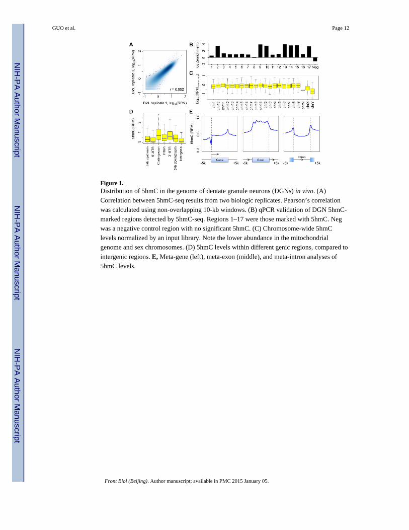
Figure 1. Distribution of 5hmC in the genome of dentate granule neurons (DGNs) in vivo. (A)
Correlation between 5hmC-seq results from two biologic replicates. Pearson’s correlation
was calculated using non-overlapping 10-kb windows. (B) qPCR validation of DGN 5hmC-
marked regions detected by 5hmC-seq. Regions 1–17 were those marked with 5hmC. Neg
was a negative control region with no significant 5hmC. (C) Chromosome-wide 5hmC
levels normalized by an input library. Note the lower abundance in the mitochondrial
genome and sex chromosomes. (D) 5hmC levels within different genic regions, compared to
intergenic regions. E, Meta-gene (left), meta-exon (middle), and meta-intron analyses of
5hmC levels.
GUO et al. Page 12
Front Biol (Beijing). Author manuscript; available in PMC 2015 January 05.
NIH
-PA
Author M
anuscriptN
IH-P
A A
uthor Manuscript
NIH
-PA
Author M
anuscript
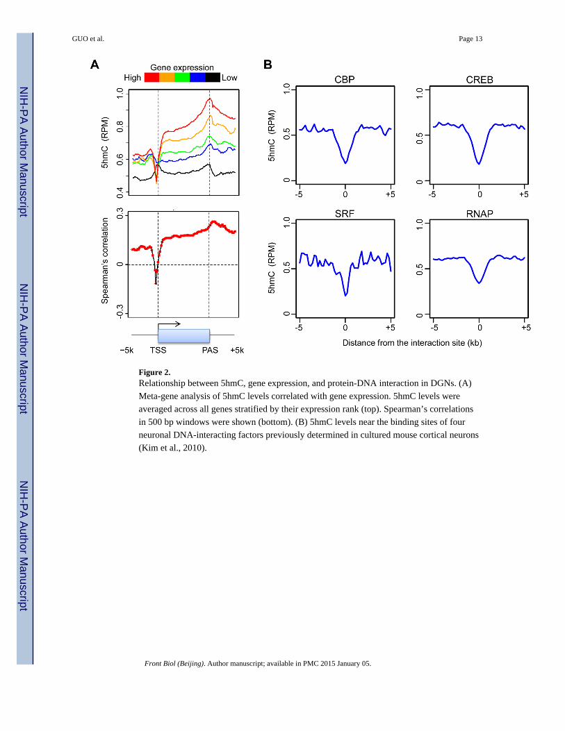
Figure 2. Relationship between 5hmC, gene expression, and protein-DNA interaction in DGNs. (A)
Meta-gene analysis of 5hmC levels correlated with gene expression. 5hmC levels were
averaged across all genes stratified by their expression rank (top). Spearman’s correlations
in 500 bp windows were shown (bottom). (B) 5hmC levels near the binding sites of four
neuronal DNA-interacting factors previously determined in cultured mouse cortical neurons
(Kim et al., 2010).
GUO et al. Page 13
Front Biol (Beijing). Author manuscript; available in PMC 2015 January 05.
NIH
-PA
Author M
anuscriptN
IH-P
A A
uthor Manuscript
NIH
-PA
Author M
anuscript

Figure 3. Comparison of 5hmC distributions between DGNs and ESCs. (A) A correlation matrix of
5hmC-seq results from two DGN replicates and on ESC sample. Pearson’s correlations were
shown. (B) A scatter plot of DGN and ESC gene-body 5hmC levels. DGN- and ESC-
specific 5hmC-marked genes were indicated by blue and red circles, respectively. (C)
Examples of DGN-specific 5hmC-marked genes (top) and ESC-specific 5hmC-marked
genes (bottom). (D) Gene ontology analysis of 1129 DGN-specific 5hmC-marked genes
(left) and 1004 ESC-specific 5hmC-marked genes (right).
GUO et al. Page 14
Front Biol (Beijing). Author manuscript; available in PMC 2015 January 05.
NIH
-PA
Author M
anuscriptN
IH-P
A A
uthor Manuscript
NIH
-PA
Author M
anuscript
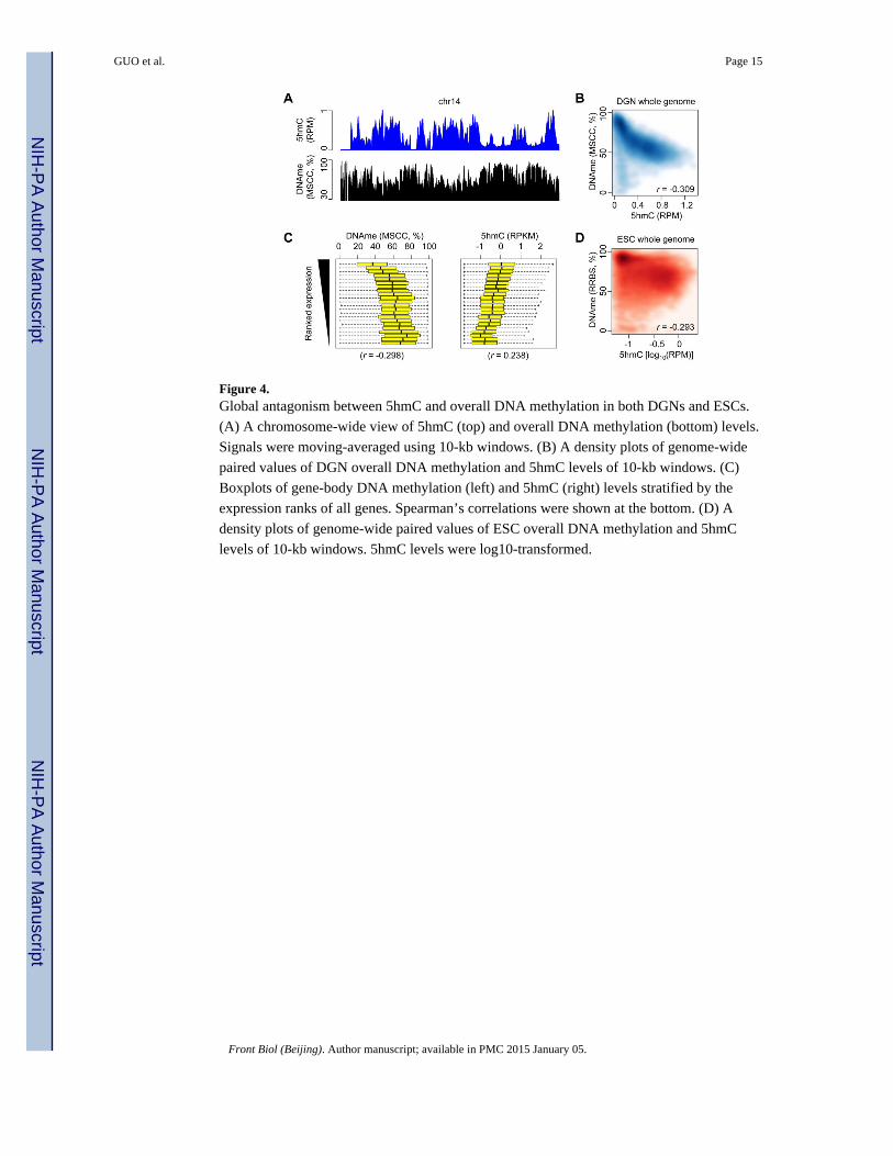
Figure 4. Global antagonism between 5hmC and overall DNA methylation in both DGNs and ESCs.
(A) A chromosome-wide view of 5hmC (top) and overall DNA methylation (bottom) levels.
Signals were moving-averaged using 10-kb windows. (B) A density plots of genome-wide
paired values of DGN overall DNA methylation and 5hmC levels of 10-kb windows. (C)
Boxplots of gene-body DNA methylation (left) and 5hmC (right) levels stratified by the
expression ranks of all genes. Spearman’s correlations were shown at the bottom. (D) A
density plots of genome-wide paired values of ESC overall DNA methylation and 5hmC
levels of 10-kb windows. 5hmC levels were log10-transformed.
GUO et al. Page 15
Front Biol (Beijing). Author manuscript; available in PMC 2015 January 05.
NIH
-PA
Author M
anuscriptN
IH-P
A A
uthor Manuscript
NIH
-PA
Author M
anuscript