Embed Size (px)
Citation preview

7/30/2019 Junq.ed.10 Indo.organ Limfoid
http://slidepdf.com/reader/full/junqed10-indoorgan-limfoid 1/60
Organ Limfoid
7/30/2019 Junq.ed.10 Indo.organ Limfoid
http://slidepdf.com/reader/full/junqed10-indoorgan-limfoid 2/60
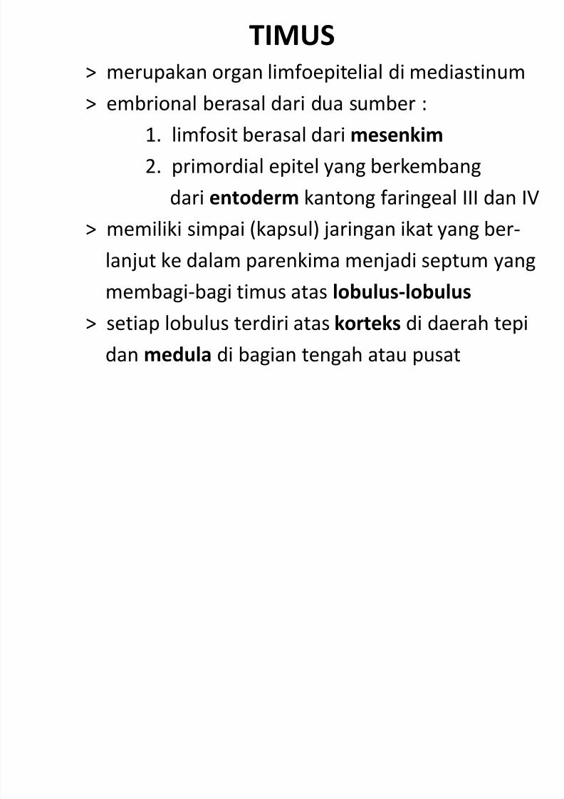
TIMUS> merupakan organ limfoepitelial di mediastinum
> embrional berasal dari dua sumber :
1. limfosit berasal dari mesenkim
2. primordial epitel yang berkembang
dari entoderm kantong faringeal III dan IV
> memiliki simpai (kapsul) jaringan ikat yang ber-
lanjut ke dalam parenkima menjadi septum yang
membagi-bagi timus atas lobulus-lobulus> setiap lobulus terdiri atas korteks di daerah tepi
dan medula di bagian tengah atau pusat
7/30/2019 Junq.ed.10 Indo.organ Limfoid
http://slidepdf.com/reader/full/junqed10-indoorgan-limfoid 3/60
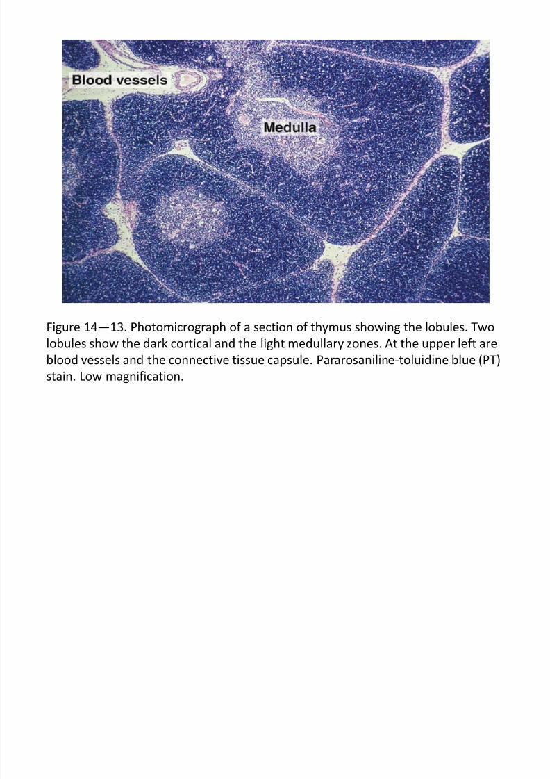
Figure 14—13. Photomicrograph of a section of thymus showing the lobules. Two
lobules show the dark cortical and the light medullary zones. At the upper left are
blood vessels and the connective tissue capsule. Pararosaniline-toluidine blue (PT)
stain. Low magnification.
7/30/2019 Junq.ed.10 Indo.organ Limfoid
http://slidepdf.com/reader/full/junqed10-indoorgan-limfoid 4/60
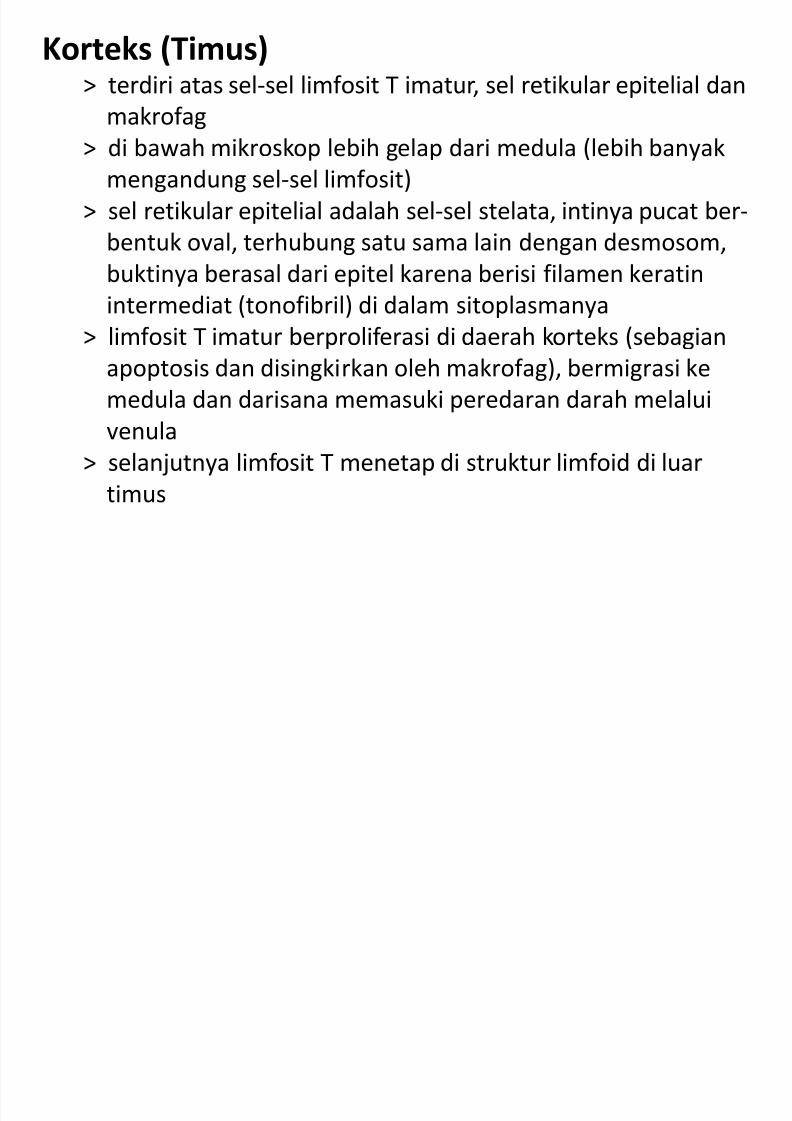
Korteks (Timus)> terdiri atas sel-sel limfosit T imatur, sel retikular epitelial dan
makrofag
> di bawah mikroskop lebih gelap dari medula (lebih banyak
mengandung sel-sel limfosit)
> sel retikular epitelial adalah sel-sel stelata, intinya pucat ber-
bentuk oval, terhubung satu sama lain dengan desmosom,
buktinya berasal dari epitel karena berisi filamen keratinintermediat (tonofibril) di dalam sitoplasmanya
> limfosit T imatur berproliferasi di daerah korteks (sebagian
apoptosis dan disingkirkan oleh makrofag), bermigrasi ke
medula dan darisana memasuki peredaran darah melalui
venula
> selanjutnya limfosit T menetap di struktur limfoid di luar
timus

7/30/2019 Junq.ed.10 Indo.organ Limfoid
http://slidepdf.com/reader/full/junqed10-indoorgan-limfoid 5/60
Figure 14—14. Thymus cortical zone showing epithelial reticular cells with visible
nucleoli (arrowheads) surrounded by dark-stained T lymphocytes undergoing
differentiation. PT stain. Medium magnification.
7/30/2019 Junq.ed.10 Indo.organ Limfoid
http://slidepdf.com/reader/full/junqed10-indoorgan-limfoid 6/60
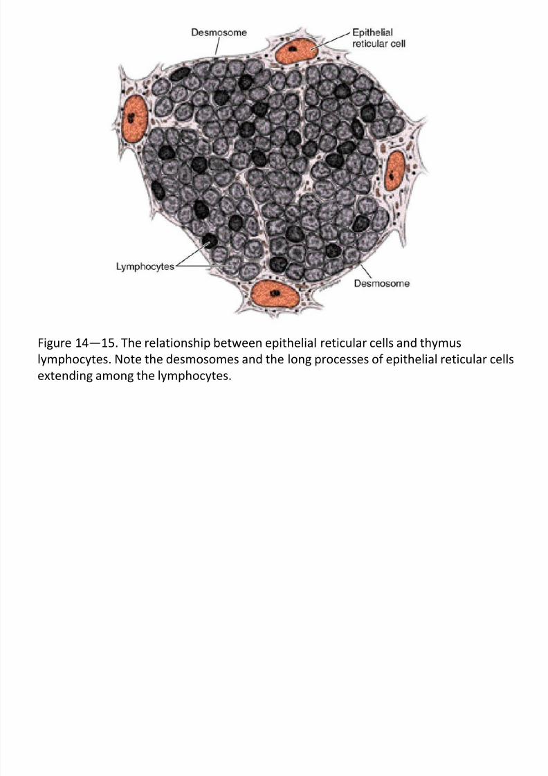
Figure 14—15. The relationship between epithelial reticular cells and thymus
lymphocytes. Note the desmosomes and the long processes of epithelial reticular cells
extending among the lymphocytes.
7/30/2019 Junq.ed.10 Indo.organ Limfoid
http://slidepdf.com/reader/full/junqed10-indoorgan-limfoid 7/60
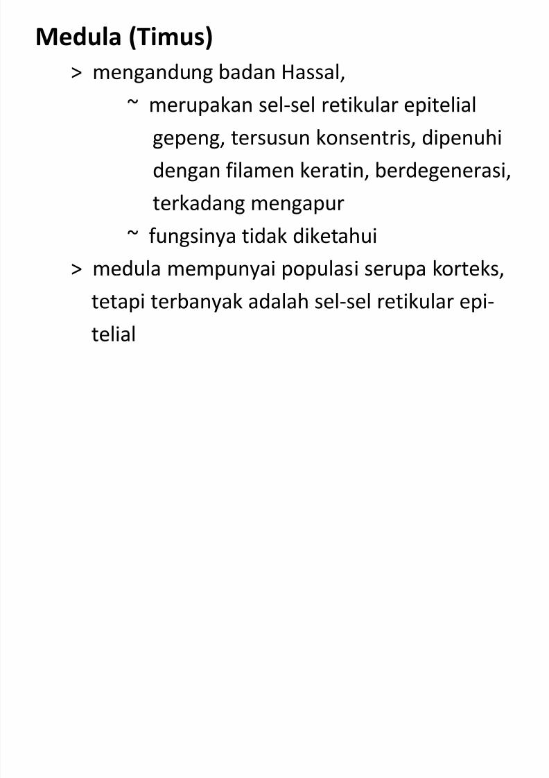
Medula (Timus)
> mengandung badan Hassal,
~ merupakan sel-sel retikular epitelial
gepeng, tersusun konsentris, dipenuhi
dengan filamen keratin, berdegenerasi,
terkadang mengapur
~ fungsinya tidak diketahui
> medula mempunyai populasi serupa korteks,
tetapi terbanyak adalah sel-sel retikular epi-
telial
7/30/2019 Junq.ed.10 Indo.organ Limfoid
http://slidepdf.com/reader/full/junqed10-indoorgan-limfoid 8/60
Figure 14—16. Photomicrograph of the medullary zone of the thymus. The large
numbers of epithelial reticular cells with their large and light-stained nuclei are
responsible for the light color of the thymus medulla. This zone also contains
mature T lymphocytes. PT stain. Medium magnification.
7/30/2019 Junq.ed.10 Indo.organ Limfoid
http://slidepdf.com/reader/full/junqed10-indoorgan-limfoid 9/60
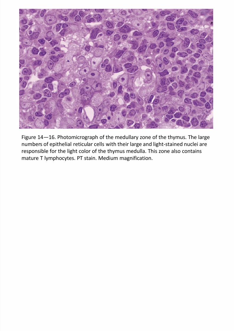
Figure 14—17. Photomicrograph of a portion of the cortical zone, identified by its
dark staining (right), and a portion of medulla, identified by its lighter staining and
the presence of a Hassall corpuscle (left). These corpuscles exist only in the
medulla. PT stain. Medium magnification.
7/30/2019 Junq.ed.10 Indo.organ Limfoid
http://slidepdf.com/reader/full/junqed10-indoorgan-limfoid 10/60
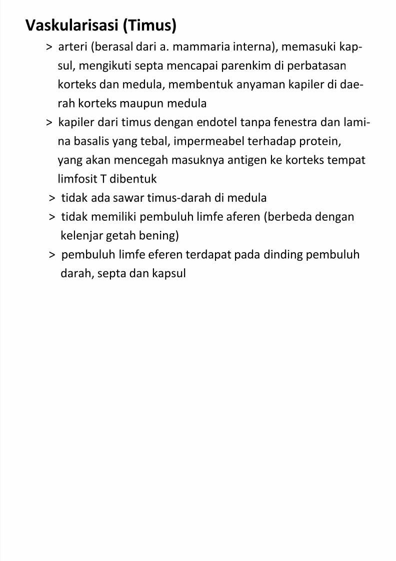
Vaskularisasi (Timus)
> arteri (berasal dari a. mammaria interna), memasuki kap-
sul, mengikuti septa mencapai parenkim di perbatasankorteks dan medula, membentuk anyaman kapiler di dae-
rah korteks maupun medula
> kapiler dari timus dengan endotel tanpa fenestra dan lami-
na basalis yang tebal, impermeabel terhadap protein,
yang akan mencegah masuknya antigen ke korteks tempat
limfosit T dibentuk
> tidak ada sawar timus-darah di medula
> tidak memiliki pembuluh limfe aferen (berbeda dengan
kelenjar getah bening)
> pembuluh limfe eferen terdapat pada dinding pembuluh
darah, septa dan kapsul
7/30/2019 Junq.ed.10 Indo.organ Limfoid
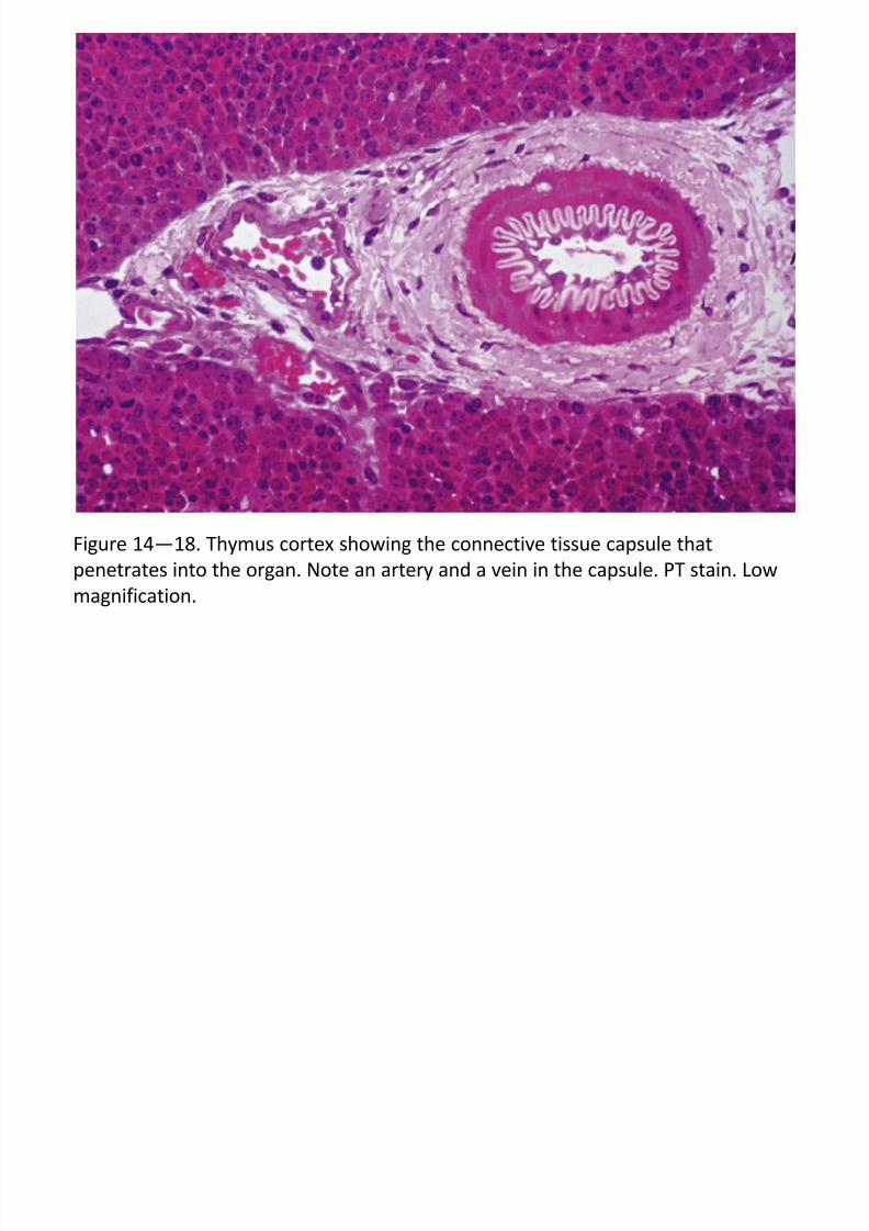
http://slidepdf.com/reader/full/junqed10-indoorgan-limfoid 11/60
Figure 14—18. Thymus cortex showing the connective tissue capsule that
penetrates into the organ. Note an artery and a vein in the capsule. PT stain. Low
magnification.
7/30/2019 Junq.ed.10 Indo.organ Limfoid
http://slidepdf.com/reader/full/junqed10-indoorgan-limfoid 12/60
Histofisiologi (Timus)
> perkembangan timus segera setelah lahir dan akan me-
ngalami involusi setelah usia pubertas> sel-sel induk dari sumsum tulang menambah populasi
sel T secara kontinu, bermigrasi ke timus selama masa
janin dan dewasa
> setelah memasuki timus, sel T atau timosit yang ber-kembang menempati bagian korteks
> selama di timus, sel T bermitosis berulang-ulang, tetapi
lebih dari 95 % disingkirkan melalui apoptosis, yaitu lim-
fosit yang tidak bereaksi terhadap antigen dan limfosityang bereaksi terhadap antigen sendiri
> bila limfosit yang bereaksi terhadap antigen sendiri tidak
disingkirkan menyebabkan terjadinya penyakit autoimun
7/30/2019 Junq.ed.10 Indo.organ Limfoid
http://slidepdf.com/reader/full/junqed10-indoorgan-limfoid 13/60
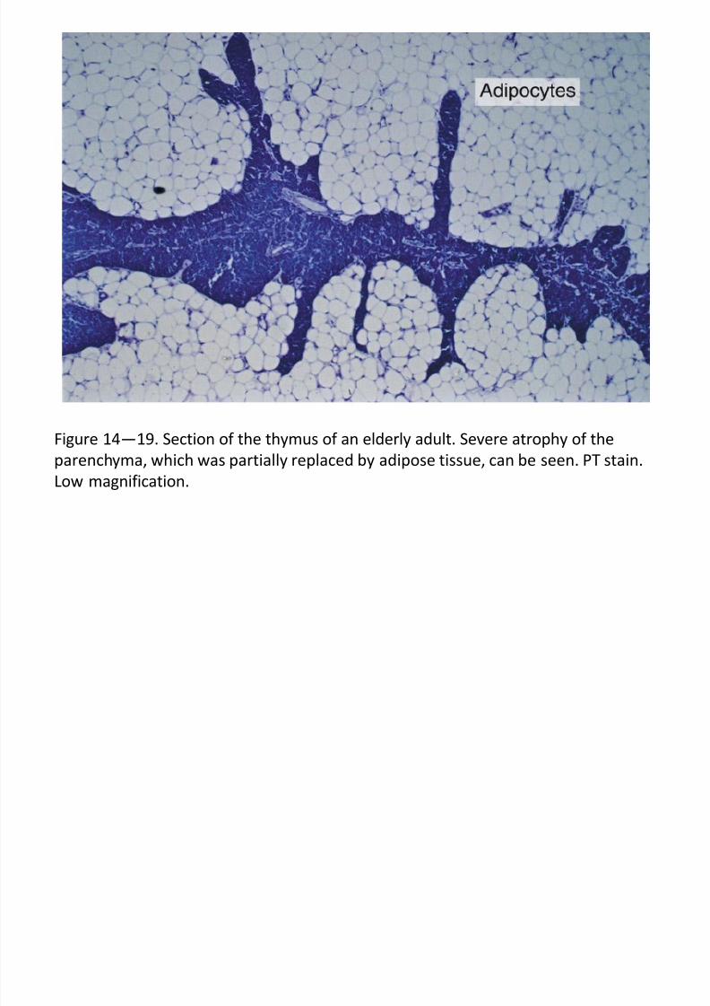
Figure 14—19. Section of the thymus of an elderly adult. Severe atrophy of the
parenchyma, which was partially replaced by adipose tissue, can be seen. PT stain.
Low magnification.
7/30/2019 Junq.ed.10 Indo.organ Limfoid
http://slidepdf.com/reader/full/junqed10-indoorgan-limfoid 14/60
Histofisiologi (Timus) (samb.)
> limfosit yang akhirnya dihasilkan oleh timus adalah
limfosit T, yang bereaksi terhadap antigen asing dan
esensial untuk reaksi imun adaptif normal
> pada mamalia, daerah ‘tergantung-timus’ (thymus-dependent organs), yang kaya sel-T adalah :
~ zona parakorteks kelenjar getah bening
~ plak Peyer
~ selubung periarterial pulpa putih limpa
7/30/2019 Junq.ed.10 Indo.organ Limfoid
http://slidepdf.com/reader/full/junqed10-indoorgan-limfoid 15/60
Histofisiologi (Timus)(samb.)> timus menghasilkan protein faktor pertumbuhan yang
merangsang proliferasi dan diferensiasi limfosit T, merupakan
produk sekresi parakrin dalam timus, yaitu :~ faktor timosin-alfa
~ timopoietin
~ timolin
~ faktor humoral timus
> timus juga merupakan sasaran sejumlah hormon,~ adrenokortikoid menyebabkan :
- pengurangan jumlah limfosit
- pengurangan kecepatan mitosis limfosit
- atrofi lapisan korteks timus
> hormon adrenokortikotropin dari hipofisis anterior mera-sang aktivitas korteks adrenal
> hormon kelamin pria dan wanita mempercepat involusi timus
> kastrasi memperlambat involusi timus
7/30/2019 Junq.ed.10 Indo.organ Limfoid
http://slidepdf.com/reader/full/junqed10-indoorgan-limfoid 16/60
KELENJAR GETAH BENING
Pengantar
> suatu organ berbentuk ginjal atau lonjong, ber-simpai, terdiri dari jaringan limfoid
> merupakan sederetan saringan untuk pertaha-
nan tubuh terhadap mikroorganisme dan pe-nyebaran sel-sel tumor
> bagian-bagian kelenjar getah bening ialah, hilus,
simpai dan trabekula> daerah-daerah dari kelenjar getah bening ialah,
korteks luar, korteks dalam dan medula
7/30/2019 Junq.ed.10 Indo.organ Limfoid
http://slidepdf.com/reader/full/junqed10-indoorgan-limfoid 17/60
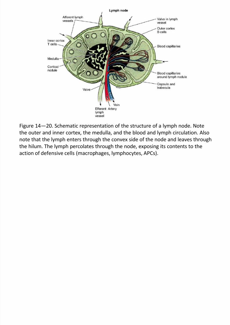
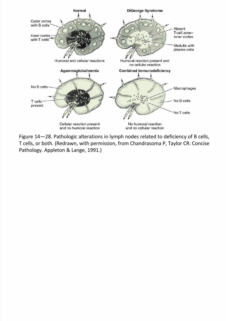
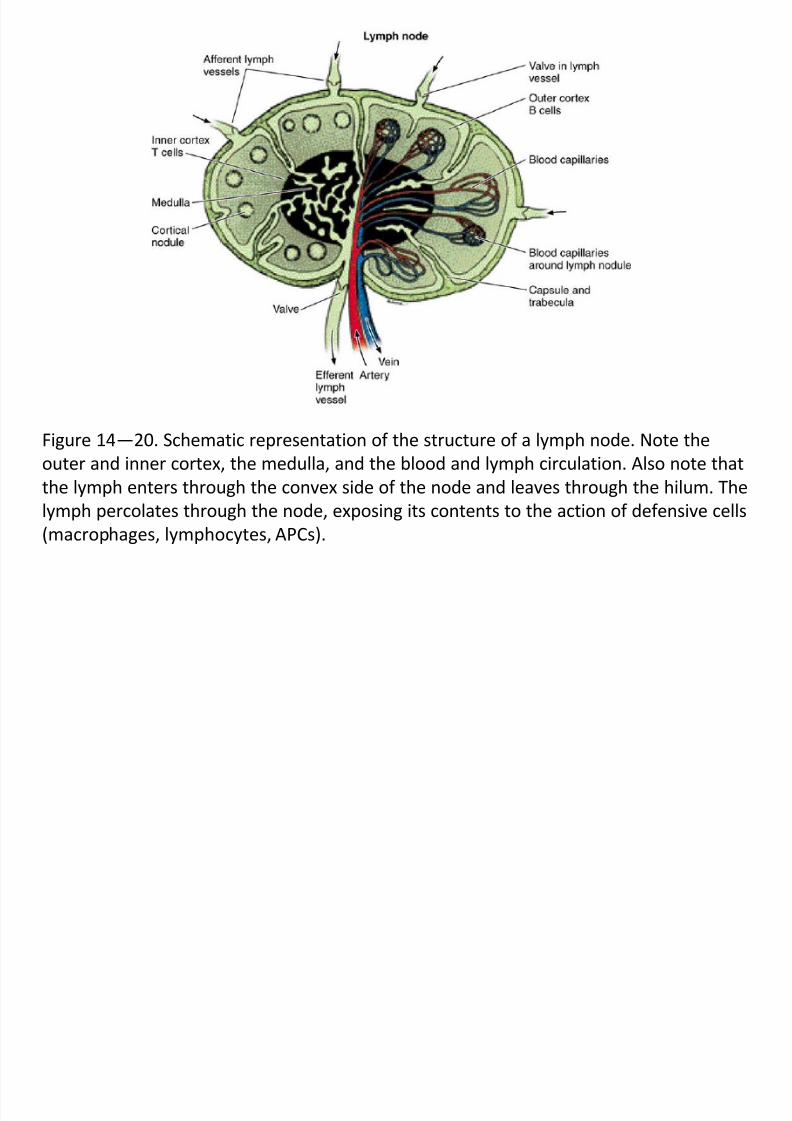
Figure 14—20. Schematic representation of the structure of a lymph node. Note
the outer and inner cortex, the medulla, and the blood and lymph circulation. Also
note that the lymph enters through the convex side of the node and leaves through
the hilum. The lymph percolates through the node, exposing its contents to the
action of defensive cells (macrophages, lymphocytes, APCs).
7/30/2019 Junq.ed.10 Indo.organ Limfoid
http://slidepdf.com/reader/full/junqed10-indoorgan-limfoid 18/60
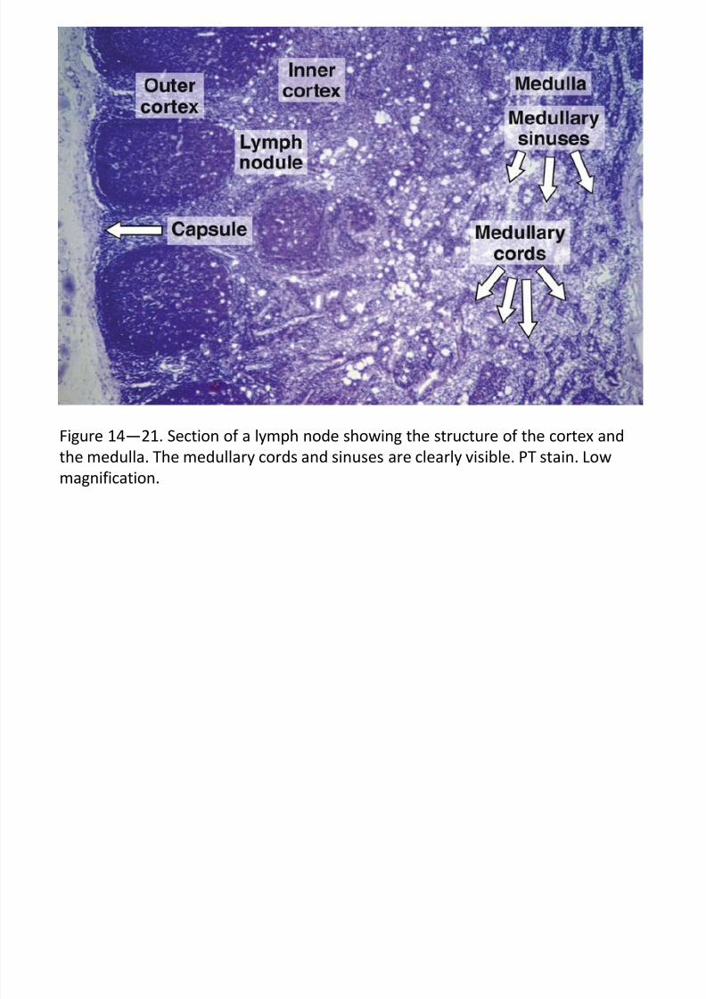
Figure 14—21. Section of a lymph node showing the structure of the cortex and
the medulla. The medullary cords and sinuses are clearly visible. PT stain. Low
magnification.
7/30/2019 Junq.ed.10 Indo.organ Limfoid
http://slidepdf.com/reader/full/junqed10-indoorgan-limfoid 19/60
KELENJAR GETAH BENING (samb.)
KORTEKS LUAR
> mengandung sinus subkapsularis, berhubungan dengan
sinus medularis melalui sinus intermediat (sinus trabekularis)
> korteks luar terdiri dari jalinan longgar makrofag, sel-sel
retikular dan serat retikulin, dipenuhi oleh sel-sel B
> mengandung struktur nodul limfoid, kaya akan limfosit B
yang bereaksi terhadap antigen
> limfosit B bertambah besar, bermitosis menghasilkan sel-
sel besar, basofilik, anak inti jelas disebut imunosit
> beberapa nodul limfoid , daerah pusatnya berwarna lebih
terang disebut pusat germinal
> pusat germinal memperlihatkan sel-sel yang bermitosis
dan banyak mengandung imunosit, menghasilkan sel plas-
ma penghasil ntibodi
7/30/2019 Junq.ed.10 Indo.organ Limfoid
http://slidepdf.com/reader/full/junqed10-indoorgan-limfoid 20/60
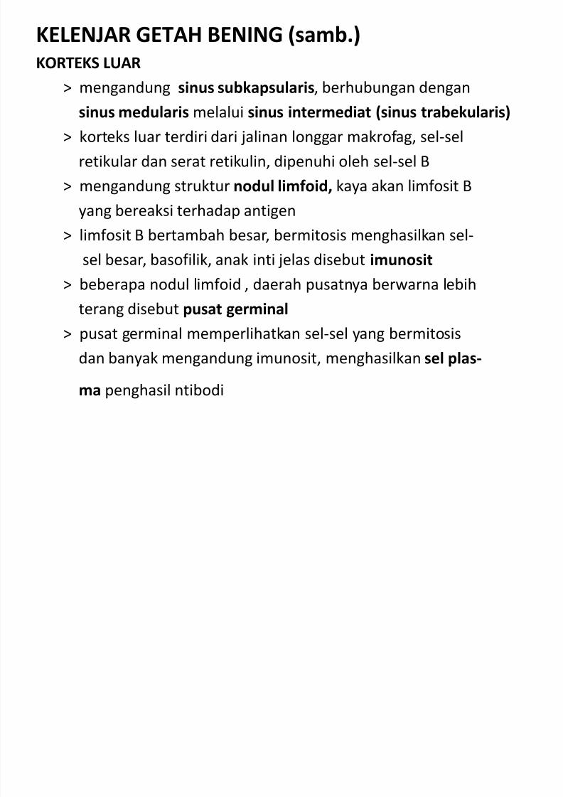
Figure 14—22. Section of a lymph node showing the capsule, subcapsular sinuses,
and a small portion of the outer cortex. Some adipose tissue is seen outside the
capsule. PT stain. Medium magnification.

7/30/2019 Junq.ed.10 Indo.organ Limfoid
http://slidepdf.com/reader/full/junqed10-indoorgan-limfoid 21/60
Figure 14—23. Section of the cortex of a lymph node showing a lymphoid nodule
that was strongly activated by the injection of an antigen. Note the large number of
macrophages (light structures) surrounded by B lymphocytes. PT stain. Medium
magnification.
7/30/2019 Junq.ed.10 Indo.organ Limfoid
http://slidepdf.com/reader/full/junqed10-indoorgan-limfoid 22/60
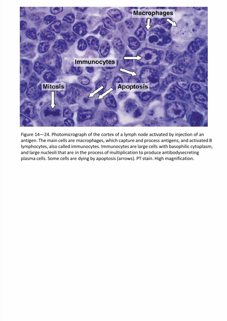
Figure 14—24. Photomicrograph of the cortex of a lymph node activated by injection of an
antigen. The main cells are macrophages, which capture and process antigens, and activated B
lymphocytes, also called immunocytes. Immunocytes are large cells with basophilic cytoplasm,
and large nucleoli that are in the process of multiplication to produce antibodysecreting
plasma cells. Some cells are dying by apoptosis (arrows). PT stain. High magnification.
7/30/2019 Junq.ed.10 Indo.organ Limfoid
http://slidepdf.com/reader/full/junqed10-indoorgan-limfoid 23/60
KELENJAR GETAH BENING (samb.)
KORTEKS DALAM
> lanjutan dari korteks luar, mengandung sedikit nodul lim-foid, banyak mengandung limfosit T
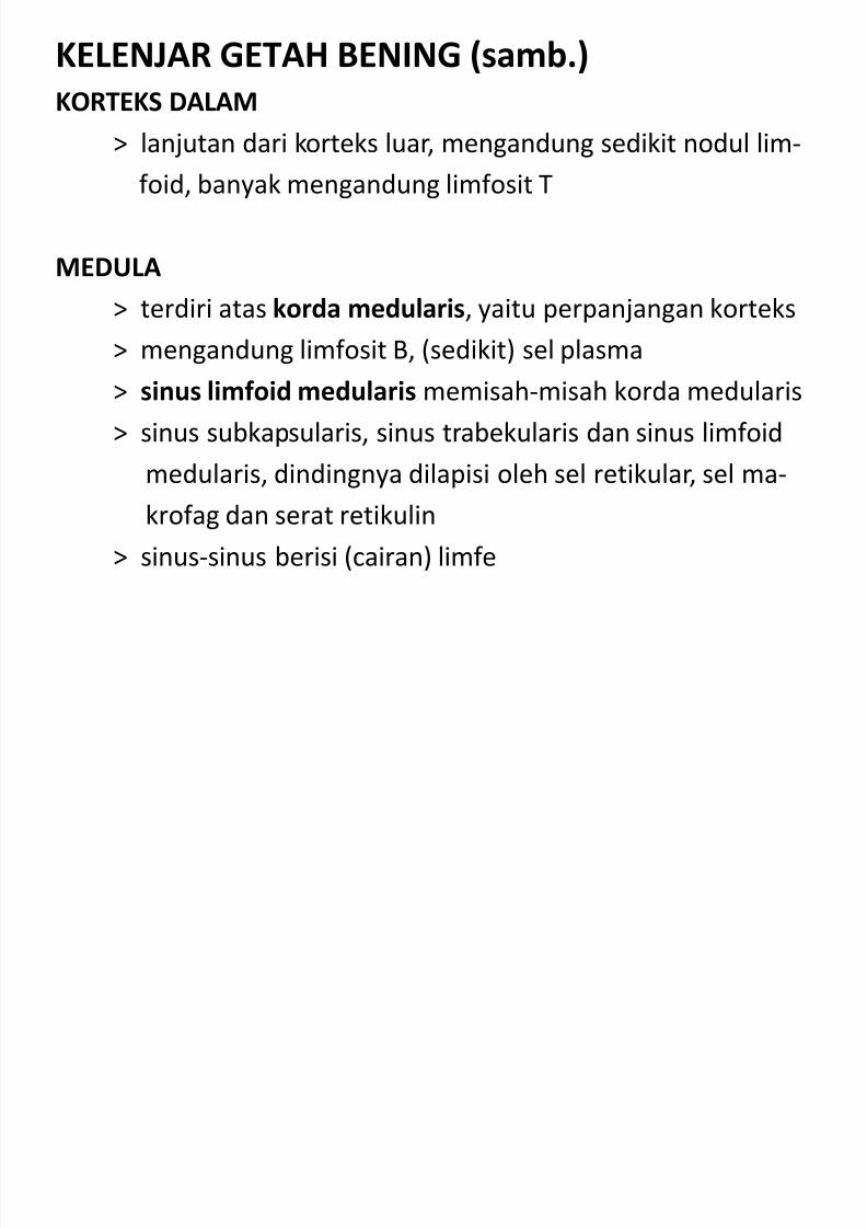
MEDULA
> terdiri atas korda medularis, yaitu perpanjangan korteks
> mengandung limfosit B, (sedikit) sel plasma
> sinus limfoid medularis memisah-misah korda medularis
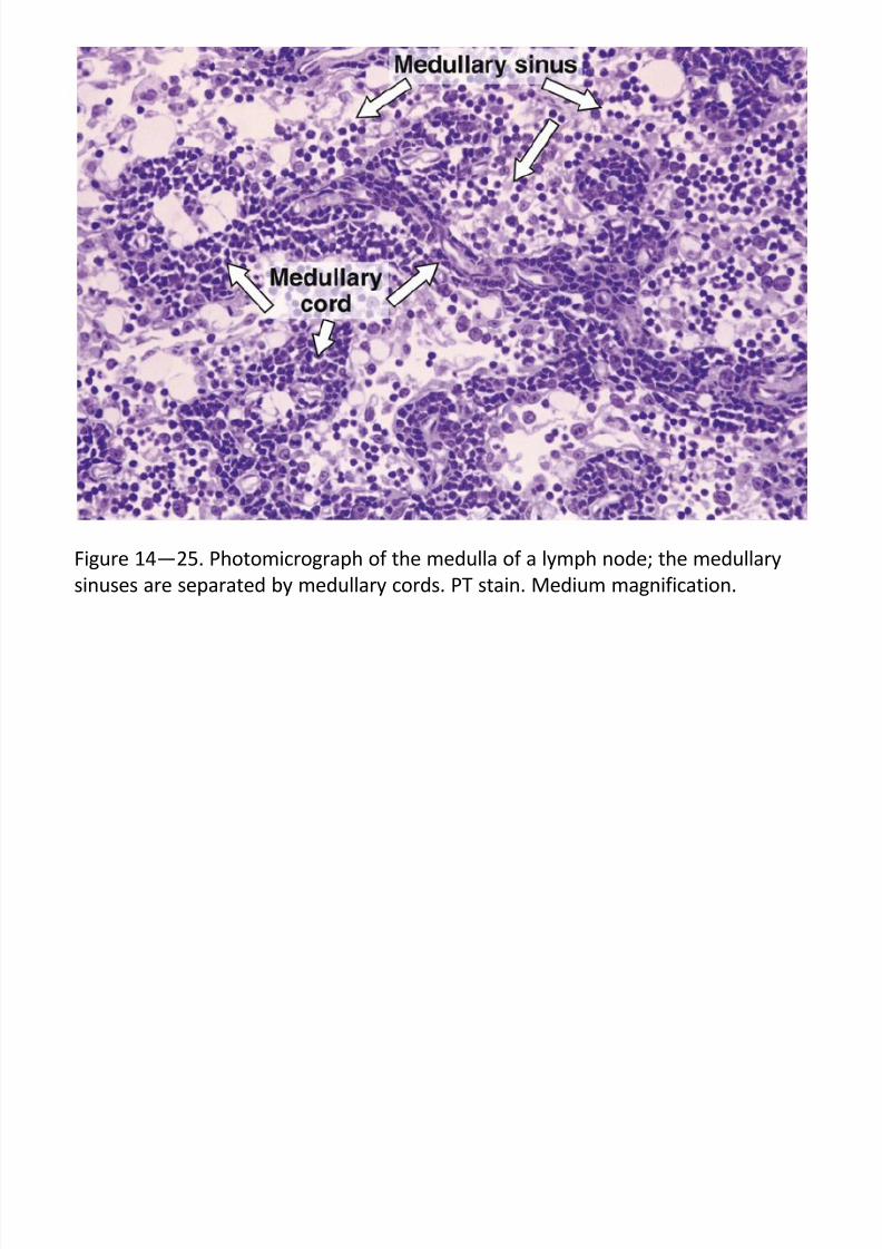
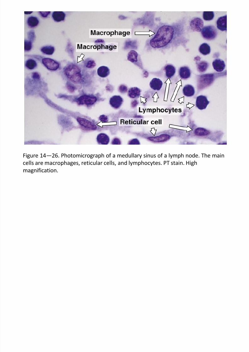
> sinus subkapsularis, sinus trabekularis dan sinus limfoid
medularis, dindingnya dilapisi oleh sel retikular, sel ma-
krofag dan serat retikulin
> sinus-sinus berisi (cairan) limfe
7/30/2019 Junq.ed.10 Indo.organ Limfoid
http://slidepdf.com/reader/full/junqed10-indoorgan-limfoid 24/60
Figure 14—25. Photomicrograph of the medulla of a lymph node; the medullary
sinuses are separated by medullary cords. PT stain. Medium magnification.
7/30/2019 Junq.ed.10 Indo.organ Limfoid
http://slidepdf.com/reader/full/junqed10-indoorgan-limfoid 25/60
Figure 14—26. Photomicrograph of a medullary sinus of a lymph node. The main
cells are macrophages, reticular cells, and lymphocytes. PT stain. High
magnification.
7/30/2019 Junq.ed.10 Indo.organ Limfoid
http://slidepdf.com/reader/full/junqed10-indoorgan-limfoid 26/60
Figure 14—27. Section of the medullary region of a lymph node stained with
picrosirius and photographed under polarized light. Note the abundance of
reticular fibers made of collagen type III. Medium magnification.
7/30/2019 Junq.ed.10 Indo.organ Limfoid
http://slidepdf.com/reader/full/junqed10-indoorgan-limfoid 27/60
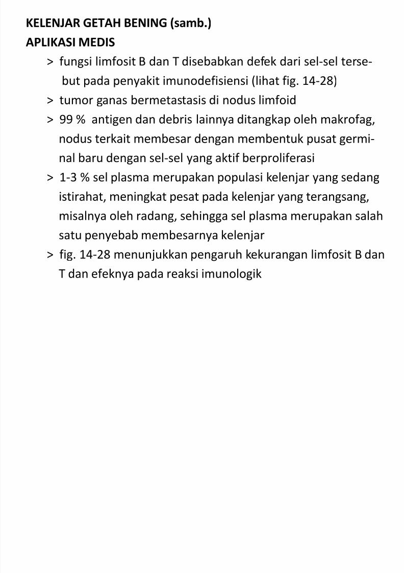
KELENJAR GETAH BENING (samb.)
APLIKASI MEDIS
> fungsi limfosit B dan T disebabkan defek dari sel-sel terse-
but pada penyakit imunodefisiensi (lihat fig. 14-28)
> tumor ganas bermetastasis di nodus limfoid
> 99 % antigen dan debris lainnya ditangkap oleh makrofag,
nodus terkait membesar dengan membentuk pusat germi-nal baru dengan sel-sel yang aktif berproliferasi
> 1-3 % sel plasma merupakan populasi kelenjar yang sedang
istirahat, meningkat pesat pada kelenjar yang terangsang,
misalnya oleh radang, sehingga sel plasma merupakan salah
satu penyebab membesarnya kelenjar
> fig. 14-28 menunjukkan pengaruh kekurangan limfosit B dan
T dan efeknya pada reaksi imunologik
7/30/2019 Junq.ed.10 Indo.organ Limfoid
http://slidepdf.com/reader/full/junqed10-indoorgan-limfoid 28/60
Figure 14—28. Pathologic alterations in lymph nodes related to deficiency of B cells,
T cells, or both. (Redrawn, with permission, from Chandrasoma P, Taylor CR: Concise
Pathology. Appleton & Lange, 1991.)
7/30/2019 Junq.ed.10 Indo.organ Limfoid
http://slidepdf.com/reader/full/junqed10-indoorgan-limfoid 29/60
KELENJAR GETAH BENING (samb.)
Sirkulasi Limfe dan Darah (Fig. 14-20)
> pembuluh limfe aferen menembus simpai, berlanjut kesinus subkapsularis, sinus trabekularis sampai ke sinus
medularis
> arsitektur sinus-sinus memperlambat aliran limfe, mem-
beri kesempatan kepada makrofag dan sel dendritik
(yaitu sel penyaji-antigen) menangkap dan mencerna
materi atau benda asing
> makrofag dan sel dendritik dibentuk dalam sumsum tu-
lang dan dibawa sirkulasi ke kelenjar getah bening
> aliran limfe keluar di hilus melalui pembuluh limfe eferen
> pembuluh darah arteri kecil membentuk anyaman kapiler
di dalam nodul limfoid, masuk dan keluar di hilus
7/30/2019 Junq.ed.10 Indo.organ Limfoid
http://slidepdf.com/reader/full/junqed10-indoorgan-limfoid 30/60
Figure 14—20. Schematic representation of the structure of a lymph node. Note the
outer and inner cortex, the medulla, and the blood and lymph circulation. Also note that
the lymph enters through the convex side of the node and leaves through the hilum. The
lymph percolates through the node, exposing its contents to the action of defensive cells
(macrophages, lymphocytes, APCs).
7/30/2019 Junq.ed.10 Indo.organ Limfoid
http://slidepdf.com/reader/full/junqed10-indoorgan-limfoid 31/60
KELENJAR GETAH BENING (samb.)
Histofisiologi
> limfe mengalir melalui kelenjar getah bening untuk penya-ringan benda asing sebelum dikembalikan ke darah
> limfe yang terbentuk di jaringan sekurang-kurangnya harus
melintasi satu kelenjar sebelum memasuki sirkulasi darah
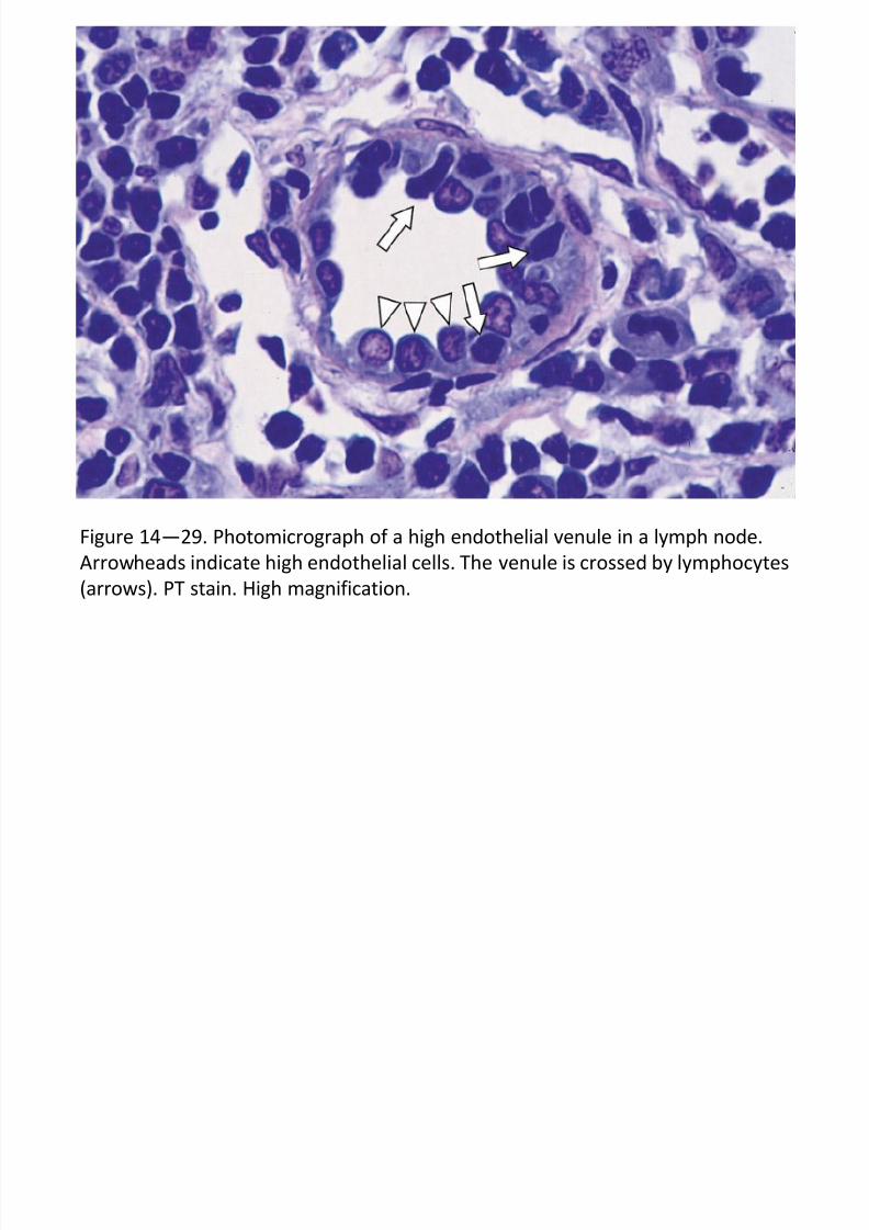
Resirkulasi Limfosit : Suatu Sistem Komunikasi
> perjalanan limfosit : kelenjar getah bening >>> pembuluh
eferen >>> sirkulasi darah >>> kembali ke kelenjar getahbening melalui pembuluh darah khusus, venula pasca-kapiler
atau venula berendotel tinggi (fig. 14-29)
7/30/2019 Junq.ed.10 Indo.organ Limfoid
http://slidepdf.com/reader/full/junqed10-indoorgan-limfoid 32/60
KELENJAR GETAH BENING (samb.)
Resirkulasi Limfosit : Suatu Sistem Komunikasi (samb.)
> venula berendotel tinggi, dengan sel endotel khusus, ber-
bentuk kuboid tinggi, tetapi sel-sel limfosit dapat menero-bos pembuluh ini di antara sel-sel tersebut
> venula berendotel tinggi terdapat juga di apendiks, tonsila
dan plak Peyer ; tidak dijumpai di limpa
> resirkulasi limfosit paling sering terjadi di kelenjar getahbening sendiri
> kembalinya limfosit disebabkan adanya molekul komple-
menter pada permukaan limfosit dan permukaan sel endo-
tel tinggi tersebut
> melalui resirkulasi limfosit ini terjadi suatu sistem komunika-
si di antara kelenjar regional dengan organ limfoid lain untuk
menyiapkan organisme untuk respons imun umum terhadap
infeksi
7/30/2019 Junq.ed.10 Indo.organ Limfoid
http://slidepdf.com/reader/full/junqed10-indoorgan-limfoid 33/60
Figure 14—29. Photomicrograph of a high endothelial venule in a lymph node.
Arrowheads indicate high endothelial cells. The venule is crossed by lymphocytes
(arrows). PT stain. High magnification.
7/30/2019 Junq.ed.10 Indo.organ Limfoid
http://slidepdf.com/reader/full/junqed10-indoorgan-limfoid 34/60
LIMPA
Struktur Umun> berperan untuk :
~ pertahanan tubuh terhadap mikroorganisme yang
memasuki peredaran darah
~ tempat penghancuran eritrosit tua
~ tempat produksi limfosit yang aktif yang memasuki
peredaran darah
~ menjadi saringan imunologis darah terhadap anti-
gen dan organ pembentuk antibodi
7/30/2019 Junq.ed.10 Indo.organ Limfoid
http://slidepdf.com/reader/full/junqed10-indoorgan-limfoid 35/60
LIMPA (samb.)
Struktur Umum (samb.)
> limpa dibungkus oleh simpai (kapsul) dan trabe-kula menyekat-nyekat parenkim ; kedua-duanya
terdiri dari jaringan ikat
> adanya hilusnya tempat arteri, vena, pembuluh
limfe (dari trabekula saja, pulpa limpa tidak me-
ngandung pembuluh limfe) dan serat saraf keluar
masuk ke dalam limpa
> simpai dan trabekula mengandung sedikit sel-selotot polos
> parenkim limpa terdiri dari anyaman jaringan reti-
kular, mengandung limfosit, makrofag dan APC
7/30/2019 Junq.ed.10 Indo.organ Limfoid
http://slidepdf.com/reader/full/junqed10-indoorgan-limfoid 36/60
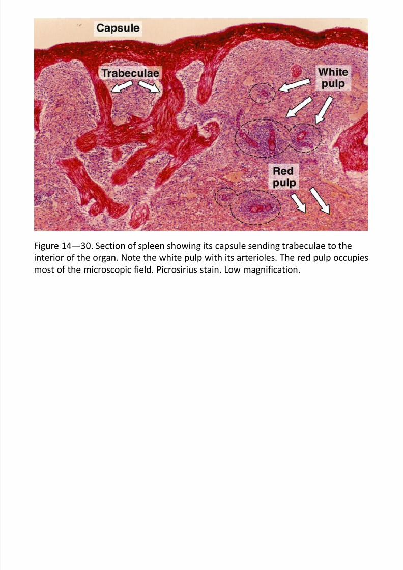
Figure 14—30. Section of spleen showing its capsule sending trabeculae to the
interior of the organ. Note the white pulp with its arterioles. The red pulp occupies
most of the microscopic field. Picrosirius stain. Low magnification.
7/30/2019 Junq.ed.10 Indo.organ Limfoid
http://slidepdf.com/reader/full/junqed10-indoorgan-limfoid 37/60
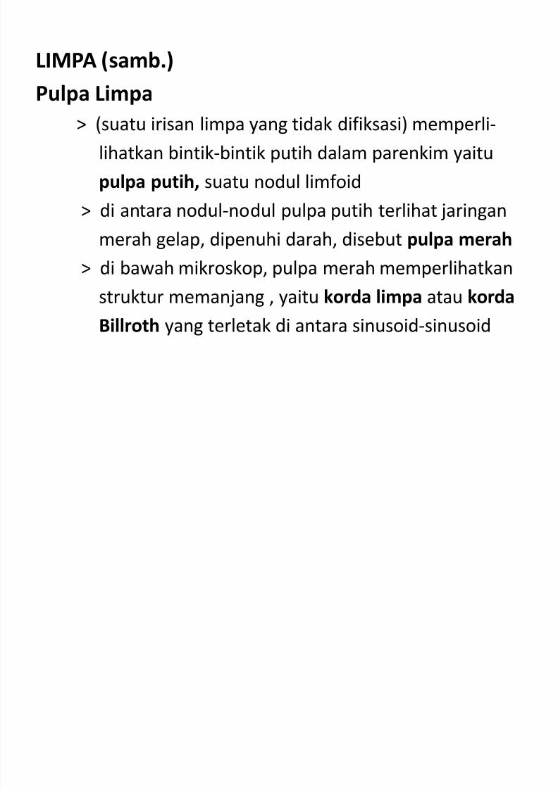
LIMPA (samb.)
Pulpa Limpa> (suatu irisan limpa yang tidak difiksasi) memperli-
lihatkan bintik-bintik putih dalam parenkim yaitu
pulpa putih, suatu nodul limfoid
> di antara nodul-nodul pulpa putih terlihat jaringan
merah gelap, dipenuhi darah, disebut pulpa merah
> di bawah mikroskop, pulpa merah memperlihatkan
struktur memanjang , yaitu korda limpa atau korda
Billroth yang terletak di antara sinusoid-sinusoid
7/30/2019 Junq.ed.10 Indo.organ Limfoid
http://slidepdf.com/reader/full/junqed10-indoorgan-limfoid 38/60
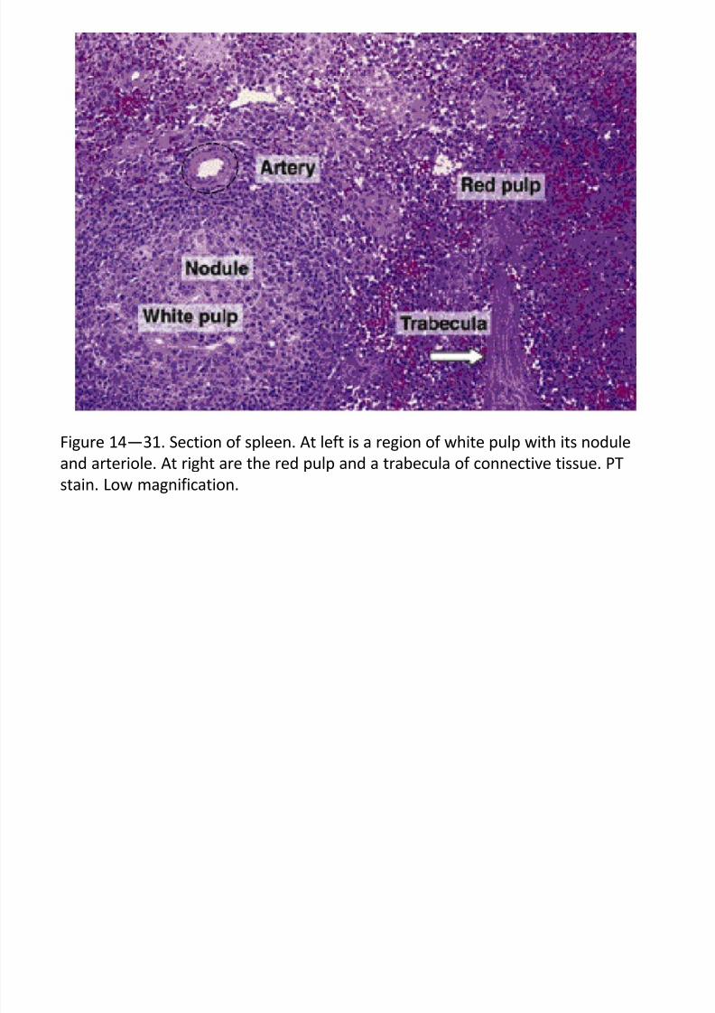
Figure 14—31. Section of spleen. At left is a region of white pulp with its nodule
and arteriole. At right are the red pulp and a trabecula of connective tissue. PT
stain. Low magnification.
7/30/2019 Junq.ed.10 Indo.organ Limfoid
http://slidepdf.com/reader/full/junqed10-indoorgan-limfoid 39/60
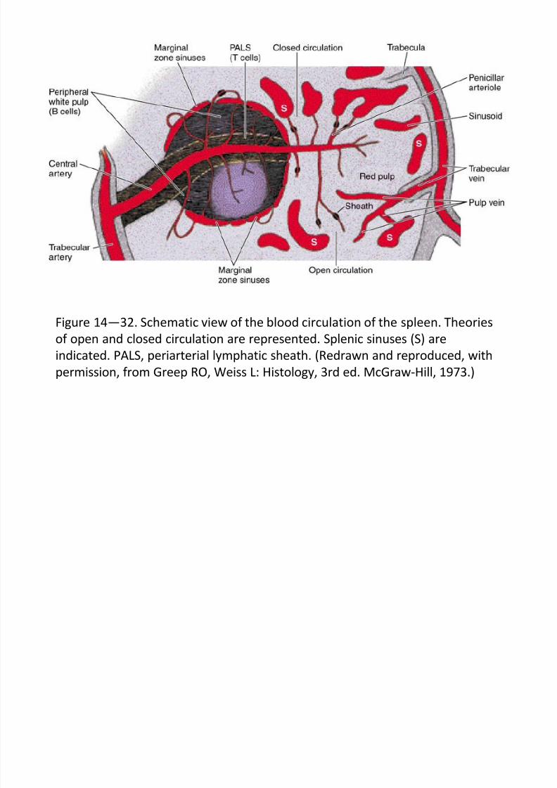
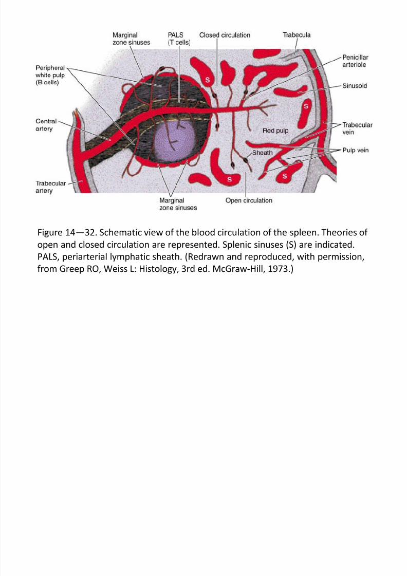
Sirkulasi Darah (Limpa)
> arteri lienalis memasuki hilus, bercabang-cabang menjadi
arteri trabekularis, memasuki parenkim menjadi arterisentralis atau arteri pulpa putih
> arteri sentralis diselubungi oleh selubung limfosit T, di-
sebut selubung limfatik periarterial (PALS)
> arteri sentralis posisinya dalam nodul limfoid pulpa putih
adalah eksentris
> setelah keluar dari pulpa putih, arteri sentralis bercabang-
cabang membentuk arteriol penisili (diameter 24 mu)
> arteriol penisili dibungkus oleh selubung dari sel-sel reti-
kular, sel limfosit dan makrofag
7/30/2019 Junq.ed.10 Indo.organ Limfoid
http://slidepdf.com/reader/full/junqed10-indoorgan-limfoid 40/60
Figure 14—32. Schematic view of the blood circulation of the spleen. Theories
of open and closed circulation are represented. Splenic sinuses (S) are
indicated. PALS, periarterial lymphatic sheath. (Redrawn and reproduced, with
permission, from Greep RO, Weiss L: Histology, 3rd ed. McGraw-Hill, 1973.)
7/30/2019 Junq.ed.10 Indo.organ Limfoid
http://slidepdf.com/reader/full/junqed10-indoorgan-limfoid 41/60
LIMPA (samb.)
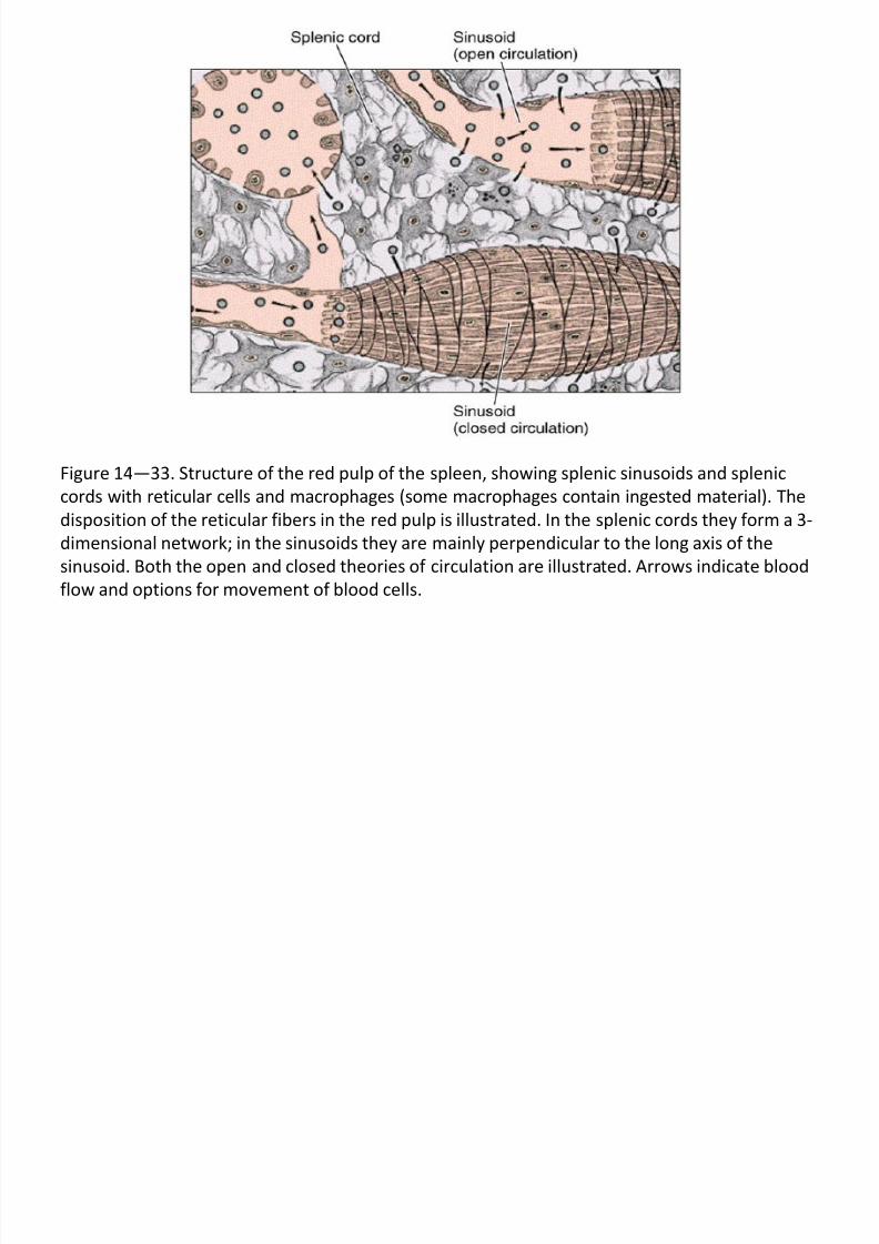
Sirkulasi Darah (samb.)> arteriol penisili berlanjut menjadi kapiler biasa dan selan-
jutnya menjadi sinusoid atau sinus pulpa merah dan
menempati celah-celah di antara korda pulpa merah
> (cara darah mengalir dari kapiler ke sinus pulpa merah
belum sepenuhnya dimengerti), ada beberapa teori :
~ teori sirkulasi tertutup, darah selamanya berada
di dalam pembuluh~ teori sirkulasi terbuka, darah mengalir melalui
celah-celah di antara sel-sel dan mencapai sinusoid
di dalam pulpa merah, pada manusia adalah jenis
sirkulasi terbuka
> dari sinusoid darah masuk ke dalam vena pulpa merah, kemudi-an menuju vena trabekularis, selanjutnya ke dalam vena lienalis
dan meninggalkan limpa melalui hilus
> vena trabekularis tidak memiliki dinding dari sel-sel otot polos
sendiri, tetapi dilapisi oleh sel endotel
7/30/2019 Junq.ed.10 Indo.organ Limfoid
http://slidepdf.com/reader/full/junqed10-indoorgan-limfoid 42/60
Figure 14—33. Structure of the red pulp of the spleen, showing splenic sinusoids and splenic
cords with reticular cells and macrophages (some macrophages contain ingested material). The
disposition of the reticular fibers in the red pulp is illustrated. In the splenic cords they form a 3-
dimensional network; in the sinusoids they are mainly perpendicular to the long axis of the
sinusoid. Both the open and closed theories of circulation are illustrated. Arrows indicate blood
flow and options for movement of blood cells.
7/30/2019 Junq.ed.10 Indo.organ Limfoid
http://slidepdf.com/reader/full/junqed10-indoorgan-limfoid 43/60
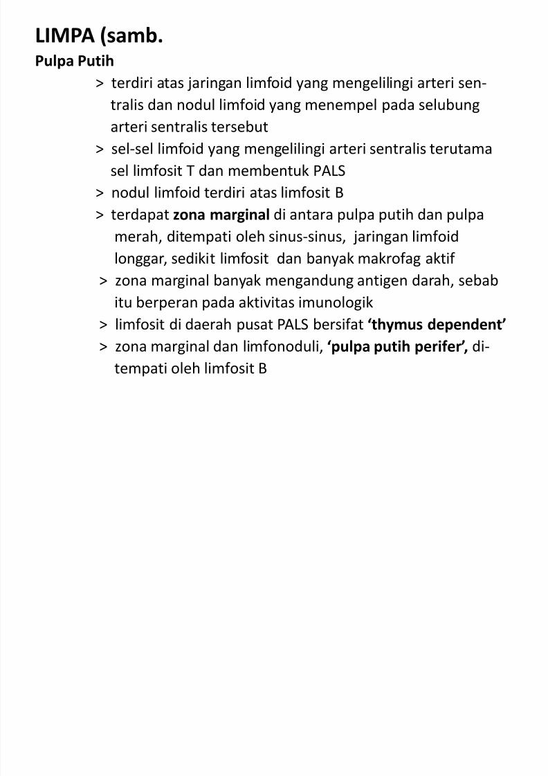
LIMPA (samb.Pulpa Putih
> terdiri atas jaringan limfoid yang mengelilingi arteri sen-
tralis dan nodul limfoid yang menempel pada selubung
arteri sentralis tersebut
> sel-sel limfoid yang mengelilingi arteri sentralis terutama
sel limfosit T dan membentuk PALS
> nodul limfoid terdiri atas limfosit B> terdapat zona marginal di antara pulpa putih dan pulpa
merah, ditempati oleh sinus-sinus, jaringan limfoid
longgar, sedikit limfosit dan banyak makrofag aktif
> zona marginal banyak mengandung antigen darah, sebab
itu berperan pada aktivitas imunologik
> limfosit di daerah pusat PALS bersifat ‘thymus dependent’
> zona marginal dan limfonoduli, ‘pulpa putih perifer’, di-
tempati oleh limfosit B
7/30/2019 Junq.ed.10 Indo.organ Limfoid
http://slidepdf.com/reader/full/junqed10-indoorgan-limfoid 44/60
Figure 14—32. Schematic view of the blood circulation of the spleen. Theories of
open and closed circulation are represented. Splenic sinuses (S) are indicated.
PALS, periarterial lymphatic sheath. (Redrawn and reproduced, with permission,
from Greep RO, Weiss L: Histology, 3rd ed. McGraw-Hill, 1973.)
7/30/2019 Junq.ed.10 Indo.organ Limfoid
http://slidepdf.com/reader/full/junqed10-indoorgan-limfoid 45/60
LIMPA (samb.)Pulpa Merah
> mengandung korda limpa dan sinusoid
> korda limpa terdiri dari anyaman longgar sel-sel retikulardengan serat retikuli (kolagen tipe III)
> korda limpa mengandung makrofag, limfosit B, limfosit T,
sel plasma dan sel-sel darah (eritrosit, granulosit dan
trombosit)> sinusoid dibatasi oleh sel endotel panjang (sumbu panjang-
nya sejajar dengan sumbu panjang sinusoid)
> sel-sel endotel ini diikat oleh serat retikulin, seperti tali
yang mengikat dinding gentong
> sel-sel makrofag menempati celah-celah di antara sel-sel
endotel
> lamina basal dari sinusoid tidak kontinu atau utuh
> diameter celah di antara sel-sel endotel sekitar 2-3 um
7/30/2019 Junq.ed.10 Indo.organ Limfoid
http://slidepdf.com/reader/full/junqed10-indoorgan-limfoid 46/60
Figure 14—33. Structure of the red pulp of the spleen, showing splenic sinusoids and
splenic cords with reticular cells and macrophages (some macrophages contain ingested
material). The disposition of the reticular fibers in the red pulp is illustrated. In the
splenic cords they form a 3-dimensional network; in the sinusoids they are mainly
perpendicular to the long axis of the sinusoid. Both the open and closed theories of
circulation are illustrated. Arrows indicate blood flow and options for movement of
blood cells.
7/30/2019 Junq.ed.10 Indo.organ Limfoid
http://slidepdf.com/reader/full/junqed10-indoorgan-limfoid 47/60
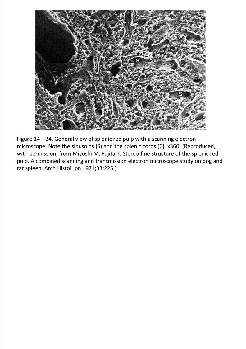
Figure 14—34. General view of splenic red pulp with a scanning electron
microscope. Note the sinusoids (S) and the splenic cords (C). x360. (Reproduced,
with permission, from Miyoshi M, Fujita T: Stereo-fine structure of the splenic red
pulp. A combined scanning and transmission electron microscope study on dog and
rat spleen. Arch Histol Jpn 1971;33:225.)
7/30/2019 Junq.ed.10 Indo.organ Limfoid
http://slidepdf.com/reader/full/junqed10-indoorgan-limfoid 48/60
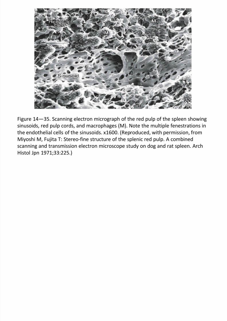
Figure 14—35. Scanning electron micrograph of the red pulp of the spleen showingsinusoids, red pulp cords, and macrophages (M). Note the multiple fenestrations in
the endothelial cells of the sinusoids. x1600. (Reproduced, with permission, from
Miyoshi M, Fujita T: Stereo-fine structure of the splenic red pulp. A combined
scanning and transmission electron microscope study on dog and rat spleen. Arch
Histol Jpn 1971;33:225.)
LIMPA ( b )
7/30/2019 Junq.ed.10 Indo.organ Limfoid
http://slidepdf.com/reader/full/junqed10-indoorgan-limfoid 49/60
LIMPA (samb.)
HISTIOFISIOLOGI
Produksi Limfosit> pulpa putih menghasilkan limfosit, limfosit bermigrasi ke pulpa merah,
masuk ke sinusoid untuk selanjutnya mengikuti edaran darah
Destruksi Eritrosit
> umur rata-rata eritrosit selama 120 hari> penurunan fleksibilitas dan perubahan membran eritrosit merupakan
tanda destruksi eritrosit (juga terjadi di dalam sumsum tulang
> makrofag dalam korda limpa mencerna eritrosit yang pe-
cah dalam ruang ekstrasel
> hemoglobin diuraikan menjadi globin, selanjutnya dihidrolisis menjadi
asam amino untuk digunakan menyintesis protein baru
> besi dilepaskan dari heme dan bersama transferin, diangkut ke sumsum
tulang untuk selanjutnya digunakan dalam prose eritropoiesis
7/30/2019 Junq.ed.10 Indo.organ Limfoid
http://slidepdf.com/reader/full/junqed10-indoorgan-limfoid 50/60
LIMPA (samb.)
Histofisiologi (samb.)
Destruksi Eritrosit (samb.)> heme bebas besi diuraikan menjadi bilirubin, diekskresi dalam empedu
oleh sel-sel hati
> setelah splenektomi, terjadi peningkatan jumlah eritrosit abnormal
(perubahan bentuk), juga peningkatan jumlah trombosit (menunjukkanlimpa juga berperan menghancurkan trombosit)
Pertahanan Terhadap Penyusup
> limpa berperan dalam pertahanan imunologis tubuh, karena mengan-
dung limfosit B, limfosit T, APC dan sel-sel fagositik> limpa juga berperan sebagai saringan bagi darah seperti halnya kelenjar
getah bening sebagai saringan bagi limfe
> sel-sel limpa adalah sel yang paling aktif dalam memfagositosis organis-
me hidup (bakteri dan virus) dan partikel mati yang masuk ke darah
7/30/2019 Junq.ed.10 Indo.organ Limfoid
http://slidepdf.com/reader/full/junqed10-indoorgan-limfoid 51/60
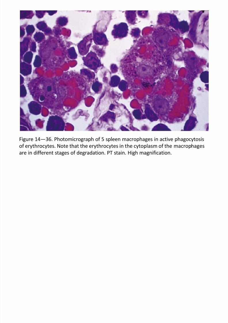
Figure 14—36. Photomicrograph of 5 spleen macrophages in active phagocytosis
of erythrocytes. Note that the erythrocytes in the cytoplasm of the macrophages
are in different stages of degradation. PT stain. High magnification.
7/30/2019 Junq.ed.10 Indo.organ Limfoid
http://slidepdf.com/reader/full/junqed10-indoorgan-limfoid 52/60
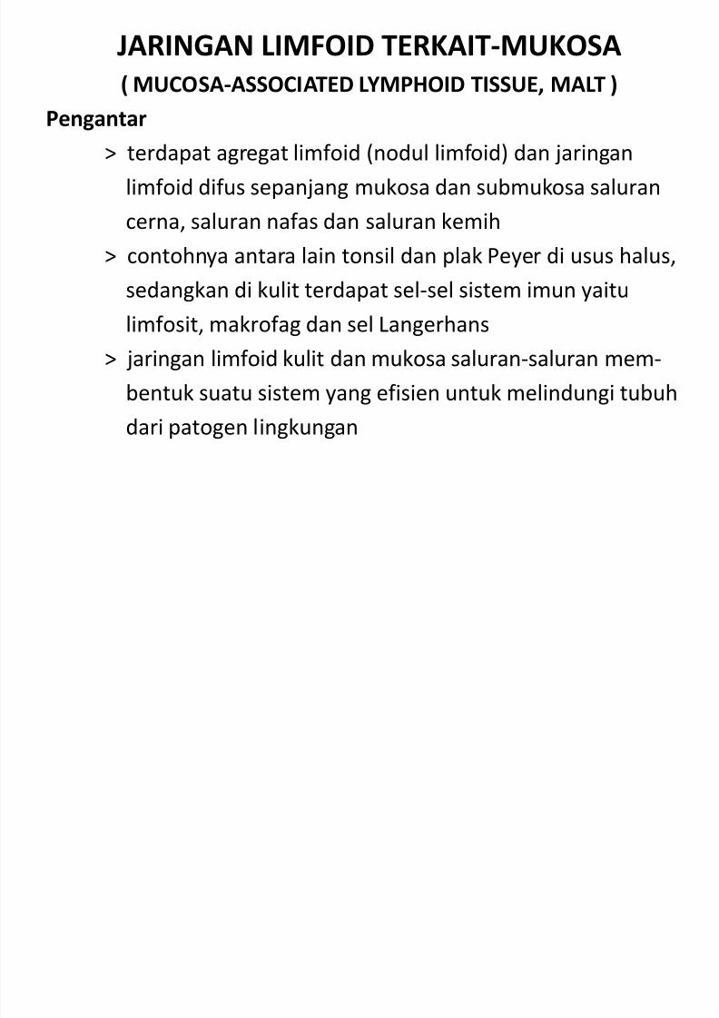
JARINGAN LIMFOID TERKAIT-MUKOSA
( MUCOSA-ASSOCIATED LYMPHOID TISSUE, MALT )
Pengantar
> terdapat agregat limfoid (nodul limfoid) dan jaringan
limfoid difus sepanjang mukosa dan submukosa saluran
cerna, saluran nafas dan saluran kemih
> contohnya antara lain tonsil dan plak Peyer di usus halus,sedangkan di kulit terdapat sel-sel sistem imun yaitu
limfosit, makrofag dan sel Langerhans
> jaringan limfoid kulit dan mukosa saluran-saluran mem-
bentuk suatu sistem yang efisien untuk melindungi tubuh
dari patogen lingkungan
7/30/2019 Junq.ed.10 Indo.organ Limfoid
http://slidepdf.com/reader/full/junqed10-indoorgan-limfoid 53/60
Figure 14—37. Section of lung showing a collection of lymphocytes in the
connective tissue of the bronchiolar mucosa, an example of mucosa-associated
lymphoid tissue (MALT). PT stain. Low magnification.
7/30/2019 Junq.ed.10 Indo.organ Limfoid
http://slidepdf.com/reader/full/junqed10-indoorgan-limfoid 54/60
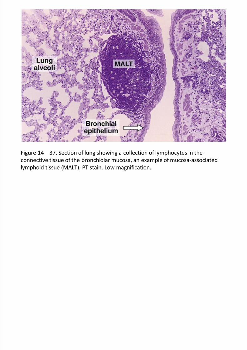
Figure 14—38. Section of Peyer’s patch of the small intestine showing the epithelial
covering of enterocytes and goblet cells (right), the intestinal lumen (center), and
the covering of the patch with a row of M cells and groups of lymphocytes (left).
The small dark nuclei belong to B and T lymphocytes, and the large pale-stained
nuclei belong to M cells. PT stain. Medium magnification.
7/30/2019 Junq.ed.10 Indo.organ Limfoid
http://slidepdf.com/reader/full/junqed10-indoorgan-limfoid 55/60
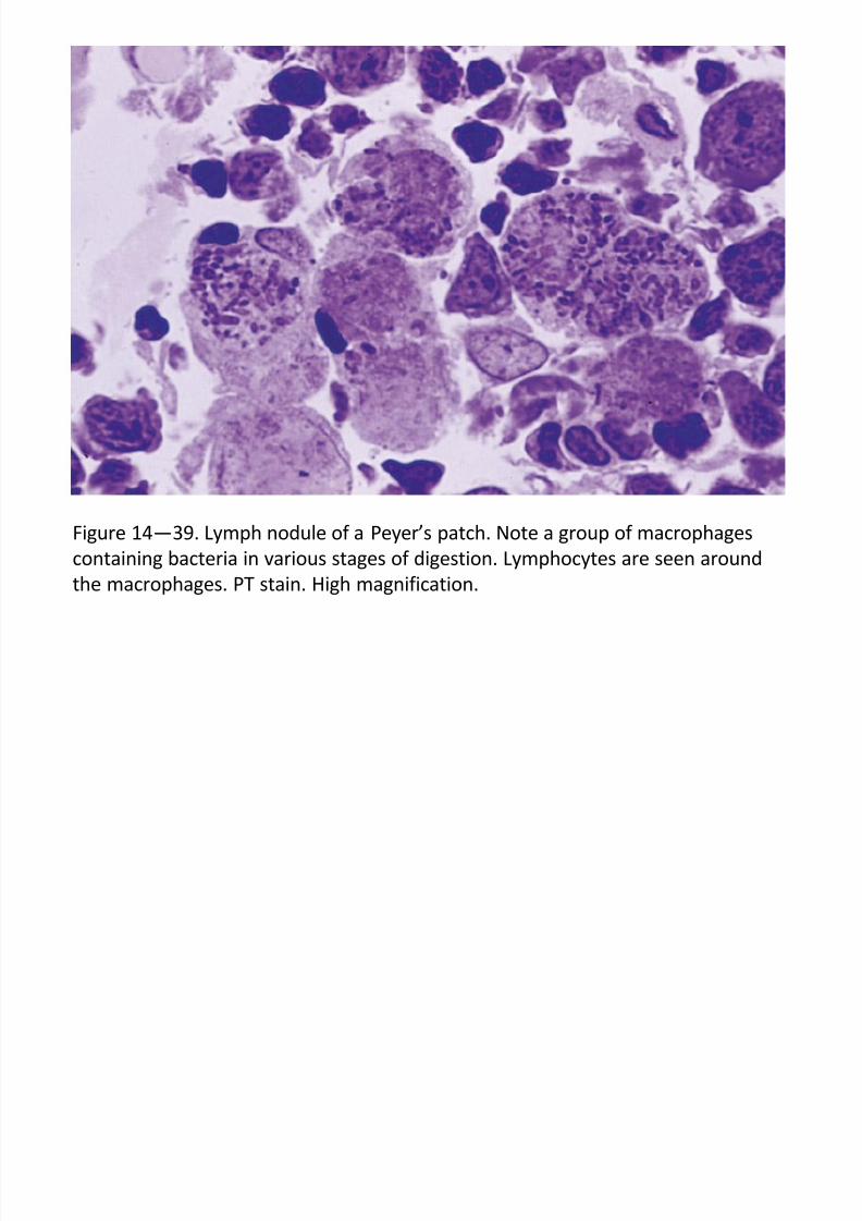
Figure 14—39. Lymph nodule of a Peyer’s patch. Note a group of macrophages
containing bacteria in various stages of digestion. Lymphocytes are seen around
the macrophages. PT stain. High magnification.
7/30/2019 Junq.ed.10 Indo.organ Limfoid
http://slidepdf.com/reader/full/junqed10-indoorgan-limfoid 56/60
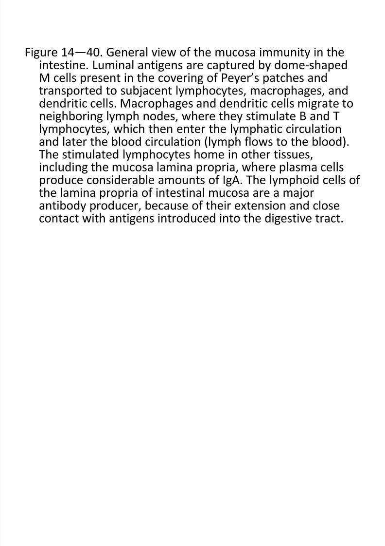
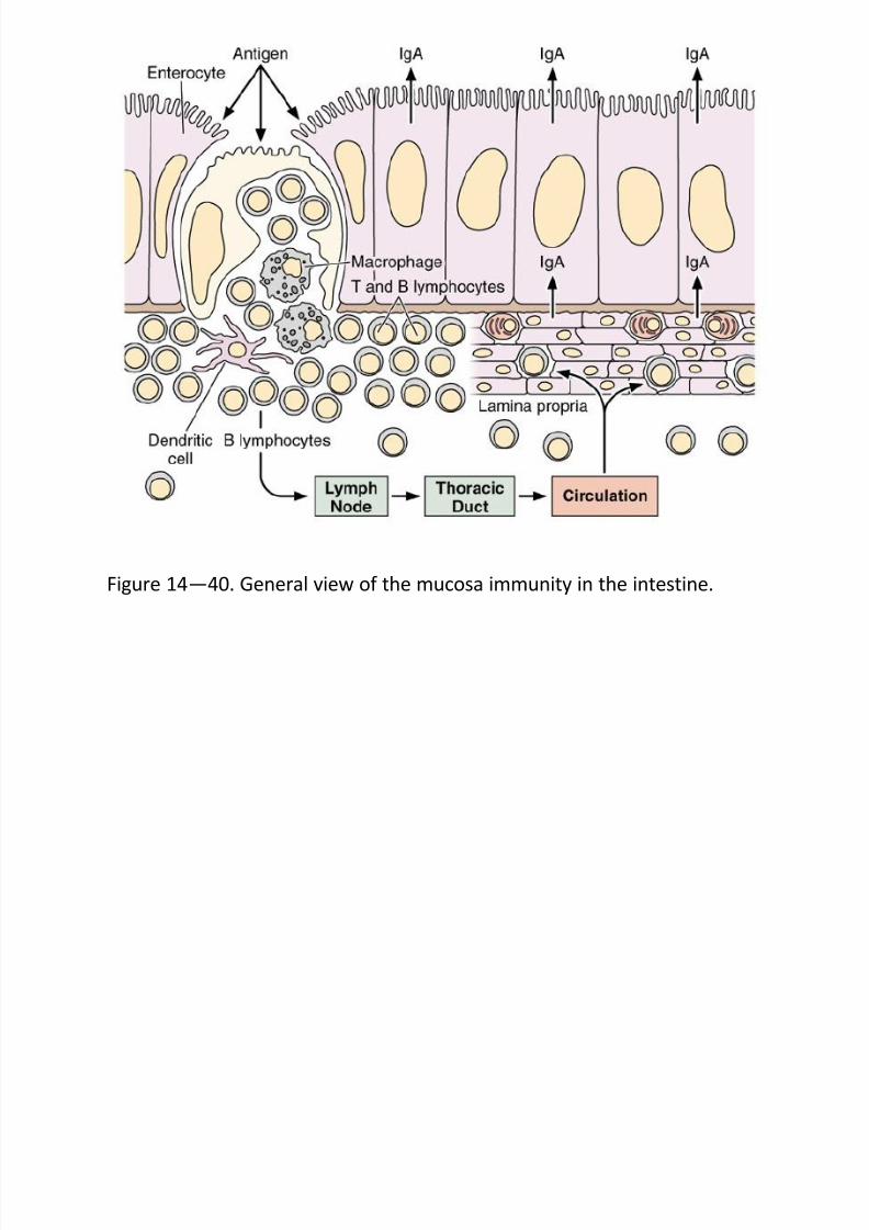
Figure 14—40. General view of the mucosa immunity in theintestine. Luminal antigens are captured by dome-shapedM cells present in the covering of Peyer’s patches andtransported to subjacent lymphocytes, macrophages, anddendritic cells. Macrophages and dendritic cells migrate toneighboring lymph nodes, where they stimulate B and Tlymphocytes, which then enter the lymphatic circulation
and later the blood circulation (lymph flows to the blood).The stimulated lymphocytes home in other tissues,including the mucosa lamina propria, where plasma cellsproduce considerable amounts of IgA. The lymphoid cells of the lamina propria of intestinal mucosa are a major
antibody producer, because of their extension and closecontact with antigens introduced into the digestive tract.
7/30/2019 Junq.ed.10 Indo.organ Limfoid
http://slidepdf.com/reader/full/junqed10-indoorgan-limfoid 57/60
Figure 14—40. General view of the mucosa immunity in the intestine.
TONSIL
7/30/2019 Junq.ed.10 Indo.organ Limfoid
http://slidepdf.com/reader/full/junqed10-indoorgan-limfoid 58/60
TONSIL
Tonsila Palatina
> sepasang agregat (organ) jaringan limfoid, terletak di
dinding lateral faring
> epitel permukaannya jenis epitel berlapis gepeng,
dengan 10-20 invaginasi epitel memasuki ke dalam pa-
renkim membentuk kriptus yang berisi sel-sel epitel
yang lepas, limfosit yang hidup maupun yang mati serta
bakteri
> di bawah lapisan epitel terdapat jaringan limfoid ber-
bentuk nodul limfoid dengan pusat-pusat germinal
> tonsil dipisahkan dengan jaringan di bawahnya dengan
simpai (kapsul) jaringan ikatnpadat dan berperan seba-
gai sawar (‘barrier’) terhadap penyebaran infeksi
TONSIL ( b )
7/30/2019 Junq.ed.10 Indo.organ Limfoid
http://slidepdf.com/reader/full/junqed10-indoorgan-limfoid 59/60
TONSIL (samb.)
Tonsila Faringea
> sebuah organ tunggal, terletak di bagian posterior-superior faring> ditutupi oleh epitel bertingkat silindris bersilia, terdiri atas lipatan
mukosa dan mengandung jaringan limfoid difus dan noduli
> tidak ada kriptus dan simpainya tipis
> hipertrofi tonsila faringea akibat radang menahun dinamakan adenoid
Tonsila Lingualis
> tonsila lingualis lebih kecil dan lebih banyak dari tonsila palatina atau
tonsila faringea
> terletak di dasar dari lidah dan permukaannya ditutupi oleh epitelberlapis gepeng
> setiap tonsila lingualis mempunyai satu kriptus
Rujukan :
7/30/2019 Junq.ed.10 Indo.organ Limfoid
http://slidepdf.com/reader/full/junqed10-indoorgan-limfoid 60/60
Rujukan :
HISTOLOGI DASAR
Teks & Atlas
Edisi 10, 2004
Luiz Carlos JUNQUEIRA
Jose CARNEIRO
Alih Bahasa : dr. Jan Tambayong
Penerbit Buku Kedokteran EGC Jakarta