Embed Size (px)
Citation preview
Juvenile Idiopathic Arthritis:When it is not “just” Growing Pains
Deborah Jane Power, D.O., MS, FACR, FACOI
Catalina Pointe Arthritis & Rheumatology Specialists, P.C.
Tucson, Arizona
23th Annual Southwestern Conference on Medicine
April 24, 2015

Disclosures
Speakers’ Bureau
– Amgen
– Abbvie
Outline
Introduction
Diagnosis of Joint Pain
Classification/Diagnosis of Juvenile
Idiopathic Arthritis (JIA)
Treatment of JIA
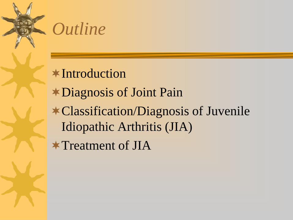
“Kids Don’t Get Arthritis”
“They will grow out of it”
“JIA is just RA in a little person”
“JIA will become RA once the child becomes an adult”
“My child has RA because the arthritis blood test was positive”
“I want to treat my child with natural remedies”
Pediatric Rheumatic Diseases
Juvenile Idiopathic Arthritis (JIA)
Connective-Tissue Diseases
– Systemic Lupus Erythematosus
– Mixed Connective Tissue Disease
– Antiphospholipid Syndrome
– Juvenile Dermatomyositis
– Scleroderma
– Henoch-Schönlein Purpura- IgA Vasculitis
– Medium- and Large-Vessel Vasculitis
Juvenile Idiopathic Arthritis
JIA describes condition of chronic synovitis in children of which there are distinct subgroups
JIA is the most frequent major rheumatic disease of children (same number as diabetes mellitus-type 1, four times more than sickle cell or cystic fibrosis)
Incidence: 57-220 cases/100,000 per year
Prevalence estimates 10,300 – 60,900 in the US population in patients under age of 16
40-45% of patients still have active disease after 10 years
Up to 50% of JIA patients have active disease that persists into adulthood
– Estimated 35,000 to 50,000 people over 16 have active JIA in UShttp://www.cdc.gov/arthritis/basic/chi
ldhood.htm
Objective Signs of Arthritis
Joint Swelling
– Synovial hypertrophy
– Increased amounts of synovial fluid
– Swelling of periarticular tissues
Joint Pain
– On motion
– On palpation (tenderness)
Loss of Joint Motion
– Stiffness of joints
Joint Warmth
Joint Erythema
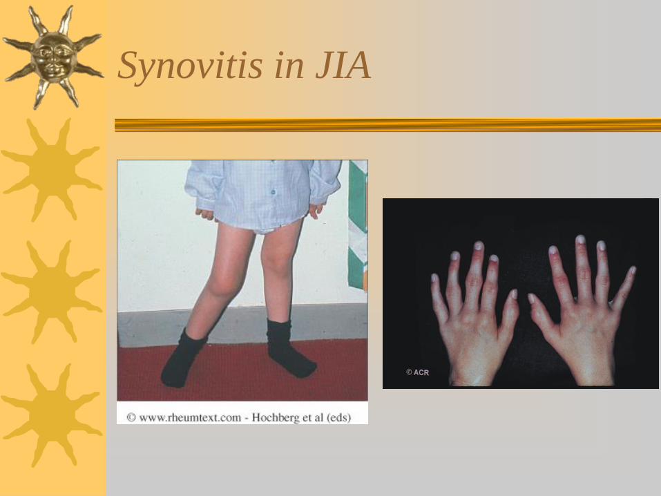
Synovitis in JIA
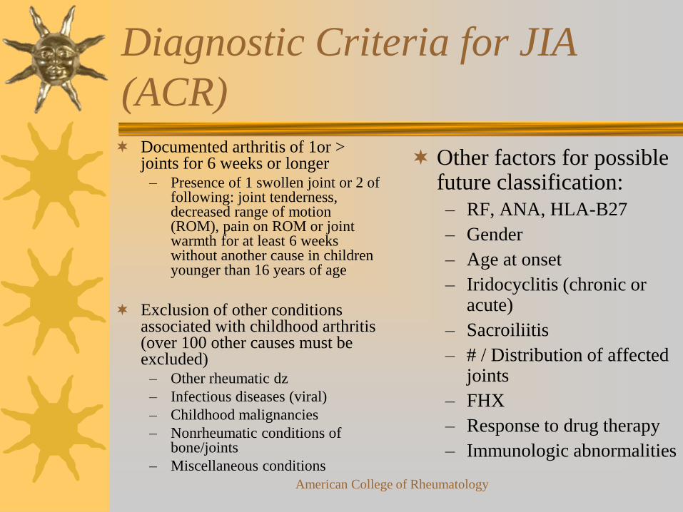
Diagnostic Criteria for JIA
(ACR) Documented arthritis of 1or >
joints for 6 weeks or longer– Presence of 1 swollen joint or 2 of
following: joint tenderness, decreased range of motion (ROM), pain on ROM or joint warmth for at least 6 weeks without another cause in children younger than 16 years of age
Exclusion of other conditions associated with childhood arthritis (over 100 other causes must be excluded)
– Other rheumatic dz
– Infectious diseases (viral)
– Childhood malignancies
– Nonrheumatic conditions of bone/joints
– Miscellaneous conditions
Other factors for possible future classification:
– RF, ANA, HLA-B27
– Gender
– Age at onset
– Iridocyclitis (chronic or acute)
– Sacroiliitis
– # / Distribution of affected joints
– FHX
– Response to drug therapy
– Immunologic abnormalities
American College of Rheumatology
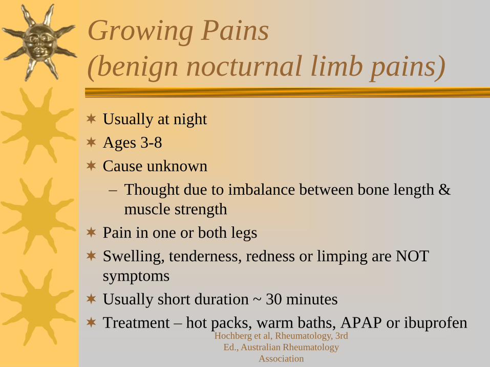
Growing Pains
(benign nocturnal limb pains)
Usually at night
Ages 3-8
Cause unknown
– Thought due to imbalance between bone length &
muscle strength
Pain in one or both legs
Swelling, tenderness, redness or limping are NOT
symptoms
Usually short duration ~ 30 minutes
Treatment – hot packs, warm baths, APAP or ibuprofenHochberg et al, Rheumatology, 3rd
Ed., Australian Rheumatology
Association
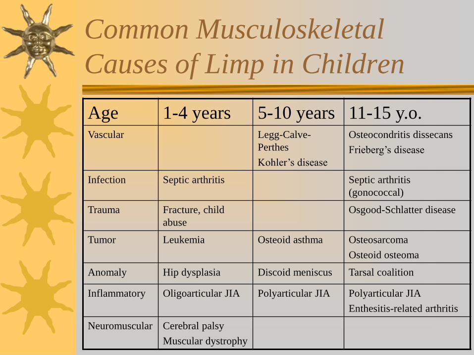
Common Musculoskeletal
Causes of Limp in Children
Age 1-4 years 5-10 years 11-15 y.o.Vascular Legg-Calve-
Perthes
Kohler’s disease
Osteocondritis dissecans
Frieberg’s disease
Infection Septic arthritis Septic arthritis
(gonococcal)
Trauma Fracture, child
abuse
Osgood-Schlatter disease
Tumor Leukemia Osteoid asthma Osteosarcoma
Osteoid osteoma
Anomaly Hip dysplasia Discoid meniscus Tarsal coalition
Inflammatory Oligoarticular JIA Polyarticular JIA Polyarticular JIA
Enthesitis-related arthritis
Neuromuscular Cerebral palsy
Muscular dystrophy
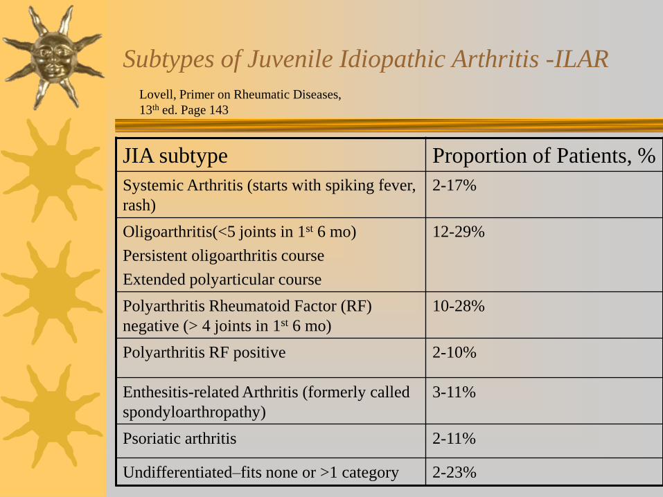
Subtypes of Juvenile Idiopathic Arthritis -ILAR
JIA subtype Proportion of Patients, %
Systemic Arthritis (starts with spiking fever,
rash)
2-17%
Oligoarthritis(<5 joints in 1st 6 mo)
Persistent oligoarthritis course
Extended polyarticular course
12-29%
Polyarthritis Rheumatoid Factor (RF)
negative (> 4 joints in 1st 6 mo)
10-28%
Polyarthritis RF positive 2-10%
Enthesitis-related Arthritis (formerly called
spondyloarthropathy)
3-11%
Psoriatic arthritis 2-11%
Undifferentiated–fits none or >1 category 2-23%
Lovell, Primer on Rheumatic Diseases,
13th ed. Page 143
Subtypes of Juvenile Idiopathic Arthritis -ILAR
A 2-year-old girl presents with history of a daily
spiking fever, salmon colored rash which is
evanescent and a pericardial effusion. She is
ANA positive and RF negative. Sedimentation
rate and platelet count are markedly elevated.

Systemic onset JIA
2-17% of JIA population
Arthritis with or preceded by daily fever of at least 2 weeks, quotidian for at least 3 days plus at least one of the following: rheumatoid rash, generalized LAD, hepato- or splenomegaly and serositis
Usually affects younger children – As young as 6-7 months old
– Peak onset 1-6 years old
Slight male predominance or equally affected
RF/ANA negative
Severe arthritis in 25%
Arthritis typically polyarticular – affecting both large & small joints
Systemic Onset Disease
50% of patients develop severe, treatment resistant-polyarticular disease
Systemic manifestations may precede arthritis by weeks or years
– Daily mono- or biphasic spiking fever pattern >101°F
– Rash: salmon pink, blanching, discrete, erythematous macules on trunk or joints, transient (min-few hours)
– Hepatosplenomegaly and/or lymphadenopathy can be pronounced
– Serositis (pericardial, pleural or peritoneal)
– Complications: DIC, pericarditis, tamponade, macrophage activation syndrome (MAS)
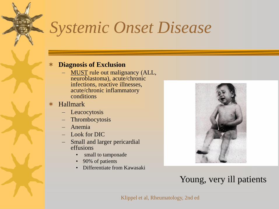
Systemic Onset Disease
Diagnosis of Exclusion– MUST rule out malignancy (ALL,
neuroblastoma), acute/chronic infections, reactive illnesses, acute/chronic inflammatory conditions
Hallmark– Leucocytosis
– Thrombocytosis
– Anemia
– Look for DIC
– Small and larger pericardial effusions
• small to tamponade
• 90% of patients
• Differentiate from Kawasaki
Young, very ill patients
Klippel et al, Rheumatology, 2nd ed
Macrophage Activation Syndrome
(MAS)
Rare, life-threatening complication of Systemic Onset –Juvenile Idiopathic Arthritis (SOJIA) but seen in all forms of rheumatic disease
– AKA: Secondary or Acquired Hemophagocytic Syndrome; Hemophagocytic Lymphohistiocytosis
Increased activation & expansion macrophages & CD8+ Tcells leading to overwhelming inflammatory response and demonstration of histiophagocytois in bone marrow
Triggers: preceding viral illness; addition of or change in medications (esp. NSAIDs, suflasalazine, more recently –etanercept)
Acutely ill – HSM, LAD, purpura, prolonged PT, PTT, elevated fibrin split products, hyperferritinemia, hypertryglyceridemia
Hochberg et al, Rheumatology, 3rd
ed
Macrophage Activation Syndrome
(MAS)
Sed rate often low (* clue to diagnosis of MAS
vs. exacerbation of SOJIA)
– Due to hypofibrinogemia secondary consumptive
coagulopathy and hepatic dysfunction
Mortality 8-22%
Treatment: pulse methoprednisolone 30 mg/kg up
to 1 gram x 3 days then 2-3 mg/kg/Q6H
– (Sawhney, Arch Dis Child, 2001)
Second line - IV cyclosporine A (2-7 mg/kg/day)
– (Mouy, J Pediatr, 1996)
Macrophage Activation Syndrome
(MAS)
Third line treatment:– Etoposide (VP-16) (Fishman, Br J Rheumatol 1995
– Anti-thymocte globulin (ATG) (Coca, Clin Immunol,
2009)
– IV Ig (Tristano, J Clin Rheumatol, 2003)
– Anti-TNF alpha agents
– Rituximab (Balamuth, J Pe diatr Hematol/Oncol, 2007)
– Anakinra (Miettuenen, Rheum, 2010)
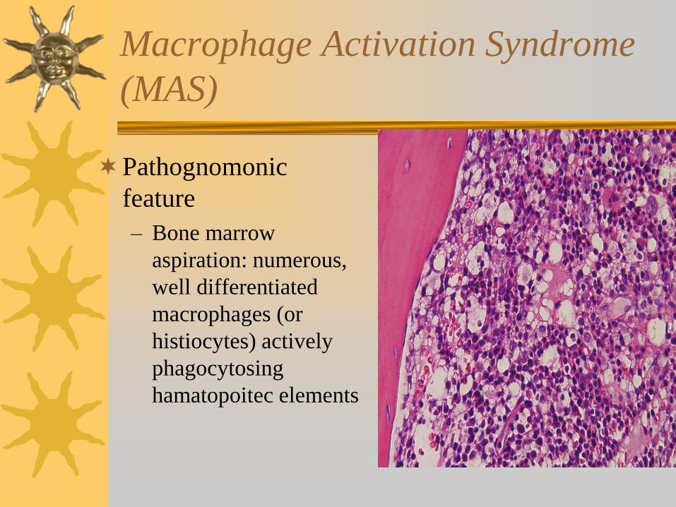
Macrophage Activation Syndrome
(MAS)
Pathognomonic
feature
– Bone marrow
aspiration: numerous,
well differentiated
macrophages (or
histiocytes) actively
phagocytosing
hamatopoitec elements

Oligoarticular JIA (oJIA)
24-58% of patients
Arthritis of few joints (knees, ankles, elbows)
Females more affected than males (4:1)
Early childhood onset (1-5)
ANA + 60%, RF-negative
At greatest risk for developing chronic eye inflammation (30-50%)
– Anterior chamber
– Minimal, if any, symptoms in 80% of children

Oligoarticular JIA (oJIA)
Radiographs of the knees of a
boy with monarthritis of the
left knee. There is marked
osteopenia on the left, with
coarsening of the trabeculae
and enlargement of the
epiphyses. The joint space is
narrowed laterally, and there is
lengthening of the left leg with
a moderate valgus deformity.
Erosions are not present
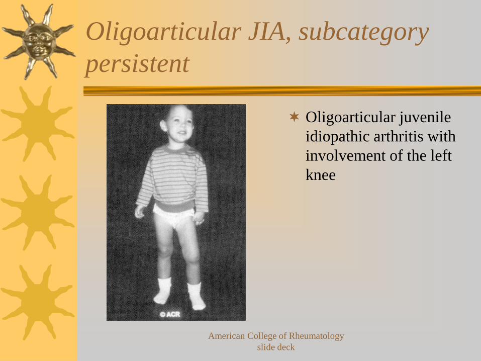
Oligoarticular JIA, subcategory
persistent
Oligoarticular juvenile
idiopathic arthritis with
involvement of the left
knee
American College of Rheumatology
slide deck
Oligoarthritis, subcategory
persistent
Arthritis in < four joints at any time during onset or course of the disease
Best overall articular outcome of all categories JIA
Up to 50% of patients with persistent oJIA will have monarticular involvement in knee
Severity joint symptoms usually mild
– Present with normal or near normal physical function, joint swelling and loss of motion in knee
Oligoarthritis, subcategory
extended
Arthritis in < four joints 1st 6 months of disease but affecting cumulative total of > 5 joints after 1st 6 months
Up to 50% of oJIA patients evolve to extended category
– 30% do so in the 2 years after disease onset
Risk factors development extended (more extensive and severe articular involvement)
– Arthritis wrists, hand or ankle
– Symmetric arthritis
– Arthritis in more than one joint
– Elevated ESR
– Positive ANA
Polyarticular JIA (poJIA)
Rheumatoid Factor Positive (2-10%)
– Arthritis > 5 joints during 1st 6 months and positive test for RF at least twice 3 months apart
– Symmetric polyarthritis of small & large joints
– Females affected more often than males
– Late childhood onset (rarely before 8 y.o.)
– ANA + in 50-75%
– Rheumatoid nodules common over pressure points (elbows, heels, 1st MTPJ, MCP’s, extensor surfaces fingers)
– Erosions more common (resembles adult RA)
– Rapidly progressive joint destruction within 6-12 months of onset – high chance of permanent disability
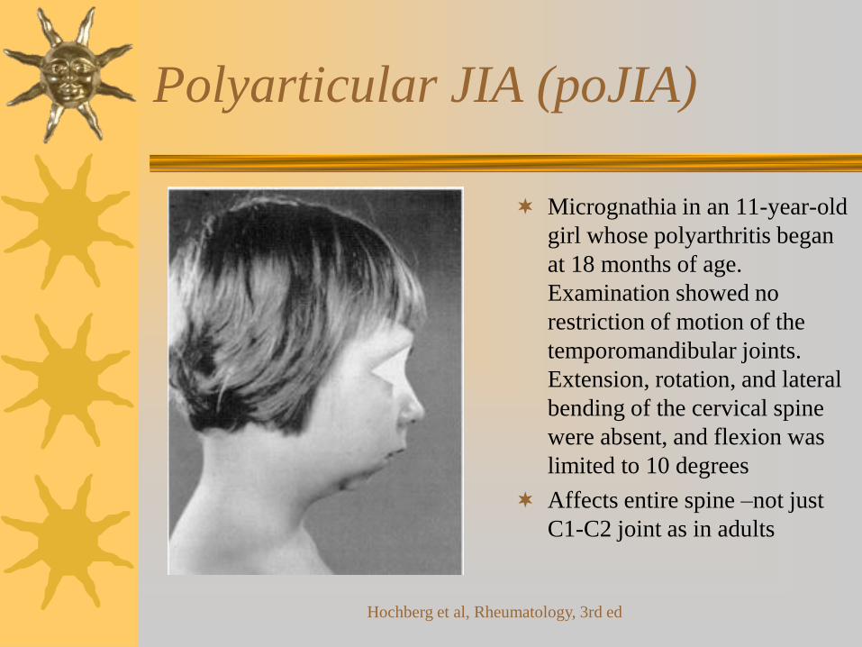
Polyarticular JIA (poJIA)
Micrognathia in an 11-year-old
girl whose polyarthritis began
at 18 months of age.
Examination showed no
restriction of motion of the
temporomandibular joints.
Extension, rotation, and lateral
bending of the cervical spine
were absent, and flexion was
limited to 10 degrees
Affects entire spine –not just
C1-C2 joint as in adults
Hochberg et al, Rheumatology, 3rd ed
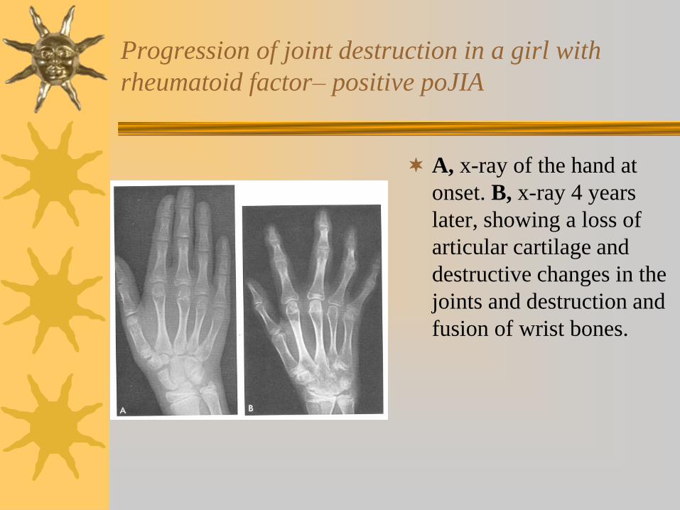
Progression of joint destruction in a girl with
rheumatoid factor– positive poJIA
A, x-ray of the hand at
onset. B, x-ray 4 years
later, showing a loss of
articular cartilage and
destructive changes in the
joints and destruction and
fusion of wrist bones.
Polyarticular JIA (poJIA)
Rheumatoid Factor Negative (10-28%)
– Arthritis > 5 joints during 1st 6 months with negative tests for RF
– Symmetric polyarthritis small & large joints
– Females more affected than males
– Early or late childhood onset
– ANA positive in 25%
– Rheumatoid nodules uncommon
– Severe arthritis in 10-15%
– Usually little joint destruction
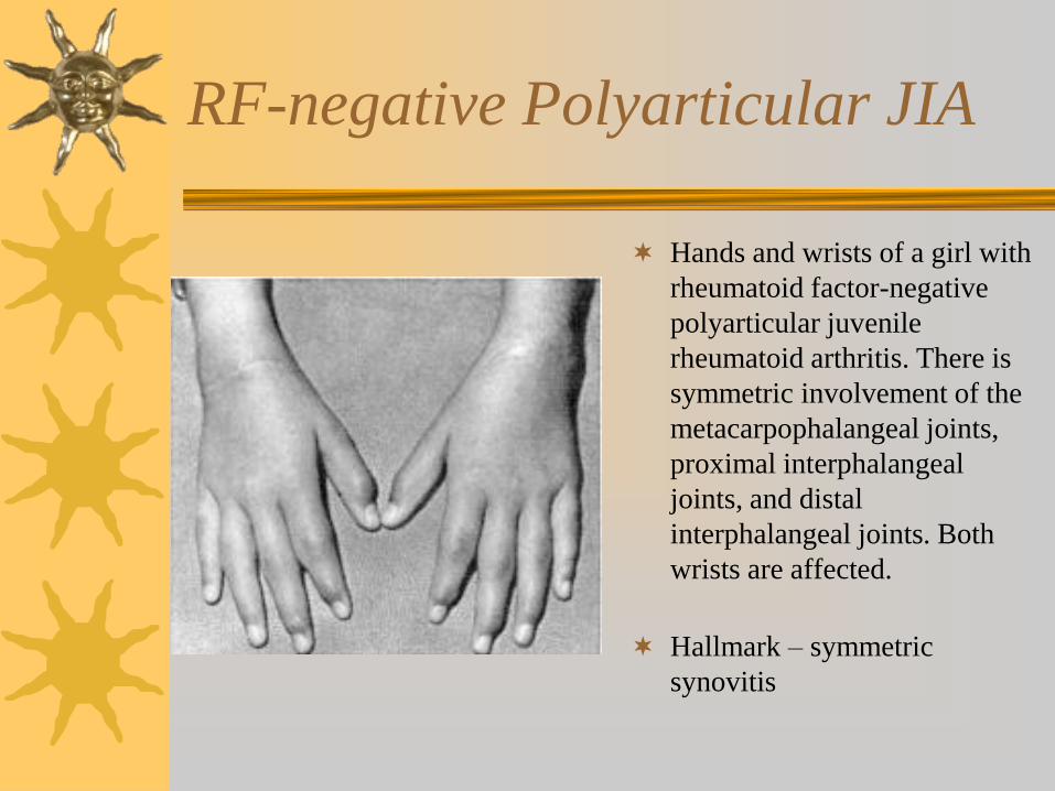
RF-negative Polyarticular JIA
Hands and wrists of a girl with
rheumatoid factor-negative
polyarticular juvenile
rheumatoid arthritis. There is
symmetric involvement of the
metacarpophalangeal joints,
proximal interphalangeal
joints, and distal
interphalangeal joints. Both
wrists are affected.
Hallmark – symmetric
synovitis
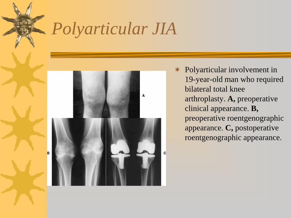
Polyarticular JIA
Polyarticular involvement in
19-year-old man who required
bilateral total knee
arthroplasty. A, preoperative
clinical appearance. B,
preoperative roentgenographic
appearance. C, postoperative
roentgenographic appearance.
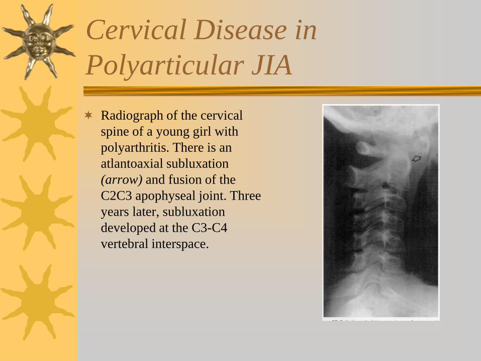
Cervical Disease in
Polyarticular JIA
Radiograph of the cervical
spine of a young girl with
polyarthritis. There is an
atlantoaxial subluxation
(arrow) and fusion of the
C2C3 apophyseal joint. Three
years later, subluxation
developed at the C3-C4
vertebral interspace.
Enthesitis-related arthritis (eJIA)
Seen in 3-11% of JIA patients
Arthritis and enthesitis or arthritis or enthesitis plus any two of the following:
– Sacroiliac joint tenderness and/or inflammatory lumbosacral pain
– + HLA-B27 Antigen
– Physician diagnosed HLA-B27-associated disease in 1st or 2nd
degree relative
– Symptomatic anterior uveitis
– Male > 6 y.o. at onset of arthritis or enthesitis
– Exclusions: Psoriasis or h/o psoriasis in patient or 1st degree relative, +RF or h/o sJIA
Psoriatic Arthritis (pJIA)
Seen in 2-11% of JIA population
Arthritis and psoriasis or arthritis and at least 2 of the following:– Physician diagnosed psoriasis in 1st degree relative
– Dactylitis
– Nail abnormalities (pitting or oncyholysis)
Only ~ 10% of these patients present with rash and arthritis at the same time– Rash may not appear for years after onset of arthritis
or may precede the arthritis
Undifferentiated JIA (uJIA)
Affects 2-23% of JIA population
Arthritis does not fulfill any of the above
categories or fits into more than one category
– 60% failed to demonstrate characteristics that fulfilled
criteria for one of the other categories
– 40% meet criteria from more than one category
• Most common overlap – Polyarticular JIA, RF negative with
either enthesitis related JIA or psoriatic JIA

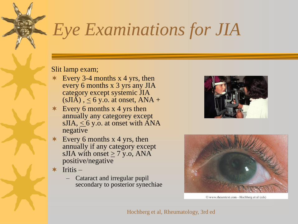
Eye Examinations for JIA
Slit lamp exam;
Every 3-4 months x 4 yrs, then every 6 months x 3 yrs any JIA category except systemic JIA (sJIA) , < 6 y.o. at onset, ANA +
Every 6 months x 4 yrs then annually any categorey except sJIA, < 6 y.o. at onset with ANA negative
Every 6 months x 4 yrs, then annually if any category except sJIA with onset > 7 y.o, ANA positive/negative
Iritis –– Cataract and irregular pupil
secondary to posterior synechiae
Hochberg et al, Rheumatology, 3rd ed

Laboratory and Diagnostic Evaluation
for Juvenile Idiopathic Arthritis
Oligoarticular and Polyarticular Onset
Antinuclear Antibody (ANA) (identify those at risk for iritis/uveitis)
CRP (ESR may be normal in active disease)
PPD
Rheumatoid Factor (RF)
HLA-B27 (for classification not diagnosis)
Slit-lamp examination
X-rays: not diagnostic, rule out other diagnoses
can be diagnostic for sacroiliitis
Systemic Onset
CXR
ECG – look for signs of tamponade
2-D ECHO
Slit-lamp examination
Consider:Bone scan, Bone marrow bx, Small bowel series
What does a positive ANA mean?
Screening test
Detects antibodies against ~ 50 different
components (proteins, DNA) of cell nucleus
Low positive ANA common in normal persons
– ~10% normal, healthy children have + ANA
– Not due to disease process and NO clinical
significance
Goals of Medical Treatment in JIA
Amelioration of disease signs/symptoms
Non-specific suppression of inflammatory response
Minimize discomfort; Maximize function
Maintain and/or improve strength and ROM
Permit optimal physical and psychological growth and development
Rationale for early, aggressive treatment based on improved outcomes in adult RA clinical trials
– Decreased rate of erosions, remission less likely if treatment delayed
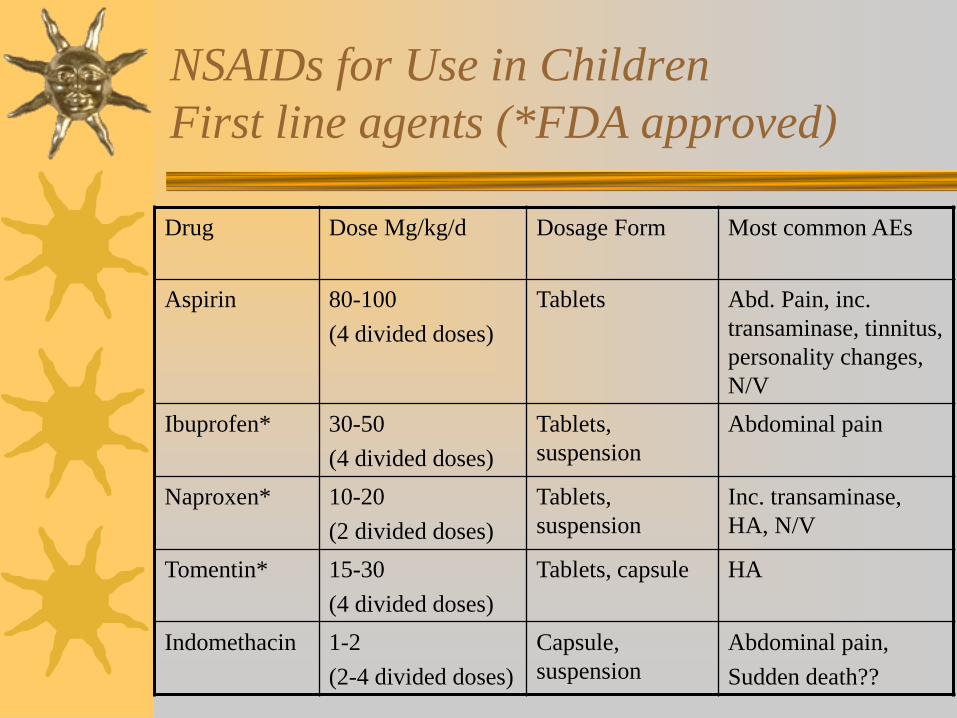
NSAIDs for Use in Children
First line agents (*FDA approved)
Drug Dose Mg/kg/d Dosage Form Most common AEs
Aspirin 80-100
(4 divided doses)
Tablets Abd. Pain, inc.
transaminase, tinnitus,
personality changes,
N/V
Ibuprofen* 30-50
(4 divided doses)
Tablets,
suspension
Abdominal pain
Naproxen* 10-20
(2 divided doses)
Tablets,
suspension
Inc. transaminase,
HA, N/V
Tomentin* 15-30
(4 divided doses)
Tablets, capsule HA
Indomethacin 1-2
(2-4 divided doses)
Capsule,
suspension
Abdominal pain,
Sudden death??

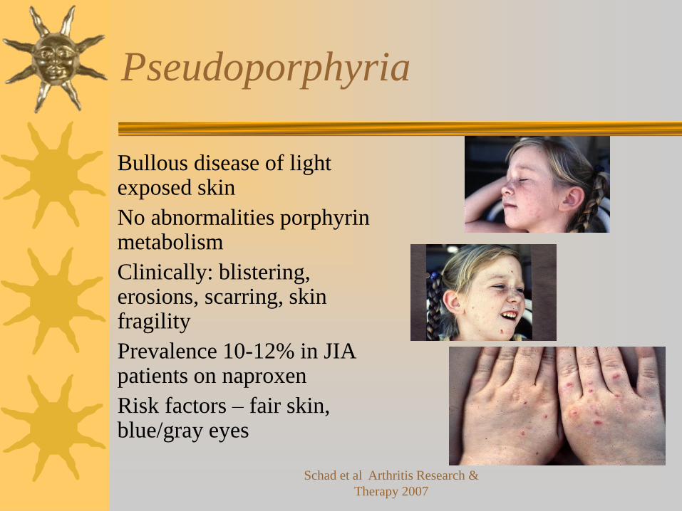
Pseudoporphyria
Bullous disease of light exposed skin
No abnormalities porphyrin metabolism
Clinically: blistering, erosions, scarring, skin fragility
Prevalence 10-12% in JIA patients on naproxen
Risk factors – fair skin, blue/gray eyes
Schad et al Arthritis Research &
Therapy 2007
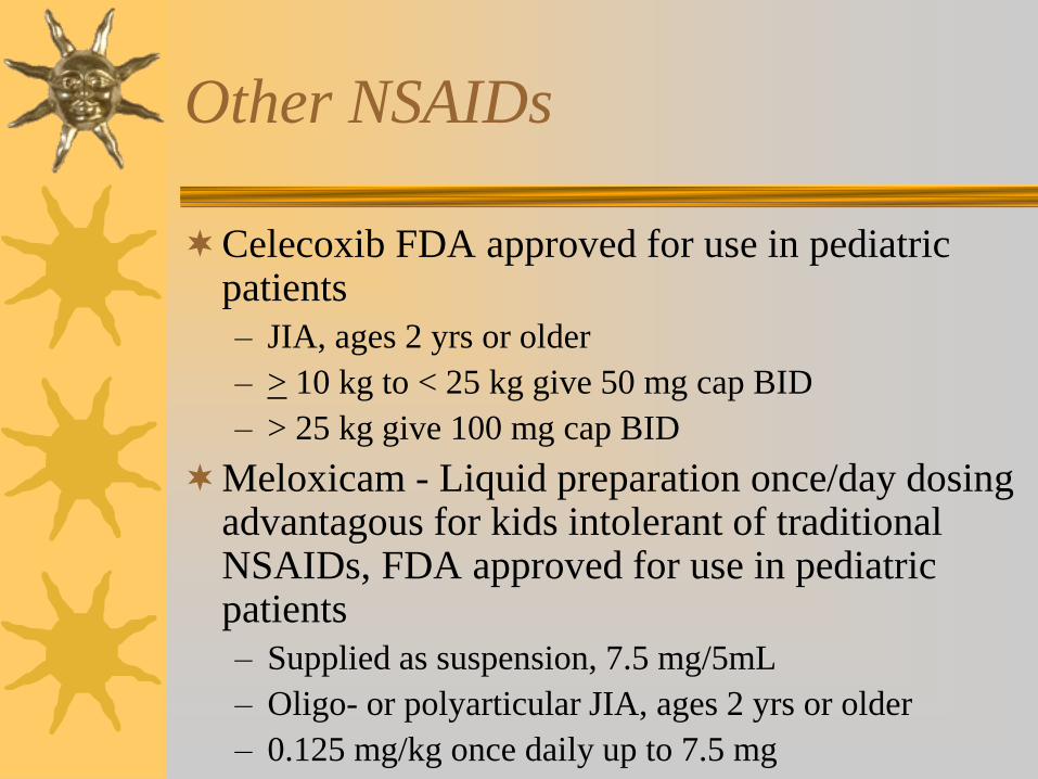
Other NSAIDs
Celecoxib FDA approved for use in pediatric patients
– JIA, ages 2 yrs or older
– > 10 kg to < 25 kg give 50 mg cap BID
– > 25 kg give 100 mg cap BID
Meloxicam - Liquid preparation once/day dosing advantagous for kids intolerant of traditional NSAIDs, FDA approved for use in pediatric patients
– Supplied as suspension, 7.5 mg/5mL
– Oligo- or polyarticular JIA, ages 2 yrs or older
– 0.125 mg/kg once daily up to 7.5 mg
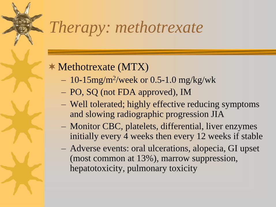
Therapy: methotrexate
Methotrexate (MTX)
– 10-15mg/m2/week or 0.5-1.0 mg/kg/wk
– PO, SQ (not FDA approved), IM
– Well tolerated; highly effective reducing symptoms and slowing radiographic progression JIA
– Monitor CBC, platelets, differential, liver enzymes initially every 4 weeks then every 12 weeks if stable
– Adverse events: oral ulcerations, alopecia, GI upset (most common at 13%), marrow suppression, hepatotoxicity, pulmonary toxicity
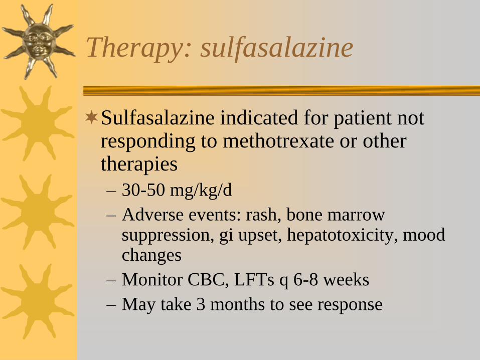
Therapy: sulfasalazine
Sulfasalazine indicated for patient not responding to methotrexate or other therapies
– 30-50 mg/kg/d
– Adverse events: rash, bone marrow suppression, gi upset, hepatotoxicity, mood changes
– Monitor CBC, LFTs q 6-8 weeks
– May take 3 months to see response
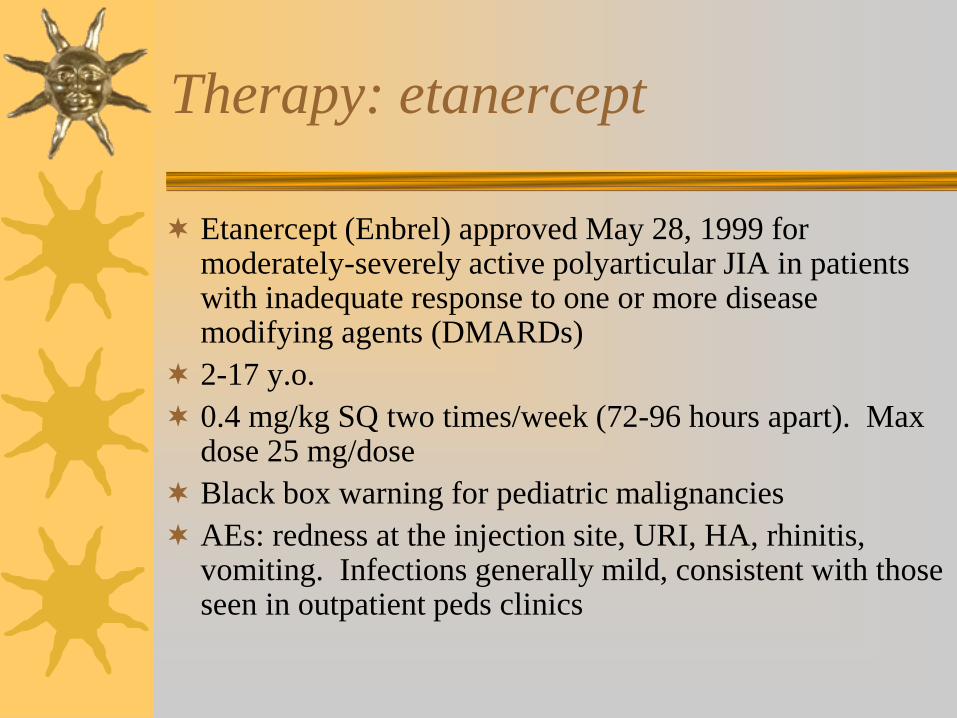
Therapy: etanercept
Etanercept (Enbrel) approved May 28, 1999 for moderately-severely active polyarticular JIA in patients with inadequate response to one or more disease modifying agents (DMARDs)
2-17 y.o.
0.4 mg/kg SQ two times/week (72-96 hours apart). Max dose 25 mg/dose
Black box warning for pediatric malignancies
AEs: redness at the injection site, URI, HA, rhinitis, vomiting. Infections generally mild, consistent with those seen in outpatient peds clinics
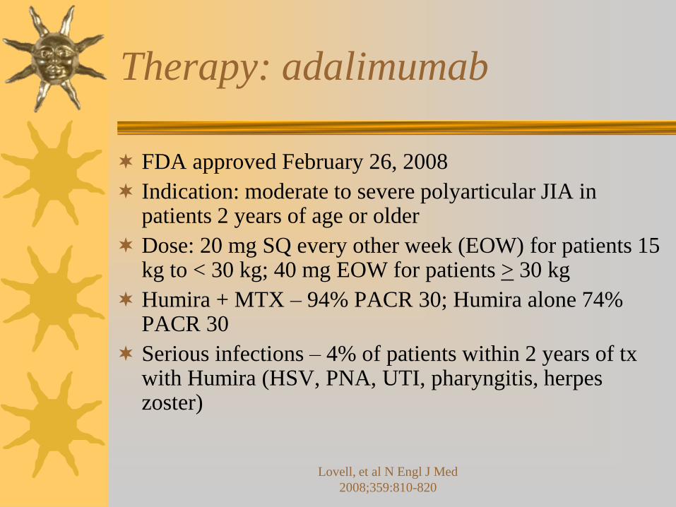
Therapy: adalimumab
FDA approved February 26, 2008
Indication: moderate to severe polyarticular JIA in patients 2 years of age or older
Dose: 20 mg SQ every other week (EOW) for patients 15 kg to < 30 kg; 40 mg EOW for patients > 30 kg
Humira + MTX – 94% PACR 30; Humira alone 74% PACR 30
Serious infections – 4% of patients within 2 years of tx with Humira (HSV, PNA, UTI, pharyngitis, herpes zoster)
Lovell, et al N Engl J Med
2008;359:810-820
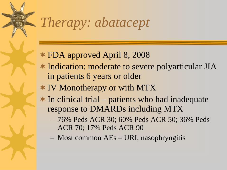
Therapy: abatacept
FDA approved April 8, 2008
Indication: moderate to severe polyarticular JIA in patients 6 years or older
IV Monotherapy or with MTX
In clinical trial – patients who had inadequate response to DMARDs including MTX
– 76% Peds ACR 30; 60% Peds ACR 50; 36% Peds ACR 70; 17% Peds ACR 90
– Most common AEs – URI, nasophryngitis
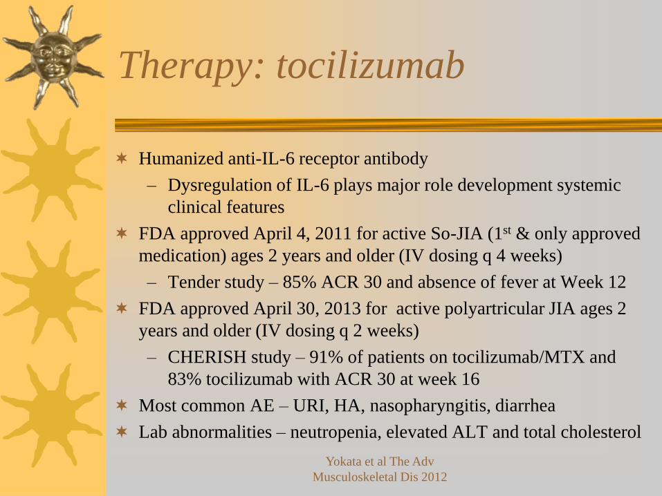
Therapy: tocilizumab
Humanized anti-IL-6 receptor antibody
– Dysregulation of IL-6 plays major role development systemic
clinical features
FDA approved April 4, 2011 for active So-JIA (1st & only approved
medication) ages 2 years and older (IV dosing q 4 weeks)
– Tender study – 85% ACR 30 and absence of fever at Week 12
FDA approved April 30, 2013 for active polyartricular JIA ages 2
years and older (IV dosing q 2 weeks)
– CHERISH study – 91% of patients on tocilizumab/MTX and
83% tocilizumab with ACR 30 at week 16
Most common AE – URI, HA, nasopharyngitis, diarrhea
Lab abnormalities – neutropenia, elevated ALT and total cholesterol
Yokata et al The Adv
Musculoskeletal Dis 2012
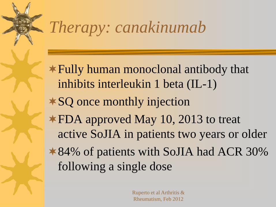
Therapy: canakinumab
Fully human monoclonal antibody that
inhibits interleukin 1 beta (IL-1)
SQ once monthly injection
FDA approved May 10, 2013 to treat
active SoJIA in patients two years or older
84% of patients with SoJIA had ACR 30%
following a single dose
Ruperto et al Arthritis &
Rheumatism, Feb 2012
Risk of malignancy with use of
TNFα blockers
FDA received reports on 48 cases of malignancy in children associated with TNFα blockers as of April 28, 2008
31 cases for infliximab, 15 cases for etanercept, and 2 cases for adalimumab
Types of malignancies– lymphoma, leukemia, melanoma, other solid tumors
Patients had either JIA or Crohn’s disease
88% of cases reported use of concomitant immunosuppressants
Number and types of malignancies reported were concerning given small number of children treated with TNFα blockers
US Food and Drug Administration

Rates of Malignancy Associated
with JIA and Its Treatment
Medicaid data 2000 through 2005 used to ID cohorts of
children with JIA and without JIA
7,812 children with JIA – Standardized incidence ratio
(SIR) 4.4 (95%CI) for probable and highly probable
malignancies
Following use of TNF inhibitors, no probable or highly
probable malignancies identified (SIR 0 (95% CI)
Increased risk of malignancy in JIA; treatment including
TNF inhibitors, not significantly associated with
development of malignancy
Beukelman et al, Arthritis &
Rheumatism, April 2012
Therapy: Corticosteroids
Corticosteroids not acceptable for long term use due to
toxicities in children
– Growth retardation most significant toxicity
– Dose of 5 mg/d usually inhibitory
1-2 mg/kg/day in once daily dosing to treat systemic
manifestations
Methylprednisolone 10-30 mg/kg IV in one pulse, or
daily for 3 days for severe JIA
Triamcinolone hexacetonide (Aristospan) 5-40 mg
(depending on joint size) for intra-articular injection
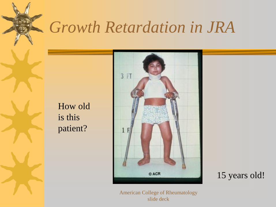
Growth Retardation in JRA
How old
is this
patient?
15 years old!
American College of Rheumatology
slide deck
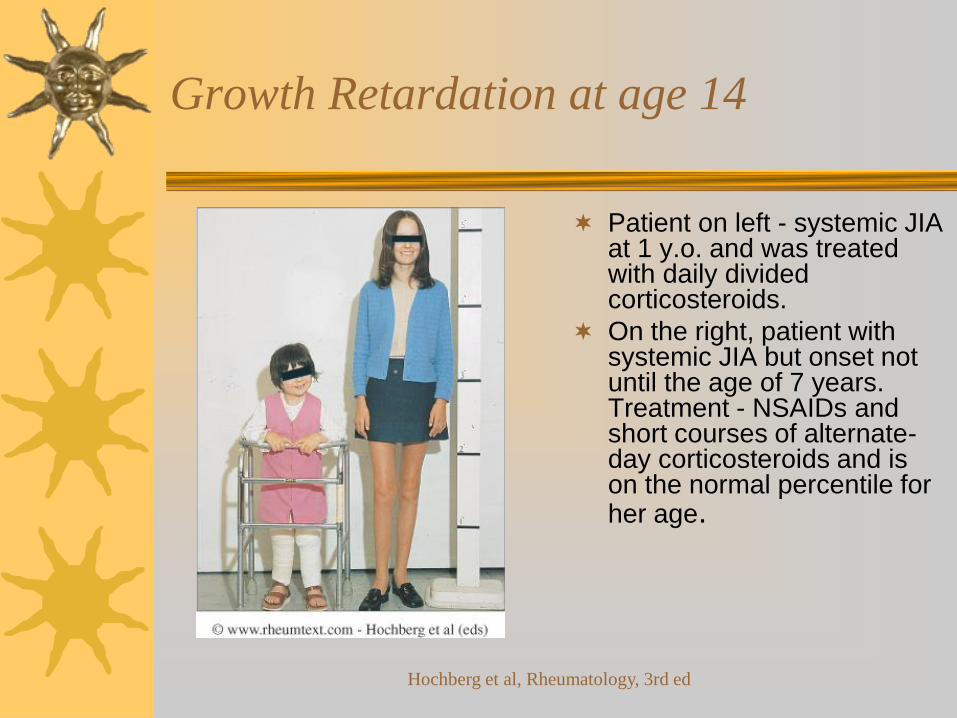
Growth Retardation at age 14
Patient on left - systemic JIA at 1 y.o. and was treated with daily divided corticosteroids.
On the right, patient with systemic JIA but onset not until the age of 7 years. Treatment - NSAIDs and short courses of alternate-day corticosteroids and is on the normal percentile for her age.
Hochberg et al, Rheumatology, 3rd ed
Prognosis
Recent outcomes studies show patients JIA often have lifelong sequelae
– Patients followed 27 years, 53% active with avg. age at diagnosis 7.5 years
Initially believed that 70-90% of patients enter adulthood without significant disability but now believed 50-70% of poly- or systemic arthritis and 40-50% of oligoarthritis will have active disease in adulthood
At greatest risk for joint destruction:
– Those with systemic onset disease
– Those with RF+ polyarthritis
Iritis/uveitis may cause permanent disability (5-16% visual defects, 16-26% cataracts, 14-24% glaucoma, 11-22% band keratopathy)
Juvenile Systemic Lupus
Erythematosus
Most common connective tissue disease in childhood– Prevalence: 5-10 per 100,000
• Adolescent females but may affect males and younger kids
ANA positive in virtually ALL children with SLE– Presence of anti-Smith Ab and hypocomplementemia
– Renal involvement common – up to 2/3 of children with SLE
– CNS involvement common cause of morbidity• Transverse myelitis, seizures, coma or subtle findings (poor
judgement, depression, short term memory deficits)
ACR criteria for SLE applies to SLE in children
May initially present with ITP and then meet criteria months or years later
Criteria for SLE Diagnosis
1. Malar rash
2. Discoid rash
3. Photosensitivity
4. Oral ulcers – or nasopharyngeal ulcers
5. Arthritis – 2 or more peripheral joints
6. Serositis – pleuritis, pericarditis
7. Renal disorder – proteinuria or cellular casts
8. Neurologic disorder – seizure, psychosis
9. Hematologic disorder –hemolytic anemia, leukopenia or lymphopenia
10. Immunologic disorder – dsDNA, anti-Smith Ab, aCL Abs, LAC, or false + VDRL
11. Antinuclear antibody
Pediatric Rheumatology
February 2007 report to the Health Resources and Services Adminstraion (HRSA_
– The Pediatric Rheumatology Workforce: A Study of the Supply and Demand for Pediatric Rheumatologists
As of 2003, less than 200 board certified practicing Pediatric Rheumatologists
As of 2003, 13 states in the United States did not have a single Pediatric Rheumatologist
300,000 children felt to have pediatric rheumatic disease in the United States
On average, patients travel 57 miles to see Peds Rheum, but less than 25 miles to see Peds Cards, Peds Endo and other specialties
One-third of medical schools and 40 percent of pediatric residency programs have no pediatric rheumatologist available to provide patient care or educate physicians in training
In 2003, only 10 fellows in Pediatric Rheumatology completed their training
Choosing Wisely®Five Things Physicians and Patients Should Question
Don’t order antibody panels unless positive antinuclear
antibodies (ANA) and evidence of rheumatic disease
– Up to 50% of children develop MS pain but no
evidence Ab testing w/o HX or PE of rheum disease
enhances DX w/ isolated MS pain
– Ab panels expensive
– Evidence demonstrates cost reduction by limiting
autoAb panel testing
American College of Rheumatology -
Pediatric Rheumatology
Choosing Wisely®Five Things Physicians and Patients Should Question
Don’t test for Lyme disease as cause of MS
symptoms w/o exposure history & appropriate
exam findings
– Lyme Dz – brief attacks of arthralgia or intermittent or
persistent episodes of arthritis in one or a few large
joints at a time, esp knee
– Diffuse arthralgias, myalgias or fibromyalgia alone are
not criteria for MS Lyme disease
American College of Rheumatology -
Pediatric Rheumatology
Choosing Wisely®Five Things Physicians and Patients Should Question
Don’t routinely perform surveillance joint x-rays
to monitor JIA disease activity
– No available data to suggest routine x-rays improves
outcomes
– Radiation exposure and costs are potential risks
– Obtain x-rays only when HX/PE raise clinical concern
about joint damage or decline in function
American College of Rheumatology -
Pediatric Rheumatology
Choosing Wisely®Five Things Physicians and Patients Should Question
Don’t perform methotrexate toxicity labs more
often than every 12 weeks on stable doses
Lab abnormalities usually mild, rarely prompt
significant changes in treatment
More frequent monitoring 1st six months of
treatment, dose escalation or risk factors for
toxicity
– Obesity, diabetes, renal disease, psoriasis, SO-JIA,
Down syndrome or use of alcohol
American College of Rheumatology -
Pediatric Rheumatology
Choosing Wisely®Five Things Physicians and Patients Should Question
Don’t repeat a confirmed positive ANA in
patients with established JIA or systemic lupus
erythematosus (SLE)
– ANA important in DX SLE and positivity guides more
frequent slit lamp exams for detection of uveitis in
children with JIA
– No evidenced that ANA is valuable for ongoing
management of SLE or JIA
– Repeat if child with JIA has evolution of sxs
suggestive of an autoimmune connective tissue disease
American College of Rheumatology -
Pediatric Rheumatology
References
Textbook of Pediatric Rheumatology, 5th
edition, Cassidy, JT, Petty et al, 2005
Rheumatology, 3rd edition, Hochberg MC,
Silman, AJ et al, 2003
Rheumatology, 2nd edition, Kippel JH,
Dieppe, PA, 1998