Embed Size (px)
Citation preview
Kampinos National Park: a risk area for spotted fevergroup rickettsioses, central Poland?
Joanna Stanczak1• Beata Biernat1
• Anna Matyjasek1,2•
Maria Racewicz1• Marta Zalewska3
• Daria Lewandowska1
Received: 25 May 2016 / Accepted: 3 August 2016 / Published online: 8 September 2016� The Author(s) 2016. This article is published with open access at Springerlink.com
Abstract Ixodid ticks are important vectors of a variety of bacterial and protozoan
pathogens which cause infections in humans. In this study, altogether 1041 questing Ixodes
ricinus (n = 305) and Dermacentor reticulatus ticks (n = 736), sympatrically occurring in
Kampinos National Park (KPN), central-east Poland, were analyzed by PCR for Rickettsia
species. Overall, the pathogen prevalence in ticks was 27.5 % for I. ricinus and 42.8 % for
D. reticulatus. Sequencing analysis showed that the first tick species was exclusively
infected with R. helvetica, whereas the latter was infected with R. raoultii. These organism
may pose a threat for populations exposed to ticks. Preliminary results of a serosurvey of
74 KPN employees, inhabitants and visitors from the same area showed a 31.1 % total
seroprevalence against SFG rickettsiae compared to 13.3 % seropositive blood donors of
the control group. Risk factors significantly associated with IgG seropositivity were:
occupational exposure to ticks (p = 0.002), frequency of tick bites (p = 0.02) and male
gender (p = 0.005). Seropositive and seronegative individuals occupationally exposed to
ticks did not differ significantly with respect to age and years of employment.
Keywords Dermacentor reticulatus � Ixodes ricinus � Rickettsia helvetica � Rickettsiaraoultii � Spotted fever group rickettsiae � Seroprevalence � Kampinos National Park �Poland
& Joanna [email protected]
1 Department of Tropical Parasitology, Institute of Maritime and Tropical Medicine, MedicalUniversity of Gdansk, Powstania Styczniowego 9B Str., 81-519 Gdynia, Poland
2 Chair and Clinic of Internal Medicine, Connective Tissue Diseases and Geriatrics, MedicalUniversity of Gdansk, Debinki 7 Str., 80-211 Gdansk, Poland
3 Department of Environmental Hazards Prevention and Allergology, Medical University of Warsaw,Banacha 1a Str., 02-091 Warsaw, Poland
123
Exp Appl Acarol (2016) 70:395–410DOI 10.1007/s10493-016-0083-9
Introduction
Spotted fever group (SFG) rickettsioses in humans are caused by small, obligate intra-
cellular Gram-negative bacteria of the genus Rickettsia (Rickettsiaceae; Rickettsiales).
Most SFG rickettsiae are tick-associated, except Rickettsia akari (mite-borne) and R. felis
(flea-borne). Maintenance of rickettsiae in tick vectors occurs by both vertical and hori-
zontal transmission. Therefore, larvae, nymphs and adults may all be infective for sus-
ceptible hosts, including humans. Rickettsiae infecting the ticks’ salivary glands are
transmitted to the host during feeding (Brouqui et al. 2007). Thus, ixodid ticks serve both
as the main vectors and reservoir hosts for pathogens.
At least eight human rickettsial pathogens circulate in ticks in different and often
overlapping parts of Europe, including R. conorii, R. massiliae, R. slovaca, R. raoultii, R.
sibirica sibirica, R. sibirica mongolotimonae, R. helvetica, R. rioja, and possibly others
(Eremeeva and Dusch 2015). Four of them (R. helvetica, R. raoultii, R. massiliae, R.
slovaca) have been so far detected in ticks in Poland (Chmielewski et al. 2009; Mierze-
jewska et al. 2015; Rymaszewska and Piotrowski 2013; Stanczak et al. 2008). In humans,
tick-borne rickettsioses (TBR) have no pathognomonic signs, but may cause a suggesting
spectrum of clinical signs: fever, headache, rash, inoculation eschar and enlarged cervical
lymph nodes. Acute febrile illness, meningitis and a fatal perimyocarditis, caused by R.
helvetica have been reported from Sweden (Nilsson 2009; Nilsson et al. 1999, 2010, 2011)
and France (Fournier et al. 2000), whereas R. monacensis has been isolated so far from
three patients with Mediterranean spotted fever-like illness in Spain and in Italy (Jado et al.
2007; Madeddu et al. 2012). Rickettsia slovaca and R. raoultii are recognized etiologic
agent of tick-borne lymphadenopathy (TIBOLA) (Lakos 1997), the disease also known as
Dermacentor-borne necrosis erythema and lymphadenopathy (DEBONEL) (Ibarra et al.
2006) or scalp eschar and neck lymphadenopathy after tick bite (SENLAT) (Angelakis
et al. 2010). Human cases due to R. slovaca or, rarely, R. raoultii infections have been
reported from Hungary, France, Spain, Portugal, Italy and Germany (Lakos 1997; Oteo
et al. 2004; Parola et al. 2009; Rieg et al. 2011; Selmi et al. 2008; de Sousa et al. 2013).
According to the Polish regulations the reporting and registration of rickettsioses are
obligatory. In 2006-2012, five cases of various SFG rickettsioses, including two imported
from South Africa, were reported in Poland. These infections have been recognized in
Mazovia (three cases) and Lower Silesia (two cases). Detected rickettsiae have been
classified as: R. conorii, R. slovaca, R. raoultii and R. africae (Maczka et al. 2013).
The purpose of this work was to evaluate risk of human exposure to Rickettsia spp.
infection by investigating these bacteria in ticks and antibodies against rickettsiae in
individuals presumably exposed to tick bites. For this purpose, a highly frequented
recreational area, Kampinos National Park (Kampinoski Park Narodowy) (KPN), was
chosen as a study area. This is an exceptional national park, as it encompasses forests
directly adjacent to Warsaw (Warszawa), the capital of Poland and, as a natural recre-
ational hinterland, is frequently visited by its inhabitants and tourists.
Materials and methods
Study area
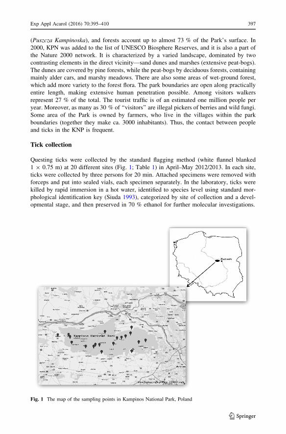
Kampinos National Park (KNP) [52�1802100N, 20�3603200E] (Fig. 1) is the largest natural
area in Poland. It covers 38,544 ha, including the ancient Kampinos Primeval Forest
396 Exp Appl Acarol (2016) 70:395–410
123
(Puszcza Kampinoska), and forests account up to almost 73 % of the Park’s surface. In
2000, KPN was added to the list of UNESCO Biosphere Reserves, and it is also a part of
the Nature 2000 network. It is characterized by a varied landscape, dominated by two
contrasting elements in the direct vicinity—sand dunes and marshes (extensive peat-bogs).
The dunes are covered by pine forests, while the peat-bogs by deciduous forests, containing
mainly alder cars, and marshy meadows. There are also some areas of wet-ground forest,
which add more variety to the forest flora. The park boundaries are open along practically
entire length, making extensive human penetration possible. Among visitors walkers
represent 27 % of the total. The tourist traffic is of an estimated one million people per
year. Moreover, as many as 30 % of ‘‘visitors’’ are illegal pickers of berries and wild fungi.
Some area of the Park is owned by farmers, who live in the villages within the park
boundaries (together they make ca. 3000 inhabitants). Thus, the contact between people
and ticks in the KNP is frequent.
Tick collection
Questing ticks were collected by the standard flagging method (white flannel blanked
1 9 0.75 m) at 20 different sites (Fig. 1; Table 1) in April–May 2012/2013. In each site,
ticks were collected by three persons for 20 min. Attached specimens were removed with
forceps and put into sealed vials, each specimen separately. In the laboratory, ticks were
killed by rapid immersion in a hot water, identified to species level using standard mor-
phological identification key (Siuda 1993), categorized by site of collection and a devel-
opmental stage, and then preserved in 70 % ethanol for further molecular investigations.
Fig. 1 The map of the sampling points in Kampinos National Park, Poland
Exp Appl Acarol (2016) 70:395–410 397
123
DNA extraction
All ticks were analysed individually. Extraction of total DNA was done by boiling crushed
specimens in ammonium hydroxide (NH4OH) (Guy and Stanek 1991; Rijpkema et al.
1996). Concentrations of DNA were measured with spectrophotometric method (Nano-
Drop 1000 spectrophotometer, Thermo Scientific, USA). The obtained lysates were stored
at -20 �C until use as templates for the molecular investigations.
Real time PCR
All tick samples were individually screened by real time PCR for the citrate synthase
encoding gene (gltA) specific for all Rickettsia spp. Primers Rick GltA-f (50-ATCCTA-
CATGCCGATCATGAGC-30) and Rick GltA-r (50-GTGAGCAGGTCCCCAAAGTG-30)were designed to target a 123-bp part of the gene with TaqMan probe (5-HEX-
ATGCTTCTACTTCAACAGTCCGAATTGCCG-BHQ1-30) (Biernat et al. 2016a).
Reaction mixtures and cycling conditions were as previously described (Biernat et al.
2016a). Negative and positive controls were included in all runs. Rickettsia-positive
control was constructed by cloning the 123-bp PCR amplicon into a circular pJet1.1
plasmids (Fermentas, USA) (Biernat et al. 2016a). Reactions were performed in the
Mx3005P Real-Time QPCR System (Stratagene, CA, USA).
Detection and identification of Rickettsia spp.
Most real time PCR-positive samples were subsequently rerun using semi nested PCR and
nested PCR assays to obtain longer amplicons for further DNA sequencing. Semi nested
PCR was conducted with three primers of which Ric and Ric U8 yielded a 1385-bp
fragment encompassing almost the complete 16S rRNA gene, while Ric and Ric Rt flanked
a 757 bp fragment (Nilsson et al. 1997). In the nested PCR, primers SLO1F/SLO1R and
SLO2F/SLO2R, targeting a fragment (355 bp) of ompA gene specific for R. slovaca and R.
raoultii, were used as the outer and inner pairs, respectively (Raoult et al. 2002). Resulted
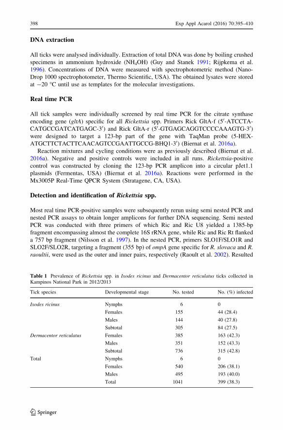
Table 1 Prevalence of Rickettsia spp. in Ixodes ricinus and Dermacentor reticulatus ticks collected inKampinos National Park in 2012/2013
Tick species Developmental stage No. tested No. (%) infected
Ixodes ricinus Nymphs 6 0
Females 155 44 (28.4)
Males 144 40 (27.8)
Subtotal 305 84 (27.5)
Dermacentor reticulatus Females 385 163 (42.3)
Males 351 152 (43.3)
Subtotal 736 315 (42.8)
Total Nymphs 6 0
Females 540 206 (38.1)
Males 495 193 (40.0)
Total 1041 399 (38.3)
398 Exp Appl Acarol (2016) 70:395–410
123
amplicons of 355 bp were considered positive. Additionally, a few of the positive samples
were analyzed by the conventional PCR using the primer pair RpCS.877p and RpCS.1258n
amplifying a fragment of the citrate synthase encoding gene (gltA), which has conserved
regions shared by all known Rickettsia species (Regnery et al. 1991). DNA products of
380 bp were considered to be positive results.
All amplifications were carried out in the GeneAmp� PCR System 9700 (Applied
Biosystems, Foster City, CA, USA) as previously described (Biernat et al. 2016b). PCR
products were separated on 2 % agarose gels stained with Midori green DNA Stain
(Nippon Genetics Europe) and visualized under UV light using the GelDoc–It, Imagine
Systems UVTM Transluminator (Upland, CA, USA). Rickettsia helvetica and R. raoultii
positive samples obtained in our previous investigations (Stanczak 2006; Stanczak et al.
2009) and confirmed by the sequence analysis of the PCR products were used as positive
controls. Nuclease-free water was added to each run as a negative control.
DNA sequencing
Chosen positive amplicons were purified using the Clean-Up purification kit (A&A
Biotechnology, Gdynia, Poland), sequenced in both directions with the same primers as
used in the semi-nested PCR and nested PCR assays with the ABI Prism� Big DyeTM
Terminator v.3.1 Cycle Sequencing Kit and then analyzed with the ABI PRISM 3130 xL
genetic Analysers (Applied Biosystem) according to the manufacturer’s protocol. Finally,
sequences were edited and compared with each other and with corresponding sequences
registered in the GenBank database using the NCBI BLAST program (U. S. National
Institutes of Health, Bethesda, Maryland) [http://blast.ncbi.nlm.nih.gov/Blast.cgi]. Then
consensus sequences were submitted to GenBank.
Seroreactivity to Rickettsia of population exposed to tick-bite
Study group
A total of 74 persons differently exposed to tick bites were examined. The group comprised
60 workers of KPN (23 females, 37 males; mean age 47.2 years, range 27–65) and 14
members of their families (9 females, 5 males; mean age: 40.1 years, range 9–65) occu-
pationally and recreationally, respectively, exposed to ticks.
As a control group, 30 blood donors (8 females, 22 males; mean age 29.8 years, range
18–60) who denied a tick bite 6 months prior to the investigation were examined. They
were city dwellers with no Lyme borreliosis history.
Blood samples were taken by venipuncture and sera separated by centrifugation. In
addition, EDTA blood samples were obtained from the study group for real time PCR
analysis. Samples were stored at -20 �C until the time of analysis.
Serological tests
Immunoenzymatic assay
The commercial ELISA kits (Spotted Fever Group Rickettsia EIA IgG and IgM Antibody
Kit, Fuller Laboratories, Fullerton, CA, USA) were used to detect IgG and IgM antibodies
Exp Appl Acarol (2016) 70:395–410 399
123
against SFG Rickettsia spp. The EIA module wells in this kit utilized a SFG-specific
lipopolysaccharide (rLPS) antigen extracted from a members of the SF group, including R.
rickettsii, R. akari, R. parkeri, R. felis, R. montanensis, and others. The tests were carried
out according manufacturer’s instructions, including the cutoff calibrator instructions.
Absorbance was measured at a wave length of 450 nm on a microtiter plate reader. The
obtained values of tested samples were divided by the mean absorbance values of Cutoff
Calibrator. The Calibrator was set at an index of 1.0. Index values from 0.8 to 1.2 were
considered equivocal (weak positive), above 1.2 as positive and those below 0.8 were
considered negative. For analysis, the weak positive results were included into positive
group.
Micro-immunofluorescence assay (MIF)
To detect IgG antibodies to selected antigens of SGF rickettsiae simultaneously, Rickettsia
2-Antigen MIF IgG Antibody Test (Fuller Laboratories) was used. Purified, acetone-fixed
antigens of R. helvetica and R. raoultii used as an individual substrate on the same slide
wells were applied as diagnostics antigens. These slides contain Vero-76 cells with 30–40
infected cells per field when using a 40X lens. The positive and negative controls of human
serum used in the procedures were contained in the MIF kits. The assays were performed
according to the manufacturer’s instructions. Positive control serum was tested in serial
dilution to determine their endpoint titer. Fluorescence of the rickettsiae with an intra-
cellular distribution and intensity pattern similar to the positive control was considered as a
positive reaction. The test titer started at 1:32 and an antibody titer of C1:64 was con-
sidered as positive reaction. All IFA slides were screened by the trained person using a
fluorescence microscope (Zeiss).
PCR assay
EDTA blood samples were screened using real time PCR. DNA extraction was carried out
with the Blood Mini kit (A&A Biotechnology, Gdynia, Poland) template preparation
according to the manufacturer’s instruction. Obtained templates were stored in -20 until
used for real time PCR assays with the same primers and probe like in the case of ticks
investigation.
Seroreactivity and association with risk factors
Each serum donor answered a questionnaire regarding gender, age, ticks exposure (oc-
cupational, recreational) and tick bites experienced during the last 12 months (‘none’, ‘1-
5’, ‘6-10’, and ‘[10’), length and character of employment in the forest, other tick-borne
diseases history, and symptoms such as: an eschar at the site of a tick bite, unexplained
fever, headache, myalgia or enlarged lymph nodes 4–6 weeks prior to investigation.
Ethical statement
The study was approved by the Bioethics Committee of Warsaw Medical University (KB/
189/2013). All individuals who agreed to participate signed their consent form and their
personal information was held by Department of Tropical Parasitology, Medical University
of Gdansk.
400 Exp Appl Acarol (2016) 70:395–410
123
Statistical analysis
All statistical analyses were performed with R (2008; http://www.R-project.org) and Excel.
Qualitative variables were presented using frequencies. Regression analysis and analysis of
variance with Tukey’s multiple comparisons of means for quantitative variables and
Fisher’s Exact test for qualitative variables were (a = 0.05).
Results
Ticks and tick infection rates
Altogether 1041 questing ticks were collected during their spring activity season (April–
May) in 2012/2013 at 20 different collection sites spread all-over KPN. Of them, 736 were
identified as D. reticulatus (70.7 %) (385 females and 351 males) and 305 as I. ricinus
(29. 3 %) (155 females, 144 males, 6 nymphs) (Table 1). Dermacentor reticulatus ticks
were found at 16 collection sites whereas I. ricinus in 18 of them (Table 2). The first
species (93.5 % collected specimens) prevailed in the open areas, meadows, pastures and
wastelands (10 collection sites). The second species (62.3 % collected specimens) was
dominant in the forested areas (8 collection sites). In two collection sites (mixed stands)
both species occurred in comparable numbers (in total 30 vs 25).
All collected ticks were individually screened for the presence of Rickettsia spp. by the
real time PCR and rickettsial DNA was detected in 38.3 % (n = 399) of them. However,
the rate of infection differed by tick species. The infection rate of I. ricinus was 27.5 %
(n = 84/305), being comparable in females and males (28.4 vs 27.8 %), whereas none of 6
nymphs was positive (Table 1). Prevalence of Rickettsia spp. in D. reticulatus ticks—
42.8 % (n = 315/736)—was significantly higher than in I. ricinus. The infection rate in
male and female ticks also were similar (43.3 vs 42.3 %) (Table 1).
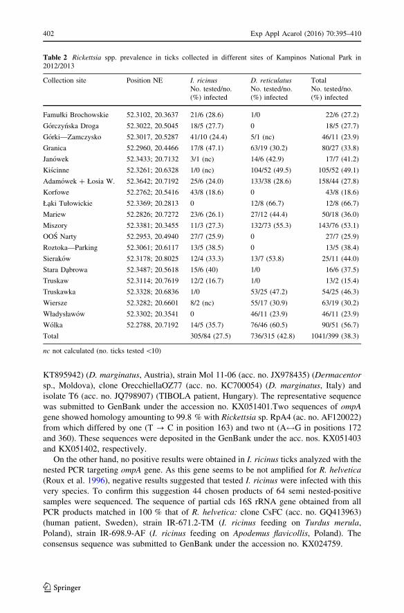
Depending on collection site, the percentage of infected I. ricinus varied between 16.7
and 47.1 %, while in the case of D. reticulatus the infection level ranged from 18.8 to
66.7 % (Table 2).
Identification of Rickettsia spp.
To identify species of Rickettsia, majority of real time PCR-positive samples were sub-
sequently rerun using nested or/and semi nested PCR targeting two genes, 16S rRNA and
ompA. As a result, among positive D. reticulatus 59 amplicons of 16S rRNA-fragment of
Rickettsia spp. and 223 amplicons of ompA-fragment were obtained. Of them, a total of 60
samples were sequenced and 55 obtained sequences were compared with those from
rickettsia species and strains deposited in GenBank database. Thirty-three sequences
showed 100 % homology with the sequences of partial cds of 16S rRNA gene of 4 R.
raoultii strains or isolates: strain RpA4 (acc. no. AF120026) (tick, Russia), strain DnS28
(acc. no. AF120024) (tick, Russia), strain Marne (acc. no. DQ365809) (D. reticulaus,
France) and isolate BL029-2 (acc. no. KJ410261) (Hyalomma asiaticum, China). The
representative sequence was submitted to GenBank under the accession no. KX024760.
Twenty sequences were 100 % identical with numerous homologous fragments of ompA
gene of R. raoultii (former Rickettsia sp. RpA4), including Rickettsia sp. RpA4 (ac. no.
AF120022) detected for the first time in hard ticks in Russia, isolate TG82 (acc. no.
Exp Appl Acarol (2016) 70:395–410 401
123
KT895942) (D. marginatus, Austria), strain Mol 11-06 (acc. no. JX978435) (Dermacentor
sp., Moldova), clone OrecchiellaOZ77 (acc. no. KC700054) (D. marginatus, Italy) and
isolate T6 (acc. no. JQ798907) (TIBOLA patient, Hungary). The representative sequence
was submitted to GenBank under the accession no. KX051401.Two sequences of ompA
gene showed homology amounting to 99.8 % with Rickettsia sp. RpA4 (ac. no. AF120022)
from which differed by one (T ? C in position 163) and two nt (A$G in positions 172
and 360). These sequences were deposited in the GenBank under the acc. nos. KX051403
and KX051402, respectively.
On the other hand, no positive results were obtained in I. ricinus ticks analyzed with the
nested PCR targeting ompA gene. As this gene seems to be not amplified for R. helvetica
(Roux et al. 1996), negative results suggested that tested I. ricinus were infected with this
very species. To confirm this suggestion 44 chosen products of 64 semi nested-positive
samples were sequenced. The sequence of partial cds 16S rRNA gene obtained from all
PCR products matched in 100 % that of R. helvetica: clone CsFC (acc. no. GQ413963)
(human patient, Sweden), strain IR-671.2-TM (I. ricinus feeding on Turdus merula,
Poland), strain IR-698.9-AF (I. ricinus feeding on Apodemus flavicollis, Poland). The
consensus sequence was submitted to GenBank under the accession no. KX024759.
Table 2 Rickettsia spp. prevalence in ticks collected in different sites of Kampinos National Park in2012/2013
Collection site Position NE I. ricinus D. reticulatus TotalNo. tested/no.(%) infected
No. tested/no.(%) infected
No. tested/no.(%) infected
Famułki Brochowskie 52.3102, 20.3637 21/6 (28.6) 1/0 22/6 (27.2)
Gorczynska Droga 52.3022, 20.5045 18/5 (27.7) 0 18/5 (27.7)
Gorki—Zamczysko 52.3017, 20.5287 41/10 (24.4) 5/1 (nc) 46/11 (23.9)
Granica 52.2960, 20.4466 17/8 (47.1) 63/19 (30.2) 80/27 (33.8)
Janowek 52.3433; 20.7132 3/1 (nc) 14/6 (42.9) 17/7 (41.2)
Kiscinne 52.3261; 20.6328 1/0 (nc) 104/52 (49.5) 105/52 (49.1)
Adamowek ? Łosia W. 52.3642; 20.7192 25/6 (24.0) 133/38 (28.6) 158/44 (27.8)
Korfowe 52.2762; 20.5416 43/8 (18.6) 0 43/8 (18.6)
Łaki Tułowickie 52.3369; 20.2813 0 12/8 (66.7) 12/8 (66.7)
Mariew 52.2826; 20.7272 23/6 (26.1) 27/12 (44.4) 50/18 (36.0)
Miszory 52.3381; 20.3455 11/3 (27.3) 132/73 (55.3) 143/76 (53.1)
OOS Narty 52.2953, 20.4940 27/7 (25.9) 0 27/7 (25.9)
Roztoka—Parking 52.3061; 20.6117 13/5 (38.5) 0 13/5 (38.4)
Sierakow 52.3178; 20.8025 12/4 (33.3) 13/7 (53.8) 25/11 (44.0)
Stara Dabrowa 52.3487; 20.5618 15/6 (40) 1/0 16/6 (37.5)
Truskaw 52.3114; 20.7619 12/2 (16.7) 1/0 13/2 (15.4)
Truskawka 52.3328; 20.6836 1/0 53/25 (47.2) 54/25 (46.3)
Wiersze 52.3282; 20.6601 8/2 (nc) 55/17 (30.9) 63/19 (30.2)
Władysławow 52.3302; 20.3541 0 46/11 (23.9) 46/11 (23.9)
Wolka 52.2788, 20.7192 14/5 (35.7) 76/46 (60.5) 90/51 (56.7)
Total 305/84 (27.5) 736/315 (42.8) 1041/399 (38.3)
nc not calculated (no. ticks tested\10)
402 Exp Appl Acarol (2016) 70:395–410
123
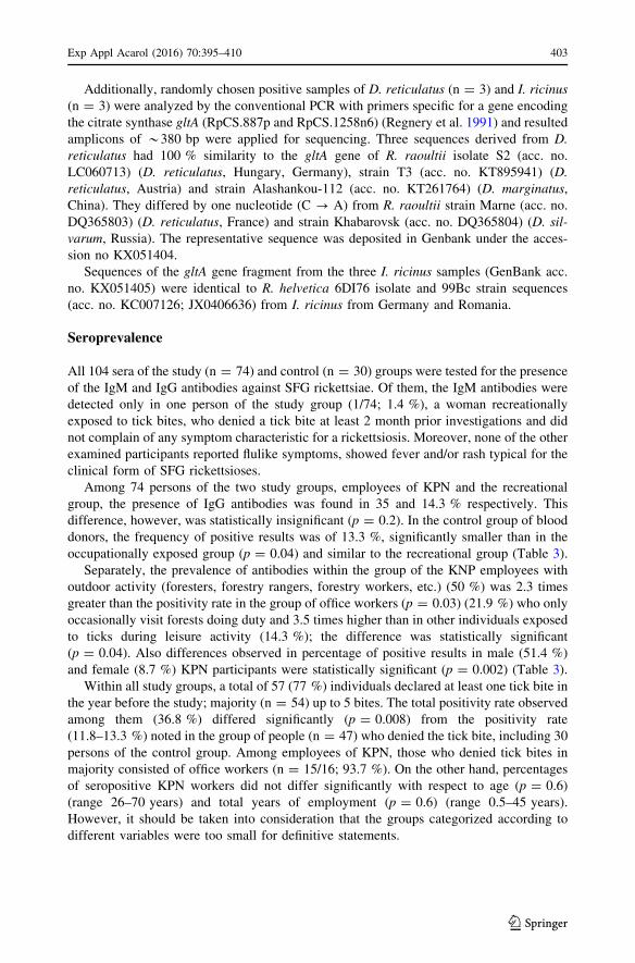
Additionally, randomly chosen positive samples of D. reticulatus (n = 3) and I. ricinus
(n = 3) were analyzed by the conventional PCR with primers specific for a gene encoding
the citrate synthase gltA (RpCS.887p and RpCS.1258n6) (Regnery et al. 1991) and resulted
amplicons of *380 bp were applied for sequencing. Three sequences derived from D.
reticulatus had 100 % similarity to the gltA gene of R. raoultii isolate S2 (acc. no.
LC060713) (D. reticulatus, Hungary, Germany), strain T3 (acc. no. KT895941) (D.
reticulatus, Austria) and strain Alashankou-112 (acc. no. KT261764) (D. marginatus,
China). They differed by one nucleotide (C ? A) from R. raoultii strain Marne (acc. no.
DQ365803) (D. reticulatus, France) and strain Khabarovsk (acc. no. DQ365804) (D. sil-
varum, Russia). The representative sequence was deposited in Genbank under the acces-
sion no KX051404.
Sequences of the gltA gene fragment from the three I. ricinus samples (GenBank acc.
no. KX051405) were identical to R. helvetica 6DI76 isolate and 99Bc strain sequences
(acc. no. KC007126; JX0406636) from I. ricinus from Germany and Romania.
Seroprevalence
All 104 sera of the study (n = 74) and control (n = 30) groups were tested for the presence
of the IgM and IgG antibodies against SFG rickettsiae. Of them, the IgM antibodies were
detected only in one person of the study group (1/74; 1.4 %), a woman recreationally
exposed to tick bites, who denied a tick bite at least 2 month prior investigations and did
not complain of any symptom characteristic for a rickettsiosis. Moreover, none of the other
examined participants reported flulike symptoms, showed fever and/or rash typical for the
clinical form of SFG rickettsioses.
Among 74 persons of the two study groups, employees of KPN and the recreational
group, the presence of IgG antibodies was found in 35 and 14.3 % respectively. This
difference, however, was statistically insignificant (p = 0.2). In the control group of blood
donors, the frequency of positive results was of 13.3 %, significantly smaller than in the
occupationally exposed group (p = 0.04) and similar to the recreational group (Table 3).
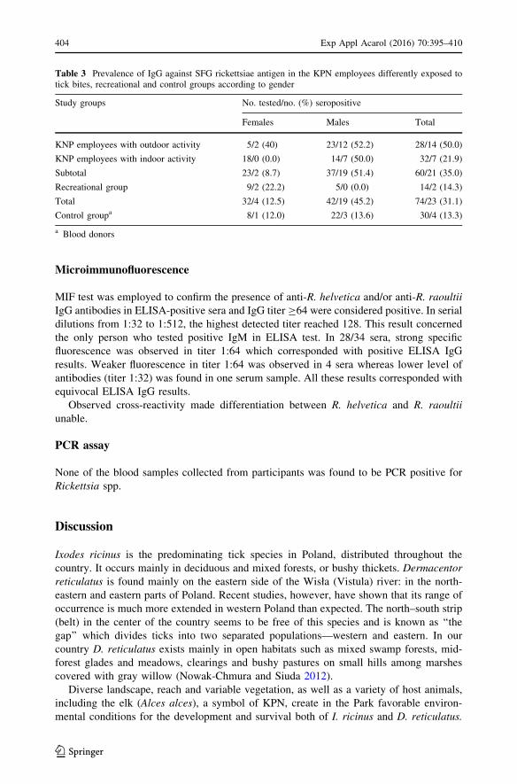
Separately, the prevalence of antibodies within the group of the KNP employees with
outdoor activity (foresters, forestry rangers, forestry workers, etc.) (50 %) was 2.3 times
greater than the positivity rate in the group of office workers (p = 0.03) (21.9 %) who only
occasionally visit forests doing duty and 3.5 times higher than in other individuals exposed
to ticks during leisure activity (14.3 %); the difference was statistically significant
(p = 0.04). Also differences observed in percentage of positive results in male (51.4 %)
and female (8.7 %) KPN participants were statistically significant (p = 0.002) (Table 3).
Within all study groups, a total of 57 (77 %) individuals declared at least one tick bite in
the year before the study; majority (n = 54) up to 5 bites. The total positivity rate observed
among them (36.8 %) differed significantly (p = 0.008) from the positivity rate
(11.8–13.3 %) noted in the group of people (n = 47) who denied the tick bite, including 30
persons of the control group. Among employees of KPN, those who denied tick bites in
majority consisted of office workers (n = 15/16; 93.7 %). On the other hand, percentages
of seropositive KPN workers did not differ significantly with respect to age (p = 0.6)
(range 26–70 years) and total years of employment (p = 0.6) (range 0.5–45 years).
However, it should be taken into consideration that the groups categorized according to
different variables were too small for definitive statements.
Exp Appl Acarol (2016) 70:395–410 403
123
Microimmunofluorescence
MIF test was employed to confirm the presence of anti-R. helvetica and/or anti-R. raoultii
IgG antibodies in ELISA-positive sera and IgG titer C64 were considered positive. In serial
dilutions from 1:32 to 1:512, the highest detected titer reached 128. This result concerned
the only person who tested positive IgM in ELISA test. In 28/34 sera, strong specific
fluorescence was observed in titer 1:64 which corresponded with positive ELISA IgG
results. Weaker fluorescence in titer 1:64 was observed in 4 sera whereas lower level of
antibodies (titer 1:32) was found in one serum sample. All these results corresponded with
equivocal ELISA IgG results.
Observed cross-reactivity made differentiation between R. helvetica and R. raoultii
unable.
PCR assay
None of the blood samples collected from participants was found to be PCR positive for
Rickettsia spp.
Discussion
Ixodes ricinus is the predominating tick species in Poland, distributed throughout the
country. It occurs mainly in deciduous and mixed forests, or bushy thickets. Dermacentor
reticulatus is found mainly on the eastern side of the Wisła (Vistula) river: in the north-
eastern and eastern parts of Poland. Recent studies, however, have shown that its range of
occurrence is much more extended in western Poland than expected. The north–south strip
(belt) in the center of the country seems to be free of this species and is known as ‘‘the
gap’’ which divides ticks into two separated populations—western and eastern. In our
country D. reticulatus exists mainly in open habitats such as mixed swamp forests, mid-
forest glades and meadows, clearings and bushy pastures on small hills among marshes
covered with gray willow (Nowak-Chmura and Siuda 2012).
Diverse landscape, reach and variable vegetation, as well as a variety of host animals,
including the elk (Alces alces), a symbol of KPN, create in the Park favorable environ-
mental conditions for the development and survival both of I. ricinus and D. reticulatus.
Table 3 Prevalence of IgG against SFG rickettsiae antigen in the KPN employees differently exposed totick bites, recreational and control groups according to gender
Study groups No. tested/no. (%) seropositive
Females Males Total
KNP employees with outdoor activity 5/2 (40) 23/12 (52.2) 28/14 (50.0)
KNP employees with indoor activity 18/0 (0.0) 14/7 (50.0) 32/7 (21.9)
Subtotal 23/2 (8.7) 37/19 (51.4) 60/21 (35.0)
Recreational group 9/2 (22.2) 5/0 (0.0) 14/2 (14.3)
Total 32/4 (12.5) 42/19 (45.2) 74/23 (31.1)
Control groupa 8/1 (12.0) 22/3 (13.6) 30/4 (13.3)
a Blood donors
404 Exp Appl Acarol (2016) 70:395–410
123
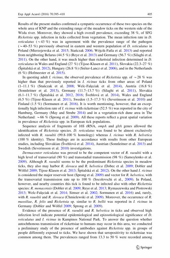
Results of the present studies confirmed a sympatric occurrence of these two species on the
whole area of KNP and the extending range of the meadow ticks on the western side of the
Wisła river. Moreover, they showed a high overall prevalence, exceeding 38 %, of SFG
Rickettsia spp. infection in ticks collected from vegetation. The mean infection rate in D.
reticulatus (*43 %) was in agreement with the prevalence range of the pathogen
(*40–53 %) previously observed in eastern and western population of D. reticulatus in
Poland (Mierzejewska et al. 2015; Stanczak 2006; Wojcik-Fatla et al. 2013) and reported
from neighboring Belarus (44.5 %) (Reye et al. 2013) and Germany (56.7 %) (Silaghi et al.
2011). On the other hand, it was much higher than rickettsial infection determined in D.
reticulatus in Wales and England (27 %) (Tijsse-Klasen et al. 2011), Slovakia (22.3–27 %)
(Spitalska et al. 2012), Hungary (26.8 %) (Sreter-Lancz et al. 2006), and in the Netherlands
(6 %) (Hofmeester et al. 2015).
In questing adult I. ricinus, the observed prevalence of Rickettsia spp. of *28 % was
higher than that previously reported in I. ricinus ticks from other areas of Poland
(1–11.1 %) (Stanczak et al. 2008; Welc-Faleciak et al. 2014), Austria (16.8 %)
(Sonnleitner et al. 2013), Germany (11.7–13.7 %) (Silaghi et al. 2011), Slovakia
(6.1–11.7 %) (Spitalska et al. 2012, 2016; Svehlova et al. 2014), Wales and England
(6.5 %) (Tijsse-Klasen et al. 2011), Sweden (1.5–17.3 %) (Severinsson et al. 2010) and
Finland (1.5 %) (Sormunen et al. 2016). It is worth mentioning, however, that an excep-
tionally high infection rate of I. ricinus with rickettsiae (52.5 %) was reported in the city of
Hamburg, Germany (May and Strube 2014) and in a vegetation-rich dune area in The
Netherland: *66 % (Sprong et al. 2009). All these reports reflect a great spatial variation
in prevalence of Rickettsia spp. in European tick populations.
Sequence analysis of fragments of 16S rRNA, ompA and gltA genes allowed the
identification of Rickettsia species. D. reticulatus was found to be almost exclusively
infected with R. raoultii (99.8-100 % homology) whereas I. ricinus with R. helvetica
(100 % identity). These findings are in accordance with results from other European
studies, including Slovakian (Svehlova et al. 2014), Austrian (Sonnleitner et al. 2013) and
Swedish (Severinsson et al. 2010) investigations.
Dermacentor reticulatus was proved to be the competent vector of R. raoultii with a
high level of transovarial (90 %) and transstadial transmission (98 %) (Samoylenko et al.
2009). Although R. raoultii seems to be the predominant Rickettsia species in meadow
ticks, they also may harbor R. slovaca and R. helvetica (Dobec et al. 2009; Dobler and
Wolfel 2009; Tijsse-Klasen et al. 2013; Spitalska et al. 2012). On the other hand I. ricinus
is considered the major reservoir host (Sprong et al. 2009) and vector for R. helvetica, with
the transovarial transmission rate up to 100 % (Socolovschi et al., 2009). In Poland,
however, and nearby countries this tick is found to be infected also with other Rickettsia
species: R. monacensis (Dobler et al. 2009; Reye et al. 2013; Rymaszewska and Piotrowski
2013; Welc-Faleciak et al. 2014; Simser et al. 2002; Sormunen et al. 2016) and, rarely,
with R. rauoltii and R. slovaca (Chmielewski et al. 2009). Moreover, the occurrence of R.
massiliae, R. felis and Rickettsia sp. similar to R. bellii was reported in I. ricinus in
Germany (Dobler and Wolfel 2009; Sprong et al. 2009).
Evidence of the presence of R. raoultii and R. helvetica in ticks and observed high
infection level indicate potential epidemiological and epizootiological significance of D.
reticulatus and I. ricinus in Kampinos National Park. To answer the question whether
autochthonous transmission of rickettsiae to humans may occur in this area, we conducted
a preliminary study of the presence of antibodies against Rickettsia spp. in groups of
people differently exposed to ticks. We have shown that seropositivity to rickettsiae was
common among them. The prevalences ranged from 13.3 to 50 % were recorded among
Exp Appl Acarol (2016) 70:395–410 405
123
KNP employees with outdoor activity and in office workers who occasionally visit forests
doing duty, in individuals exposed to ticks during leisure activity and in the group of blood
donors. This suggests that contacts between ticks and humans, and transmission of Rick-
ettsia spp. is frequent. Moreover, persons who denied tick bites at least 6 month prior to the
investigation had a seropositivity rate for IgG of 11.8–13.3 %, that confirms once more the
well-known clinical observation that tick bites often go unnoticed.
The overall seropositivity rate (31.1 %) detected in the present study is comparable with
that reported in studies performed in eastern Poland, were total 36 % of forestry and
agricultural workers were found to be positive (Zajac et al. 2013). As arthropod bites and
arthropod-borne infections are the frequent occupational hazards among forestry workers,
in both studies the seroprevalence was found to be the highest among forestry workers with
outdoor activity—50 and 50.7 %, respectively. These results were significantly higher than
the result obtained for forestry workers from northeastern and southern Poland (14.7 %)
(Podsiadły et al. 2011) as well as from north-eastern Italy (3.9 %) and Alsace in France
(9.2 %) (Cinco et al. 2006; Fournier et al. 2000). In Germany, antibodies against different
Rickettsia spp. were found in 9.1 % hunters (Jansen et al. 2008). Survey of another risk
group, military recruits during their field training period in the highly tick endemic area of
Gotland in Sweden, showed that 22.9 % of them had antibodies against R. helvetica
(Nilsson et al. 2005). The latter result is comparable with the seroprevalence of KPN
employees with the advantage of indoor activity.
Moreover, a serosurvey conducted in Danish patients seropositive for Lyme borreliosis
showed that 12.5 % of them had positive antibody titers to R. helvetica (Nielsen et al.
2004). Finally, in southern Sweden, 10 % patients with erythema migrans (EM) and/or
general signs of infection following a tick bite had antibodies against the same rickettsial
species (Lindblom et al. 2013). These results correspond with the seropositivity level
among KPN visitors, recreationally exposed to the tick bites.
On the other hand, SFG Rickettsia antibody prevalence in blood donors varies as well.
In Sweden approximately 1 % of them were seroreactive (Lindblom et al. 2013), whereas
in Tyrol, Austria, seroprevalence ranged from 4.8 to 10.6 % (Sonnleitner et al. 2013), and
in our control group reached 13.3 %.
To explain so high seroprevalence in people occupationally exposed to ticks in eastern
Poland, 36 % of the total, Zajac et al. (2013) suggested that they lived in the area where
over 50 % of D. reticulatus ticks harbored R. raoultii (Wojcik-Fatla et al. 2013) and thus
were under significantly increased risk of infection with these rickettsiae. The only case of
autochthonous spotted fever described in Poland was cause the most probably by R.
raoultii (Switaj et al. 2012), what may support this suggestion. Also in KNP prevalence of
D. reticulatus infected with R. raoultii exceeded 40 %. However, D. reticulatus rarely bites
humans. None of the individuals surveyed in our study declared to be bitten by this species,
although some of them occasionally found these ticks crawling on their clothes. In con-
trary, majority of them reported I. ricinus bites with a frequency of 1–5 per year. This tick
species shows a high affinity for humans and in Poland, of SFG rickettsiae, is almost
exclusively infected with R. helvetica (Stanczak et al. 2008). Interestingly, prevalence of R.
helvetica in I. ricinus in KPN (27.5 %) was comparable or lower than seroprevalence rates
in humans differently exposed to tick bites (range 14.3–50 %). This may suggest that
seroprevalence in humans does not directly reflect the prevalence in ticks.
In conclusion, on multivariate regression analysis risk factors significantly associated
with SFG rickettsiae infection (prevalence of antibodies) were: occupational exposure to
ticks (p = 0.02), male gender (p = 0.004) and frequency tick bites (p = 0.02).
406 Exp Appl Acarol (2016) 70:395–410
123
Unfortunately, results of the R. helvetica- and R. raoultii-specific MIF test did not
answer the question which of these two rickettsiae evoke positive immune response in
studied groups. MIF has been the serologic gold standard, originally being the means of
determining separate species, however SFG rickettsiae cause cross-reactions within the
group (Fournier et al. 2000; Hechemy et al. 1989). We did not observe a significant
difference in specific fluorescence between R. helvetica and R. raoultii, and uncertainty
about the source of the infection remained. Interestingly, Podsiadły et al. (2011) detected
by MIF antibodies to R. massiliae in 79 % of the seropositive forestry workers from
northeastern and southern Poland. However, R. massiliae is commonly found in Rhipi-
cephalus sanguineus or R. turanicus ticks (Cardenosa et al. 2003) which are absent in
Poland. Recently it has been detected in Haemaphysalis punctata (Tijsse-Klasen et al.
2013), which may attack humans and is recognized as existing permanently in Poland, but
in West Pomerania province (Nowak-Chmura and Siuda 2012).
The lack of IGM and the presence of IgG antibodies against SFG Rickettsia in studied
groups in titer 1:64 reflect infection acquired at an undetermined time. The past infection
was also confirmed by negative results of the real time PCR assay. Actually, a rickettsemia
has been demonstrated to occur on the first stage of the disease. None of the forestry
workers and other individuals in this study reported any tick bite-related symptoms at least
6 weeks prior to the investigation, however, subclinical infection should not be excluded.
These findings confirm that rickettsial tick-transmitted agents are widely distributed in
Poland and suggest that they should be taken into consideration in the differential diagnosis
of febrile patients with a recent history of tick bite in the investigated area and other
regions of Poland. The prevalence of rickettsial diseases in Poland is probably underes-
timated. To prove, however, that spotted fever rickettsioses occur in the country, the
isolation of the agents from patients is needed. The results also demonstrate a need for
further, more extensive studies.
Acknowledgments This study was financially supported by the Grant of Ministry of Science and HigherEducation No. N404 179 040. We are grateful to Mirosława Dabert (Adam Mickiewicz University, Poznan,Poland) for sharing an ABI Prism 3130xL Genetic Analyzer for DNA sequencing.
Compliance with ethical standards
Conflict of interest The authors declare that they have no conflict of interest.
Ethical approval All procedures performed in studies involving human participants were in accordancewith the ethical standards of the institutional and/or national research committee and with the 1964 Helsinkideclaration and its later amendments or comparable ethical standards.
Open Access This article is distributed under the terms of the Creative Commons Attribution 4.0 Inter-national License (http://creativecommons.org/licenses/by/4.0/), which permits unrestricted use, distribution,and reproduction in any medium, provided you give appropriate credit to the original author(s) and thesource, provide a link to the Creative Commons license, and indicate if changes were made.
References
Angelakis E, Pulcini C, Waton J, Imbert P, Socolovschi C, Edouard S, Dellamonica P, Raoult D (2010)Scalp eschar and neck lymphadenopathy caused by Bartonella henselae after tick bite. Clin Infect Dis50:549–551. doi:10.1086/650172
Biernat B, Stanczak J, Michalik J, Sikora B, Cieniuch S (2016a) Rickettsia helvetica and R. monacensisinfections in immature Ixodes ricinus ticks derived from sylvatic passerine birds in west-centralPoland. Parasitol Res. doi:10.1007/s00436-016-5110-6
Exp Appl Acarol (2016) 70:395–410 407
123
Biernat B, Stanczak J, Michalik J, Sikora B, Wierzbicka A (2016b) Prevalence of infection with Rickettsiahelvetica in Ixodes ricinus ticks feeding on non-rickettsiemic rodent hosts in sylvatic habitats of west-central Poland. Ticks Tick Borne Dis 7:135–141
Brouqui P, Parola P, Fournier PE, Raoult D (2007) Spotted fever rickettsioses in southern and easternEurope. FEMS Immunol Med Microbiol 29:2–12
Cardenosa N, Sequara F, Raoult D (2003) Serosurvey among Mediterranean spotted fever patients of a newspotted fever group rickettsial strain (Bar29). Eur J Epidemiol 18:351–356
Chmielewski T, Podsiadły E, Karbowiak G, Tylewska-Wierzbanowska S (2009) Rickettsia spp. in ticks,Poland. Emerg Infect Dis 15:486–488
Cinco M, Luzzatti R, Mascioli M, Floris R, Brouqui P (2006) Serological evidence of Rickettsia infection inforestry rangers in north-eastern Italy. Clin Microbiol Infect 12:493–495
de Sousa R, Pereira PI, Nazareth C, Cabral S, Ventura C, Crespo P, Marques N, da Cunha S (2013)Rickettsia slovaca infection in humans, Portugal. Emerg Infect Dis 19:1627–1629
Dobec M, Golubic D, Punda-Polic V, Kaeppeli F, Sievers M (2009) Rickettsia helvetica in Dermacentorreticulatus tick. Emerg Infect Dis 15:98–100
Dobler G, Wolfel R (2009) Typhus and other rickettsioses. Emerging infections in Germany. Dtsch ArzteblInt 106:348–354
Dobler G, Essbauer S, Wolfel R (2009) Isolation and preliminary characterisation of ‘Rickettsia monacensis’in south-eastern Germany. Clin Mirobiol Infect 15(Suppl 2):263–264. doi:10.1111/j.1469-0691.2008.02227.x
Eremeeva ME, Dusch GA (2015) Challenges posed by tick-borne rickettsiae: eco-epidemiology and publichealth implications. Front Publ Health 3:55. doi:10.3389/fpubh.2015.00055
Fournier PE, Grunnenberger F, Jaulhac B, Gastinger G, Raoult D (2000) Evidence of Rickettsia helveticainfection in humans, eastern France. Emerg Infect Dis 6:389–92
Guy E, Stanek G (1991) Detection of Borrelia burgdorferi in patients with Lyme disease by the polymerasechain reaction. J Clin Pathol 44:610–611
Hechemy KE, Raoult D, Fox J, Han Y, Elliott LB, Rawlings J (1989) Cross-reaction of immune sera frompatients with rickettsial disease. J Med Microbiol 29:199–202
Hofmeester TR, van der Lei PB, van Leeuven AD, Sprong H, van Wieren SE (2015) New foci ofHaemaphysalis punctate and Dermacentor reticulatus in the Netherlands. Ticks Tick Borne Dis7:367–370
Ibarra V, Oteo JA, Portillo A, Santibanez S, Blanco JR, Metola L, Eiros JM, Perez-Martinez L, Sanz M(2006) Rickettsia slovaca infection: DEBONEL/TIBOLA. Ann N Y Acad Sci 1078:206–214. doi:10.1196/annals.1374.040
Jado I, Oteo JA, Aldamiz M, Gil H, Escudero R, Ibarra V, Portu J, Portillo A, Lezaun MJ, Garcıa-Amil C,Rodrıguez-Moreno I, Anda P (2007) Rickettsia monacensis and human disease, Spain. Emerg InfectDis 13:1405–1407. doi:10.3201/eid1309.060186
Jansen A, La Scola B, Raoult D, Lierz M, Wichmann O, Schneider T (2008) Antibodies against Rickettsiaspp. in hunters, Germany. Emerg Infect Dis 15:1961–1963
Lakos A (1997) Tick-borne lymphadenopathy—a new rickettsial disease? Lancet 350:1006. doi:10.1016/S0140-6736(05)64072-X
Lindblom A, Wallmenius K, Nordberg M, Forsberg P, Eliasson I, Pahlson C, Nilsson K (2013) Seroreac-tivity for spotted fever rickettsiae and co-infections with other tick-borne agents among habitants incentral and southern Sweden. Eur J Clin Microbiol Infect Dis 32:317–323
Maczka I, Roguska U, Tylewska-Wierzbanowska S (2013) Prevalence of rickettsioses in Poland in2006–2012. Przegl Epidemiol 67:633–636
Madeddu G, Mancini F, Caddeo A, Ciervo A, Babudieri S, Maida I, Fiori ML, Rezza G, Mura MS (2012)Rickettsia monancensis as cause of Mediterranean spotted fever-like illness, Italy. Emerg Infect Dis18:702–704
May K, Strube C (2014) Prevalence of Rickettsiales (Anaplasma phagocytophilum and Rickettsia spp.) inhard ticks (Ixodes ricinus) in the city of Hamburg, Germany. Parasitol Res 113:2169–2175. doi:10.1007/s00436-014-3869-x
Mierzejewska E, Pawełczyk A, Radkowski M, Welc-Faleciak R, Bajer A (2015) Pathogens vectored by thetick, Dermacentor reticulatus, in endemic regions and zones of expansion in Poland. Parasites Vectors8:490. doi:10.1186/s13071-015-1099-4
Nielsen H, Fournier PE, Pedersen IE, Krarup H, Ejlertsen T, Raoult D (2004) Serological and molecularevidence of Rickettsia helvetica in Denmark. Scand J Infect Dis 36:559–563
Nilsson K (2009) Septicaemia with Rickettsia helvetica in a patient with acute febrile illness, rash andmyasthenia. J Infect 58:79–82
408 Exp Appl Acarol (2016) 70:395–410
123
Nilsson K, Jaenson TGT, Uhnoo I, Lindquist O, Pettersson B, Uhlen M, Friman G, Pahlson C (1997)Characterization of the spotted fever group rickettsia from Ixodes ricinus ticks in Sweden. J ClinMicrobiol 35:243–247
Nilsson L, Lindquist O, Pahlson C (1999) Association of Rickettsia helvetica with chronic perimyocarditisin sudden cardiac death. Lancet 354:1169–1173
Nilsson K, Lukinius A, Pahlson C, Moron C, Hajem N, Olsson B, Lindquist O (2005) Evidence of Rickettsiaspp. infections in Sweden: a clinical, ultrastructural and serological study. APMIS 113:126–134
Nilsson K, Elfving K, Pahlson C (2010) Rickettsia helvetica in patients with meningitis, Sweden, 2006.Emerg Infect Dis 16:490–492. doi:10.3201/eid1603.090184
Nilsson L, Wallmenius K, Pahlson C (2011) Coinfection with Rickettsia helvetica and herpes simplex virus2 in a young woman with meningoencephalitis. Case Rep Infect Dis. doi:10.1155/2011/469194
Nowak-Chmura M, Siuda K (2012) Ticks of Poland. Review of contemporary issues and latest research.Ann Parasitol 58:125–155
Oteo JA, Ibarra V, Blanco JR, Martinez de Artola V, Marquez FJ, Portillo A, Raoult D, Anda P (2004)Dermacentor-borne necrosis erythema and lymphadenopathy: clinical and epidemiological features ofa new tick-borne disease. Clin Microbiol Infect 10:327–331. doi:10.1111/j.1198-743X.2004.00782.x
Parola P, Rovery C, Rolain JM, Brouqui P, Davoust B, Raoult D (2009) Rickettsia slovaca and R. raoultii intick-borne rickettsioses. Emerg Infect Dis 15:1105–1108. doi:10.3201/eid1507.081449
Podsiadły E, Chmielewski T, Karbowiak G, Kedra E, Tylewska-Wierzbanowska S (2011) The occurrence ofspotted fever rickettsioses and other tick-borne infection in forest workers in Poland. Vector BorneZoonotic Dis 7:985–989
R Development Core Team (2008) R: a language and environment for statistical computing. R Foundationfor Statistical Computing, Vienna, Austria. ISBN 3-900051-07-0
Raoult D, Lakos A, Fenollar F, Beytout J, Brouqui P, Fournier PE (2002) Spotless rickettsiosis caused byRickettsia slovaca and associated with Dermacentor ticks. Clin Infect Dis 34:1331–1336
Regnery RL, Spruill CL, Plikyatis BD (1991) Genotypic identification of rickettsiae and estimation ofinterspecies sequence divergence for portions of two rickettsial genes. J Bacteriol 173:1576–1589
Reye AL, Stegniy V, Mishaeva NP, Velhin S, Hubschen JM, Ignatyev G, Muller CP (2013) Prevalence oftick-borne pathogens in Ixodes ricinus and Dermacentor retculatus ticks from different geographicallocations in Belarus. PLoS One 8:e54476. doi:10.1371/journal.pone.0054476
Rieg S, Schmold S, Theilacker C, de With K, Wolfe S, Kern WV, Dobler G (2011) Tick-borne lym-phadenopathy (TIBOLA) acquired in southwestern Germany. (Case study). BMC Infect Dis 11:167.doi:10.1186/1471-2334-11-167
Rijpkema S, Golubic D, Molkenboer M, Verbreek-De Kruif N, Schellekens J (1996) Identification of fourgroups of Borrelia burgdorferi sensu lato in Ixodes ricinus ticks collected in a Lyme borreliosisendemic region of northern Croatia. Exp Appl Acarol 20:23–30
Roux V, Fournier PE, Raoult D (1996) Differentiation of spotted fever group rickettsiae by sequencing andanalysis of restriction fragment length polymorphism of PCR-amplified DNA of the gene encoding theprotein rOmpA. J Clin Microbiol 34:2058–2065
Rymaszewska A, Piotrowski M (2013) Use of DNA sequences for Rickettsia identification in Ixodes ricinusticks: the first detection of Rickettsia monacensis in Poland. Microbes Infect 15:140–146
Samoylenko I, Shpynov S, Raoult D, Rudakov N, Fournier PE (2009) Evaluation of Dermacentor speciesnaturally infected with Rickettsia raoultii. Clin Microbiol Infect 15(Suppl 2):305–306
Selmi M, Bertolotti L, Tomassone L, Mannelli A (2008) Rickettsia slovaca in Dermacentor marginatus and tick-borne lymphadenopathy, Tuscany, Italy. Emerg Infect Dis 14:817–820. doi:10.3201/eid1405.070976
Severinsson K, Jaenson TG, Pettersson J, Falk K, Nilsson K (2010) Detection and prevalence of Anaplasmaphagocytophilum and Rickettsia helvetica in Ixodes ricinus ticks in seven study areas in Sweden.Parsites Vectors 3:66. doi:10.1186/1756-3305-3-66
Silaghi C, Hamel D, Thiel C, Pfister K, Pfeffer M (2011) Spotted fever group rickettsiae in ticks, Germany.Emerg Infect Dis 17:890–892
Simser JA, Palmer AT, Fingerle V, Wilske B, Kurtti TJ, Munderloh UG (2002) Rickettsia monacensis sp.nov., a spotted fever group Rickettsia, from ticks (Ixodes ricinus) collected in a European city park.Appl Environ Microbiol 68:4559–4566
Siuda K (1993) Kleszcze Polski (Acari: Ixodida) [Ticks (Acari: Ixodida) of Poland. Part II taxonomy anddistribution], Polskie Towarzystwo Parazytologiczne, Warszawa
Socolovschi C, Mediannikov O, Raoult D, Parola P (2009) The relationship between spotted fever groupRickettsiae and ixodid ticks. Vet Res 40:34. doi:10.1051/vetres/2009017
Sonnleitner ST, Simeoni J, Lang S, Dobler G, Speck S, Zelger R, Schennach H, Lass-Florl C, Walder G(2013) Spotted fever group—rickettsiae in the Tyrols: evidence by seroepidemiology and PCR. ZoonPubl Health 60:284–290
Exp Appl Acarol (2016) 70:395–410 409
123
Sormunen JJ, Penttinen R, Klemola K, Hanninen J, Vuorinen I, Laaksonen M, Saaksjarvi IE, Ruohomaki K,Vesterinen J (2016) Tick-borne bacterial pathogens in southwestern Finland. Parasites Vectors 9:168.doi:10.1186/s13071-016-1449-x
Spitalska E, Stefanidesova K, Kocianova E, Boldis V (2012) Rickettsia slovaca and Rickettsia raoultii inDermacentor marginatus and Dermacentor reticulatus ticks from Slovak Republic. Exp Appl Acarol57:189–197. doi:10.1007/s10493-012-9539-8
Spitalska E, Stanko M, Mosansky L, Kraljik J, Miklisova D, Marhıkova L, Bona M, Kazımirova M (2016)Seasonal analysis of Rickettsia species in ticks in agricultural site of Slovakia. Exp Appl Acarol68:315–324
Sprong H, Wielinga PR, Fonville M, Reusken C, Brandenburg AH, Borgsteede F, Gaasenbeek C, van derGiesen JWB (2009) Ixodes ricinus ticks are reservoir hosts for Rickettsia helvetica and potentiallycarry flea-borne Rickettsia species. Parasites Vectors 2:4. doi:10.1186/1756-3305-2-41
Sreter-Lancz Z, Szell Z, Kovacs G, Egyed L, Marialigeti K, Sreter T (2006) Rickettsiae of the spotted fevergroup in ixodid ticks from Hungary: identification of a new genotype (‘Candidatud Rickettsia Kot-lanii’). Ann Trop Med Parasitol 100:229–236
Stanczak J (2006) Detection of spotted fever group (SFG) rickettsiae in Dermacentor reticulatus (Acari:Ixodidae) in Poland. Int J Med Microbiol 296(Suppl 40):144–148
Stanczak J, Racewicz M, Michalik J, Buczek A (2008) Distribution of Rickettsia helvetica in Ixodes ricinustick populations in Poland. Int J Med Microbiol 298(Suppl 1):231–234
Stanczak J, Racewicz M, Michalik J, Cieniuch S, Sikora B, Skoracki M (2009) Prevalence of infection withRickettsia helvetica in feeding ticks and their hosts in western Poland. Clin Microbiol Infect 15(Suppl2):328–329
Svehlova A, Berthova L, Sallay B, Boldis V, Sparagano OAE, Spitalska E (2014) Sympatric occurrence ofIxodes ricinus, Dermacentor reticulatus and Haemaphysalis concinna ticks and Rickettsia and Babesiaspecies in Slovakia. Ticks Tick Borne Dis 5:600–605
Switaj K, Chmielewski T, Borkowski P, Tylewska-Wierzbanowska S, Olszynska-Krowicka M (2012)Spotted fever rickettsiosis caused by Rickettsia raoultii—case report. Przegl Epidem 66:347–350
Tijsse-Klasen E, Jameson LJJ, Fonville M, Leach S, Sprong H, Medlock YM (2011) First detection ofspotted fever group rickettsiae in Ixodes ricinus and Dermacentor reticulatus ticks in the UK. Epi-demiol Infect 139:524–529
Tijsse-Klasen E, Hansford KM, Jahfari S, Phipps P, Sprong H, Medlock YM (2013) Spotted fever grouprickettsiae in Dermacentor reticulatus and Haemaphysalis punctata ticks in the UK. Parasites Vectors6:2012. http://www.parasitesandvectors.com/content/6/1/212
Welc-Faleciak R, Kowelec M, Karbowiak G, Bajer A, Behnke JM, Sinski E (2014) Rickettsiaceae andAnaplasmataceae infections in Ixodes ricinus ticks from urban and natural forested areas of Poland.Parasites Vectors 7:121. http://www.parasitesandvectors.com/content/7/1/121
Wojcik-Fatla A, Cisak E, Zajac V, Sroka J, Sawczyn A, Dutkiewicz J (2013) Study on tick-borne rickettsiaein eastern Poland: I. Prevalence in Dermacentor reticulatus (Acari: Amblyommidae). Ann AgricEnviron Med 20:276–279
Zajac V, Wojcik-Fatla A, Cisak E, Sroka J, Sawczyn E, Dutkiewicz J (2013) Study on tick-borne rickettsiaein eastern Poland. II. Serological response of occupationally exposed population. Ann Agric EnvironMed 20:280–282
410 Exp Appl Acarol (2016) 70:395–410
123