Embed Size (px)
Citation preview
Circulation JournalOfficial Journal of the Japanese Circulation Societyhttp://www.j-circ.or.jp
rugada syndrome (BrS) is a hereditable disorder char-acterized by ST-segment elevations in the right pre-cordial electrocardiogram (ECG) leads, associated with
a high incidence of syncope or sudden death due to ventricular tachyarrhythmias, which mostly affects men. BrS is genetically heterogeneous, and has been linked to mutations in genes that perturb cardiac ion currents (INa, ICa, IK-ATP and Ito) contributing to the early phase of action potential (AP).1–12 Among the BrS-causing genes, mutations in SCN5A (encoding the pore-form-ing α-subunit of the cardiac voltage-gated sodium channel) have accounted for the major form of BrS in approximately 20% of cases, and other gene mutations for approximately 10%,1–12 thus around 70% of BrS cases remain to be geneti-
cally elucidated.The transient outward potassium current (Ito) in the heart
functions mainly during the early phase of AP because it acti-vates and inactivates rapidly on membrane depolarization. Pre-dominant expression of Ito in ventricular epimyocardium com-pared to endomyocardium, especially in the right ventricle, contributes to the rapid repolarization and the initial plateau formation of the AP (AP notch).13–15 Experimental studies sug-gested that the gain of function of Ito leads to augmentation of the AP notch in epimyocardium but not in endomyocardium, thus resulting in the enhancement of transmural voltage gradi-ent during the ventricular repolarization, which is thought to be responsible for the ST-segment elevation in the right pre-
B
Received April 25, 2012; revised manuscript received July 26, 2012; accepted August 7, 2012; released online September 13, 2012 Time for primary review: 28 days
Department of Medicine and Biological Science, Gunma University Graduate School of Medicine, Maebashi (T.N., Y.K., T.I., M.K.); Department of Cardiovascular and Respiratory Medicine (J.W., T.A., S.O., M.H.), Department of Physiology (W.-G.D., H.M.), Shiga University of Medical Science, Otsu, Japan; and Department of Pharmacology, Medical School of Xi’an Jiaotong University, Xi’an, Shaanxi (J.W.), China
The first two authors contributed equally to this work (T.N., J.W.).Mailing address: Minoru Horie, MD, PhD, Department of Cardiovascular and Respiratory Medicine, Shiga University of Medical Science,
Seta Tsukinowa-cho, Otsu 520-2192, Japan. E-mail: [email protected] doi: 10.1253/circj.CJ-12-0551All rights are reserved to the Japanese Circulation Society. For permissions, please e-mail: [email protected]
KCNE3 T4A as the Genetic Basis of Brugada-Pattern Electrocardiogram
Tadashi Nakajima, MD, PhD; Jie Wu, PhD; Yoshiaki Kaneko, MD, PhD; Takashi Ashihara, MD, PhD; Seiko Ohno, MD, PhD; Tadanobu Irie, MD;
Wei-Guang Ding, MD, PhD; Hiroshi Matsuura, MD, PhD; Masahiko Kurabayashi, MD, PhD; Minoru Horie, MD, PhD
Background: Brugada syndrome (BrS) is genetically heterogeneous. In Japanese BrS patients, except for SCN5A and KCNE5, mutations in the responsible genes have not yet been identified, and therefore the genetic heterogene-ity remains poorly elucidated.
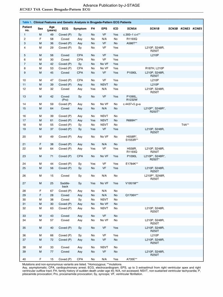
Methods and Results: Forty consecutive patients with Brugada-pattern electrocardiogram (ECG) underwent com-prehensive genetic analysis of BrS-causing genes including SCN5A, SCN1B, SCN3B, CACNA1C, CACNB2, KCNE3 and KCNE5. Besides identifying 8 SCN5A mutations in the present cohort, a KCNE3 T4A mutation was found in a 55-year-old male patient who had experienced several episodes of syncope. A head-up tilt test during passive tilt provoked both hypotension and bradycardia, followed by syncope. He was therefore diagnosed with neurally mediated syncope (NMS). To characterize the functional consequence of the mutant, electrophysiological experiments using whole-cell patch-clamp methods and computer simulations using human right ventricular wall model were carried out. It was found that KCNE3 T4A increased Ito recapitulated by heterologously coexpressing Kv4.3 + KChIP2b + KCNE3-wild type or KCNE3-T4A in CHO cells.
Conclusions: A KCNE3 T4A mutation was identified in a Japanese patient presenting Brugada-pattern ECG and NMS. Its functional consequence was the gain of function of Ito, which could underlie the pathogenesis of Brugada-pattern ECG. The data provide novel insights into the genetic basis of Japanese BrS.
Key Words: Brugada syndrome; Ito; KCNE3; Mutation; Neurally mediated syncope
Advance Publication by-J-STAGE
NAKAJIMA T et al.
cordial ECG leads (Brugada-pattern ECG) in BrS.14 Further augmentation of the AP notch in epimyocardium causes the loss of AP plateau phase (dome), which consequently leads to arrhythmogenesis in BrS defined as phase 2 reentry.14 Ito in human ventricle is thought to be consist of α-subunit Kv4.3 (encoded by KCND3) and β-subunits including Kv channel interacting protein (KChIP), KCNE, diaminopeptidyl transfer-ase-like protein (DPP) and Kvβ families.16–24 Accordingly, mu-tations in the related genes that increase Ito may underlie the pathogenesis of BrS.
KCNE3, 1 member of the KCNE gene family (KCNE1–5), modulates the function of Kv4.3 as an inhibitory β-subunit.19,20 Recently, Delpón et al identified a KCNE3 R99H mutation in 1 BrS family.6 Functional analysis using the heterologous ex-pression system that recapitulates Ito by coexpression of Kv4.3 with KCNE3-wild type (WT)/R99H showed that KCNE3 R99H causes a gain of function of Ito by a dominant-positive effect, thus precipitating the development of BrS.6 Moreover, muta-tions in Kv4.3 and KCNE5, which also functions as an inhibi-tory β-subunit of Ito, were identified in BrS patients.9,10 Func-tional analysis of these mutations showed that they increase Ito.9,10 Therefore, it was established that the gain of function of Ito by mutations in genes that encode Ito could be one of the causes of BrS.
We have previously reported the identification of 8 muta-tions in SCN5A among 30 consecutive Japanese patients with Brugada-pattern ECG.25 Considering that the genetic hetero-geneity of BrS is poorly elucidated in Japan,10,26,27 we further conducted genetic screening of BrS-causing genes among the 30 consecutive patients and another 10 new patients with Brugada-pattern ECG, and identified a KCNE3 T4A mutation in a patient presenting with neurally mediated syncope (NMS). In the present study, we describe the clinical phenotype of the KCNE3 T4A carrier, and characterize the functional conse-quence of the Ito recapitulated by heterologously coexpressing Kv4.3 + KChIP2b + KCNE3-T4A in CHO cells. Furthermore, we performed computer simulations based on the Ito obtained in electrophysiological recordings, and showed that KCNE3-T4A recapitulated the ECG phenotype.
MethodsSubjectsThe present subjects were 40 consecutive patients (probands; 35 male, 47±16 years of age) with Brugada-pattern ECG who were referred to Gunma University Hospital between April 2002 and September 2010. All patients, except for patient 27, presented with coved-type ST-elevation in the right precordial ECG leads with or without provocation of Na channel blocker (pilsicainide: 1 mg/kg, or procainamide 5 mg/kg), although it is still under debate whether patients with drug-induced Brugada-pattern ECG have poor prognosis.28,29 Echocardiography and conventional left catheterization, if performed, indicated no structural heart disease in all the patients. Thirty-two patients underwent electrophysiological assessment. Up to 3 extra stim-uli (minimum coupling interval: 180 ms) were delivered from 2 ventricular sites (right ventricular apex and right ventricular outflow tract). A head-up tilt (HUT) test was performed using the same protocol as described previously.30 Clinical features of the subjects are listed in Table 1.
Genetic AnalysisAfter obtaining appropriate approval from the institution re-view board and written informed consent from the patient, ge-nomic DNA was extracted from peripheral blood lymphocytes
using the standard protocol of the QIAamp DNA Blood Midi Kit (QIAGEN, Hilden, Germany). All coding exons of SCN5A, SCN1B, KCNE3, SCN3B and KCNE5, and their splice sites were amplified on polymerase chain reaction (PCR) using primers flanking the intronic sequences as reported previous-ly.1,5–7,25,31 The PCR products were purified and directly se-quenced using an ABI PRISM 3130 Genetic Analyzer (Ap-plied Biosystems, Foster City, CA, USA). Regarding patient 18, CACNA1C and CACNB2 were also analyzed.4 The muta-tion was analyzed twice on independent PCR amplification and sequencing. KCNE3 T4A was not identified in 528 control alleles.
Heterologous Expression of hKv4.3 and β-Subunits in CHO CellsFull-length cDNA fragment of WT KCNE3 in pCR3.1 vector was subcloned into pIRES-CD8 vector that is useful in cell selection. The KCNE3 mutant (T4A) was constructed using a Quick Change II XL site-directed mutagenesis kit according to the manufacturer’s instructions (Stratagene, La Jolla, CA, USA) and subcloned to the same vector. The KCNE3 mutant was fully sequenced (ABI PRISM 3130 Genetic analyzer) to ensure fidelity. Full-length cDNA encoding the short isoform of human Kv4.3 (hKv4.3) subcloned into the pIRES-GFP (Clontech, Palo Alto, CA, USA) expression vector was kind-ly provided by Dr GF Tomaselli (Johns Hopkins University). Full-length cDNA encoding Kv channel-interacting protein 2b (KChIP2b) subcloned into the PCMV-IRS expression vector was a kind gift from Dr GN Tseng (Virginia Commonwealth University). Kv4.3 was transiently transfected into CHO cells together with KChIP2b and KCNE3-WT (or T4A) cDNA at equimolar ratio (Kv4.3, 1.0 μg; KChIP2b, 1.0 μg; KCNE3, 1.0 μg) using Lipofectamine (Invitrogen Life Technologies, Carlsbad, CA, USA) according to the manufacturer’s instruc-tions. In a subset of experiments, 0.5 μg KCNE3-WT and 0.5 μg KCNE3-T4A were co-transfected into cells with 1.0 μg Kv4.3 and 1.0 μg KChIP2b. The transfected cells were then cultured in Ham’s F-12 medium (Nakalai Tesque, Kyoto, Japan) as de-scribed previously.24
Electrophysiologic Recording and Data AnalysisAfter 48 h of transfection, a coverslip with cells was transferred to a 0.5-ml bath chamber at 25°C on an inverted microscope stage and perfused at 1–2 ml/min with extracellular solution containing the following (in mmol/L): 140 NaCl, 5.4 KCl, 1.8 CaCl2, 0.5 MgCl2, 0.33 NaH2PO4, 5.5 glucose, and 5.0 HEPES; pH 7.4 with NaOH. Cells that emitted green fluorescence were chosen for patch-clamp experiments. If coexpressed with KCNE3 (or its mutant), the cells were incubated with polysty-rene microbeads precoated with anti-CD8 antibody (Dynabeads M450, Dynal, Norway) for 15 min. In these cases, cells that emitted green fluorescence and had attached beads were cho-sen for electrophysiologic recording. Whole-cell membrane currents were recorded with an EPC-8 patch-clamp amplifier (HEKA, Lambrecht, Germany), and data were low-pass filtered at 1 kHz, acquired at 5 kHz through an LIH-1600 analog-to-digital converter (HEKA), and stored on hard disk using Puls-eFit software (HEKA). Patch pipettes had a resistance of 2.5–5.0 mol/LΩ when filled with the following pipette solution (in mmol/L): 70.0 potassium aspartate, 50.0 KCl, 10.0 KH2PO4, 1.0 MgSO4, 3.0 Na2-ATP (Sigma, Japan, Tokyo), 0.1 Li2-GTP (Roche Diagnostics, Mannheim, Germany), 5.0 EGTA, and 5.0 HEPES (pH 7.2).
Whole cell currents were elicited in a series of depolarizing voltage steps from a holding potential of –80 mV. The time
Advance Publication by-J-STAGE
KCNE3 T4A Causes Brugada-Pattern ECG
Table 1. Clinical Features and Genetic Analysis in Brugada-Pattern ECG Patients
Patient no. Sex Age
(years) ECG Symptom FH EPS ICD SCN5A SCN1B SCN3B KCNE3 KCNE5
1 M 49 Coved (P) Sy No VF Yes c.393–1 c>t**
2 F 58 Coved Asy No N/A No R1193Q
3 M 36 Coved (P) Asy No VF No A586T**
4 M 29 Coved (P) Sy No VF Yes L210P, S248R, R250T
5 M 56 Coved CPA No VF Yes L210P
6 M 30 Coved CPA No VF Yes
7 M 42 Coved (P) Sy No No VF Yes
8 M 53 Coved (P) CPA No No VF Yes R187H, L210P
9 M 45 Coved CPA No VF Yes P1090L L210P, S248R, R250T
10 M 47 Coved (P) CPA No VF Yes L210P
11 M 39 Coved (P) Asy No NSVT No L210P
12 M 32 Coved Asy Yes N/A Yes L210P, S248R, R250T
13 M 42 Coved (Pro)
Sy No VF Yes P1090L, R1232W
14 M 59 Coved (P) Asy No No VF No c.4437+5 g>a
15 M 64 Coved Asy No N/A No L210P*, S248R*, R250T*
16 M 39 Coved (P) Asy No NSVT No
17 M 61 Coved (P) Asy Yes NSVT No R689H**
18 M 55 Coved (P) Sy No NSVT No T4A**
19 M 37 Coved (P) Sy Yes VF Yes L210P, S248R, R250T
20 M 49 Coved (P) Asy No No VF No H558R*, S1553R**
21 F 38 Coved (P) Asy No N/A No
22 M 64 Coved (P) Asy Yes VF Yes H558R, R1193Q
L210P, S248R, R250T
23 M 71 Coved (P) CPA No No VF Yes P1090L L210P*, S248R*, R250T*
24 M 44 Coved (P) Sy Yes VF Yes E1784K**
25 M 56 Coved (P) Sy No VF Yes L210P, S248R, R250T
26 M 15 Coved Sy No N/A No L210P*, S248R, R250T
27 M 25 Saddle-back
Sy Yes No VF Yes V1951M**
28 F 67 Coved (P) Asy No N/A No
29 F 28 Coved Asy No N/A No Q1706H**
30 M 38 Coved Sy No NSVT No
31 M 30 Coved (P) Asy No No VF No
32 M 63 Coved (P) Asy No NSVT No L210P, S248R, R250T
33 M 43 Coved Asy No VF No
34 M 57 Coved Asy No No VF No L210P, S248R, R250T
35 M 40 Coved (P) Sy No VF Yes L210P, S248R, R250T
36 M 66 Coved (P) Sy No VF Yes L210P
37 M 72 Coved (P) Asy No VF No L210P, S248R, R250T
38 M 33 Coved Asy No NSVT No
39 M 77 Coved Asy No VF No L210P, S248R, R250T
40 F 15 Coved (P) CPA No N/A Yes A735E**
Mutations and non-synonymous variants are listed. *Homozygous; **mutations.Asy, asymptomatic; CPA, cardiopulmonary arrest; ECG, electrocardiogram; EPS, up to 3 extrastimuli from right ventricular apex and right ventricular outflow tract; FH, family history of sudden death under age 45; N/A, not accessed; NSVT, non-sustained ventricular tachycardia; P, pilsicainide provocation; Pro, procainamide provocation; Sy, syncope; VF, ventricular fibrillation.
Advance Publication by-J-STAGE

NAKAJIMA T et al.
interval between each voltage pulse was 10 s. The difference between the peak current amplitude and the current at the end of a test pulse was referred to as the transient outward current. To control for cell size variability, currents are expressed as densities (pA/pF) as described previously.24 Steady-state acti-vation curves were obtained by plotting the normalized con-ductance as a function of peak outward potentials. Steady-state inactivation curves were generated by a standard 2-pulse pro-tocol with a conditioning pulse of 500 ms and obtained by plot-ting the normalized current as a function of the test potential. Steady-state inactivation/activation kinetics were fitted to the following Boltzmann equation:
Y(V) = 1 ⁄ (1 + exp[(V1/2 – V) / k]),
where Y = normalized conductance or current, V1/2 = potential for half-maximum inactivation or activation, respectively, and k = slope factor.
Data relative to inactivation time constants, time to peak, and mean current levels were obtained using current data re-corded at +50 mV. Recovery from inactivation was assessed using a standard paired-pulse protocol: a 1-s test pulse to +50 mV (P1) followed by a variable recovery interval at –80 mV and then a second test pulse to +50 mV (P2). Both the inactivation time constants and the time constant for recovery from inacti-vation were determined by fitting the data to a single exponen-tial:
I(t) (or P2/P1) = A + Bexp(–t / τ),
where I(t) = current amplitude at time t, A and B = constants, and τ = inactivation time constant or time constant for recovery from inactivation. For measurement of recovery from inacti-vation, the plot of P2/P1 instead of I(t) was used.
All data are given as mean ± SEM. Statistical comparisons between 2 groups were analyzed using Student’s unpaired t-test. Comparisons among multiple groups were analyzed using
analysis of variance followed by Dunnett test. P<0.05 was con-sidered significant.
Computer SimulationTo confirm the exact role of the KCNE3 T4A mutation, we conducted simulations of paced propagation in a 0.5-cm 1-D bidomain myocardial model with transverse conductivity, mim-icking transmural section of right ventricular wall. Membrane kinetics were represented by the Priebe-Beuckelmann human ventricular model,32 of which original Ito was replaced by the Ito with KCNE3-WT or KCNE3-T4A mutation obtained in electrophysiologic recording.
To obtain the transmural gradient in the right ventricular wall, we defined endocardial and epicardial tissues as each of length 0.25 cm, and we set the conductances of the slowly ac-tivating component of the delayed rectifier potassium channel (IKs), the inward rectifier potassium channel (IK1), and Ito in the endocardial layers to 46%, 82%, and 29%, respectively, of those in the epicardial layers. Pacing stimuli of 2 ms and strength twice-diastolic threshold were applied transmembranously to the endocardial end at a cycle length of 1,000 ms. The time and spatial discretization steps were 10 μs and 50 μm, respectively. Other model parameters and the numerical approach have been described elsewhere.33
ResultsGenetic AnalysisWe conducted comprehensive genetic analysis of BrS-causing genes including SCN5A, SCN1B, KCNE3, SCN3B and KCNE5, among 40 consecutive patients with Brugada-pattern ECG. Besides identifying 8 SCN5A mutations in the present cohort, we also found a T4A mutation in KCNE3 in the patient 18 who had no mutations in the other genes (including CACNA1C and CACNB2) associated with BrS (Figure 1A; Table 1).
Figure 1. (A) Direct sequencing of the KCNE3 gene in the index patient. Nucleotide and amino acid substitutions are indicated below. (B) Twelve-lead ECG recorded 2 months before admission. (C) V1–3 lead electrocardiogram before (baseline) and after pilsicainide provocation (Pilsicainide). ics, intercostal space.
Advance Publication by-J-STAGE
KCNE3 T4A Causes Brugada-Pattern ECG
Clinical PresentationA 55-year-old man (patient 18) was referred to hospital to examine the cause of syncope. He had experienced several epi-sodes of syncope under specific conditions, such as when sit-ting at a funeral, and standing up after drinking alcohol, since his 30 s. He had no previous history of illness except for syn-copal episodes, and no family history of sudden cardiac death. A physical examination, chest X-ray, and blood test showed no remarkable abnormalities. His 12-lead ECG, recorded 2 months before admission, showed saddle-back-type ST-seg-ment elevation in the right precordial ECG leads (Figure 1B). The QTc interval was 414 ms. A coved-type ST-segment ele-vation in the right precordial ECG leads at the second inter-costal space appeared after provocation with pilsicainide (Figure 1C). Signal-averaged ECG showed no late potentials. Transthoracic echocardiography showed no apparent structural heart disease.
The patient underwent electrophysiological assessment. Up to 3 extrastimuli induced non-sustained polymorphic ventricu-lar tachycardia, but not ventricular fibrillation. An HUT test was performed because syncopal episodes had occurred under specific conditions that could evoke NMS. The HUT test dur-ing passive tilt provoked both hypotension and bradycardia, followed by syncope. Therefore, the patient was diagnosed as having NMS. The patient was not prescribed medication or implanted with an implantable cardioverter defibrillator.
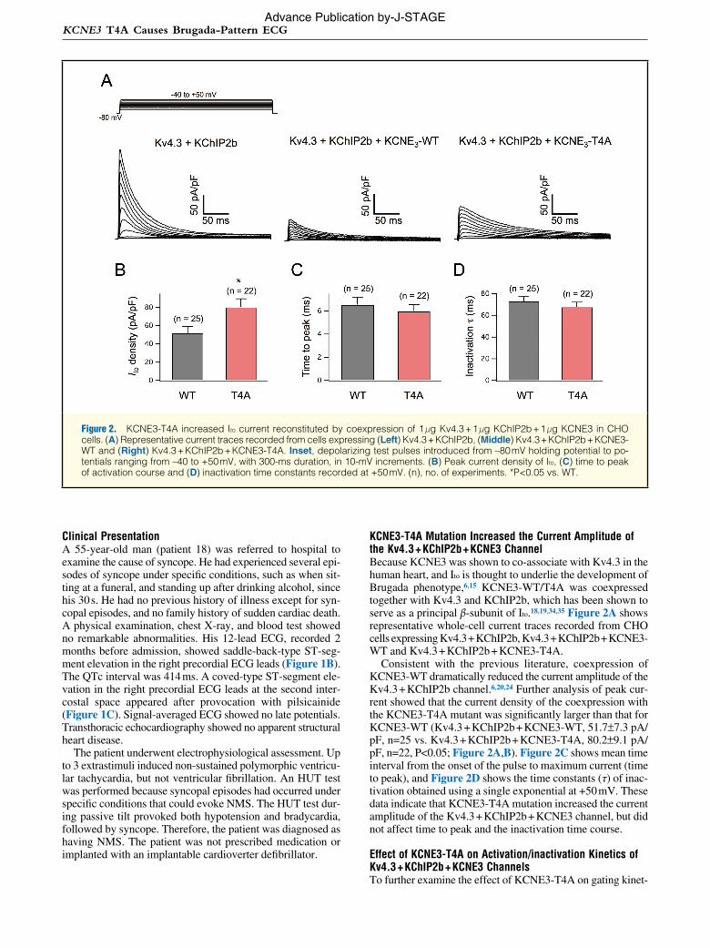
KCNE3-T4A Mutation Increased the Current Amplitude of the Kv4.3 + KChIP2b + KCNE3 ChannelBecause KCNE3 was shown to co-associate with Kv4.3 in the human heart, and Ito is thought to underlie the development of Brugada phenotype,6,15 KCNE3-WT/T4A was coexpressed together with Kv4.3 and KChIP2b, which has been shown to serve as a principal β-subunit of Ito.18,19,34,35 Figure 2A shows representative whole-cell current traces recorded from CHO cells expressing Kv4.3 + KChIP2b, Kv4.3 + KChIP2b + KCNE3-WT and Kv4.3 + KChIP2b + KCNE3-T4A.
Consistent with the previous literature, coexpression of KCNE3-WT dramatically reduced the current amplitude of the Kv4.3 + KChIP2b channel.6,20,24 Further analysis of peak cur-rent showed that the current density of the coexpression with the KCNE3-T4A mutant was significantly larger than that for KCNE3-WT (Kv4.3 + KChIP2b + KCNE3-WT, 51.7±7.3 pA/pF, n=25 vs. Kv4.3 + KChIP2b + KCNE3-T4A, 80.2±9.1 pA/pF, n=22, P<0.05; Figure 2A,B). Figure 2C shows mean time interval from the onset of the pulse to maximum current (time to peak), and Figure 2D shows the time constants (τ) of inac-tivation obtained using a single exponential at +50 mV. These data indicate that KCNE3-T4A mutation increased the current amplitude of the Kv4.3 + KChIP2b + KCNE3 channel, but did not affect time to peak and the inactivation time course.
Effect of KCNE3-T4A on Activation/inactivation Kinetics of Kv4.3 + KChIP2b + KCNE3 ChannelsTo further examine the effect of KCNE3-T4A on gating kinet-
Figure 2. KCNE3-T4A increased Ito current reconstituted by coexpression of 1 μg Kv4.3 + 1 μg KChIP2b + 1 μg KCNE3 in CHO cells. (A) Representative current traces recorded from cells expressing (Left) Kv4.3 + KChIP2b, (Middle) Kv4.3 + KChIP2b + KCNE3-WT and (Right) Kv4.3 + KChIP2b + KCNE3-T4A. Inset, depolarizing test pulses introduced from –80 mV holding potential to po-tentials ranging from –40 to +50 mV, with 300-ms duration, in 10-mV increments. (B) Peak current density of Ito, (C) time to peak of activation course and (D) inactivation time constants recorded at +50 mV. (n), no. of experiments. *P<0.05 vs. WT.
Advance Publication by-J-STAGE
NAKAJIMA T et al.
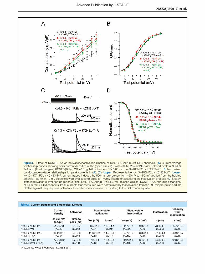
Figure 3. Effect of KCNE3-T4A on activation/inactivation kinetics of Kv4.3 + KChIP2b + KCNE3 channels. (A) Current–voltage relationship curves showing peak current densities of the (open circles) Kv4.3 + KChIP2b + KCNE3-WT, (closed circles) KCNE3-T4A and (filled triangles) KCNE3-(0.5 μg WT +0.5 μg T4A) channels. *P<0.05 vs. Kv4.3 + KChIP2b + KCNE3-WT. (B) Normalized conductance-voltage relationships for peak currents in (A). (C) (Upper) Representative Kv4.3 + KChIP2b + KCNE3-WT, (Lower) Kv4.3 + KChIP2b + KCNE3-T4A current traces induced by 500-ms pre-pulses from –90 mV to +50 mV applied from the holding potential –80 mV in 10-mV steps followed by a second pulse to +40 mV (fixed) for assessing the inactivation process. (D) Steady-state inactivation curves for the (open circles) Kv4.3 + KChIP2b + KCNE3-WT, (closed circles) KCNE3-T4A, and (filled triangles) KCNE3-(WT + T4A) channels. Peak currents thus measured were normalized by that obtained from the –90 mV pre-pulse and are plotted against the pre-pulse potentials. Smooth curves were drawn by fitting to the Boltzmann equation.
Table 2. Current Density and Biophysical Kinetics
Current density Activation Steady-state
activation Steady-state inactivation Inactivation
Recovery from
inactivation
At + 50 mV (pA/pF)
Time to peak (ms) V1/2 (mV) k (mV) V1/2 (mV) k (mV) τ (ms) τ (ms)
Kv4.3 + KChIP2b + KCNE3-WT
51.7±7.3 (n=25)
6.6±0.7 (n=25)
–6.5±2.0 (n=21)
17.2±1.1 (n=21)
–52.7±1.7 (n=22)
–9.9±1.7 (n=22)
73.0±4.2 (n=25)
65.7±10.2 (n=9)
Kv4.3 + KChIP2b + KCNE3-T4A
80.2±9.1† (n=22)
6.0±0.6 (n=22)
–11.6±1.2† (n=19)
14.2±0.8 (n=19)
–54.7±1.6 (n=15)
–8.8±2.1 (n=15)
67.1±4.1 (n=22)
66.9±10.7 (n=9)
Kv4.3 + KChIP2b + KCNE3-(WT + T4A)
77.7±8.6† (n=11)
6.7±0.8 (n=11)
–7.2±1.1 (n=10)
15.4±0.8 (n=10)
–52.2±2.0 (n=10)
–6.1±1.1 (n=10)
64.3±9.8 (n=11)
72.8±12.6 (n=6)
†P<0.05 vs. Kv4.3 + KChIP2b + KCNE3-WT.
Advance Publication by-J-STAGE

KCNE3 T4A Causes Brugada-Pattern ECG
ics of Kv4.3 + KChIP2b + KCNE3 channel, we assessed the current-voltage (I–V) relationship of the Kv4.3 + KChIP2b + KCNE3-T4A and Kv4.3 + KChIP2b + KCNE3-WT + KCNE3-T4A channels. Coexpression of KCNE3-T4A (at –20 to +50 mV) or KCNE3-WT + KCNE3-T4A (at 0 to +50 mV) sig-nificantly increased peak current densities (Figure 3A; Table 2). Meanwhile, coexpression of KCNE3-T4A, but not KCNE3-(T4A + WT), also caused a negative shift (approximately –5 mV) of voltage dependence of steady-state activation as assessed by plotting the normalized conductance as a function of test potentials (Figure 3B; Table 2).
Figure 3C shows the representative current traces elicited by a double-step pulse method (inset) used to evaluate steady-state inactivation. Peak currents recorded at various levels of pre-pulse (Figure 3C) were normalized by that measured after a 500-ms pre-pulse at –90 mV and plotted as a function of pre-pulse test potentials (Figure 3D). Coexpression of KCNE3-T4A or KCNE3-(WT + T4A) with Kv4.3 + KChIP2b did not significantly modify the steady-state inactivation of Kv4.3 + KChIP2b + KCNE3 channels (Figure 3D; Table 2).
Because the changes in the time course of reactivation can also affect Ito current, a double-pulse protocol (Figure 4A) was used to test the effect of KCNE3-T4A or KCNE3-(WT + T4A) coexpression on the time course for recovery from inac-tivation. Figures 4A,B shows the representative current traces for coexpression of KCNE3-WT and KCNE3-T4A. Figure 4C shows the time courses of recovery of KCNE3-WT, KCNE3-T4A and KCNE3-(WT + T4A) coexpression together with
Kv4.3 + KChIP2b. Time constants (τ) of recovery from inac-tivation are listed in Table 2. Coexpression of KCNE3-T4A or KCNE3-(WT + T4A) did not affect the time course of re-covery from inactivation.
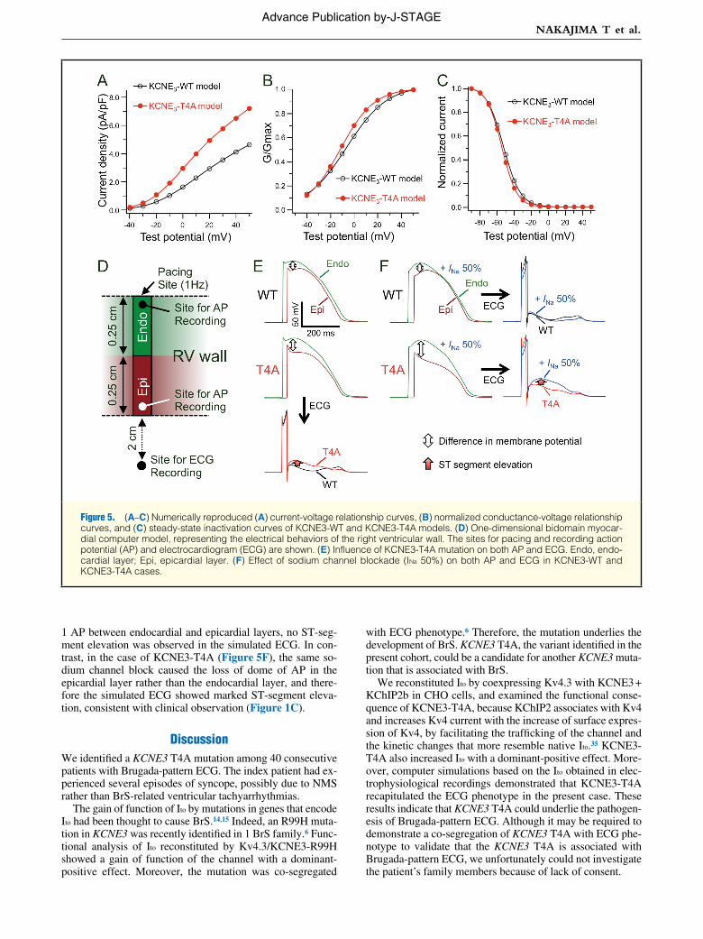
Phenotype of KCNE3-T4A Mutation in a Simulated Human Right Ventricular Wall ModelTo clarify whether the gain of function of Ito resulting from KCNE3-T4A mutation is indeed responsible for the Brugada-pattern ECG, we performed computer simulations using the 1-D myocardial model of human right ventricular wall. Based on the Ito obtained in electrophysiologic recording (Figures 3A,B,D), we numerically reproduced the current-volt-age relationship curves (Figure 5A), the normalized conduc-tance-voltage relationship curves (Figure 5B), and the steady-state inactivation curves (Figure 5C) of the KCNE3-WT and KCNE3-T4A channels. The numerically reproduced Ito was incorporated into the 1-D right ventricular wall model, con-sisting of endocardial and epicardial layers (Figure 5D). We found that the difference in the phase 2 AP between endocar-dial and epicardial layers in the KCNE3-T4A model was larg-er than that in the KCNE3-WT model, and therefore the simu-lated right precordial ECG showed ST-segment elevation in the case of KCNE3-T4A (Figure 5E).
To examine the adequacy of the KCNE3-T4A model, we additionally conducted simulations with sodium channel block-ade. In the case of KCNE3-WT (Figure 5F), because sodium channel block (INa 50%) did not increase the difference in phase
Figure 4. Effects of KCNE3-T4A on re-covery from inactivation of the Kv4.3 + KChIP2b + KCNE3 channel. A double step-pulse method was used to as-sess the recovery from inactivation. (A, inset) Protocol. A 1-s test pulse to +50 mV (P1) followed by a variable interval at –80 mV, then a second test pulse to +50 mV (P2). (A,B) Represen-tative results from cells transfected with (A) Kv4.3 + KChIP2b + KCNE3-WT and (B) Kv4.3 + KChIP2b + KCNE3-T4A. (C) The time course of recovery of the (open circles) Kv4.3 + KChIP2b + KCNE3- WT, (closed circles) KCNE3-T4A, and (filled triangles) KCNE3-(WT + T4A) channels. The ratio to peak current amplitude (P2/P1) is plotted as a func-tion of the inter-pulse (P1–P2) interval.
Advance Publication by-J-STAGE
NAKAJIMA T et al.
1 AP between endocardial and epicardial layers, no ST-seg-ment elevation was observed in the simulated ECG. In con-trast, in the case of KCNE3-T4A (Figure 5F), the same so-dium channel block caused the loss of dome of AP in the epicardial layer rather than the endocardial layer, and there-fore the simulated ECG showed marked ST-segment eleva-tion, consistent with clinical observation (Figure 1C).
DiscussionWe identified a KCNE3 T4A mutation among 40 consecutive patients with Brugada-pattern ECG. The index patient had ex-perienced several episodes of syncope, possibly due to NMS rather than BrS-related ventricular tachyarrhythmias.
The gain of function of Ito by mutations in genes that encode Ito had been thought to cause BrS.14,15 Indeed, an R99H muta-tion in KCNE3 was recently identified in 1 BrS family.6 Func-tional analysis of Ito reconstituted by Kv4.3/KCNE3-R99H showed a gain of function of the channel with a dominant-positive effect. Moreover, the mutation was co-segregated
with ECG phenotype.6 Therefore, the mutation underlies the development of BrS. KCNE3 T4A, the variant identified in the present cohort, could be a candidate for another KCNE3 muta-tion that is associated with BrS.
We reconstituted Ito by coexpressing Kv4.3 with KCNE3 + KChIP2b in CHO cells, and examined the functional conse-quence of KCNE3-T4A, because KChIP2 associates with Kv4 and increases Kv4 current with the increase of surface expres-sion of Kv4, by facilitating the trafficking of the channel and the kinetic changes that more resemble native Ito.35 KCNE3-T4A also increased Ito with a dominant-positive effect. More-over, computer simulations based on the Ito obtained in elec-trophysiological recordings demonstrated that KCNE3-T4A recapitulated the ECG phenotype in the present case. These results indicate that KCNE3 T4A could underlie the pathogen-esis of Brugada-pattern ECG. Although it may be required to demonstrate a co-segregation of KCNE3 T4A with ECG phe-notype to validate that the KCNE3 T4A is associated with Brugada-pattern ECG, we unfortunately could not investigate the patient’s family members because of lack of consent.
Figure 5. (A–C) Numerically reproduced (A) current-voltage relationship curves, (B) normalized conductance-voltage relationship curves, and (C) steady-state inactivation curves of KCNE3-WT and KCNE3-T4A models. (D) One-dimensional bidomain myocar-dial computer model, representing the electrical behaviors of the right ventricular wall. The sites for pacing and recording action potential (AP) and electrocardiogram (ECG) are shown. (E) Influence of KCNE3-T4A mutation on both AP and ECG. Endo, endo-cardial layer; Epi, epicardial layer. (F) Effect of sodium channel blockade (INa 50%) on both AP and ECG in KCNE3-WT and KCNE3-T4A cases.
Advance Publication by-J-STAGE
KCNE3 T4A Causes Brugada-Pattern ECG
The precise molecular mechanisms of the reverse of KCNE3-induced suppression of Kv4.3 by KCNE3-T4A remain to be elucidated. Delpón et al demonstrated, using coimmunopre-cipitation techniques, that KCNE3 coassociates with Kv4.3 in human atrial myocardium and rat ventricular myocardium.6 In addition, Lundby and Olesen reported that KCNE3 has an inhibitory effect on Kv4.3 independent of the presence of KChIP2 in a heterologous expression system.20 Therefore, although we recapitulated Ito in the presence of KChIP2b, KCNE3-T4A might reverse the KCNE3-induced suppression of Kv4.3 in the absence of KChIP2b, as is the case with KCNE3-R99H reported by Delpón et al.6 Interestingly, Lundby and Olesen also reported that delayed injection of KCNE3 into Xenopus oocytes can almost completely inhibit Kv4.3 current, suggesting that KCNE3 transcription can act as a regulatory mechanism of the Kv4.3 current density.20 Further studies of the transcription, trafficking and turnover rate of K4.3 channel and channel complexes in the presence or absence of KChIP2b and KCNE3-WT/T4A, would be required to elucidate the mech-anisms of the reverse of KCNE3-induced suppression of Ito by KCNE3-T4A.
Yokokawa et al reported that 35% of patients with Brugada-pattern ECG had positive responses during the HUT test,30 demonstrating a high prevalence of NMS among individuals with Brugada-pattern ECG. Although the autonomic nervous system may play an important role in the development of both BrS and NMS, the precise pathophysiological link between BrS and NMS remains to be elucidated. The identification of a novel SCN5A Q55X mutation in a patient with Brugada-pat-tern ECG presenting as NMS and the expression of SCN5A in both myocardial cells and intracardiac ganglia raise the pos-sibility of a genetic association between BrS and NMS.36,37 KCNE3 (MiRP2) is also expressed in not only the heart but also the central nervous system, and it modulates delayed rec-tifier currents in mammalian neurons by forming native chan-nel complexes with Kv2.1 and Kv3.1b.38 Taken together, KCNE3 T4A may be associated with both phenotypes of BrS and NMS.
In contrast, we have previously reported that KCNE3 muta-tions (R99H and T4A) are associated with long QT syndrome (LQTS).39 A functional analysis of KCNQ1 + KCNE3-R99H coexpression demonstrated a reduction of the repolarizing po-tassium current, thus supporting the proposition that KCNE3 R99H could be a cause of LQTS. A functional analysis of KCNQ1 + KCNE3-T4A, however, could not demonstrate sig-nificant functional abnormalities. Regarding the present case, the QTc interval was not prolonged. Therefore, further studies would be necessary to establish the association between KCNE3 T4A and LQTS. Along with this line, 2 KCNE3 T4A carriers we previously reported had no apparent spontaneous ST-seg-ment elevation in the right precordial ECG leads.39 One car-rier was a 16-year-old boy, much younger than most affected patients, and another was an old woman. Pharmacological provocation tests were not performed to study whether they had Brugada-pattern ECG.
ConclusionsWe identified a KCNE3 T4A mutation in a Japanese patient with Brugada-pattern ECG presenting as NMS. Its functional consequence was the gain of function of Ito, which could un-derlie the pathogenesis of Brugada-pattern ECG. The data pro-vide novel insight into the genetic basis of Japanese BrS. Further studies are required to clarify whether the KCNE3 T4A mutation is also associated with NMS and/or LQTS.
AcknowledgmentsWe thank the patients for their participation in this study. We thank Takako Kobayashi and Yukiyo Tosaka for helping with the genetic anal-ysis. This work was supported, in part, by a Grant-in-Aid for Scientific Research from the Ministry of Education, Culture, Sports, Science and Technology (to T.N. and M.H.). This work was also supported by grants from the Uehara Memorial Foundation and the Ministry of Health, Labor and Welfare of Japan for Clinical Research on Measures for Intractable Diseases (to M.H.), and the National Natural Science Foundation of China (#81273501 to J.W. and W.G.D.).
DisclosuresConflict of interest to declare: None.
References 1. Chen Q, Kirsch GE, Zhang D, Brugada R, Brugada J, Brugada P, et al.
Genetic basis and molecular mechanism for idiopathic ventricular fi-brillation. Nature 1998; 392: 293 – 296.
2. London B, Michalec M, Mehdi H, Zhu X, Kerchner L, Sanyal S, et al. Mutation in glycerol-3-phosphate dehydrogenase 1 like gene (GPD1-L) decreases cardiac Na+ current and causes inherited arrhythmias. Circulation 2007; 116: 2260 – 2268.
3. Van Norstrand DW, Valdivia CR, Tester DJ, Ueda K, London B, Makielski JC, et al. Molecular and functional characterization of novel glycerol-3-phosphate dehydrogenase 1 like gene (GPD1-L) mutations in sudden infant death syndrome. Circulation 2007; 116: 2253 – 2259.
4. Antzelevitch C, Pollevick GD, Cordeiro JM, Casis O, Sanguinetti MC, Aizawa Y, et al. Loss-of-function mutations in the cardiac calcium channel underlie a new clinical entity characterized by ST-segment elevation, short QT intervals, and sudden cardiac death. Circulation 2007; 115: 442 – 449.
5. Watanabe H, Koopmann TT, Le Scouarnec S, Yang T, Ingram CR, Schott JJ, et al. Sodium channel beta1 subunit mutations associated with Brugada syndrome and cardiac conduction disease in humans. J Clin Invest 2008; 118: 2260 – 2268.
6. Delpón E, Cordeiro JM, Núñez L, Thomsen PE, Guerchicoff A, Pollevick GD, et al. Functional effects of KCNE3 mutation and its role in the development of Brugada syndrome. Circ Arrhythm Elec-trophysiol 2008; 1: 209 – 218.
7. Hu D, Barajas-Martinez H, Burashnikov E, Springer M, Wu Y, Varro A, et al. A mutation in the beta 3 subunit of the cardiac sodium chan-nel associated with Brugada ECG phenotype. Circ Cardiovasc Genet 2009; 2: 270 – 278.
8. Kattygnarath D, Maugenre S, Neyroud N, Balse E, Ichai C, Denjoy I, et al. MOG1: A new susceptibility gene for Brugada syndrome. Circ Cardiovasc Genet 2011; 4: 261 – 268.
9. Giudicessi JR, Ye D, Tester DJ, Crotti L, Mugione A, Nesterenko VV, et al. Transient outward current (I(to)) gain-of-function mutations in the KCND3-encoded Kv4.3 potassium channel and Brugada syn-drome. Heart Rhythm 2011; 8: 1024 – 1032.
10. Ohno S, Zankov DP, Ding WG, Itoh H, Makiyama T, Doi T, et al. KCNE5 (KCNE1L) variants are novel modulators of Brugada syn-drome and idiopathic ventricular fibrillation. Circ Arrhythm Electro-physiol 2011; 4: 352 – 361.
11. Barajas-Martinez H, Hu D, Ferrer T, Onetti CG, Wu Y, Burashnikov E, et al. Molecular genetic and functional association of Brugada and early repolarization syndromes with S422L missense mutation in KCNJ8. Heart Rhythm 2012; 9: 548 – 555.
12. Antzelevitch C. Genetic, molecular and cellular mechanisms under-lying the J wave syndromes. Circ J 2012; 76: 1054 – 1065.
13. Nabauer M, Beuckelmann DJ, Uberfuhr P, Steinbeck G. Regional differences in current density and rate-dependent properties of the transient outward current in subepicardial and subendocardial myo-cytes of human left ventricle. Circulation 1996; 93: 168 – 177.
14. Yan GX, Antzelevitch C. Cellular basis for the Brugada syndrome and other mechanisms of arrhythmogenesis associated with ST-segment elevation. Circulation 1999; 100: 1660 – 1666.
15. Antzelevitch C. Brugada syndrome. Pacing Clin Electrophysiol 2006; 29: 1130 – 1159.
16. Dixon JE, Shi W, Wang HS, McDonald C, Yu H, Wymore RS, et al. Role of the Kv4.3 K+ channel in ventricular muscle: A molecular correlate for the transient outward current. Circ Res 1996; 79: 659 – 668.
17. Kuo HC, Cheng CF, Clark RB, Lin JJ, Lin JL, Hoshijima M, et al. A defect in the Kv channel-interacting protein 2 (KChIP2) gene leads to a complete loss of I(to) and confers susceptibility to ventricular tachycardia. Cell 2001; 107: 801 – 813.
Advance Publication by-J-STAGE

NAKAJIMA T et al.
18. Decher N, Uyguner O, Scherer CR, Karaman B, Yuksel-Apak M, Busch AE, et al. hKChIP2 is a functional modifier of hKv4.3 potas-sium channels: Cloning and expression of a short hKChIP2 splice variant. Cardiovasc Res 2001; 52: 255 – 264.
19. Radicke S, Cotella D, Graf EM, Banse U, Jost N, Varro A, et al. Func-tional modulation of the transient outward current Ito by KCNE beta-subunits and regional distribution in human non-failing and failing hearts. Cardiovasc Res 2006; 71: 695 – 703.
20. Lundby A, Olesen SP. KCNE3 is an inhibitory subunit of the Kv4.3 potassium channel. Biochem Biophys Res Commun 2006; 346: 958 – 967.
21. Radicke S, Cotella D, Graf EM, Ravens U, Wettwer E. Expression and function of dipeptidyl-aminopeptidase-like protein 6 as a puta-tive beta-subunit of human cardiac transient outward current encoded by Kv4.3. J Physiol 2005; 565: 751 – 756.
22. Aimond F, Kwak SP, Rhodes KJ, Nerbonne JM. Accessory Kvbeta1 subunits differentially modulate the functional expression of voltage-gated K+ channels in mouse ventricular myocytes. Circ Res 2005; 96: 451 – 458.
23. Niwa N, Nerbonne JM. Molecular determinants of cardiac transient outward potassium current (I(to)) expression and regulation. J Mol Cell Cardiol 2010; 48: 12 – 25.
24. Wu J, Shimizu W, Ding WG, Ohno S, Toyoda F, Itoh H, et al. KCNE2 modulation of Kv4.3 current and its potential role in fatal rhythm disorders. Heart Rhythm 2010; 7: 199 – 205.
25. Nakajima T, Kaneko Y, Saito A, Irie T, Tange S, Iso T, et al. Identi-fication of six novel SCN5A mutations in Japanese patients with Brugada syndrome. Int Heart J 2011; 52: 27 – 31.
26. Ogawa R, Kishi R, Takagi A, Sakaue I, Takahashi H, Matsumoto N, et al. A novel microsatellite polymorphism of sodium channel beta1-subunit gene (SCN1B) may underlie abnormal cardiac excitation manifested by coved-type ST-elevation compatible with Brugada syn-drome in Japanese. Int J Clin Pharmacol Ther 2010; 48: 109 – 119.
27. Makiyama T, Akao M, Haruna Y, Tsuji K, Doi T, Ohno S, et al. Mutation analysis of the glycerol-3 phosphate dehydrogenase-1 like (GPD1L) gene in Japanese patients with Brugada syndrome. Circ J 2008; 72: 1705 – 1706.
28. Shimizu A. Is this a philosophic issue? Do patients with drug-induced Brugada type ECG have poor prognosis? (Pro) Circ J 2010; 74: 2455 – 2463.
29. Nishizaki M, Sakurada H, Yamawake N, Ueda-Tatsumoto A, Hiraoka M. Low risk for arrhythmic events in asymptomatic patients with drug-induced type 1 ECG. Do patients with drug-induced Brugada type ECG have poor prognosis? (Con) Circ J 2010; 74: 2464 – 2473.
30. Yokokawa M, Okamura H, Noda T, Satomi K, Suyama K, Kurita T, et al. Neurally mediated syncope as a cause of syncope in patients with Brugada electrocardiogram. J Cardiovasc Electrophysiol 2010; 21: 186 – 192.
31. Nakajima T, Kaneko Y, Irie T, Takahashi R, Kato T, Iijima T, et al. Compound and digenic heterozygosity in desmosome genes as a cause of arrhythmogenic right ventricular cardiomyopathy in Japanese pa-tients. Circ J 2012; 76: 737 – 743.
32. Priebe L, Beuckelmann DJ. Simulation study of cellular electric prop-erties in heart failure. Circ Res 1998; 82: 1206 – 1223.
33. Tsuji-Wakisaka K, Akao M, Ishii TM, Ashihara T, Makiyama T, Ohno S, et al. Identification and functional characterization of KCNQ1 muta-tions around the exon 7-intron 7 junction affecting the splicing pro-cess. Biochim Biophys Acta 2011; 1812: 1452 – 1459.
34. Wang S, Bondarenko VE, Qu Y, Morales MJ, Rasmusson RL, Strauss HC. Activation properties of Kv4.3 channels: Time, voltage and [K+]o dependence. J Physiol 2004; 557: 705 – 717.
35. An WF, Bowlby MR, Betty M, Cao J, Ling HP, Mendoza G, et al. Modulation of A-type potassium channels by a family of calcium sen-sors. Nature 2000; 403: 553 – 556.
36. Makita N, Sumitomo N, Watanabe I, Tsutsui H. Novel SCN5A mu-tation (Q55X) associated with age-dependent expression of Brugada syndrome presenting as neurally mediated syncope. Heart Rhythm 2007; 4: 516 – 519.
37. Scornik FS, Desai M, Brugada R, Guerchicoff A, Pollevick GD, Antzelevitch C, et al. Functional expression of “cardiac-type” Nav1.5 sodium channel in canine intracardiac ganglia. Heart Rhythm 2006; 3: 842 – 850.
38. McCrossan ZA, Lewis A, Panaghie G, Jordan PN, Christini DJ, Lerner DJ, et al. MinK-related peptide 2 modulates Kv2.1 and Kv3.1 potas-sium channels in mammalian brain. J Neurosci 2003; 23: 8077 – 8091.
39. Ohno S, Toyoda F, Zankov DP, Yoshida H, Makiyama T, Tsuji K, et al. Novel KCNE3 mutation reduces repolarizing potassium current and associated with long QT syndrome. Hum Mutat 2009; 30: 557 – 563.
Advance Publication by-J-STAGE