Embed Size (px)
Citation preview
1
KDOQI CLINICAL PRACTICE GUIDELINE FOR VASCULAR ACCESS: 2018
AJKD SUBMISSION DRAFT April 2019
2
DISCLAIMER
SECTION I: USE OF THE CLINICAL PRACTICE GUIDELINE
This Clinical Practice Guideline document is based upon the best information available as of April 2017*. It is designed to provide
information and assist decision-making. It is not intended to define a standard of care, and should not be construed as one, nor should
it be interpreted as prescribing an exclusive course of management. Variations in practice will inevitably and appropriately occur when
clinicians take into account the needs of individual patients, available resources, and limitations unique to an institution or type of
practice. Every health-care professional making use of these recommendations is responsible for evaluating the appropriateness of
applying them in the setting of any particular clinical situation. The recommendations for research contained within this document are
general and do not imply a specific protocol.
*Commissioned evidence review included articles published through April 2017. Consensus opinion statements use literature published
though August 2018.
SECTION II: DISCLOSURE
Kidney Disease Outcomes Quality Initiative (KDOQI) makes every effort to avoid any actual or reasonably perceived conflicts of interest
that may arise as a result of an outside relationship or a personal, professional, or business interest of a member of the Work Group. All
members of the Work Group are required to complete, sign, and submit a disclosure and attestation form showing all such
relationships that might be perceived or actual conflicts of interest. All reported information will be printed in the final publication and
are on file at the National Kidney Foundation (NKF).
3
Table of Contents
Table of Tables ................................................................................................................................................................................................... 5
Abbreviations and Acronyms .............................................................................................................................................................................. 6
Glossary ............................................................................................................................................................................................................. 7
CKD Nomenclature Used by KDOQI ................................................................................................................................................................... 11
Work Group Membership ................................................................................................................................................................................. 12
Organization Leadership ................................................................................................................................................................................... 13
Abstract ........................................................................................................................................................................................................... 14
Foreword ......................................................................................................................................................................................................... 14
Introduction ..................................................................................................................................................................................................... 15
Methods .......................................................................................................................................................................................................... 15
Summary of Guideline Statements .................................................................................................................................................................... 18
KDOQI Clinical Practice Guideline for Vascular Access ....................................................................................................................................... 33
GUIDELINE 1. PATIENT FIRST: ESKD LIFE-PLAN ............................................................................................................................................... 33
Statements: ESKD Life-Plan and Vascular Access Choice ............................................................................................................................................. 33
GUIDELINE 2. VASCULAR ACCESS TYPES ......................................................................................................................................................... 35
GUIDELINE 3. VASCULAR ACCESS LOCATIONS ................................................................................................................................................ 39
GUIDELINE 4. NOVEL MATERIALS AND AV-ACCESS ......................................................................................................................................... 42
GUIDELINE 5. CVC CONFIGURATION AND MATERIALS .................................................................................................................................... 44
GUIDELINE 6. TIMING, PREPARATION & PLANNING FOR CREATION/INSERTION OF DIALYSIS ACCESS .............................................................. 46
Statements for Vessel Preservation ............................................................................................................................................................................. 48
Statements for Multidisciplinary Team Approach ....................................................................................................................................................... 49
GUIDELINE 7. PATIENT AND VESSEL EXAMINATIONS PREOPERATIVE CONSIDERATIONS .................................................................................. 51
Statements for Patient Clinical Examination ................................................................................................................................................................ 51
Statements for Vessel Mapping for Vascular Access ................................................................................................................................................... 52
Statements for Optimal vessel size for artery and vein for AV-access creation .......................................................................................................... 52
GUIDELINE 8. AV-ACCESS CREATION .............................................................................................................................................................. 54
Statement: Use of Anesthesia for AV-access creation ................................................................................................................................................. 54
Statement: AV-Access Anastomotic Configuration and Apposition Methods ............................................................................................................. 54
Statement: AV-access Anastomotic Suture Technique ................................................................................................................................................ 54
Statements: Use of intra-operative assisted maneuvers for AV-access maturation ................................................................................................... 54
Statement: AV-access Anastomotic Suture Technique ................................................................................................................................................ 54
Statement: Use of Anesthesia for AV-access creation ................................................................................................................................................. 54
Statement: AV-Access Anastomotic Configuration and Apposition Methods ............................................................................................................. 55
Statements: Use of intra-operative assisted maneuvers for AV-access maturation ................................................................................................... 56
Statement: AV-access Anastomotic Suture Technique ................................................................................................................................................ 59
Statement: AV-access Anastomotic Suture Technique ................................................................................................................................................ 59
GUIDELINE 9. CATHETER INSERTION .............................................................................................................................................................. 61
Statements: Techniques and other considerations for placement .............................................................................................................................. 61
GUIDELINE 10. POST AV-ACCESS CREATION / CVC INSERTION CONSIDERATIONS ............................................................................................ 63
Statements: AV-access Early Post-operative Considerations (0-30 days) – Early AV-access complications ............................................................... 63
Statements: Post-operative AV- Access Maturation .................................................................................................................................................... 64
Statements: Timing of removal (CVC) .......................................................................................................................................................................... 67
GUIDELINE 11. VASCULAR ACCESS USE .......................................................................................................................................................... 69
Statement for Vascular Access General Monitoring .................................................................................................................................................... 69
Statements: AV-Access Cannulation ............................................................................................................................................................................ 69
Statements: Catheter system connect and disconnect procedure considerations ........................................................................................... 72
GUIDELINE 12 AV-ACCESS CANNULATION COMPLICATIONS ........................................................................................................................... 76
4
Statements: AV-Access Cannulation Complications .................................................................................................................................................... 76
GUIDELINE 13. AV-ACCESS FLOW DYSFUNCTION+ - MONITORING/SURVEILLANCE ........................................................................................... 78
Statements: Appropriate Use of Monitoring/Surveillance for AV-Access Flow Dysfunction ...................................................................................... 78
GUIDELINE 14. AV-ACCESS FLOW DYSFUNCTION+ - PREVENTION .................................................................................................................... 86
Statements for the Prevention of AV-Access (AVF or AVG) Flow Dysfunction+ .......................................................................................................... 86
GUIDELINE 15. AV-ACCESS FLOW DYSFUNCTION - TREATMENT ...................................................................................................................... 89
Statement: Radiographic Confirmation of Clinically Significant AV-access Stenosis ................................................................................................... 89
Statement: Treatment of Thrombosed AV-Access....................................................................................................................................................... 90
GUIDELINE 16. AV-ACCESS INFECTION ........................................................................................................................................................... 97
Statements for AV-Access Infections ........................................................................................................................................................................... 97
Monitoring and Prevention .......................................................................................................................................................................................... 97
GUIDELINE 17. AV-ACCESS ANEURYSMS ........................................................................................................................................................ 99
Statements for AV-access Aneurysms .......................................................................................................................................................................... 99
GUIDELINE 18. AV-ACCESS STEAL ................................................................................................................................................................ 103
Statements ................................................................................................................................................................................................................. 103
GUIDELINE 19. OTHER AV-ACCESS COMPLICATIONS ..................................................................................................................................... 105
Statement: Management of AVG seroma .................................................................................................................................................................. 105
Statement: Management of high flow AV-access ...................................................................................................................................................... 106
GUIDELINE 20. TREATMENT AND PREVENTION OF CVC COMPLICATIONS ..................................................................................................... 106
Statement for the Monitoring/Surveillance of catheter complications .................................................................................................................... 106
GUIDELINE 21. CATHETER-RELATED DYSFUNCTION ...................................................................................................................................... 107
Statement: Definition of CVC dysfunction ................................................................................................................................................................. 107
Statements: Pharmacologic Prevention of CVC* Dysfunction ................................................................................................................................... 108
GUIDELINE 22: TREATMENT AND MANAGEMENT OF CVC DYSFUNCTION ..................................................................................................... 112
Statements: Medical management of CVC dysfunction ............................................................................................................................................ 112
Statements: Mechanical management of CVC dysfunction ....................................................................................................................................... 114
GUIDELINE 23. CATHETER-RELATED INFECTION ........................................................................................................................................... 115
Statement of Definitions of Catheter-Related Infections .......................................................................................................................................... 115
GUIDELINE 24: PREVENTION OF CVC INFECTION .......................................................................................................................................... 118
Statement for Infection Surveillance Programs and Infection Control Teams .......................................................................................................... 118
Statement for the monitoring of catheter complications: Infection ......................................................................................................................... 118
Statements on Methods to Prevent CRBSI ................................................................................................................................................................. 119
GUIDELINE 25. TREATMENT OF CVC-RELATED INFECTION ............................................................................................................................ 123
Statement: Management of the patient with a catheter-related infection .............................................................................................................. 123
Statement: Management of the catheter in a patient with a catheter-related infection ......................................................................................... 123
GUIDELINE 26. OTHER VASCULAR ACCESS-RELATED COMPLICATIONS .......................................................................................................... 125
Statement for intervention of asymptomatic central venous stenosis ..................................................................................................................... 125
Statement for Investigation and treatment of symptomatic central venous stenosis .............................................................................................. 127
Statement for management of CVC fibrin sheath associated with clinical problems ............................................................................................... 128
RESEARCH RECOMMENDATIONS................................................................................................................................................................. 129
GOALS AND TARGETS ................................................................................................................................................................................. 138
Biographical and Disclosure Information ......................................................................................................................................................... 142
References ..................................................................................................................................................................................................... 145
5
Table of Tables
Table I1: Grade for Strength of Recommendation ............................................................................................................................................................ 17
Table I2: Grade for Quality of Evidence ............................................................................................................................................................................ 17
Table 2.1 Access Type Comparison Studies Reviewed ...................................................................................................................................................... 37
Table 3.1 Vessel location by distal to proximal sites ......................................................................................................................................................... 41
Table 6.1 Suggested indications for Creation/Insertion of a Vascular Access in Peritoneal Dialysis Patients .................................................................. 47
Table 7.1 Focused pre-operative physical exam for vascular access planning and creation ............................................................................................ 51
Table 11.1. Circumstances where buttonhole cannulation may be acceptable ............................................................................................................... 71
Table 11.2 Procedure for CVC connect and disconnect procedures ................................................................................................................................. 74
Table 13.2: Clinical indicators (signs and symptoms) found in AV-access monitoring, suggestive of an underlying clinically significant lesion ............. 83
Table 13.4 Performance and Agreement Beyond Chance between Physical Examination and Ultrasonography ........................................................... 85
Table 14.1 Primary Outcomes Fish Oil versus Placebo ..................................................................................................................................................... 87
Table 15.1 Outcomes Paclitaxel-Coated Balloon Angioplasty in AVGs and AVFs ............................................................................................................. 92
Table 15.2 Indications for Stent-Graft Use in AV-Access* ................................................................................................................................................ 94
Table 17.1: Physical examination findings that are clinically relevant to differentiate between aneurysm/pseudoaneurysm that do not require
urgent intervention and those of urgent concern. ......................................................................................................................................................... 102
Table 18.1 Strategies to Reduce the Incidence of AV-Access Steal................................................................................................................................. 103
Table 18.2 Clinical Predictors of AV-Access Steal ............................................................................................................................................................ 103
Table 18.3: Society for Vascular Surgery Reporting Standards for AV-access Steal{Sidawy, 2002 #517} ....................................................................... 104
Table 18.4 Treatment Options for AV-Access Steal ........................................................................................................................................................ 104
Table 23.1 Definitions of Catheter Related Blood Stream Infections ............................................................................................................................. 116
Table 24.1 Intraluminal Strategies: Effect of antibiotic and antimicrobial catheter lock solutions on catheter-related bloodstream infections (CRBSI):
summary of randomized clinical trials ............................................................................................................................................................................ 121
Table 26.1 Signs and Symptoms of Central Venous Stenosis ........................................................................................................................................ 125
Table of Potential Future Research for Hemodialysis Vascular Access and Related Topics ......................................................................................... 130
GOALS AND TARGETS .................................................................................................................................................................................................... 138
Table GT1. Interventions ............................................................................................................................................................................................... 140
6
Abbreviations and Acronyms
Vascular Access Acronyms and Abbreviations
ABIM American Board of Internal Medicine
ACGME Accreditation Council of Graduate Medical Education
ACPP access circuit primary patency
AEC Allogenic endothelial cells
aHR Adjusted hazard ratio
aOR Adjusted odds ratio
AUC Area under the curve
AV Arteriovenous
AV-access Arteriovenous access – Refers to both a hemodialysis arteriovenous fistula and arteriovenous graft
AVF Arteriovenous fistula
AVG Arteriovenous graft
BAM Balloon assisted maturation
BFR Blood flow rate
BP Blood pressure
CDC Centers for Disease Control and Prevention
CI Confidence interval
CKD Chronic kidney disease
CMS Centers for Medicare & Medicaid Services
CPG Clinical Practice Guideline
CQI Continuous quality improvement
CRBSI Catheter-related bloodstream infection
CrCl Creatinine clearance
CVC Central venous catheter; if referring to hemodialysis catheter, will assume tunneled, cuffed central venous catheter unless
otherwise stated
NT-CVC Non-tunneled, non-cuffed central venous catheter
CVD Cardiovascular disease
DDU Duplex Doppler ultrasound
DOPPS Dialysis Outcomes and Practice Patterns Study
DRIL Distal revascularization interval ligation
DVP Dynamic venous pressure
FDA Food and Drug Administration
GFR Glomerular filtration rate
HD Hemodialysis
HTN Hypertension
HR Hazard ratio
KDOQI Kidney Disease Outcomes Quality Initiative
KRT Kidney replacement therapy
NKF National Kidney Foundation
NS Not significant
PBA Primary balloon angioplasty
PD Peritoneal dialysis
PICC Peripherally inserted central catheter
PTA Percutaneous balloon angioplasty
PTFE Polytetrafluoroethylene
Qa Access blood flow
Qb Blood pump flow delivered to the dialyzer
7
QOL Quality of life
RCT Randomized controlled trial
RR Relative risk
rTPA Recombinant tissue plasminogen activator
SVC Superior vena cava
SVP – Static venous pressure
TAPP Treatment area primary patency
URR Urea reduction ratio
US Ultrasonography
USRDS United States Renal Data System
VA Vascular access
VAC Vascular access coordinator
VAT Vascular access team
Glossary
Acronecrosis: Gangrene occurring in the distal part of the extremities, usually fingertips and toes.
Anastomosis: A connection between an artery and a vein by surgical, endovascular, traumatic, or pathological means
Aneurysm: An abnormal blood-filled dilation of a blood vessel wall resulting from disease or trauma of the vessel wall.
Pseudoaneurysm: An abnormality (i.e. hole) in the vessel or prosthetic AV-access that is walled off or contained by the surrounding soft tissue.
Antibiotic lock: Interdialytic instillation and dwelling (“locking”) of an antibiotic containing solution into the lumen of a dialysis catheter.
Antimicrobial: Any agent capable of destroying or inhibiting the growth of microorganisms.
Antimicrobial lock: Interdialytic instillation and dwelling (“locking”) of an antimicrobial solution into the lumen of a dialysis catheter
Antiseptic: Any agent capable of preventing infection by inhibiting the growth of microorganisms.
AV-access abandonment – when a vascular access can no longer be used for one or two needle, prescribed dialysis as it may be unable to provide adequate flows and or is deemed unsafe for the patient, and the associated problem cannot be corrected by any intervention, including medical, surgical, or radiological interventions or rest.1 A checklist can be used to confirm abandonment as follows:
Please check the following:
Yes No
The fistula/graft does not have a pulsation (even with augmentation) at the anastomosis or the access body
The fistula/graft does not have a palpable thrill at the anastomosis or the access body
The fistula/graft does not have an audible bruit anywhere along the anastomosis or body (up to 10 cm from
the anastomosis)
A radiological intervention such as angioplasty, thrombolysis, stenting, embolization or other will not salvage
the access to be useable
A surgical revision will not salvage the access to be useable.
Note: A revision means that it is a revision of the current access and not a surgical procedure that will
effectively create new access
A reasonable effort has been made to improve the condition of the access in order for it to be used; for
example, adequate elevation and time for rest of an infiltrated fistula
The access is viable but there are complications that require the abandonment of the access e.g. high cardiac
output failure or severe steal syndrome
Other, indicate: _______________________________________
I confirm that the fistula or graft is officially abandoned for further use and is not safely salvageable
8
AV- access creation: the connection of an artery and vein for the purposes of establishing hemodialysis access
Cannulation: The insertion of cannulate (a needle with a lumen) or angiocaths into a vascular vessel.
Buttonhole technique cannulation: The cannulation into the exact same puncture site and needle track tunnel developed by repeated
cannulation at the same location, angle and depth between the skin and access vein. The scar tissue tunnel track allows the needle to
pass through to the outflow vessel of the fistula following the same path with each cannulation time. Typically used in autogenous AVF
and may be acceptable in grafts made of non-autogenous biologic material such as bovine. Should not be used for accessing AVG made
of synthetic material such as PTFE.
Constant-site technique cannulation – another term for buttonhole cannulation above. This should NOT be confused with “general
area” cannulation. General area cannulation is neither rope ladder or buttonhole cannulation, whereby new arterial and venous needle
insertions are chronically inserted within close proximity (e.g. millimeters) of prior insertions each time, i.e. always in the same general
areas. This poor technique leads to Av-access aneurysms and damage and should be avoided.
Catheter: A device providing access to the central veins or right atrium, permitting high volume flow rates.
Clinically significant lesion: is one that contributes to clinical signs and symptoms without other cause, with or without a sustained
change in surveillance measurements (e.g. change in blood flow (Qa) or venous pressures) in the dialysis access circuit. Such a lesion is
found during monitoring of vascular access (surveillance findings are supplementary).
Clinical Monitoring: Monitoring refers to the examination and evaluation of the access by means of physical examination or check to
detect clinical signs that suggest the presence of AV- access flow dysfunction, other dysfunction or pathology. These abnormal clinical
signs may include arm swelling, changes in the access bruit or thrill or prolonged bleeding post dialysis (See Tables 13.1 and
13.2). Supplementing this patient physical exam can include concurrent dialysis measures such as those indicating recirculation (when
needle placement is correctly spaced and placed) or other measures of reduced dialysis adequacy (e.g. urea reduction ratio or Kt/V), in
the absence of other contributing factors.
Complications
Thrombotic Flow – Related complications: those specifically related to the risk of or occurrence of thrombosis that leads to the
clinically important reduction in intra-access flow which threatens the required access patency to achieve prescribed dialysis
and/or results in clinical signs and symptoms (e.g. stenosis or thrombosis)
Non-thrombotic flow-related complications: such complications may or may not threaten flow or patency but are associated with
clinical signs and symptoms e.g. AV-access aneurysms, steal syndrome
Infectious Complications – any infection involving the vascular access (intraluminal/access, extraluminal/access, peri-access i.e.
cannulation or entry site) that results in clinically important infectious signs and symptoms
Cumulative Patency: a duration of time measuring intra-access patency that starts from the date of vascular access creation (AV-access)
or insertion (CVC) to the date of vascular access abandonment.
Diagnostic testing: Specialized testing that is prompted by some abnormality or other medical indication and that is undertaken to
diagnose the cause of the vascular access problem.
Dialysis usability – a dialysis access that can reliably and safely provide prescribed dialysis, per definition of mature fistula or graft
Distal revascularization—interval ligation (DRIL): A surgical procedure to reduce ischemia to the hand caused by steal syndrome. See
Figure 18.3
Dysfunction: AV-access or vascular access dysfunction has been replaced by 3 terms:
1) Thrombotic Flow-Related Complications; 2) Non-Thrombosis Flow-related; 3) Infectious
Exit site: The location on the skin that the catheter exits through the skin surface. Insertion site: the location at which the catheter
enters the vein, for example, the right internal jugular vein is the preferred insertion site.
Failure to mature – an access that, despite radiological or surgical intervention, cannot be used successfully for dialysis by 6 months
following its creation1

9
Fistula (plural, fistulae or fistulas): Autologous arteriovenous fistula, also referred to as native fistula.
Brescia-Cimino (radiocephalic) fistula: An autologous fistula constructed between the radial artery and the cephalic vein at the
wrist.
Endovascular fistula (or endoAVF): an autologous fistula created by endovascular techniques, originally described by
anastomosis of the proximal ulnar artery and proximal ulnar vein
Gracz fistula: An autologous fistula constructed between the brachial artery and a branch of the medial antecubital vein, the
perforating vein, below the elbow.
Snuff-box fistula: An autologous fistula constructed between a branch of the radial artery and an adjacent vein in the anatomic
snuff box of the hand.
Fistula maturation: The process by which a fistula becomes suitable for providing prescribed dialysis.
Unassisted Fistula (or Unassisted AVF): refers to an AVF that matures and is used without the need for endovascular or surgical
interventions, such as angioplasty. A pre-planned vessel superficialization is acceptable and not considered an additional
intervention.
Flow: The amount of blood flowing through a system.
Qa: Intra-access blood flow
Qb: Blood pump flow delivered to the dialyzer
Graft: A conduit of synthetic or biological material connecting artery to vein.
Synthetic: Made of plastic polymers, such as polytetrafluoroethylene (PTFE), polyurethane (PU).
Biological: Made of biological materials, such as bovine carotid artery, cryopreserved human femoral veins, biologically
engineered vessels, etc.
Tapered: Grafts for which internal diameter varies from the arterial to the venous end.
Untapered: Grafts with a uniform diameter, usually 6 mm.
Clinically significant lesion: is one that contributes to clinical signs and symptoms (see -AV- access Monitoring Table 13.2) without
other cause, with or without a sustained change in measurements (e.g. change in access flow (Qa) or venous pressures) in the dialysis
access circuit. Such a lesion is found during, monitoring of vascular access (surveillance findings are supplementary).
Infiltration injury - Infiltration injury is vessel injury related to cannulation or the dialysis procedure and can be categorized as below:
a) Minor cannulation injury – an injury that may result in bleeding infiltration and swelling that may be treated with
conservative measures such as ice and rest for 1-2 days but cannulation can be re-attempted for the next dialysis session. The
access should be successfully re-cannulated with 2 needles in < 7 days.2 Note that even a minor cannulation injury may require
the use of a temporary catheter.
b) Major cannulation injury- an injury that results in significant bleeding infiltration and swelling that requires recovery for >7
days. 2
c) Severe cannulation injury - an injury that results in significant bleeding complications that requires one of: blood transfusion,
emergency room visit, hospitalization, radiological or surgical intervention.
Magnetic resonance angiography (MRA): A technique to visualize the arterial and venous systems using a radiological contrast material,
usually gadolinium, as the imaging agent.
Mature fistula – a mature fistula can be defined as physiologically mature or functionally mature.2-4 In these guidelines a mature fistula
is one that can provide prescribed dialysis consistently with 2 needles for >2/3 dialysis sessions within 4 consecutive weeks. Note the
criteria of 2/3 is used to include studies referenced in these guidelines; however it must be emphasized that truly mature AV-access
should provide reliable prescribed dialysis most times, given expert cannulation and lack of cannulation technical complications.
Mature graft - In these guidelines a mature fistula is one that can provide prescribed dialysis consistently with 2 needles for >2/3
dialysis sessions within 4 consecutive weeks.1 Note the criteria of 2/3 is used to include studies referenced in these guidelines; however
it must be emphasized that truly mature AV-access should provide reliable prescribed dialysis most times, given expert cannulation and
lack of cannulation technical complications.
Monitoring: See “Clinical Monitoring”
Neointimal hyperplasia: The myoendothelial proliferation of cells and matrix that produces stenosis in AV-accesses.
Operator discretion: when the operator carefully considers both the patient’s individual circumstances and the operator’s own clinical
experience and expertise (i.e. reasonable capabilities and limitations)
10
Percutaneous transluminal angioplasty: The endoluminal repair of a lesion, usually with a balloon that can be inflated to pressures up
to 30 atmospheres.
Physical examination (of the vascular access): Inspection, palpation, and auscultation of the vascular access.
Primary failure: the following terms in the literature: primary failure, failure to mature, early failure, late failure, and mature fistula,
have been inconsistently defined1. These guidelines have attempted to avoid discussion of “primary failure” due to these
inconsistencies; however, when they appear, they are defined by the original study from which they are discussed.
Primary Patency – a duration of time measuring intra-access patency that starts from the date of vascular access creation (AV-access)
or insertion (CVC) to the date of one of the following events (whichever one comes first): thrombosis or any intervention to facilitate,
maintain, or re-establish patency (e.g. angioplasty).
Recirculation: The return of dialyzed blood to the systemic circulation without full equilibration.
Cardiopulmonary recirculation: Resulting from the return of dialyzed blood without full equilibration with all systemic venous
return.
Access recirculation: Resulting from the admixture of dialyzed blood with arterial access blood without equilibration with the
systemic arterial circulation. Occurs under conditions in which blood pump flow is greater than intra-access flow.
Steal syndrome: Compromised perfusion and ischemia of tissue following construction of an AV-access due to diversion of arterial blood flow
into the AV-access away from the peripheral system, leading to a range of signs and symptoms e.g. mild numbness to severe motor
impairment, skin ulceration to gangrene necessitating major amputation
Stenosis: A constriction or narrowing of a duct or passage; a stricture.
Cephalic arch stenosis: A common site for stenosis of the cephalic vein; the location of narrowing occurs in the cephalic vein as
it arches over the shoulder in the region of the deltopectoral groove before the vein junction with the axillary vein.
Surveillance: refers to the periodic evaluation of the vascular access by using device-based methods or tests which involve special
instrumentation beyond clinical exam and for which an abnormal test result suggests the presence of thrombotic flow – related
complications (defined above). One example is attempting to detect stenosis by measurement of access blood flow. Access blood flow (Qa)
can be measured by a number of different techniques including estimation of flow by doppler ultrasound, dilution techniques such as ultrasound
dilution, differential conductivity, glucose infusion, ionic dialysance and timed ultrafiltration methods or by magnetic resonance angiography
(MRA). Other surveillance methods include static venous pressure (note: dynamic venous pressure is considered monitoring, not surveillance)
Tissue plasminogen activator (tPA): A natural (endogenous) lytic used to dissolve fibrin or nonorganized thrombus. rTPA is the
exogenous recombinant form used for vascular access intervention
Transposition: The movement of a vein from its normal position by elevation and/or by lateral movement to bring the vein closer to the
skin to permit improved maturation and/or easier cannulation or use for dialysis.
Ultrasound: The use of ultrasonic waves for diagnostic or therapeutic purposes, specifically to image an internal body structure.
Doppler ultrasound (DU): Ultrasound that uses the Doppler effect to measure movement or flow in the body and especially
blood flow
Duplex Doppler ultrasound (DDU): Combines Doppler and B-mode (grayscale) imaging to provide quantitative color velocity
assessment (AV-access flow) as well as anatomic visualization of stenosis/abnormality.
Systolic velocity ratio (SVR): The ratio of velocity in an abnormal vessel relative to a normal vessel.
Urokinase: A natural lytic used to dissolve fibrin or nonorganized thrombus.
Vascular access coordinator (VAC): An individual knowledgeable in dialysis access who coordinates vascular access care of the patient.
This is achieved by patient and vascular access assessment, facilitating communication between the vascular access team (VAT)
members, organizing /managing required VA tests, treatments, and required follow-up vascular access –related appointments. Often
responsible for managing vascular access database and has a role in associated inputs, analysis, interpretation and outputs. Usually
critically involved in quality improvement projects.
Vascular access team (VAT): Patient and group of professionals involved in management of vascular access (includes caregivers who
construct, cannulate, monitor, detect problems in, and repair vascular accesses). Caregivers include nephrologist, nephrology nurse,
patient care technician, nurse practitioner, physician assistant, interventionalist, surgeons, and vascular access coordinator (VAC).
11
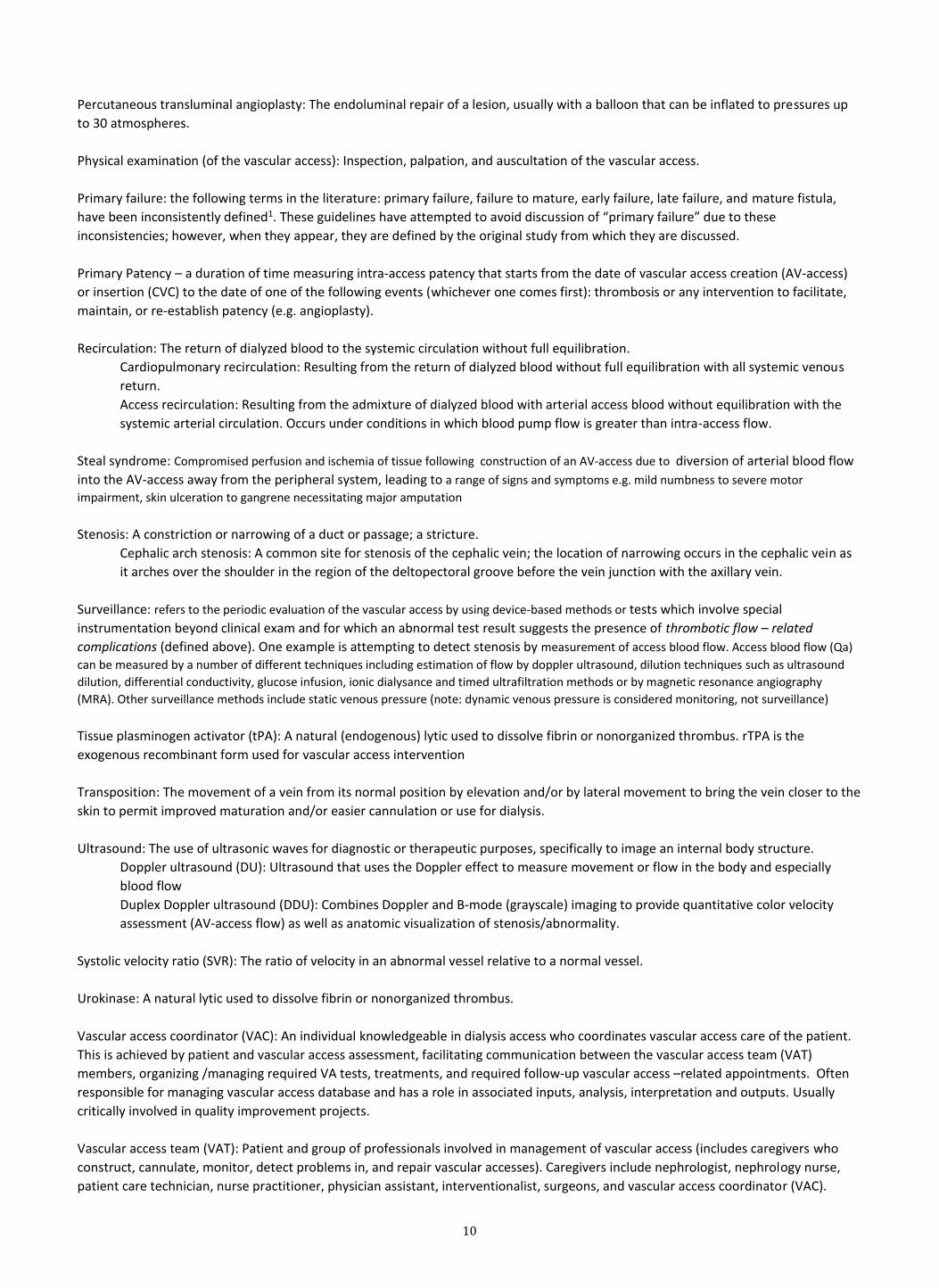
CKD Nomenclature Used by KDOQI
Prognosis of CKD by GFR and Albuminuria Categories
Green: low risk (if no other markers of kidney disease, no CKD); Yellow: moderately increased risk; Orange: high risk; Red, very high risk.
From: KDIGO 2012 Clinical Practice Guideline for the Evaluation and Management of Chronic Kidney Disease.5

12
Work Group Membership
Charmaine Lok, MD (Chair)
Professor of Medicine, University of Toronto
Director of Multi-care Kidney Clinics and Hemodialysis Program,
University Health Network
Senior Scientist, Toronto General Research Institute
Kenneth Abreo, MD, FASDIN
Professor of Medicine, Chief, Division of Nephrology,
and Vice Chairman, Department of Medicine
LSU Health Shreveport School of Medicine
Michael Allon, MD
Professor of Medicine, Associate Director for Clinical Affairs,
and Medical Director of Dialysis Operations, Division of Nephrology
University of Alabama at Birmingham
Arif Asif MD, MHCM
Professor of Medicine, Chairman, Department of Medicine, Jersey Shore Medical Center
Seton Hall-Hackensack Meridian School of Health
Neptune, New Jersey
Brad Astor, PhD
Associate Professor, Departments of Medicine and Population Health Sciences
University of Wisconsin School of Medicine and Public Health
Marc Glickman, MD
Assistant Professor (retired)
Eastern Virginia Medical School, Norfolk VA
Chief Medical Officer
Hancock Jaffe Laboratories
Irivne, California
Janet Graham, MHScN, CNeph(C)
Clinical Director of Nephrology, The Ottawa Hospital
Regional Director of Nephrology, Champlain LHIN Ontario Renal Network
Louise Moist, MD
Professor of Medicine, Epidemiology and Biostatistics
Associate Chair, Division of Nephrology
Surendra Shenoy, MD
(Vice Chair, Guideline Scope)
Professor of Surgery and Director of the Living
Donor Transplantation Program, Washington
University School of Medicine, Saint Louis
Alexander Yevzlin, MD
(Vice Chair, Guideline Scope)
Professor of Medicine and Director of
Interventional Nephrology, University of
Michigan
Thomas S. Huber, MD, PhD
(Editorial Committee)
Professor of Medicine and Chief, Division of
Vascular Surgery, University of Florida College of
Medicine
Timmy Lee, MD
(Editorial Committee)
Professor of Medicine, Department of Medicine,
Division of Nephrology, and Department of
Biomedical Engineering, University of Alabama at
Birmingham
13
Western University, ON
Dheeraj Rajan, MD
Professor of Medicine and Division Chief of Vascular and Interventional Radiology
University of Toronto
Cindy Roberts, RN, CNN
Renal Research Institute, University of North Carolina at Chapel Hill
Nephrology and Hypertension Division
Tushar Vachharajani, MD
Director, Interventional Nephrology, Director Global Nephrology
Cleveland Clinic, Ohio
Rudolph Valentini, MD
Professor of Pediatrics, Pediatric Nephrology, Chief Medical Officer, Children’s Hospital of Michigan (CHM), Wayne State University School of
Medicine
Pediatric Nephrology
Evidence Review Team
University of Minnesota Department of Medicine
Minneapolis VA Center for Chronic Disease Outcomes Research, Minneapolis, MN, USA
Michelle Brasure, PhD, MSPH, MLIS, Program Manager and Medical Librarian
Nancy Greer, PhD, Senior Research Associate and Program Manager
Areef Ishani, MD, MS, Professor of Medicine, Chief of Primary Care at the Minneapolis VA Health Care System and University of Minnesota
Roderick MacDonald, MS, Senior Research Assistant
Victoria A Nelson, MSc, Research Fellow and Study Coordinator
Carin Olson, MD, MS, Medical Editor and Writer
Yelena Slinin, MD, MS, Nephrologist at Kaiser Permanente
Timothy J. Wilt, MD, MPH, Professor of Medicine and Project Director
Organization Leadership
KDOQI Leadership
Michael Rocco, MD, MSCE
KDOQI Chair
Holly Kramer, MD
Vice Chair, Research, and President, National Kidney Foundation
Bernard Jaar, MD MPH
Vice Chair, Education
Michael J. Choi, MD
Vice Chair, Policy, and Past President, National Kidney Foundation
KDOQI Guideline Development Staff
Kerry Willis, PhD, Chief Scientific Officer
Jessica Joseph, MBA, Vice President, Scientific Activities

14
Laura Brereton, MSc, KDOQI Project Director
Abstract
(add)
Foreword
This third update of the KDOQI Vascular Access Guideline represents a complete revamping of the last guideline that was released in 2006. This comprehensive update was performed due to the significant growth in the evidentiary database for vascular access, which was comprehensive reviewed by the evidence review team based at the University of Minnesota in Minneapolis, Minnesota. A number of important randomized clinical trials have been performed in long-term hemodialysis patients since the publication of 2006 guidelines. More than 4600 manuscripts were reviewed to develop this guideline, for which 286 manuscripts were included in the evidence tables used to develop the 26 guideline statements and the research recommendations.
Hemodialysis access issues are managed by a number of different medical professionals. Thus, the workgroup that wrote this guideline is
multidisciplinary, with members representing not only the clinic-based nephrologist but also interventional nephrologists, radiologists, surgeons
and vascular access nurses, including the past presidents of both the American Society of Diagnostic and Interventional Nephrology (ASDIN) and the
Vascular Access Society of the Americas (VASA).
An important new concept introduced in this Vascular Access guideline update is that of the “ESKD Life Plan”. This individualized and
comprehensive map for dialysis modalities and vascular access for the lifetime of the patient is documented in this guideline, as well as the
implementation tools for this guideline that will be developed by the National Kidney Foundation.
This document is the culmination of thousands of hours of volunteer time by the guideline workgroup members as well as by those health care
professionals and patients who participated in the internal and external reviewers of this guideline. The National Kidney Foundation extends its
deepest appreciation to all of those volunteers who contributed their time and effort in developing this guideline. Special gratitude is expressed to
Dr. Charmaine Lok of the University of Toronto, the work group chair, for her tireless efforts to bring this document to fruition, as well as to the two
guideline vice-chairs, Dr. Surendra Shenoy of the Washington University School of Medicine and Dr. Alexander Yevzlin of the University of Michigan
and to the two editorial committee members, Dr. Thomas Huber of the University of Florida and Dr. Timmy Lee of the University of Alabama. It is
their commitment and dedication to the KDOQI process that has made this guideline document possible.
Michael V. Rocco, MD MSCE
Chair, NKF KDOQI

15
Introduction Hemodialysis continues to be the single most prevalent modality of kidney replacement therapy in the United States.6 Longevity on dialysis is
directly proportional to the quality of dialysis, and that quality in turn depends on the reliability and integrity of the access to the patient’s vascular
system. This crucial connection is known as the hemodialysis vascular access. The ideal hemodialysis vascular access is one that provides reliable,
complication-free access to deliver prescribed dialysis, that is also concurrently suitable for a given patient’s needs. The last revision of the NKF
Kidney Disease Outcomes Quality Initiative (KDOQI) Clinical Practice Guidelines for Vascular Access was completed in 2006. Since then
improvements in ESKD care, changes in patient demographics and increasing patient longevity have resulted in a renewed interest in vascular
access management. There is a need to readdress some of the practices previously considered ‘best practices’ that have evolved as a result of
updated data derived from clinical research and changing ESKD care delivery patterns.
The 2018 guidelines represent a fresh approach to vascular access care. While they are grounded in rigorous and sophisticated evaluation and
integration of data accumulated
over the last several decades, the resulting guidance statements reflect the Workgroup’s thoughtful and practical application to support
multidisciplinary care providers in meeting the dialysis access needs of individual patients. These guidelines emphasize a more patient-focused
approach and recommend development of an ESKD Life-Plan, taking each patient’s needs and preferences into consideration when choosing an
access and planning up front for the likely complications and remediations of the current access along with the transition plan to the next access.
Thus, the focus is away from the prior “Fistula First” approach and urges providers to think not only about what access is first, but “what’s next”
during the planning of the first access. Indeed the first access may be a PD catheter access, so the ESKD Life-Plan encourages a comprehensive
evaluation of the patient’s lifetime with ESKD and kidney replacement therapy options. This will have many benefits, including to help preserve
vessels needed for successful future AV-access creation and use and to avoid unnecessary procedures and complications. To summarize, KDOQI has
refocused on a P-L-A-N for each patient: Patient Life-Plan first, followed by his or her corresponding Access Needs.
Moreover, we did not update the previous guidelines number for number, but rather used the new and existing evidence to reframe our approach
to the topic. New or more rigorous evidence has reshaped some prior recommendations. For example, there is a de-emphasis on the need for AV-
access surveillance but a greater emphasis on the need for improved training and application of vascular access monitoring. We address the
preparation for and creation of vascular accesses, the care and management of each type of vascular access, and the prevention and treatment of
complications
These guidelines are less prescriptive in targeting the fine details within each of these areas, recognizing differences in practice patterns, but still
emphasize the need for high quality standards. As a result, we present only three primary targets for use in tracking performance. One target
reinforces that each patient has a regularly updated Life-Plan designed with their goals in mind to achieve the most suitable dialysis access type
despite changes in circumstances. Other chosen targets for each access type aim to limit the major known complications associated with that
access type (e.g. an infection rate target for central venous catheters). We chose to limit the number of targets in order to reasonably enable and
encourage achievement. Our focus is on supporting the actions that will lead to the ideal vascular access as defined above e.g. “reliable”
“complication-free… to deliver prescribed dialysis” and “concurrently suitable for a given patient’s needs”.
Lastly, we recognize the gaps in knowledge and evidence in vascular access care, and provide suggestions for future research. We highlight the need
for continual re-evaluation within each area of care and corresponding section of the guidelines, making room for new evidence and innovations in
dialysis access and its affiliated therapies.
This manuscript is the result of three years of work, consisting of a substantial literature review, months of evidence analysis and discussion, and
multidisciplinary integration of the resulting data into practical guidance for CKD and ESKD care providers. We see these guidelines as a
recalibration and evolution of the previous recommendations. We hope that they will be valuable to our colleagues, helpful for policy makers, and
influential in improving the lives of those living with ESKD.
Methods
The KDOQI guideline development process begins with selection of the topic and refinement of the Guideline Scope followed by a comprehensive literature review of the available evidence. The Guidelines Scope was led by the Chair and Vice Chairs of the Guidelines Scope Committee with refinement after input from the entire multidisciplinary Work Group. The literature review and evidence analysis for this update was carried out by the University of Minnesota Evidence-based Practice Center, at the Minneapolis VA Center for Chronic Disease Outcomes Research. Members of a multidisciplinary Work Group were chosen by the Guideline Chair and Guideline Scope Vice Chairs based on their content and methodology expertise and representation of fields including nephrology (adult and pediatric), nursing, vascular surgery, interventional nephrology, interventional radiology, epidemiology and biostatistics. The analysis and strength of evidence provided by the evidence review team (ERT) was reviewed and discussed by this Work Group using a formal GRADE evidence to decision format7,8 . The Workgroup, through use of standardized work sheets, a series of conference calls, email correspondence, and two in-person meetings, developed Guidelines statements and accompanying strength of recommendations. After statements were agreed upon, guideline sections were drafted by individual members of the Work Group. Once all sections were drafted, they were re-reviewed by the entire multidisciplinary team via weekly-monthly teleconferences until consensus was achieved. If none was achieved, the statement went to a vote, with majority vote being the resulting statement; these have been identified in the guidelines document. The guideline Chair and two editorial committee members (TH and TL) made editorial revisions to the text for flow and comprehensiveness.
16
Literature Review and Evidence Analysis
Data Sources and Searches The ERT searched bibliographic databases including MEDLINE and Embase via Ovid; and the Cochrane Library to identify studies published from January 2000 through October 2016. Search strategies are available in Appendix 2. They supplemented bibliographic database searches with citation searching of identified studies. Study Selection They included trials and prospective observational studies with parallel groups that compared vascular access interventions and reported outcomes preselected for their review. Relevant interventions included those related to different vascular access types. They excluded studies enrolling predominately (<75%) pediatric or acute kidney injury participants; studies enrolling predominately (<75%) participants with vascular accesses created prior to 2000; studies not reporting outcomes relevant to their review; and studies not published in English. Two investigators independently reviewed titles and abstracts of search results to identify potentially eligible references. Two investigators independently screened full text of those references to determine if they met inclusion criteria. A third investigator resolved discrepancies. Data Extraction and Quality Assessment
One reviewer extracted population and comparison characteristics from all eligible studies. Risk of bias was independently assessed for each eligible study by two investigators using methods outlined by the Agency for Healthcare Research and Quality. 9 Risk of bias was assessed as low, medium, or high based upon selection of the exposed and unexposed populations, similarity of surveillance for the outcome, measurement of and adjustment for prognostic imbalance, and attrition. Studies assessed as low or medium risk of bias were included in their analyses. They extracted data in a hierarchical manner to efficiently capture the most relevant data and avoid duplication of samples (when more than one study used the same dataset). If there was an RCT for a comparison, they did not extract data from observational studies, unless the observational study reported a unique outcome. When a comparison was only addressed with observational studies, they identified and extracted data from large registry studies first. If there were multiple studies using the same database, such as the US Renal Data System (USRDS) or same patient population and reporting the same outcomes, they extracted only the study with the most recent data. If studies reported data that were included in the registry studies, they extracted data from these studies only if they reported a different outcome from registry studies. They did not extract data from studies if their contribution to the total population analyzed for that comparison was less than 3%. When studies used multivariate analysis, they extracted the most fully adjusted models and listed confounders adjusted for in the evidence table. Data Synthesis and Analysis They grouped studies by comparison and independently analyzed statistical significance of the results. Heterogeneity in study populations and methods prevented data pooling. They assessed quality of evidence using GRADE.10 Evidence quality was rated high, moderate, low, or very low.
Guideline Statements
The Work Group drafted clinical practice guideline statements based on the evidence analyzed provided by the ERT. Some statements are similar to those of the previous guidelines published in 200611 but have been re-emphasized or re-interpreted in light of new data. For each of the guideline statements, the quality of the evidence and the strength of the recommendations were graded separately using the Grading of Recommendations Assessment, Development, and Evaluation (GRADE) approach criteria.7,12 The Work Group used an adapted version of these scales using High to Very Low for quality of the evidence and Strong or Conditional for the strength of the recommendation. Recommendation strength includes assessment of the statement’s potential clinical impact (Tables I1 and I2). The guideline statements were based on a consensus within the Work Group that the strength of the evidence provided by the ERT was sufficient to make definitive statements about appropriate clinical practice. When the strength of the evidence was not sufficient to make graded statements, but the subject or intervention was deemed important for inclusion, the Work Group identified the statement as “There is inadequate evidence for KDOQI to make a recommendation…”, and then identified the important issue. This was felt important to communicate to the community that the issue has been identified but that further research is required. Finally, there were many topics that are important topics that were excluded from the ERT as they did not make the ERT search criteria or criteria for analysis. Due to their importance, the Work Group offered Guidelines statements based on the best available evidence, independent of the ERT. Such statements begin with “KDOQI considers it reasonable to consider” and are labeled ‘Expert Opinion’. Expert Opinion statements are the consensus opinion of the Work Group based on best available evidence, and are ungraded. Phrasing and definitions presented were also decided on by a formal, full group vote. The statements and definitions that required voting included the classification and working definition of vascular access complications; the wording of Guideline 3 Statement B on vascular access location order; the wording of the Guideline 7 statement on routine preoperative ultrasound; and strength of recommendation of the Guideline 11 statement on AV-access cannulation technique. Regarding all of the guidelines, clinicians should be aware that each patient and their circumstances are unique and require careful thought and individualization; thus, best clinical judgment must be used with careful application of a guideline statement which may infrequently stray from the recommendations of the Work Group to allow for optimal patient outcomes.

17
Table I1: Grade for Strength of Recommendation
Evidence
Base
Grade Implications
Patients Clinicians Policy
ERT derived Strong
recommendation:
“We Recommend”
Most people in your
situation would want the
recommended course of
action and only a small
proportion would not.
Most patients should
receive the
recommended course
of action.
The
recommendation
can be adopted as
policy in most
situations.
ERT derived Conditional
recommendation/
suggestion):
“We Suggest”
The majority of people in
your situation would want
the recommended course
of action, but many would
not.
Different choices will
be appropriate for
different patients.
Each patient needs
help to arrive at a
management decision
consistent with her or
his values and
preferences.
The
recommendation is
likely to require
substantial debate
and involvement of
stakeholders
before policy can
be determined.
ERT derived There is inadequate
evidence
The quality of the evidence was insufficient to make a suggestion or
recommendation but important enough to acknowledge as an area for
future study
Workgroup
derived
Ungraded
““KDOQI considers it
reasonable”
Ungraded recommendations are based on Work Group consensus and The
Literature not found through the formal ERT literature review.
Adapted from Uhlig et al.12
Note: Expert opinion statements that allow for the use of “the clinician’s discretion and best clinical judgment” means that there is currently no rigorous evidence to recommend a therapy, device, or strategy over another. The Workgroup expects ERT derived evidence based statements will ultimately replace expert opinion based statements once such rigorous evidence becomes available.
Table I2: Grade for Quality of Evidence
High quality of evidence. We are confident that the true effect lies close to that of the estimate of the effect.
Moderate quality of evidence. The true effect is likely to be close to the estimate of the effect, but there is a possibility
that it is substantially different.
Low quality of evidence. The true effect may be substantially different from the estimate of the effect.
Very low quality of evidence. The estimate of effect is very uncertain, and often will be far from the truth.
18
Summary of Guideline Statements
GUIDELINE 1. PATIENT FIRST: ESKD LIFE-PLAN
Statements: ESKD Life-Plan and Vascular Access Choice 1.1 KDOQI considers it reasonable that each patient with progressive CKD and/or has an eGFR 15-20 ml/min or already on kidney replacement
therapy should have an individualized ESKD Life-Plan that is regularly reviewed, updated, and documented on their medical record (Expert Opinion)
1.2 KDOQI considers it reasonable to conduct an annual review and update of each patient’s individualized ESKD Life-Plan, together with their healthcare team (Expert Opinion).
1.3 KDOQI considers it reasonable that, in addition to regular monitoring, a minimum quarterly overall review and update of each patient’s vascular access functionality, complication risks, and potential future dialysis access options, be done together with their healthcare team (Expert Opinion).
GUIDELINE 2. VASCULAR ACCESS TYPES
What They Are and Indications
Statements: AV-ACCESS: Indications for use 2.1 KDOQI considers it reasonable to have an AV-access (AVF or AVG) * in a patient requiring hemodialysis, when consistent with their ESKD Life-
Plan and overall goals of care. (Expert Opinion) *See specific sections on incident and prevalent patients and the choice of AV-access type and their appropriate locations
Statements: Central Venous Catheters (CVC): Indications for use
2.2 KDOQI considers it reasonable in valid clinical circumstances to use tunneled CVCs for short-term or long-term durations, as follows (Expert Opinion): Short-term duration: o AVF or AVG created but not ready for use and dialysis is required o Acute transplant rejection or other complications requiring dialysis o PD patient with complications that require time-limited peritoneal rest or resolution of complication e.g. pleural leak o Patient has a living donor transplant confirmed with OR date in near future (e.g. < 90 days) but requires dialysis o AVF or AVG complication such as major infiltration injury or cellulitis that results in temporary non-use until problem is resolved*
* In special, limited circumstances where temporary CVC is required to manage a vascular access complication (e.g. <10 days), it may be acceptable to use a non-tunneled CVC (below)
Long-term or Indefinite duration: o Multiple prior failed AV-accesses with no available options (see anatomical restrictions below) o Valid patient preference whereby use of an AV-access would severely limit QOL or achievement of life goals and after the patient has been
properly informed of patient specific risks and benefits of other potential and reasonable access options for that patient (if available) o Limited life expectancy o Patient anatomy in terms of the combination of the inflow artery and outflow vein precludes the creation of an AVF or AVG (e.g. severe
arterial occlusive disease, non-correctable central venous outflow occlusion, infants/children with diminutive vessels). o Special medical circumstances
Statements: Vascular Access for Incident Patients The statements below are in the context of the ESKD Life-Plan and associated Access Algorithms and their considerations e.g. patient comorbidities,
circumstances etc. 2.3 KDOQI suggests an AV-access (AVF or AVG) in preference to a catheter in most incident and prevalent hemodialysis patients due to the
lower infection risk associated with AV-access use (Conditional recommendation, low quality of evidence).
2.4 KDOQI considers it reasonable that the choice of AV-access (AVF or AVG) be based on the operator’s/ clinician’s best clinical judgment that considers the vessel characteristics, patient comorbidities, health circumstances and patient preference (Expert opinion)
2.5 KDOQI suggests that if sufficient time and patient circumstances* are favorable for a mature, usable AVF, such a functioning AVF is preferred to an AVG in incident hemodialysis patients due to fewer long-term vascular access events (e.g. thrombosis, loss of primary patency, interventions) associated with unassisted AVF use (Conditional recommendation, low quality of evidence). *Patient circumstances refer to vessel characteristics, patient comorbidities, health circumstances and patient preference *Unassisted AVF use refers to an AVF that matures and is used without the need for endovascular or surgical interventions, such as angioplasty. A pre-planned vessel superficialization is acceptable and not considered an additional intervention.
19
2.6 KDOQI suggests that most incident HD patients starting dialysis with a central venous catheter should convert to either a AVF or AVG, if possible, to reduce their risk of infection/ bacteremia, infection-related hospitalizations and adverse consequences (Conditional recommendation, Very low-moderate quality of evidence).
2.7 There is inadequate evidence for KDOQI to make recommendations on choice of incident vascular access type based on associations with all-cause hospitalizations or mortality.
2.8 There is inadequate evidence for KDOQI to make a recommendation on choice of AVF vs. AVG for incident vascular access based on associations with infections, all-cause hospitalizations or patient mortality.
2.9 There is inadequate evidence for KDOQI to make a recommendation for incident hemodialysis patients using a CVC on converting to an AV-access (AVF or AVG) within the first year of dialysis initiation, solely to reduce their risk of mortality.
2.10 KDOQI considers it reasonable to use tunneled CVC in preference to non-tunneled CVC due to the lower infection risk with tunneled CVC (Expert Opinion)
2.11 KDOQI considers it reasonable to use non-tunneled internal jugular CVC only for temporary purposes for a limited time period (< 2 weeks
or per individual facility policy) to limit infection risk (Expert Opinion) Statements: Vascular Access in Prevalent HD Patients 2.12 There is inadequate evidence for KDOQI to make a recommendation on the type of vascular access preferred in prevalent hemodialysis
patients based on vascular access outcomes, patient hospitalizations or mortality.
2.13 KDOQI considers it reasonable that prevalent HD patients use an AV-access (AVF or AVG) in preference to a central venous catheter, if possible, due to the association with lower vascular access related events (e.g. infection, thrombotic and non-thrombotic complications) (Expert Opinion)
2.14 KDOQI considers it reasonable that if clinical circumstances* are favorable for a mature, usable AVF, such a functioning AVF is preferred
to AVG in prevalent hemodialysis patients (Expert Opinion). *Clinical circumstances refer to patient’s vessel characteristics, comorbidities, health circumstances, potential exposure time to catheter use and patient preference
2.15 KDOQI considers it reasonable in valid clinical circumstances to use tunneled CVCs for short-term or long-term durations, as follows (Expert Opinion):
Short-term duration: o AVF or AVG created but not ready for use and dialysis is required o Acute transplant rejection or other complications requiring dialysis o PD patient with complications that require time-limited peritoneal rest or resolution of complication e.g. pleural leak o Patient has a living donor transplant confirmed with OR date in near future (e.g. < 90 days) but requires dialysis o AVF or AVG complication* such as major infiltration injury or cellulitis that results in temporary non-use until problem is resolved
* In special, limited circumstances where temporary CVC is required to manage a vascular access complication (e.g. <10 days), it may be acceptable to use a non-tunneled CVC (see below)
Long-term or Indefinite duration: o Multiple prior failed AV-accesses with no available options (see anatomical restrictions below) o Valid patient preference whereby use of an AV-access would severely limit QOL or achievement of life goals and after the patient has
been properly informed of patient specific risks and benefits of other potential and reasonable access options for that patient (if available)
o Limited life expectancy o Patient anatomy in terms of the combination of the inflow artery and outflow vein precludes the creation of an AVF or AVG (e.g. severe
arterial occlusive disease, non-correctable central venous outflow occlusion, infants/children with diminutive vessels). o Special medical circumstances
GUIDELINE 3. VASCULAR ACCESS LOCATIONS
Statements: AV-access Locations The statements below are in the context of the ESKD Life-Plan and associated Access Algorithms and their considerations e.g. feasible anatomy etc. 3.1 KDOQI considers it reasonable to choose the site (location) of the AV-access after careful consideration of the patient’s ESKD Life-Plan (see
Figures 1.1-1.6), potentially following the below paths (Expert Opinion):
A) A patient’s ESKD Life-Plan includes an anticipated long duration e.g.> 1 year on HD: o Forearm AVF (Snuffbox or distal radiocephalic or transposed radiobasilic) o Forearm loop AVG or proximal forearm AVF (e.g. proximal radiocephalic, perforator-combinations) or brachiocephalic, per operator
discretion o Brachiobasilic AVF or Upper arm AVG, per operator discretion
B) A patient’s ESKD Life-Plan includes an anticipated limited duration (e.g. <1 year) on HD: o Forearm loop AVG or brachiocephalic AVF (with high likelihood of unassisted maturation)
20
o Upper arm AVG
C) A patient urgently starts HD without prior sufficient time to plan for and/or create an AV-access and has an anticipated limited duration (e.g. <1 year) on HD:
o Early or standard cannulation loop AVG (forearm or upper arm location+), or CVC, per operator discretion and patient’s clinical needs D) A patient urgently starts HD without prior sufficient time to plan for and/or create an AV-access and has an anticipated long duration e.g.> 1 year on HD: o PD catheter, and follow above algorithm (A) if PD not a long-term option or o Forearm early cannulation loop graft; when AVG fails, follow above algorithm (A) or o CVC if high likelihood of rapid AVF maturation and usability success, then follow above algorithm (A)
E) All AV-access options in the upper extremity have been exhausted and patient’s ESKD Life-Plan includes a long duration e.g.> 1 year on HD, the following may be considered based on individual patient circumstances and operators best clinical judgment and expertise: o Lower extremity AVF or AVG or Hero Graft
+ The choice of upper extremity location of an AVG should be based upon the operator’s discretion and best clinical judgment considering the
patients ESKD Life-Plan, due to inadequate evidence to demonstrate a difference between forearm versus upper arm AVG patency or
complication outcomes.*
*includes infections, hospitalizations, mortality
Statements: Catheter Locations 3.2 KDOQI considers it reasonable to choose the site (location) of the CVC after careful consideration of the patient’s ESKD Life-Plan as follows:
o Upper extremity before lower extremity, only if choices are equivalent o There are valid reasons for CVC use (see Guideline 2) and its duration of use is expected to be limited (e.g. < 3 months) with anticipated
use of AV-access (transplant is considered as a potential option (i.e. preserve iliac vessels)): ▪ Tunneled cuffed CVC in opposite extremity to anticipated AV-access
(see below guidance for more detail)
o Some experts support that in urgent dialysis start situations, under limited use circumstances (e.g. < 1 month) and transplant is not an option, use of a tunneled, cuffed femoral CVC is acceptable (unless contraindicated*) until the AV-access or PD catheter can be quickly created and used. Use of the femoral vein preserves the upper extremity vessels for future AV-access creation.
*Contraindications to femoral vein CVC include: femoral or iliac vessel pathology or prior surgery/reconstruction; hygienic reasons e.g. chronic unresolved diarrhea; morbid obesity (BMI>35) or other difficult vein access
o When there are valid reasons for CVC use (see Guideline 2) and duration of use is expected to be prolonged (e.g. > 3 months) without anticipated use of AV-access, CVC may be placed in the following locations in order of preference:
▪ Internal jugular ▪ External jugular ▪ Femoral ▪ Subclavian ▪ Lumbar
o In the absence of prior pathology (e.g. central stenosis) or intervention (e.g. pacemaker) the right side is preferable to left side due to
more direct anatomy; however, the side without pathology or intervention is priority
o While a suggested stepwise approach to AV-access site selection is provided (See Figures 1.1-1.6), modification of the approach is encouraged as necessary to consider the individual’s ESKD Life-Plan and circumstances, and follow the below key principles, given available suitable vessels:
▪ Distal first to proximal next approach ▪ Always preserve the integrity of vessels for future vascular access options ▪ Non-dominant extremity in preference to dominant, only if choices are equivalent
GUIDELINE 4. NOVEL MATERIALS AND AV-ACCESSS
Statements: Novel Materials and AV-Access 4.1 KDOQI suggests that the choice of material for an AVG should be based upon the clinician’s /surgeon’s discretion and best clinical judgment
since the current evidence does not demonstrate that one graft material or modification thereof is associated with improved outcomes in terms of patency or complications (Conditional recommendation, Low quality of evidence).
4.2 KDOQI considers it reasonable to use early cannulation grafts as a catheter sparing strategy in appropriate patients, considering their ESKD Life-Plan (Expert opinion)
GUIDELINE 5. CVC CONFIGURATION AND MATERIALS
Statements: CVC configuration and materials
21
5.1 KDOQI suggests that the choice of tunneled hemodialysis catheter type and design be based on the clinician’s discretion and best clinical judgment (Conditional recommendation, Low quality evidence).
GUIDELINE 6. TIMING, PREPARATION & PLANNING FOR CREATION/INSERTION OF DIALYSIS ACCESS
Statements: Education on ESKD Modalities and Dialysis Access 6.1 KDOQI considers it reasonable for adult and pediatric patients with an eGFR < 30 mL/min/1.73 m2 (CKD stage G4) with progressive decline
in kidney function, to be educated on all modalities of kidney replacement therapy (KRT) options, including transplantation, so that timely referral can be made for the appropriate modality and creation of a functional dialysis access, if necessary (Expert Opinion)
6.2 KDOQI considers it reasonable for adult and pediatric patients with a kidney transplant with an eGFR* < 30 mL/min/1.73 m2 with progressive decline in kidney function, to be educated on all modalities of kidney replacement therapy (KRT) options, including potential re-transplantation, so that timely referral can be made for the appropriate modality and creation of a functional dialysis access, if necessary (Expert Opinion) * if pediatric patient, estimate eGFR by Schwartz formula
6.3 KDOQI considers it reasonable for PD patients with complications refractory to therapy and/or with circumstances that make PD less
conducive than HD*, to be educated on all kidney transplant and HD options, so that timely referral can be made for the appropriate modality preparation and creation of a functional dialysis access, if necessary (Expert Opinion) *see Special discussions
6.4 KDOQI considers it reasonable for the predetermined dialysis access to be usable to provide the prescribed dialysis when the patient is ready to initiate dialysis (Expert Opinion)
Statements: Referral for AV-access In some facilities, referral of the patient for assessment by the vascular access team /surgeon for appropriate dialysis access is a different process than referral for the actual creation/insertion. However, for simplicity, the Guideline recommendations have been combined, keeping in mind variable timeframes between assessment for and creation of vascular access. Non-Dialysis CKD Patients 6.5 KDOQI considers it reasonable that in non-dialysis CKD patients* with progressive decline in kidney function, referral for dialysis access
assessment and subsequent creation should occur when eGFR is 15-20 ml/min. Earlier referral should occur in patient with unstable and/or rapid rates of eGFR decline (e.g. >10ml/min/year)+ (Expert Opinion)
*CKD, including patient with a failing transplant + In urgent situations (regardless of rate of progression), note that some newer graft materials may be cannulated immediately (<72 hours) after placement and can be considered along with the Patient’s ESKD Life-Plan
Dialysis Patients
6.6 KDOQI considers it reasonable that in HD patients with recurrent vascular access problems*, prompt referral for assessment and creation of a new AV-access should be made to allow adequate time for specialist consult and follow-up, and possible AV-access failure and correction; and should consider individual patient circumstances and competing risk of death (Expert Opinion).
* includes recurrent need for CVC use and/or >3 corrective interventions/6 months
When PD is the modality of choice:
6.7 KDOQI considers it reasonable and ideal to place a peritoneal dialysis (PD) catheter at least 2 weeks before the anticipated need of the PD treatments (Expert Opinion)
6.8 KDOQI considers it reasonable for an urgent peritoneal dialysis (PD) catheter to be placed for immediate PD as necessary under the direction and care of experienced personnel in conducive environments (Expert Opinion)
Statements for Vessel Preservation 6.9 KDOQI considers it reasonable to protect all central and peripheral arteries and veins from damage whenever possible, including the
avoidance of peripherally inserted venous catheters* and unnecessary venipunctures, for patients on dialysis or with chronic kidney disease where dialysis access is expected in the future (Stage G4 -G5) (Expert Opinion).
* other scenarios where vessel (artery or vein) damage may occur that should be avoided include: 1) radial artery access for coronary interventions, 2) Venous cardiovascular implantable electronic devices – alternatives such as epicardial/leadless pacing should be considered whenever possible.
Statements for Multidisciplinary Team Approach 6.10 KDOQI considers it reasonable to educate on, coordinate and manage all aspects of dialysis access using a multidisciplinary team within the
resource capacities and feasibilities of each facility (Expert Opinion)
6.11 There is inadequate evidence for KDOQI to make a recommendation on the use of a multidisciplinary team to reduce the rate of CVC or
increase the use of AVF
22
GUIDELINE 7. PATIENT AND VESSEL EXAMINATIONS and PREOPERATIVE CONSIDERATIONS
Statements for Patient Clinical Examination 7.1 KDOQI recommends that a physical examination focused on vascular anatomy be the basis for the initial assessment and planning of
vascular access creation (Conditional recommendation, Very low quality of evidence)
7.2 KDOQI considers it reasonable to have greater emphasis* on and more training in preoperative clinical examination to assess patients and their vessels to determine the type and location of their vascular access (Expert Opinion)
Statements for Vessel Mapping for Vascular Access 7.3 KDOQI suggests selective preoperative ultrasound in patients at high risk of AV-access failure rather than routine vascular mapping in all
patients (Conditional recommendation, Low quality of evidence)
7.4 KDOQI considers it reasonable to use various imaging studies as needed to evaluate the suitability of vessels for AV-access creation e.g. ultrasonography for peripheral vessels and venography for suspected central vein occlusion, while considering the patients clinical circumstances and residual kidney function
Statements for Optimal vessel size for artery and vein for AV-access creation 7.5 KDOQI considers it reasonable to have no minimal vein and arterial diameter threshold to create an AVF; however, vessels <2 mm should
have further careful evaluation for feasibility and quality to create a functioning AVF (Expert Opinion).
7.6 KDOQI considers it reasonable to evaluate multiple characteristics of vessel quality for AVF creation (size, distensibility, flow etc.) (Expert Opinion)
GUIDELINE 8. AV-ACCESS CREATION Statement: Use of Anesthesia for AV-access creation 8.1 KDOQI suggests that the choice of anesthesia for AVF creation should be based on the operator’s discretion and best clinical judgment as
current evidence shows no difference between regional block or local anesthesia in terms of AVF usability, patency, interventions or patient experience (Conditional recommendation, Low-Moderate level of evidence).
Statement: AV-Access Anastomotic Configuration and Apposition Methods 8.2 KDOQI considers it reasonable that the choice of anastomotic configuration and apposition method (e.g. vascular clips, sutures) for AVF
creation be based on surgeon’s discretion and best clinical judgment, as there is insufficient evidence to prefer one configuration or
apposition method over another (Expert Opinion).
Statement: AV-access Anastomotic Suture Technique
8.3 In the opinion of the KDOQI workgroup, it is reasonable to consider that the choice of suture technique for AV-access creation should be based on the surgeon’s discretion and best clinical judgment as there is insufficient evidence that any anastomotic suture technique is advantageous in terms of AV-access patency or complications (Expert Opinion).
Statements: Use of intra-operative assisted maneuvers for AV-access maturation 8.4 KDOQI does not suggest the use of allogenic endothelial implants to improve AVF maturation, patency, clinical usability or reduce
thrombosis or to improve AVG graft patency (Conditional recommendation, very low quality of evidence)
8.5 KDOQI does not suggest the use of pancreatic elastase to improve the patency and clinical use of AVF or AVG (Conditional recommendation, Moderate quality of evidence).
8.6 KDOQI considers it reasonable to have a careful individualized approach to operator enhanced (surgical or endovascular) maneuvers during AV-access creation to facilitate AV-access maturation, based on the operators best clinical judgment and expertise (Expert Opinion)
GUIDELINE 9. CATHETER INSERTION
Statements: Techniques and other considerations for placement 9.1 KDOQI recommends the use of image guided CVC insertions to improve success of insertions (Conditional recommendation, Moderate
quality of evidence).
9.2 KDOQI considers it reasonable that after tunnelled CVC insertion* alternative imaging is used to ensure that the CVC tip has been correctly placed, if fluoroscopy is unavailable (Expert Opinion)
23
GUIDELINE 10. POST AV-ACCESS CREATION / CVC INSERTION CONSIDERATIONS
Statements: AV-access Early Post-operative Considerations (0-30 days) – Early AV-access complications 10.1 KDOQI considers it reasonable for AV-access (AVF and AVG) to be evaluated by a surgeon/operator* for post-operative complications
within two weeks and for an appropriate member of the VAT to evaluate for AVF maturation by four-six weeks after AV-access creation (Expert Opinion)
* ideally the surgeon/operator who created the AV-access
Statements: Post-operative AV- Access Maturation Patient Enhanced 10.2 There is inadequate evidence for KDOQI to make a recommendation on the use of upper extremity exercise to facilitate postoperative AVF
maturation 10.3 KDOQI recommends the use of whole arm rather than finger exercise, if exercise is used to facilitate AVF maturation. (Conditional
recommendation, Moderate-High quality of evidence)
Pharmacologic Intervention 10.4 KDOQI does not suggest the use of heparin as an adjuvant therapy in the peri-operative period to improve primary patency or initial use
of AV-access (AVF or AVG) (Conditional recommendation; Low quality of evidence) 10.5 KDOQI does not suggest the use of adjuvant Clopidogrel monotherapy initiation in the perioperative period to improve AVF maturation
and reduce the likelihood of primary failure. (Conditional recommendation, Low quality of evidence) 10.6 KDOQI does not suggest the use of Glyceryl-trinitrate to enhance AVF maturation. (Conditional recommendation, Low quality of evidence)
10.7 KDOQI does not suggest the use of Cholecalciferol to enhance AVF maturation (Conditional recommendation, Moderate quality of evidence)
10.8 There is inadequate evidence for KDOQI to make a recommendation on the use of clopidogrel-prostacyclin (iloprost) for AVF usability or 12-month primary or secondary patency
Endovascular and Surgical Intervention 10.9 There is inadequate evidence for KDOQI to make a recommendation on the preferred use of surgical or endovascular techniques for
postoperative maturation. It is reasonable to consider a careful individualized approach for using either surgical techniques or endovascular techniques when needing to intervene on an AV-access to enhance maturation postoperatively
Statements: Timing of removal (CVC) Non-cuffed, non-tunneled CVC 10.10 KDOQI considers it reasonable to limit the use of temporary, non-cuffed, non-tunneled dialysis catheters to a maximum of 2 weeks due to
increased risk of infection, and should be only considered in patients in need of emergent access (Expert Opinion). Cuffed, tunneled CVC 10.11 KDOQI considers it reasonable that in HD patients for whom a cuffed, tunneled CVC is the most appropriate permanent dialysis access*,
there is no maximum time limit to catheter use but regular evaluation is required to determine if the CVC remains the most appropriate dialysis access (Expert Opinion).
*Note: the above re: appropriate use of catheter is discussed in Guideline 2
GUIDELINE 11. VASCULAR ACCESS USE
Statement for Vascular Access General Monitoring 11.1 KDOQI considers it reasonable to assess or check the vascular access* and surrounding area by physical exam prior to every cannulation (if
AV-access) or connection (if CVC) for potential complications (Expert Opinion) Statements: AV-Access Cannulation 11.2 KDOQI recommends rope ladder cannulation as the preferred cannulation technique for AVFs (Conditional recommendation, moderate
quality of evidence)
11.3 KDOQI considers it reasonable to limit buttonhole cannulation only to special circumstances given the associated increased risks of infection and related adverse consequences (Expert Opinion).
11.4 KDOQI suggests that when select buttonhole cannulation is performed, the use of buttonhole cannulation devices to facilitate cannulation
should be at the discretion and expertise of the cannulator (Conditional recommendation, low quality of evidence). 11.5 KDOQI considers it reasonable to use trained cannulators with high rates of cannulation success for initial AV-access cannulations to help
avoid primary infiltration injury of the AV-access (Expert Opinion). 11.6 KDOQI considers it reasonable to have structured training and supervision of dialysis technicians and nurses prior to their first
cannulation, and regular training updates to maintain cannulation competency (Expert Opinion).

24
Note: to be clear, any consideration of buttonhole cannulation refers only to AVF. AVG made of PTFE should not be accessed by buttonhole cannulation, due to risks of one-siteitis and its serious consequences
11.7 KDOQI considers it reasonable to avoid buttonhole cannulation in synthetic PTFE grafts due to potential serious consequences (Expert Opinion)
Statements: Catheter system connect and disconnect procedure considerations
11.8 KDOQI suggests the use of a catheter care protocol for exit site and hub care to reduce catheter related bloodstream infections and treatment of catheter dysfunction (Strong recommendation, Moderate quality of evidence,)
11.9 KDOQI considers it reasonable to use aseptic technique, correct hand washing, and masks for patients and staff performing catheter
connection and disconnection procedures (Expert Opinion). 11.10 KDOQI considers it reasonable to cleanse the catheter hub when connecting and disconnecting the catheter with a Chlorhexidine based
solution. If chlorhexidine is contraindicated (e.g., sensitivity, allergy), povidone-iodine solution is a reasonable substitute and should be used (Expert Opinion).
11.11 KDOQI considers it reasonable that at the time of catheter dressing change, to cleanse the skin surrounding the catheter exit site with a
chlorhexidine based solution. If Chlorhexidine is contraindicated (e.g., sensitivity, allergy), povidone-iodine solution is a reasonable substitute and should be used (Expert Opinion)
11.12 There is inadequate evidence for KDOQI make a recommendation on the Chlorhexidine formulation to use for infection prophylaxis and this should be based on the clinician’s best clinical judgment, and local practical considerations.
11.13 There is inadequate evidence to demonstrate a difference in catheter-related infections with the use of transparent film dressing compared with non-transparent dressing; thus, the choice of catheter dressing material should be based on the clinician’s discretion that considers the patient’s circumstances and uses best clinical judgment.
11.14 KDOQI considers it reasonable to use a topical antiseptic or antibiotic barrier at the catheter exit site in addition to cleansing until the exit site is healed to reduce the risk of catheter-related infection (Expert Opinion).
11.15 There is inadequate evidence to demonstrate a difference in catheter-related infections between the use of various antiseptic or antibiotic topical exit site barriers; thus, the choice of topical exit site barrier should be based on the clinician’s discretion and best clinical judgment
11.16 KDOQI considers the following catheter care practices reasonable to follow:
o The frequency of catheter dressing change should be based on the clinician’s discretion and best clinical judgment, with a minimum of 1x/week
o Catheter dressings should be protected against wet and dirty environments, particularly when the exit site is not yet fully healed (e.g. avoid swimming and unprotected showering)
GUIDELINE 12 AV-ACCESS CANNULATION COMPLICATIONS
Statements: AV-Access Cannulation Complications 12.1 KDOQI considers the following therapeutic interventions for cannulation injury reasonable to follow:
o Any size infiltration: apply ice for a minimum of 10 minutes and refrain from maximizing the blood pump speed (Expert Opinion)
o If the infiltration is moderate, the needle should be withdrawn and manual pressure held over the infiltration site (Expert Opinion)
o If the infiltration is significantly large, in addition to the above, a decision on the necessity for dialysis that day is required – if dialysis is
required, a site above the infiltration injury should be cannulated; if this is not possible, reattempt at the area of injury should not proceed until manual pressure and ice is applied for 30 minutes (Expert Opinion)
o If a hematoma develops, close assessment of the site, the AV-access and the adjacent extremity should be made, including measurement of swelling, assessment of the presence of flow in the AV-access both above and below the hematoma and circulation to the associated extremity (Expert Opinion)
12.2 KDOQI considers it reasonable to use ultrasound to help determine direction of flow and proper needle placement in select patients as
needed and performed by trained operators, to prevent cannulation complications (Expert Opinion)
25
GUIDELINE 13. AV-ACCESS FLOW DYSFUNCTION+ - MONITORING/SURVEILLANCE + “AV-access flow dysfunction” refers to clinically significant abnormalities in AV-access (AVF or AVG) flow or patency due to underlying stenosis, thrombosis or related pathology. This is in distinction to other types of AV-access complications Statements: Appropriate Use of Monitoring/Surveillance for AV-Access Flow Dysfunction Physical exam (monitoring): 13.1 KDOQI recommends regular physical examination or check of the AVF, by a knowledgeable and experienced health practitioner, to detect
flow dysfunction of the AVF (Conditional/Strong recommendation, Moderate quality of evidence)
13.2 KDOQI recommends regular physical examination or check of the AVG, by a knowledgeable and experienced health practitioner, to detect flow dysfunction of the AVG (Conditional/Strong recommendation, Moderate quality of evidence)
13.3 KDOQI considers it reasonable for nephrology trainees and health practitioners involved with clinical hemodialysis patient care to be properly trained in physical examination of the AV-access to monitor for and detect AV-access flow dysfunction (Expert Opinion)
Surveillance to facilitate patency: 13.4 There is inadequate evidence for KDOQI to make a recommendation on routine AVF surveillance by measuring access blood flow, pressure
monitoring, or imaging for stenosis, that is additional to routine clinical monitoring, to improve access patency.
13.5 KDOQI does not suggest routine AVG surveillance by measuring access blood flow, pressure monitoring, or imaging for stenosis, that is additional to regular clinical monitoring, to improve AVG patency. (Conditional recommendation, Low quality of evidence).
Note: In other words, monitoring of vascular access is primary while surveillance findings are supplementary, and action should not be based
solely on surveillance findings.
Surveillance and Pre-emptive Intervention for AV-Access Stenosis, Not Associated with Clinical Indicators
Endovascular Intervention to improve patency:
13.6 KDOQI does not recommend pre-emptive angioplasty of AVFs with stenosis, not associated with clinical indicators, to improve access patency (Conditional recommendation, moderate quality evidence).
13.7 KDOQI does not recommend pre-emptive angioplasty of AVGs with stenosis, not associated with clinical indicators, to improve access patency (Conditional recommendation, Moderate quality evidence).
13.8 KDOQI considers it reasonable for patients with consistently persistent clinical indicators to undergo pre-emptive angioplasty of their AV-access to reduce the risk of thrombosis and AV-access loss (Expert opinion)
Surgical Intervention to improve patency: 13.9 There is inadequate evidence for KDOQI to make a recommendation on pre-emptive surgical interventions in AVFs with stenosis, not
associated with clinical indicators, to improve access patency.

26
GUIDELINE 14. AV-ACCESS FLOW DYSFUNCTION+ - PREVENTION
Statements for the Prevention of AV-Access (AVF or AVG) Flow Dysfunction+ +“AV-access flow dysfunction” refers to clinically significant abnormalities in AV-access (AVF or AVG) flow or patency due to underlying stenosis, thrombosis or related pathology. This is in distinction to other types of AV-access complications. Pharmacological Primary and Secondary Prevention of AV-access flow dysfunction
Fistulas 14.1 KDOQI suggests that the use of adjuvant far-infrared therapy to improve AVF primary patency be based on individual circumstances,
feasibility and the clinician’s best judgment and expertise (Conditional recommendation, Moderate quality evidence)
14.2 KDOQI does not suggest the use of fish oil or aspirin to prevent AVF flow dysfunction (Conditional recommendation; low-moderate quality of evidence)
14.3 There is inadequate evidence for KDOQI to make a recommendation on the use of oral simvastatin and ezetimibe to reduce AVF interventions or thrombosis
14.4 There is inadequate evidence for KDOQI to make a recommendation on the use of clopidogrel-prostacyclin to improve AVF primary failure
Grafts
14.5 KDOQI suggests careful consideration of potential individual patient benefits, risks and circumstances prior to the use of combination dipyridamole (200 mg) and aspirin (25 mg) twice daily to improve AVG primary unassisted patency (Conditional recommendation, High quality evidence)
14.6 KDOQI suggests the use of oral fish oil supplementation, in patients with newly created AV grafts, to reduce patient morbidity (i.e. reduce
frequency of thrombosis and related corrective interventions) (Conditional recommendation; Moderate quality of evidence) 14.7 There is inadequate evidence for KDOQI to make a recommendation on the use of oral fish oil supplementation to prolong AVG cumulative
patency
14.8 There is inadequate evidence for KDOQI to make a recommendation on the use of oral simvastatin and ezetimibe for reducing dialysis AVG interventions and thrombosis.
GUIDELINE 15. AV-ACCESS FLOW DYSFUNCTION – TREATMENT
Statement: Radiographic Confirmation of Clinically Significant AV-access Stenosis*
15.1 KDOQI considers it reasonable that when clinical monitoring suspects clinically significant AV-access stenosis*, further timely and
confirmatory evaluation should proceed, including imaging of the dialysis access circuit (Expert Opinion)
15.2 KDOQI considers it reasonable to use the smallest volume of iodinated contrast or non-iodinated contrast agents (e.g. CO2 gas) by
operators knowledgeable in their uses, contraindications and risks to obtain the best possible image in all patients with CKD to preserve
residual kidney function (Expert Opinion)
15.3 KDOQI considers it reasonable that when further confirmatory imaging studies reveals a culprit lesion responsible for clinical signs and
symptoms, that the clinically significant lesion* is promptly treated (Expert Opinion).
*a clinically significant lesion is one that contributes to clinical signs and symptoms (see AV access Monitoring Table 13.2) without other
cause, with or without a sustained change in measurements (e.g. change in blood flow (Qa) or venous pressures) in the dialysis access
circuit.
Statement: General Treatment of Clinically Significant Stenosis or Thrombosed AV-access
15.4 KDOQI considers it reasonable to use a careful individualized approach to the treatment of failing or thrombosed AVF and AVG (surgical or
endovascular), based on the operators best clinical judgment and expertise** considering the patient’s ESKD Life-Plan (Expert Opinion)
** ** It considers both the patient’s individual circumstances and the operator’s clinical experience and expertise (i.e. reasonable
capabilities and limitations); preferably discussed and agreed upon by the team managing the patient’s vascular access, including but not
limited to the patient and one or more of: nephrologist, interventionalist, surgeon, VAC, cannulators (nurse or technician
Statement: Treatment of Clinically Significant AV-access Stenosis
Angioplasty
15.5 KDOQI considers it reasonable to use balloon angioplasty (with high pressure as needed) as primary treatment of AVF and AVG stenotic
lesions that are both clinically and angiographic ally significant *(Expert Opinion)
*angiographically present stenosis without accompanying clinical signs and symptoms is inadequate to treat/intervene upon
27
15.6 There is inadequate evidence for KDOQI to make a recommendation regarding the use of specialized balloons (drug-coated or cutting)
versus standard high-pressure balloons in the primary treatment of AVF and AVG stenosis.
15.7 There is inadequate evidence for KDOQI to make a recommendation regarding the optimal duration of balloon inflation time during
angioplasty to improve intervention primary patency in the treatment of AVF or AVG stenosis.
15.8 KDOQI considers it reasonable that a careful patient-individualized approach to the choice of balloon type for angioplasty of clinically
significant AVF and AVG stenosis be based on the operators best clinical judgment and expertise (Expert Opinion)
Stents
15.9 KDOQI suggests the appropriate use* of self-expanding stent-grafts in preference to angioplasty alone to treat clinically significant graft-
vein anastomotic stenosis in AVG when the goal is overall better 6-month post intervention outcomes+ after carefully considering the
patient’s ESKD Life-Plan (Conditional recommendation, Moderate quality of evidence).
+overall better 6-month outcomes refer to reduced recurrent AVG restenosis +/- patency
* Appropriate use avoids cannulation segments
15.10 KDOQI considers it reasonable to first consider the consequences of placement of a stent-graft on future AV-access options according to the
patient’s ESKD Life-Plan, with consultation with the vascular access team if necessary, prior to its placement (Expert Opinion)
15.11 KDOQI suggests that the use of an appropriately placed stent-graft* is preferred to angioplasty alone for the treatment of in-stent
restenosis in AVG and AVF for overall better 6-month post intervention outcomes+. (Conditional recommendation, Moderate quality of
evidence)
+overall better 6-month outcomes refer to reduced recurrent AVG and AVF restenosis +/- patency
15.12 KDOQI considers it reasonable to avoid the use of bare metal stents for the treatment of clinically and /or angiographically significant AVG and AVF stenotic lesions (Expert Opinion)
Statement: Treatment of Thrombosed AV-Access
15.13 KDOQI considers it reasonable that management of each episode of AV-access thrombosis involves the consideration of the patient’s
dialysis access Succession Plan that is consistent with the ESKD Life-Plan, given the compromised AV-access patency after either
endovascular or surgical treatment (Expert Opinion)
**Operators discretion carefully considers both the patient’s individual circumstances and the operator’s own clinical experience and expertise
(i.e. reasonable capabilities and limitations)
15.14 KDOQI considers it reasonable to surgically treat a failing AV-access in the following circumstances: 1) endovascular failures, 2) clinically
significant lesions not amenable to endovascular treatment, 3) situations in which the surgical outcomes are deemed markedly better+.
(Expert Opinion) + situations when surgical outcomes are anticipated to be better than alternative options should be first discussed and agreed upon by
the team managing the patient’s vascular access, including but not limited to the patient and one or more of: nephrologist,
interventionalist, surgeon, VAC, cannulation expert, if possible
GUIDELINE 16. AV-ACCESS INFECTION
Statements for AV-Access Infections Monitoring and Prevention 16.1 KDOQI considers it reasonable to check the vascular access and surrounding area prior to every cannulation for signs and symptoms of
infection* (Expert Opinion). * this should be done by patient and cannulator (if patient does not self cannulate)
16.2 KDOQI considers it reasonable to educate the patient on washing the access arm using antiseptic to clean the skin prior to every cannulation (Expert opinion)
Diagnosis 16.3 KDOQI considers it reasonable to use radiologic imaging* to help confirm the diagnosis of AV-access infection; however, physical
examination remains the hallmark for assessing for infection** (Expert opinion) *imaging includes duplex ultrasound, +/- CT, PET, and nuclear medicine scans (e.g. indium scan) **This includes erythema, skin breakdown, purulent discharge, presence of exposed graft
Treatment
16.4 KDOQI considers it reasonable to obtain cultures and sensitivities of the blood and any available infected AV-access vessel/material, surrounding tissue or drainage prior to initiating antibiotic therapy (Expert Opinion)
28
16.5 KDOQI considers it reasonable to consider for infected AV-access the rapid initiation of empiric broad spectrum antibiotics and timely referral to a surgeon knowledgeable in the management of vascular access complications (Expert Opinion)
16.6 KDOQI considers it reasonable to consider strict follow-up of culture results with the appropriate change in antibiotics based on organism sensitivities, with antibiotic duration according to extent of vascular access infection and surgical intervention (Expert Opinion)
16.7 KDOQI considers it reasonable that the specific surgical treatment for AV-access infections (with concurrent antibiotics) should be based
on the patient’s individual circumstances considering the extent of infection, offending organism, and future vascular access options (Expert Opinion)
Prevention
16.8 KDOQI considers it reasonable to conduct a careful physical exam by the surgeon and managing team prior to surgery to identify infection risks that should first be managed before proceeding with AV-access surgery (e.g. poor dentition with dental infection, malnutrition etc.) (Expert Opinion)
Special considerations from Guideline 11:
11.2 KDOQI recommends rope ladder cannulation as the preferred cannulation technique for AVFs (Conditional recommendation, Moderate quality of evidence)
11.3 KDOQI considers it reasonable to limit buttonhole cannulation only to special circumstances given the associated increased risks of
infection and related adverse consequences (Expert Opinion).
11.6 KDOQI considers it reasonable to have structured training and supervision of dialysis technicians and nurses prior to their first cannulation, and regular training updates to maintain cannulation competency (Expert Opinion).
16.9 KDOQI considers it reasonable to investigate* and closely monitor for metastatic complications (e.g. endocarditis, spinal abscesses, septic
arthritis) in patients with buttonhole infection from particularly dangerous organisms such as S. aureus, gram negative bacteria and fungal organisms.
*investigations include 2D echocardiography, MRI, joint aspirate and other, as appropriate
GUIDELINE 17. AV-ACCESS ANEURYSMS Statements for AV-access Aneurysms
Recognition and Diagnosis
17.1 KDOQI considers it reasonable to routinely check AV-access for aneurysm/pseudoaneurysms at each dialysis session by knowledgeable care providers, including but not limited to dialysis technicians, nurses, nephrologists and vascular access coordinator (Expert Opinion).
17.2 KDOQI considers it is reasonable to obtain proactive surgical assessment when clinical findings suggest an AV-access
aneurysm/pseudoaneurysm to be at risk of complications* (Expert Opinion) An aneurysm/pseudoaneurysm that is considered at risk of complications are those with evidence of associated symptoms or skin breakdown
17.3 KDOQI considers it is reasonable to obtain emergent surgical assessment and treatment for AV-access aneurysm/pseudoaneurysm
complications such as erosion or hemorrhage (Expert Opinion)
17.4 KDOQI considers it reasonable to use Duplex ultrasound to corroborate the physical examination suggesting an AV-access aneurysm/pseudoaneurysm and to obtain information on the size, presence of stenosis/thrombus, and impact on the AV-access ( including flow rate (Qa), status of the arterial inflow and the venous outflow) (Expert Opinion).
Management
17.5 KDOQI considers it reasonable that the presence of an aneurysm/pseudoaneurysm alone in the absence of symptoms (i.e. asymptomatic) is
not an indication for definitive treatment (Expert Opinion).
17.6 KDOQI considers it reasonable to avoid cannulating the access segment(s) that involve the aneurysm/pseudoaneurysm if there are
alternative sites. In the rare scenario where there are no suitable alternative sites, the side of the aneurysm/pseudoaneurysm should be
cannulated (Expert Opinion).
17.7 KDOQI considers it reasonable to obtain appropriate imaging of the arterial inflow and venous outflow to assess volume flow or stenotic
problems that may need correction prior to or during definitive treatment of symptomatic aneurysm/pseudoaneurysm (Expert Opinion).
17.8 KDOQI considers it reasonable that surgical management is the preferred treatment for patients with symptomatic, large or rapidly
expanding AV-access aneurysm/ pseudoaneurysm (see below “definitive treatment”) (Expert Opinion)
17.9 KDOQI considers it reasonable that a definitive surgical treatment is usually required for anastomotic aneurysms/pseudoaneurysms (Expert
Opinion).
29
GUIDELINE 19. OTHER AV-ACCESS COMPLICATIONS Statement: Management of AVG seroma 19.1 KDOQI considers it reasonable to carefully monitor for complications of AVG seroma and management based on the patient’s individual
circumstances and the clinician’s best judgment (Expert Opinion)
Statement: Management of high flow AV-access 19.2 KDOQI considers it reasonable to closely monitor for* and prophylactically manage AV-access with high flows to avoid serious or
irreversible complications (e.g. high output cardiac failure), based on the patient’s individual circumstances and the clinician’s best judgment (Expert Opinion) * close monitoring refers to physical exam and history on routine dialysis rounds and determination of Qa/CO every 6-12 months
GUIDELINE 20. TREATMENT AND PREVENTION OF CVC COMPLICATIONS Statement for the Monitoring/Surveillance of catheter complications 20.1 KDOQI considers it reasonable to perform a basic medical history focused on signs & symptoms of catheter-related complications (e.g.
dysfunction, infection) and physical examination or check of the dialysis catheter, exit site, tunnel and surrounding area at each catheter dressing change or dialysis session (Expert Opinion).
GUIDELINE 21. CATHETER-RELATED DYSFUNCTION Note: Expert opinion statements that allow for the use of “the clinician’s discretion and best clinical judgment” means that there is currently no rigorous evidence to recommend a therapy, device, or strategy over another. The Workgroup expects ERT derived evidence based statements will ultimately replace expert opinion based statements once such rigorous evidence becomes available.
Treatment – Definitive
17.10 KDOQI considers it reasonable that open surgical treatment should be deemed the definitive treatment for AV-access
aneurysms/pseudoaneurysms with the specific approach determined based upon the local expertise (Expert Opinion).
17.11 KDOQI considers it reasonable to use covered intraluminal stents (stent grafts) as an alternative to open surgical repair of AV-access
aneurysms/pseudoaneurysms, only in special circumstances such as specific patient contraindication to surgery or lack of surgical option,
due to the associated risk of infection in this scenario (Expert Opinion).
17.12 KDOQI considers it reasonable that should a stent graft be used to treat aneurysms/pseudoaneurysm, that cannulation over the stent graft segment be avoided when possible (Expert Opinion)
*the use of stent grafts to manage aneurysms/pseudoaneurysms is not an FDA approved indicat
Prevention
17.13 KDOQI considers it reasonable that appropriate cannulation techniques should be implemented to reduce the occurrence of aneurysms/pseudoaneurysms (see Guideline 11) (Expert Opinion).
GUIDELINE 18. AV-ACCESS STEAL Statements
18.1 KDOQI considers it reasonable that strategies to both prevent and treat AV-access steal should be developed and implemented before AV-access creation, to reduce the risk of AV-access steal and related morbidity, respectively (Expert Opinion)
18.2 KDOQI considers it reasonable that postoperatively, patients should be monitored closely for signs and symptoms associated with AV-access steal and managed appropriately with consideration of individual circumstances as follows (Expert Opinion):
o Mild to moderate signs and symptoms require close monitoring for progression of ischemia and worsening of signs and symptoms o Moderate to severe signs and symptoms often require urgent treatment to correct the hemodynamic changes and prevent any
longer-term disability
18.3 KDOQI considers it reasonable that patients with signs and symptoms consistent with AV-access steal should be referred urgently to a
surgeon/interventionist familiar with the diagnosis and options for the definitive treatment of AV-access complications, particularly AV-access steal (Expert Opinion)
18.4 KDOQI considers it reasonable that the optimal treatment of AV-access steal should be determined based upon the patient’s clinical presentation and resources. (Expert Opinion)
30
Statement: Definition of CVC dysfunction 21.1 KDOQI considers it reasonable to assess for CVC dysfunction during each HD session using the updated KDOQI VA 2018 definition of catheter
dysfunction, which is: failure to maintain the prescribed extracorporeal blood flow required for adequate hemodialysis without lengtheningthe prescribed HD treatment. (Expert Opinion)
Statements: Pharmacologic Prevention of CVC* Dysfunction CVC connectors to prevent CVC dysfunction 21.2 KDOQI considers it reasonable to have an individualized approach to use special CVC connectors based on the clinician’s discretion and best
clinical judgment (Expert Opinion)
21.3 KDOQI considers it reasonable to consider an antimicrobial barrier cap to help reduce CRBSI in high-risk patients or facilities; the choice of connector should be based on clinician’s discretion and best clinical judgment (Expert Opinion)
Intraluminal agents to prevent CVC dysfunction 21.4 KDOQI considers it reasonable that the choice to use citrate or heparin as a CVC locking solution be based on the clinician’s discretion and
best clinical judgment as there is inadequate evidence to demonstrate a difference in CVC survival or complications between these locking solutions (Expert Opinion)
21.5 KDOQI suggests the use of low concentration citrate (<5%) CVC locking solution, if feasible, to help prevent CRBSI and CVC dysfunction (Conditional recommendation, low quality evidence)
21.6 KDOQI suggests that r-TPA may be prophylactically used as a CVC locking solution once per week to help reduce CVC dysfunction (Conditional recommendation, low quality evidence)
21.7 There is inadequate evidence for KDOQI to make a recommendation on the comparative use of the following CVC locking agents for CVC dysfunction or infection prophylaxis: tinzaparin vs. unfractionated heparin; taurolidine/ citrate vs. heparin with or without gentamicin;
neutral valve connector (Tego) vs. citrate (46.7%) locking solution
Systemic agents to prevent CVC dysfunction 21.8 KDOQI recommends against the routine use of prophylactic systemic anticoagulants (e.g. warfarin) for the sole purpose of maintaining or
improving CVC patency as there is inadequate evidence of benefit for CVC patency but suggestion of increased risk of harm (Conditional/Strong recommendation, Low quality of evidence)
21.9 KDOQI suggests that low dose aspirin may be used to maintain tunneled CVC patency in patients with low bleeding risk (Conditional recommendation, Low quality evidence).
*Recall: CVC refers tunneled hemodialysis CVCs unless otherwise specified
GUIDELINE 22: TREATMENT AND MANAGEMENT OF CVC DYSFUNCTION Statements: Medical management of CVC dysfunction Conservative maneuvers
22.1 KDOQI considers it reasonable for a conservative bedside approach to managing CVC dysfunction prior to other medical or mechanical interventions (Expert Opinion)
Pharmacologic maneuvers 22.2 KDOQI recommends intraluminal administration of thrombolytic agent in each catheter port to restore function of dysfunctional
hemodialysis catheters due to thrombosis (Conditional recommendation, Moderate quality of evidence)
22.3 KDOQI recommends the use of Alteplase or urokinase plus citrate 4% per limb for restoring intraluminal catheter blood flow in an occluded hemodialysis catheter (Conditional recommendation, Moderate quality of evidence)
22.4 KDOQI suggests intraluminal administration of alteplase 2 mg in preference to alteplase 1 mg in each catheter port to restore function of
dysfunctional hemodialysis catheters due to thrombosis (Conditional recommendation, Moderate quality of evidence)
22.5 KDOQI suggests administering alteplase by the dwell or push method to treat catheter dysfunction (Conditional recommendation, Low quality of evidence)
Statements: Mechanical management of CVC dysfunction
22.6 KDOQI considers it reasonable that the decision to perform fibrin sheath disruption during CVC exchange for CVC dysfunction, be based on the operator’s discretion and best clinical judgment (Expert opinion)
22.7 There is inadequate evidence for KDOQI to make a recommendation on the efficacy of or method of fibrin sheath disruption based on CVC patency outcomes.
22.8 KDOQI considers it reasonable that CVC removal followed by replacement at a different site should be the last resort after conservative, medical, and other mechanical (e.g. angioplasty, CVC exchange) strategies have all failed to treat CVC dysfunction (Expert Opinion).

31
GUIDELINE 23. CATHETER-RELATED INFECTION Statement of Definitions of Catheter-Related Infections
23.1 KDOQI considers it reasonable to consistently use standardized definitions for hemodialysis catheter related infections to allow for comparisons across programs/jurisdictions (Expert Opinion)
23.2 KDOQI considers it reasonable to use the KDOQI VA-2018 definitions of catheter related infections that considers the unique circumstances of a hemodialysis patient* (Expert Opinion) *In order to harmonize definitions, the KDOQI VA-2018 definitions encompass those of other societies, such as the Centers for Disease Control and Prevention
GUIDELINE 24: PREVENTION OF CVC INFECTION
General Prevention of CVC Infection Statement for Infection Surveillance Programs and Infection Control Teams 24.1 KDOQ considers it reasonable for an infection control program to include an infection surveillance team to monitor, track (in an electronic
database), prevent and evaluate outcomes of vascular access infections and in particular, catheter – related infections (Expert opinion)
Specific Prevention of CVC Infection Statement for the monitoring of catheter complications: Infection 20.1 KDOQI considers it reasonable to perform a basic medical history focused on signs & symptoms of catheter-related complications (e.g. dysfunction, infection) and physical examination or check of the dialysis catheter, exit site, tunnel and surrounding area at each catheter dressing change or dialysis session (Expert Opinion). Statement for the surveillance of catheter colonization and preemptive CRBSI management 24.2 There is inadequate evidence for KDOQI to support routine catheter surveillance cultures for colonization and subsequent pre-emptive
antibiotic lock installation if culture is positive.
Statements on Methods to Prevent CRBSI Extraluminal strategies – See Guidelines 11, 22,23 on “Catheter system connect and disconnect procedure considerations” and section on
prevention of CVC Dysfunction
Intraluminal strategies
24.3 KDOQI suggests that the selective use of prophylactic antibiotic locks* can be considered in patients in need of long-term CVC who are at high risk of CRSBI+ (e.g. multiple prior CRSBI), especially in facilities with high rates of CRBSI (e.g. >3.5/1000 days). *Under these circumstances and given the current data, KDOQI considers it reasonable for prophylactic use of cefotaxime, gentamicin or cotrimoxazole (TMP-SMX). KDOQI cannot support the routine prophylactic use of antibiotic locks with very low supporting evidence (Table 24.1). (Conditional recommendation, level of evidence low-moderate)
24.4 KDOQI suggests that the selective use of prophylactic antimicrobial locks* can be considered in patients in need of long-term CVC who are at high risk of CRSBI, especially in facilities with high rates of CRBSI (e.g. >3.5/1000 days). *Under these circumstances and given the current data, KDOQI can support the prophylactic use of methylene blue. KDOQI cannot support the routine prophylactic use of antimicrobial locks with very low supporting evidence (Table 24.1). (Conditional recommendation, Low-Moderate quality of evidence)
24.5 KDOQI suggests that the selective use of once weekly prophylactic CVC locking with thrombolytic agent (recombinant TPA)* can be
considered in patients in need of long-term CVC who are at high risk of CRSBI+, especially in facilities with high rate of CRBSI (e.g. >3.5/1000 days). *Under these circumstances and given the current data, KDOQI can support the prophylactic use of recombinant TPA. (Conditional recommendation, Moderate quality of evidence)
+high risk patients refers to those with prior CRSBI, S. aureus nasal carriers
GUIDELINE 25. TREATMENT OF CVC-RELATED INFECTION Statement: Management of the patient with a catheter-related infection 25.1 KDOQI considers it reasonable and necessary to obtain appropriate cultures prior to initiating empiric antibiotics for the treatment of
suspected catheter-related infection, with a change in antibiotics according to culture sensitivities* (Expert Opinion)
Statement: Management of the catheter in a patient with a catheter-related infection 25.2 KDOQI considers it reasonable to have an individualized approach to the management of an infected catheter based on the patient’s health,
dialysis and vascular access circumstances. Options include CVC exchange via guidewire, CVC removal and reinsertion, CVC salvage and
concurrent antibiotic lock (particularly if the CVC is deemed to be the patient’s final access)*. (Expert Opinion)
* see detailed justification for further detailed guidance

32
GUIDELINE 26. OTHER VASCULAR ACCESS-RELATED COMPLICATIONS Treatment of other AV-access or Catheter related complications Statement for intervention of asymptomatic central venous stenosis -
26.1 KDOQI considers it reasonable that if asymptomatic central venous stenosis is identified and/or associated with the prior or current presence of a CVC it should not be treated (Expert Opinion).
Statement for Investigation and treatment of symptomatic central venous stenosis
26.2 Same as guidelines for “Treatment of Access flow dysfunction and Clinically Significant Stenosis” (See Guideline 15)
Statement for management of CVC fibrin sheath associated with clinical problems 26.3 KDOQI considers it reasonable that when a catheter fibrin sheath is associated with adverse clinical manifestations (CVC dysfunction and/or
infection), to perform a catheter exchange with or without balloon disruption of the fibrin sheath (Expert Opinion).

33
KDOQI Clinical Practice Guideline for Vascular Access
GUIDELINE 1. PATIENT FIRST: ESKD LIFE-PLAN
Statements: ESKD Life-Plan and Vascular Access Choice
1.1 KDOQI considers it reasonable that each patient with progressive CKD and/or has an eGFR 15-20 ml/min or already on kidney replacement therapy should have an individualized ESKD Life-Plan that is regularly reviewed, updated, and documented on their medical record (Expert Opinion)
1.2 KDOQI considers it reasonable to conduct an annual review and update of each patient’s individualized ESKD Life-Plan, together with their healthcare team (Expert Opinion).
1.3 KDOQI considers it reasonable that, in addition to regular monitoring, a minimum quarterly overall review and update of each patient’s vascular access functionality, complication risks, and potential future dialysis access options, be done together with their healthcare team (Expert Opinion).
Rationale/Background
Who needs consideration for hemodialysis vascular access?
Patients with CKD who are either preparing to initiate hemodialysis (predialysis), transitioning from another kidney replacement modality (peritoneal dialysis (PD) or failing/failed kidney transplant), or are already on hemodialysis with a failing arteriovenous (AV) access or hemodialysis catheter (CVC) will need consideration for hemodialysis vascular access. However, the actual need for hemodialysis vascular access (versus a peritoneal dialysis access or preparation for kidney transplantation) depends on the patient’s current individualized ESKD Life-Plan and the corresponding renal replacement therapy (RRT) modality choice and dialysis access (below). No decision about a single vascular access creation or placement should be made in isolation or independent of the patient’s overall ESKD life-plan13.
What is the ESKD Life-Plan?
The ESKD Life-Plan is a strategy for living with ESKD, ideally made together by the patient and a coordinated chronic kidney disease (CKD) management team. For the purposes of dialysis access, this team should include but is not limited to the following professionals and supportive members: nephrologist, access surgeon, radiologist, nurse, patient family member or other supporter. The ESKD Life-Plan is a strategy that should start in the predialysis period and encompasses a continuum of care model for CKD to ESKD. It aims to maximize ESKD modality choices and utilization for a specific patient’s foreseeable lifespan and specifically considers the patient’s current medical situation, their current and future life goals, their preferences, social support, functional status, logistic and other practical feasibilities14. The ESKD Life-Plan maps out an individualized plan for ESKD modalities for a patient; in doing so, the dialysis access strategy is concurrently considered (See Figure 1.1). It is an iterative process as events occur in one’s life that may change the patient’s medical, social and other life circumstances and goals, with corresponding alterations in the patient’s ESKD Life-Plan.
Detailed Justification
Prior guidelines and initiatives have emphasized a “fistula first” approach to vascular access choice due to the arteriovenous fistula’s (AVF) associations with superior patency and lower complications compared with other vascular access types11,15. However, more recent data have challenged these associations because of the high complication rates of AVF maturation failure requiring interventions and therefore have prompted a re-evaluation of this “fistula first’ approach16-21. A patient-centered approach to hemodialysis vascular access that considers multiple aspects of a patient’s needs and dialysis access eligibility have been emphasized18,22-25. Vascular access remains a significant challenge for patients with ESKD – we need to be creative in not only thinking about how to prepare for, create and preserve durable long-term access, but equally important, be proactive in planning for the protection, creation, and preservation of the NEXT access, long before the current one fails. This chain of careful, continual consideration of dialysis access lifelines as it pertains to the individual patient’s circumstances, needs and preferences is the essence of the ESKD Life-Plan.
Special Discussions
This concept of the ESKD Life-Plan relies on clear, timely, and effective communication between the patient and their family/personal supporters, and key team members. As such, a dialysis access and kidney replacement modality short- and long-term plan (ESKD Life-Plan) should be updated on a regular basis. The frequency of re-evaluation depends on the patient’s circumstances, but a minimum annual basis is expected.
34
Implementation Considerations
Choice of Dialysis Access
Since the dialysis access strategy reflects the ESKD Life-Plan, whereby the appropriate dialysis access aligns with the modality for kidney replacement therapy (dialysis or transplant), it must be individualized to help each patient achieve his or her life goals safely. For example, a young active predialysis patient with residual kidney function might best initiate peritoneal dialysis with a PD catheter, in anticipation for a living donor kidney transplant and as that transplant eventually fails, this patient will receive a native AVF in view of starting home hemodialysis and so on. In contrast, an older more medically complex but functionally active patient may have started hemodialysis urgently with a central venous catheter, with plans to have a native AVF created, with a back-up plan of a synthetic AVG if the AVF fails to mature in a timely fashion, to avoid prolonged CVC dependence. Lastly, a palliative patient may best be served with an early cannulation graft or central venous catheter for shorter-term hemodialysis. Guidance is provided by Figures 1.1-1.6 The choice of vascular access based on patient and vessel characteristics / circumstances is available here- insert mobile app/internet link. Monitoring and Evaluation
Who should update the ESKD Life-Plan and associated dialysis accesses? Ideally, the ESKD Life-Plan should be discussed with the patient within a multidisciplinary team framework (nephrologist, surgeon, interventionalist). If not feasible, the nephrologist should discuss modality options with the patient, with referral to surgeon/interventionalist for input on the appropriate dialysis access that corresponds to the chosen KRT modality.
Who should document the ESKD Life-Plan and associated access changes?
The documentation of the ESKD Life-Plan should be the responsibility of the nephrologist and accompany the patient’s medical record/chart. A sample template can be found in Appendix 1: ESKD Life Plan – Patient-Physician Shared Documentation.
Monitoring and Evaluation
Attainment of the “right access, in the right patient, at the right time, for the right reasons”, is a more patient centered approach to care, where population measures, such as percentage with AVF created or used or the percentage with hemodialysis CVC is unhelpful and counterproductive for patient-centered goals. How does one achieve “right access, in the right patient, at the right time, for the right reasons”? The Work Group suggests considering 1) the patient first, followed by 2) dialysis access – this is done by creating their individual P-L-A-N. See Figure 1.1 Patient Life-Plan Access Vascular Access Creation Plan- What access, where, when, by whom (Guidelines 2,3,6-9) Needs Vascular Access Contingency Plan – What are the remedial measure when problematic (Guidelines 10, 12-27)
Vascular Access Succession Plan – What access is next when the current one fails? (Guidelines 1,2,3,6-9) Concurrently, there must always be a Vessel Preservation Plan, to ensure viability for future access (Guideline 6.11). Therefore, for each vascular access, the Access Needs must include 4 plans or VPICCS: Vessel Preservation plan, Insertion/Creation plan, Contingency plan and Succession plan. A number of measures can be used to monitor a dialysis facility, including the percentage of patients with a P-L-A-N on record. Specific measures for each vascular access type can be found in Goals and Targets section.
Future Research, Goals and Targets
The use of the ESKD Life-Plan strategy must be evaluated. Key outcomes should include:
• Patient satisfaction with their dialysis access, using a validated instrument
• Rate of unnecessary dialysis access creations/placements
• Rate of vascular access procedures
• Rate of vascular access infections
ESKD Life-Plan Discussed above
35
GUIDELINE 2. VASCULAR ACCESS TYPES
GUIDELINE 2. VASCULAR ACCESS TYPES
What They Are and Indications
Statements: AV-ACCESS: Indications for use 2.1 KDOQI considers it reasonable to have an AV-access (AVF or AVG) * in a patient requiring hemodialysis, when consistent with their ESKD
Life-Plan and overall goals of care. (Expert Opinion) *See specific sections on incident and prevalent patients and the choice of AV-access type and their appropriate locations
Statements: Central Venous Catheters (CVC): Indications for use
2.2 KDOQI considers it reasonable in valid clinical circumstances to use tunneled CVCs for short-term or long-term durations, as follows (Expert Opinion): Short-term duration: o AVF or AVG created but not ready for use and dialysis is required o Acute transplant rejection or other complications requiring dialysis o PD patient with complications that require time-limited peritoneal rest or resolution of complication e.g. pleural leak o Patient has a living donor transplant confirmed with OR date in near future (e.g. < 90 days) but requires dialysis o AVF or AVG complication such as major infiltration injury or cellulitis that results in temporary non-use until problem is resolved*
* In special, limited circumstances where temporary CVC is required to manage a vascular access complication (e.g. <10 days), it may be acceptable to use a non-tunneled CVC (below)
Long-term or Indefinite duration: o Multiple prior failed AV-accesses with no available options (see anatomical restrictions below) o Valid patient preference whereby use of an AV-access would severely limit QOL or achievement of life goals and after the patient has been
properly informed of patient specific risks and benefits of other potential and reasonable access options for that patient (if available) o Limited life expectancy o Patient anatomy in terms of the combination of the inflow artery and outflow vein precludes the creation of an AVF or AVG (e.g. severe
arterial occlusive disease, non-correctable central venous outflow occlusion, infants/children with diminutive vessels). o Special medical circumstances
Statements: Vascular Access for Incident Patients The statements below are in the context of the ESKD Life-Plan and associated Access Algorithms and their considerations e.g. patient comorbidities,
circumstances etc. 2.3 KDOQI suggests an AV-access (AVF or AVG) in preference to a catheter in most incident and prevalent hemodialysis patients due to the
lower infection risk associated with AV-access use (Conditional recommendation, Low quality of evidence).
2.4 KDOQI considers it reasonable that the choice of AV-access (AVF or AVG) be based on the operator’s/ clinician’s best clinical judgment that considers the vessel characteristics, patient comorbidities, health circumstances and patient preference (Expert Opinion)
2.5 KDOQI suggests that if sufficient time and patient circumstances* are favorable for a mature, usable AVF, such a functioning AVF is preferred to an AVG in incident hemodialysis patients due to fewer long-term vascular access events (e.g. thrombosis, loss of primary patency, interventions) associated with unassisted AVF use (Conditional recommendation, Low quality of evidence]. *Patient circumstances refer to vessel characteristics, patient comorbidities, health circumstances and patient preference *Unassisted AVF use refers to an AVF that matures and is used without the need for endovascular or surgical interventions, such as angioplasty. A pre-planned vessel superficialization is acceptable and not considered an additional intervention.
2.6 KDOQI suggests that most incident HD patients starting dialysis with a central venous catheter should convert to either an AVF or AVG, if
possible, to reduce their risk of infection/ bacteremia, infection-related hospitalizations and adverse consequences (Conditional recommendation, Very low-moderate quality of evidence).
2.7 There is inadequate evidence for KDOQI to make recommendations on choice of incident vascular access type based on associations with all-cause hospitalizations or mortality.
2.8 There is inadequate evidence for KDOQI to make a recommendation on choice of AVF vs. AVG for incident vascular access based on associations with infections, all-cause hospitalizations or patient mortality.
2.9 There is inadequate evidence for KDOQI to make a recommendation for incident hemodialysis patients using a CVC on converting to an AV-access (AVF or AVG) within the first year of dialysis initiation, solely to reduce their risk of mortality.
2.10 KDOQI considers it reasonable to use tunneled CVC in preference to non-tunneled CVC due to the lower infection risk with tunneled CVC (Expert Opinion)
36
2.11 KDOQI considers it reasonable to use non-tunneled internal jugular CVC only for temporary purposes for a limited time period (< 2 weeks or per individual facility policy) to limit infection risk (Expert Opinion)
Statements: Vascular Access in Prevalent HD Patients 2.12 There is inadequate evidence for KDOQI to make a recommendation on the type of vascular access preferred in prevalent hemodialysis
patients based on vascular access outcomes, patient hospitalizations or mortality.
2.13 KDOQI considers it reasonable that prevalent HD patients use an AV-access (AVF or AVG) in preference to a central venous catheter, if possible, due to the association with lower vascular access related events (e.g. infection, thrombotic and non-thrombotic complications) (Expert Opinion)
2.14 KDOQI considers it reasonable that if clinical circumstances* are favorable for a mature, usable AVF, such a functioning AVF is preferred to AVG in prevalent hemodialysis patients (Expert Opinion). *Clinical circumstances refer to patient’s vessel characteristics, comorbidities, health circumstances, potential exposure time to catheter use and patient preference
2.15 KDOQI considers it reasonable in valid clinical circumstances to use tunneled CVCs for short-term or long-term durations, as follows (Expert Opinion):
Short-term duration: o AVF or AVG created but not ready for use and dialysis is required o Acute transplant rejection or other complications requiring dialysis o PD patient with complications that require time-limited peritoneal rest or resolution of complication e.g. pleural leak o Patient has a living donor transplant confirmed with OR date in near future (e.g. < 90 days) but requires dialysis o AVF or AVG complication* such as major infiltration injury or cellulitis that results in temporary non-use until problem is resolved
* In special, limited circumstances where temporary CVC is required to manage a vascular access complication (e.g. <10 days), it may be acceptable to use a non-tunneled CVC (see below)
Long-term or Indefinite duration: o Multiple prior failed AV-accesses with no available options (see anatomical restrictions below) o Valid patient preference whereby use of an AV-access would severely limit QOL or achievement of life goals and after the patient has
been properly informed of patient specific risks and benefits of other potential and reasonable access options for that patient (if available)
o Limited life expectancy o Patient anatomy in terms of the combination of the inflow artery and outflow vein precludes the creation of an AVF or AVG (e.g. severe
arterial occlusive disease, non-correctable central venous outflow occlusion, infants/children with diminutive vessels). o Special medical circumstances
Rationale/Background: Incident Patients
Ideally, a CKD patient will have been properly educated about KRT modalities, and will have made an informed choice in a timely manner to allow for planned HD start with the appropriate dialysis access – to achieve the right access in the right patient at the right time for the right reasons. Yet, prior to executing the plan to create or insert the chosen vascular access, the “contingency access plan and succession strategy” for the chosen dialysis access must be considered that incorporates the patient’s various ESKD Life-Plan options (see Guideline 1). The contingency plan is a plan to remediate a failing dialysis access while the succession strategy is the preparation and planning of the next dialysis access so that it can be used when the current one fails, to ensure continual functioning dialysis access throughout the patient’s life. Incident dialysis patients are often overwhelmed by dialysis initiation and the lifestyle changes required. If the dialysis initiation is urgent or emergent, the patient may not have had a choice of vascular access and initiated with a CVC. However, once circumstances are stabilized, long-term vascular access must be carefully considered and pursued. The long-term choice of vascular access should consider the patient’s current and future ESKD Life-Plan (see Guideline 1) Given the above considerations, we recognize that prior guidelines generally supported the notion that the AVF was associated with improved outcomes (superior patency, lower complications, lowest cost)11 compared with an AVG or CVC. The previous guideline statements were based on older evidence and analytic concepts. We now recognize the significant biases associated with the prior data, given newer data and analytic considerations. As an example, prior patency comparisons of AVFs vs. AVGs evaluated only AVFs that were successfully used and excluded AVFs that had primary failure or were abandoned without use26,27 28. The impact of concluding AVF “superiority” led to the creation of many AVF that were not usable for dialysis29-31). Any vascular access not usable for dialysis should be considered a complication attributable to that vascular access. Thus, this bias in analysis impacts the findings of both AVF patency superiority as well as complication rate. Additionally, multiple biases exist in comparisons of CVC with either an AVF or AVG32-34. The most apparent is the selection bias found in patients who have significant comorbidity and poor vessels who may not be eligible for an AV-access or their social, functional or other circumstances so poor that an emergent dialysis start with CVC is necessary – these confounding factors may be the very responsible factors for poor outcomes and not necessarily the use of the CVC per se. The repercussions of such biases are many, including the potentially misleading association of mortality and VA type, with CVC associated with the worse mortality. While indeed each VA type has unique advantages and disadvantages - and the disadvantages of CVC may contribute to poor patient outcomes, the true magnitude of this effect is not certain in view of the selection bias and confounding effects, inherent in the observational data which informed the previous guidelines. The past decade has brought insight regarding the effect of changing patient characteristics; health care environments; processes, measurements and definitions surrounding dialysis delivery; and data analytic techniques – all which influence hemodialysis vascular access choice and
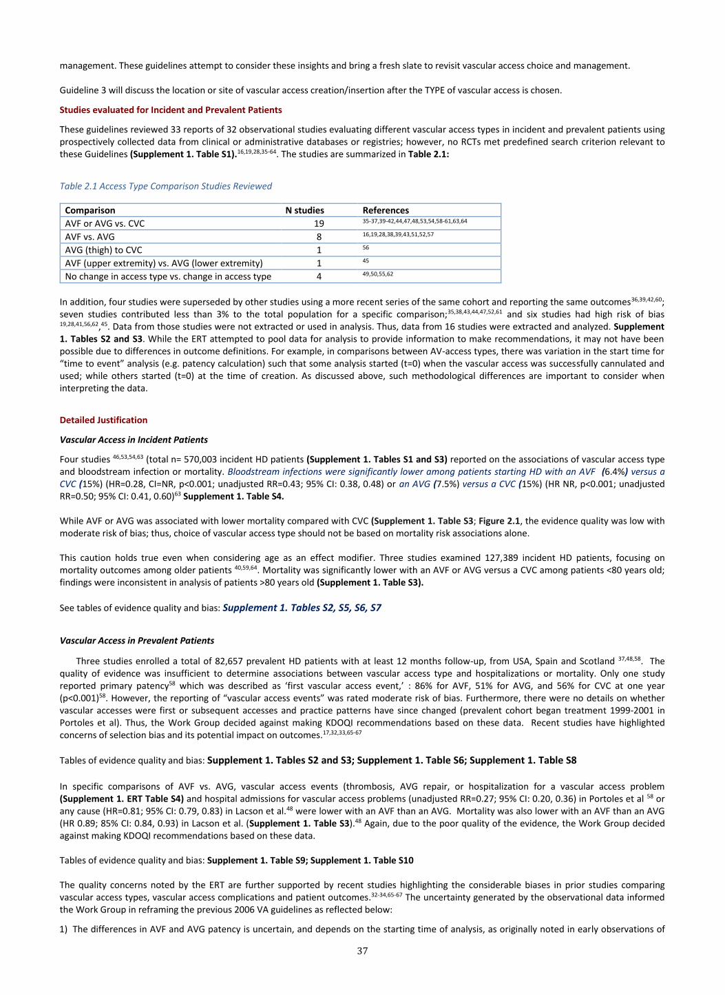
37
management. These guidelines attempt to consider these insights and bring a fresh slate to revisit vascular access choice and management.
Guideline 3 will discuss the location or site of vascular access creation/insertion after the TYPE of vascular access is chosen.
Studies evaluated for Incident and Prevalent Patients
These guidelines reviewed 33 reports of 32 observational studies evaluating different vascular access types in incident and prevalent patients using prospectively collected data from clinical or administrative databases or registries; however, no RCTs met predefined search criterion relevant to these Guidelines (Supplement 1. Table S1).16,19,28,35-64. The studies are summarized in Table 2.1:
Table 2.1 Access Type Comparison Studies Reviewed
Comparison N studies References
AVF or AVG vs. CVC 19 35-37,39-42,44,47,48,53,54,58-61,63,64
AVF vs. AVG 8 16,19,28,38,39,43,51,52,57
AVG (thigh) to CVC 1 56
AVF (upper extremity) vs. AVG (lower extremity) 1 45
No change in access type vs. change in access type 4 49,50,55,62
In addition, four studies were superseded by other studies using a more recent series of the same cohort and reporting the same outcomes36,39,42,60; seven studies contributed less than 3% to the total population for a specific comparison;35,38,43,44,47,52,61 and six studies had high risk of bias 19,28,41,56,62,45. Data from those studies were not extracted or used in analysis. Thus, data from 16 studies were extracted and analyzed. Supplement 1. Tables S2 and S3. While the ERT attempted to pool data for analysis to provide information to make recommendations, it may not have been possible due to differences in outcome definitions. For example, in comparisons between AV-access types, there was variation in the start time for “time to event” analysis (e.g. patency calculation) such that some analysis started (t=0) when the vascular access was successfully cannulated and used; while others started (t=0) at the time of creation. As discussed above, such methodological differences are important to consider when interpreting the data.
Detailed Justification
Vascular Access in Incident Patients
Four studies 46,53,54,63 (total n= 570,003 incident HD patients (Supplement 1. Tables S1 and S3) reported on the associations of vascular access type and bloodstream infection or mortality. Bloodstream infections were significantly lower among patients starting HD with an AVF (6.4%) versus a CVC (15%) (HR=0.28, CI=NR, p<0.001; unadjusted RR=0.43; 95% CI: 0.38, 0.48) or an AVG (7.5%) versus a CVC (15%) (HR NR, p<0.001; unadjusted RR=0.50; 95% CI: 0.41, 0.60)63 Supplement 1. Table S4. While AVF or AVG was associated with lower mortality compared with CVC (Supplement 1. Table S3; Figure 2.1, the evidence quality was low with moderate risk of bias; thus, choice of vascular access type should not be based on mortality risk associations alone. This caution holds true even when considering age as an effect modifier. Three studies examined 127,389 incident HD patients, focusing on mortality outcomes among older patients 40,59,64. Mortality was significantly lower with an AVF or AVG versus a CVC among patients <80 years old; findings were inconsistent in analysis of patients >80 years old (Supplement 1. Table S3).
See tables of evidence quality and bias: Supplement 1. Tables S2, S5, S6, S7
Vascular Access in Prevalent Patients
Three studies enrolled a total of 82,657 prevalent HD patients with at least 12 months follow-up, from USA, Spain and Scotland 37,48,58. The quality of evidence was insufficient to determine associations between vascular access type and hospitalizations or mortality. Only one study reported primary patency58 which was described as ‘first vascular access event,’ : 86% for AVF, 51% for AVG, and 56% for CVC at one year (p<0.001)58. However, the reporting of “vascular access events” was rated moderate risk of bias. Furthermore, there were no details on whether vascular accesses were first or subsequent accesses and practice patterns have since changed (prevalent cohort began treatment 1999-2001 in Portoles et al). Thus, the Work Group decided against making KDOQI recommendations based on these data. Recent studies have highlighted concerns of selection bias and its potential impact on outcomes.17,32,33,65-67
Tables of evidence quality and bias: Supplement 1. Tables S2 and S3; Supplement 1. Table S6; Supplement 1. Table S8 In specific comparisons of AVF vs. AVG, vascular access events (thrombosis, AVG repair, or hospitalization for a vascular access problem (Supplement 1. ERT Table S4) and hospital admissions for vascular access problems (unadjusted RR=0.27; 95% CI: 0.20, 0.36) in Portoles et al 58 or any cause (HR=0.81; 95% CI: 0.79, 0.83) in Lacson et al.48 were lower with an AVF than an AVG. Mortality was also lower with an AVF than an AVG (HR 0.89; 85% CI: 0.84, 0.93) in Lacson et al. (Supplement 1. Table S3).48 Again, due to the poor quality of the evidence, the Work Group decided against making KDOQI recommendations based on these data. Tables of evidence quality and bias: Supplement 1. Table S9; Supplement 1. Table S10 The quality concerns noted by the ERT are further supported by recent studies highlighting the considerable biases in prior studies comparing vascular access types, vascular access complications and patient outcomes.32-34,65-67 The uncertainty generated by the observational data informed the Work Group in reframing the previous 2006 VA guidelines as reflected below:
1) The differences in AVF and AVG patency is uncertain, and depends on the starting time of analysis, as originally noted in early observations of
38
AVF patency68. Subsequent studies have demonstrated equivalency of AVF and AVG patency28,69-71 2) There are few RCTs informing the comparisons of AVF vs. AVG; those that have been done in fact demonstrate there may be benefit of AVG,
including in patients with poor vessels and greater comorbidity than in those with AVF72,73 3) The statement that AVFs may have fewer complications compared to AVGs, may not be generalizable to all AVF and AVG and may further
depend on what constitutes a complication. The increase in AVF creation has identified a significant and variably high rate of AVF failure to mature or non-usability for dialysis (20-60%)12,15,22,28,31,74. This non-usability for dialysis should be considered a complication, particularly as it requires interventions (e.g. angioplasty/BAM/ ligations etc.) to facilitate its use and/or CVC insertion if dialysis is needed. Any need for an unintended intervention, including CVC insertion should be considered a complication. This is well known for AVG thrombosis and the required corrective interventions (angioplasty, thrombectomy or surgical revision). In general, complications occur early in AVFs and later in AVGs – the total rate of complications for AVFs or AVGs has not been compared. This is particularly important when considering the required lifespan of an AV-access to serve the patient, according to their ESKD Life-Plan. See case examples.
Special discussions
When analyzing vascular access outcomes, it is important to clarify the “incident/prevalent” designation. In some studies, the “incident/prevalent” status refers to the PATIENT’s status – NOT the vascular access status. The vascular access used in an incident dialysis patient may not necessarily mean it was the patient’s first access. For example, an incident patient starting dialysis with a CVC may have had a prior failed AVF. Similarly, an incident HD patient using an AVF may have had a prior failed AVF; the behavior of a first versus a subsequent access and the impact on the patient may differ. Clearly, the implications of both the patient status (as incident or prevalent on dialysis) and of the vascular access (as first or subsequent) are related; however, their relative contributions to the outcome of interest are unclear and likely to depend on clinical circumstances. For example, at time of analysis, a prevalent hemodialysis patient who has a successful functioning AVF as their “first” vascular access is different than a prevalent dialysis patient who has had multiple failed AVF who is now analyzed with a CVC as their “prevalent” (or “subsequent”) vascular access – both patient and vascular access outcomes would likely differ, even though they are both considered “prevalent”. These analyses do not provide information in that regard and the associative findings should be considered in that light. To further complicate matters, the definitions of important vascular access outcomes (such as “patency”) have been highly inconsistent, making it challenging, if not impossible to make valid comparisons between vascular access types and between studies1,75. For example, as discussed under vascular access for incident patients, prior patency comparisons of AVFs vs. AVGs evaluated only AVF that were successfully used and excluded AVF that had primary failure or abandoned without use27,31. The impact of concluding AVF superiority on this premise led to the creation of many AVF that were not usable for dialysis28,31,74. Any VA not usable for dialysis should be considered a complication attributable to that vascular access. Thus, this bias in analysis impacts the findings of both AVF patency superiority as well as complication rate. The current KDOQI guidelines on the choice of vascular access in prevalent patients reflect the prior inconsistencies in definitions and biases in analyses and interpretation in the existing literature. Future research is recommended to provide the necessary data required to make recommendations on type of vascular access for incident and prevalent patients in need of first or subsequent accesses. Please see section on future research recommendations. Implementation Considerations See Figure 2.2 ESKD Life-Plan Case Examples The outcomes of AV-accesses that have required interventions prior to use may have a different “natural history” than those not needing interventions (unassisted matured AV-accesses). Further comparative study is required; until such time, caution should be made in generalizing the outcomes of AVF and AVG noted in prior literature that have not taken this into consideration1
Special populations: Pediatrics The choice of vascular access for pediatric HD patients must take a number of factors into account. For incident patients with an unplanned dialysis initiation and who desire HD for KRT, a CVC will be necessary irrespective of age, body habitus, venous anatomy, or comorbidities. Placement of CVC may be required in patients with small or diminutive vessels that are not suitable for an AVF or AVG, but this should be considered a “bridge” or mid-term solution until the vessels sizes are adequate. There is no specific size cut off in terms of weight or vessel diameter in this setting, and, thus, the timing for access creation (AVF or AVG) should be based upon patient circumstances and local expertise. The use of CVC is associated with higher risk of bloodstream infections, greater hospitalization rates and antibiotic usage with potential nephrotoxic effects on residual kidney function, and damage to central vessels making future AV-access creation difficult. As such, pediatric dialysis centers must be committed to insightful best care for their patients- this current year and next as an integral part of their ESKD Life-Plan, which will include HD, transplant, and PD. The choice of AVF is preferred over AVG, but may be dictated by available vascular anatomy, other clinical factors and local surgical expertise for AV-access creation and interventional radiology expertise for maintaining AV-access function. The benefit margin of AVF over CVC in pediatrics has been well documented.76,77 However, evidence demonstrating the superiority of AVF over AVG, is less available in the pediatric hemodialysis patient.78 The best first strategy is CVC avoidance followed by CVC minimization. Further, for patients/families who are eligible and desire PD (even if needed acutely), urgent placement of a PD catheter is recommended to avoid the risks and long-term morbidities associated with CVC placement, even if only used for a few weeks to months.79
. Future Research
• First vs. subsequent accesses
• RCT of various types – this is critical but challenging and may be impossible?
• Consider complication rate as an outcome (define what is viewed as a complication e.g. for AVF that FTM, use of a CVC may be deemed a complication; etc.)
• Validation of standardized definitions for patency and complications
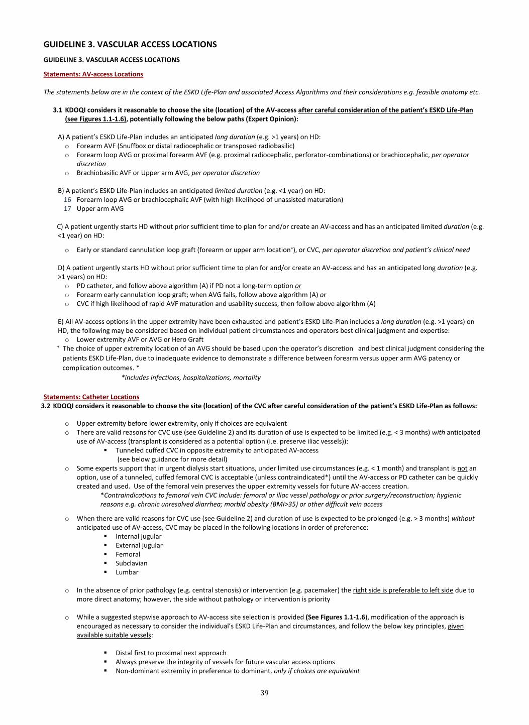
39
GUIDELINE 3. VASCULAR ACCESS LOCATIONS
GUIDELINE 3. VASCULAR ACCESS LOCATIONS
Statements: AV-access Locations The statements below are in the context of the ESKD Life-Plan and associated Access Algorithms and their considerations e.g. feasible anatomy etc.
3.1 KDOQI considers it reasonable to choose the site (location) of the AV-access after careful consideration of the patient’s ESKD Life-Plan (see Figures 1.1-1.6), potentially following the below paths (Expert Opinion):
A) A patient’s ESKD Life-Plan includes an anticipated long duration (e.g. >1 years) on HD: o Forearm AVF (Snuffbox or distal radiocephalic or transposed radiobasilic) o Forearm loop AVG or proximal forearm AVF (e.g. proximal radiocephalic, perforator-combinations) or brachiocephalic, per operator
discretion o Brachiobasilic AVF or Upper arm AVG, per operator discretion
B) A patient’s ESKD Life-Plan includes an anticipated limited duration (e.g. <1 year) on HD:
16 Forearm loop AVG or brachiocephalic AVF (with high likelihood of unassisted maturation) 17 Upper arm AVG
C) A patient urgently starts HD without prior sufficient time to plan for and/or create an AV-access and has an anticipated limited duration (e.g.
<1 year) on HD:
o Early or standard cannulation loop graft (forearm or upper arm location+), or CVC, per operator discretion and patient’s clinical need D) A patient urgently starts HD without prior sufficient time to plan for and/or create an AV-access and has an anticipated long duration (e.g. >1 years) on HD: o PD catheter, and follow above algorithm (A) if PD not a long-term option or o Forearm early cannulation loop graft; when AVG fails, follow above algorithm (A) or o CVC if high likelihood of rapid AVF maturation and usability success, then follow above algorithm (A)
E) All AV-access options in the upper extremity have been exhausted and patient’s ESKD Life-Plan includes a long duration (e.g. >1 years) on HD, the following may be considered based on individual patient circumstances and operators best clinical judgment and expertise: o Lower extremity AVF or AVG or Hero Graft
+ The choice of upper extremity location of an AVG should be based upon the operator’s discretion and best clinical judgment considering the
patients ESKD Life-Plan, due to inadequate evidence to demonstrate a difference between forearm versus upper arm AVG patency or
complication outcomes. *
*includes infections, hospitalizations, mortality
Statements: Catheter Locations 3.2 KDOQI considers it reasonable to choose the site (location) of the CVC after careful consideration of the patient’s ESKD Life-Plan as follows:
o Upper extremity before lower extremity, only if choices are equivalent o There are valid reasons for CVC use (see Guideline 2) and its duration of use is expected to be limited (e.g. < 3 months) with anticipated
use of AV-access (transplant is considered as a potential option (i.e. preserve iliac vessels)): ▪ Tunneled cuffed CVC in opposite extremity to anticipated AV-access
(see below guidance for more detail) o Some experts support that in urgent dialysis start situations, under limited use circumstances (e.g. < 1 month) and transplant is not an
option, use of a tunneled, cuffed femoral CVC is acceptable (unless contraindicated*) until the AV-access or PD catheter can be quickly created and used. Use of the femoral vein preserves the upper extremity vessels for future AV-access creation.
*Contraindications to femoral vein CVC include: femoral or iliac vessel pathology or prior surgery/reconstruction; hygienic reasons e.g. chronic unresolved diarrhea; morbid obesity (BMI>35) or other difficult vein access
o When there are valid reasons for CVC use (see Guideline 2) and duration of use is expected to be prolonged (e.g. > 3 months) without anticipated use of AV-access, CVC may be placed in the following locations in order of preference:
▪ Internal jugular ▪ External jugular ▪ Femoral ▪ Subclavian ▪ Lumbar
o In the absence of prior pathology (e.g. central stenosis) or intervention (e.g. pacemaker) the right side is preferable to left side due to
more direct anatomy; however, the side without pathology or intervention is priority
o While a suggested stepwise approach to AV-access site selection is provided (See Figures 1.1-1.6), modification of the approach is encouraged as necessary to consider the individual’s ESKD Life-Plan and circumstances, and follow the below key principles, given available suitable vessels:
▪ Distal first to proximal next approach ▪ Always preserve the integrity of vessels for future vascular access options ▪ Non-dominant extremity in preference to dominant, only if choices are equivalent
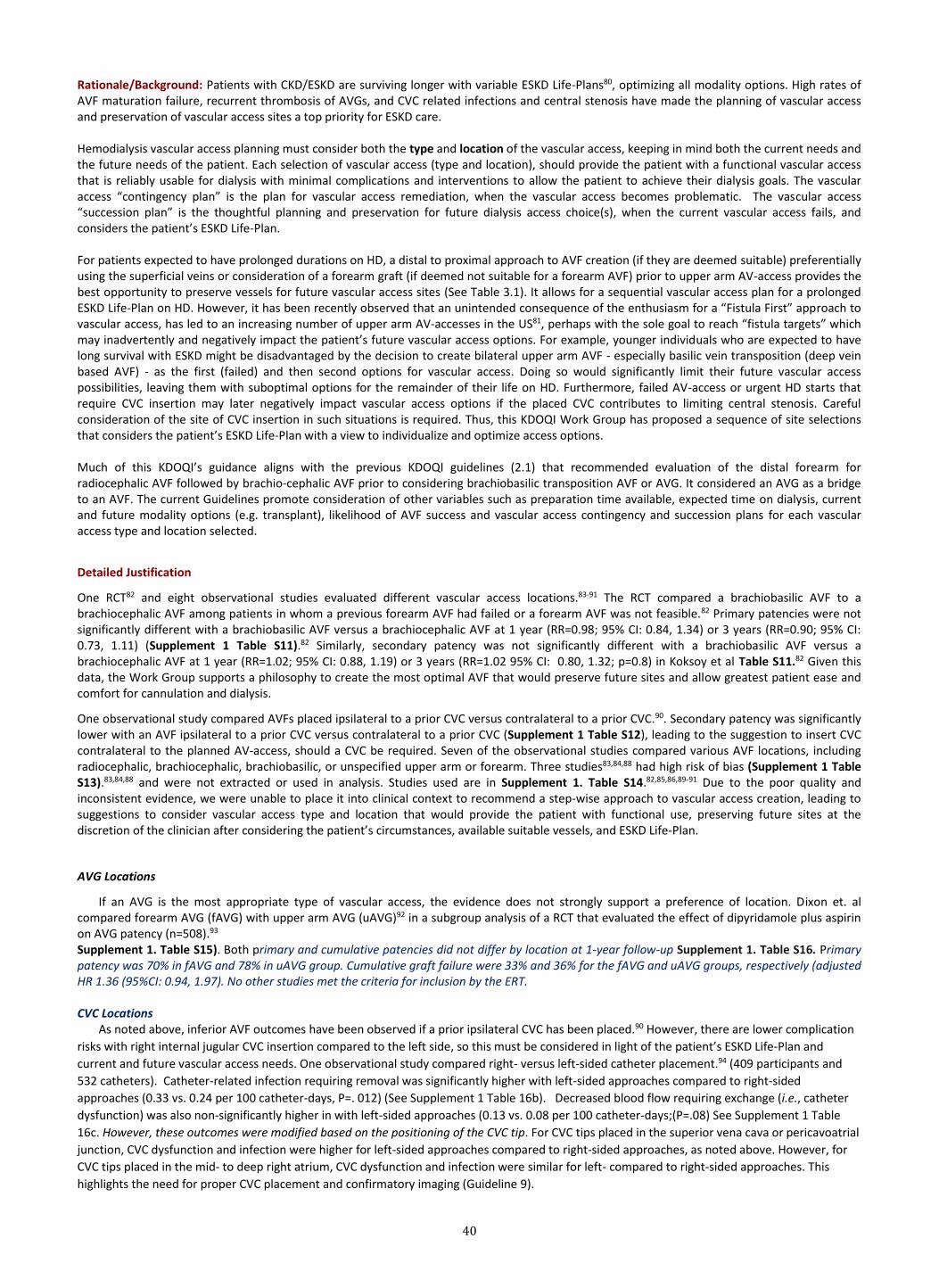
40
Rationale/Background: Patients with CKD/ESKD are surviving longer with variable ESKD Life-Plans80, optimizing all modality options. High rates of AVF maturation failure, recurrent thrombosis of AVGs, and CVC related infections and central stenosis have made the planning of vascular access and preservation of vascular access sites a top priority for ESKD care. Hemodialysis vascular access planning must consider both the type and location of the vascular access, keeping in mind both the current needs and the future needs of the patient. Each selection of vascular access (type and location), should provide the patient with a functional vascular access that is reliably usable for dialysis with minimal complications and interventions to allow the patient to achieve their dialysis goals. The vascular access “contingency plan” is the plan for vascular access remediation, when the vascular access becomes problematic. The vascular access “succession plan” is the thoughtful planning and preservation for future dialysis access choice(s), when the current vascular access fails, and considers the patient’s ESKD Life-Plan. For patients expected to have prolonged durations on HD, a distal to proximal approach to AVF creation (if they are deemed suitable) preferentially using the superficial veins or consideration of a forearm graft (if deemed not suitable for a forearm AVF) prior to upper arm AV-access provides the best opportunity to preserve vessels for future vascular access sites (See Table 3.1). It allows for a sequential vascular access plan for a prolonged ESKD Life-Plan on HD. However, it has been recently observed that an unintended consequence of the enthusiasm for a “Fistula First” approach to vascular access, has led to an increasing number of upper arm AV-accesses in the US81, perhaps with the sole goal to reach “fistula targets” which may inadvertently and negatively impact the patient’s future vascular access options. For example, younger individuals who are expected to have long survival with ESKD might be disadvantaged by the decision to create bilateral upper arm AVF - especially basilic vein transposition (deep vein based AVF) - as the first (failed) and then second options for vascular access. Doing so would significantly limit their future vascular access possibilities, leaving them with suboptimal options for the remainder of their life on HD. Furthermore, failed AV-access or urgent HD starts that require CVC insertion may later negatively impact vascular access options if the placed CVC contributes to limiting central stenosis. Careful consideration of the site of CVC insertion in such situations is required. Thus, this KDOQI Work Group has proposed a sequence of site selections that considers the patient’s ESKD Life-Plan with a view to individualize and optimize access options. Much of this KDOQI’s guidance aligns with the previous KDOQI guidelines (2.1) that recommended evaluation of the distal forearm for radiocephalic AVF followed by brachio-cephalic AVF prior to considering brachiobasilic transposition AVF or AVG. It considered an AVG as a bridge to an AVF. The current Guidelines promote consideration of other variables such as preparation time available, expected time on dialysis, current and future modality options (e.g. transplant), likelihood of AVF success and vascular access contingency and succession plans for each vascular access type and location selected.
Detailed Justification
One RCT82 and eight observational studies evaluated different vascular access locations.83-91 The RCT compared a brachiobasilic AVF to a brachiocephalic AVF among patients in whom a previous forearm AVF had failed or a forearm AVF was not feasible.82 Primary patencies were not significantly different with a brachiobasilic AVF versus a brachiocephalic AVF at 1 year (RR=0.98; 95% CI: 0.84, 1.34) or 3 years (RR=0.90; 95% CI: 0.73, 1.11) (Supplement 1 Table S11).82 Similarly, secondary patency was not significantly different with a brachiobasilic AVF versus a brachiocephalic AVF at 1 year (RR=1.02; 95% CI: 0.88, 1.19) or 3 years (RR=1.02 95% CI: 0.80, 1.32; p=0.8) in Koksoy et al Table S11.82 Given this data, the Work Group supports a philosophy to create the most optimal AVF that would preserve future sites and allow greatest patient ease and comfort for cannulation and dialysis.
One observational study compared AVFs placed ipsilateral to a prior CVC versus contralateral to a prior CVC.90. Secondary patency was significantly lower with an AVF ipsilateral to a prior CVC versus contralateral to a prior CVC (Supplement 1 Table S12), leading to the suggestion to insert CVC contralateral to the planned AV-access, should a CVC be required. Seven of the observational studies compared various AVF locations, including radiocephalic, brachiocephalic, brachiobasilic, or unspecified upper arm or forearm. Three studies83,84,88 had high risk of bias (Supplement 1 Table S13).83,84,88 and were not extracted or used in analysis. Studies used are in Supplement 1. Table S14.82,85,86,89-91 Due to the poor quality and inconsistent evidence, we were unable to place it into clinical context to recommend a step-wise approach to vascular access creation, leading to suggestions to consider vascular access type and location that would provide the patient with functional use, preserving future sites at the discretion of the clinician after considering the patient’s circumstances, available suitable vessels, and ESKD Life-Plan.
AVG Locations
If an AVG is the most appropriate type of vascular access, the evidence does not strongly support a preference of location. Dixon et. al compared forearm AVG (fAVG) with upper arm AVG (uAVG)92 in a subgroup analysis of a RCT that evaluated the effect of dipyridamole plus aspirin on AVG patency (n=508).93 Supplement 1. Table S15). Both primary and cumulative patencies did not differ by location at 1-year follow-up Supplement 1. Table S16. Primary patency was 70% in fAVG and 78% in uAVG group. Cumulative graft failure were 33% and 36% for the fAVG and uAVG groups, respectively (adjusted HR 1.36 (95%CI: 0.94, 1.97). No other studies met the criteria for inclusion by the ERT. CVC Locations
As noted above, inferior AVF outcomes have been observed if a prior ipsilateral CVC has been placed.90 However, there are lower complication
risks with right internal jugular CVC insertion compared to the left side, so this must be considered in light of the patient’s ESKD Life-Plan and
current and future vascular access needs. One observational study compared right- versus left-sided catheter placement.94 (409 participants and
532 catheters). Catheter-related infection requiring removal was significantly higher with left-sided approaches compared to right-sided
approaches (0.33 vs. 0.24 per 100 catheter-days, P=. 012) (See Supplement 1 Table 16b). Decreased blood flow requiring exchange (i.e., catheter
dysfunction) was also non-significantly higher in with left-sided approaches (0.13 vs. 0.08 per 100 catheter-days;(P=.08) See Supplement 1 Table
16c. However, these outcomes were modified based on the positioning of the CVC tip. For CVC tips placed in the superior vena cava or pericavoatrial
junction, CVC dysfunction and infection were higher for left-sided approaches compared to right-sided approaches, as noted above. However, for
CVC tips placed in the mid- to deep right atrium, CVC dysfunction and infection were similar for left- compared to right-sided approaches. This
highlights the need for proper CVC placement and confirmatory imaging (Guideline 9).

41
Special discussions
Surgeons: Practical considerations of choice of vessels
Where appropriate, consider use of superficial veins before deep veins. Recall that venous drainage in the extremity is from superficial (veins superficial to deep fascia) to deep venous system (veins deep to deep fascia). Deep veins tend to converge in a single vein as it approaches the heart 95. Obliteration or stenoses in the deep veins impair the availability for more distal access sites. Thus, evaluation of potential vascular access sites could consider first utilizing the superficial veins for the outflow. Forearm and upper arm cephalic and the forearm basilic veins are considered superficial veins 96. The Work Group felt that it is reasonable to consider a forearm loop graft in the sequence as a bridge to future AVF when the superficial vein based options are exhausted. Importantly, the AVF location should consider the maturation of the outflow vein, facilitating future cannulation. An adequate cannulation vein segment should be long enough to accommodate: 1) needle separation of at least 1 inches (2.54 cm) or 2 finger widths, and 2) rotation of cannulation sites to avoid aneurysm development. Furthermore, it should be easily and comfortably accessible for the patient. Having segments on the underside of the arm can be uncomfortable or very painful. Segments that are too deep may be challenging to cannulate without ultrasound guidance, which may not be readily available and/or may lead to patient and nursing/technician frustration. While superficialization of such deep AVF may be possible, it is important to note that AVF use for dialysis is typically delayed due to the need for a planned secondary procedure. See also commentary in Guideline 8.
Special considerations for femoral vein catheterization
In select patients and limited circumstances, tunneled cuffed femoral vein catheterization may be reasonable. For example, in situations where patients urgently start dialysis and are not transplant candidates but are in clinical environments that are conducive to quick dialysis access creation/insertion (i.e. within 1 month), tunneled cuffed femoral CVC may serve several beneficial purposes. Firstly, it will preserve central veins to ensure AV-access is feasible (assuming central veins not previously damaged or stenosed by other process). Secondly, use of femoral vein CVC reminds patients and providers that CVC is only for temporary purposes. This strategy is only effective if the facility can coordinate rapid AV-access creation for patients destined for HD (e.g. early cannulation AVG or AVF in a patient with high likelihood of maturation success) or PD catheter insertion for patients who have chosen PD. Issues such as proper placement of the exit site to ensure patient comfort and dignity must be considered (Figure 3.1) . Of note, RCTs, metanalysis and systematic reviews have demonstrated equivalent or superior outcomes with regards to CVC thrombosis and infection, using tunneled femoral vein CVC. However, precaution with femoral CVC use, as with any CVC use must be taken (Guidelines 20-25)97-99
Table 3.1 Vessel location by distal to proximal sites
Vessel location/ Cannulation location
AVF AVG
Forearm /Forearm Snuffbox or distal radiocephalic Forearm loop
Forearm /Upper arm Proximal radiocephalic Antecubital perforator combinations
Upper Arm/Upper arm Brachiocephalic Upper arm straight Upper arm loop Brachiobasilic
Implementation considerations
It is important to reiterate that the primary goal of vascular access creation and planning is to create a functional vascular access that can provide reliable prescribed dialysis, with minimal interventions and complications. The planning must consider contingency plans for remediation when the vascular access becomes problematic and a succession plan for future dialysis access options when the vascular access eventually fails. This applies to all populations in need of dialysis access, including CKD patients with a failing transplant, a PD patient who requires transition to HD, pediatric CKD patients, other established CKD patients or patients needing an urgent start to dialysis. A proactive approach to the pediatric CKD patient with failing eGFR is imperative to avoid CVC placement for hemodialysis initiation. This means allowing adequate maturation time to allow the AV-access to be usable on the day of HD initiation- typically several months of lead time for AVF placement and a few weeks for an AVG placement. CVC avoidance for elective hemodialysis is achievable with adherence to ESKD Life-Plan. It is imperative that health care providers communicate the ESKD Life-Plan, vascular access creation, contingency and succession plans to provide continuity in KRT and dialysis access care.
Future research
• Medium sized peripheral arteries (radial and ulnar) > 2 mm can develop AVF to support adequate dialysis 100. Future research can help determine if larger arteries with higher baseline flows are associated with greater risk of flow-associated problems.
• Weighing the potential options for access type and location, as discussed, will require more accurate patient-specific estimates of the predicted duration of HD and predicted probability of AVF maturation.
42
GUIDELINE 4. NOVEL MATERIALS AND AV-ACCESS
Statements: Novel Materials and AV-Access 4.1 KDOQI suggests that the choice of material for an AVG should be based upon the clinician’s /surgeon’s discretion and best clinical
judgment since the current evidence does not demonstrate that one graft material or modification thereof is associated with improved outcomes in terms of patency or complications (Conditional recommendation, Low quality of evidence).
4.2 KDOQI considers it reasonable to use early cannulation grafts as a catheter sparing strategy in appropriate patients, considering their ESKD Life-Plan (Expert opinion)
Rationale/Background
Effective hemodialysis requires a suitable access for the withdrawal and return of blood. The ideal hemodialysis access should provide adequate flow rates to sustain prescribed dialysis, should be easy to access/cannulate, inexpensive, acceptable to patients and associated with excellent long-term patency and minimal complications. A mature, functional autogenous arteriovenous access or arteriovenous fistula (AVF) may be the most ideal access although not all patients have suitable veins for an AVF and not all of the veins mature sufficient for cannulation, thereby negating their potential advantages. Furthermore, in the culture of patient-centered care, an AVF may not always be the appropriate access, given individual circumstances. Acceptable arteriovenous access can be created using a variety of grafts (arteriovenous graft - AVG) including prosthetic, biologic and autogenous materials. Expanded polytetrafluoroethylene (PTFE) has been the most commonly used graft material since its original description and has served as the comparator or “gold standard” for numerous randomized trials. A variety of other prosthetic graft materials are commercially available although none have proven to be superior to PTFE in terms of the patency or complications. Similarly, a variety of graft modifications have been made to improve AVG performance including changes in wall thickness, surface modifications, diameter changes, and external support, but none have proven superior to the standard wall 6 mm PTFE graft. The use of autogenous and biological grafts had largely been abandoned with the introduction of PTFE although some limited recent evidence suggests that non-autogenous saphenous vein and bovine carotid artery biological grafts may be associated with fewer infectious complications and improved patency, respectively. The current KDOQI recommendations are consistent with the previous 2006 NKF-KDOQI VA guidelines.
Detailed Justification
PTFE has been the most widely used prosthetic material for AVGs since its introduction over 40 years ago.101. There have been a variety of RCTs and observational studies (see Supplement 1. Table 17) evaluating various AVG materials and configurations. For example, there have been comparisons of tetrafluoroethylene (Dacron)102,103 or polyurethane104,105 to PTFE although none have proven superior in terms of patency or complication rates. Similarly, the brand of PTFE does not appear to make a difference in terms of outcome.106,107
There have been numerous modifications of the prosthetic AVGs, predominantly PTFE, to improve patency and reduce complications although none of these modifications have proven in RCTs to be consistently superior to the standard wall 6 mm PTFE AVG. These modifications have included the thickness of the AVG material (i.e. thin wall PTFE),108 the elasticity of the AVG material (i.e. stretch PTFE),109 external reinforcement (e.g. removal external rings), tapered AVG diameter (i.e. 4 – 7 mm taper PTFE, 6 – 8 mm taper PTFE),110,111 addition of a cuff to the venous end of the AVG,112,113 surface modification with heparin,114 and modification of the external surface.115 Notably, Polo et al.111 reported that the 6 – 8 mm tapered PTFE upper arm AVGs had better primary and primary assisted patency rates than the 6 mm grafts without an increased incidence of AV-access steal although the patient population was limited to non-diabetics < 71 years of age. Dammers et al.110 reported no difference in the patency or incidence of steal symptoms between the 4 – 7 mm tapered and 6 mm PTFE AVGs. The 4 – 7 mm tapered grafts may theoretically reduce the incidence of steal symptoms due to the smaller diameter and resultant lower flow rates, however, the flow rates through the tapered grafts in their study were actually greater than the non-tapered ones. The RCTs examining the benefit of a venous outflow cuff have been contradictory with Sorum et al.113 reporting improved patency and increased flow rates for the cuffed AVGs. Of note, none of the cuffed AVGs failed as a result of a venous outflow stenosis. Data from a more recent study by Ko et al.112 found that primary patency was not significantly different with cuffed versus non-cuffed graft at 1 or 2 years (63% vs. 50% at one year, RR 1.23; 95% CI 0.71, 2.13; 45% versus 32% at 2 years, RR=1.56; 95% CI: 0.39, 6.19) (Supplement 1. Table 18).
Surface treatment of AVG with heparin affords a theoretical advantage in terms of thrombosis and patency although the available evidence failed to demonstrate any significant differences.114 Specifically, the patency (1 year primary patency with heparin-bonded (14%) versus standard AVG (12%) at 1 year (RR=1.10; 95% CI: 0.50, 2.44; 2 year secondary patency with heparin-bonded AVG (83%) versus standard AVG (73%) at 2 years (RR=1.14; 95% CI, 0.96, 1.34)), and complications rates were comparable between groups.
In terms of timing of graft cannulation, Aitken et al.116 reported that early cannulation PTFE grafts (i.e. modification of middle layer) were associated with reduced bacteremia and mortality when compared with tunneled dialysis CVC. A systematic review by Al Shakarchi et al.117 suggested that the newer generation early cannulation grafts can be safely cannulated without detriment and were associated with comparable patency and complications with standard wall PTFE although the number of studies that comprised the review was limited.
The HeRO graft (Merit Medical, South Jordan, UT), is a non-traditional vascular access comprised of a hybrid PTFE graft – catheter system that represents a variation of the prosthetic graft modifications outlined above. A recent RCT comparing the HeRO graft to standard wall PTFE demonstrated no difference in patency (primary patency 35% HERO versus 28% standard AVGs; RR=1.25; 95% CI: 0.54, 2.89 and secondary patency 66% HERO versus 56% standard AVGs at one year; RR=1.19; 95% CI: 0.75, 1.89) mortality (HeRO (2%) versus standard graft (0%) (RR=1.19; 95% CI: 0.44, 3.23; RD=0.02; 95% CI: - 0.02, 0.06) or complications.118 Notably, this trial was designed to compare the safety and efficacy of the HeRO graft in a cohort of patients without central vein occlusions or stenosis. However, the HeRO graft may have its greatest applicability in this subset of patients.
The role of autogenous and non-autogenous biological AVGs has evolved. Their use was largely abandoned with the introduction of PTFE (see above). Two recent trials suggest that biologic AVGs may confer some advantage although the sample sizes were relatively small (< 60 patients). Kennealey et al.119 reported that the bovine carotid artery AVG might be associated with improved outcome. Notably, the primary patency (bovine carotid artery AVG (61%) versus PTFE AVG (10%) at 1 year; RD=51%; 95% CI: 39% - 61%; p=0.006 by Kaplan-Meier analysis; however, the numbers at risk at each time point was not indicated), thrombosis rates (0.34 / patient-year) versus PTFE AVG (0.77/patient year) (RR=0.44; 95% CI: 0.29 -
43
0.67; p=0.01 by Poisson regression analysis) and re-intervention rates (1.45 / patient-year) versus a PTFE AVG (1.99 / patient-year) RR=0.73; 95% CI: 0.58, 0.91) were significantly lower when compared to PTFE AVG although there were no differences in the secondary patency ((64%) versus a PTFE graft (59%) at 2 years; P=NS) or other complications. Mousavi et al.120 reported that the infectious complication rates with non-autogenous human saphenous vein AVG were lower than PTFE although there were no differences in patency.
The saphenous and femoral veins have both been used as autogenous AVG materials.121,122 The saphenous vein is relatively thick walled and does not usually dilate when used for an AVF, similar to the cephalic or basilic veins traditionally used for upper extremity autogenous access. Furthermore, the saphenous vein diameter is not usually large enough to meet the minimal criteria for effective dialysis (i.e. > 4 mm, flow rate > 500 mL/min).123 The mean diameter of the femoral vein is sufficient for effective cannulation (i.e. 7 mm) and the length of the femoropopliteal segment is adequate for brachial-axillary access although its use in this configuration is associated with a modest incidence of wound complications and AV-access steal.121,124 A variety of novel techniques and AVGS are currently under investigation including a totally percutaneous or endovascular arteriovenous graft, fistula125 and a bioengineered graft comprised of human tissues.126 Although no RCTS have been performed to assess their efficacy, they may play a role in the future. Also see tables: Supplement 1. ERT Table 19. Risk of Bias Assessments: Novel Devices Supplement 1. ERT Table 20. Description of Eligible Studies: Novel Vascular Access Devices Special discussions
The Work Group discussed the need for the guidelines to be updated in an iterative fashion to allow for new hemodialysis access options and the ability to properly evaluate them and incorporate worthy options in future guidelines
Implementation considerations: See Guideline 1 Monitoring and evaluation: See Guidelines 11, 12, 14-20
Future research
• Further define the optimal non-autogenous graft material for an AV-access.
• Further validate the benefits identified for the current biologic grafts.
• Further develop novel techniques and graft materials for the creation of autogenous and non-autogenous AV-access.
44
CVC CONFIGURATIONS AND MATERIALS
GUIDELINE 5. CVC CONFIGURATION AND MATERIALS
Statements: CVC configuration and materials 5.1 KDOQI suggests that the choice of tunneled hemodialysis catheter type and design be based on the clinician’s discretion and best clinical
judgment (conditional recommendation, low quality evidence).
Rationale/Background
Identifying the type of CVC with the lowest complication rate is important for patients who are dependent upon CVC for chronic HD-whether for short term or long-term use.
However, RCTs comparing various CVC types and designs do not consistently demonstrate, with moderate or high-quality evidence, significant differences in primary assisted patency, CRBSI or thrombosis rates. Catheter types evaluated in these clinical trials include:
• Tesio-Cath twin catheter system versus LifeCath Twin catheter system127
• Palindrome Symmetric Tip versus HemoStar Long-Term CVCs (staggered tips), 128
• Palindrome Symmetric Tip versus Medcomp (step-tip) CVCs,129
• Ash Split (split-tip) versus PermCath or Optiflow (step-tip) catheters130,131
Detailed Justification
The below summarizes key evidence-based CVC comparisons: Twin catheter systems: Tesio vs. LifeCath Twin tunneled CVCs: One RCT (n=80) compared the Tesio-Cath (ovoid cuff) vs. the LifeCath Twin (cuboidal cuff); both CVC types have 2 free-floating lumens and were placed in the right internal jugular vein. Study follow-up was 12 months.127 There was no significant difference in CVC survival (92% for the Tesio-Cath group vs. 86% for the LifeCath Twin group), or CRBSI [0.40/1000 CVC days (Tesio group) and 0.51/1000 CVC days (LifeCath Twin group)] (P=0.7). Rates of hospitalization for infection and CVC-related mechanical complications were higher in the LifeCath Twin group compared with the Tesio-Cath group (0.94 versus 0.24 per 1000 CVC days, respectively (P=. 02)). This was associated with the greater need for urokinase infusions in the LifeCath Twin group (0.51 per 1000 CVC days) versus none in the Tesio-Cath group. (See, Supplement 1. Tables 21-23)).
Palindrome symmetric tipped vs. HemoStar staggered tipped tunneled CVC A number of studies found no significant difference in primary assisted patency, CRBSI or thrombosis rates when comparing Palindrome symmetric tipped vs. HemoStar staggered tipped tunneled CVC as follows: One RCT (302 CVCs inserted in 239 participants)128 reported no statistically significant difference in infection and thrombosis-free CVC survival (following censoring for CVC design–unrelated removal), or CRBSI (0.24 and 0.10 per 1000 CVC days (HR 2.26 [95%CI 0.44, 11.96]) in the Palindrome and HemoStar CVC groups, respectively). While rates of thrombosis or CVC removal due to thrombosis did not differ between groups, the need for a urokinase infusion was lower for the Palindrome group vs. the HemoStar group, 17 versus 35 (HR 0.58 [95%CI 0.49, 0.68]). (Supplement 1. Tables 22-24)
Note that urokinase is rarely used in the United States today. It is unknown whether the decreased urokinase use in Palindrome CVCs would also apply with other more commonly used thrombolytic agents-such as recombinant tissue plasminogen activator (rTPA).
Palindrome symmetric tipped vs. Medcomp stepped-tipped catheter One RCT (n=97) of 2 months duration showed CVC survival at 2 months was significantly higher in the symmetric tip group compared with the step-tip group, 91% versus 69% (P=0.02).129 For the indication of decreased blood flow rate, fewer CVCs were removed in the symmetric tip CVC group compared with the step-tip CVC group (6% versus 22% (P=. 04) (Supplement 1. Tables 22,23,25, 26).
Ash split-tip catheter and Permcath or Optiflow step-tip catheters There were two RCTs.130,131 One study compared the Ash Split with the PermCath catheter (N=69).130 CVC survival at 12 months following censoring for renal recovery, fistula formation, peritoneal dialysis, and transplantation was higher in the PermCath group compared with the Ash Split group (74% versus 49%) with no difference between groups in CVC removal due to sepsis, rates of infection, or CVC occlusion requiring removal. The other study compared the Ash Split to the Optiflow catheter (n=132).131 In this study, overall CVC survival at 180 days was higher in the Ash Split CVC (75%) compared to the Optiflow CVC (55%) (P=. 02).
Quality of Evidence and Risk of Bias: See Supplement 1. Tables 27-29). Special discussions
• Catheter coatings are addressed in a separate section
Future research
• More clinical studies are needed to identify the ideal hemodialysis CVC that meets the criteria of long survival times, low CVC complication rates (dysfunction and CRBSI), and potentially lower patient complication rates (vascular stenosis and thrombosis). Scientific advances will need to focus on vascular biology and the design of biocompatible CVC materials to minimize complications of hemodialysis CVC for the select patients who are not candidates for an AVF or AVG.
• Future CVC designs should also focus on impact of CVC on development of central stenosis, vessel injury and/or thrombosis.
• All CVC design studies should report results using consistent definitions and outcomes.

45
CVC CONFIGURATIONS AND MATERIALS
46
GUIDELINE 6. TIMING, PREPARATION & PLANNING FOR CREATION/INSERTION OF DIALYSIS ACCESS
Statements: Education on ESKD Modalities and Dialysis Access 6.1 KDOQI considers it reasonable for adult and pediatric patients with an eGFR < 30 mL/min/1.73 m2 (CKD stage G4) with progressive decline
in kidney function, to be educated on all modalities of kidney replacement therapy (KRT) options, including transplantation, so that timely referral can be made for the appropriate modality and creation of a functional dialysis access, if necessary (Expert Opinion)
6.2 KDOQI considers it reasonable for adult and pediatric patients with a kidney transplant with an eGFR* < 30 mL/min/1.73 m2 with progressive decline in kidney function, to be educated on all modalities of kidney replacement therapy (KRT) options, including potential re-transplantation, so that timely referral can be made for the appropriate modality and creation of a functional dialysis access, if necessary (Expert Opinion) * if pediatric patient, estimate eGFR by Schwartz formula
6.3 KDOQI considers it reasonable for PD patients with complications refractory to therapy and/or with circumstances that make PD less
conducive than HD*, to be educated on all kidney transplant and HD options, so that timely referral can be made for the appropriate modality preparation and creation of a functional dialysis access, if necessary (Expert Opinion) *see Special discussions
6.4 KDOQI considers it reasonable for the predetermined dialysis access to be usable to provide the prescribed dialysis when the patient is ready to initiate dialysis (Expert Opinion)
Statements: Referral for AV-access In some facilities, referral of the patient for assessment by the vascular access team /surgeon for appropriate dialysis access is a different process than referral for the actual creation/insertion. However, for simplicity, the Guideline recommendations have been combined, keeping in mind variable timeframes between assessment for and creation of vascular access. Non-Dialysis CKD Patients 6.5 KDOQI considers it reasonable that in non-dialysis CKD patients* with progressive decline in kidney function, referral for dialysis access
assessment and subsequent creation should occur when eGFR is 15-20 ml/min. Earlier referral should occur in patient with unstable and/or rapid rates of eGFR decline (e.g. >10ml/min/year)+ (Expert Opinion)
*CKD, including patient with a failing transplant + In urgent situations (regardless of rate of progression), note that some newer graft materials may be cannulated immediately (<72 hours) after placement and can be considered along with the Patient’s ESKD Life-Plan
Dialysis Patients
6.6 KDOQI considers it reasonable that in HD patients with recurrent vascular access problems*, prompt referral for assessment and creation of a new AV-access should be made to allow adequate time for specialist consult and follow-up, and possible AV-access failure and correction; and should consider individual patient circumstances and competing risk of death (Expert Opinion).
* includes recurrent need for CVC use and/or >3 corrective interventions/6 months
When PD is the modality of choice:
6.7 KDOQI considers it reasonable and ideal to place a peritoneal dialysis (PD) catheter at least 2 weeks before the anticipated need of the PD treatments (Expert Opinion)
6.8 KDOQI considers it reasonable for an urgent peritoneal dialysis (PD) catheter to be placed for immediate PD as necessary under the direction and care of experienced personnel in conducive environments (Expert Opinion)
Statements for Vessel Preservation 6.9 KDOQI considers it reasonable to protect all central and peripheral arteries and veins from damage whenever possible, including the
avoidance of peripherally inserted venous catheters* and unnecessary venipunctures, for patients on dialysis or with chronic kidney disease where dialysis access is expected in the future (Stage G4 -G5) (Expert Opinion).
* other scenarios where vessel (artery or vein) damage may occur that should be avoided include: 1) radial artery access for coronary interventions, 2) Venous cardiovascular implantable electronic devices – alternatives such as epicardial/leadless pacing should be considered whenever possible.
Statements for Multidisciplinary Team Approach 6.10 KDOQI considers it reasonable to educate on, coordinate and manage all aspects of dialysis access using a multidisciplinary team within
the resource capacities and feasibilities of each facility (Expert Opinion)
6.11 There is inadequate evidence for KDOQI to make a recommendation on the use of a multidisciplinary team to reduce the rate of CVC or
increase the use of AVF
Rationale/Background
Timely referral for appropriate KRT modality education and choice is necessary to allow adequate time for preparation, creation and use of a functional dialysis access when it is needed at dialysis initiation. It is the first of many important steps required to attain “the right access, in the

47
right patient, at the right time, for the right reasons”, the guiding principles of these Guidelines.
These 2018 Guideline statements regarding modality and vascular access education are almost identical to the 2006 NKF-KDOQI VA guidelines,11 although these Guidelines emphasize the Expert Opinion basis due to the paucity of rigorous evidence to support recommendations.
Key differences in the statements for timing of education and referral for vascular access creation/insertion include using referral criteria that do not depend solely on GFR criteria and recognizing the challenges of creating vascular access based on a predicted time of dialysis start. A range of patient populations that may need vascular access is also recognized.
Detailed Justification Appropriate and well-balanced education and preparation is required for patients to choose the most suitable KRT modality and corresponding access, suitable for each patient’s own circumstances (see ESKD Life-Plan).
It has become increasingly evident that despite clinician’s best efforts, that predicting when a patient will initiate dialysis is challenging at best, and mostly inaccurate. Several studies have demonstrated that neither GFR alone nor a time duration before dialysis start is unreliable and using such criteria to base vascular access decisions leads to variable results132. Patient and process of care factors play contributing roles of varying degrees, depending on the different circumstances associated with each combination of interactions. These Guidelines attempted to find a balance between achieving a functional AV-access at dialysis initiation with potential unnecessary procedures that may occur if a predialysis patient’s kidney function does not decline or due to the competing risk of death. The competing risk of transplant or transfer to PD should be less of an issue if appropriate and timely modality education and preparation occurs, corresponding to the patients ESKD Life-Plan. Given the lack of prospective studies evaluating different GFR thresholds for vascular access creation, a well conducted simulation study supports an eGFR referral of 15-20 ml/min/1.73m2 133 Studies have also demonstrated increasing post creation procedure rates the longer time away from starting dialysis beyond 6-9 months, with no greater proportion of patients initiating dialysis with an AVF134. Such a time frame makes intuitive clinical sense given the time now required to ensure AVF maturation and usability, with an average time to cannulation of 132 days in the United States (USRDS 2017; Chapter 3) – for AVF that do not fail.135 The corresponding AVF that failed to mature was 35.9%. A 6-9 month time frame from dialysis initiation would allow time to create a new AV-access to be used for dialysis start should the first one fail; the challenge again, is being able to predict that start time. Thus, it is critical to allow time for surgical referral and creation, plan for risk of AV-access failure and correction, and consider trajectory of progression (if pre-dialysis) or CVC exposure time (if dialysis), along with the competing risk of death when referring for AV-access creation. Special Discussions Patients transferring to hemodialysis, after failing peritoneal dialysis, frequently start hemodialysis with a catheter.136 Special effort to examine the reasons for this and reduce CVC use in this population is required. Suggested indications for creation/insertion of a vascular access in peritoneal dialysis patients (See Table 6.1), are provided and based on Expert Opinion
Table 6.1 Suggested indications for Creation/Insertion of a Vascular Access in Peritoneal Dialysis Patients
Referral for HD Vascular Access in PD Patients
• Recurrent peritonitis, especially if due to poor connection technique (gram positive) or bowel leak (mixed gram negative)
• Ultrafiltration failure, especially in the presence of oligoanuria and persistent volume overload
• Decline in physical status considered to be the result of under dialysis (adherence issues or loss of residual kidney function)
o Note: This is a relative indication and should not be construed as implying that loss of RKF is itself an indication for vascular access creation
o Caution: The patient nonadherent to PD may very well be nonadherent to HD and vascular access creation
• Change in status of the patient, such that self-dialysis is no longer feasible o E.g. intercurrent event, e.g. stroke, death or other loss of a caregiver
• Significant non-infectious complication o E.g. Recurrent hernias, dialysate leaks including hydrothorax
Future research
• Validation of suggested criteria using both eGFR criteria and progressive decline in kidney function is required.
• Validation of criteria to refer PD patients for HD vascular access.
• Use of prediction equations to assist with timing of access? Does it really help?
• Develop and validate approaches to reduce potential damage to central and peripheral vessels by providers throughout the health care system.

48
Statements for Vessel Preservation
Rationale/Background
Central and peripheral vessel damage may contribute to CVC dysfunction and limit future options for AV-access creation and usability in CKD and ESKD patients. Due to the growing age and comorbidities of this population, the risk of and exposure to potentially vessel damaging conditions and interventions are high. For example, venipuncture itself and infusion of sclerosing medications though peripheral intravenous catheters may result in damage and/or thrombosis of peripheral vein segments that may later be used for AV-access creation or catheter insertion. In addition, catheterization of central veins for non-HD requirements such as internal jugular and subclavian lines for acute and chronic care, transvenous pacemakers and defibrillators are associated with stenosis or loss of central vein patency. Central stenosis is a serious complication that may prohibit successful AV-access creation, maturation or use. PICC’s are known to damage veins with subsequent thrombosis in many cases. There is also the risk of clot propagation into the central veins. Loss of these veins can have a dramatic impact on patient morbidity and mortality with loss of current and future hemodialysis vascular access sites137-139. Patients should be educated regarding these risks and encouraged to advocate for their own vessel protection, along with the nephrology community’s support. An informative website on saving veins can be found: https://www.saveyourvein.org, serving as an example of a public platform educating on the importance of saving veins. It can be used by patients and professionals. Indeed, caregivers involved in the delivery of care to CKD stage 4/5 patients should be educated regarding the implications of venous and arterial access use and institutional policies directed towards addressing these concerns are strongly suggested.
Detailed Justification
A case controlled study (N=120) demonstrated that the prior placement of a PICC was associated with a subsequent lower frequency of AVF use139 (odds ratio, 3.2; p<0.001,) even following adjustment for patient sex, artery and vein diameters, and prior CVC insertion. An observational study using USRDS data found that of 6487 HD patients with PICC’s placed within two years prior to and after AV-access creation were independently associated with lower likelihoods of transition to any working AV- access137. One study examining the presence of central vein stenosis (CVS) in association with PICCs reported a 7% rate of overall CVS140. However, this is likely an underestimation of the true incidence of CVS associated with PICCs, as the study was limited to patients undergoing serial extremity venography. In a prospective study examining thrombosis associated with PICC placement, all patients underwent ultrasound examination of the arm at 28 days after PICC insertion or at time of PICC removal and found an overall thrombosis rate of 71.9% (partial or complete obliteration of vessel lumen)141. Unfortunately, the national effort to reduce the use of PICC lines among CKD and ESKD patients has not achieved its objective. Notably, McGill et al.142 reported from their urban teaching hospital that > 30% of patients with CKD have had a PICC line and that > 50% were placed in their non-dominant arm. Angiographic comparisons of stenosis rates between subclavian and jugular hemodialysis CVC insertions found both were associated with stenosis, with 42% of subclavian veins stenosed after CVC insertion143. Lastly, the use of a CVC (including PICCs and catheters inserted at the subclavian and jugular veins) was found to be associated with a 14-fold increased risk of an upper extremity DVT144. Vein loss may be extrapolated from DVT formation. In a consensus statement regarding cardiovascular implantable devices in CKD and ESKD patients, it was noted that such patients derive a reduced survival benefit from ICD treatment compared to patients with normal renal function with a 2.7 fold higher risk of mortality145. In regard to central vein stenosis, 64% of patients developed stenosis146. Options to avoid upper body central veins include femoral vein placement (with its own risks of infradiaphragmatic venous damage), epicardial placement, subcutaneous ICD placement, leadless pacers, and possibly wearable defibrillators.147,148 Lastly, in regard to radial artery access for cardiac interventions, a meta-analysis of the literature noted a radial artery occlusion rate of <1%-33%. The conclusion was that radial artery occlusion was common149. Given that radial artery patency is integral to future radiocephalic AVF creation, femoral arterial access for coronary interventions should be strongly considered.
Special discussions
• Alternative options to PICCs are needed – potentially options suggested were pediatric internal jugular vein catheters/small bore catheter; however, whether or not their use translates into less vessel damage and central vein stenosis is unknown
• The “Choosing wisely” campaign should be fully supported in CKD/ESKD patients to “save the vein” and avoid PICCs whenever possible.
Implementation considerations
• Strategies to avoid PICCs and vessel damage e.g. venipuncture in back of wrist
• Avoid PICCs for < 7 days of infusion – use a peripheral IV preferably on the back of the hand
• Consider femoral venous access for central vein access
• CQI within/across institutions
• PICC placement in CKD patients in hospital requires approval by Nephrology
Monitoring and evaluation
• Use of PICCs when there are no other options
Future research
• Feasibility and use of other options for blood access
• Radial access impact on future VA creation
• Midline Catheter insertion effect on future VA creation
49
• Does use of small-bore internal jugular catheter reduce central venous stenosis?
• Rigorously evaluate the impact of radial artery access for cardiovascular and other procedural interventions on the creation and outcomes of AV-access for hemodialysis
Statements for Multidisciplinary Team Approach
Rationale/Background
As described previously (Guideline 1), the ESKD Life-Plan is a strategy for living with ESKD, ideally formulated by the patient and a multidisciplinary
team (coordinated chronic kidney disease (CKD) management team). A patient-centered approach to hemodialysis vascular access considers
multiple aspects of a patient’s needs and dialysis access eligibility specifically considering the patient’s current medical situation, their current and
future life goals, their preferences, social support, functional status, logistic and other practical feasibilities. For the purposes of dialysis access,
this team should include but is not limited to the following professionals and supportive members: nephrologist, access surgeon, radiologist,
nurse, social worker, patient family member or other supporter.
Placement of any dialysis access but particularly an AV-access requires education of the patient and family, as well as coordination of visits around
timely surgical consultation and follow-up visits. Given the limited ability of individual nephrologists to attend to the multiple and complex
aspects of vascular access care, most kidney care programs have established multidisciplinary vascular access teams.150-152 The multidisciplinary
VA team ensures timely referral for vascular access when appropriate, tracking, and monitoring patients’ vascular accesses for complications and
outcomes. Often central to the team is a vascular access coordinator (VAC) who coordinates and/or schedules the patient for procedures. In
addition, the VAC is key to educating patients on the risks and benefits of each vascular access choice, and addressing any patient concerns, and
facilitating appropriate use of the vascular access.153
Detailed Justification
While the multidisciplinary team approach appears ideal, the majority of studies of multidisciplinary team care were at high risk of bias154-162. Only
one study that evaluated a care coordinator.163 and one study evaluating multidisciplinary care,160 and one study evaluating patient education,164
were extracted and analyzed (Supplement 1. Table 30).
Multidisciplinary Care Wilson et al.160 (n=3636) analyzed patients starting hemodialysis in the DaVita system with follow-up for 360 days. An Incident Management of Patient, Actions Centered on Treatment (IMPACT) program for patients before and early in dialysis was instituted at selected clinics to manage predialysis and early dialysis issues, including initiating HD with an AV-access. The multidisciplinary team included a nephrologist, nurses, dietitians, social workers, and clinical care providers, involved structured intake, referral services, patient education, patient management checklists and timelines, and monitoring outcomes. Usual care also included patient education and other interventions, but not in a formalized manner. Patients in the IMPACT program were propensity-matched to patients receiving usual care in a 1:2 ratio -treatment groups were well-balanced for baseline characteristics. Use of an AVF or AVG versus a CVC was not significantly different among IMPACT patients (50%) versus usual-care patients (47%) (RR=1.06; 95% CI, 0.99, 1.14) at 90 days160. However, use of an AVF or AVG versus a CVC was significantly higher among IMPACT patients (63%) versus usual-care patients (48%) (RR=1.31; 95% CI, 1.22, 1.41) at 360 days.160
The success of the multidisciplinary team has historically been measured by increasing the rate of use of the AVF.165-168 However, there are a
number of other important outcomes, including the rate of VA-related procedures and complications. Recently Gill et al. performed an
observational study of vascular access outcomes in the first year of hemodialysis treatment before (2004-2005, pre-team period) and after
implementation of an access team (2006-2008 early team period; 2009-2011, late team period).169 Access team implementation did not affect the
probability of catheter-free use of the AVF (OR, 0.87; 95%confidence interval, 0.52 to 1.43, for the early; and 0.89; 0.52 to 1.53, for the late team
versus pre team period). Patients underwent an average of 4–5 total access-related procedures during the first year of hemodialysis, with higher
rates in women and in people with comorbidities. Catheter-related procedure rates were similar before and after team implementation; relative
to the pre team period: AVF-related procedure rates were 40% (20%–60%) and 30% (10%–50%) higher in the early-team and late-team periods,
respectively. One difficulty in interpreting the results of this study, is that concurrent with the implementation of the access team was the
widespread implementation of Fistula First policies in the study centers. The use of AVG was very low (<1%) in the centers involved in the study.
While the CVC rates were similar, the rates of procedures increased in AVF of women and those with greater comorbidities, factors associated
with poor AVF maturation and outcomes. It is unclear if results would have differed if those high-risk patients had received AVG instead. This
highlights the need to evaluate the use of multidisciplinary vascular access team to help patients delineate an appropriate ESKD Life-Plan with the
corresponding appropriate vascular access, including evaluation of feasibility for AVF.
Vascular access coordinator
Data on the true impact of a VAC for improving VA outcomes is lacking. Sixty percent of hemodialysis programs reported having at least one vascular access coordinator in a 2013 DOPPS survey of 72 United States sites, but they did not report whether an access team was present165,169. The percentage of patients using an AVF or a CVC did not differ across facilities with or without at least one vascular access coordinator. It is important to note that the percentage of use of one type of vascular access or another is insufficient for evaluating the usefulness of a VAC. It is not only the percentage use that matters, but of those using a particular vascular access, whether or not that use is appropriate, safe, provides patient satisfaction and supports their dialysis to help them achieve their own goals. Having a vascular access coordinator has been shown to

50
increase the odds of successful unassisted fistulas maturation, the ideal form of fistula maturation. Indeed, vascular access coordinators have the expertise, skills and capacity to build relationships with patients and multiple team members to educate, coordinate, guide and manage vascular access for the patient170,171 –typically much better than sole physicians and surgeons. For example, having a dedicated vascular access nurse or coordinator to assist physicians and staff with management of CVC-related infections has been reported to reduce CVC treatment failure rates and death from sepsis172-174 However, further research is required to generate more specific evidence to support vascular outcomes that can be attributable to the work of vascular access coordinators.
Future research Further research is needed to identify new strategies to deliver optimal, personalized, and patient-centered vascular access care. Data on the true
benefits of a multidisciplinary vascular access team are limited. The success of a multidisciplinary vascular access team is dependent on the
defined outcome measure. If it is increasing the use of AVFs there is conflicting data from observational studies using different compositions of
teams and roles of the team members. Additionally, these types of studies require accurate information regarding vascular access use across a
patient’s ESKD life span which is challenging and requires high-quality data assessing time periods of vascular access use with and without a
catheter.
Currently there is no data on the benefit of multidisciplinary teams on patient trust and confidence in making dialysis access related decisions, in
their quality of life or satisfaction with their dialysis access. Futures studies may also determine the impact of VAC and multidisciplinary teams on
health care spending, which may or may not show benefit by shifting care from physicians to lower-cost providers. This may also enhance patient
engagement and promote shared decision-making. These important outcome measures should be evaluated in future studies.
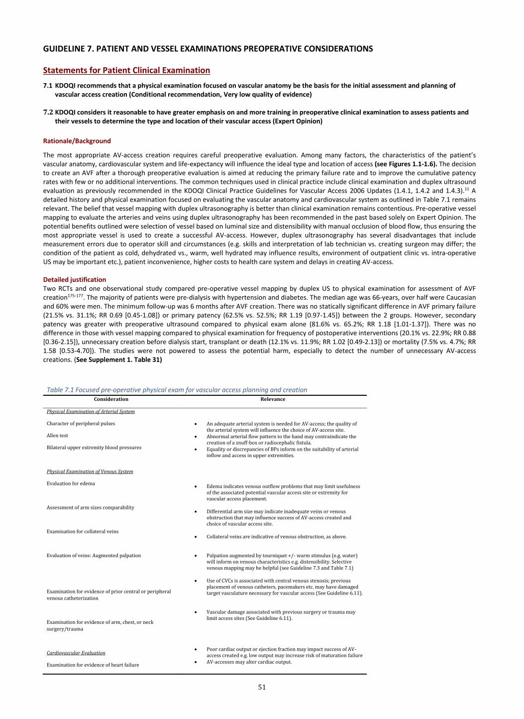
51
GUIDELINE 7. PATIENT AND VESSEL EXAMINATIONS PREOPERATIVE CONSIDERATIONS
Statements for Patient Clinical Examination
7.1 KDOQI recommends that a physical examination focused on vascular anatomy be the basis for the initial assessment and planning of vascular access creation (Conditional recommendation, Very low quality of evidence)
7.2 KDOQI considers it reasonable to have greater emphasis on and more training in preoperative clinical examination to assess patients and their vessels to determine the type and location of their vascular access (Expert Opinion)
Rationale/Background
The most appropriate AV-access creation requires careful preoperative evaluation. Among many factors, the characteristics of the patient’s vascular anatomy, cardiovascular system and life-expectancy will influence the ideal type and location of access (see Figures 1.1-1.6). The decision to create an AVF after a thorough preoperative evaluation is aimed at reducing the primary failure rate and to improve the cumulative patency rates with few or no additional interventions. The common techniques used in clinical practice include clinical examination and duplex ultrasound evaluation as previously recommended in the KDOQI Clinical Practice Guidelines for Vascular Access 2006 Updates (1.4.1, 1.4.2 and 1.4.3).11 A detailed history and physical examination focused on evaluating the vascular anatomy and cardiovascular system as outlined in Table 7.1 remains relevant. The belief that vessel mapping with duplex ultrasonography is better than clinical examination remains contentious. Pre-operative vessel mapping to evaluate the arteries and veins using duplex ultrasonography has been recommended in the past based solely on Expert Opinion. The potential benefits outlined were selection of vessel based on luminal size and distensibility with manual occlusion of blood flow, thus ensuring the most appropriate vessel is used to create a successful AV-access. However, duplex ultrasonography has several disadvantages that include measurement errors due to operator skill and circumstances (e.g. skills and interpretation of lab technician vs. creating surgeon may differ; the condition of the patient as cold, dehydrated vs., warm, well hydrated may influence results, environment of outpatient clinic vs. intra-operative US may be important etc.), patient inconvenience, higher costs to health care system and delays in creating AV-access. Detailed justification Two RCTs and one observational study compared pre-operative vessel mapping by duplex US to physical examination for assessment of AVF creation175-177. The majority of patients were pre-dialysis with hypertension and diabetes. The median age was 66-years, over half were Caucasian and 60% were men. The minimum follow-up was 6 months after AVF creation. There was no statically significant difference in AVF primary failure (21.5% vs. 31.1%; RR 0.69 [0.45-1.08]) or primary patency (62.5% vs. 52.5%; RR 1.19 [0.97-1.45]) between the 2 groups. However, secondary patency was greater with preoperative ultrasound compared to physical exam alone (81.6% vs. 65.2%; RR 1.18 [1.01-1.37]). There was no difference in those with vessel mapping compared to physical examination for frequency of postoperative interventions (20.1% vs. 22.9%; RR 0.88 [0.36-2.15]), unnecessary creation before dialysis start, transplant or death (12.1% vs. 11.9%; RR 1.02 [0.49-2.13]) or mortality (7.5% vs. 4.7%; RR 1.58 [0.53-4.70]). The studies were not powered to assess the potential harm, especially to detect the number of unnecessary AV-access creations. (See Supplement 1. Table 31)
Table 7.1 Focused pre-operative physical exam for vascular access planning and creation Consideration Relevance
Physical Examination of Arterial System
Character of peripheral pulses
Allen test
Bilateral upper extremity blood pressures
Physical Examination of Venous System
Evaluation for edema
Assessment of arm sizes comparability
Examination for collateral veins
Evaluation of veins: Augmented palpation
Examination for evidence of prior central or peripheral
venous catheterization
Examination for evidence of arm, chest, or neck
surgery/trauma
Cardiovascular Evaluation
Examination for evidence of heart failure
• An adequate arterial system is needed for AV-access; the quality of the arterial system will influence the choice of AV-access site.
• Abnormal arterial flow pattern to the hand may contraindicate the creation of a snuff-box or radiocephalic fistula.
• Equality or discrepancies of BPs inform on the suitability of arterial inflow and access in upper extremities.
• Edema indicates venous outflow problems that may limit usefulness of the associated potential vascular access site or extremity for vascular access placement.
• Differential arm size may indicate inadequate veins or venous obstruction that may influence success of AV-access created and choice of vascular access site.
• Collateral veins are indicative of venous obstruction, as above.
• Palpation augmented by tourniquet +/- warm stimulus (e.g. water) will inform on venous characteristics e.g. distensibility. Selective venous mapping may be helpful (see Guideline 7.3 and Table 7.1)
• Use of CVCs is associated with central venous stenosis; previous placement of venous catheters, pacemakers etc. may have damaged target vasculature necessary for vascular access (See Guideline 6.11).
• Vascular damage associated with previous surgery or trauma may limit access sites (See Guideline 6.11).
• Poor cardiac output or ejection fraction may impact success of AV-access created e.g. low output may increase risk of maturation failure
• AV-accesses may alter cardiac output.
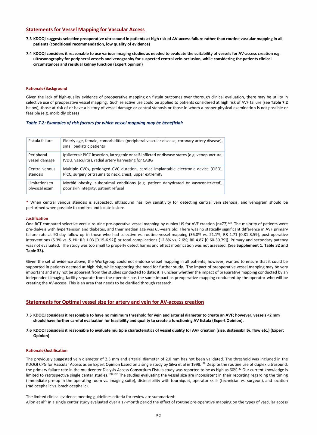
52
Statements for Vessel Mapping for Vascular Access
7.3 KDOQI suggests selective preoperative ultrasound in patients at high risk of AV-access failure rather than routine vascular mapping in all patients (conditional recommendation, low quality of evidence)
7.4 KDOQI considers it reasonable to use various imaging studies as needed to evaluate the suitability of vessels for AV-access creation e.g. ultrasonography for peripheral vessels and venography for suspected central vein occlusion, while considering the patients clinical circumstances and residual kidney function (Expert opinion)
Rationale/Background
Given the lack of high-quality evidence of preoperative mapping on fistula outcomes over thorough clinical evaluation, there may be utility in selective use of preoperative vessel mapping. Such selective use could be applied to patients considered at high risk of AVF failure (see Table 7.2 below), those at risk of or have a history of vessel damage or central stenosis or those in whom a proper physical examination is not possible or feasible (e.g. morbidly obese)
Table 7.2: Examples of risk factors for which vessel mapping may be beneficial:
Risk Factors
Fistula failure Elderly age, female, comorbidities (peripheral vascular disease, coronary artery disease), small pediatric patients
Peripheral vessel damage
Ipsilateral: PICC insertion, iatrogenic or self-inflicted or disease states (e.g. venepuncture, IVDU, vasculitis), radial artery harvesting for CABG
Central venous stenosis
Multiple CVCs, prolonged CVC duration, cardiac implantable electronic device (CIED), PICC, surgery or trauma to neck, chest, upper extremity
Limitations to physical exam
Morbid obesity, suboptimal conditions (e.g. patient dehydrated or vasoconstricted), poor skin integrity, patient refusal
* When central venous stenosis is suspected, ultrasound has low sensitivity for detecting central vein stenosis, and venogram should be performed when possible to confirm and locate lesions Justification One RCT compared selective versus routine pre-operative vessel mapping by duplex US for AVF creation (n=77)178. The majority of patients were pre-dialysis with hypertension and diabetes, and their median age was 65-years old. There was no statically significant difference in AVF primary failure rate at 90-day follow-up in those who had selective vs. routine vessel mapping (36.0% vs. 21.1%; RR 1.71 [0.81-3.59], post-operative interventions (5.3% vs. 5.1%; RR 1.03 [0.15-6.92]) or total complications (12.8% vs. 2.6%; RR 4.87 [0.60-39.79]). Primary and secondary patency was not evaluated. The study was too small to properly detect harms and effect modification was not assessed. (See Supplement 1. Table 32 and Table 33). Given the set of evidence above, the Workgroup could not endorse vessel mapping in all patients; however, wanted to ensure that it could be supported in patients deemed at high risk, while supporting the need for further study. The impact of preoperative vessel mapping may be very important and may not be apparent from the studies conducted to date; it is unclear whether the impact of preparative mapping conducted by an independent imaging facility separate from the operator has the same impact as preoperative mapping conducted by the operator who will be creating the AV-access. This is an area that needs to be clarified through research.
Statements for Optimal vessel size for artery and vein for AV-access creation
7.5 KDOQI considers it reasonable to have no minimum threshold for vein and arterial diameter to create an AVF; however, vessels <2 mm should have further careful evaluation for feasibility and quality to create a functioning AV fistula (Expert Opinion).
7.6 KDOQI considers it reasonable to evaluate multiple characteristics of vessel quality for AVF creation (size, distensibility, flow etc.) (Expert Opinion)
Rationale/Justification
The previously suggested vein diameter of 2.5 mm and arterial diameter of 2.0 mm has not been validated. The threshold was included in the KDOQI CPG for Vascular Access as an Expert Opinion based on a single study by Silva et al in 1998.179 Despite the routine use of duplex ultrasound, the primary failure rate in the multicenter Dialysis Access Consortium Fistula study was reported to be as high as 60%.29 Our current knowledge is limited to retrospective single center studies.180-182 The studies evaluating the vessel size are inconsistent in their reporting regarding the timing (immediate pre-op in the operating room vs. imaging suite), distensibility with tourniquet, operator skills (technician vs. surgeon), and location (radiocephalic vs. brachiocephalic). The limited clinical evidence meeting guidelines criteria for review are summarized: Allon et al26 in a single center study evaluated over a 17-month period the effect of routine pre-operative mapping on the types of vascular access
53
placed and their outcome. The minimum vein diameter of > 2.5 mm and arterial diameter of >2.0 mm for AVF creation and vein diameter of >4.0 mm for AVG creation was implemented. The study did not report their outcomes based on different tertiles of vessel size. Overall compared to historical controls the study reported an increment in AVF creation rate from 34% to 64% with greatest improvement in women and diabetic patients. The overall increment in the usability of AVF to support dialysis from the historical cohort was statistically insignificant (46% to 54%; p=0.34). However, there was substantial increase in the usability of forearm AVFs, though not statistically significant (34 to 54% p=0.06). Dageforde et al183 reported in a retrospective study of 158 patients (mean age 54-year, diabetes 56% and BMI 32) with brachiocephalic or brachiobasilic AVF. The study cohort was divided into quartiles based on the vein diameter with lowest group vein diameter of <2.7 mm and highest group with >4.1 mm. Patients with minimum vein diameter of >3.4 mm had a higher maturation rate compared to those created with vein diameter of < 3.2 mm (79% vs. 90%) and 6-, 12- and 24- month patency of 77, 55, 49 vs. 90, 67, 58% respectively. The variability in reported parameters limits the clinical evidence necessary to make any recommendations on minimal lumen size. Furthermore, variables other than vessel size may be important to consider, such as distensibility (defined as increase in internal vein diameter with proximal compression), arterial wall thickness and resistance index to reactive hyperemia was used in a study of 116 patients by Malvorh184. Primary patency rate in patients with increment in internal venous diameter (from 0.226 cm +/- 0063 to 0.335cm +/- 0.115) was 80.2% compared to 19.2% amongst patients with internal venous diameter increment from 0.219cm +/- 0.097 to 0.245cm +/-0.126 cm). The study also evaluated internal arterial diameter, baseline arterial blood flow and resistance index with reactive hyperemia with forearm AVFs. The group with higher maturity had internal arterial diameter of 0.264 cm +/- 0.065 vs. 0.162 +/- 0.066, baseline blood flow of 54.5 ml/min +/- 2.81 vs. 24.11 +/- 16.81 and RI at hyperemia of 0.50 +/-0.13 vs. 0.70 +/-0.17. However, the usability was not reported.
Future Research and Educational Needs
There is a paucity of training in vascular access physical exam for the preparation or use of vascular access. The Work Group members felt that there should be a much greater emphasis on and more training in preoperative clinical examination to assess patients and their vessels to determine the type and location of their vascular access. The impact of preoperative vessel mapping may be very important and may not be apparent from the studies conducted to date; it is unclear whether the impact of preparative mapping conducted by an independent imaging facility separate from the operator has the same impact as preoperative mapping conducting by the operator who will be creating the AV-access. This is an area that needs to be clarified through research.
54
GUIDELINE 8. AV-ACCESS CREATION
Statement: Use of Anesthesia for AV-access creation
8.1 KDOQI suggests that the choice of anesthesia for AVF creation should be based on the operator’s discretion and best clinical judgment as current evidence shows no difference between regional block or local anesthesia in terms of AVF usability, patency, interventions or patient experience (Conditional recommendation, Low-moderate level of evidence).
Statement: AV-Access Anastomotic Configuration and Apposition Methods
8.2 KDOQI considers it reasonable that the choice of anastomotic configuration and apposition method (e.g. vascular clips, sutures) for AVF
creation be based on surgeon’s discretion and best clinical judgment, as there is insufficient evidence to prefer one configuration or
apposition method over another (Expert Opinion).
Statement: AV-access Anastomotic Suture Technique
8.3 In the opinion of the KDOQI workgroup, it is reasonable to consider that the choice of suture technique for AV-access creation should be based on the surgeon’s discretion and best clinical judgment as there is insufficient evidence that any anastomotic suture technique is advantageous in terms of AV-access patency or complications (Expert Opinion).
Statements: Use of intra-operative assisted maneuvers for AV-access maturation
8.4 KDOQI does not suggest the use of allogenic endothelial implants to improve AVF maturation, patency, clinical usability or reduce thrombosis or to improve AVG graft patency (Conditional recommendation, Very low quality of evidence)
8.5 KDOQI does not suggest the use of pancreatic elastase to improve the patency and clinical use of AVF or AVG (Conditional recommendation, Moderate quality of evidence).
8.6 KDOQI considers it reasonable to have a careful individualized approach to operator enhanced (surgical or endovascular) maneuvers
during AV-access creation to facilitate AV-access maturation, based on the operator’s best clinical judgment and expertise (Expert Opinion)
Statement: AV-access Anastomotic Suture Technique
8.8 In the opinion of the KDOQI workgroup, it is reasonable to consider that the choice of suture technique for AV-access creation should be based on the surgeon’s discretion and best clinical judgment as there is insufficient evidence that any anastomotic suture technique is advantageous in terms of AV-access patency or complications (Expert Opinion).
Statement: Use of Anesthesia for AV-access creation
8.1.KDOQI suggests that the choice of anesthesia for AVF creation should be based on the operator’s discretion and best clinical judgment as current evidence shows no difference between regional block or local anesthesia in terms of AVF usability, patency, interventions or patient experience (Conditional recommendation, Low-moderate level of evidence).
Rationale/Background
A majority of AV-access creation procedures require subcutaneous dissection and minimal deep dissection in a circumscribed area of the extremity. Conventionally these procedures have been performed using local anesthesia and intravenous sedation to increase patient ease. More significant procedures that require extensive tissue dissection such as aneurysm repair or superficialization are usually performed under general anesthesia or regional blocks, based on the operator’s discretion185. Both regional and general anesthetic have the potential for inducing vasodilation by altering the vascular autonomic tone186; the resultant larger vessels (up to 25%)187 may improve operative ease. Such observations have prompted investigators to evaluate whether the method (type and location) of anesthesia helps to increase AV-access creation rates, AV-access patency, AVF maturation, patient satisfaction, and operator ease. This topic is new to the current NKF-KDOQI VA guidelines.
Detailed Justification
The success of AVF maturation following creation29,188 may be associated with the vein diameter used for anastomosis100. It is hypothesized that dilation of veins with increased caliber and flow might facilitate and increase AVF creation189,190 , particularly distal AVF creation. However, it is uncertain whether anesthetic induced vein dilation and AVF creation is associated with improved AVF maturation and use. In particular, regional block anesthesia has a propensity to dilate superficial veins and increase the blood flow to the limb during the intra and immediate post-operative period191. It has been suggested that the resultant increased vein dilation and flow might lead to more distal AV-access site selection and creation, and that the increased flow in the perioperative phase might increase AVF maturation189,190 The greater availability of duplex-Doppler ultrasonography has increased the ease, safety and efficacy of administering regional (supraclavicular and infraclavicular) block for surgical procedures performed in the upper limb192. Six RCTs and 3 observational studies compared the effectiveness and harms of different anesthesia techniques on AV-access outcomes187,193-195;
55
however, 1 RCT and all 3 observational studies were excluded due to high risk of bias. The evaluated studies showed no difference in AV-access patency or failure, patient satisfaction and complications between groups receiving regional anesthetic using different techniques compared to those receiving local anesthetic during radiocephalic or brachiocephalic AVF creation. Yildirim et al. (n=100) compared a stellate ganglion block versus local anesthesia for patients undergoing radiocephalic AVF creation.196 Patients were followed-up until their AVF was sufficiently mature for cannulation. Successful cannulation was not significantly different with a stellate ganglion block versus local anesthesia (RR=1.58; 95% CI 0.996, 2.52). However, the mean maturation time was significantly shorter with stellate ganglion block (mean, 41.4 days) versus local anesthesia (mean, 77.1 days) (mean difference= -36 days; 95% CI: - 41, -31). Three RCTs compared brachial plexus block versus local anesthesia187,194,195. In patients receiving radiocephalic AVF, pooled analysis showed no significant difference in AV-access patency, failure or complications (See Supplement 1. Table 34) In the study by Aitken et.al,193 the ability to use the AVF was significantly higher with a brachial plexus block versus local anesthesia among patients who had a radiocephalic AVF created. (RR: 1.83; 95% CI: 1.07, 3.12) (See Supplement 1. Table 35). For those receiving brachiocephalic AVF, the type of anesthesia did not differ in AVF patency, need for secondary interventions, ability to cannulate the AVF or patient satisfaction. Reasonably good procedural safety for different regional block techniques performed with ultrasound guidance has been shown192. However,
regional blocks are also associated with small but significant risks of short- and long-term complications.197 Thus, the choice of anesthesia
technique for AVF creation should be based on institutional experience, operative technique and patient characteristics to optimize successful
AVF creation. See Supplement 1 Tables 36-38
Special discussions
The Work Group discussed that creating distal AVF when appropriate, helps preserve more proximal sites. Vasodilation caused by regional
anesthesia may provide an opportunity to identify vessels that were missed or deemed not suitable due to size during initial evaluation. It is
worthwhile to investigate if there is a subgroup of patients who may benefit from the use of regional anesthetic. Central venous stenosis is an
important cause for long-term failure of AVF. Any irritation in and around these veins precipitates development of this problem over time. Long
term effects of regional anesthetic which is infiltrated in the neurovascular bundle that contain the ipsilateral central vein are not known.
Future research
● Identify a patient population who may benefit from the use of regional anesthesia ● Evaluate the long-term effect of regional anesthesia on central veins.
Statement: AV-Access Anastomotic Configuration and Apposition Methods
8.2 KDOQI considers it reasonable that the choice of anastomotic configuration and apposition method (e.g. vascular clips, sutures) for AVF
creation be based on surgeon’s discretion and best clinical judgment, as there is insufficient evidence to prefer one configuration or
apposition method over another (Expert Opinion).
Rationale/Background
Anastomotic configuration is one of many factors considered for its potential influence on AV-access primary failure and outcomes. An acute
increase in blood flow and a configurational change in the outflow vein are two permanent changes that follow AVF creation198. Studies have
demonstrated that wall shear stress, caused by the altered flow pattern following AV-access creation, may lead to venous neointimal
hyperplasia199,200, which is associated with juxta-anastomotic stenosis (JAS). JAS is consistently seen at the AVG-vein anastomosis of an AVG and
AV anastomosis in an AVF201,202, and is associated with AV-access thrombosis and AV-access maturation failure4. An optimal anastomotic
configuration or apposition method might mitigate adverse flow patterns and shear stress199,203 of the created AV-access but may also have
unintended consequences, such as venous hypertension.
Detailed Justification
Studies have evaluated anastomosis techniques and tools for AVF creation204-206. Mozaffar et al. compared side-to-side with end-vein-to-side-
artery anastomosis.205 Primary AVF failure at 6 months was not significantly different with side-to-side (20%) versus end-to-side anastomosis
(17%) (RR 1.20; 95% CI 0.41, 3.51) (See Supplement 1. Tables 39-42).
A French observational study by Sadaghianloo et al compared radial artery deviation and reimplantation (RADAR) technique (n=53) with historical
control standard end-vein-to-side-artery anastomosis (n=73).206 Primary AVF patency at 6 months was significantly better with the RADAR
technique (93%) versus end-vein-to-side artery anastomosis (52%) (RR=1.81; 95% CI: 1.29, 2.55). Other outcomes, including secondary AVF
patency and interventions also favored the RADAR technique.
In terms of apposition methods, anastomotic vascular clips have been compared with sutures. Walker et al. (n=31) compared the U-clip device
(Medtronic) with continuous 6/0 Prolene suture (Johnson and Johnson) for anastomosis.207). Zeebregts et al. (n=98) compared the VCS Clip
Applier System (Tyco Health/Auto Suture Company) with continuous 6/0 Prolene (Johnson and Johnson) suture for anastomosis.208 The ERT was
able to pool data from these 2 studies for analysis. Primary AVF patency at 6 months was not statistically different between clips (71%) and
sutures (72%) in a pooled analysis (RR=0.98; 95% CI 0.73 to 1.32) (.207,208 Secondary patency at 6 months was also not statistically different
between clips (86%) and sutures (69%) (RR=1.25; 95% CI 0.96, 1.64).
Tables of evidence quality and risks of bias: See Supplement 1. Tables 39-42
56
Special discussions
The Work Group discussed ERT recommendations related to RADAR technique. The technique necessitates radial artery transection and is an end
artery to side vein anastomosis. While bidirectional flow in the cephalic vein may be a reason for increased patency similar to side to side AVF, it
also poses the risk for development of segmental venous HTN in the long term; thus, the main reason for selective use of end to side anastomosis
over side to side anastomosis. Transection of the radial artery is a future ischemic risk for patients failing the procedure. While the RADAR study
showed benefits in the short term the long-term outcome remains to be seen206,209. CFD (what is CFD?) modeling with similar arterial
configuration in end to end anastomosis have suggested the possibility of developing arterial stenosis related to flow stress.210,211
While the ERT found low to moderate quality of evidence for end-artery-to-side-vein anastomosis over end-vein to side-artery anastomosis for
AVF creation for better primary patency and need for intervention, the Work Group had reservations. The Work Group felt that in addition to
methodological issues (non-generalizable i.e. only radiocephalics were evaluated in the study, limited short term follow-up, etc.), clinical concerns
such as potential loss of the artery for future vascular access, potential for venous hypertension and hand ischemia, hampered the Work Group’s
enthusiasm for this technique.
Implementation considerations
AVOID - Excessive Depth and the Inability to Cannulate Regardless of anastomotic technique, AV-accesses that lie too deep relative the surface of the skin can lead to problems with cannulation. This scenario typically arises for obese patients with brachiocephalic AVFs but can occur with the other AV-access types and configurations depending upon the distribution of fat in the forearm for the radiocephalic AVF and the depth of which the various AVGs and brachiobasilic AVFs are tunneled. This concern is more relevant for the non-maturing AV-access although it can present later in the life cycle of the AV-access. There are a variety of open surgical techniques to elevate or “superficialize” the AVF relative to the surface of the skin.212-216 An incision can be made over the course of the AVF, typically the outflow vein for a brachiocephalic AVF, and the vein can be elevated by re-approximating the underlying subcutaneous tissue. Bronder et al.212 have reported an extensive 7-year experience with 296 consecutive patients with excellent long-term results. The subcutaneous tissue overlying the AVF can be resected, effectively resulting in “elevating” the AVF by decreasing the distance between the skin and the AVF. Alternatively, the vein can be transposed or re-routed rather than simply elevated, thereby reducing the likelihood of the vein being exposed if the wound were to breakdown. Bourquelot et al.213 have described making a series of transverse incisions over the course of the conduit and undermining the skin to facilitate resecting the excess subcutaneous tissue while others have described using suction lipectomy.217,218 Future research The preponderance of stenosis in specific anatomic locations suggest local hemodynamics play a significant role in its development. The
availability of better imaging technique and CFD provides an opportunity to model hemodynamics and shear stress patterns219. Areas of bends
and curves tend to have maximum shear stress gradient that predisposes VNH 210,219. Hull et.al studied the end to side to side to side CFD models
found a configuration, called the pSLOT, to have more uniform wall shear stress patterns compared to several other angles.220 Future rigorous
study of this and other techniques based on hemodynamic parameters will be insightful to help limit the development of JAS and consequent
stenosis, in the hopes to improve AV-access longevity
• Study of hemodynamics (flow, shear stress) as a causative factor of AVF dysfunction outcomes e.g. outflow vein stenosis, JAS,
• Developing AV-access specific CFD modeling
• Clinical evaluation of different anastomotic configurations – identify stress zones and mitigate or avoid them (prevention of stenosis)
• A ‘piggyback’ straight-line onlay technique has been described but was excluded from ERT appraisal due to the high risk of bias204; further rigorous study of this technique may be insightful.
• Further rigorous research and long-term follow up of RADAR technique
Statements: Use of intra-operative assisted maneuvers for AV-access maturation
8.3 KDOQI does not suggest the use of allogenic endothelial implants to improve AVF maturation, patency, clinical usability or reduce thrombosis or to improve AVG graft patency (Conditional recommendation, Very low quality of evidence)
8.4 KDOQI does not suggest the use of pancreatic elastase to improve the patency and clinical use of AVF or AVG (Conditional recommendation, Moderate quality of evidence).
8.5 KDOQI considers it reasonable to have a careful individualized approach to operator enhanced intraoperative maneuvers (surgery) to
facilitate AV-access maturation, based on the operators best clinical judgment and expertise (Expert Opinion)
8.6 KDOQI considers it reasonable to have a careful individualized approach to intraoperative endovascular maneuvers for AV-access maturation, based on the operators best clinical judgment and expertise (Expert Opinion)
Rationale/Background AVF maturation failure and AVG thrombosis remain major clinical problems for hemodialysis patients. Over 60% of AVFs created fail to mature successfully for dialysis use. Thrombosis accounts for almost 80% of AVG failures221,222 due to an underlying stenosis at the venous anastomosis223-
225. At present there are few, if any, effective therapies to enhance AVF maturation and reduce AVG stenosis and thrombosis. Pharmacologic therapies to assist AVF maturation were not addressed in 2006 KDOQIguidelines11. Since the 2006 NKF-KDOQI guidelines, there have been several RCTs evaluating novel biological therapies to assist AVF maturation and prevent AVG thrombosis

57
Detailed Justification
The role of adjunctive non-pharmaceutical treatment for AVF and AVG creation specifically focuses on allogeneic endothelial cell implants and pancreatic elastase type I. See Supplement 1 Tables 43-46.
Non-pharmaceutical biologics to enhance maturation
Allogenic endothelial cell implants vs. placebo in AVF 226,227 Data from Conte et al. includes 65 maintenance HD patients needing AVF (n= 31) or AVG (n=34) 226 from combined Phase I (allogeneic endothelial cell implants (AEC) safety trial) and Phase II (RCT of AEC versus placebo sponges in a 2:1 ratio) studies. Patients treated with AEC received two Vascugel sponges containing human endothelial cells suspended in gelatin medium placed adjacent to the venous outflow vein and anastomosis with some patients receiving a third sponge for placement next to the arterial anastomosis. Primary AVF patency was not statistically different with AEC (60%) compared to placebo (62%) at 24 weeks (RR= 0.97; 95% CI 0.52, 1.83)226. There were no thrombosis events for either the AEC or placebo groups226. Local wound infections and need for intervention to correct a complication was not significantly different with AEC (4%) versus placebo (0%) (RD= -0.04; 95% CI -0.20, 0.13)226. Allogenic endothelial cell implants vs. placebo in AVG 226
In this study by Conte et al.226, all patients (n=34) with AVG had two Vascugel sponges placed adjacent to the venous anastomosis and outflow vein and were followed for 24 weeks. Phase II AVG patients (n=30) received a third sponge adjacent to the arterial anastomosis, for a total of three implanted sponges. There were no differences in thrombosis or patency between those receiving AEC or placebo. Thrombosis at 30 days was 9% in AEC vs. 18% in placebo (RR 0.48; 95% CI 0.08, 2.96) and primary AVG patency at 24 weeks was 38% in the Vascugel group compared to 23% assigned to placebo (RR=1.44; 95% CI 0.48, 4.27). Local wound infection, thrombosis, and need for intervention to correct a complication at 30 days showed ≤ 2 events for either treatment arm. See S1 Table 47. Pancreatic elastase type 1 in AVF 228,229
Hye et al. and Peden et al. (n=229) randomized participants with AVF to pancreatic elastase Type I (PRT-201) or placebo228,229 and followed them for 1 year (See S1 Table 47). Data was available in 188 participants for this detailed justification. The drug was administered directly to the inflow artery, anastomosis, and outflow vein during surgery, over the course of 10 minutes immediately after AVF creation. Primary AVF failure at 2 weeks229 and secondary AVF patency was not significantly different between the two groups228 . Unassisted AVF maturation, defined as maturation with no prior procedure to restore or maintain patency, at 3 months was higher with PRT-201 (68%) versus placebo (46%) (RR 1.48; 95% CI 1.02, 2.15)228. However, hemodynamically significant lumen stenosis at 3 months was not statistically different (RR 0.93; 95% CI 0.19, 3.43). In pooled analysis of the two trials, primary AVF patency at 1 year was not statistically different with PRT-201 versus placebo (RR=1.21; 95% CI 0.87, 1.68). 228,229 At 1 year, primary AVF patency, the ability to use the AVF (59% for PRT-201 versus 53% for placebo; RR 1.11; 95% CI: 0.82, 1.51)228 and thrombosis of the AVF was not statistically different (RR=0.86; 95% CI 0.31, 2.38) 228,229. Neither trial showed differences in any reported harm (stenosis, thrombosis, hypoesthesia, steal syndrome, and need for intervention). (See Supplement 1 Table 48.) Pancreatic elastase type 1 in AVG230 Dwivedi et al230 enrolled 89 patients with AVG and followed them for 6 months. There were 9 study groups grouped by dose levels (low, medium, high). Pancreatic Elastase Type I (PRT-201) was administered as a series of drops directly to the graft-vein anastomosis and adjacent outflow vein over the course of 10 minutes immediately after AVG creation. At 6 months, AVG thrombosis (PRT-201 (42%) versus placebo (46%) (RR=0.90; 95% CI 0.48, 1.67) and venous stenosis ((42% for PRT-201 versus 32% for placebo; RR 1.30; 95% CI 0.63, 2.65)) was not different between groups230. At 1 year, primary AVG patency was not statistically different between PRT-201 (range of 17-21%, depending on dose) and placebo groups (19%) at all dose levels. Secondary AVG patency (range 59-71%, depending on dose) was also not significantly different230. Rates of thrombosis, stenosis, and need for intervention to correct a complication at 12 months were not significantly different between groups.
To summarize the above, two RCTS evaluated device or topical therapies that were delivered intraoperatively following AV-access creation: pancreatic elastase and allogenic endothelial cells. These therapies are currently not commercially available for use as they are in phase III studies. Moreover, the current data from these studies does not suggest benefit at this time in AVF or AVG. See Supplement 1. Tables 49-53
Intraoperative devices and operator enhanced intra-operative maneuvers
The OptiFlow anastomotic connection device implanted at the time of AVF creation has been evaluated in single arm studies with comparison with historical control groups showing similar unassisted AVF maturation rates231,232. The Optiflow device has not been studied in Phase III studies and is not commercially available.
Other surgical maneuvers
There is very little literature in the area of operator enhanced (surgery) maneuvers for AV-access maturation (see Guidelines 8.5 and 8.6). Whether a one stage vs. two-stage basilic vein transposition is “more beneficial” when creating an upper arm AVF, is a common clinical question at the time of AVF creation. Only observational studies are available to address this question that largely do not show a benefit of one technique versus the other233,234. Further studies comparing these two techniques will be necessary before recommendations can be made. Until this issue is resolved, an individualized approach is suggested.
Intraoperative endovascular maneuvers
There has been one observational study evaluating the role of intraoperative primary balloon angioplasty (PBA) as a technique to upgrade small-diameter veins during AVF creation in combination with sequential balloon assisted maturation (BAM) 235. This study reported a >90% use in this
58
center with PBA235. However, this study did not have a comparator group and did not report the number of interventions required to maintain patency after AVF use, and was also non-randomized. PBA with BAM warrants further scrutiny before further adoption as standard practice. Special discussions
• Pancreatic elastase is currently in phase 3 clinical trials and not available on the market
• The Phase III clinical trial involving allogenic endothelial implants was ended before completion of enrollment
• Balloon assisted maturation was discussed but literature was not extracted in this area of intraoperative maneuvers for AVF maturation. Implementation considerations None of these studies are available for use at present because the phase III studies were terminated before completion and/or currently being evaluated in ongoing phase III studies
Future research
• The role of the Fistula Assist Device in AVF Maturation is currently in early phase studies and may play a role in AVF maturation. No formal data is available regarding this technology.
• The role of balloon assisted maturation needs to be studied further in the setting of RCTs
• One stage versus two stage basilic vein transpositions need to be studied further in the setting of randomized clinical trials
59
Statement: AV-access Anastomotic Suture Technique
8.7 In the opinion of the KDOQI workgroup, it is reasonable to consider that the choice of suture technique for AV-access creation should be based on the surgeon’s discretion and best clinical judgment as there is insufficient evidence that any anastomotic suture technique is advantageous in terms of AV-access patency or complications (Expert Opinion).
Rationale/Background Creation of vascular access is associated with a significant risk of primary failure.236 Suture techniques are one of the many factors that have been studied to evaluate their influence on the surgical outcome of AV-access creation. Vessel apposition by suturing can be performed in a continuous or an interrupted fashion. Studies have shown hemodynamic advantage for interrupted anastomosis in the presence of pulsatile flow.237 Due to difficulties in execution and lack of adequate devices available to perform interrupted anastomosis, continuous suturing is the most prevalent anastomotic technique. Two new devices238,239 have been evaluated and may facilitate surgeons to utilize interrupted suturing to perform vascular access anastomosis. Detailed Justification
Continuous vs. interrupted anastomosis have different hemodynamic profile and compliance patterns at the para-anastomotic site of small vessel anastomosis.237 The current literature does not provide clarity on the effect of this altered hemodynamic and compliance on long-term AV-access outcomes. A recent RCT (n=78) reported improved primary patency for a partially interrupted anastomosis in radiocephalic AVF but no difference in functional patency240 compared to continuous sutures. Another retrospective study compared the outcomes of continuous vs. interrupted anastomosis in 334 procedures performed in a large cohort of veteran patients and observed an equivalent outcome.241 A larger multicenter (17 centers) retrospective study reported the long-term performance of 398 AVF (199 clips and 199 sutures) and 740 AVG (401 clips, 344 sutures). Improvements in bleeding complications, primary patency, and cumulative patency was associated with clipped anastomosis in AVF that were patent and used, and in an intention to treat analysis improved patency was also associated with clipped anastomosis in AVG.
Two RCT’s assessed the comparative effectiveness and harms of using continuous versus interrupted suture techniques in AVF. Zeebregts et.al208 compared the outcome of VCS clips (Tyco Health/Auto Suture Company) versus 6-0 Prolene (Johnson and Johnson) sutures in 98 patients followed up to 2 years. Walker et al.207 compared the U-clip device (Medtronic) with continuous 6-0 Prolene suture (Johnson and Johnson) for anastomosis in 31 patients. Pooled data from these two trials failed to show a statistically significant impact for interrupted anastomosis on primary and secondary patency at 6 months’ time to primary failure. Special discussions While interrupted anastomosis appear to have a better hemodynamic profile at the anastomotic site, studies have failed to show a significant impact on AV-access outcome. One retrospective study242 with a large number of AVF anastomosis (n=1345) with a long term follow up (2 years) found significant patency advantage for clips (anastoclips) suggesting that the beneficial effect of a better hemodynamic profile is possibly small. The theoretical disadvantage of interrupted anastomosis is the potential for the anastomosis to expand with time, possibly leading to “mega fistula”. While this was not demonstrated as a major factor in coarctation repair in cardiac surgery,243 it may be possible in AVF construction on larger vessels that have a higher propensity of developing flow related problems. Further study of anastomotic techniques is required.
Statement: AV-access Anastomotic Suture Technique
8.8 In the opinion of the KDOQI workgroup, it is reasonable to consider that the choice of suture technique for AV-access creation should be based on the surgeon’s discretion and best clinical judgment as there is insufficient evidence that any anastomotic suture technique is advantageous in terms of AV-access patency or complications (Expert Opinion).
Rationale/Background Creation of vascular access is associated with a significant risk of primary failure.236 Suture techniques are one of the many factors that have been studied to evaluate their influence on the surgical outcome of AV-access creation. Vessel apposition by suturing can be performed in a continuous or an interrupted fashion. Studies have shown hemodynamic advantage for interrupted anastomosis in the presence of pulsatile flow.237 Due to difficulties in execution and lack of adequate devices available to perform interrupted anastomosis, continuous suturing is the most prevalent anastomotic technique. Two new devices238,239 have been evaluated and may facilitate surgeons to utilize interrupted suturing to perform vascular access anastomosis.
60
Detailed Justification
Continuous vs. interrupted anastomosis have different hemodynamic profile and compliance patterns at the para-anastomotic site of small vessel anastomosis.237 The current literature does not provide clarity on the effect of this altered hemodynamic and compliance on long-term AV-access outcomes. A recent RCT (n=78) reported improved primary patency for a partially interrupted anastomosis in radiocephalic AVF but no difference in functional patency240 compared to continuous sutures. Another retrospective study compared the outcomes of continuous vs. interrupted anastomosis in 334 procedures performed in a large cohort of veteran patients and observed an equivalent outcome.241 A larger multicenter (17 centers) retrospective study reported the long-term performance of 398 AVF (199 clips and 199 sutures) and 740 AVG (401 clips, 344 sutures). Improvements in bleeding complications, primary patency, and cumulative patency was associated with clipped anastomosis in AVF that were patent and used, and in an intention to treat analysis improved patency was also associated with clipped anastomosis in AVG.
Two RCT’s assessed the comparative effectiveness and harms of using continuous versus interrupted suture techniques in AVF. Zeebregts et.al208 compared the outcome of VCS clips (Tyco Health/Auto Suture Company) versus 6-0 Prolene (Johnson and Johnson) sutures in 98 patients followed up to 2 years. Walker et al.207 compared the U-clip device (Medtronic) with continuous 6-0 Prolene suture (Johnson and Johnson) for anastomosis in 31 patients. Pooled data from these two trials failed to show a statistically significant impact for interrupted anastomosis on primary and secondary patency at 6 months’ time to primary failure. Special discussions While interrupted anastomosis appear to have a better hemodynamic profile at the anastomotic site, studies have failed to show a significant impact on AV-access outcome. One retrospective study242 with a large number of AVF anastomosis (n=1345) with a long term follow up (2 years) found significant patency advantage for clips (anastoclips) suggesting that the beneficial effect of a better hemodynamic profile is possibly small. The theoretical disadvantage of interrupted anastomosis is the potential for the anastomosis to expand with time, possibly leading to “mega fistula”. While this was not demonstrated as a major factor in coarctation repair in cardiac surgery,243 it may be possible in AVF construction on larger vessels that have a higher propensity of developing flow related problems. Further study of anastomotic techniques is required.
61
CVC INSERTION
GUIDELINE 9. CATHETER INSERTION
Statements: Techniques and other considerations for placement
9.1 KDOQI recommends the use of image guided CVC insertions to improve success of insertions (Conditional recommendation, Moderate quality of evidence).
9.2 KDOQI considers it reasonable that after CVC insertion alternative imaging is used to ensure that the CVC tip has been correctly placed, if fluoroscopy is unavailable (Expert Opinion)
Rationale/Background
Central venous catheters (CVC) were traditionally placed using anatomic landmarks including vessel pulsation, and typically inserted in the internal jugular vein (right side preferred over left side) or the femoral vein (for short term use). More recently, image guided placement of CVCs by both fluoroscopic and ultrasound imaging have been assessed and used. The advantages to imaging guided insertion of CVCs by trained individuals are increased successful insertion and reduced complication rates. Main complications include hematomas and inadvertent arterial puncture.
The current 2018 NKF KDOQI clinical practice guidelines concur with the prior 2006 NKF KDOQI guidelines for vascular access that recommended that CVC insertion should be performed in centers where ultrasound guidance and fluoroscopy are available. Ultrasound guidance should be used for all tunneled, cuffed CVC insertions to minimize insertion complications such as inadvertent arterial cannulation. Furthermore, fluoroscopic localization of the catheter tip was also encouraged to allow for ideal CVC tip location for attainment of maximal hemodialysis blood flow.11 Detailed Justification
Successful placement of a CVC requires attainment of proper location within the vascular system and sufficient hemodialysis blood flow rates to achieve prescribed adequate dialysis. Imaging allows the trained operator to localize the target vein and its surrounding structures-thereby detecting any variant anatomy and ensuring patency (as intraluminal thrombosis is not uncommon)244. Because the anatomic relationship of the right internal jugular vein to the common carotid artery can vary, ultrasound guidance for CVC insertion may reduce the likelihood of inadvertent arterial puncture.245,246 The 2018 KDOQI recommendations are primarily based on the greater quality of evidence that image guided placement of CVC results in a higher likelihood of successful insertion with weaker evidence that it reduces complications (below). In this section imaging refers to fluoroscopic and ultrasound guidance. A single center RCT (n=110) compared the success of ultrasound guided versus traditional insertion (using anatomic landmarks without ultrasound) of uncuffed femoral vein CVCs and accounted for physician experience-comparing those with < 6 years of experience with those with > 6 years of experience. The success rate (defined as insertion of the CVC after no more than 3 attempts) was significantly higher with ultrasound (98% vs. 80%, p=. 002), as was success on the first attempt (86% vs. 55%, P<. 001).247 Significantly fewer attempts were required to achieve catheterization with ultrasound (mean of 1.16 vs. 1.51, p=. 001). Fewer complications (hematoma or arterial puncture) occurred in the ultrasound group compared to the anatomical landmark group (5.5% vs. 18.2%, p=. 04).247
One observational study (n=202) compared fluoroscopy-guided CVC placement (n=136) to CVC placement without imaging (using a slightly modified traditional technique of introducer insertion without the rigid dilator; n=66).244 All CVCs were placed in the internal jugular vein with the majority (83%) placed on the right side. There was a significantly higher success rate (defined as placement and use of the CVC with adequate blood flow) with fluoroscopy (98% vs. 92%, p=. 03). Significantly more CVCs were placed on the right side in the non-imaging group (91% vs. 80%, p=. 02). This study reported bleeding events and showed no significant difference between groups for major, minor, or total bleeding events. Total bleeding rate was 1.5% in the fluoroscopy-guided placement group and 3.0% in the non-imaging placement group. See Supplement 1. Tables 54, 55 Overall, KDOQI cannot suggest the use of image guided CVC insertion based solely on reducing complication rates, as that the complication rates are not significantly different with ultrasound guided versus traditional placement techniques. Given the strength of the available evidence, further high-quality study on image guided CVC insertions and complication rates are needed. Post insertion imaging should be considered to avoid malpositioning of CVCs.248,249 Proper location of the CVC tip is at the mid right atrium to avoid vessel and right atrial trauma and consequent complications. Malpositioning can lead to vascular perforation, venous thrombosis, catheter malfunction and cerebral migration. Such complications have been documented to occur in 1% of CVCs placed into the right internal jugular vein via ultrasound guidance in a retrospective single center study where 75% of CVCs were placed by trainees (residents/fellows).248,249 In a prospective study, 1.4% of right internal jugular vein CVCs were malpositioned when placed by anesthesiology with the aid of anatomic landmarks without image guidance.249 Factors associated with tunneled CVC tip malpositioning include: change in position from supine to upright position, which may be accentuated in obese patients and female patients.250 Consistent with the 2006 KDOQI clinical practice guidelines, it is reasonable to consider imaging for correct CVC location when catheter dysfunction persists that is unresponsive to conservative maneuvers and rTPA administration.11 See Supplement 1. Tables 56-59. Special discussions
The preferential order of CVC insertion (e.g. right internal jugular vein, left internal jugular vein, etc.) is addressed in a separate section (Guideline 3).

62
CVC INSERTION
Implementation considerations
To ensure competency for procedural skills to insert non-tunneled (uncuffed) CVC, nephrology fellowship programs have incorporated simulation-based learning. The focus has shifted from tallying the number of procedures performed to a more focused measurement of the necessary components to the process of successful performance of this procedure. Procedural training, which includes placement of non-tunneled HD CVC, is also a requirement of the American Board of Internal Medicine (ABIM) and the Accreditation Council of Graduate Medical Education (ACGME).251 Such training is highly desirable for all jurisdictions and personnel – at any level of training or practice - who need to insert CVC for hemodialysis. Given the global nature of HD and travel of HD patients, a minimum level of procedural training for CVC insertion would provide standardization of care. Future research Additional clinical studies are needed to identify the ideal means to insert a CVC for hemodialysis. Imaging appears to favorably impact successful placement and may reduce complication rates. Both ultrasound and fluoroscopy have been studied thus far, with the former being most utilized for placement of the CVC and the latter being used primarily to ensure appropriate CVC tip localization. Newer technologies include vein localizing tools based on near infrared spectroscopy which is absorbed by hemoglobin and reflected by neighboring tissues, thereby outlining the vascular tree and allowing visualization of the vessels through the overlying skin.252 This technology has been used to increase successful placement of peripherally inserted central catheters in neonates.253 Whether this technology has application in adults for CVC placement, and whether it warrants comparison to real time ultrasound as an imaging technique to enhance CVC placement would be a subject for future studies.
63
POST VASCULAR ACCESS CREATION/ INSERTION MANAGEMENT
GUIDELINE 10. POST AV-ACCESS CREATION / CVC INSERTION CONSIDERATIONS
Statements: AV-access Early Post-operative Considerations (0-30 days) – Early AV-access complications
10.1 KDOQI considers it reasonable for AV-access (AVF and AVG) to be evaluated by a surgeon/operator* for post-operative complications within two weeks and for an appropriate member of the VAT to evaluate for AVF maturation by four-six weeks after AV-access creation (Expert Opinion)
* ideally the surgeon/operator who created the AV-access
Rationale/Background
After creation of an AV-access, there are two issues that need to be addressed: early surgical complications and usability or maturation. Early surgical complications need immediate attention and are best addressed by the surgeon or operator who created the vascular access. A knowledgeable member of the VAT (e.g. nephrologists and/or nurses skilled in AVF examination) can determine usability and maturation. Typically, an AVG can be used immediately (early stick grafts) or at two weeks after creation. An AVF should be evaluated for maturity at four–six weeks after creation; if concerned, arrangements must be made for further investigations, such as an ultrasound examination.
Detailed Justification Surgeons see patients within two weeks after construction of the vascular access to assess wound healing and evaluate for potential complications. Early complications of the vascular access are thrombosis, immaturity, infection, pain, numbness, weakness and edema of the ipsilateral arm/hand254,255. Infections are more common after AVG than AVF placement. The majority of AVG infections occur in the first month after placement256. Hand or finger tingling and numbness can occur from soft tissue swelling and hematomas compressing nerves but usually resolves within 4 weeks255. Pain, numbness, weakness and paralysis of the hand and fingers with a warm hand, often occurring immediately after surgery suggests ischemic nerve damage, a condition called ischemic monomelic neuropathy that typically requires immediate ligation of the vascular access257. Hand and finger pain with a cold and blue hand suggests ischemia that needs surgical measures to re-perfuse the hand258,259 (See Guidelines 18-19). Limb edema could result from the surgical procedure itself but if persistent (>2 weeks) suggests central vein stenosis especially if the patient currently has or had ipsilateral CVC (s) or other central vein damage/manipulation260.
AVF examination for maturity is important especially if patients are dialyzing with a CVC. The earlier AVF immaturity is detected and addressed, the shorter the time the patient receives hemodialysis with a CVC. An experienced and knowledgeable member of the VAT, such as dialysis nurses and nephrologists skilled in AVF examination, can accurately assess maturity in most AVFs at 4 weeks after creation4,123. In one study, experienced dialysis nurses were able to predict with 80% accuracy the ability to use AVFs for hemodialysis123. The major causes of AVF failure to mature can be detected by physical examination of the AVF261-263. The tools provided by the Fistula First Catheter Last Initiative have not been validated. The arm-raising test assesses adequacy of venous outflow, and the augmentation test assess adequacy of arterial inflow. In addition, the examination may reveal a large accessory vein or collateral vein and stenosis contributing to AVF immaturity. If the physical examination is equivocal, an appropriate follow-up investigation, such as ultrasound examination with Doppler, should be performed. This will help evaluate for causative abnormality and assess AVF maturation progress via AVF diameter and blood flow (brachial artery and AVF). Minimum ultrasound criteria have been established for AVF maturity at 4 weeks using vessel diameter and flow parameters (vessel diameter 4-5 mm and blood flow 400-500 ml/min)123,184,262,264,265 Special discussions Tools provided by Fistula First Catheter Last have not been validated but were created by a group of multidisciplinary experts in hemodialysis vascular access. Implementation considerations Routine re-training of relevant health team members (e.g. annual) on post-creation/post-insertion evaluation and monitoring for complications of AV-access and CVC, respectively. Monitoring and Evaluation Evaluate whether 2 - 4 week timelines suggested need to be altered according to each facilities logistics and practice patterns, and what impact it has on patient’s AV-access complications and usability
Future research Validate tools provided by Fistula First Catheter Last for AVF maturation and the impact on AVF usability
64
POST VASCULAR ACCESS CREATION/ INSERTION MANAGEMENT
Statements: Post-operative AV- Access Maturation
Statements: Post-operative AV- Access Maturation Patient Enhanced
10.2 There is inadequate evidence for KDOQI to make a recommendation on the use of upper extremity exercise to facilitate postoperative AVF maturation
10.3 KDOQI recommends the use of whole arm rather than finger exercise, if exercise is used to facilitate AVF maturation. (Conditional recommendation; Moderate-high quality of evidence)
Pharmacologic Intervention 10.4 KDOQI does not suggest the use of heparin as an adjuvant therapy in the peri-operative period to improve primary patency or initial use
of AV-access (AVF or AVG) (Conditional recommendation; Low quality of evidence) 10.5 KDOQI does not suggest the use of adjuvant Clopidogrel monotherapy initiation in the perioperative period to improve AVF maturation
and reduce the likelihood of primary failure. (Conditional recommendation, Low quality of evidence) 10.6 KDOQI does not suggest the use of glyceryl-trinitrate to enhance AVF maturation. (Conditional recommendation, Low quality of
evidence)
10.7 KDOQI does not suggest the use of cholecalciferol to enhance AVF maturation (Conditional recommendation, Moderate quality of evidence)
10.8 There is inadequate evidence for KDOQI to make a recommendation on the use of Clopidogrel-prostacyclin (iloprost) for AVF usability or 12-month primary or secondary patency
Endovascular and Surgical Intervention 10.9 There is inadequate evidence for KDOQI to make a recommendation on the preferred use of surgical or endovascular techniques for
postoperative maturation. It is reasonable to consider a careful individualized approach for using either surgical techniques or endovascular techniques when needing to intervene on an AV-access to enhance maturation postoperatively (Expert Opinion)
Rationale/Background
Arteriovenous fistula (AVF) maturation failure and arteriovenous graft (AVG) thrombosis remain major clinical problems for hemodialysis patients. Over 60% of AVFs created fail to mature successfully for dialysis use. Thrombosis accounts for almost 80% of AVG failures221,222 due to an underlying stenosis at the venous anastomosis223-225. At present there are few, if any, effective therapies to enhance AVF maturation and reduce AVG stenosis and thrombosis. Patient enhanced and pharmacologic therapies to assist post-operative AV-access maturation and stenosis/thrombosis prevention were not addressed in 2006 KDOQI guidelines.11 Moreover, the type of intervention (e.g. surgical or endovascular) also was not addressed in the 2006 KDOQI vascular access guidelines. Since the 2006 KDOQI guidelines, there have been several RCTs evaluating therapies to assist AVF maturation and prevent AVG thrombosis in the early postoperative period.
Detailed Justification
Patient enhanced postoperative maturation
Among the postoperative interventions to promote AVF maturation that were reviewed, one patient enhanced postoperative procedure and one pharmacologic therapy demonstrated significant benefit to improve postoperative maturation. Although commonly practiced, there is inadequate evidence demonstrating the benefit of arm exercises for AVF maturation. Arm exercises following AVF creation should be positively considered, despite some literature suggesting no benefit vs. control, in part because it is in part non-invasive, has little to no harm, and requires minimal costs.
Directed upper extremity (Elbow/Wrist/Hand) Postoperative Exercise program versus routine postoperative care
Fontsere et al.266 (n=72) compared isometric exercises along the whole arm postoperatively to usual routine post-operative care, allocating 39 patients to the exercise and 33 patients to the control group. The exercise intervention included repetitive exercise of the elbow, wrist and hand using a flex band, for a duration of 1 month7 following AVF creation. The control group was not asked to perform specific exercises. Potentially relevant drug use (e.g. anticoagulant) was similar among treatment groups. Clinical AVF maturation, defined as an easily palpable vein by physical examination, with a straight-superficial segment, length more than 10 cm, sufficient diameter, and good palpable thrill, at 1 month was not statistically different between the exercise group (95%) versus the control group (81%) (RR 1.18; 95% CI 0.97, 1.42).266 This non-significant result was consistent with that found using ultrasound criteria for AVF maturation, defined as a draining vein diameter ≥5 mm, skin–vein distance ≤6 mm, and brachial blood flow rate (BFR) ≥500 mL/min of the exercise (82%) and control groups (74%), respectively, (RR 1.10; 95% CI 0.85, 1.42)7. There was no difference between exercise and controls groups for 1) change in mean brachial artery flow - +389 ml/min (exercise) versus 431 ml/min (control) (p=0.99); 2) change in mean venous diameter +2.1 mm (exercise) versus +2.5 mm (control) (p=0.30)7. Harms, including potential repetitive stress injury, were not assessed. AVF location as an effect modifier, was analyzed. Proximal AVF (brachial-cephalic, brachial-basilic) was 50% (19/38) in the exercise group and 68% (21/31) in the control group, with no difference in clinical maturation. Clinical maturation of distal AVF was achieved by 95% in the exercise group and 60% in the control group. However, the effect of location (proximal versus distal AVF) on clinical maturation was not significant (OR 3.78; 95% CI 0.74, 19.16). The effect of location on maturation became statistically significant when ultrasound measures were used (OR 6.82; 95% CI 1.76, 26.40). AVF maturation defined by ultrasound criteria for proximal AVF was achieved by 95% in the exercise group and 86% in the control group7 and in distal AVF, in 69% of the exercise group versus 50% in the control group. Clinical usability was not assessed in this study.

65
POST VASCULAR ACCESS CREATION/ INSERTION MANAGEMENT
Arm Exercise Versus Finger Exercise
In a single center study, Salimi et al.267 compared isometric exercises of the whole arm (N=25) to limited finger movements (N=25). The intervention group performed exercises at home, using a tourniquet placed 15 cm above the AVF incision site. It included a defined number of exercise repetitions over specific time frames-four times a day for 2 weeks after AVF creation that involved the hand, lower arm, elbow, and upper arm. Exercise difficulty progressed over the study period by making the exercises more challenging and adding in the use of light dumbbells (0.5 kg) and flex bands. The comparison group did not have a specified routine but were asked to routinely open and close their fingers.267 Originally, 55 patients (n=25 in control group and n=30 in intervention group) were recruited but 5 participants in the intervention group who did not comply and were not analyzed and not reported. Clinical maturation, defined as a palpable, relatively straight with >10 cm length of the superficial vein and had a uniform thrill on palpation, at 2 weeks improved with arm exercises (52%) versus finger exercise (20%) (RR 2.60; 95% CI 1.09, 6.20). Maturation by ultrasound criteria, defined as draining vein diameter ≥ 6 mm, skin-vein distance ≤ 6mm and blood flow rate ≥ 600 mL/min were considered as AVF ultrasonographic maturation criteria, at two weeks was not statistically different between the whole arm (88%) and finger (68%) exercise groups (RR 1.29; 95% CI 0.95, 1.76). The change in the flow rate at 2 weeks was not statistically different with arm exercise (+431 mL/min) versus finger exercise (+316mL/min) (Mean Difference 114 ml/min; 95% CI -41.0, 269). There was no difference between groups for: 1) mean change in the draining vein diameter -2.32 mm (whole arm exercise) versus +1.63mm (finger exercise) (mean difference +0.72 ml/min; 95% CI -0.20, +1.64) or mean change in the skin to vein distance: -1.95mm (whole arm exercise) versus 1.80mm (finger exercise) (Mean difference -0.15; 95% CI -1.01, 0.71).267
Pharmaceutical Treatment Post-operative Maturation
Heparin in AVF and AVG
Four trials with a total of 336 participants268-271 evaluated heparin for AV-access maturation - statistics provided below are for this combined cohort. D’Ayala et al, Bhomi et al, and Wang et al were eligible to be pooled together and resulted in a study population with mean age of 52, and 54% male. Primary patency was reported in 3 trials:268,270,271 Short-term primary (<60 days), was not different with heparin or no adjunctive treatment (RR=1.01; 95% CI 0.64, 1.60). The ability to use the AVF for dialysis at 3 months did not differ between participants who received heparin versus no adjunctive treatment (RR 1.13; 95% CI 0.82, 1.57) Harms reporting were inconsistent across studies. Two trials reported hematomas.268,271 Bhomi et al had no hematomas in either group while there was no statistical difference in hematomas with heparin (12%) versus no heparin (5%) in Wang et al (RR 2.68; 95% CI 0.30, 24.1). AVF thrombosis was reported by Chen et al, and occurred in 13% of heparin treated group and 17% in the no heparin group (RR 0.80; 95% CI 0.34, 1.89). None of the trials assessed the effect modification of patient, vessel, or care delivery characteristics. Clopidogrel versus Placebo
Two trials with low risk of bias enrolled a total of 970 participants272 Dember et al.29 randomized 877 patients to Clopidogrel 300 mg the first day after AVF creation followed by Clopidogrel 75 mg daily or placebo treatment, for 6 weeks and were followed up until 150 to 180 days after fistula creation or 30 days after initiation of dialysis, whichever occurred later. Ghorbani et al.272 randomized 93 patients in this study, 46 to Clopidogrel 75 mg daily versus 47 to placebo control daily, 7-10 days prior to AVF creation up to 6 weeks postoperatively. These 2 studies were eligible for pooling that resulted in a study population with mean age of 51, and 58% male. (Primary failure, defined as thrombosis 6 weeks after AVF creation, favored the treatment group at 6 weeks in Dember et al, RR 0.63 (95% CI 0.46- 0.86).29 At 8 weeks, the primary failure outcome in Ghorbani et al showed no statistical difference between treatment arms; RR 0.26 (95% CI 0.06, 1.14). The pooled failure rate that was not statistically different with Clopidogrel (11%) versus placebo (19%); RR=0.55 (95% CI 0.29-1.03). The ability to use the AVF was not different with Clopidogrel (38%) versus placebo (41%) at 6 weeks (RR 0.94; 95% CI 0.79, 1.13) or at 6 months (Clopidogrel (52%) versus placebo (51%); (RR 1.02; 95% CI 0.69, 1.51)). Serious harms, bleeding, and thrombosis were reported by one or both studies29,272 and were not statistically different with Clopidogrel versus placebo. Dember et al. reported no statistical difference in surgical or percutaneous interventions with Clopidogrel (1.6%) versus placebo (2.3%) (RR 0.69; 95% CI 0.27, 1.81). No trial assessed the effect modification of patient, vessel, or care delivery characteristics. Glyceryl-Trinitrate Versus Placebo
Field et al’s single center trial randomized 101 to the placebo group and 99 to the glyceryl-trinate group. Of these, 167 patients completed surgery and 86 received glyceryl-trinitrate transdermal patch and 81 received a placebo patch applied near the anastomosis/incision site and then dressed, immediately after surgery.273 Patients were told to remove the patch after 24 hours and were followed for 6 weeks. Radiocephalic or brachiocephalic AVF were included. Data from 167 of the 200 randomized patients was used in the analysis. Primary fistula failure at 6 weeks was not significantly different with glyceryl-trinitrate patch (28%) versus placebo (23%) (RR 1.19; 95% CI 0.71, 2.0). Venous diameter mean change showed no statistical difference between the glyceryl- trinitrate patch (+2.2mm) versus placebo (+2.3mm) (Mean Difference -0.10; 95% CI -0.66, 0.46).273 Cholecalciferol versus placebo
Wasse et al274 randomized 52 prevalent HD patients scheduled to have AVF creation within 4 weeks, to oral vitamin D3(cholecalciferol) 200,000 IU weekly (n=25) or placebo (N=27) for 3 weeks and followed them for 6 months. Eight participants died, were lost of follow-up, or never received permanent access, leaving 44 participants in the study. Nine (20%) of these 44 participants ended up receiving AV grafts instead of AV fistulas. The ability to use the AVF at 6 months was not statistically different between cholecalciferol (45%) and placebo (54%) (RR=0.83; 95% CI 0.45, 1.53). Clopidogrel and Iloprost Versus Placebo
Abacilar et al. randomized 96 participants to a combination of Clopidogrel and iloprost (N=40) or placebo (N=46) and followed them for 1 year275. Those on treatment were given 75 mg/day of Clopidogrel and 200 mg/day iloprost starting 7-10 days prior to surgery and continuing for 52 weeks. Primary failure was lower with drug combination (8%) over placebo (30.4%) at 4 weeks (RR 0.26 95% CI (0.09-0.74). Primary patency was higher with drug combination (85%) versus placebo (68%) at 3 months (RR = 1.28: 95% CI 1.02, 1.61). Maturation was better with Clopidogrel and
66
POST VASCULAR ACCESS CREATION/ INSERTION MANAGEMENT
iloprost combination therapy (87%) versus placebo (67%) at three months (RR 1.28; 95% CI 1.01, 1.61). There was no statistical difference in rates of adverse events with Clopidogrel/iloprost combination therapy (18%) versus placebo (13%) (RR 1.38 95% CI 0.52-3.58) or reoperations (0% in Clopidogrel/iloprost versus 4% in placebo (RR 0.20; 95% CI 0.01, 4.06)). 275 While Clopidogrel and Iloprost in one study shows significant benefit to improve primary patency and maturation,275 consideration of use should be individualized in the elderly and frail population. Detailed evaluation of the study by Work Group members revealed study inconsistencies that raised caution and led the group to lower its recommendation. Furthermore, a much larger RCT with over 900 patients29 evaluating Clopidogrel only vs. placebo did not demonstrate significant benefit to improve clinical AVF use, but did demonstrate significant benefit to reduce early thrombosis. Thus, the true benefit of Clopidogrel to improve AVF maturation needs further evaluation. Other interventions Data extraction on the type of intervention, endovascular versus surgery, used to intervene on a non-maturing AV-access was not performed. Formal comparative studies directly evaluating these two methods in salvaging non-maturing AVFs are lacking. The studies published to-date, primarily in AVFs, has shown conflicting results276-280. Randomized controlled trials will be needed to determine the appropriate type of intervention used to intervene on a non-maturing AVF. Moreover, recently, a more aggressive approach to AVF maturation failure in which repeated long-segment angioplasty procedures (balloon-assisted maturation [BAM]) has been used to sequentially dilate up the peri-anastomotic venous segments. Two single center studies evaluating BAM techniques to salvage non-maturing AVFs have been published. These two studies demonstrated that a large proportion of immature AVFs could be salvaged for successful use on dialysis, but post-intervention primary unassisted patency rates were poor and the number of post-maturation angioplasties per year to maintain function was high281,282. Prospective and randomized studies of BAM compared to standard of care need to be performed in the future. See Supplement 1 Tables. 60-87
Special Discussions Additional Work Group discussion determined that: (1) There is a need to provide a uniform recommendation on when to abandon an AV-access, (2) There is a need to determine the best methods to evaluate for post-operative maturation including physical exam techniques and imaging, (3) There is a need to provide uniform definitions of AV- access maturation and patency, that are relevant for both clinical and study purposes, and (4) There is a need for consensus guidelines to determine the best protocols to intervene on non-maturing AVF (e.g. less than and greater than 6 weeks) and early AVG problems. The KDOQI Work Group discussed the Clopidogrel/iloprost study and felt that a larger, rigorously designed and conducted RCT to evaluate this drug regimens effect to improve AV-access outcomes would be needed.
Implementation considerations
While some of these studies did not meet the pre-specified primary outcome, several important secondary outcomes were achieved; thus, use of these therapies must be considered on an individual basis. Moreover, therapies such as Clopidogrel, which is an antiplatelet agent, require special consideration for use in the elderly and frail population who has high risk of falls or other bleeding events. Future research The Work Group determined several areas for critical future research in this area which include: (1) The role of surgical versus endovascular procedures to enhance AVF maturation, (2) The potential role of protocol driven AVF maturation (e.g. balloon assisted maturation); in particular, the short and long-term risks and benefits of this procedure, (3) the potential role of external assistance e.g. the Fistula Assist Device in AVF maturation and others, that may be in development or early phase clinical studies, which may be promising.
67
Statements: Timing of removal (CVC)
Non-cuffed, non-tunneled CVC
10.10 KDOQI considers it reasonable to limit the use of temporary, non-cuffed, non-tunneled dialysis catheters to a maximum of 2 weeks due to increased risk of infection, and should be only considered in patients in need of emergent access (Expert Opinion).
Cuffed, tunneled CVC 10.11 KDOQI considers it reasonable that in HD patients for whom a cuffed, tunneled CVC is the most appropriate permanent dialysis access*,
there is no maximum time limit to catheter use but regular evaluation is required to determine if the CVC remains the most appropriate dialysis access (Expert Opinion).
*Appropriate uses of a cuffed, tunneled CVC for chronic hemodialysis include the following: 1) All other AV-access options have been exhausted (after thorough multidisciplinary evaluation);
2) Temporary switch from another modality (e.g. PD, due to PD related complications such as pleural leak, transplant – acute rejection, etc.) but the patient is expected to return to that modality after the complication is adequately resolved;
3) Awaiting live-donor kidney transplant with established surgical date (< 6 months);
4) Very limited life expectancy (e.g. <6-12 months);
5) Clinical conditions that would worsen with AV-access (e.g. HF with EF<15%, non-treatable skin lesions where cannulation/scratching significantly increases infection or rupture risk, etc.);
6) Patient choice after proper informed consent (e.g. competent, >85 yo elderly woman with high risks of AV-access failure, needle phobia and unknown life expectancy)
*Note: the above regards appropriate use of catheter is discussed in Guideline 2 Rationale/ Background
In patients where an AV-access has not been created, is not ready for use or is not possible, hemodialysis can be performed using a hemodialysis catheter. In the United States 80% of patients were using a CVC at hemodialysis initiation in 2015, which has changed little since 2005135. At 90 days after initiation of dialysis, 68.5% of hemodialysis patients were still using a CVC135. The type and duration of CVC use during this time must be considered as it may have implications for patient well-being.
Non-tunneled, non-cuffed catheters (NT-CVC) serve a temporary role for acute clinical situations where immediate hemodialysis is needed and/or there are immediate barriers or contra-indications to placement of a cuffed tunneled catheter (CVC). Sepsis, a lack of image guidance, and uncorrectable coagulopathy are examples of such barriers. It is known that cuffed, tunneled CVCs have a lower risk of infection than NT-CVC283,284 . Therefore, NT-CVC use should be limited. A related question of when to switch from a non-tunneled CVC to a tunneled CVC in patients who do not recover from AKI has not been addressed by prospective studies. However, one study noted that need for dialysis in AKI often exceeded three weeks285.
The prior 2006 NKF-KDOQI VA guideline (2.4) recommended that there should be a plan to: i) discontinue, or ii) convert any short-term catheter (NT-CVC) to a long-term catheter (CVC) within 1 week. As for maximum CVC dwell time, there may be concerns about CVC durability over time and of the CVC becoming incorporated or scarred into the venous wall over a prolonged period of time. Complications related to these concerns have been reported such as breakage and migration of catheter parts, incorporated and retained CVC sections that have caused fatal sepsis, etc.286. While resistant adherent CVC, also known as “stuck catheters” can be removed by endovascular techniques, if unsuccessful, it may require open heart surgery to extract scarred in CVCs 287 . Despite these reports, no studies have ascertained “ideal” dwell time. Furthermore, other opinion- based guidelines 288,289 now recommend against “routine change” of CVCs including the prior 2006 NFK-KDOQI VA guidelines. Furthermore, there are newly described techniques that allow minimally invasive removal of “stuck” catheters290,291 in situations where the cuffed, tunneled catheter is a valid choice for vascular access but needs removal or exchange.
Detailed Justification
In terms of short term use, in one study, the infection rate increases exponentially after one week with actuarial analysis of 272 catheters (37 CVC versus 235 NT-CVC) showed a difference in infection rates by 2 weeks283. Also, infection rates per 1,000 days at risk for NT-CVC were more than 5 times as great as with internal jugular CVC and almost 7 times greater with femoral NT-CVC 283. In HD catheters for AKI or urgent/emergent HD starts, a prospective multicenter RCT comparing NT-CVC to CVC (internal jugular vein location) in critically ill patients found a significant reduction in CVC-related sepsis with CVC 292. A single center retrospective study found that mean CVC dwell time in patients who eventually recovered from AKI was 34 days with only 15 of 76 (20%) patients recovering kidney function within 1 week285. In terms of long-term use of HD catheters, although there are concerns regarding stuck HD CVC with possible embolization of fragments during removal attempts, there have been recent techniques that now allow for extraction percutaneously. These include laser extraction similar to pacemaker lead extraction and intraluminal CVC dilation that dislodges the catheter from vessel walls290,291

68
Special Discussion
While uncommon, complications such as resistant adherent CVCs, also known as a “stuck catheters” may require operators with prior specialized experience to manage.
Monitoring and Evaluation
Regular re-evaluation of patient’s use of CVC required, including whether patient should receive conservative care Future research
• Testing long term durability of dialysis catheters
• Materials to prevent resistant adherent CVCs (“stuck catheter”); do newer removal techniques prevent stuck catheters in the long term?
69
GUIDELINE 11. VASCULAR ACCESS USE
Statement for Vascular Access General Monitoring
11.1 KDOQI considers it reasonable to assess or check the vascular access* and surrounding area by physical exam prior to every cannulation (if AV-access) or connection (if CVC) for potential complications (Expert Opinion)
Statements: AV-Access Cannulation
11.2 KDOQI recommends rope ladder cannulation as the preferred cannulation technique for AVFs (Conditional recommendation, Moderate quality of evidence)
11.3 KDOQI considers it reasonable to limit buttonhole cannulation only to special circumstances given the associated increased risks of infection and related adverse consequences (Expert Opinion).
11.4 KDOQI suggests that when select buttonhole cannulation is performed, the use of buttonhole cannulation devices to facilitate
cannulation should be at the discretion and expertise of the cannulator (Conditional recommendation, Low quality of evidence). 11.5 KDOQI considers it reasonable to use trained cannulators with high rates of successful cannulation for initial AV-access cannulations to
help avoid primary infiltration injury of the AV-access (Expert Opinion). 11.6 KDOQI considers it reasonable to have structured training and supervision of dialysis technicians and nurses prior to their first
cannulation, and regular training updates to maintain cannulation competency (Expert Opinion). Note: to be clear, any consideration of buttonhole cannulation refers only to AVF. AVG made of PTFE should not be accessed by buttonhole cannulation, due to risks of one-siteitis and its serious consequences
11.7 KDOQI considers it reasonable to avoid buttonhole cannulation in synthetic PTFE grafts due to potential serious consequences (Expert
Opinion)
Rationale/Background
Once an AV-access is created and nurtured to “readiness for cannulation”, the next important step is AV-access cannulation. The first cannulation
is often a source of anxiety for the patient, and often for the cannulator as well. Problems with cannulation can lead to a range of complications
anywhere from mild infiltration injury to major hematomas or blood loss requiring blood transfusions and even loss of the AV-access. Mild
infiltration injury can occur as frequently as >50% of all AVF and major infiltrations of 5-7%2. Thus, it is critical to ensure that cannulators have
received adequate training and mentoring to facilitate and achieve successful cannulation. A successful cannulation is one where 2 needles of
adequate size are inserted into the AV-access at the right depth and angle to facilitate prescribed dialysis and is achieved with minimal pain or no
complications. Different cannulation techniques can be applied effectively293 but require skilled cannulators who have received adequate initial
and ongoing training294 and require continued close monitoring of the AV-access.
Detailed Justification
Technique There were 5 unique RCT and 7 observational studies reviewed on cannulation technique. Three trials (total n= 265) 295-297 compared buttonhole cannulation (BH) versus rope-ladder cannulation (RL). Neither AVF survival nor pain with cannulation was statistically different with buttonhole versus rope-ladder cannulation. For example, MacRae et al. showed no difference in AVF survival (RR=1.04; 95% CI: 0.81, 1.34). While pain was not different between techniques, lower use of lidocaine with buttonhole cannulation was reported. Patient satisfaction was also not significantly different with BH versus RL 297. Chow et al reported the Kidney Disease Quality of Life (KD-QoL) scale, which includes the Short Form (SF) Health Survey. The SF-12 physical and mental composite scales were described as showing no significant difference between groups (The SF-12 physical composite score had mean 35.80 (BH) vs. 33.88 (RL); the SF-12 mental composite score had mean 42.58 (BH) vs. 44.39 (RL)).
The need for surgical or endovascular intervention was also not significantly different between BH and RL.296 At 1 year, MacRae et al. found the rate of surgical intervention with BH (0.09 per patient-year at risk) versus RL (0.11 per patient-year at risk) (RR= 0.79; 95% CI: 0.33, 1.89); and the rate of endovascular intervention with BH (0.90 per patient-year at risk) versus RL (0.72 per patient-year at risk) (RR=1.28; 95% CI: 0.78, 2.10). Evidence quality was very low (No other intermediate outcomes were reported. Hematomas were reported with varying results. 295,297 MacRae et al found significantly fewer hematomas per dialysis session with BH (295 per 1000 dialysis sessions) versus RL (436 per 1000 dialysis session) at 8-weeks (RR=0.68; 95% CI: 0.58, 0.79). 295 In contrast, Chow et al reported significantly more hematomas with BH (12%) versus RL (0%) at 26-weeks (RD= 0.12; 95% CI: 0.01, 0.23)297. There was no difference with thrombosis or exit site infections. For example, pooled analysis of three trials showed exit site infections with buttonhole cannulation had RR= 4.41 (95% CI: 0.16, 123.5).296-298 See Figure 11.1 BH Infection
Of importance was the risk of serious AV-access related infections. Staphylococcus aureus bacteremia was significantly more frequent with BH (13%) versus RL (0%) at 1-year (RR=19; 95% CI: 8, 46; RD= 0.13; 95% CI: 0.05, 0.21).296 The quality of evidence for this harm was strong compared with the quality of evidence for other findings of cannulation technique.
See Supplement 1. Tables 88-95
70
Aids to buttonhole cannulation technique
Two trials examined cannulation aids (total n=226) and assessed outcomes at 1 year. Vascular access failures were significantly lower with
buttonhole (BH)-peg cannulation (0%) versus usual care (different-site) (13%) in one trial (RR 0.06; 95% CI, 0.03, 0.15; RD= - 0.13; 95% CI: - 0.21, -
0.05).299 Total interventions (fistuloplasty or thrombectomy) were significantly lower with BH-peg cannulation (19%) versus usual care (39%) in
one trial (RR=0.48; 95% CI: 0.26 to 0.89).299
Vaux et al reported enlargement of existing aneurysm was significantly lower with BH-peg cannulation (23%) versus different-site technique (67%)
(RR=0.34; 95% CI: 0.12, 0.99).299. However, development of a new aneurysm did not differ: BH-peg cannulation (4%) versus different-site
technique (17%) (RR=0.27; 95% CI: 0.06, 1.17) 299
Of note, using a buttonhole cannulation aid was associated with increased risk of cannulation site infection (while not using one was not – above). Exit-site infections were significantly higher with BH-peg cannulation (3%) versus different-site technique (0%) in a pooled analysis of data from the two trials (RR=4.60; 95% CI: 2.31, 9.18).299, 300 Again the quality of this evidence was stronger than the other outcomes.
Special discussions
Given the high risk of infectious harms with buttonhole cannulation (e.g. S. aureus bacteremia) and low evidence of clinical benefit with BH (e.g.
no difference in AV-access survival, surgical, and endovascular interventions or patient comfort), the Work Group agreed on a cautious approach
to accessing the patient’s life-line with a decision to prefer rope-ladder cannulation technique. However, they recognized that circumstances do
exist where BH cannulation may be necessary (see Table 11.1). The Work Group commented that in home hemodialysis patients who cannot
cannulate using rope ladder technique, that the infection risk associated with BH cannulation is similar to a well-cared for CVC301-305; thus the
decision to continue with an AVF with BH cannulation or insert CVC must be carefully considered, weighting the risks and benefits or both in the
context of the patient’s ESKD Life-Plan. The potential risks of “one-siteitis” and its consequences in synthetic grafts made of materials such as
PTFE can be serious; therefore, the workgroup took a cautious approach to avoid buttonhole cannulation in these circumstances. The use of
buttonhole cannulation in other non-autogenous graft materials, such as bovine or other biological material is limited,306 so the workgroup cannot
make commentary. In the absence of evidence, the workgroup considers it reasonable to use the same precautions as with native AVF
cannulation until data is available.
In such situations where buttonhole cannulation is appropriate, careful establishment of the BH with proper training, retraining, and close
monitoring is required. While the BH-peg has potential advantages, it is also associated with increased risks of cannulation site infection, whereby
BH without aids does not appear to share that risk (but has a high risk of bacteremia). Thus, the decision to use a BH aid must be carefully
considered in an individualized manner considering the characteristics and skills of both the patient and cannulator, respectively.
The skill and experience of a cannulator is intuitively important for cannulation outcomes, although there are no rigorous studies reporting on
this. The use of “expert cannulators” to perform initial AVF cannulations was discussed; however, there are no widely accepted or standardized
definitions of “expert cannulator”. Ideally, all nurses, technicians, patients and physicians cannulating an AV-access should have a level of
proficiency where both new and established AV-accesses can be cannulated with the same degree of comfort, reliability, and success.
The specific details on the mechanics of accessing the AV-access e.g. clean or aseptic technique was not discussed but should follow universal
infection control guidelines and adhere to specific dialysis unit/institutional policies. The use of graduated needle sizes, advancing blood flow
rates etc. should be individualized to meet the prescribed dialysis needs to achieve patient goals. Here, consideration of the ESKD Life-Plan is very
important. For example, a patient on home nocturnal dialysis may be self-cannulating with needles and blood pump speeds quite different than a
patient being cannulated by a technician with 14 gauge needs to satisfy a QB of 450 ml/min in a 3-4 hours in-center dialysis session.
Furthermore, since the last Guidelines, the prior “Rule of 6s” has been assessed by the NIH Hemodialysis Fistula Maturation Study. While blood
flow, vein diameter, and depth were shown to be important, when using a criteria of fistula blood flow of 600 ml/min, vein diameter of 6 mm and
depth of 2 mm below the skin, the likelihood of maturation success was approximately 50%, with greater depths having poorer maturation
outcomes.307 Thus cannulation criterion needs to be revisited and studied further. The principals of having a vein of adequate length and diameter
that is easily accessible (i.e. not too deep and properly located to allow for comfortable needle cannulation), continue to hold. Cannulation
criterion may, in part, be dependent on needle size and blood pump speed needed to achieve adequate dialysis. This is supported by the
observed variation in cannulation times and successes around the world in studies comparing cannulation practices patterns.81,308,309
Lastly, the use of ultrasound-guided cannulation was discussed at length amongst Work Group members. Due to the reliance on operator
expertise, resources required, mixed patient and user feedback, and the paucity of literature to support its widespread use, the Work Group
supports its use in select patients until further research is available. Select patients where US may be useful include first or new AVF cannulation
where a cannulator experienced and competent with US guided cannulation deems it may be additionally helpful or an AVF with prior infiltration
injury.
Implementation considerations
Training and retraining of cannulators is critical to ensure maintenance of competency of cannulation skills. The use of cannulation simulation
may be beneficial and should be studied
Monitoring and evaluation
New or altered cannulation protocols should be monitored in a CQI manner to establish change has made an improvement e.g. reduction in rate
of infiltration injury.
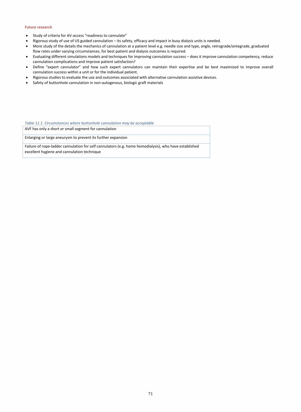
71
Future research
• Study of criteria for AV-access “readiness to cannulate”
• Rigorous study of use of US guided cannulation – its safety, efficacy and impact in busy dialysis units is needed.
• More study of the details the mechanics of cannulation at a patient level e.g. needle size and type, angle, retrograde/antegrade, graduated flow rates under varying circumstances, for best patient and dialysis outcomes is required.
• Evaluating different simulations models and techniques for improving cannulation success – does it improve cannulation competency, reduce cannulation complications and improve patient satisfaction?
• Define “expert cannulator” and how such expert cannulators can maintain their expertise and be best maximized to improve overall cannulation success within a unit or for the individual patient.
• Rigorous studies to evaluate the use and outcomes associated with alternative cannulation assistive devices.
• Safety of buttonhole cannulation in non-autogenous, biologic graft materials
Table 11.1. Circumstances where buttonhole cannulation may be acceptable
AVF has only a short or small segment for cannulation
Enlarging or large aneurysm to prevent its further expansion
Failure of rope-ladder cannulation for self cannulators (e.g. home hemodialysis), who have established
excellent hygiene and cannulation technique
72
Statements: Catheter system connect and disconnect procedure considerations
Note: While CVC procedures are extremely important, many statements were based on Expert Opinion (supported by available evidence) since the available evidence did not meet inclusion criteria by the Evidence Review Team (ERT), to allow for an ERT evidence based statement.
11.8 KDOQI suggests the use of a catheter care protocol for exit site and hub care to reduce catheter related bloodstream infections and treatment of catheter dysfunction (Strong recommendation, Moderate quality of evidence,)
11.9 KDOQI considers it reasonable to use aseptic technique, correct hand washing, and masks for patients and staff performing catheter
connection and disconnection procedures (Expert Opinion). 11.10 KDOQI considers it reasonable to cleanse the catheter hub when connecting and disconnecting the catheter with a Chlorhexidine-
based solution. If Chlorhexidine is contraindicated (e.g., sensitivity, allergy), povidone-iodine solution is a reasonable substitute and should be used (Expert Opinion).
11.11 KDOQI considers it reasonable that at the time of catheter dressing change, to cleanse the skin surrounding the catheter exit site with a
Chlorhexidine based solution. If Chlorhexidine is contraindicated (e.g., sensitivity, allergy), povidone-iodine solution is a reasonable substitute and should be used (Expert Opinion)
11.12 There is inadequate evidence for KDOQI to make a recommendation on the Chlorhexidine formulation to use for infection prophylaxis and this should be based on the clinician’s best clinical judgment, and local practical considerations.
11.13 There is inadequate evidence to demonstrate a difference in catheter-related infections with the use of transparent film dressing compared with non-transparent dressing; thus, the choice of catheter dressing material should be based on the clinician’s discretion that considers the patient’s circumstances and uses best clinical judgment.
11.14 KDOQI considers it reasonable to use a topical antiseptic or antibiotic barrier at the catheter exit site in addition to cleansing until the exit site it healed to reduce the risk of catheter-related infection (Expert Opinion).
11.15 There is inadequate evidence to demonstrate a difference in catheter-related infections between the use of various antiseptic or antibiotic topical exit site barriers; thus, the choice of topical exit site barrier should be based on the clinician’s discretion and best clinical judgment
11.16 KDOQI considers the following catheter care practices reasonable to follow:
o The frequency of catheter dressing change should be based on the clinician’s discretion and best clinical judgment, with a minimum of 1x/week
o Catheter dressings should be protected against wet and dirty environments, particularly when the exit site is not yet fully healed (e.g. avoid swimming and unprotected showering)
Rationale/Background
Hemodialysis central venous catheters (CVC), when compared to AV-access, are associated with a 2-3 fold higher risk of infection-related
hospitalization and associated costs due to catheter-related blood stream infections (CRBSI)65,310,311. Contamination of the external and internal
catheter surface through both the extraluminal and intraluminal pathways, respectively, involves the transfer of organisms during CVC
manipulation, such as during dressing changes or connection and disconnection of the CVC. The transfer of organisms are from either the health
providers’ or patients’ hands (if patient does own CVC care), the patient’s skin or surrounding clothing, to the CVC exit site opening and tunnel
(extraluminal pathway of organism entry) or to the external or internal surfaces of CVC hubs or caps (extraluminal or intraluminal pathway)289,312
(See Figure 11.2 The pathogenesis of biofilm formation). As such, some guidelines have recommended: 1) minimal manipulation of the CVC to
reduce the risk of infection; 2) limiting access of hemodialysis vascular access to personnel who are adequately trained, and 3) need for regular re-
training of individuals handling hemodialysis catheters (or any dialysis access)313. As a result, many dialysis units restrict accessing the CVC to the
provision of hemodialysis, unless in emergency situations. The prior 2006 NKF-KDOQI VA guidelines recommended CVC dressing changes at each
dialysis session312. The current guidelines differ by suggesting only the necessary minimal manipulation of CVC until the exit site and tunnel are
healed to reduce the risk of infection. These guidelines suggest the preferred use of Chlorhexidine for CVC and exit site cleansing, unless
contraindicated, similar to prior recommendations.
Detailed Justification
See Supplement 1 Tables 96-107
Vigilant CVC care is required, including regular inspection of the CVC, tunnel and exit site, and adherence to CVC care protocols. Both the patient
and the HD staff should follow universal precautions and hygienic measures. The 2006 KDOQI vascular access guideline recommended that staff
manipulating CVCs should wear a mask and clean or sterile disposable gloves; however, the data supporting masks are extrapolated from studies of
their use during CVC insertion314,315. During CVC insertion, the risk of infection is high due to direct exposure of the vessel and bloodstream to the
external environment. Whether or not masks are required is unclear in situations where the exit site is healed. However, extra protection is
theoretically afforded if the health care provider or patient inadvertently sneezes, coughs or spits (e.g. while talking) during CVC care. The use of
sterile gloves (versus new clean gloves) is also controversial, particularly when the use of no touch techniques is strictly enforced.
73
Hub Care
Risks to contamination of the hub and infection include: 1) contact of the exposed CVC hub with a non-sterile surface (e.g. bedside desk) or object
(e.g. hand); 2) prolonged exposure to the air; 3) improper cleansing of the hub; 4) patient or provider breathing on the exposed hub316. Thus, it is
important to reduce the hub exposure time and diligent hub cleansing is required. Doing so can result in a marked (almost four-fold) decrease in
CRBSI ra317tes, and approach a rate of 1 episode/1000 catheter days.317 Thus, adopting protocols that incorporate limiting hub exposure time and
increasing protection by vigorously “scrubbing the hub” is very important and supported by these and other guidelines288,289.
Catheter and Exit Site Cleansing
One cluster RCT (n=422 facilities) compared a new CVC care protocol involving exit site disinfection with 2% Chlorhexidine and 70% alcohol swab
sticks and 70% alcohol pads for hub care to current practice (no specific disinfectant, no scrubbing of CVC hubs).318). Significantly fewer blood
stream infections were reported in the intervention facilities. A patient-level analysis, adjusted for a cluster effect, yielded an RR of 0.79 (95%CI
0.78, 0.81). Adverse events were reported only for the intervention facilities, which included Chlorhexidine sensitivity. A total of 184 local, non-life-
threatening events were reported in 82 study participants.
In a pilot RCT (n=105), a 2% Chlorhexidine in 70% isopropyl alcohol solution was compared to Chlorhexidine solutions (either 0.5% Chlorhexidine in
alcohol [81% of control group] or 0.05% aqueous Chlorhexidine [19% of control group]) for CVC exit site antisepsis.319 Follow-up was 12 months.
Overall, fewer CVC-related infections were noted in the intervention group compared to the control group but the difference was not significant
(RR 0.49 [95%CI 0.18, 1.34]). There were also no significant differences between groups for specific types of infection (i.e., CRBSI or exit site). Skin
sensitivity reaction was reported in 4 participants in the 2% Chlorhexidine in 70% alcohol group (Fisher exact test P=. 12)
Given the above evidence, the Work Group strongly suggests using a CVC care protocol that involves Chlorhexidine, and if circumstances permit,
using Chlorhexidine gluconate 1% or 2% and 70% alcohol solution.288,289 Additional research by the Work Group found Chlorhexidine skin cleansing
to be superior to povidone-iodine and alcohol in the prevention of CVC-related infection320-322. If Chlorhexidine is contraindicated (e.g., skin
sensitivity or allergy), povidone-iodine 10% in 70% ethanol should be use.323 The antiseptic solution should be applied using friction, for at least 30
seconds and allowed to air-dry without wiping or blotting, to promote adherence of the dressing material to the skin and reduce the likelihood of
skin breakdown and infection. Multidose cleansing solution bottles are discouraged due to the risk of cross contamination. All cleansing solutions
should be single use, for example, disposable swab stick or pad.289
Exit Site Barriers and Dressings
The routine application of topical antiseptic/antibiotic ointments at the CVC exit site has been shown to be associated with a 75–93% reduction in
the risk of CRBSI324-326. Topical ointments that have been studied include mupirocin, povidone-iodine, and polysporin triple antibiotic ointment. A 6-
year prospective follow-up study using a polysporin triple ointment application at the exit site of HD catheters has not demonstrated microbial
resistance or loss of efficacy for infection prophylaxis, with bacteremia rates consistently <1.0/1000 catheter days305. Medical grade honey has been
shown to have equivalent efficacy to mupirocin for CRBSI.327 Such findings are supported by the ERT. For example, bacteremia occurred in 12% with
medi-honey and 10% with mupirocin prophylactic application at the CVC exit site, P=. 78.328 Bacteremia-free survival also did not differ between
these groups (HR 0.94 [95%CI 0.27, 3.24]).
Given the reduction in CRBSI using topical antiseptic or antibiotic barriers compared with placebo or no barrier, but no significant differences
between types of antiseptic/antibiotic barrier, the Work Group suggests the choice of barrier based on the clinician’s best clinical judgment and
local practices. Whether the benefit exists after exit sites are fully healed is unclear. However, it is important to recall that exit sites require re-
healing after each CVC exchange. As with the use of any intraluminal solutions, any cleansing solution or antiseptic/antibiotic used topically should
be compatible with the CVC material.
All dressing material should be applied using no touch or aseptic technique. No touch technique means that all open ports and ends of CVCs are
not touched with hands or gloves. Recent data indicate that there is no significant difference between transparent, semi-permeable dressings, and
standard gauze dressings with respect to CVC exit site colonization or CRBSI329-331. This is also supported by the ERT. For example, one small RCT
(n=66) compared 2 dressings – a sterile transparent film and a traditional sterile gauze and hypoallergenic micropore dressing. 332 No significant
difference in CVC-related infection (12% intervention, 9% control, P=. 69) was found.332
Both patient and environmental factors should be considered when selecting dressing type and the frequency of change (discussed above).
Dressings should not be submerged in water and should be changed when they become damp, loose, soiled, nonocclusive, or nonadherent, and
only trained dialysis care providers (including patients) should change CVC dressings. Regular retraining is highly desirable to maintain competency
and reduce the risk of infection.
Assessment of CVC Patency
Routine flushing with 0.9% normal saline (NS) is used to maintain CVC patency, and has become a standard of practice when accessing and de-
accessing CVCs. Flushing of CVC lumens is intended to prevent the mixing of incompatible medications or solutions within the CVC lumen and
assists in clearing the CVC of blood) or fibrin build up333. Typically, a 5-10 ml syringe is used; given the lack of evidence for syringe size, use of CVC
manufactures instructions may be used as a guide. However, the clinician is expected to use their best clinical judgment and follow local CVC care
protocols.
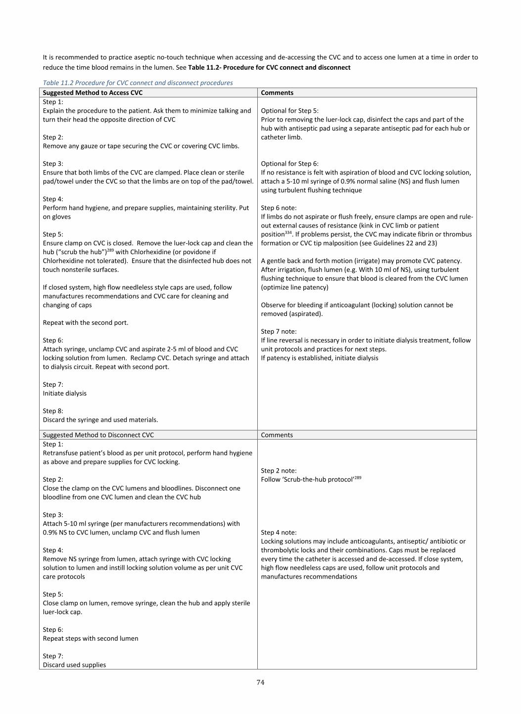
74
It is recommended to practice aseptic no-touch technique when accessing and de-accessing the CVC and to access one lumen at a time in order to
reduce the time blood remains in the lumen. See Table 11.2- Procedure for CVC connect and disconnect
Table 11.2 Procedure for CVC connect and disconnect procedures
Suggested Method to Access CVC Comments
Step 1: Explain the procedure to the patient. Ask them to minimize talking and turn their head the opposite direction of CVC Step 2: Remove any gauze or tape securing the CVC or covering CVC limbs. Step 3: Ensure that both limbs of the CVC are clamped. Place clean or sterile pad/towel under the CVC so that the limbs are on top of the pad/towel. Step 4: Perform hand hygiene, and prepare supplies, maintaining sterility. Put on gloves Step 5: Ensure clamp on CVC is closed. Remove the luer-lock cap and clean the hub (“scrub the hub”)289 with Chlorhexidine (or povidone if Chlorhexidine not tolerated). Ensure that the disinfected hub does not touch nonsterile surfaces. If closed system, high flow needleless style caps are used, follow manufactures recommendations and CVC care for cleaning and changing of caps Repeat with the second port. Step 6: Attach syringe, unclamp CVC and aspirate 2-5 ml of blood and CVC locking solution from lumen. Reclamp CVC. Detach syringe and attach to dialysis circuit. Repeat with second port. Step 7: Initiate dialysis Step 8: Discard the syringe and used materials.
Optional for Step 5: Prior to removing the luer-lock cap, disinfect the caps and part of the hub with antiseptic pad using a separate antiseptic pad for each hub or catheter limb. Optional for Step 6: If no resistance is felt with aspiration of blood and CVC locking solution, attach a 5-10 ml syringe of 0.9% normal saline (NS) and flush lumen using turbulent flushing technique Step 6 note: If limbs do not aspirate or flush freely, ensure clamps are open and rule-out external causes of resistance (kink in CVC limb or patient position334. If problems persist, the CVC may indicate fibrin or thrombus formation or CVC tip malposition (see Guidelines 22 and 23) A gentle back and forth motion (irrigate) may promote CVC patency. After irrigation, flush lumen (e.g. With 10 ml of NS), using turbulent flushing technique to ensure that blood is cleared from the CVC lumen (optimize line patency) Observe for bleeding if anticoagulant (locking) solution cannot be removed (aspirated). Step 7 note: If line reversal is necessary in order to initiate dialysis treatment, follow unit protocols and practices for next steps. If patency is established, initiate dialysis
Suggested Method to Disconnect CVC Comments
Step 1: Retransfuse patient’s blood as per unit protocol, perform hand hygiene as above and prepare supplies for CVC locking. Step 2: Close the clamp on the CVC lumens and bloodlines. Disconnect one bloodline from one CVC lumen and clean the CVC hub Step 3: Attach 5-10 ml syringe (per manufacturers recommendations) with 0.9% NS to CVC lumen, unclamp CVC and flush lumen Step 4: Remove NS syringe from lumen, attach syringe with CVC locking solution to lumen and instill locking solution volume as per unit CVC care protocols Step 5: Close clamp on lumen, remove syringe, clean the hub and apply sterile luer-lock cap. Step 6: Repeat steps with second lumen Step 7: Discard used supplies
Step 2 note: Follow ‘Scrub-the-hub protocol’289 Step 4 note: Locking solutions may include anticoagulants, antiseptic/ antibiotic or thrombolytic locks and their combinations. Caps must be replaced every time the catheter is accessed and de-accessed. If close system, high flow needleless caps are used, follow unit protocols and manufactures recommendations
75
Care of Hemodialysis Central Venous Catheters and Patient Education
To facilitate CVC care, the following actions should be strongly considered:4
1. Educate patients (see below), healthcare personnel and relevant administrators regarding the acceptable appropriate indications for CVC use
(see Guidelines 1, 2), proper procedures for safe and optimal CVC care and use, including prophylactic measures for CVC-related infections
2. Periodically assess knowledge of and adherence to guidelines for all individuals (including patients and relevant family/ patient supporters)
involved in CVC access, use and maintenance
3. Designate only trained individuals who demonstrate competence for the access, use and maintenance of CVC.
Patient teaching and instructions should include, but are not limited to the importance of:
• Initial CVC care, including when sutures should be removed
• Frequent hand washing
• Avoid pulling, tugging, or using sharp objects (e.g. scissors) around the catheter
• What to do if the dressing becomes soiled or wet
• What to do if bleeding occurs
• What to do if the CVC falls out
• What to do if a limb clamp breaks and falls off
• What to do in the case of pain, fever or chills, or redness or discharge seen at the CVC exit site or tunnel
• Who to call with questions or concerns, with the correct contact details
Patients should be provided written instructions on CVC care and up-to-date contact information for reporting any vascular access concerns e.g.
bleeding or signs of infection (fever, chills, and purulent discharge)
Implementation considerations
• The concern of the often rapid turn-over of dialysis technicians, leading to inadequate training and/or competency to care of CVC (and all
types of vascular access)
• The use of any intraluminal or extraluminal cleansing solutions, antiseptics, antibiotics, medicines or anticoagulants should be compatible
with the CVC material
Monitoring and Evaluation
• Monitor for CVC exit site healing
Future research
• Are sterile gloves required if strict “no touch” technique is used for CVC manipulation and care?
• Are masks required for each CVC dressing change and manipulation, if the patient has head turned away and both patient and provider
do not talk. Exception is if patient or provider has respiratory signs/symptoms
• Is there benefit of topical barriers after exit site and tunnel fully endotheliaized and healed
• Validation of tests to determine exit site healing
• Can a patient safely shower when the exit site and tunnel are fully healed?
• Can a patient safely eliminate the use of dressings altogether when the exit site and tunnel are fully healed, if the patient properly
cleanses?
76
GUIDELINE 12 AV-ACCESS CANNULATION COMPLICATIONS
Statements: AV-Access Cannulation Complications
12.1 KDOQI considers the following therapeutic interventions for cannulation injury reasonable to follow: o Any size infiltration: apply ice for a minimum of 10 minutes and refrain from maximizing the blood pump speed (Expert Opinion)
o If the infiltration is moderate, the needle should be withdrawn and manual pressure held over the infiltration site (Expert Opinion)
o If the infiltration is significantly large, in addition to the above, a decision on the necessity for dialysis that day is required – if dialysis is
required, a site above the infiltration injury should be cannulated; if this is not possible, reattempt at the area of injury should not proceed until manual pressure and ice is applied for 30 minutes (Expert Opinion)
o If a hematoma develops, close assessment of the site, the AV-access and the adjacent extremity should be made, including measurement of swelling, assessment of the presence of flow in the AV-access both above and below the hematoma and circulation to the associated extremity (Expert Opinion)
12.2 KDOQI considers it reasonable to use ultrasound to help determine direction of flow and proper needle placement in select patients as
needed and performed by trained operators, to prevent cannulation complications (Expert Opinion)
Rationale/Background
Despite cannulation being an integral component of the hemodialysis procedure, there is very limited evidence to support practice. Knowledge, skill and ability among cannulating nurses/dialysis personnel varies significantly across jurisdictions with little to no minimum standards or standardization. Basic knowledge of vascular anatomy, patient-centered care and the importance of provider-patient therapeutic relationships should be components of all cannulation training programs to reduce the frequency of complications. The complications that occur as a result of cannulation not only cause significant discomfort and distress to patients but in some cases, may negatively impact their ability to have a functioning dialysis access in the future. For example, a single infiltration injury that occurs before successful 2 needle cannulation is associated with 56% lower odds of overall AVF maturation.153 Infiltration injury is associated with frequent imaging, interventions, and prolonged need for a catheter. 2 Indeed, unsuccessful cannulation attempts are associated with poorer AVF maturation success and outcomes.153 Currently, complications related to cannulation are not consistently or well documented as part of the dialysis treatment. Cannulation site bleeding can usually be corrected by direct pressure but occasionally requires the placement of a skin suture. Pressure should be applied directly to the bleeding site and care should be exercised not to occlude the AV-access outflow distal to the bleeding site because of the potential to increase the intraluminal AV-access pressure to arterial levels. A “bad stick” that results in a significant hematoma often requires placement of a CVC and deferral of further AV-access cannulations until the hematoma is resolved, a period that may last up to 3 months.2 Bleeding from a vascular access needle site that needs a skin suture or results in a very large hematoma is very suggestive of a venous outflow stenosis and requires a referral for an angiogram.
Detailed Justification
Complications and management
• Infiltration of the vein can occur when a needle is inserted and the tip is inadvertently advanced beyond the vein perforating the side or back wall resulting in some degree of swelling, bruising and/or pain.
• Hematomas can develop as a result of an infiltration of the vein or due to leaking of blood around the puncture site during cannulation, during
a dialysis treatment or post removal of a needle at the end of a dialysis treatment. The size of hematomas can vary significantly from a small
diffuse area to a large, firm mass that can potentially compress the vessel resulting in thrombosis of the AV-access. The development of
significant hematomas can also result in the development of stenosis at the site of hematoma. Every effort should be made to avoid these.
• Pain of various degrees is common and can occur in various sites, at various times and be of various intensity. Pain can develop at the time of
cannulation, develop during the dialysis treatment at the site of or surrounding a site of cannulation, or along the arm or hand and continue
post dialysis as a result of a hematoma, infiltration of the vein or irritation of an adjacent nerve.
• Management of cannulation complications such as infiltration and resulting hematoma is dependent on the extent of swelling and associated
pain and anxiety to the patient. The sharing of knowledge and provision of support to patient and family cannot be minimized. Providing
comfort measures to analgesic administration must be assessed on an individual basis.
Prevention of cannulation complications
• AV-access check or assessment prior to cannulation is required. Ensuring that flow is present in the AV-access and determining the direction of
flow to ensure optimal dialysis is required prior to needle placement. Observation of the full AV-access and adjacent limb and auscultation and
palpation along the AV-access can detect many defects and aid in appropriate selection of sites for cannulation. The use of beside ultrasound
to aid in cannulation has been associated with increase in nurse confidence, patient comfort (2) and can help determine direction of flow
within the AV-access with a trained operator but there is limited evidence at this point to recommend use in all patients.
• Appropriate selection of cannulation sites based on assessment and input from patients can help avoid associated complications.
77
• Use of experienced cannulators to cannulate AV-accesses at risk of complications should be attempted whenever possible. Cannulators should
determine at the time of assessment if they anticipate any challenges with cannulation and seek expert advice and guidance. If a cannulator is
unsuccessful cannulating an AV-access, a maximum of 2 attempts is recommended before seeking expert advice.
• The use of smaller gauge and Teflon needles should be considered when cannulating smaller and more fragile vessels. Although these needles
may limit blood flow, consideration of longer, more frequent or individualized prescription based on patient’s dialysis needs and goals may
enable the non-complicated use of the AV-access and avoid insertion of a CVC.
When appropriate and possible, self-cannulation in well-trained patients may be beneficial. Documentation of complications of cannulation should be reviewed as part of the cannulator training and evaluation. Special discussions
• Centers with limited opportunities for staff to cannulate dialysis accesses face particular challenges. Accessing on-line resources and expertise should be considered to ensure the provision of adequate care to patients.
• The Work Group acknowledged that there is limited evidence to support the recommendations related to cannulation and management of cannulation related complications, limiting them to Expert Opinion only. Even when reviewing the literature in areas such as peripheral venipuncture and arterial cannulation, the evidence is opinion based.
• When appropriate and possible, self-cannulation in well trained patients may be beneficial to improving patient comfort and reduce anxiety in cannulating the AV-access
Future research
• Rigorous studies examining cannulation practices, challenges to achieve complication free cannulation, and strategies to mitigate barriers to successful AV-access cannulation
• To study the role of cannulation complication documentation and review on whether or not it improves quality of cannulation and AV-access outcomes.
78
GUIDELINE 13. AV-ACCESS FLOW DYSFUNCTION+ - MONITORING/SURVEILLANCE
+ “AV-access flow dysfunction” refers to clinically significant abnormalities in AV-access (AVF or AVG) flow or patency due to underlying stenosis, thrombosis or related pathology. This is in distinction to other types of AV-access complications
Statements: Appropriate Use of Monitoring/Surveillance for AV-Access Flow Dysfunction
Physical exam (monitoring): 13.1 KDOQI recommends regular physical examination or check of the AVF, by a knowledgeable and experienced health practitioner, to
detect flow dysfunction of the AVF (Conditional/Strong recommendation, Moderate quality of evidence)
13.2 KDOQI recommends regular physical examination or check of the AVG, by a knowledgeable and experienced health practitioner, to detect flow dysfunction of the AVG (Conditional/Strong recommendation, Moderate quality of evidence)
13.3 KDOQI considers it reasonable for nephrology trainees and health practitioners involved with clinical hemodialysis patient care to be properly trained in physical examination of the AV-access to monitor for and detect AV-access flow dysfunction (Expert Opinion)
Surveillance to facilitate patency: 13.4 There is inadequate evidence for KDOQI to make a recommendation on routine AVF surveillance by measuring access blood flow,
pressure monitoring, or imaging for stenosis, that is additional to routine clinical monitoring, to improve access patency.
13.5 KDOQI does not suggest routine AVG surveillance by measuring access blood flow, pressure monitoring, or imaging for stenosis, that is additional to regular clinical monitoring, to improve AVG patency. (Conditional recommendation/Low quality of evidence).
Note: In other words, monitoring of vascular access is primary while surveillance findings are supplementary, and action should not be based
solely on surveillance findings.
Statements: Surveillance and Pre-emptive Intervention for AV-Access Stenosis, Not Associated with Clinical Indicators Endovascular Intervention to improve patency: 13.6 KDOQI does not recommend pre-emptive angioplasty of AVFs with stenosis, not associated with clinical indicators, to improve access
patency (Conditional recommendation, Moderate quality evidence). 13.7 KDOQI does not recommend pre-emptive angioplasty of AVGs with stenosis, not associated with clinical indicators, to improve access
patency (Conditional recommendation, Moderate quality evidence). 13.8 KDOQI considers it reasonable for patients with consistently persistent clinical indicators to undergo pre-emptive angioplasty of their AV-
access to reduce the risk of thrombosis and AV-access loss (Expert opinion)
Surgical Intervention to improve patency: 13.9 There is inadequate evidence for KDOQI to make a recommendation on pre-emptive surgical interventions in AVFs with stenosis, not
associated with clinical indicators, to improve access patency.
Rationale/Background
In these Guidelines, “AV-access flow dysfunction” refers to clinically significant abnormalities in AV-access (AVF or AVG) flow or patency due to underlying stenosis, thrombosis or related pathology. This was previously referred to as “AV-access dysfunction", which encompassed many forms of dysfunction (or abnormalities of AV-access); however, the Work Group wanted to distinguish AV-access dysfunction due to stenosis or thrombosis from other causes of dysfunction, such as aneurysms. Thus, the terminology AV-access flow dysfunction is used in distinction to other types of AV-access complications. AV-access flow dysfunction is a common problem, typically associated with underlying stenosis and/or thrombosis. The development of progressive vascular access stenosis with subsequent failure of the vascular access contributes significant morbidity to patients and costs to the health care system335. The fundamental principle for performing routine vascular access monitoring and surveillance is to detect and correct the stenosis to minimize or avoid reduced dialysis clearance (dialysis dose protection), reduce the rate of thrombosis, and improve access function. Indeed ‘vascular access function” was globally ranked a top priority for patients and healthcare providers clinically and as a research target, so much so that it has become the core outcome measure for vascular access clinical trials.336
Rationale for Physical Examination/Monitoring
A variety of methods have been proposed for screening the AV-access for early detection of stenosis before the AV-access becomes dysfunctional337-342. Clinical monitoring strategies include physical examination (inspection, palpation, and auscultation) of the vascular access to detect signs that suggest the presence of pathology. This monitoring is ideally conducted when the patient is not on dialysis. Abbreviated forms, such as the “One Minute Check”, is rapid, effective and can be conducted by patients and providers prior to dialysis.343 Such monitoring can be supplemented by review of routine laboratory studies regularly obtained in the dialysis unit, dialysis adequacy (urea reduction ratio or Kt/V, documented recirculation), difficulties in cannulation or achieving hemostasis after needle withdrawal, and other clinical signs. Although these different techniques and methods are available for identifying vascular access flow dysfunction, the scientific evidence for the optimal methodology

79
is lacking. A small number of RCTs have been performed evaluating different monitoring techniques. Dynamic venous pressure (DVP) can be measured on most dialysis machines; however, the utility of DVP at blood flows (Qb) 150–200 mL/min in detecting stenosis or predicting AV-access thrombosis is very limited as DVP is dependent on a variety of factors, such as the needle gauge and the length of the dialysis needle344. AV-access recirculation and declines in URR or Kt/V may help indicate AV-access stenosis, but by themselves cannot be sole indicators as many variables affect their measurements, and they have not been rigorously studied. Physical examination of the AV- access by an experienced individual has high sensitivity and specificity261,345,346. The physical exam is easily available, requiring minimal training, is cost efficient and takes minimal equipment and time. Several studies have examined the use of the physical exam to detect AV-access flow dysfunction (i.e. stenosis within the AVF261,346, and AVG342,347). The value of physical exam (monitoring) is that if AV-access flow dysfunction is suspected with appropriate matching clinical indicators, further investigation and preventive intervention, such as with angioplasty could be performed to improve outcomes (e.g. preventing thrombosis).
Rationale for Surveillance
The rationale underlying AV-access surveillance is to detect and correct stenosis within the AV-access prior to the development of thrombosis, to improve the patency of the AV-access by reducing the rate of thrombosis. Clinical indicators associated with AV-access stenosis (Table 13.2) include reduced dialysis clearance without other known cause, excessive bleeding post needle withdrawal, high venous and arterial pressures at the prescribed blood flow348 and indicators on physical exam (see above on AV-access monitoring). Surveillance procedures, requiring specialized equipment and operator skills, have been studied to detect stenosis prior to the development of a clinical indicator. These include AV-access flow (Qa) measurement339,349,350 by a variety of methods including ultrasound dilution method (UDM)), and duplex Doppler ultrasound to measure Qa and visualize anatomic abnormalities351,352. The use of dynamic and static venous pressure has also been used as a surveillance tool353; dynamic venous pressure measures are now considered as supplementary to clinical monitoring. Several important questions arise when considering AV-access surveillance to reduce thrombosis and improve AV-access patency: i) what is the diagnostic value of surveillance for detection of stenosis, ii) what are the validated diagnostic thresholds by surveillance indicators (e.g. Qa, change in Qa/time) that accurately diagnose stenosis that will lead to future thrombosis, iii) what factors, other than stenosis contribute to abnormal surveillance indicators, iv) what is the appropriate intervention when stenosis is detected by surveillance in the absence of clinical indicators, v) what is the AV-access patency outcome after intervention of stenosis not associated with a clinical indicator and does this differ from intervention of stenosis detected by clinical exam? What is the diagnostic value of surveillance for detection of stenosis?
Surveillance methods can detect stenosis; however, there are valid issues regarding the accuracy and reproducibility of this detection, which may vary based on the method of surveillance and the indicator used. Stenosis within the AV-access is thought to be reflected by reductions in AV-access flow and alterations in AV-access circuit pressures. However, flows and pressures are influenced by factors other than the presence of stenosis, including location and degree of the stenosis, variations in the hemodynamics over the course of dialysis (e.g. timing, blood pressure) and cannulation technique and AV-access characteristics353-355. The location of the stenosis can influence the diagnostic characteristics of the surveillance tool and an AV-access can have multiple stenosis, not reliably detected with a single surveillance tool, especially if measured in isolation356. Additionally, the AVG and the AVF have different pathophysiology for development of stenosis, occurring at different rates, locations and with different hemodynamic consequences based on their configurations357. What are the validated diagnostic thresholds by surveillance indicators (e.g. access flow rate, change in access flow rate) that accurately diagnose stenosis that will lead to future thrombosis?
The various surveillance techniques available to detect AV- access stenosis differ in their ability to accurately and reliably detect AV-access flow dysfunction beyond clinical exam11,358. In addition, when using a given surveillance method to detect “significant” stenosis that would prompt intervention, there is still a need for standardized diagnostic thresholds with sufficient sensitivity to detect clinically significant stenosis but with good specificity to avoid unnecessary interventional procedures. Issues regarding surveillance reliability and reproducibility (above in i)) add to the challenge of defining and validating diagnostic thresholds. As such, the required validated thresholds for intervention have not been established for all methods of AV-access surveillance. What factors, other than stenosis contribute to abnormal surveillance indicators
Variables to consider are discussed above in i) that include factors related to the hemodialysis procedure and the patients’ response to it (e.g. hypotension), the AV-access and AV-access circuit anatomy and physiology353,354. In addition, the expertise and reliability of the operator obtaining the measurement contribute to abnormal surveillance indicators. These guidelines focus on the evidence for or the lack thereof, in answering the remaining patient important and clinically relevant questions iv and v above: iv) what is the appropriate intervention when stenosis is detected by surveillance in the absence of clinical indicators, and v) what is the AV-access patency outcome after intervention of stenosis not associated with a clinical indicator and does this differ from intervention of stenosis detected by clinical exam? The brief answers are: What is the appropriate intervention when stenosis is detected by surveillance in the absence of clinical indicators: do nothing – do not intervene in the absence of clinical indicators. What is the AV-access patency outcome after intervention of stenosis not associated with a clinical indicator and does this differ from intervention of stenosis detected by clinical exam: a) for AVF: the data is unclear and more study is required; b) for AVG: the data does not demonstrate improved patency with surveillance and subsequent pre-emptive intervention on AVG with no clinical indicators, compared with routine clinical exam.
See Supplement 1 Tables 99, 108-129

80
Detailed Justification
The 2006 KDOQI Vascular Access Work Group previously recommended AV-access surveillance with pre-emptive angioplasty of stenosis to improve AV-access outcomes11. This was based on the best available evidence at that time that supported the premise that use of surveillance for the early detection of AV- access stenosis and its preemptive correction compared to deferred correction would improve AV-access outcomes. There has been controversy over the interpretation of this recommendation based on previous and more current evidence. Over time, the nephrology community has recognized that surveillance for stenosis in isolation is not clinically meaningfully without also examining the effect of the subsequent intervention on clinically important outcomes that, in this case, would include AV-access thrombosis, patency and intervention rates. Many studies have been underpowered to show either benefit or harm from surveillance strategies. Multiple factors discussed above contribute to the heterogeneity of the results and interpretations of the studies in this area and have contributed to the change in the recommendations for AV-access surveillance.
Physical Examination:
The physical exam is easily available, requiring minimal training, is cost efficient and takes minimal equipment and time. Several studies have examined the use of the physical exam to detect AV-access flow dysfunction (e.g. stenosis within the AVF11,355,356, and AVG352,353).
Diagnostic accuracy of the physical exam to detect stenosis in the AVF
Two observational studies compared physical exam with angiography for monitoring and diagnosing dysfunction with AVF.261,345 Coentrao et al. analyzed data resulting from physical examinations by 11 general nephrologists and by one nephrology resident who received 6-months training on conducting physical exams to identify AV-access flow dysfunction.345 Diagnostic physical exam results were compared to that of angiography in detecting AV-access stenosis. Clinical criteria for AV-access flow dysfunction prompting angiography were applied according to the prior KDOQI 2006 guidelines11. Physical examination included inspection of the arm, chest, neck and face, palpation of the entire AVF tract, arm elevation, pulse and thrill abnormalities and pulse augmentation tests. AVF dysfunction was classified into four major disorders: inflow stenosis, outflow stenosis, co-existing inflow–outflow stenosis and AVF thrombosis. Stenosis by angiography was defined as 50% luminal narrowing compared to the normal vascular segment located adjacent to the stenosis according to KDOQI 200611. Thrombosis of the AVF was ascertained by the presence of clots in the arterial and/or venous sides of the AVF. The agreement beyond chance between the nephrologists’ physical exam and angiography for the assessment of AVF dysfunction was moderate (kappa=0.49, 95% CI 0.40–0.57). and near-perfect when the physical exam was done by the trained nephrology resident (kappa = 0.86, 95% CI 0.77–0.95). The agreement was similar for both forearm and upper arm AVFs. Asif et al. 261 (n=147) compared results of a complete physical exam of the AV-access conducted by an interventional nephrologist to angiography. Similar criteria for AV-access flow dysfunction was used as in the Coentrao study above. The diagnostic accuracy of physical exam was poor for central vein and AVF body stenosis; moderate for inflow and coexisting inflow and outflow stenosis; and good for outflow stenosis.
Diagnostic accuracy of the physical exam to detect stenosis in the AVG
Leon et al347 conducted pre-procedure physical exams on 43 consecutive cases referred for angiography to manage AVG dysfunction. Physical exam findings and diagnosis were each recorded and secured in a sealed envelope. Angiography from the feeding artery to the right atrium was performed. The angiographic images were reviewed by an independent interventionalist with expertise in AV-access procedures who was blinded to the physical exam results. The agreement beyond chance between physical exam and angiographic findings were strong for the diagnosis of vein-graft anastomotic stenosis (kappa=0.52); moderate for intragraft stenosis (kappa=0.43) and inflow stenosis (kappa = 0.40). The findings of this study demonstrate that physical examination can assist in the detection and localization of stenoses in AVGs.
See S1. Tables 99-
Surveillance:
Surveillance of the AVF The use of physical exam is an accepted, standard, evidence based practice for assessment and monitoring of the AV-access for access flow dysfunction, infection and vascular integrity. Only one study (compared the addition of surveillance methods to routine clinical examination for detecting stenosis and reported AVF outcomes, with variable results, depending on the type of surveillance and outcome examined.359 The need to address whether or not surveillance methods can detect AVF dysfunction and stenosis and its incremental benefit beyond clinical exam (monitoring) is important in order to focus efforts for education, training and the attainment of expertise in the relevant method for detecting AVF dysfunction. i. Clinical Monitoring plus Blood Flow (Qa) Surveillance vs. Clinical Monitoring alone A single center RCT (n=137) of AVF surveillance compared clinical monitoring plus intra-access blood flow (Qa) surveillance using UDM to clinical monitoring alone338. The clinical monitoring group was referred to angiography if stenosis was clinically suspected. Participants in the UDM Qa surveillance group were referred to angiography if stenosis was clinically suspected or Qa was <500 ml/min at baseline or if Qa fell by >20% once Qa was <1000 ml/min. Patients in the UDM Qa surveillance group were twice as likely to have stenosis detected compared to the clinical monitoring alone group (HR 2.27 95%C.I.0.85-5.98, p=0.09(NS)), with a trend for the stenosis to be detected earlier in the UDM Qa surveillance group. The area under the curve (AUC) demonstrated moderate prediction of >50% stenosis (0.78, 95% C.I. 0.63-0.0.94, p=0.006) in the UDM
surveillance group. The study primary outcome was the time to detection of an angiographically significant AVF stenosis [defined as a 50% reduction of the normal vessel luminal diameter on angiography accompanied by a hemodynamic (in the case of the Qa surveillance group), functional or clinical abnormality (in the case of the control group)] was not significantly different with clinical monitoring plus UDM Qa surveillance versus clinical monitoring alone (p=0.20). In summary, the use of surveillance methods in addition to clinical monitoring in AVF appears to increase the rate of detection of AVF stenosis and the rate of AVF intervention.
81
Surveillance of the AVG Several previous studies have examined the effect of surveillance in addition to clinical monitoring in AVG342,351,355,360. Most of the studies did not specifically examine the rate of detection of stenosis but rather a clinical outcome of thrombosis or patency. The Evidence Review Team did not include these studies as their publication dates were outside the prespecified window of dates for data extraction. However, the Work Group felt it was important to discuss previous studies that examined clinical monitoring alone to clinical monitoring plus surveillance in AVGs. One blinded, RCT (n=112) compared clinical monitoring (dynamic venous pressure (DVP) and physical examination) plus monthly Qa measurement by UDM to clinical monitoring alone342. The Qa surveillance group was referred for angiogram if Qa <650 ml/min or a 20% decrease in Qa from baseline, while the clinical monitoring alone group were referred for significant changes in dialysis adequacy, physical exam or high DVP. Percutaneous angioplasty was performed for stenosis ≥50% compared to the adjacent vessel. The rate of AVG thrombosis per patient-year at risk was 0.51 and 0.41 in the surveillance and clinical monitoring groups, respectively (P= 0.57). Stenosis was detected more frequently in the surveillance group due to the increased use of angiograms and was accompanied by an increase in interventions: 51 interventions (0.93/patient-years at risk) in the Qa surveillance group versus 31 interventions (0.61/patient-years at risk) in the clinical monitoring alone group. In summary, the use of surveillance methods in addition to clinical monitoring in AVG appears to increase the rate of detection of AVG stenosis and the rate of AVG intervention. Surveillance and Preemptive Repair: The aim of interventions to prevent AV-access flow dysfunction (i.e. to improve patency or access function) is to improve patient and vascular access outcomes, with greater net benefit than harm, compared to no interventions (see above background/rationale). There is limited evidence to provide guidance on the role of appropriate methods to prevent clinically insignificant stenosis (i.e. stenosis that may be associated with abnormal findings on surveillance but without accompanying clinical indicators) in AVFs and AVGs. The available literature for AVF and AVG is provided in the subsequent sections below. Surveillance and Preemptive Repair in Asymptomatic AVFs
One observational study using data from the Fresenius Medical Care North America centers (n=35,716) compared elective percutaneous angioplasty (PTA) with no treatment for the prevention of AVF and AVG (AV-access) dysfunction. Patients were referred for PTA based on Qa < 400 ml/min or change >30% for AVF and < 600 ml/min for AVG. In the AVF subgroup, patients who received angiography and PTA were matched to controls not receiving an intervention for access type (matched on vascular access type, access age, intra access blood flow rate using the method of ionic online clearance and spKt/V. Follow-up was for 1 year after intervention with angiography and PTA. The key findings are361: (1) secondary patency was not significantly different with elective angiography and PTA versus no treatment and (2) thrombosis was not significantly different with elective PTA versus no treatment. The primary outcome was 1-year AV-access survival (cumulative patency) from date of first intervention, and was not significantly different with elective PTA versus no treatment (54.8 vs. 47.8 per 100 access years; HR 1.06; 95% CI: 0.98 to 1.15). Several secondary outcomes were evaluated. Embolism with upper-arm thrombosis was not significantly different with elective PTA versus no treatment for AVF and AVG combined (0.86% vs. 0.03% events per procedure; attributable risk increase 0.83%; 95% CI: 0.56 to 1.12). This outcome was not reported separately by AV-access type. Other harms were not reported for this comparison. In contrast, a prospective study was conducted in patients who did not meet prior KDOQI Qa criteria for intervention but had abnormal clinical monitoring findings e.g. on physical exam. A single open RCT of AVFs (n=58) assessed AVF with subclinical stenosis and Qa>500 ml/min (but abnormal physical exam, Qa<900 ml/min and elevated SVP) who had prophylactic repair (pre-emptive intervention group) versus observation (repair at the time of AVF thrombosis) 340. Subclinical stenosis was angiographically defined as >50% reduction in vessel diameter compared with the adjacent segment. Prophylactic repair in the pre-emptive intervention group was by either PTA or surgery while the observation arm underwent stenosis repair only after the onset of AVF dysfunction (defined as clinical abnormality) or a Qa <400 mL/min (n=3). AVF loss was lower in the pre-emptive PTA group than observation alone, 5 (18%) versus 13 (43%), respectively (corresponding AVF loss rates (event/AVF-year) were 0.066 and 0.186 (P=. 041). Thrombosis rates were significantly lower in the pre-emptive PTA group than observation alone group, 21% versus 50%. The use of temporary CVC and associated CVC infection rates were not significantly different between groups340. In addition to the studies above extracted by e Evidence Review Team, when finalizing the KDOQI guideline statements, the Work Group considered other RCTs 338,339,362,363 in this area outside the pre-specified window of data extraction. The Work Group felt that these previous RCTs help place the above data in the appropriate context. There is further discussion about these specific studies in the “Special Discussions” section below. See Supplement 1 Tables 130-142 Surveillance and Preemptive Repair in Asymptomatic AVGs
There was one RCT (n=64) in AVG comparing pre-emptive intervention of asymptomatic stenosis versus observation (repair at the time of thrombosis)364. AVGs with elevated static venous pressure (SVPR, a ratio of static venous pressure/systolic blood pressure measured at dialysis machine blood flow rate of 0 mL/min) values underwent diagnostic angiography. If there was a stenosis producing at least a 50% reduction in lumen diameter and the SVPR ratio was ≥0.4, patients were then randomized to pre-emptive intervention arm (prophylactic repair by PTA or surgery [if PTA was unable to be performed or was unsuccessful]). The observation arm had intervention only after an AVG thrombosis event or if there was clinical evidence of AVG dysfunction. Participants were followed 3.5 years. Mortality (19% in the pre-emptive intervention arm and 13% in observation arm), AVG loss (14 patients (44%) in each arm) and time to AVG abandonment did not differ between groups. The rates of thrombosis were significantly lower with pre-emptive intervention than with observation (44% vs. 72%). Dember et al.364 reported no other differences between groups. In AVGs, one observational study by Chan et. al 361 (n=35,716), used data from the Fresenius Medical Care North America centers to compare elective angioplasty versus no treatment for prevention of AVF dysfunction (described above), but also included a subset of patients with AVG who had elective angioplasty versus no treatment. Similar to the AVF cohort, in AVGs, follow-up was for 1 year after intervention with angiography and PTA. This study had these main findings: (1) secondary AVG patency was not significantly different with elective angiography and PTA versus no treatment and (2) thrombosis was not significantly different with elective angioplasty versus no treatment. The primary outcome was 1-year access
82
survival (cumulative patency) from date of first intervention, and was not significantly different with elective PTA versus no treatment (51.7 vs. 52.7 per 100 access years; HR 0.95; 95% CI: 0.86 to 1.05). Similar to our discussion in the above AVF section, in addition to the studies above extracted by the Evidence Review Team, when finalizing the KDOQI guideline statements, the Work Group considered other RCTs 342,351,364-367 in this area outside the pre-specified window of data extraction. The Work Group felt that these previous RCTs help place the above data in the appropriate context. There is further discussion about these specific studies in the “Special Discussions” section below. In summary, in clinical practice, many centers utilize surveillance techniques with the intention to detect early dysfunction in AVF and AVG with the premise that early identification and correction of stenosis may prevent clinically significant dysfunction, such as a thrombotic event. However, in clinical practice it is difficult to predict which stenosis (anatomic abnormality) will progress into a clinically significant functional abnormality, such as an occlusive thrombosis. Intervening on stenoses that are clinically asymptomatic may lead to unnecessary interventions, and subsequently more interventions to maintain patency – which does not appear to be improved with pre-emptive intervention364. Thus, given the evidence available from the literature, our recommendations are not to intervene on AVF and AVGs that do not have clinically relevant dysfunction when detected on routine clinical monitoring (e.g. abnormal physical examination findings, low Kt/Vs, blood flow rates, high venous pressure during dialysis, etc.) See Supplement 1. Tables 143-154 Special discussions
Overall, there were very few studies that evaluated methods to prevent AVF and AVG dysfunction that were of high-quality evidence. The Work Group discussed and considered previous RCTs338,339,342,351,362,364-368 not included in the data extraction (Table 13.3 below) in developing the guidelines, and determined these previous RCTs were important in the overall context of developing the statement guideline. Only one previous study demonstrated improved longevity with surveillance in AVGs367, with the remaining studies showing no statistical benefit compared to clinical monitoring in improving thrombosis or cumulative survival. In AVFs several studies showed benefit of surveillance techniques compared to clinical monitoring; however, they too, had methodological concerns (group contamination; generalizability). There was no evidence grade literature on evaluating multidisciplinary care in prevention of AV-access flow dysfunction (primary and secondary prevention). However, given that a multidisciplinary effort is essential to achieve a successful vascular access, including disciplines such as nephrology, surgery, interventionalist, and dialysis nursing, a multidisciplinary team is also likely crucial to identify clinically important AV-access flow dysfunction, refer for intervention and proceed in a timely manner, if necessary.
Implementation considerations
A multidisciplinary program comprised of nephrologists, surgeons, interventionalist, vascular access coordinators, and dialysis nurses to provide coordinated care is likely essential to prevent asymptomatic AV-access complications. Development of curriculum for implementation into training programs for nephrology fellows and health practitioners accessing the AV-access.
Monitoring and Evaluation
Important considerations when implementing a new monitoring and surveillance programs include considering the processes of care and practice patterns at each individual dialysis center for monitoring and surveillance may be different. Thus, changes in implementation of monitoring or surveillance within a dialysis unit may impact usual established protocols. Future research
The Work Group proposed the following topics as important areas for further research: (1) randomized clinical trials, with adequate methodology and power to delineate the role, if any, of the current and future surveillance and intervention methods, within a strategy to improve AV-access patency (2) specific studies evaluating the impact of pre-emptive intervention in patients with clinical indicators (3) more comparative studies of physical exam / clinical monitoring and imaging studies in various clinical scenarios (e.g. on/off dialysis) to better define its sensitivity, specificity, predictive values and accuracy of detecting clinically significant lesions, to expand upon data (example of data in Table 13.4) (4) validation of the indicators on clinical monitoring of clinically significant stenosis, (5) identification of sub-groups of patients who may benefit from earlier angiographic intervention, (6) indications for endovascular versus surgical intervention, and (7) potential impact of multidisciplinary care for improved AV-access patency.
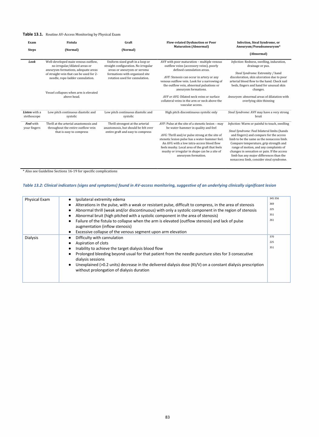
83
Table 13.1. Routine AV-Access Monitoring by Physical Exam
Exam
Steps
Fistula
(Normal)
Graft
(Normal)
Flow-related Dysfunction or Poor
Maturation (Abnormal)
Infection, Steal Syndrome, or
Aneurysm/Pseudoaneurysm*
(Abnormal)
Look Well-developed main venous outflow,
no irregular/dilated areas or
aneurysm formations, adequate areas
of straight vein that can be used for 2-
needle, rope-ladder cannulation.
Vessel collapses when arm is elevated
above head.
Uniform sized graft in a loop or
straight configuration. No irregular
areas or aneurysm or seroma
formations with organized site
rotation used for cannulation.
AVF with poor maturation – multiple venous
outflow veins (accessory veins), poorly
defined cannulation areas.
AVF: Stenosis can occur in artery or any
venous outflow vein. Look for a narrowing of
the outflow vein, abnormal pulsations or
aneurysm formations.
AVF or AVG: Dilated neck veins or surface
collateral veins in the arm or neck above the
vascular access.
Infection: Redness, swelling, induration,
drainage or pus.
Steal Syndrome: Extremity / hand
discoloration, skin ulceration due to poor
arterial blood flow to the hand. Check nail
beds, fingers and hand for unusual skin
changes.
Aneurysm: abnormal areas of dilatation with
overlying skin thinning
Listen with a
stethoscope
Low pitch continuous diastolic and
systolic
Low pitch continuous diastolic and
systolic
High pitch discontinuous systolic only Steal Syndrome: AVF may have a very strong
bruit
Feel with
your fingers
Thrill at the arterial anastomosis and
throughout the entire outflow vein
that is easy to compress
Thrill strongest at the arterial
anastomosis, but should be felt over
entire graft and easy to compress
AVF: Pulse at the site of a stenotic lesion – may
be water-hammer in quality and feel
AVG: Thrill and/or pulse strong at the site of
stenotic lesion pulse has a water-hammer feel.
An AVG with a low intra-access blood flow
feels mushy. Local area of the graft that feels
mushy or irregular in shape can be a site of
aneurysm formation.
Infection: Warm or painful to touch, swelling
Steal Syndrome: Feel bilateral limbs (hands
and fingers) and compare for the access
limb to be the same as the nonaccess limb.
Compare temperature, grip strength and
range of motion, and any complaints of
changes in sensation or pain. If the access
limb has any major differences than the
nonaccess limb, consider steal syndrome.
* Also see Guideline Sections 16-19 for specific complications
Table 13.2: Clinical indicators (signs and symptoms) found in AV-access monitoring, suggestive of an underlying clinically significant lesion
Physical Exam ● Ipsilateral extremity edema ● Alterations in the pulse, with a weak or resistant pulse, difficult to compress, in the area of stenosis ● Abnormal thrill (weak and/or discontinuous) with only a systolic component in the region of stenosis ● Abnormal bruit (high pitched with a systolic component in the area of stenosis) ● Failure of the fistula to collapse when the arm is elevated (outflow stenosis) and lack of pulse
augmentation (inflow stenosis) ● Excessive collapse of the venous segment upon arm elevation
345 356
369
225
351
261
Dialysis ● Difficulty with cannulation ● Aspiration of clots ● Inability to achieve the target dialysis blood flow ● Prolonged bleeding beyond usual for that patient from the needle puncture sites for 3 consecutive
dialysis sessions ● Unexplained (>0.2 units) decrease in the delivered dialysis dose (Kt/V) on a constant dialysis prescription
without prolongation of dialysis duration
370
225
351
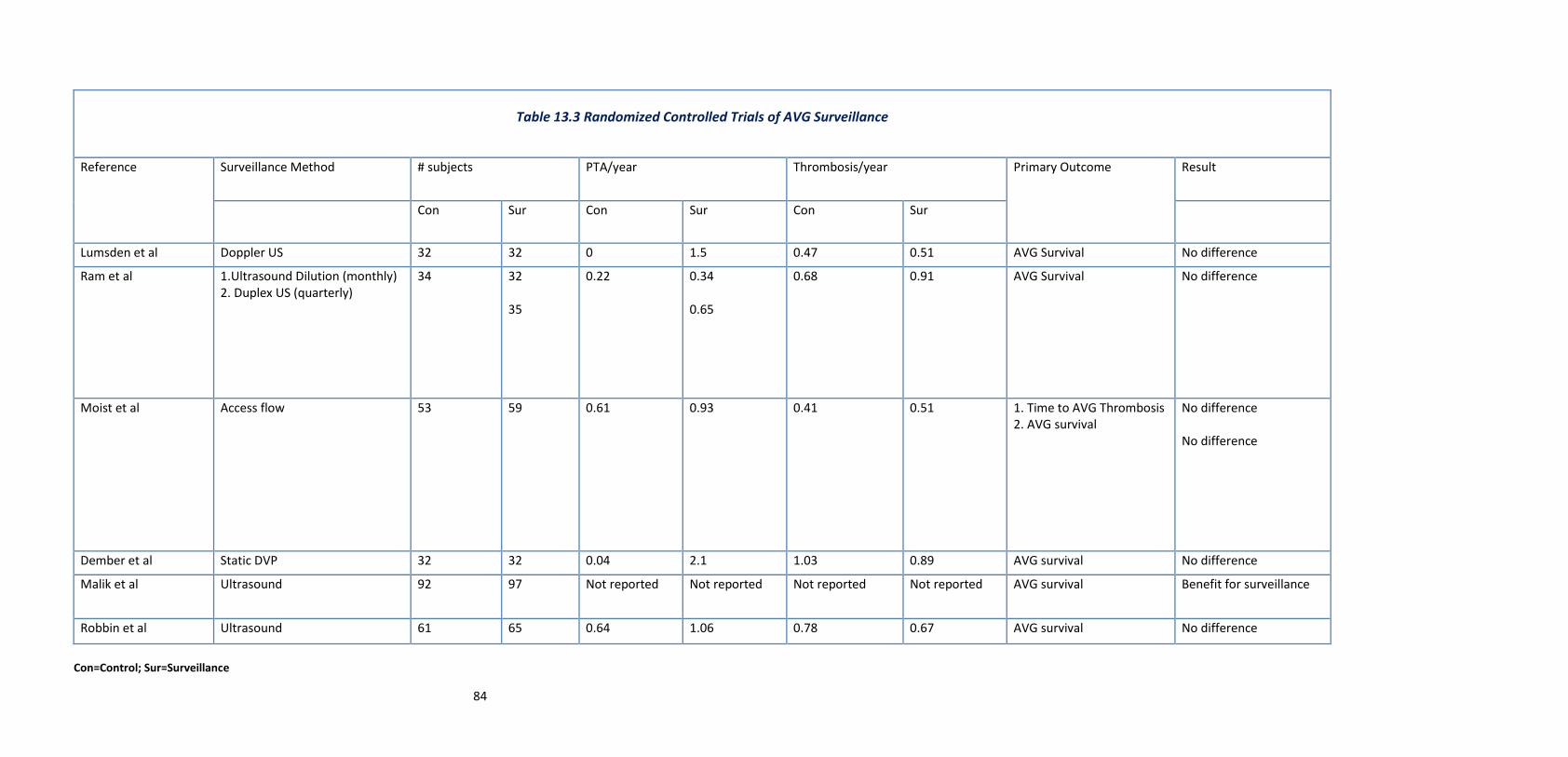
84
Table 13.3 Randomized Controlled Trials of AVG Surveillance
Reference Surveillance Method # subjects PTA/year Thrombosis/year Primary Outcome Result
Con Sur Con Sur Con Sur
Lumsden et al Doppler US 32 32 0 1.5 0.47 0.51 AVG Survival No difference
Ram et al 1.Ultrasound Dilution (monthly) 2. Duplex US (quarterly)
34 32 35
0.22 0.34 0.65
0.68 0.91 AVG Survival No difference
Moist et al Access flow 53 59 0.61 0.93 0.41 0.51 1. Time to AVG Thrombosis 2. AVG survival
No difference No difference
Dember et al Static DVP 32 32 0.04 2.1 1.03 0.89 AVG survival No difference
Malik et al Ultrasound 92 97 Not reported Not reported Not reported Not reported AVG survival Benefit for surveillance
Robbin et al Ultrasound 61 65 0.64 1.06 0.78 0.67 AVG survival No difference
Con=Control; Sur=Surveillance
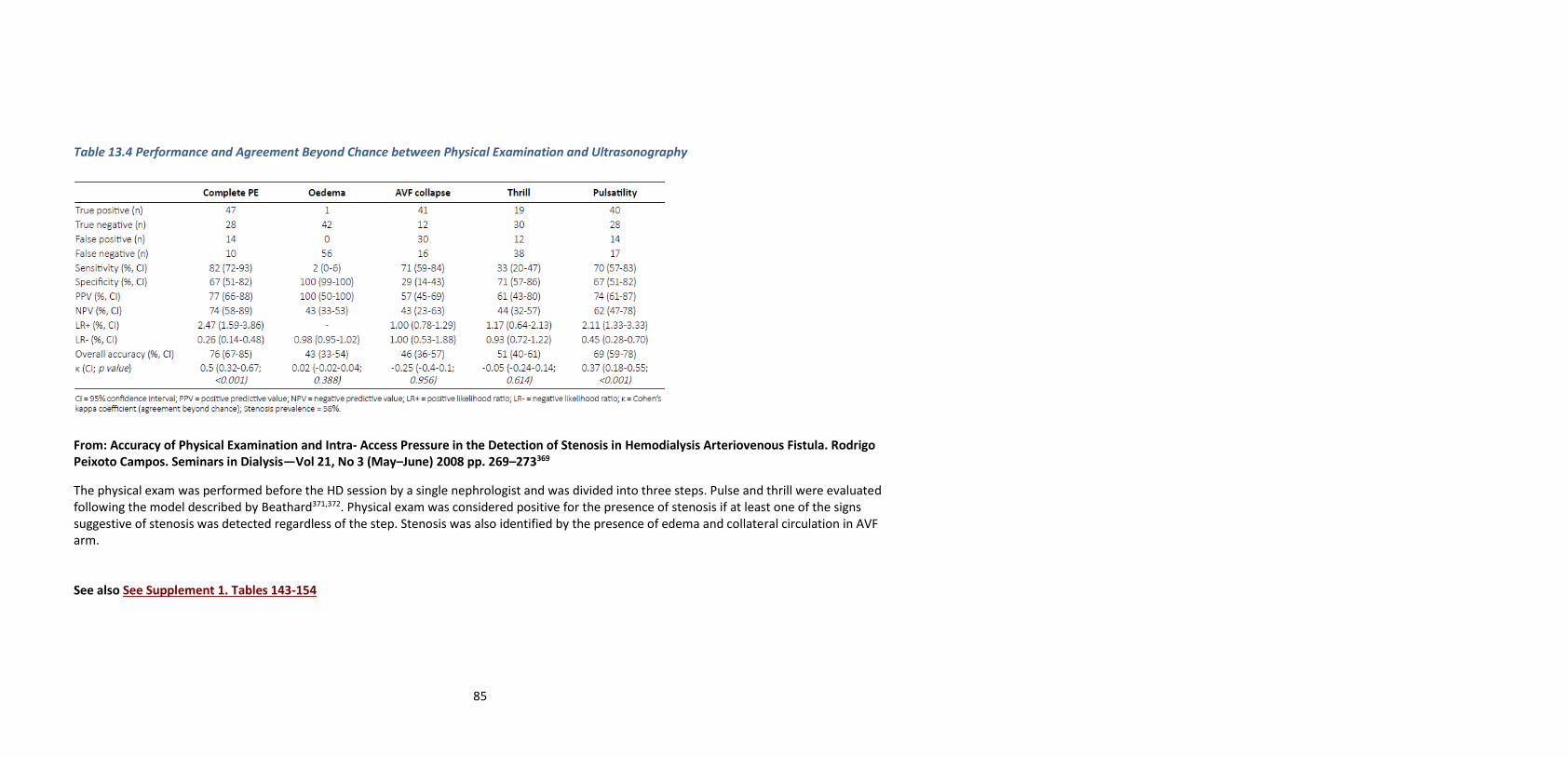
85
Table 13.4 Performance and Agreement Beyond Chance between Physical Examination and Ultrasonography
From: Accuracy of Physical Examination and Intra- Access Pressure in the Detection of Stenosis in Hemodialysis Arteriovenous Fistula. Rodrigo Peixoto Campos. Seminars in Dialysis—Vol 21, No 3 (May–June) 2008 pp. 269–273369
The physical exam was performed before the HD session by a single nephrologist and was divided into three steps. Pulse and thrill were evaluated following the model described by Beathard371,372. Physical exam was considered positive for the presence of stenosis if at least one of the signs suggestive of stenosis was detected regardless of the step. Stenosis was also identified by the presence of edema and collateral circulation in AVF arm. See also See Supplement 1. Tables 143-154
86
GUIDELINE 14. AV-ACCESS FLOW DYSFUNCTION+ - PREVENTION
Statements for the Prevention of AV-Access (AVF or AVG) Flow Dysfunction+
+“AV-access flow dysfunction” refers to clinically significant abnormalities in AV-access (AVF or AVG) flow or patency due to underlying stenosis, thrombosis or related pathology. This is in distinction to other types of AV-access complications. Pharmacological Primary and Secondary Prevention of AV-access flow dysfunction
Fistulas 14.1 KDOQI suggests that the use of adjuvant far-infrared therapy to improve AVF primary patency be based on individual circumstances,
feasibility and the clinician’s best judgment and expertise (Conditional recommendation, Moderate quality evidence)
14.2 KDOQI does not suggest the use of fish oil or aspirin to prevent AVF flow dysfunction (Conditional recommendation; Low-Moderate quality of evidence)
14.3 There is inadequate evidence for KDOQI to make a recommendation on the use of oral simvastatin and ezetimibe to reduce AVF interventions or thrombosis
14.4 There is inadequate evidence for KDOQI to make a recommendation on the use of Clopidogrel-prostacyclin to improve AVF primary failure
Grafts
14.5 KDOQI suggests careful consideration of potential individual patient benefits, risks and circumstances prior to the use of combination dipyridamole (200 mg) and aspirin (25 mg) twice daily to improve AVG primary unassisted patency (Conditional recommendation; High quality evidence)
14.6 KDOQI suggests the use of oral fish oil supplementation, in patients with newly created AV grafts, to reduce patient morbidity (i.e.
reduce frequency of thrombosis and related corrective interventions) (Conditional recommendation; Moderate quality of evidence) 14.7 There is inadequate evidence for KDOQI to make a recommendation on the use of oral fish oil supplementation to prolong AVG
cumulative patency
14.8 There is inadequate evidence for KDOQI to make a recommendation on the use of oral simvastatin and ezetimibe for reducing dialysis AVG interventions and thrombosis.
Rationale/Background
Arteriovenous Fistula
AVF maturation failure remains an important clinical problem for hemodialysis patients and has been reported in observational studies to range from 20-60%373,374. In fact, a multicenter RCT in the United States reported that up to 60% of AVFs created failed to mature successfully for dialysis use236. At present there are very few effective therapies to prevent AVF dysfunction. The 2006 NKF-KDOQI guidelines recommended interventions in AVF in the presence of inadequate flow to support dialysis, hemodynamically significant venous stenosis, aneurysm formation, and ischemia11. These guidelines recommended a percutaneous transluminal angioplasty (PTA) or surgical revision in an AVF with a greater than 50% stenosis in either the venous outflow or arterial inflow, in conjunction with clinical or physiological abnormalities11. The previous 2006 NKF-KDOQI VA guidelines did not address pharmacologic interventions to prevent AVF dysfunction11. Since the 2006 NKF-KDOQI VA guidelines, there has been newly published literature evaluating pharmacologic and other therapies to prevent AVF dysfunction.
Far-Infrared Therapy 375-378
Four studies (n=763), all conducted in Taiwan, compared far-infrared radiation with no treatment for prevention of AVF dysfunction375-378. One study recruited patients not yet on hemodialysis for whom a new AVF was created377, two studies recruited patients already on hemodialysis via AVF for at least 6 months without AVF interventions for at least 3 months375,376, and one recruited patients on hemodialysis via AVF who required two or more percutaneous interventions in the past378. Participants were followed for 1 year. All four studies as well as the pooled results reported statistically higher rates of primary patency at 1 year with far-infrared radiation compared to no treatment (RR=1.24; 95% CI: 1.07 to 1.45). Lin et al. in 2013 reported significantly lower occlusion in the far infrared radiation group (5%) versus no treatment group (18%) (p=0.03)377. Lin et al. in 2007 found no difference in thrombosis at 1 year (p=0.15)375. There was no difference in the need for angioplasty between treatment arms; Lin et al. in 2013 reported angioplasty in 23% versus 25% (p=0.87)375,376. In another study, Lin et al. reported 0.11 vs. 0.29 angioplasties per day per patient per year (p=0.10)377. Lai et al.378 and Lin et al. 377 reported no complications or infections in both groups. Lin et al. 377 reported shorter hospital stays with the far-infrared radiation versus no treatment (0.40 vs. 1.35 days/patient/year; p<0.01) See Supplement 1. Table 155 Fish oil 379
A single RCT by Irish et al RCT (n=567) compared fish oil versus placebo379 for prophylaxis of AVF dysfunction. A subset of participants in both groups without indication for aspirin (n=406) was also randomized to aspirin or placebo. It is unclear how many in each group were concurrently taking aspirin but analyses controlled for aspirin status. Follow-up was 6 months for adverse events, hospitalizations/ED visits and mortality and 1 year for primary outcomes (primary AVF failure and thrombosis). Study participants were on average 55 years old, 63% were men, 53% were
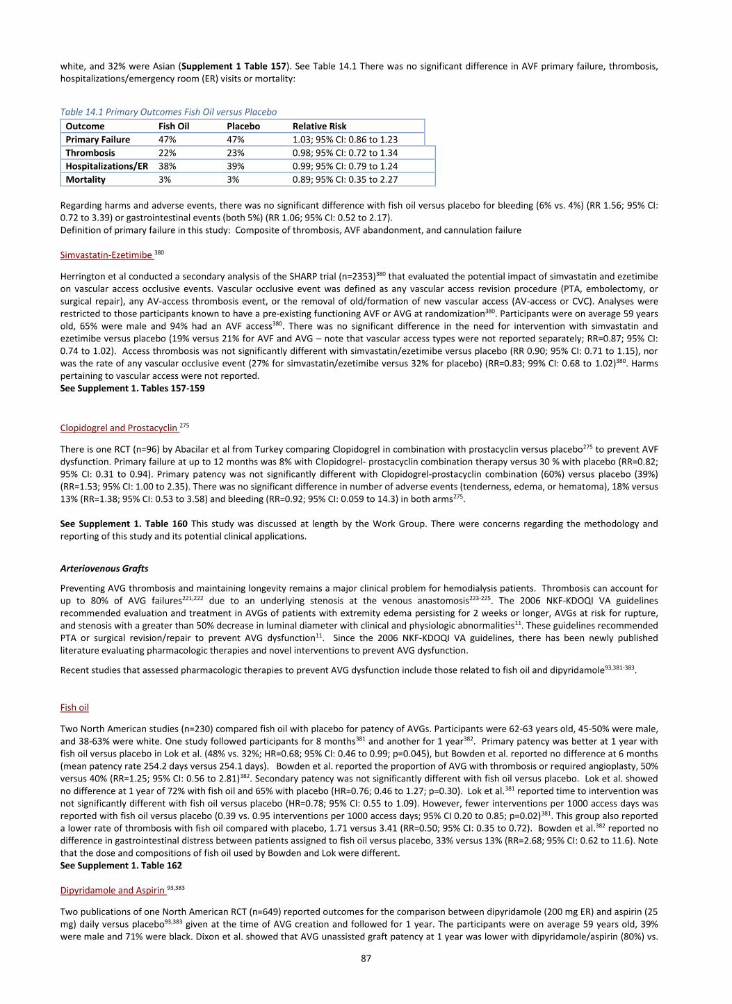
87
white, and 32% were Asian (Supplement 1 Table 157). See Table 14.1 There was no significant difference in AVF primary failure, thrombosis, hospitalizations/emergency room (ER) visits or mortality:
Table 14.1 Primary Outcomes Fish Oil versus Placebo
Outcome Fish Oil Placebo Relative Risk
Primary Failure 47% 47% 1.03; 95% CI: 0.86 to 1.23
Thrombosis 22% 23% 0.98; 95% CI: 0.72 to 1.34
Hospitalizations/ER 38% 39% 0.99; 95% CI: 0.79 to 1.24
Mortality 3% 3% 0.89; 95% CI: 0.35 to 2.27
Regarding harms and adverse events, there was no significant difference with fish oil versus placebo for bleeding (6% vs. 4%) (RR 1.56; 95% CI: 0.72 to 3.39) or gastrointestinal events (both 5%) (RR 1.06; 95% CI: 0.52 to 2.17). Definition of primary failure in this study: Composite of thrombosis, AVF abandonment, and cannulation failure Simvastatin-Ezetimibe 380
Herrington et al conducted a secondary analysis of the SHARP trial (n=2353)380 that evaluated the potential impact of simvastatin and ezetimibe on vascular access occlusive events. Vascular occlusive event was defined as any vascular access revision procedure (PTA, embolectomy, or surgical repair), any AV-access thrombosis event, or the removal of old/formation of new vascular access (AV-access or CVC). Analyses were restricted to those participants known to have a pre-existing functioning AVF or AVG at randomization380. Participants were on average 59 years old, 65% were male and 94% had an AVF access380. There was no significant difference in the need for intervention with simvastatin and ezetimibe versus placebo (19% versus 21% for AVF and AVG – note that vascular access types were not reported separately; RR=0.87; 95% CI: 0.74 to 1.02). Access thrombosis was not significantly different with simvastatin/ezetimibe versus placebo (RR 0.90; 95% CI: 0.71 to 1.15), nor was the rate of any vascular occlusive event (27% for simvastatin/ezetimibe versus 32% for placebo) (RR=0.83; 99% CI: 0.68 to 1.02)380. Harms pertaining to vascular access were not reported. See Supplement 1. Tables 157-159
Clopidogrel and Prostacyclin 275
There is one RCT (n=96) by Abacilar et al from Turkey comparing Clopidogrel in combination with prostacyclin versus placebo275 to prevent AVF dysfunction. Primary failure at up to 12 months was 8% with Clopidogrel- prostacyclin combination therapy versus 30 % with placebo (RR=0.82; 95% CI: 0.31 to 0.94). Primary patency was not significantly different with Clopidogrel-prostacyclin combination (60%) versus placebo (39%) (RR=1.53; 95% CI: 1.00 to 2.35). There was no significant difference in number of adverse events (tenderness, edema, or hematoma), 18% versus 13% (RR=1.38; 95% CI: 0.53 to 3.58) and bleeding (RR=0.92; 95% CI: 0.059 to 14.3) in both arms275. See Supplement 1. Table 160 This study was discussed at length by the Work Group. There were concerns regarding the methodology and reporting of this study and its potential clinical applications.
Arteriovenous Grafts
Preventing AVG thrombosis and maintaining longevity remains a major clinical problem for hemodialysis patients. Thrombosis can account for up to 80% of AVG failures221,222 due to an underlying stenosis at the venous anastomosis223-225. The 2006 NKF-KDOQI VA guidelines recommended evaluation and treatment in AVGs of patients with extremity edema persisting for 2 weeks or longer, AVGs at risk for rupture, and stenosis with a greater than 50% decrease in luminal diameter with clinical and physiologic abnormalities11. These guidelines recommended PTA or surgical revision/repair to prevent AVG dysfunction11. Since the 2006 NKF-KDOQI VA guidelines, there has been newly published literature evaluating pharmacologic therapies and novel interventions to prevent AVG dysfunction.
Recent studies that assessed pharmacologic therapies to prevent AVG dysfunction include those related to fish oil and dipyridamole93,381-383. Fish oil
Two North American studies (n=230) compared fish oil with placebo for patency of AVGs. Participants were 62-63 years old, 45-50% were male, and 38-63% were white. One study followed participants for 8 months381 and another for 1 year382. Primary patency was better at 1 year with fish oil versus placebo in Lok et al. (48% vs. 32%; HR=0.68; 95% CI: 0.46 to 0.99; p=0.045), but Bowden et al. reported no difference at 6 months (mean patency rate 254.2 days versus 254.1 days). Bowden et al. reported the proportion of AVG with thrombosis or required angioplasty, 50% versus 40% (RR=1.25; 95% CI: 0.56 to 2.81)382. Secondary patency was not significantly different with fish oil versus placebo. Lok et al. showed no difference at 1 year of 72% with fish oil and 65% with placebo (HR=0.76; 0.46 to 1.27; p=0.30). Lok et al.381 reported time to intervention was not significantly different with fish oil versus placebo (HR=0.78; 95% CI: 0.55 to 1.09). However, fewer interventions per 1000 access days was reported with fish oil versus placebo (0.39 vs. 0.95 interventions per 1000 access days; 95% CI 0.20 to 0.85; p=0.02)381. This group also reported a lower rate of thrombosis with fish oil compared with placebo, 1.71 versus 3.41 (RR=0.50; 95% CI: 0.35 to 0.72). Bowden et al.382 reported no difference in gastrointestinal distress between patients assigned to fish oil versus placebo, 33% versus 13% (RR=2.68; 95% CI: 0.62 to 11.6). Note that the dose and compositions of fish oil used by Bowden and Lok were different. See Supplement 1. Table 162 Dipyridamole and Aspirin 93,383
Two publications of one North American RCT (n=649) reported outcomes for the comparison between dipyridamole (200 mg ER) and aspirin (25 mg) daily versus placebo93,383 given at the time of AVG creation and followed for 1 year. The participants were on average 59 years old, 39% were male and 71% were black. Dixon et al. showed that AVG unassisted graft patency at 1 year was lower with dipyridamole/aspirin (80%) vs.
88
placebo (84%) (HR=0.82; 95% CI: 0.68 to 0.98). They reported median primary patency of 5.8 months with dipyridamole/aspirin vs. 4.3 months with placebo (i.e. a difference of 6 weeks); however, statistical significance was not reported and data was otherwise insufficient to calculate significance or assess quality of evidence. There was no difference in hospitalizations between groups (54% vs. 52%; HR 0.93; 95% CI 0.75 to 1.15)93,383. The study did not report further harms of dipyridamole and aspirin combination. Tables of study and evidence quality: See Supplement 1. Table 163-164 Summary: Arteriovenous Fistula In AVFs, Clopidogrel and prostacyclin has been the only pharmacologic therapy suggested by RCT evidence to be effective to prevent AVF dysfunction275. This study reported that Clopidogrel and prostacyclin therapy improved primary AVF failure. While reviewed in a separate section, a multicenter RCT by Dember et al.236 reported Clopidogrel significantly reduced 6-week AVF thrombosis but did not improve AVF suitability for dialysis. A recent RCT evaluating fish oil and ASA in new AVFs did not show any benefit to improve primary failures, thrombosis, abandonment, or cannulation failure in the fish oil group379. A subset of participants without aspirin indication (n=406) in both groups were also randomized to aspirin (ASA) or placebo379. It is unclear how many in each group were concurrently taking aspirin but analyses controlled for ASA status. The results from the ASA subset were not graded for level of evidence. However, in the ASA only analysis, ASA did not improve primary or secondary outcomes. Also, in a subsequent independent publication, fish oil was found to be associated with reduced rates of AV-access interventions.336 Finally, the far-infrared therapy studies demonstrated significantly improved primary AVF patency rates compared to no treatment. While several of these pharmacologic and technologic therapies have shown benefit, we would recommend an individualized approach towards implementing these therapies as the sample size was small in several of these studies. Arteriovenous Grafts There were two major studies evaluating pharmacologic therapies in AVG, fish oil381 and dipyridamole/ASA93. The fish oil study improved primary but not secondary AVG patency. Dipyridamole/ ASA showed benefit to reduce loss of primary patency. Clinical practice implementation based on the results of these studies need to be individualized. While in combined studies fish oil did not show significant benefit for AVF patency it was associated with reduced frequency (rate) of thrombosis and interventions, and prolonged the time to thrombosis. Overall, fish oil has low risk profile; risks and benefits can be considered within the patient’s ESKD Life-Plan. On the other hand, while dipyridamole/ASA showed a significant statistical benefit to reduce loss of primary patency, the benefit of the therapy realistically was very modest, primary unassisted patency in dipyridamole/ASA group was 28% vs. 23% in placebo group (i.e. taking dipyridamole/ASA delayed loss of primary patency by 6 weeks)93. However, the risks of potential complications of frailty, falls, and bleeding must be individualized before considering use of this therapy. Although not reviewed by the ERT for these guidelines, the efficacy of the combination of aspirin and Clopidogrel in the prevention of graft thrombosis was evaluated in a Veterans Affairs Cooperative study384. The study was a randomized, double-blind trial conducted at 30 hemodialysis units at Veterans Affairs medical centers. Participants undergoing hemodialysis with an AVG were randomized to receive either double placebos or aspirin (325 mg) and Clopidogrel (75 mg) daily. Participants were to be monitored while receiving study medications for a minimum of 2 years. The study was stopped after randomization of 200 participants by the Data Safety and Monitoring Board because of a significantly increased risk of bleeding among the participants receiving aspirin and Clopidogrel therapy. The cumulative incidence of bleeding events was significantly greater for those participants, compared with participants receiving placebos [hazard ratio, 1.98; 95% confidence interval (CI), 1.19 to 3.28; P = 0.007]384. Among the vascular access outcomes data reported, there was no significant benefit of active treatment in the prevention of thrombosis (hazard ratio, 0.81; 95% CI, 0.47 to 1.40; P = 0.45)384. Similar increased major bleeding risks were noted in a well-conducted double blind RCT of low dose warfarin (target INR 1.4-1.9) versus placebo to reduce hemodialysis graft thrombosis385. While these studies did now show benefit of an antiplatelet agent or anticoagulant to reduce AVG thrombosis, they more importantly highlight the need for assessing the risks for potential complications of using antiplatelet or anticoagulant agents in an elderly and frail population, where the benefit of the therapy may only be modest or absent. Special Discussions
Overall, there were very few studies that evaluated therapies to prevent AVF and AVG dysfunction that was of high quality evidence. Even in those studies of high quality, careful consideration should be made before using additional therapies based on statistically significant results in the absence of important clinically meaningful differences. Implementation considerations
• Some of these therapies such as far-infrared therapy may not be widely available outside of Asia.
• Antiplatelet and anticoagulant agents may increase risk of bleeding and their use needs to be considered carefully considered in the elderly population and those patients with high bleeding risks
Monitoring and Evaluation
• Since a number of these therapies have been performed in smaller studies and often non-randomized studies, adverse events need to be closely monitored and evaluated.
• A number of these agents have adverse events such as bleeding, so these complications must be closely monitored and evaluated.
Future research
• Far-Infrared therapies for AVF and AVG dysfunction in populations outside of Taiwan
• FISH study did not reach its enrolment target, additional studies of omega-3 fatty acids, including those with different formulations and/or different doses may provide insight to potential benefits for AV-access outcomes.
89
GUIDELINE 15. AV-ACCESS FLOW DYSFUNCTION - TREATMENT
Statement: Radiographic Confirmation of Clinically Significant AV-access Stenosis
15.1 KDOQI considers it reasonable that when clinical monitoring suspects clinically significant AV-access stenosis, further timely and
confirmatory evaluation should proceed, including imaging of the dialysis access circuit *+ (Expert Opinion)
*Dialysis access circuit is defined as: the continuum from the heart and the arterial inflow through the AV-access to the venous outflow
back to the heart. +The timeframe, choice and extent of imaging studies for further evaluation is dependent on local resources and the severity of findings
on clinical monitoring; a timeframe of less than 2 weeks was deemed reasonable by the KDOQI Work Group.
15.2 KDOQI considers it reasonable to use the smallest volume of iodinated contrast or non-iodinated contrast agents (e.g. CO2 gas) by
operators knowledgeable in their uses, contraindications and risks to obtain the best possible image in all patients with CKD to preserve
residual kidney function (Expert Opinion)
15.3 KDOQI considers it reasonable that when further confirmatory imaging studies reveals a culprit lesion responsible for clinical signs and
symptoms, that the clinically significant lesion* is promptly treated (Expert Opinion).
*a clinically significant lesion is one that contributes to clinical signs and symptoms (see AV access Monitoring Table 13.2) without other
cause, with or without a sustained change in measurements (e.g. change in blood flow (Qa) or venous pressures) in the dialysis access
circuit.
Statement: General Treatment of Clinically Significant Stenosis or Thrombosed AV-access
15.4 KDOQI considers it reasonable to use a careful individualized approach to the treatment of failing or thrombosed AVF and AVG (surgical
or endovascular), based on the operators best clinical judgment and expertise** considering the patient’s ESKD Life-Plan (Expert
Opinion)
** It considers both the patient’s individual circumstances and the operator’s clinical experience and expertise (i.e. reasonable
capabilities and limitations); preferably discussed and agreed upon by the team managing the patient’s vascular access, including but not
limited to the patient and one or more of: nephrologist, interventionalist, surgeon, VAC, cannulators (nurse or technician
Statement: Treatment of Clinically Significant AV- Access Stenosis
Angioplasty
15.5 KDOQI considers it reasonable to use balloon angioplasty (with high pressure as needed) as primary treatment of AVF and AVG stenotic
lesions that are both clinically and angiographically significant *(Expert Opinion)
*angiographically present stenosis without accompanying clinical signs and symptoms is insufficient to treat/intervene upon
15.6 There is inadequate evidence for KDOQI to make a recommendation regarding the use of specialized balloons (drug-coated or cutting)
versus standard high-pressure balloons in the primary treatment of AVF and AVG stenosis.
15.7 There is inadequate evidence for KDOQI to make a recommendation regarding the optimal duration of balloon inflation time during
angioplasty to improve intervention primary patency in the treatment of AVF or AVG stenosis.
15.8 KDOQI considers it reasonable that a careful patient-individualized approach to the choice of balloon type for angioplasty of clinically
significant AVF and AVG stenosis be based on the operator’s best clinical judgment and expertise. (Expert Opinion)
Stents
15.9 KDOQI suggests the appropriate use* of self-expanding stent-grafts in preference to angioplasty alone to treat clinically significant graft-
vein anastomotic stenosis in AVG when the goal is overall better 6-month post intervention outcomes+ after carefully considering the
patient’s ESKD Life-Plan (Conditional recommendation, Moderate quality of evidence).
+overall better 6-month outcomes refer to reduced recurrent AVG restenosis +/- patency
* Appropriate use avoids cannulation segments
15.10 KDOQI considers it reasonable to first consider the consequences of placement of a stent-graft on future AV-access options according to
the patient’s ESKD Life-Plan, in consultation with the vascular access team if necessary, prior to its placement (Expert Opinion)
15.11 KDOQI suggests that the use of an appropriately placed stent-graft is preferred to angioplasty alone for the treatment of in-stent
restenosis in AVG and AVF for overall better 6-month post intervention outcomes+. (Conditional recommendation, Moderate quality of
evidence)
+overall better 6-month outcomes refer to reduced recurrent AVG and AVF restenosis +/- patency
15.12 KDOQI considers it reasonable to avoid the use of bare metal stents for the treatment of clinically and /or angiographically significant
AVG and AVF stenotic lesions (Expert Opinion)
90
Statement: Treatment of Thrombosed AV-Access
15.13 KDOQI considers it reasonable that management of each episode of AV-access thrombosis involves the consideration of the patient’s
dialysis access Succession Plan that is consistent with the ESKD Life-Plan, given the compromised AV-access patency after either
endovascular or surgical treatment (Expert Opinion)
**Operators discretion carefully considers both the patient’s individual circumstances and the operator’s own clinical
experience and expertise (i.e. reasonable capabilities and limitations)
15.14 KDOQI considers it reasonable to surgically treat a failing AV-access in the following circumstances: 1) endovascular failures, 2) clinically
significant lesions not amenable to endovascular treatment, 3) situations in which the surgical outcomes are deemed markedly better+.
(Expert Opinion) + situations when surgical outcomes are anticipated to be better than alternative options should be first discussed and agreed upon
by the team managing the patient’s vascular access, including but not limited to the patient and one or more of: nephrologist,
interventionalist, surgeon, VAC, cannulation expert, if possible
Statements 15.1-15.12 Rationale/Background
Maintaining effective functioning AV-access is an ongoing challenge that frequently requires remedial treatment for problematic AV-accesses (See
Treatment of AV access flow dysfunction and Table 13.2, Clinical Signs and Symptoms Associated with Dialysis Access flow dysfunction), especially
if AV-access thrombosis occurs as a terminal event. Several of these conditions are addressed in separate sections of the Guidelines (See
Guidelines 16-19). The generic approach to these various failure modes includes appropriate monitoring and imaging to identify the underlying
cause and then definitive treatment of the clinically significant culprit lesion. Various endovascular approaches have largely replaced the open,
surgical approach and should be used as the first line of treatment with the surgical approach reserved for endovascular failures, lesions not
amenable to endovascular treatment and those select lesions in which the surgical approach is deemed far more durable.
Stenosis is the most common complication following AV-access (AVF and AVG) creation.386 Stenosis is caused by neointimal hyperplasia387 and
leads to several serious sequelae in both AVF and AVG. Stenosis causes AVF non-maturation with the attendant reliance on CVCs. AVF maturation
failure has been reported in observational studies to range from 20-60%373,374. A multicenter randomized clinical trial in the United States
reported that up to 60% of AVF failed to mature successfully for dialysis use29. Stenosis also causes dysfunction in mature AVF resulting in
inadequate HD clearance, thrombosis, and eventual abandonment. AVG are also susceptible to stenosis developing at the graft-vein junction
leading to thrombosis in 80% of AVG221,222,224,225,388. The previous NKF KDOQI Clinical Practice Guideline for Vascular Access (2006) maintained that
percutaneous balloon angioplasty (PTA) is the first-line treatment for stenosis in the access circuit.11 While this generally remains the case in the
modern era of vascular access, several important clinical trials and device innovations are considered that may alter the indications for angioplasty
and the types of balloons and stents for treatment of stenosis. The current recommendations update those from the previous KDOQI Guidelines.
Detailed Justification
Maintaining vascular access function required to provide adequate goal-directed dialysis is an ongoing challenge for all access types and
configurations; all have a limited life expectancy. The complication and remediation rates (e.g. infection, sepsis, thrombectomy) vary by the
dialysis access type (CVC – 0.7 and 2.1 episodes/year; AVF – 0.14 and 0.44 episodes/year; AVG – 0.25 and 0.77, episodes/year, respectively).389,390
and are detailed in various sections of these Guidelines. In addition to the significant morbidity associated with dialysis access complications391,
procedures to confirm and manage AV-access flow-related problems have been deemed the most burdensome to patients, often leading to
patient dissatisfaction336. It is the responsibility of the treating team to ensure appropriate detection of failing AV-access and judicious use of
invasive procedures in the diagnosis and treatment of culprit lesions. To provide greater clarity, the Work Group has replaced the term “AV-access
dysfunction” with 3 more specific terms describing AV-access complications: 1) thrombotic-flow related complications; 2) non-thrombotic-flow
related complications and 3) infection-related complications (see Glossary). It is incumbent upon all health care providers who care for
hemodialysis patients to be familiar with the various vascular access complications and failure modes. It is critical to emphasize that reducing
complications and vascular access failure begins before a vascular access is created with proper patient selection and an appropriate vascular
access plan (vascular access creation plan) that considers the entire life cycle of the vascular access (vascular access contingency and succession
plans) with the appropriate monitoring and care within the dialysis unit.
Clinically Significant Stenosis and Related Complications
Detection and Diagnosis
A clinically significant stenosis within the hemodialysis access circuit is associated with patient signs and symptoms and can lead to abnormal
dialysis indicators (13.2). The hemodialysis vascular access circuit should be viewed as a continuum from the left side of the heart through the
peripheral arterial inflow to the anastomosis and back again through the peripheral veins (or graft) to the central veins and ultimately to the right
side of the heart. AV-accesses can develop lesions, especially stenosis, at any point within this circuit although they tend to develop at fairly
predictable locations. AVGs typically developing stenoses at the graft-venous anastomoses while AVFs develop stenoses in the juxta-anastomotic
region and the swing segment for brachial-basilic AVF and the cephalic arch for brachial-cephalic AVF.392-395 As noted earlier in the Guidelines
(Guideline 15), the indication for further evaluation and definitive treatment requires the presence of a clinically symptomatic, significant lesion
and not simply the presence of a lesion (i.e. asymptomatic stenosis).396,397
91
Once routine monitoring reveals a clinically important vascular access problem (see Guideline 15), the diagnostic approach is contingent upon the
clinical suspicion of the underlying lesion (i.e. arterial inflow, anastomosis, venous outflow). Duplex ultrasound is effective for examining the AV-
access from the arterial anastomosis throughout the peripheral venous section (or graft). However, it is ineffective for interrogating the central
venous outflow given the limitations of ultrasound and the bony thoracic cavity. Arterial duplex ultrasound can be helpful to image the peripheral
arteries, but is again limited for assessing the great vessels in the chest; of note, stenoses at the origin of the left subclavian artery are common in
elderly patients. Catheter-based venography and arteriography are invasive procedures but can definitively image the AV-access from the
anastomosis to the heart (venography) or the whole AV-access circuit (arteriography) while serving as a platform for therapeutic interventions. CT
arteriography and venography are alternative diagnostic studies for most peripheral vascular interventions but may have practical limitations. 398
These diagnostic procedures may employ iodinated contrast that is potentially nephrotoxic and should be used judiciously in patients with
CKD/ESKD and residual kidney function – on or off dialysis. Indeed, the choice of contrast needs to be factored into the imaging algorithm. Carbon
dioxide (CO2) can be used as an alternative non-nephrotoxic contrast agent for catheter-based procedures in select cases although the quality of
the imaging may not be sufficient to guide interventions and caution should be exercised to avoid intra-arterial injection in the upper extremity
due to the potential of the gas to pass into the cerebral arterial circulation and cause neurologic events.399 Intravascular ultrasound can also be
helpful in patients with severe contrast allergies.400 Magnetic resonance imaging (MRI) is also an alternative to CT arteriography although the
published experience is limited and the use of the MRI contrast agent gadolinium is contraindicated due to the risk of nephrogenic fibrosis.398,401
Appropriate imaging of the dialysis circuit should identify the culprit lesion and suggest definitive treatment strategies. Venous stenoses
throughout the peripheral vein for AVFs and at the venous anastomoses for AVGs are typically treated with endovascular therapies given their less
invasive nature. Notably, these stenoses are typically dense, fibrous lesions from intimal hyperplasia and are distinctly different from arterial
atherosclerotic lesions. Anastomotic lesions can likewise be treated with an endovascular-first approach although they may not be as amenable to
angioplasty, and, thus, more suited for open repair. The choice of treatment for atherosclerotic arterial inflow lesions should be treated based
upon their natural history, independent of the fact that they involved vessels providing inflow to the AV-access. Accordingly, subclavian orifical
lesions are typically treated with a combination of balloon angioplasty and intraluminal stenting with the more peripheral lesions (i.e. axillary,
brachial, radial, ulnar) treated with by endovascular methods or open surgical repair using bypass or patch angioplasty.
Treatment
Angioplasty
High-Pressure Balloon Angioplasty: Venous stenosis from neointimal hyperplasia (NH) is characterized by a concentric thickening of the internal
elastic lamina with a lumen diameter reduction caused by myointimal cell proliferation402 While there are several putative mechanisms that may
promote NH, including an inflammatory reaction secondary to the AVF creation surgery, hemodynamic shear stress, trauma related to needle
punctures, and possibly as a direct result of uremia403 a durable solution to NH induced stenosis remains elusive. Re-stenosis at the initial lesion or
elsewhere in the previously treated dialysis circuit occurs up to 60% of the time at six months404. What is more, the usual percutaneous
angioplasty (PTA) solution to AV-access stenosis is the very mechanism by which NH is induced in the porcine model and vessels progressively
injured in HD patients.405 As a result of these fundamental limitations in the efficacy of standard PTA, several innovations have been put forth that
were evaluated in these guidelines. Aside from the special circumstances described herein, standard high-pressure balloon angioplasty remains
the treatment of choice for the majority of anatomic and clinically significant AV access stenotic lesions. A clinically significant AV access stenotic
lesion is one that is accompanied by clinical signs and symptoms (refer to section Guideline 13 and Table 13.2) and shows >50% narrowing by
angiography or ultrasound.
Paclitaxel-Coated Balloon Angioplasty in AVF: A RCT (n=40) evaluated treatment with paclitaxel drug-coated balloons (PCBs) in AVF (Supplement
1. Table 176) to high-pressure balloons for treatment of single stenotic lesions in AVFs406,407. Patients were followed to one year with clinical
assessments every two months. Both received oral aspirin 100mg daily after angioplasty. Primary patency at 200 days was not significantly
different with PCB versus high-pressure balloons (RR 2.25; 95% CI: 0.83 to 6.13). However, treatment lesion restenosis-free survival was
significantly superior in the PCB group compared with high pressure balloon group (PCB, 308 d; HPB, 161; HR, 0.47; 95% CI, 0.23–0.96; P = .03) as
was access circuit primary patency (median days); 270 versus 161 days; HR 0.48; 95% CI: 0.23 to 0.97). Thrombosis at 1 year did not differ (RR 2.0;
95% CI: 0.2 to 20.3)407. Another small RCT (n=10) compared PCB with plain balloon versus plain balloon alone for treatment of stenotic AVF where
each access circuit had two lesions, one treated with PCB and the other without. Each patient acted as their own control and was followed for 1
year.408 A high-pressure balloon was used if a plain balloon did not adequately treat the target lesion. Primary patency at 1 year was not
significantly different with PCB and plain balloons versus plain balloon alone (20% versus 0; p=0.47). Angioplasty-free days were significantly
greater with PCB and plain balloons versus plain balloon alone (251 versus 103 days; p<0.01).
Paclitaxel-Coated Balloon Angioplasty in AVG: One RCT (n=40) compared paclitaxel-coated balloon versus plain balloon for PTA in stenotic 14 AVF and 26 AVG at 1 year See Supplement 1. Table 165. See Table 15.1 Outcomes Paclitaxel-Coated Balloon Angioplasty in AVGs and AVFs.
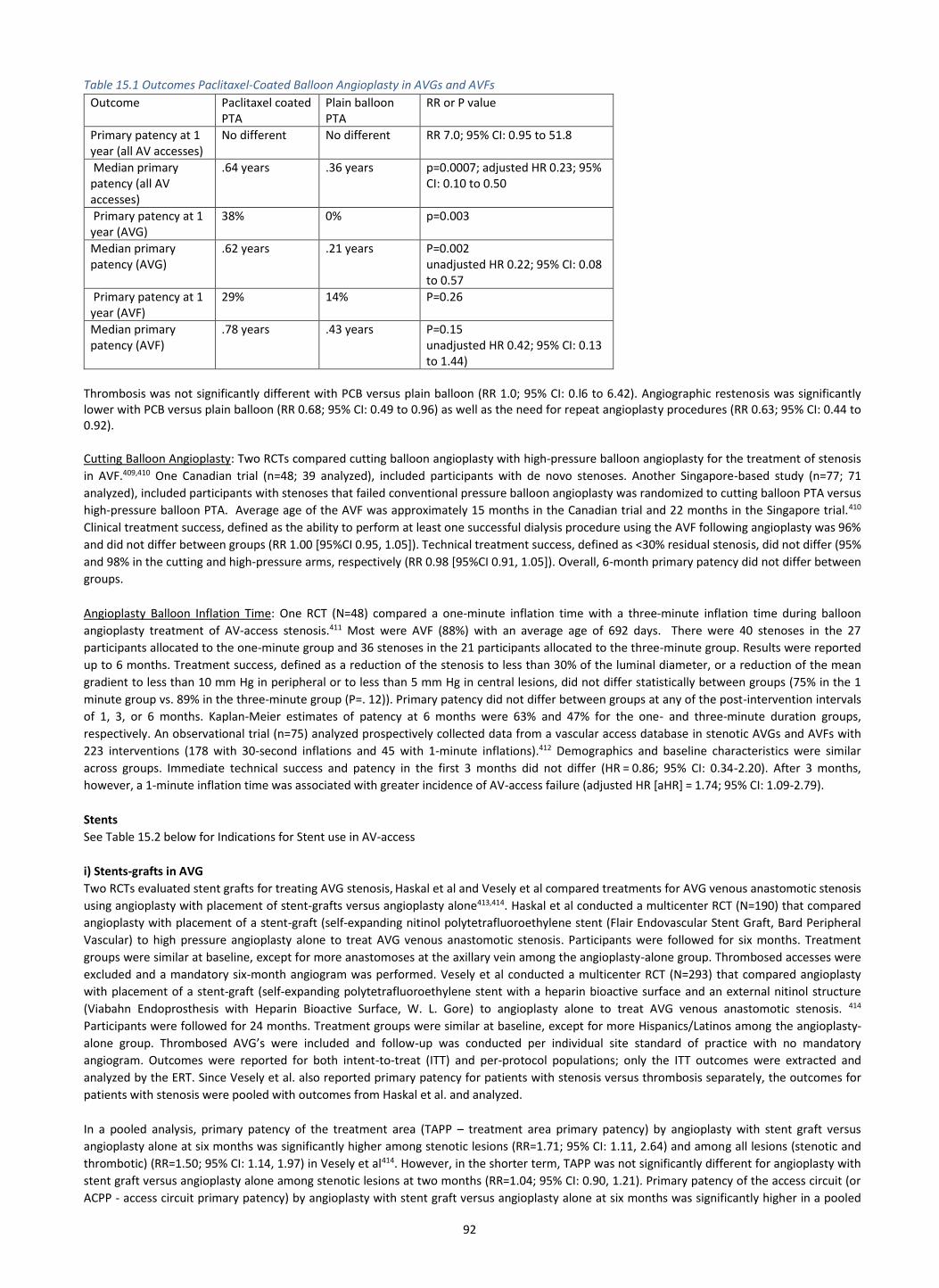
92
Table 15.1 Outcomes Paclitaxel-Coated Balloon Angioplasty in AVGs and AVFs
Outcome Paclitaxel coated PTA
Plain balloon PTA
RR or P value
Primary patency at 1 year (all AV accesses)
No different No different RR 7.0; 95% CI: 0.95 to 51.8
Median primary patency (all AV accesses)
.64 years .36 years p=0.0007; adjusted HR 0.23; 95% CI: 0.10 to 0.50
Primary patency at 1 year (AVG)
38% 0% p=0.003
Median primary patency (AVG)
.62 years .21 years P=0.002 unadjusted HR 0.22; 95% CI: 0.08 to 0.57
Primary patency at 1 year (AVF)
29% 14% P=0.26
Median primary patency (AVF)
.78 years .43 years P=0.15 unadjusted HR 0.42; 95% CI: 0.13 to 1.44)
Thrombosis was not significantly different with PCB versus plain balloon (RR 1.0; 95% CI: 0.l6 to 6.42). Angiographic restenosis was significantly lower with PCB versus plain balloon (RR 0.68; 95% CI: 0.49 to 0.96) as well as the need for repeat angioplasty procedures (RR 0.63; 95% CI: 0.44 to 0.92).
Cutting Balloon Angioplasty: Two RCTs compared cutting balloon angioplasty with high-pressure balloon angioplasty for the treatment of stenosis
in AVF.409,410 One Canadian trial (n=48; 39 analyzed), included participants with de novo stenoses. Another Singapore-based study (n=77; 71
analyzed), included participants with stenoses that failed conventional pressure balloon angioplasty was randomized to cutting balloon PTA versus
high-pressure balloon PTA. Average age of the AVF was approximately 15 months in the Canadian trial and 22 months in the Singapore trial.410
Clinical treatment success, defined as the ability to perform at least one successful dialysis procedure using the AVF following angioplasty was 96%
and did not differ between groups (RR 1.00 [95%CI 0.95, 1.05]). Technical treatment success, defined as <30% residual stenosis, did not differ (95%
and 98% in the cutting and high-pressure arms, respectively (RR 0.98 [95%CI 0.91, 1.05]). Overall, 6-month primary patency did not differ between
groups.
Angioplasty Balloon Inflation Time: One RCT (N=48) compared a one-minute inflation time with a three-minute inflation time during balloon
angioplasty treatment of AV-access stenosis.411 Most were AVF (88%) with an average age of 692 days. There were 40 stenoses in the 27
participants allocated to the one-minute group and 36 stenoses in the 21 participants allocated to the three-minute group. Results were reported
up to 6 months. Treatment success, defined as a reduction of the stenosis to less than 30% of the luminal diameter, or a reduction of the mean
gradient to less than 10 mm Hg in peripheral or to less than 5 mm Hg in central lesions, did not differ statistically between groups (75% in the 1
minute group vs. 89% in the three-minute group (P=. 12)). Primary patency did not differ between groups at any of the post-intervention intervals
of 1, 3, or 6 months. Kaplan-Meier estimates of patency at 6 months were 63% and 47% for the one- and three-minute duration groups,
respectively. An observational trial (n=75) analyzed prospectively collected data from a vascular access database in stenotic AVGs and AVFs with
223 interventions (178 with 30-second inflations and 45 with 1-minute inflations).412 Demographics and baseline characteristics were similar
across groups. Immediate technical success and patency in the first 3 months did not differ (HR = 0.86; 95% CI: 0.34-2.20). After 3 months,
however, a 1-minute inflation time was associated with greater incidence of AV-access failure (adjusted HR [aHR] = 1.74; 95% CI: 1.09-2.79).
Stents
See Table 15.2 below for Indications for Stent use in AV-access
i) Stents-grafts in AVG
Two RCTs evaluated stent grafts for treating AVG stenosis, Haskal et al and Vesely et al compared treatments for AVG venous anastomotic stenosis
using angioplasty with placement of stent-grafts versus angioplasty alone413,414. Haskal et al conducted a multicenter RCT (N=190) that compared
angioplasty with placement of a stent-graft (self-expanding nitinol polytetrafluoroethylene stent (Flair Endovascular Stent Graft, Bard Peripheral
Vascular) to high pressure angioplasty alone to treat AVG venous anastomotic stenosis. Participants were followed for six months. Treatment
groups were similar at baseline, except for more anastomoses at the axillary vein among the angioplasty-alone group. Thrombosed accesses were
excluded and a mandatory six-month angiogram was performed. Vesely et al conducted a multicenter RCT (N=293) that compared angioplasty
with placement of a stent-graft (self-expanding polytetrafluoroethylene stent with a heparin bioactive surface and an external nitinol structure
(Viabahn Endoprosthesis with Heparin Bioactive Surface, W. L. Gore) to angioplasty alone to treat AVG venous anastomotic stenosis. 414
Participants were followed for 24 months. Treatment groups were similar at baseline, except for more Hispanics/Latinos among the angioplasty-
alone group. Thrombosed AVG’s were included and follow-up was conducted per individual site standard of practice with no mandatory
angiogram. Outcomes were reported for both intent-to-treat (ITT) and per-protocol populations; only the ITT outcomes were extracted and
analyzed by the ERT. Since Vesely et al. also reported primary patency for patients with stenosis versus thrombosis separately, the outcomes for
patients with stenosis were pooled with outcomes from Haskal et al. and analyzed.
In a pooled analysis, primary patency of the treatment area (TAPP – treatment area primary patency) by angioplasty with stent graft versus
angioplasty alone at six months was significantly higher among stenotic lesions (RR=1.71; 95% CI: 1.11, 2.64) and among all lesions (stenotic and
thrombotic) (RR=1.50; 95% CI: 1.14, 1.97) in Vesely et al414. However, in the shorter term, TAPP was not significantly different for angioplasty with
stent graft versus angioplasty alone among stenotic lesions at two months (RR=1.04; 95% CI: 0.90, 1.21). Primary patency of the access circuit (or
ACPP - access circuit primary patency) by angioplasty with stent graft versus angioplasty alone at six months was significantly higher in a pooled
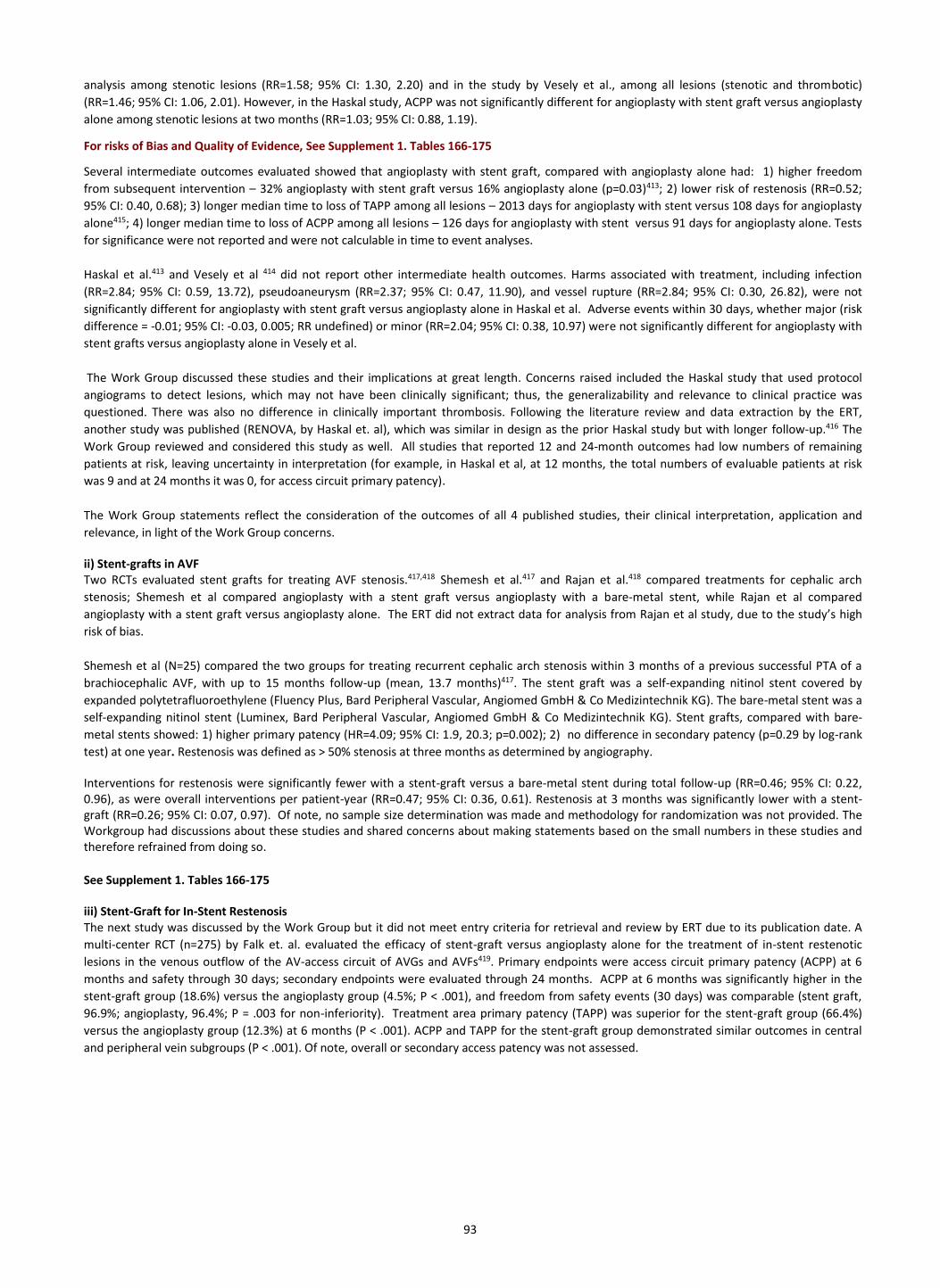
93
analysis among stenotic lesions (RR=1.58; 95% CI: 1.30, 2.20) and in the study by Vesely et al., among all lesions (stenotic and thrombotic)
(RR=1.46; 95% CI: 1.06, 2.01). However, in the Haskal study, ACPP was not significantly different for angioplasty with stent graft versus angioplasty
alone among stenotic lesions at two months (RR=1.03; 95% CI: 0.88, 1.19).
For risks of Bias and Quality of Evidence, See Supplement 1. Tables 166-175
Several intermediate outcomes evaluated showed that angioplasty with stent graft, compared with angioplasty alone had: 1) higher freedom
from subsequent intervention – 32% angioplasty with stent graft versus 16% angioplasty alone (p=0.03)413; 2) lower risk of restenosis (RR=0.52;
95% CI: 0.40, 0.68); 3) longer median time to loss of TAPP among all lesions – 2013 days for angioplasty with stent versus 108 days for angioplasty
alone415; 4) longer median time to loss of ACPP among all lesions – 126 days for angioplasty with stent versus 91 days for angioplasty alone. Tests
for significance were not reported and were not calculable in time to event analyses.
Haskal et al.413 and Vesely et al 414 did not report other intermediate health outcomes. Harms associated with treatment, including infection
(RR=2.84; 95% CI: 0.59, 13.72), pseudoaneurysm (RR=2.37; 95% CI: 0.47, 11.90), and vessel rupture (RR=2.84; 95% CI: 0.30, 26.82), were not
significantly different for angioplasty with stent graft versus angioplasty alone in Haskal et al. Adverse events within 30 days, whether major (risk
difference = -0.01; 95% CI: -0.03, 0.005; RR undefined) or minor (RR=2.04; 95% CI: 0.38, 10.97) were not significantly different for angioplasty with
stent grafts versus angioplasty alone in Vesely et al.
The Work Group discussed these studies and their implications at great length. Concerns raised included the Haskal study that used protocol
angiograms to detect lesions, which may not have been clinically significant; thus, the generalizability and relevance to clinical practice was
questioned. There was also no difference in clinically important thrombosis. Following the literature review and data extraction by the ERT,
another study was published (RENOVA, by Haskal et. al), which was similar in design as the prior Haskal study but with longer follow-up.416 The
Work Group reviewed and considered this study as well. All studies that reported 12 and 24-month outcomes had low numbers of remaining
patients at risk, leaving uncertainty in interpretation (for example, in Haskal et al, at 12 months, the total numbers of evaluable patients at risk
was 9 and at 24 months it was 0, for access circuit primary patency).
The Work Group statements reflect the consideration of the outcomes of all 4 published studies, their clinical interpretation, application and
relevance, in light of the Work Group concerns.
ii) Stent-grafts in AVF Two RCTs evaluated stent grafts for treating AVF stenosis.417,418 Shemesh et al.417 and Rajan et al.418 compared treatments for cephalic arch
stenosis; Shemesh et al compared angioplasty with a stent graft versus angioplasty with a bare-metal stent, while Rajan et al compared
angioplasty with a stent graft versus angioplasty alone. The ERT did not extract data for analysis from Rajan et al study, due to the study’s high
risk of bias.
Shemesh et al (N=25) compared the two groups for treating recurrent cephalic arch stenosis within 3 months of a previous successful PTA of a
brachiocephalic AVF, with up to 15 months follow-up (mean, 13.7 months)417. The stent graft was a self-expanding nitinol stent covered by
expanded polytetrafluoroethylene (Fluency Plus, Bard Peripheral Vascular, Angiomed GmbH & Co Medizintechnik KG). The bare-metal stent was a
self-expanding nitinol stent (Luminex, Bard Peripheral Vascular, Angiomed GmbH & Co Medizintechnik KG). Stent grafts, compared with bare-
metal stents showed: 1) higher primary patency (HR=4.09; 95% CI: 1.9, 20.3; p=0.002); 2) no difference in secondary patency (p=0.29 by log-rank
test) at one year. Restenosis was defined as > 50% stenosis at three months as determined by angiography.
Interventions for restenosis were significantly fewer with a stent-graft versus a bare-metal stent during total follow-up (RR=0.46; 95% CI: 0.22, 0.96), as were overall interventions per patient-year (RR=0.47; 95% CI: 0.36, 0.61). Restenosis at 3 months was significantly lower with a stent-graft (RR=0.26; 95% CI: 0.07, 0.97). Of note, no sample size determination was made and methodology for randomization was not provided. The Workgroup had discussions about these studies and shared concerns about making statements based on the small numbers in these studies and therefore refrained from doing so.
See Supplement 1. Tables 166-175
iii) Stent-Graft for In-Stent Restenosis The next study was discussed by the Work Group but it did not meet entry criteria for retrieval and review by ERT due to its publication date. A
multi-center RCT (n=275) by Falk et. al. evaluated the efficacy of stent-graft versus angioplasty alone for the treatment of in-stent restenotic
lesions in the venous outflow of the AV-access circuit of AVGs and AVFs419. Primary endpoints were access circuit primary patency (ACPP) at 6
months and safety through 30 days; secondary endpoints were evaluated through 24 months. ACPP at 6 months was significantly higher in the
stent-graft group (18.6%) versus the angioplasty group (4.5%; P < .001), and freedom from safety events (30 days) was comparable (stent graft,
96.9%; angioplasty, 96.4%; P = .003 for non-inferiority). Treatment area primary patency (TAPP) was superior for the stent-graft group (66.4%)
versus the angioplasty group (12.3%) at 6 months (P < .001). ACPP and TAPP for the stent-graft group demonstrated similar outcomes in central
and peripheral vein subgroups (P < .001). Of note, overall or secondary access patency was not assessed.
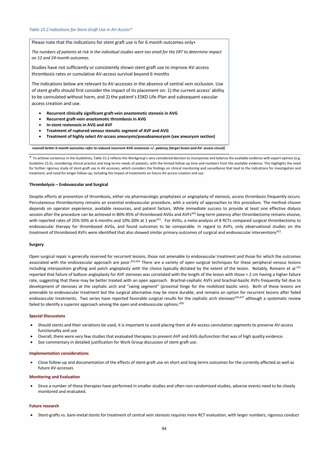
94
Table 15.2 Indications for Stent-Graft Use in AV-Access*
Please note that the indications for stent graft use is for 6 month outcomes only+
The numbers of patients at risk in the individual studies were too small for the ERT to determine impact
on 12 and 24-month outcomes.
Studies have not sufficiently or consistently shown stent graft use to improve AV-access
thrombosis rates or cumulative AV-access survival beyond 6 months
The indications below are relevant to AV-accesses in the absence of central vein occlusion. Use
of stent grafts should first consider the impact of its placement on: 1) the current access’ ability
to be cannulated without harm, and 2) the patient’s ESKD Life-Plan and subsequent vascular
access creation and use.
• Recurrent clinically significant graft-vein anastomotic stenosis in AVG
• Recurrent graft-vein anastomotic thrombosis in AVG
• In-stent restenosis in AVG and AVF
• Treatment of ruptured venous stenotic segment of AVF and AVG
• Treatment of highly select AV-access aneurysm/pseudoaneurysm (see aneurysm section)
+overall better 6-month outcomes refer to reduced recurrent AVG restenosis +/- patency (target lesion and AV- access circuit)
* To achieve consensus in the Guidelines, Table 15.2 reflects the Workgroup’s very considered decision to incorporate and balance the available evidence with expert opinion (e.g.
Guideline 15.5), considering clinical practice and long terms needs of patients, with the limited follow-up time and numbers from the available evidence. This highlights the need
for further rigorous study of stent-graft use in AV-accesses, which considers the findings on clinical monitoring and surveillance that lead to the indications for investigation and
treatment, and need for longer follow-up, including the impact of treatments on future AV-access creation and use.
Thrombolysis – Endovascular and Surgical
Despite efforts at prevention of thrombosis, either via pharmacologic prophylaxis or angioplasty of stenosis, access thrombosis frequently occurs.
Percutaneous thrombectomy remains an essential endovascular procedure, with a variety of approaches to this procedure. The method chosen
depends on operator experience, available resources, and patient factors. While immediate success to provide at least one effective dialysis
session after the procedure can be achieved in 80%-95% of thrombosed AVGs and AVFs420 long-term patency after thrombectomy remains elusive,
with reported rates of 25%-50% at 6 months and 10%-20% at 1 year421. For AVGs, a meta-analysis of 8 RCTs compared surgical thrombectomy to
endovascular therapy for thrombosed AVGs, and found outcomes to be comparable. In regard to AVFs, only observational studies on the
treatment of thrombosed AVFs were identified that also showed similar primary outcomes of surgical and endovascular interventions422.
Surgery
Open surgical repair is generally reserved for recurrent lesions, those not amenable to endovascular treatment and those for which the outcomes
associated with the endovascular approach are poor.423,424 There are a variety of open surgical techniques for these peripheral venous lesions
including interposition grafting and patch angioplasty with the choice typically dictated by the extent of the lesion. Notably, Romann et al.425
reported that failure of balloon angioplasty for AVF stenoses was correlated with the length of the lesion with those > 2 cm having a higher failure
rate, suggesting that these may be better treated with an open approach. Brachial-cephalic AVFs and brachial-basilic AVFs frequently fail due to
development of stenoses at the cephalic arch and “swing segment” (proximal hinge for the mobilized basilic vein). Both of these lesions are
amenable to endovascular treatment but the surgical alternative may be more durable, and remains an option for recurrent lesions after failed
endovascular treatments. Two series have reported favorable surgical results for the cephalic arch stenoses426,427 although a systematic review
failed to identify a superior approach among the open and endovascular options.428
Special Discussions
• Should stents and their variations be used, it is important to avoid placing them at AV-access cannulation segments to preserve AV-access functionality and use
• Overall, there were very few studies that evaluated therapies to prevent AVF and AVG dysfunction that was of high quality evidence.
• See commentary in detailed justification for Work Group discussion of stent-graft use. Implementation considerations
• Close follow-up and documentation of the effects of stent-graft use on short and long terms outcomes for the currently affected as well as future AV-accesses
Monitoring and Evaluation
• Since a number of these therapies have performed in smaller studies and often non-randomized studies, adverse events need to be closely monitored and evaluated.
Future research
• Stent-grafts vs. bare-metal stents for treatment of central vein stenosis requires more RCT evaluation, with larger numbers, rigorous conduct
95
and analysis
• More RCT evaluation of stent grafts for vascular access management (primary or secondary) with clinical (rather than angiogram) based outcomes are urgently required
• Study is needed in AVFs for multiple modalities of treatment e.g. stent grafts, drug eluting balloons, etc.
• Comparative methods of AV-access thrombolysis (e.g. surgical versus endovascular) with a variety of short and longer term AVF and AVG outcomes
• Anything for which there is inadequate evidence for, including: o The use of specialized balloons (drug-coated or cutting) versus standard high pressure balloons in the primary treatment of AVF and AVG
stenosis. o The optimal duration of balloon inflation time during angioplasty to improve intervention primary patency in the treatment of AVF or
AVG stenosis. o The secondary use of drug-coated balloons following successful angioplasty with high-pressure balloons for treatment of stenosis in AVF
and AVG
• Impact of timing of recurrence of stenosis on choice of treatment modality
• Use of stent grafts in locations other than graft-vein anastomosis or cephalic arch in brachiocephalic fistulas
• Optimal treatment of in-stent stenosis that occurs in stent-grafts
• Study outcomes with surgically corrected occluded accesses that are followed by a completion angiogram/imaging +/- further corrective procedure. How does this strategy compare with historical surgical correction without completion imaging or with endovascular management?
Statements 15.13-15.14 Rationale/Background
Fully occlusive thrombosis represents the terminal event for the “failing” AV-access and accounts for 65 – 85% of all access abandonments.429 While the contributory cause of the terminal thrombotic event is usually obvious (e.g. low flow related to venous outflow stenosis in AVG), patients can present with de novo thrombosis without any clear failure mechanism.430 The clinical priorities include providing necessary effective dialysis, clearing the intraluminal thrombus, and definitively treating the underlying cause of the failure. The diagnosis of a thrombosed AV-access is usually obvious on physical examination but can be confirmed as necessary with duplex ultrasound. Patients with an emergent need for dialysis (e.g. volume overload, hyperkalemia) should be dialyzed through a temporary non-tunneled CVC prior to definitive treatment of their AV-access thrombosis.
Detailed Justification
Both endovascular and open surgical approaches for treating thrombosed AVGs are acceptable with the choice determined by the local expertise, referral patterns, underlying condition, and patient preference. While earlier reports suggested superior outcomes with the surgical approach, more recent reports have suggested that both approaches for AVGs are comparable, likely reflecting the overall evolution in the endovascular therapies.422,431,432 Furthermore, there are a number of different endovascular approaches although none has proven superior.429 Importantly, open surgical and endovascular approaches should not be viewed as competing, but rather complementary since there are a variety of hybrid approaches that involve both (e.g. open surgical thrombectomy with combined balloon angioplasty of central vein stenosis).433,434 There has been a gradual evolution in the approach to thrombosed AVFs with the earlier version of the 2006 NKF-KDOQI VA guidelines recommending abandoning the AVF to more recent attempts at AVF thrombectomy and AV-access salvage.388,435 A meta-analysis by Kuhan et al.432 emphasized the lack of long-term data with little quality evidence to guide the management of thrombosed AVFs. The contraindications to AV-access thrombectomy include pulmonary hypertension and right-left intracardiac shunts due to the potential risk of embolization and AV-access infection. Regardless of the approach, thrombectomy should be performed in a timely fashion relative to the event, particularly for AVFs, given the pathophysiology of the thrombotic process and the underlying inflammatory response. While it is possible to remove AVG thrombus up to 30 days, early thrombectomy has been associated with better long-term results.436 Sadaghianloo et al.437 reported that thrombectomy within < 6 hours was associated with a better technical outcome and improved mid-term results. Practically, early thrombectomy helps minimize and/or eliminate the need for dialysis with a CVC. However, patients may still require a CVC for dialysis to manage volume overload or electrolyte abnormalities prior to AV-access thrombectomy. Although the reported technical success rates for AV-access thrombectomy, both endovascular and surgical have been good, the longer term patency rates have been poor, underscoring the fact that it is important to consider the next AV-access option (i.e. AV-access Succession Plan) with each thrombotic event.429,438,439 Simoni et al.440 reported from a large device registry that the technical success rate of the AngioJet (Boston Scientific, Marlborough, MA) percutaneous thrombectomy device was 92% although the patency rate for AVGs and AVFs was 53% and 86%, respectively, at 3 months. Similarly, Quencer et al.429 reported from a review article that the patency rates after thrombectomy ranged from 25 – 50% at 6 months and 10 – 20% at 12 months for both AVGs and AVFs. Accordingly, repeated thrombectomy of the same AV-access is likely not justified. Definitive treatment requires identifying and correcting the underlying cause of the AV-access thrombosis. Completion imaging should be done after either endovascular or surgical thrombectomy to confirm that the culprit lesion is corrected. AV-accesses thrombosis within the early postoperative period (< 30 days) is often due to technical issues (e.g. anastomotic stenosis) or the choice of an inadequate artery/vein combination (e.g. diminutive vein) for an AVF. AV-access thrombectomy and/or revision is/are possible if there is an identified technical defect although definitive treatment usually requires constructing a new AV-access. Chemical thrombolysis is a relative contraindication in the early postoperative period due to the associated bleeding risk.
96
Implementation Considerations All of the hemodialysis vascular access types and configurations have a limited life expectancy and, accordingly, will ultimately fail or start to fail
(i.e. “failing access”). It is incumbent upon the various health care providers that care for dialysis patients to recognized these dysfunctional
accesses and be familiar with the various failure modes (AV-access complications). Rosenburg et al.441 reported that it is possible to teach non-
medical professionals the requisite physical examination skills to recognize failing AV-access in a short duration of time. Furthermore, it is
important to engage the patients themselves and challenge them to assume responsibility for the care and maintenance of their vascular access.
The ultimate goal is to maintain safe, effective dialysis with the fewest number of interventions in a patient-centric, cost-effective manner. Any
intervention should consider the impact on current and future accesses; for example, viable/useful cannulation segments should be avoided when
using stent grafts. It is the hope that early recognition and definitive treatment of the failing and thrombosed vascular access will result in improved
vascular access functional survival.
Monitoring and Evaluation The identification of the failing and thrombosed AV-access should be part of the routine monitoring protocols (See Guidelines 13 and 14). It is
important for the various health care providers to be cognizant of the various failure modes such that they can initiate the appropriate referral
and/or remedial treatment, as part of the vascular access contingency plan. At that time, consideration and planning for the next dialysis access or
kidney replacement modality (as appropriate) should begin in accordance to the individual patient’s ESKD Life-Plan strategy.
Future research
• Define and validate the optimal monitoring strategies for the dysfunctional AV-access
• Define and validate the thresholds for intervention for the dysfunctional AV-access
• Define the optimal open and endovascular treatment for the dysfunctional and thrombosed AV- access
• Define outcome metrics for the dysfunctional and thrombosed AV-access
• Determine outcomes with surgically corrected occluded accesses that are followed by a completion angiogram/imaging +/- further corrective procedure. How does this strategy compare with historical surgical correction without completion imaging or with endovascular management?
97
GUIDELINE 16. AV-ACCESS INFECTION
Statements for AV-Access Infections Monitoring and Prevention
16.1 KDOQI considers it reasonable to check the vascular access* and surrounding area prior to every cannulation for signs and symptoms of infection (Expert Opinion). * this should be done by patient and cannulator (if patient does not self cannulate)
16.2 KDOQI considers it reasonable to educate the patient on washing the access arm using antiseptic to clean the skin prior to every cannulation (Expert opinion)
Diagnosis
16.3 KDOQI considers it reasonable to use radiologic imaging* to help confirm the diagnosis of AV-access infection; however, physical examination remains the hallmark for assessing for infection** (Expert Opinion). * imaging includes duplex ultrasound, +/- CT, PET, and nuclear medicine scans (e.g. indium scan) ** this includes erythema, skin breakdown, purulent discharge, presence of exposed graft
Treatment
16.4 KDOQI considers it reasonable to obtain cultures and sensitivities of the blood and any available infected AV-access vessel/material, surrounding tissue or drainage prior to initiating antibiotic therapy (Expert Opinion)
16.5 KDOQI considers it reasonable to consider for infected AV-access the rapid initiation of empiric broad spectrum antibiotics and timely referral to a surgeon knowledgeable in the management of vascular access complications (Expert Opinion)
16.6 KDOQI considers it reasonable to consider strict follow-up of culture results with the appropriate change in antibiotics based on organism sensitivities, with antibiotic duration according to extent of vascular access infection and surgical intervention (Expert Opinion)
16.7 KDOQI considers it reasonable that the specific surgical treatment for AV-access infections (with concurrent antibiotics) should be
based on the patient’s individual circumstances considering the extent of infection, offending organism, and future vascular access options (Expert Opinion)
Prevention 16.8 KDOQI considers it reasonable to conduct a careful physical exam by the surgeon and managing team prior to surgery to identify
infection risks that should first be managed before proceeding with AV-access surgery (e.g. poor dentition with dental infection, malnutrition etc.) (Expert Opinion)
Special considerations from Guideline 11, as they are relevant to preventing AVF cannulation related infection:
11.2 KDOQI recommends rope ladder cannulation as the preferred cannulation technique for AVFs (Conditional recommendation, Moderate quality of evidence)
11.3 KDOQI considers it reasonable to limit buttonhole cannulation only to special circumstances given the associated increased risks of
infection and related adverse consequences (Expert Opinion). 11.6 KDOQI considers it reasonable to have structured training and supervision of dialysis technicians and nurses prior to their first
cannulation, and regular training updates to maintain cannulation competency (Expert Opinion).
16.9 KDOQI considers it reasonable to investigate* and closely monitor for metastatic complications (e.g. endocarditis, spinal abscesses, septic arthritis) in patients with buttonhole infection from particularly dangerous organisms such as S. aureus, gram negative bacteria and fungal organisms.
*investigations include 2D echocardiography, MRI, joint aspirate and other, as appropriate
Rationale/Background
Both AVFs and AVGs (collectively referred to as AV-access) can become infected. The underlying mechanisms are somewhat different, but
the management principles including systemic antibiotics and source control are similar and grounded on standard principles of managing
infected vascular prostheses. The incidence of AV-access infections is relatively low, particularly for AVFs. However, the spectrum of
potential sequelae of AV-access infections are broad and range from mild limited cellulitis to extensive graft involvement mandating total
explant; the systemic consequences can range from localized pain and fever to overwhelming sepsis and death. Treatment requires early
recognition and management to prevent sequelae. The definitive treatment of AV-access infections should be chosen within the context of
the ESKD’s Life-Plan’s dialysis access needs, with consideration to preserve/maintain future vascular access options. Prevention and
monitoring for ongoing or recurrent infection is critical. The current recommendations are consistent with the recommendations from the
previous KDOQI Guidelines.
98
Detailed Justification
AVF and AVG infections are a major clinical problem, often leading to hospitalization and increased mortality.389,442,443 AVG infections have
been reported to occur in up to 1.6% – 35% of patients with an overall incidence of positive blood cultures of 0.31/1000 days.444,445 The
incidence of AVF infections is typically, but not always lower446, and this may be modified by different cannulation techniques, with
buttonhole cannulation putting AVF at greater risk of infection295,302,447. AVG that have been abandoned in the past are also sources for
infection. AVFs have a median infectious complication rate of 0.11/1000 days 448 and positive blood culture rate of 0.08/1000 days449
The underlying mechanisms responsible for the development of AV-access infections are likely multifactorial and include both patient and
system-related issues (Supplement 2- AV Access Infections). Skin organisms, Staphylococcus aureus and S. epidermidis account for 70 –
90% of AV-access infections 450-452 in the upper extremity with a higher incidence of gram negative organisms in lower extremity AV-
accesses 453. AV-access infection may be polymicrobial, with a variety of causative organisms, including fungi.258,451,454-457. Attaining blood
and relevant AV-access cultures (wound, soft tissue, tunnel or drainage) before initiating antibiotics is critical.
Patients with AV-access infections require timely detection, diagnosis and management to prevent poor outcomes. Presenting signs and
symptoms range from mild cellulitis around the cannulation site to bacteremia and overwhelming sepsis.
The physical examination and routine laboratory studies are usually sufficient to establish the diagnosis. However, additional imaging can
help corroborate the diagnosis and define the extent of AV-access involvement. In particular, Duplex ultrasound can be used to confirm
patency, interrogate the integrity of the AV-access wall, confirm the presence of any aneurysms/pseudoaneurysms, document the presence
of fluid around the vein/non-autogenous access and help determine the extent of the infection258,458 Ultrasound cannot differentiate the
type of fluid around the AV-access (i.e. hematoma vs. seroma vs. abscess) which requires clinical corroboration. Other imaging studies can
be used, including CT, PET, indium scans, and technetium scans although they are rarely necessary and not as universally
available.258,445,458,459 PET and nuclear medicine scans may help exclude an occult infection in a thrombosed, non-functional AVG.
General treatment
Blood cultures should always be obtained when an AV-access infection is suspected. Cultures of the infected skin, soft tissue, AV-access
site or tunnel (e.g. obvious abscess or purulent drainage) may help confirm causative organism and infection source. Cultures should also
be obtained at the time of any surgical intervention or AV-access excision. Antibiotic therapy should be altered accordingly to culture
sensitivity results. Most antibiotic regimens can be administered with dialysis, avoiding longer term intravenous AV-access. Consultation
with infectious diseases experts to find the most convenient and appropriate antibiotic regimen per culture results may be helpful. PICCs
should be avoided in CKD and ESKD patients as highlighted throughout the guidelines (Guideline 6).
Treatment of AV-access infections includes immediate initiation of broad spectrum antibiotics (after attaining relevant cultures) for both
gram-positive and gram-negative organism coverage (e.g. vancomycin, piperacillin/tazobactam) and control of the infectious nidus (i.e.
source control). Ideally, the infected AV-access should be avoided for hemodialysis, although possible if the infection is minimal.
Otherwise, a CVC should be inserted and used until the AV-access infection is resolved or if, not salvageable, until a new AV-access can be
established.
Specific medical management
Antibiotics alone may be adequate treatment for limited localized AV-access infections (e.g. buttonhole track infection in AVF). However,
the optimal treatment should be patient individualized, and requires a multidisciplinary team approach with early involvement of an
experienced AV-access surgeon (Supplement 2-AV Access Infections). Notably, AVG infections can rarely be treated with antibiotics alone
since once the prosthetic materials becomes infected it is almost impossible to clear the infection.
The optimal duration of antibiotic therapy depends on the patient’s circumstances and extent of infection. For example, extensive AV-
access infections involving the artery and vein should likely undergo an extended course of parenteral antibiotics similar to the treatment
of patients with endocarditis (e.g. >= 6 weeks); input from infectious diseases experts is suggested for guidance in complex cases.
Specific surgical management
Definitive surgical treatment of AV-access infection requires careful individualized consideration of the extent of the infection, the
offending organism, the type of AV-access (i.e. AVF vs. AVG), the location of the infection (i.e. anastomotic vs. non-anastomotic), the extent
of the infection (i.e. localized vs. diffuse), the presence of systemic signs, the presence of bleeding, and importantly, future dialysis access
options. Both AVF and AVG infections can lead to erosion of the skin and life-threatening hemorrhage, underscoring the importance of a
timely, definitive surgical treatment as needed. Surgical treatment options can be broadly categorized as strategies designed to salvage the
AV-access and those designed to excise the AV-access (Supplement 2) AV-access salvage options include those for “in situ” and “extra-
anatomic” reconstruction (with the reference being the anatomic course of the infected AV-access) for localized infections, where close
follow-up and surveillance is mandatory, given the risk of recurrent infection and the potential for anastomotic disruption and significant
bleeding. AV-access excision options include strategies for both complete and subtotal AV-access excision and are considered for
infections involving the full length of the AVG (See Supplement 2 for surgical details).
Key Surgical Management Points to Remember:
o Salvage of the AV-access may be possible if the infection is localized; in an AVG infection, this may be possible only if the adjacent
segments are uninvolved and well incorporated.
o If the infected segment is limited, graft replacement* through an uninfected field with excision of the involved segment, may be
possible
99
GUIDELINE 17. AV-ACCESS ANEURYSMS
Statements for AV-access Aneurysms
Recognition and Diagnosis
17.1 KDOQI considers it reasonable to routinely check AV-access for aneurysms/pseudoaneurysms at each dialysis session by knowledgeable care providers, including but not limited to dialysis technicians, nurses, nephrologists and vascular access coordinator (Expert Opinion).
17.2 KDOQI considers it is reasonable to obtain proactive surgical assessment when clinical findings suggest an AV-access
aneurysm/pseudoaneurysm to be at risk of complications* (Expert Opinion) *An aneurysm/pseudoaneurysm that is considered at risk of complications are those with evidence of associated symptoms or skin
breakdown 17.3 KDOQI considers it is reasonable to obtain emergent surgical assessment and treatment for AV-access aneurysm/pseudoaneurysm
complications such as erosion or hemorrhage (Expert Opinion) 17.4 KDOQI considers it reasonable to use Duplex ultrasound to corroborate the physical examination suggesting an AV-access
aneurysm/pseudoaneurysm and to obtain information on the size, presence of stenosis/thrombus, and impact on the AV-access (including flow rate (Qa), status of the arterial inflow and the venous outflow) (Expert Opinion).
Management
17.5 KDOQI considers it reasonable that the presence of an aneurysm/pseudoaneurysm alone in the absence of symptoms ((i.e.
asymptomatic) is not an indication for definitive treatment (Expert Opinion).
17.6 KDOQI considers it reasonable to avoid cannulating the access segment(s) that involve the aneurysm/pseudoaneurysm if there are
alternative sites. In the rare scenario where there are no suitable alternative sites, the side of the aneurysm/pseudoaneurysm
should be cannulated (Expert Opinion).
17.7 KDOQI considers it reasonable to obtain appropriate imaging of the arterial inflow and venous outflow to assess volume flow or
stenotic problems that may need correction prior to or during definitive treatment of symptomatic aneurysm/pseudoaneurysm
(Expert Opinion).
17.8 KDOQI considers it reasonable that surgical management is the preferred treatment for patients with symptomatic, large or
rapidly expanding AV-access aneurysm/pseudoaneurysm (see below “definitive treatment”) (Expert Opinion)
* preferable to replace with non-synthetic material such as femoral vein
o Extensive AV-access infections, especially AVG, require total graft excision with definitive treatment* of the arterial and venous
anastomosis.
* Potential options include vein patch angioplasty, vein bypass and ligation, depending on extent of infection and
circumstances
Preventative strategies should always be employed and include avoiding or limiting CVCs, optimizing the use of AVFs where appropriate,
avoiding the creation of lower extremity AV-accesses whenever possible, use of autogenous or non-autogenous biologic grafts in patients
at high risk for infection, use of the appropriate prophylactic perioperative antibiotics, strict sterile technique at the time of AV-access
creation, appropriate cannulation techniques (see Guideline 11) and mandatory routine AV-access monitoring with a low threshold for
intervention with any evidence of infection.
Buttonhole cannulation has been associated with significantly higher risks of infection compared with rope-ladder cannulation technique
and should be avoided whenever possible. Buttonhole infections are more likely to be gram positive and dangerous (e.g. S. aureus), often
with serious metastatic consequences such as endocarditis, septic arthritis or spinal abscesses. Should metastatic complications be
detected, rapid treatment with consultation from infectious diseases experts is necessary to prevent serious or catastrophic outcomes.
Special discussions
While the above refers to all AV-accesses, great attention must be paid to lower extremity AV-accesses due their higher complication rate,
including infectious complications (more likely to involve gram negative organisms).56,453,460,461
Early post- AV-access creation (< 1 month) soft tissue infections, are commonly associated with the inherent surgical trauma from their
creation and/or any break in sterile technique. They can typically be treated with a course of systemic antibiotics but require long term
monitoring.
While the current recommendations build upon those from the previous KDOQI Guidelines, the evidence-based review team did not
identify relevant publications for the treatment of AV-access infections using their search criteria. The previous guidelines defined outcome
measures for lifetime use of AVF (< 1%) and AVG (<10%); however, the current guidelines have not defined similar measures due to the lack
of appropriate evidence, especially those using current measures e.g. rates/1000 access days versus % use.
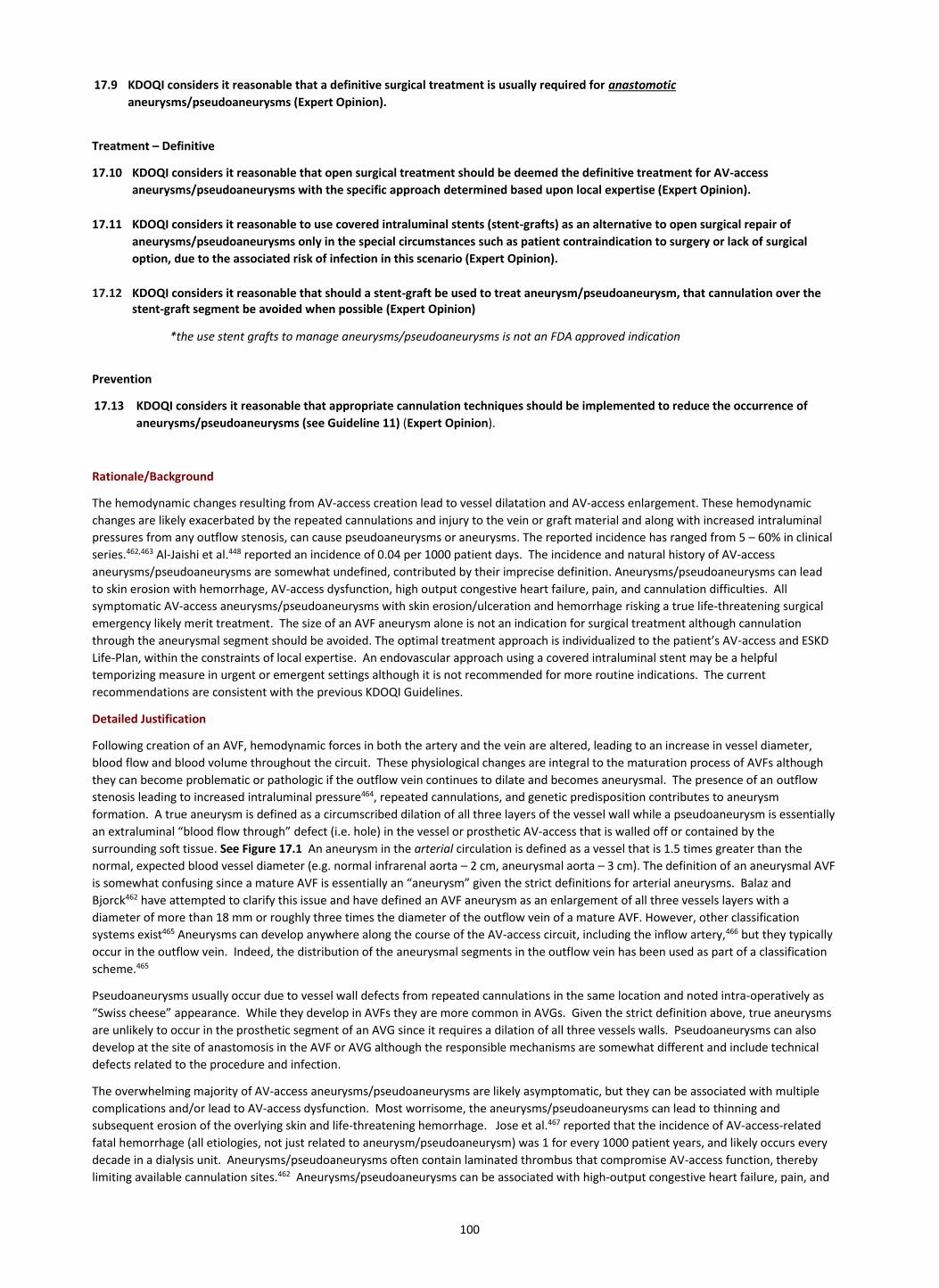
100
17.9 KDOQI considers it reasonable that a definitive surgical treatment is usually required for anastomotic
aneurysms/pseudoaneurysms (Expert Opinion).
Treatment – Definitive
17.10 KDOQI considers it reasonable that open surgical treatment should be deemed the definitive treatment for AV-access
aneurysms/pseudoaneurysms with the specific approach determined based upon local expertise (Expert Opinion).
17.11 KDOQI considers it reasonable to use covered intraluminal stents (stent-grafts) as an alternative to open surgical repair of
aneurysms/pseudoaneurysms only in the special circumstances such as patient contraindication to surgery or lack of surgical
option, due to the associated risk of infection in this scenario (Expert Opinion).
17.12 KDOQI considers it reasonable that should a stent-graft be used to treat aneurysm/pseudoaneurysm, that cannulation over the stent-graft segment be avoided when possible (Expert Opinion)
*the use stent grafts to manage aneurysms/pseudoaneurysms is not an FDA approved indication
Prevention
17.13 KDOQI considers it reasonable that appropriate cannulation techniques should be implemented to reduce the occurrence of
aneurysms/pseudoaneurysms (see Guideline 11) (Expert Opinion).
Rationale/Background
The hemodynamic changes resulting from AV-access creation lead to vessel dilatation and AV-access enlargement. These hemodynamic
changes are likely exacerbated by the repeated cannulations and injury to the vein or graft material and along with increased intraluminal
pressures from any outflow stenosis, can cause pseudoaneurysms or aneurysms. The reported incidence has ranged from 5 – 60% in clinical
series.462,463 Al-Jaishi et al.448 reported an incidence of 0.04 per 1000 patient days. The incidence and natural history of AV-access
aneurysms/pseudoaneurysms are somewhat undefined, contributed by their imprecise definition. Aneurysms/pseudoaneurysms can lead
to skin erosion with hemorrhage, AV-access dysfunction, high output congestive heart failure, pain, and cannulation difficulties. All
symptomatic AV-access aneurysms/pseudoaneurysms with skin erosion/ulceration and hemorrhage risking a true life-threatening surgical
emergency likely merit treatment. The size of an AVF aneurysm alone is not an indication for surgical treatment although cannulation
through the aneurysmal segment should be avoided. The optimal treatment approach is individualized to the patient’s AV-access and ESKD
Life-Plan, within the constraints of local expertise. An endovascular approach using a covered intraluminal stent may be a helpful
temporizing measure in urgent or emergent settings although it is not recommended for more routine indications. The current
recommendations are consistent with the previous KDOQI Guidelines.
Detailed Justification
Following creation of an AVF, hemodynamic forces in both the artery and the vein are altered, leading to an increase in vessel diameter,
blood flow and blood volume throughout the circuit. These physiological changes are integral to the maturation process of AVFs although
they can become problematic or pathologic if the outflow vein continues to dilate and becomes aneurysmal. The presence of an outflow
stenosis leading to increased intraluminal pressure464, repeated cannulations, and genetic predisposition contributes to aneurysm
formation. A true aneurysm is defined as a circumscribed dilation of all three layers of the vessel wall while a pseudoaneurysm is essentially
an extraluminal “blood flow through” defect (i.e. hole) in the vessel or prosthetic AV-access that is walled off or contained by the
surrounding soft tissue. See Figure 17.1 An aneurysm in the arterial circulation is defined as a vessel that is 1.5 times greater than the
normal, expected blood vessel diameter (e.g. normal infrarenal aorta – 2 cm, aneurysmal aorta – 3 cm). The definition of an aneurysmal AVF
is somewhat confusing since a mature AVF is essentially an “aneurysm” given the strict definitions for arterial aneurysms. Balaz and
Bjorck462 have attempted to clarify this issue and have defined an AVF aneurysm as an enlargement of all three vessels layers with a
diameter of more than 18 mm or roughly three times the diameter of the outflow vein of a mature AVF. However, other classification
systems exist465 Aneurysms can develop anywhere along the course of the AV-access circuit, including the inflow artery,466 but they typically
occur in the outflow vein. Indeed, the distribution of the aneurysmal segments in the outflow vein has been used as part of a classification
scheme.465
Pseudoaneurysms usually occur due to vessel wall defects from repeated cannulations in the same location and noted intra-operatively as
“Swiss cheese” appearance. While they develop in AVFs they are more common in AVGs. Given the strict definition above, true aneurysms
are unlikely to occur in the prosthetic segment of an AVG since it requires a dilation of all three vessels walls. Pseudoaneurysms can also
develop at the site of anastomosis in the AVF or AVG although the responsible mechanisms are somewhat different and include technical
defects related to the procedure and infection.
The overwhelming majority of AV-access aneurysms/pseudoaneurysms are likely asymptomatic, but they can be associated with multiple
complications and/or lead to AV-access dysfunction. Most worrisome, the aneurysms/pseudoaneurysms can lead to thinning and
subsequent erosion of the overlying skin and life-threatening hemorrhage. Jose et al.467 reported that the incidence of AV-access-related
fatal hemorrhage (all etiologies, not just related to aneurysm/pseudoaneurysm) was 1 for every 1000 patient years, and likely occurs every
decade in a dialysis unit. Aneurysms/pseudoaneurysms often contain laminated thrombus that compromise AV-access function, thereby
limiting available cannulation sites.462 Aneurysms/pseudoaneurysms can be associated with high-output congestive heart failure, pain, and

101
become cosmetically unacceptable to patients.
The diagnosis of an AV-access aneurysm/pseudoaneurysm is made by physical examination and can be corroborated with Duplex
ultrasound. Ultrasound can also detect peri-access fluid, intraluminal thrombus, inflow/outflow stenoses and quantitate
aneurysm/pseudoaneurysm diameter and intra- access flow rates. It is critical to determine if the aneurysm/pseudoaneurysm is
symptomatic and/or at risk for ulceration and rupture. Therefore, it is important to have routine, objective measure of
aneurysm/pseudoaneurysm growth and development, and with greater frequency if worrisome signs or symptoms develop (Table 17.1).
Some signs and “symptoms” such as AV-access flow dysfunction, thrombosis, unacceptable cosmetic appearance, pain and high-flow
cardiac output are either obvious or addressed elsewhere in the Guidelines.
All symptomatic AV-access aneurysms/pseudoaneurysms should be evaluated and managed. Patients with threatened skin or ulceration
overlying their aneurysms/pseudoaneurysms should be treated urgently or emergently to prevent significant hemorrhage. Patients with
aneurysm/pseudoaneurysm related AV-access flow dysfunction, thrombus, or limited cannulation sites require treatment to maintain AV-
access to provide effective dialysis. Patients with high output congestive heart failure merit treatment to potentially reverse any
contribution from the AV-access. Patients with pain and/or unacceptable cosmetic disfigurement due to their aneurysm/pseudoaneurysm
need an individualized treatment plan that considers the risks associated with potential loss of their AV-access. However,
aneurysm/pseudoaneurysm size alone is likely not an indication for treatment in the absence of symptoms or threatened skin.462,463 AV-
access aneurysms/pseudoaneurysms do not usually spontaneously rupture like aneurysms in the arterial circulation. However, rapidly
enlarging AV-access aneurysms/pseudoaneurysms merit closer follow-up and a lower threshold for intervention. Large, asymptomatic AVG
pseudoaneurysms may require careful monitoring and active treatment since they are essentially fed by a large opening in the prosthetic
material that is walled off or contained by the surrounding tissue. Lazarides et al.468 have suggested that 12 mm AVG pseudoaneurysms be
repaired since they exceed the typical 6 mm graft diameter two-fold. All anastomotic aneurysms/pseudoaneurysm require evaluation and
active consideration for repair as their etiology and natural history are likely different that those involving the outflow vein or length of the
graft. Lastly, all infected AV-access aneurysms/pseudoaneurysm merit treatment (See Guideline 16).
The treatment goals for patients with symptomatic access aneurysms/pseudoaneurysms should be to prevent acute complications by
managing the precipitating cause (e.g. high inflow or an venous outflow stenosis) and modifying the involved AV-access segment(s), to
maintain AV-access function, prevent further complications and avoid CVC use. Unfortunately, the quality of the available evidence to
guide treatment is limited and there are no randomized trials. Open, surgical repair remains the standard of care and it is feasible to
salvage the AV-access in the majority of cases; patency rates after revision range 52 – 100% at 1 year.462,463,468,469 The optimal approach and
timing is dictated by the extent of the aneurysm/pseudoaneurysm (i.e. focal versus diffuse), the quality of the involved tissue, and the
presence of any infection. Alternative treatments include aneurysmorraphy and interposition grafting with the choices among the latter
including both autogenous and prosthetic conduits tunneled in situ or “extra-anatomic” (i.e. relative to the course of the initial AVG). In
certain instances (e.g. two involved segments), a staged approach may be used to allow continued access use and avoid a CVC. A variety of
creative aneurysmorraphy techniques have been described involving diameter reduction over a catheter mandril,470 endo staples,471 and
reinforcement with an external mesh wrap.472 Note that aneurysmorraphy may not be feasible if the vein wall comprising the involved
segment is severely degenerated or calcified while in situ interposition grafting with prosthetic material is contraindicated in an infected
field. AV-access ligation is an appropriate alternative to AV-access salvage in certain situations but usually requires excision of the
aneurysm/pseudoaneurysm due to the potential to develop thrombophlebitis and the cosmetic appearance of the thrombosed segment.463
A full assessment of the arterial inflow and venous outflow of the AV-access and correction of any contributory lesions is integral to the
treatment algorithm given the high prevalence of associated lesions.443,462 Indeed, any AV-access dysfunction present may be related to
culprit high inflow or outflow stenosis rather than the associated aneurysm/pseudoaneurysm.
Intraluminal covered stents have been used to treat AV-access aneurysms/pseudoaneurysms469,473,474 This endovascular approach is
minimally invasive and may play a significant role as a temporizing measure for patients with active bleeding or ulceration without
compromising future definitive treatment. The potential down sides to the use of an intraluminal stent include the potential size match
between the inflow/outflow sections, potential insertion in an infected field (e.g. acute placement for ulceration/bleeding), potential loss of
cannulation zone (secondary to stent as well as surrounding non resorbable chronic thrombus with propensity for infection), and lack of
incorporation within the aneurysm/pseudoaneurysm. Additionally, covered stents were not designed for repeated cannulation and they
are not approved by the Food and Drug Administration for AV-access salvage. Zink et al.474 reported a complication rate of 29% with the use
of covered stents for salvaging AV-access, including aneurysms/pseudoaneurysms, with the specific complications including migration,
fracture, erosion and rupture.
The optimal “treatment” for the majority of asymptomatic aneurysms/pseudoaneurysms is expectant management or no intervention. The
involved segment of the AV-access should not be used for cannulation and the cannulation technique should be optimized as outlined
elsewhere in the Guidelines (Guideline 11). Indeed, optimal cannulation techniques are critical through the life cycle of the AV-access to
help prevent aneurysm/pseudoaneurysm formation. In the rare scenario that no other cannulation sites are available, the side of the
aneurysms/pseudoaneurysm should be used. There are no published recommendations for surveillance or serial imaging although the AV-
access should be monitored by physical exam at each dialysis session per routine as detailed below.
The definitive treatment for anastomotic aneurysms/pseudoaneurysms is dictated by their etiology. Anastomotic disruption in the early
postoperative period due to a technical reason (e.g. suture fracture, inadequate suture bite) should be treated by revision of the
anastomosis.
Those related to infection, typically AVGs, require removal of all infected material and then arterial repair or revascularization as outlined
elsewhere in the Guidelines (See Guideline 16).
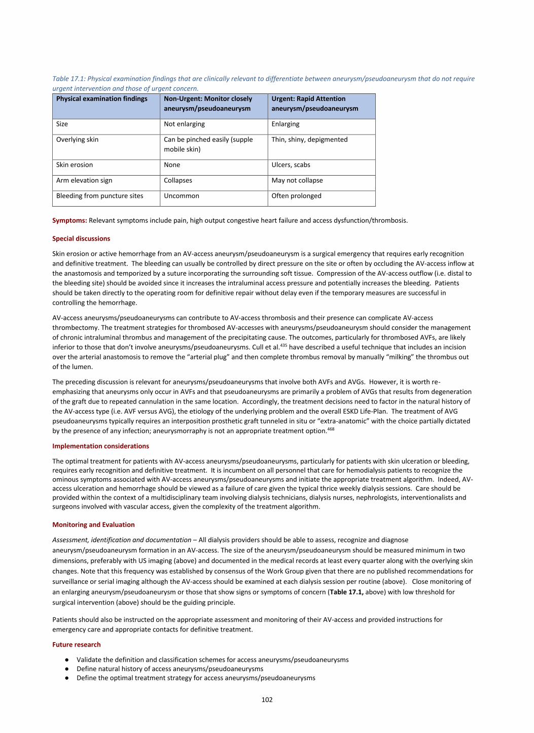
102
Table 17.1: Physical examination findings that are clinically relevant to differentiate between aneurysm/pseudoaneurysm that do not require
urgent intervention and those of urgent concern.
Physical examination findings Non-Urgent: Monitor closely
aneurysm/pseudoaneurysm
Urgent: Rapid Attention
aneurysm/pseudoaneurysm
Size Not enlarging Enlarging
Overlying skin Can be pinched easily (supple
mobile skin)
Thin, shiny, depigmented
Skin erosion None Ulcers, scabs
Arm elevation sign Collapses May not collapse
Bleeding from puncture sites Uncommon Often prolonged
Symptoms: Relevant symptoms include pain, high output congestive heart failure and access dysfunction/thrombosis. Special discussions
Skin erosion or active hemorrhage from an AV-access aneurysm/pseudoaneurysm is a surgical emergency that requires early recognition
and definitive treatment. The bleeding can usually be controlled by direct pressure on the site or often by occluding the AV-access inflow at
the anastomosis and temporized by a suture incorporating the surrounding soft tissue. Compression of the AV-access outflow (i.e. distal to
the bleeding site) should be avoided since it increases the intraluminal access pressure and potentially increases the bleeding. Patients
should be taken directly to the operating room for definitive repair without delay even if the temporary measures are successful in
controlling the hemorrhage.
AV-access aneurysms/pseudoaneurysms can contribute to AV-access thrombosis and their presence can complicate AV-access
thrombectomy. The treatment strategies for thrombosed AV-accesses with aneurysms/pseudoaneurysm should consider the management
of chronic intraluminal thrombus and management of the precipitating cause. The outcomes, particularly for thrombosed AVFs, are likely
inferior to those that don’t involve aneurysms/pseudoaneurysms. Cull et al.435 have described a useful technique that includes an incision
over the arterial anastomosis to remove the “arterial plug” and then complete thrombus removal by manually “milking” the thrombus out
of the lumen.
The preceding discussion is relevant for aneurysms/pseudoaneurysms that involve both AVFs and AVGs. However, it is worth re-
emphasizing that aneurysms only occur in AVFs and that pseudoaneurysms are primarily a problem of AVGs that results from degeneration
of the graft due to repeated cannulation in the same location. Accordingly, the treatment decisions need to factor in the natural history of
the AV-access type (i.e. AVF versus AVG), the etiology of the underlying problem and the overall ESKD Life-Plan. The treatment of AVG
pseudoaneurysms typically requires an interposition prosthetic graft tunneled in situ or “extra-anatomic” with the choice partially dictated
by the presence of any infection; aneurysmorraphy is not an appropriate treatment option.468
Implementation considerations
The optimal treatment for patients with AV-access aneurysms/pseudoaneurysms, particularly for patients with skin ulceration or bleeding, requires early recognition and definitive treatment. It is incumbent on all personnel that care for hemodialysis patients to recognize the ominous symptoms associated with AV-access aneurysms/pseudoaneurysms and initiate the appropriate treatment algorithm. Indeed, AV-access ulceration and hemorrhage should be viewed as a failure of care given the typical thrice weekly dialysis sessions. Care should be provided within the context of a multidisciplinary team involving dialysis technicians, dialysis nurses, nephrologists, interventionalists and surgeons involved with vascular access, given the complexity of the treatment algorithm. Monitoring and Evaluation
Assessment, identification and documentation – All dialysis providers should be able to assess, recognize and diagnose
aneurysm/pseudoaneurysm formation in an AV-access. The size of the aneurysm/pseudoaneurysm should be measured minimum in two
dimensions, preferably with US imaging (above) and documented in the medical records at least every quarter along with the overlying skin
changes. Note that this frequency was established by consensus of the Work Group given that there are no published recommendations for
surveillance or serial imaging although the AV-access should be examined at each dialysis session per routine (above). Close monitoring of
an enlarging aneurysm/pseudoaneurysm or those that show signs or symptoms of concern (Table 17.1, above) with low threshold for
surgical intervention (above) should be the guiding principle.
Patients should also be instructed on the appropriate assessment and monitoring of their AV-access and provided instructions for
emergency care and appropriate contacts for definitive treatment.
Future research
● Validate the definition and classification schemes for access aneurysms/pseudoaneurysms ● Define natural history of access aneurysms/pseudoaneurysms ● Define the optimal treatment strategy for access aneurysms/pseudoaneurysms
103
GUIDELINE 18. AV-ACCESS STEAL
Statements
18.1 KDOQI considers it reasonable that strategies to both prevent and treat AV-access steal should be developed and implemented before AV-access creation, to reduce the risk of AV-access steal and related morbidity, respectively (Expert Opinion)
18.2 KDOQI considers it reasonable that postoperatively, patients should be monitored closely for signs and symptoms associated with AV-access
steal and managed appropriately with consideration of individual circumstances as follows:
o Mild to moderate signs and symptoms require close monitoring for progression of ischemia and worsening of signs and symptoms o Moderate to severe signs and symptoms often require urgent treatment to correct the hemodynamic changes and prevent any longer-
term disability
18.3 KDOQI considers it reasonable that patients with signs and symptoms consistent with AV-access steal should be referred urgently to a
surgeon/interventionist familiar with the diagnosis and options for the definitive treatment of AV-access complications, particularly AV-access steal (Expert Opinion)
18.4 KDOQI considers it reasonable that the optimal treatment of AV-access steal should be determined based upon the patient’s clinical presentation and resources. (Expert Opinion) Rationale/Background
The construction of an AV-access can compromise the perfusion of the extremity distal to the anastomosis resulting in symptoms consistent with
acute or chronic ischemia, commonly referred to as “steal syndrome”. This occurs most commonly after brachial artery-based AV-accesses
although it can occur after radial artery-based or lower extremity accesses. It may occur shortly after AV-access creation or several years later, with
an increase in intra-access flow or with development of arterial disease of the inflow and outflow vessels. It is important to consider strategies to
reduce the incidence of steal syndrome (Table 18.1 see Supplement 3: Surgical Approaches to the Treatment of Steal.)
The clinical symptoms of the "steal syndrome” can range from mild numbness to severe motor compromise, and from skin ulceration to gangrene that necessitates major amputation. Several preoperative clinical predictors can identify patients at high risk of steal (see Table 18.2) who may benefit from mitigation strategies. It is important to be familiar with clinical signs and symptoms (see Table 18.3) as the diagnosis is largely a clinical one although findings can be corroborated with noninvasive testing to confirm the hemodynamic changes. The natural history of the steal syndrome is poorly defined but moderate to severe symptoms rarely resolve without definitive treatment. Treatment options (see Figure 18.1 and Table 18.4), aimed to correct the hemodynamic changes and potentially reverse the symptoms should be viewed as complementary with the optimal choice based upon the clinical scenario. Timely recognition and remediation are crucial to avoid long-term complications with the ideal goal to reverse symptoms and salvage the AV-access. The importance of early recognition of AV-access steal and early referral to an appropriate surgeon (i.e. knowledgeable in the management of AV-access steal) were part of the previous KDOQI Guidelines.11 Steal may occur after AV-access placement or several years after access placement as an increase in access flow or development of arterial disease of the inflow and outflow vessels. See also Figure 18.1
Table 18.1 Strategies to Reduce the Incidence of AV-Access Steal
Strategies to Reduce the Incidence of AV-Access Steal
Assessment of arterial inflow imaging with correction of inflow stenoses
Correct inflow stenosis or use contralateral extremity
Avoid distal brachial artery-based procedures
Avoid large conduits
Table 18.2 Clinical Predictors of AV-Access Steal
Clinical Predictors of AV-Access Steal
Advanced age
Female gender
Diabetes mellitus
Peripheral vascular disease
Large outflow conduits
Multiple prior permanent access procedures
Distal brachial artery-based procedures (i.e. near antecubital fossa)

104
Prior episode of AV-access steal
Table 18.3: Society for Vascular Surgery Reporting Standards for AV-access Steal475
Table 18.4 Treatment Options for AV-Access Steal
Special discussions
While the major focus of the preceding discussion was primarily on the remedial treatments after AVFs construction, the same considerations are
relevant for AVGs. However, the limited longer-term durability of the AVG is likely more relevant for the remedial strategies designed to salvage
the AVG and reverse the steal symptoms. The flow limiting strategies and PAI are appropriate for patients with moderate to severe steal symptoms
with an AVG
AV-access steal occurs less frequently after radial artery-based AV-accesses than after brachial-based procedures.476,477 Given the dual blood supply
to the hand through the radial and ulnar arteries, the resultant hemodynamic changes may differ. Notably, the AVF can serve as a pressure sink
that can “steal” blood from the hand or, more specifically, retrograde flow through the AVF from ulnar artery and palmar arch can compromise
digital perfusion. The available treatment options are dictated by the underlying causes of the hemodynamic changes but include correction of any
inflow lesions, flow reduction, ligation (or embolization) of the distal radial artery to prevent retrograde perfusion, and AV-access ligation.476-479.
The creation of an AV-access can result in severe sensorimotor dysfunction distal to the AV-access in the setting of only mild to moderate ischemia,
termed ischemic monomelic neuropathy.480-482 Although this is likely within the spectrum of AV-access steal, it is a distinct entity that has been
attributed to the development of severe ischemic neuropathy despite adequate skin and muscle perfusion. The published experience is somewhat
limited although the condition appears to be increased among patients with diabetes, peripheral vascular disease, and preexisting peripheral
neuropathy. Optimal treatment includes immediate recognition and treatment, typically with AV-access ligation, observation, although the
sensorimotor symptoms may not be reversible in any approach.483
Grade Severity Clinical Presentation Treatment
0 None None None
1 Mild Cool extremity with few symptoms None
2 Moderate Intermittent symptoms during dialysis, claudication Intervention sometimes
3 Severe Ischemic rest pain, tissue loss Intervention mandatory
Treatment Options for AV-Access Steal
Ligation (if symptoms are severe, limb loss at risk or no other option available)
Correction of arterial inflow stenosis
Flow limiting or banding
Proximalization of the arterial inflow (PAI)
Revision using distal inflow (RUDI)
Distal revascularization and interval ligation (DRIL)

105
Implementation considerations
The development of AV-access steal symptoms is one of the most worrisome complications associated with AV-access creation given its
potential to compromise hand function and potential for digital ischemia. Accordingly, providers constructing AV- access should be familiar
with and assume complete responsibility of every phase of its management including preoperative predictors, diagnosis, strategies to reduce
its incidence, and definitive treatment.
Monitoring and evaluation
Given the potential for AV-access steal to compromise hand function, all health care providers managing AV-access should be familiar with its
clinical presentation and appropriate initial management, including timely referral to a surgeon familiar with the various remedial treatments.
Patients should be assessed for the signs and symptoms of AV-access steal as part of routine AV-access monitoring and counseled about the
symptoms of steal and the need to share them with their healthcare providers.
Future research
• Further define the pathophysiologic changes that lead to the development of steal
• Further define and establish the predictors for AV-access steal.
• Further define and establish strategies to reduce the incidence of AV-access steal.
• Further define the natural history of mild to moderate symptoms related to AV-access steal.
• Further define the diagnostic criteria for AV-access steal.
• Further define the optimal remedial treatment for AV-access steal. Further define ischemic monomelic neuropathy as a distinct entity from AV-access steal
GUIDELINE 19. OTHER AV-ACCESS COMPLICATIONS
Statement: Management of AVG seroma
19.1 KDOQI considers it reasonable to carefully monitor for complications of AVG seroma and management based on the patient’s individual circumstances and the clinician’s best judgment (Expert Opinion) Detailed Justification Fluid collections or seromas can develop around prosthetic AVGs in the early postoperative period, frequently near the anastomoses.484 They may resemble an aneurysms/pseudoaneurysms (See Figure 19.1). The seromas are likely due to the transudation of fluid through the graft material itself or disruption of the surrounding lymphatic channels. Daria et al.485 reported the incidence to be 1.7% among 535 AVGS, with higher incidence among those with upper arm (versus forearm) AVGs. The presence of seroma/fluid around the AVG can be confirmed with an ultrasound. The majority of these early seromas is self-limited and tends to resolve without any consequence; however, they can serve as a nidus for infection given the protein rich nature of the fluid. Seromas that persist or develop after the early postoperative period may necessitate AVG removal and replacement, preferably with a different material tunneled through a different anatomic course. Fortunately, the majority of the AVGs can be salvaged and the longer-term results are reasonable.485

106
Statement: Management of high flow AV-access
19.2 KDOQI considers it reasonable to closely monitor for* and prophylactically manage AV-access with high flows to avoid serious or irreversible complications (e.g. high output cardiac failure), based on the patient’s individual circumstances and the clinician’s best judgment (Expert Opinion)
* close monitoring refers to physical exam and history on routine dialysis rounds and determination of Qa/CO every 6-12 months Detailed Justification High-flow AV-access
Increased flow rates through an AV-access can lead to a host of problems including high output congestive heart failure, pulmonary hypertension, central venous stenosis, venous hypertension, aneurysmal degeneration of the AVF, and AV-access-related hand ischemia. The exact threshold to define high-flow access has not been rigorously validated or universally accepted, though an AV-access flow rate (Qa) of 1 – 1.5 L/min or Qa > 20% of the cardiac output (Qa/CO) has been suggested.486 AV-access can exacerbate underlying symptoms of congestive heart failure and even lead to high output failure, similar to traumatic AVF.487 Basile et al.488 reported that Qa > 2.0 L/min were associated with the occurrence of high-output cardiac failure with a sensitivity of 89% and specificity of 100% while a Qa/CO of > 20% had a sensitivity of 100% and a specificity of 75%. Not surprisingly, symptoms of congestive heart failure may develop at lower thresholds in patients with underlying heart disease. The frequency of determination of Qa/CO is not established; however the Work Group felt a 2D echo every 6-12 months to help evaluate for cardiac decompensation and changes in Qa/CO, depending on the patient’s circumstances and local resources, was reasonable. AV-access flow rates can be reduced using a variety of “flow reducing therapies” or “banding” (Guidelines 18).489-492 Predictably, occlusion of the high-flow AV-access results in a decrease in the cardiac output and improved oxygen delivery.493
GUIDELINE 20. TREATMENT AND PREVENTION OF CVC COMPLICATIONS
Statement for the Monitoring/Surveillance of catheter complications
20.1 KDOQI considers it reasonable to perform a basic medical history focused on signs & symptoms of catheter-related complications (e.g. dysfunction, infection) and physical examination or check of the dialysis catheter, exit site, tunnel and surrounding area at each catheter dressing change or dialysis session (Expert Opinion).
Rationale/Background
While no studies have explicitly studied the value of history, physical examination, check or inspection for CVC dysfunction, infection and migration, they are fundamental tools for assessing the dialysis patient. For example, a patient history of signs or symptoms of bacteremia/septicemia may be indicative of CVC-related infection. It is common for a patient to appear relatively well, but then complain of symptoms of headache, nausea, dizziness and chills; and exhibit signs of vomiting, rigors, and fever 15-30 minutes following dialysis initiation when a CVC-related infection is present. These signs and symptoms may be related to endotoxin release from a CVC-related infection biofilm, perturbed by the flow incurred by the initiation of dialysis. Inspection of the catheter exit site may reveal cuff migration that places the catheter at risk of infection and also physical loss of the catheter. Exit-site infection is indicated by the presence of erythema, swelling, tenderness and purulent drainage around the CVC exit and the part of the tunnel external to the cuff. Signs of tunnel infection are swelling, erythema, fluctuance and tenderness over the CVC tunnel central or proximal to the cuff. Inspection of the patient on the side of the CVC may also indicate central venous stenosis/ occlusion. Common findings are not limited to but include dilated subcutaneous veins and limb swelling.
Special Discussions
Special discussions from Work Group included: (1) the frequency required to evaluate the CVC for adequate prevention of CVC dysfunction and (2) noted that prior KDOQI 2006 guidelines recommends against exchange of CVC.
Implementation considerations
The Work Group discussed implementation considerations, which included: (1) standardizing the use of clinical questions to ask patients pertaining to CVC complications; (2) training and update of dialysis nurses/technicians to identify physical signs of infection, occlusion, stenosis; (3) identifying key medical personnel to convey information and facilitate a subsequent action plan if an abnormality was identified.
Monitoring and Evaluation
More frequent monitoring and follow-up required if abnormality suspected or found. Future research
• Cost benefit of prophylactic vascular access related history/physical examination or check
• Testing long-term durability of dialysis catheters.

107
GUIDELINE 21. CATHETER-RELATED DYSFUNCTION
Statement: Definition of CVC dysfunction
21.1 KDOQI considers it reasonable to assess for CVC dysfunction during each HD session using the updated KDOQI VA 2018 definition of catheter dysfunction, which is: failure to maintain the prescribed extracorporeal blood flow required for adequate hemodialysis without lengthening the prescribed HD treatment. (Expert opinion).
Background/Detailed Justification
Cuffed, tunneled, dual-lumen central venous catheters (CVC) have become an acceptable form of hemodialysis (HD) vascular access when an AV-access is not suitable or available, despite the associated high complication rates and mortality risk65,446. Maintenance of CVC patency is paramount to providing adequate HD in patients requiring CVCs on either a temporary or long-term basis. A common complication of the CVC is CVC dysfunction, which is associated with reduced dialysis adequacy, increased risk of CRBSI494 and mortality. Although several studies have reported on CVC dysfunction, the rate of occurrence, the time from CVC insertion to dysfunction, and its effect on patients quality of life, there is no consistency in the definitions used for CVC dysfunction. 494-496
There are several factors that contribute to the delivery of adequate dialysis beyond blood flow, including but not limited to the duration, frequency and/or type of dialysis, type of CVC, ultrafiltration variables, degree of recirculation and weight of the patient. In fact, blood flow may be misleading as an indicator of clearance if there is a significant presence of recirculation, particularly if the lines are reversed497. Past definitions of CVC dysfunction have included specific criteria pertaining to blood flow rates, venous and arterial pressure limits, dialysis adequacy based on urea kinetics, and other parameters. The 2006 NKF KDOQI VA guidelines defined CVC dysfunction as failure to maintain extracorporeal blood flow of >300 ml⁄min at a pre-pump arterial pressure more negative than -250 mmHg or failure to attain and maintain an extracorporeal blood flow sufficient to perform HD without significantly lengthening the treatment time11. Although it is important to standardize outcomes within a guideline, this recommendation was opinion based and has been interpreted as the need to maintain blood flows > 300 ml/min to ensure adequate dialysis. As a result of these blood flow recommendations, CVCs are often run in the reverse configuration, thrombolytic agents such as tissue plasminogen activator (t-PA) are administered, or CVCs are exchanged when blood flow is consistently ≤300 ml/min. Previous comparisons of CVC mean blood flows of 250, 275, and 300 ml/min did not reliably predict inadequate dialysis. This was particularly relevant in patients with weights below 70 kg498. Furthermore, blood flows of < 300 ml/min are often standard in patients receiving long duration dialysis e.g. nocturnal dialysis, or frequent dialysis where HD adequacy is excellent.
Additionally, there is uncertainty around the frequency of “abnormality”, i.e. the number of dialysis sessions to include, before defining CVC dysfunction. Patients often experience a single “poor run” with inadequate clearance and then spontaneously resume “good runs”, providing adequate clearance, without an intervention. These intermittent decreases in clearance can be related to the position of the patient, transient BP drop, interdialytic weight gain, higher levels of hemoglobin, transient partial thrombosis of the CVC that autolyze and factors not fully understood. The consequences of such intermittent episodes of reduced clearance is unclear, likely due to its variations in frequency and etiologies499,500. However, DOPPS does report an association between number of inadequate dialysis sessions and increased risk of hospitalizations and mortality; but this data is confounded by dialysis inadequacy being attributed to shortened dialysis sessions501. It is unknown if a single session of dialysis with reduced adequacy, as measured by urea clearance but with maintained duration of dialysis is associated with the same risk. The rate of HD CVC dysfunction is variable depending on the specifics of the definitions. Using a combined set of 3,364 patients and 268,363 CVC-dependent dialysis sessions from Da Vita and the USRDS, CVC dysfunction, defined as a blood flow (Qb) < 300ml/min occurred in 7.1% of dialysis sessions and almost two-thirds of patients had ≥1 CVC dysfunction session; 30% had ≥1 CVC dysfunction session per month502. In a RCT of heparin versus t-PA, CVC dysfunction, defined as peak blood flow of <200 ml/min for 30 minutes during a dialysis treatment, mean blood flow of <250 ml/min during two consecutive dialysis treatments, or inability to initiate dialysis owing to inadequate blood flow, occurred in 20.0% of patients in the r-TPA group and 34.8% in the heparin group. Others have reported rates varying between 0.5 to 3.0 events per 1000 catheter-days503. Implementation Considerations
The KDOQI Work Group offers the above definition as it allows for intra-patient comparisons and respects differences in definitions of dialysis adequacy used by various studies, institutions and jurisdictions. It allows for flexibility required when considering various HD prescriptions according to type, duration and frequencies of the dialysis regime. Dialysis units may want to consider establishing local definitions of CVC dysfunction that incorporates their prescription goals to meet adequacy targets (thereby being consistent with KDOQI definitions), document episode(s) of dysfunction at a predetermined frequency or interval, to allow for trending and appropriate intervention Future research In order to accurately document the rate of CVC dysfunction for comparisons within institutions, across jurisdictions and treatment regimens it is necessary to standardize the definition of CVC dysfunction. There is an urgent need for the nephrology community to re-evaluate the definition of CVC dysfunction with supporting validation studies, e.g. what are the sensitivity, specificity, predictive value of various markers, and timing of intervention for catheter dysfunction. Is earlier intervention better?
108
Statements: Pharmacologic Prevention of CVC* Dysfunction
CVC connectors to prevent CVC dysfunction 21.2 KDOQI considers it reasonable to have an individualized approach to use special CVC connectors based on the clinician’s discretion and
best clinical judgment (Expert Opinion) 21.3 KDOQI considers it reasonable to consider an antimicrobial barrier cap to help reduce CRBSI in high-risk patients or facilities; the choice of
connector should be based on clinician’s discretion and best clinical judgment (Expert Opinion) Intraluminal agents to prevent CVC dysfunction 21.4 KDOQI considers it reasonable that the choice to use citrate or heparin as a CVC locking solution be based on the clinician’s discretion and
best clinical judgment as there is inadequate evidence to demonstrate a difference in CVC survival or complications between these locking solutions (Expert Opinion)
21.5 KDOQI suggests the use of low concentration citrate (<5%) CVC locking solution, if feasible, to help prevent CRBSI and CVC dysfunction (Conditional recommendation, Low quality of evidence)
21.6 KDOQI suggests that r-TPA may be prophylactically used as a CVC locking solution once per week to help reduce CVC dysfunction (Conditional recommendation, Low quality of evidence)
21.7 There is inadequate evidence for KDOQI to make a recommendation on the comparative use of the following CVC locking agents for CVC
dysfunction or infection prophylaxis: tinzaparin vs. unfractionated heparin; taurolidine/citrate vs. heparin with or without gentamicin;
neutral valve connector (Tego) vs. citrate (46.7%) locking solution
Systemic agents to prevent CVC dysfunction 21.8 KDOQI recommends against the routine use of prophylactic systemic anticoagulants (e.g. warfarin) for the sole purpose of maintaining or
improving CVC patency as there is inadequate evidence of benefit for CVC patency but suggestion of increased risk of harm (Conditional/strong recommendation, low quality of evidence)
21.9 KDOQI suggests that low dose aspirin may be used to maintain tunneled CVC patency in patients with low bleeding risk (Conditional recommendation, Low quality evidence).
*Recall: CVC refers tunnelled hemodialysis CVCs unless otherwise specified Background/Rationale
Among incident HD patients in North America, 80% continue to use a CVC504, despite the well-known complications of central vein stenosis505,
increased risk of subsequent AVF failure90,503, and persistent use of the CVC beyond ninety days506,507.504. CVC dysfunction is a common problem
and often requires medical or surgical intervention508. Use of thrombolytic agents and/or CVC exchange occurs in 20% to 40% of patients
dialyzing with CVCs494. Both of the latter interventions are associated with high costs509. In many cases, CVC dysfunction (previously defined by
KDOQI 200611 as “failure to attain and maintain an extracorporeal blood flow of 300 mL/min or greater at a prepump arterial pressure more
negative than –250 mm Hg”) persists. Besides increases in arterial and venous pressures registered by the dialyzer that necessitate a decrease
in blood flow, CVC dysfunction can result in significant recirculation leading to poor clearance and lower Kt/V. Left untreated, such CVCs
require premature removal when they become nonfunctional510 (i.e., with one or both lumens that cannot be aspirated).
The onset of CVC dysfunction can occur early or late, which may help in determining the etiology of the problem and guide subsequent
management. Dysfunction noted immediately after the CVC placement is likely due to the positioning of the CVC, preexisting vascular
abnormalities (e.g., central venous stenosis) or mechanical damage to the CVC (e.g., tight suture or perforation). Dysfunction developing after
successful initial use is usually due to intraluminal or pericatheter thrombosis, fibrin sheath formation around the CVC, mural thrombus
adhering to the CVC tip, or new central venous stenosis.
The following section discusses studies evaluating pharmacologic prevention of CVC dysfunction.
Detailed Justification
Connector maneuvers to prevent CVC dysfunction
A closed-system connector has theoretical benefits of limiting environmental contact with microorganisms, reducing biofilm formation and consequent CVC dysfunction and infection. It is left in place during dialysis sessions and changed every week. A RCT (n=66) compared a closed-system connector, flushed with saline and attached to the CVC hubs, to 46.7% trisodium citrate lock to evaluate a composite study endpoint of CVC dysfunction (requiring fibrinolytic therapy or mean blood flow ≤250 mL/min during 2 consecutive dialysis sessions) and CVC-related bacteremia511. There was no difference in the composite outcome (48% [connector] vs. 55% [trisodium citrate], RR 0.72 [95%CI 0.37, 1.42]). The 2 groups also did not differ on the individual components of the composite outcome – dysfunction or bacteremia. Need for urokinase to treat thrombotic dysfunction did not differ (14 participants from the connector group, 9 from the trisodium citrate group, P=0.20). CVC survival rates (free of dysfunction or bacteremia) at 1 year were 0.43 in the connector group and 0.37 in the trisodium citrate group. The study did not report harms. Therefore, given these neutral results, KDOQI suggests that the use of the neutral valve connector be at the discretion of the clinician.
Due to literature search timelines, the following study and data was not retrieved or reviewed by the ERT; however the KDOQI Work Group felt
it was important to include in this document. Brunnelli et al conducted a large 13-month cluster randomized, comparative-effective study that
evaluated ClearGuard HD barrier cap (single piece device that applies antimicrobial inside and outside the hub) versus Tego + Curos (two-
109
piece device that applies antimicrobial to the outside of the Tego cap only). The study outcome was positive blood culture rate as an indicator
of bloodstream infection rate. A total of 40 dialysis facilities were enrolled (20 facilities in each group). After a 3-month run-in phase, 1671
patients (826 CG, 845 Tego + Curos) qualified for the 13-month intervention phase. The Clearguard group had 23 positive blood cultures
compared to 75 in the Tego + Curos group (83,064 vs. 100,042 CVC days, 0.28 vs. 0.75 per 1000 CVC days, respectively). The incidence rate
ratio for CRBSI analysis and access related blood stream analysis favored the ClearGuard group (0.37 (p=0.003) and 0.32 (p<0.001),
respectively).512
See Supplement 1. Tables 177-180.
Intraluminal agents to prevent CVC dysfunction
Anticoagulant lock to prevent CVC dysfunction
i) Citrate versus Heparin
Three European RCTs compared citrate to heparin CVC lock (total N= 542)513-515. Follow-up was 6 months in 2 studies; one study reported
results in CVC days515. Interventions varied across the studies. One compared a 5% citrate lock to a 5,000 U/ml heparin lock514 (standard
concentrations). Two trials evaluated higher concentrations of citrate, one compared 30% citrate lock to a 5,000 U/ml heparin lock515 and one
compared 46.7% citrate lock to a 5% heparin lock513. There were more episodes of non-occlusive clot per session in the 5% citrate group
compared to the 5,000 U/ml heparin group (14% vs. 7%, P<.0001)514. All studies reported on treatment required for CVC dysfunction. Overall,
there was no statistically significant difference between groups (RR 1.25 [95%CI 0.53, 1.96]). One high concentration lock study and one
standard concentration lock study reported no significant difference between groups for needing treatment with urokinase514,515. One study
reported no bleeding complications513 and one study reported significantly lower incidences of major bleeding episodes (3% vs. 11%, P=.01)
and lower persistent bleeding after insertion (4% vs. 13%, P=.005) in the 30% citrate group compared to the heparin group515.513-515.
ii) Doses of citrate
There is inadequate evidence for KDOQI to make a recommendation on the use of high versus low concentration citrate as a locking solution
for hemodialysis CVCs. This was not included in the summary of statements to avoid confusion.
iii) Doses of heparin
One RCT516 (n=75) and one observational study517 (retrospective review of prospectively collected data; n=223) compared different heparin
concentrations. The RCT compared 5,000 U/ml to 2,500 U/ml and reported outcomes to 24 hours516. The observational study compared 3
different heparin locks (5,000; 1,000; and 500 U/ml) after CVC insertion with follow-up to 30 days517. In the RCT, there were no episodes of
thrombosis within 24 hours in either group516. The observational study found no significant differences between heparin concentrations in 2-
or 30-day CVC-related infection-free survival or CVC517, or incidence of blood flow less than 250 ml/min517.
See Supplement 1. Tables 181-191
Thrombolytic lock to prevent CVC dysfunction
Recombinant tissue plasminogen activator (r-TPA) has primarily been used in the treatment of CVC thrombosis. A RCT comparing r-TPA once
per week (with 5000 U/ml heparin lock after the other 2 dialysis sessions) to heparin lock 3 times per week, enrolled 225 participants with
both incident (first ever) and prevalent CVCs.518 For 61% of the participants, the CVC was their first dialysis CVC. Follow-up was 6 months with
extended follow-up for those who experienced the primary outcome of CVC dysfunction. The primary study outcome of CVC dysfunction was
defined as the first occurrence of decreased blood flow (e.g., peak blood flow of <200 ml/min for 30 minutes during dialysis, mean blood flow
of <250 ml/min during 2 consecutive dialysis treatments, or inability to initiate dialysis due to inadequate blood flow) after protocolized
attempts to re-establish patency. There was a significantly higher occurrence of CVC dysfunction in the heparin only group (40/115, 35%)
compared to the r-TPA group (22/110, 20%) (HR 1.91 [95%CI 1.13, 3.22]) Among CVCs that experienced dysfunction, the need for r-TPA for
immediate management was significantly higher in the heparin group (50% vs. 18%, P=.01). No significant differences were observed
between the r-TPA and heparin only groups for reversal of CVC lines for immediate management of dysfunction (r-TPA 3/22 (59%), heparin
14/40 (35%), P=.07) (or for any serious adverse event (21% [r-TPA], 30% [heparin], P=.14) or any bleeding (P=.93). There is a paucity of
rigorous studies comparing various methods of installation of thrombolytic agents (e.g. dwell, infusion, push techniques) and their impact on
CVC patency outcomes and complications.
See Supplement 1. Tables 192-205
Other intraluminal agents to prevent CVC dysfunction i) Taurolidine and Citrate vs. Heparin
2 RCTs (total n=168) that compared a combined taurolidine and citrate lock (taurolidine/citrate) to heparin lock519,520 and one RCT that compared taurolidine/citrate to a combined gentamicin and heparin lock (gentamicin/heparin) for prevention of CVC complications were identified.521 Of the 2 RCTs of taurolidine/citrate versus heparin lock, one trial was conducted in the UK (n=110),520 the other in the Netherlands (n=58).519 The UK study was of tunneled, cuffed CVC with follow up of 8129 and 9642 CVC days in the taurolidine/citrate and heparin groups, respectively.520 In the Dutch trial, the majority (76%) of the catheters were for temporary use and non-tunneled; 519 the median CVC use was 158 days for tunneled CVCs and 28 days for non-tunneled CVCs in the jugular or subclavian vein, and 7 days for CVCs inserted in the femoral vein.519 In both trials, the intervention locks contained taurolidine 1.35% and citrate 4% and the heparin lock control contained 5000 u/mL. In both trials, removal of CVC due to thrombosis/occlusion did not differ between treatment groups. In the study of all tunneled CVCs, need for thrombolytic therapy at least once was twice as great in the taurolidine/citrate group compared with the heparin group, 53% versus 26% (HR
110
2.5 [95%CI 1.3, 5.2])).520 Solomon et al conducted a non-randomized, 3-arm study (n=174), as an extension of the RCT conducted in the UK and compared taurolidine (1.35%), citrate (4%), and heparin (500 U⁄ml) (TCH) locks versus taurolidine/citrate vs. heparin (5000 U/mL) locks.520 There was no significant difference for CVC survival between group.520 Need for thrombolysis was reduced with the TCH lock (n=106) compared to the taurolidine/citrate lock (n=34) (HR 0.2 [95%CI 0.06, 0.5]) but not the heparin 5000 U/ml lock (n=34) (HR 1.4 [95%CI 0.5, 3.9]).522 The UK trial involving tunneled CVCs reported heparin-induced thrombocytopenia led to CVC removal in 1 participant in the taurolidine/citrate group and none in the heparin group.520 The other study reported no adverse events associated with the use of taurolidine and citrate lock solution.519 See Supplement 1. Tables 206-215
ii) Taurolidine and Citrate vs. Gentamicin and Heparin
There was one RCT (n=119) evaluating combined taurolidine 1.35% and citrate 4% lock (taurolidine/citrate) versus combined gentamicin 40 mg/ml and heparin 5000 U/ml lock (gentamicin/heparin).521 Follow-up was 90 days.
There was no significant difference in the development of thromboses in CVCs, 9 (12%) in the citrate/taurolidine group versus 11 (15%) in the gentamicin/heparin group (P=.63).
iii) Tinzaparin vs. Heparin One cross-over RCT from Canada (n=42) compared tinzaparin lock to heparin lock523. Participants received each of the locks for 7 weeks. All had been using CVCs for more than 3 weeks at the time of enrollment. No significant differences between treatment arms were observed for CVC removal due to failure. Use of alteplase for CVC dysfunction was significantly lower in the tinzaparin group (3.2% of dialysis sessions vs. 6.0% of sessions, P=.008). Supplement 1. Tables 183 and 188
Systemic Agents to Prevent CVC Dysfunction
Five studies of systemic anticoagulants for prevention of CVC complications were identified. Three RCTs and one observational study compared a systemic antiplatelet or anticoagulant agent to placebo or no intervention.524-527 One of the RCTs evaluated an antiplatelet drug (aspirin)526 and one RCT and the observational study evaluated an anticoagulant (warfarin).525,527 One of the RCTs compared an antiplatelet (aspirin) and an anticoagulant (warfarin) to no intervention and to each other.524 One additional RCT compared systemic anticoagulation following CVC placement to anticoagulant delayed until the first CVC thrombosis or dysfunction event.528 Supplement 1. Tables 216-230
Aspirin versus Placebo or No Intervention There are 2 RCTs (total n=223) evaluating aspirin compared to either a placebo or no intervention (control group) in participants with CVC. 524,526 One trial was conducted in Iran (n=185; follow-up 12 months),526 the other in Saudi Arabia (n=38; follow-up not indicated).524 The Iranian trial compared low dose aspirin (80 mg/day) to placebo526 while the Saudi study compared low dose aspirin (81 mg/day) to no intervention524.
Results were mixed for CVC survival outcomes following systemic antiplatelet therapy versus control. The Iranian study reported overall mean CVC survival time to be longer in the aspirin group compared to the placebo group, 5.3 versus 3.9 months (mean difference 1.40 [95%CI 0.28, 2.52]).526 The Saudi study reported 68% of the 19 CVC in the aspirin group to be dysfunction-free at 12 months compared to 37% of the 19 CVC in the no intervention group. The authors reported that the difference between groups was significant but the ERT re-analyzed using a Fisher’s exact test and found no statistically significant difference.524.
In the Saudi trial, there were fewer participants with at least one CVC dysfunction due to CVC thrombosis in the aspirin group than in the no intervention group (21% versus 47%), but when reanalyzed by the ERT using a Fischer’s exact test, the difference was not statistically significant.524
Adverse events did not differ between the aspirin and placebo groups in the Iranian trial.526 The Saudi study reported that no participant in either group experienced a major bleeding event.524.
Warfarin versus Placebo or No Anticoagulant Two RCTs (total n=213) compared warfarin to placebo or no intervention.524,527 One trial was conducted in Canada (n=174).527 The second trial represents the other intervention arm of the study from Saudi Arabia (n=39) described above.524 The follow-up periods were 709 to 722 participant-months in the Canadian study and 12 months in the Saudi study.524 Most of the participants received tunneled cuffed CVCs (24% received non-tunneled in the Canadian trial). A low dose of warfarin, adjusted to an international normalized ratio (INR) of 1.4 to 1.9, was compared to placebo in the Canadian study.527 The Saudi study compared a warfarin dose of 2 to 5 mg/day, targeting an INR of 1.5 to 2.0, to no intervention.524.
Both studies reported CVC survival outcomes: The Canadian study reported that CVC removal for any reason did not differ between the warfarin and placebo groups (HR 0.87 [95%CI 0.42, 1.81]).527 The Saudi study reported dysfunction-free CVC survival at 12 months was higher in the warfarin group (75%) compared to no intervention (37%) (reported as P<.01 in the publication and P=.02 using a Fisher’s exact test analyzed by ERT;).524 The Saudi trial also reported 4 (20%) had at least one CVC dysfunction due to CVC thrombosis in the warfarin group versus 9 (47%) with no intervention, but the difference was not statistically significant based on the ERT’s re-analysis with a Fisher’s exact test.524 In the Canadian study, need for an intervention for CVC dysfunction did not differ between the warfarin and placebo groups, 46% versus 47%, respectively (HR 0.90 [95%CI 0.57, 1.38]) .527
The Canadian trial reported no difference in major bleeds between treatment groups:527 10 (12%) in the warfarin group and in 7 (8%) in the
111
heparin group (RR 1.43 [95%CI 0.57, 3.58]). The Saudi study reported that no participant in either group experienced a major bleeding event.524
One observational study from 2 units in the UK (n=112 participants with 194 femoral CVCs)525 observed a total of 20,021 CVC days of use. Prophylactic systemic anticoagulation, typically warfarin with a target INR of 1.5-2.5, was compared with a “no anticoagulation” control group. The control group restricted anticoagulation to patients with CVC dysfunction requiring repeated treatment with urokinase locks.
CVC survival, assessed as CVC removal due to occlusion, did not differ: 33% in the anticoagulation group and 27% in the control (P=.49).
There was no difference in the number of CVC-related thromboses between groups, 9% in the prophylactic group compared to 11% (adjusted HR 0.66 [95%CI 0.25, 1.72]). Major bleeding occurred in 5 (6%) of 80 CVCs in the prophylactic group and in 4 (4%) of 108 CVCs in the no anti-coagulation group with an adjusted HR of 1.65 [95%CI 0.44, 6.22]. Corresponding rates were 0.7 and 0.4 per 1000 CVC days.
Warfarin vs. Aspirin There is inadequate evidence for KDOQI to make a recommendation or suggestion on the use of systemic warfarin versus aspirin for the
prevention or treatment of CVC dysfunction. This was not included in the summary of statements to avoid confusion. The detailed justification
is as follows:
The RCT from Saudi Arabia by Abdul-Rahman et al, with warfarin and aspirin arms compared to no intervention (described above), also provided data comparing warfarin to aspirin.524 The comparative effectiveness study compared a warfarin group (n=20) that received a dose of 2 to 5 mg/day, targeting an INR of 1.5 to 2.0, with an aspirin group (n=19) that received 81 mg/day. CVC were followed for 12 months.
There was no difference between groups for dysfunction-free CVC survival at 12 months: 75% (warfarin) versus 68% (aspirin) (P=.65) or for at least one CVC dysfunction due to CVC thrombosis: 20% (warfarin) versus 21% (aspirin). No participant in either group experienced a major bleeding event.
Warfarin Initiated after CVC Placement versus Warfarin Initiated after first Thrombosis or CVC Dysfunction Event One Italian RCT (total n=144) evaluated warfarin initiated 12 hours after CVC placement (post-placement warfarin group) to warfarin initiated after the first thrombosis or dysfunction (post-event warfarin group).528 In both groups, warfarin was adjusted to an INR of 1.8 to 2.5; follow up was 12 months for these CVCs. Fewer participants in the post-placement warfarin group experienced a thrombosis/dysfunction event, 10 (12%; 0.16 events per patient-year) versus 33 (52%; 1.65 events per patient-year) in the post-event warfarin group. Also, fewer participants in the post-placement warfarin group required a CVC replacement due to thrombosis compared with the post-event warfarin group (2% versus 17%; RR 0.14 [95%CI 0.03, 0.62]). No participant in either group experienced a major bleeding event. Special discussions
• Several of these solutions such as citrate and taurolidine are not widely available for use in the outpatient setting in the USA
• The cost-effectiveness of using prophylactic thrombolytic locking solutions has not been extensively evaluated
• Regarding the uses of systemic anticoagulants, the lack of benefit and the potential risks of harm was deemed by the Work Group to be a concern regardless of whether an INR is targeted (e.g. in cases where warfarin is used), especially given the known challenges of consistently achieving a target INR and maintaining within the therapeutic range in hemodialysis patients.
Implementation considerations
• Must consider whether each individual dialysis unit has the resources to implement changes in preventive locking solutions.
• The use of any intraluminal or extraluminal cleansing solutions, antiseptics, antibiotics, medicines or anticoagulants should be compatible with the CVC material
• When the evidence extracted and analyzed by the ERT was inadequate to make a recommendation and there were no associated harms, the Work Group felt it important to support use based on the clinician’s discretion and best clinical judgment. The Work Group did not want to place restrictions and instead encourage future research to provide the necessary evidence to assist with future clinical practice guidelines development on these topics.
Monitoring and Evaluation
Close monitoring of adverse events related to locking solutions such as citrate
Future research
• In order to prevent CVC dysfunction, better diagnostic tools are required to detect etiology of dysfunction. For example, further research can evaluate the role of 2D echo to diagnose a catheter related atrial thrombus.
• The relationship between size of intraluminal or mural thrombus and symptoms
• Larger multicenter studies evaluating citrate and taurolidine in RCT
• The role of ethanol as a CVC locking solution
• The role of nitroglycerin-based CVC lock solution
• The benefit of anticoagulant lock over normal saline for CVC patency has been raised and could be further researched, given the potential for increased risk of adverse events with anticoagulant locks529-532
• The cost effectiveness of various doses of prophylactic rTPA in high risk patients e.g. 2 mg vs. 1 mg installation
• Rigorous studies comparing various methods of installation of thrombolytic agents (e.g. dwell, infusion, push techniques) and their impact on CVC patency outcomes and complications
112
GUIDELINE 22: TREATMENT AND MANAGEMENT OF CVC DYSFUNCTION
Statements: Medical management of CVC dysfunction
Conservative maneuvers 22.1 KDOQI considers it reasonable for a conservative bedside approach to managing CVC dysfunction prior to other medical or mechanical
interventions (Expert Opinion)
Pharmacologic maneuvers 22.2 KDOQI recommends intraluminal administration of thrombolytic agent in each catheter port to restore function of dysfunctional
hemodialysis catheters due to thrombosis (Conditional recommendation, Moderate quality of evidence)
22.3 KDOQI recommends the use of alteplase or urokinase plus citrate 4% per limb for restoring intraluminal catheter blood flow in an occluded hemodialysis catheter (Conditional recommendation, Moderate quality of evidence)
22.4 KDOQI suggests intraluminal administration of alteplase 2 mg in preference to alteplase 1 mg in each catheter port to restore function
of dysfunctional hemodialysis catheters due to thrombosis (Conditional recommendation, Moderate quality of evidence)
22.5 KDOQI suggests administering alteplase by the dwell or push method to treat catheter dysfunction (Conditional recommendation, Low quality of evidence)
Rationale/Background Despite the well-known complications of CVC use90,503,505-507, they are used in 80% of all incident HD patients504. CVC dysfunction is a common problem and often requires medical, endovascular or surgical intervention508 in 20% to 40% of patients dialyzing with CVC494. This is inconvenient for patients and costly to the healthcare system509. In the 2006 NKF-KDOQI VA Guidelines, 11 CVC catheter dysfunction was defined as “failure to attain and maintain an extracorporeal blood flow of 300 mL/min or greater at a prepump arterial pressure more negative than –250 mm Hg”. Besides increases in arterial and venous pressures registered by the dialyzer that necessitate a decrease in blood flow, CVC dysfunction can result in significant recirculation leading to poor clearance and lower Kt/V. Left untreated, such CVC require premature removal when they become nonfunctional510 (i.e., with one or both lumens that cannot be aspirated and dialysis cannot be delivered).
The onset of CVC dysfunction can present as early or late after its insertion, which may help determine the etiology of the problem and guide subsequent management. Dysfunction noted immediately after CVC insertion is likely due to CVC malpositioning, preexisting vascular abnormalities (e.g., central venous stenosis) or mechanical damage to the CVC (e.g., tight suture or perforation). Dysfunction developing after successful initial use is usually due to intraluminal or pericatheter thrombosis, fibrin sheath formation around the CVC, mural thrombus adhering to the CVC tip, or new central venous stenosis. To facilitate the current NKF-KDOQI guideline recommendations, interventions to prevent and treat CVC dysfunction are categorized by the type of intervention as medical and mechanical interventions. Medical interventions are further subdivided into conservative maneuvers and pharmacologic interventions per se (e.g. rTPA use). Mechanical interventions are similarly subdivided into fibrin sheath disruption, catheter exchange, and CVC removal with replacement (See Figure 23.1. Algorithm: Treatment for CVC Infection).
Conservative maneuvers
Due to complex anatomy of the thoracic veins, CVC malposition is common533 ( Guideline 9). In the settings of superior venacava (SVC) stenosis (itself common in HD patients) and venous aberrations that follow, such as dilatation of the azygous vein, the likelihood of incorrect CVC positioning is even higher534. Even if initially appropriately positioned, CVCs can migrate spontaneously, most commonly in the contralateral innominate vein generating an array of complications535,536.
Catheter dysfunction in these cases is due to direct contact of the CVC tip or its’ side holes with the vessel wall causing obstruction of blood flow. A CVC placed through the left internal jugular vein may induce thrombus formation even if its tip only moves up into the upper portion of the SVC535,536, because of the 90 degree turn the CVC has to take from the left brachiocephalic vein into the SVC. If the CVC length is too short, its tip will be abutted against the right lateral wall of the SVC, irritating endothelium. If the CVC is found to be malpositioned within the first week of placement, it should be exchanged for one of proper size /length and properly situated. Older CVCs will likely have fibrous tissue formed around the cuff necessitating subcutaneous dissection and subsequent exchange for a new CVC.
Catheter occlusion, a common cause of poor blood flow, may also be caused by kinking or malpositioning. In these cases, CVC dysfunction generally emerges during the first HD session and can be resolved by repositioning if poor position is the culprit.
Use of intraluminal thrombolytic
The initial management of CVC dysfunction involves bedside maneuvers. Bedside management includes repositioning the patients (Trendelenburg position) or the use of rapid saline flushes to dislodge a possible thrombus537. Reversal of lumens may provide temporary respite and allow for completion of the HD treatment. If these initial bedside maneuvers are unsuccessful, and the CVC remains dysfunctional (i.e. cannot be used to provide prescribed hemodialysis), consideration should be made for subsequent medical or mechanical intervention.
Persistent CVC dysfunction may be due to intraluminal or pericatheter thrombus or development of a fibrin sheath. Fibrin sheaths may start to develop at the time of CVC insertion, recruiting platelets and other coagulation factors and promoting leukocyte adherence538. Over a period of days to months, collagen is deposited near the tip from the venous vessel wall where the CVC is located. If clotting exceeds the endogenous fibrinolytic system’s capacity, subsequent CVC thrombosis will ensue538. The three main types of types of catheter-related thrombi include intraluminal thrombus, catheter-tip thrombus, and fibrin sheath-thrombus, the most common type of thrombus. Management is directed at these types of thrombus.
113
The evidence supporting the use of thrombolytic agents for the medical management of CVC dysfunction follows:
One RCT (n=106) compared intraluminal administration of the recombinant tissue plasminogen activator (rTPA) alteplase (1 mg/mL) to urokinase (5000 IU/mL) in completely occluded tunneled hemodialysis CVC539; both thrombolytics were allowed to dwell over 40 minutes. The mean duration since CVC insertion was 246 days (Supplement 1. Tables 192 and 197) and 31% were femoral CVC. Outcomes were assessed after one and 10 hemodialysis sessions following the initial alteplase and urokinase administrations. Treatment success, defined as blood flow rate >200 mL/min after dwell, was not significantly different between the thrombolytic groups. Treatment success after one dose of alteplase was 95% versus 82% in the urokinase group (P=.06). After 10 hemodialysis sessions, CVC function was maintained in 93% of alteplase recipients and 86% of urokinase recipients (P=.23). Catheter removal due to treatment failure occurred in one from the alteplase group (3%) compared with 7 (13%) in the urokinase group. No serious harms occurred.
A second RCT (n=151) compared tenecteplase 2 mg with placebo (administered over a dwell time of one hour) in dysfunctional CVC, defined as a blood flow rate <300 ml/min.540. Outcomes were assessed after one treatment session. Treatment success was defined as blood flow rate (Qb) ≥300 ml/min and an increase of 25 mL/min from baseline. Success was greater in the tenecteplase group compared with the placebo group (22% versus 5% (Supplement 1. Table 200). The absolute mean difference was 17% (95%CI 6%, 27%; P=. 004). Rates of CRB were low and did not significantly differ between groups, 1% and 4% in the tenecteplase and placebo groups, respectively. There were no significant harms in either group. Dose of intraluminal thrombolytic
One observational study (n=237) compared high-dose alteplase (2mg) to low-dose alteplase (1 mg) for the treatment of CVC dysfunction.541 The treatments were administered over a dwell time of 30 minutes. The time-to-event outcome was defined as the time interval between the first alteplase session until CVC removal due to occlusive thrombus. More CVCs were removed due to unresolved thrombus-related dysfunction in the low-dose group compared to the high-dose group, 19% versus 10%. Following adjustment for potential confounding variables, the risk for CVC removal was much greater in the low-dose group compared to the high-dose group with an adjusted HR of 2.75 (95%CI 1.25, 6.04). Mean survival times were 955 days for the high-dose group and 782 days for the low-dose group (P=.019).
One RCT (n=81) compared higher-dose urokinase (100,000 IU lock) with lower-dose urokinase (25,000 IU lock) in participants who experienced CVC thrombosis/malfunction during a 3-year study period.542 542 The treatments were administered over a dwell time of one hour. All participants received warfarin therapy to prevent CVC-related thrombosis. Nine study participants with functioning CVC died and were excluded from analysis. Higher-dose urokinase was more effective than lower-dose urokinase in restoring adequate blood flow. Over the 3-year period, 36 and 29 thrombotic events were reported for the higher and lower-dose urokinase groups, respectively. In the higher-dose group, adequate blood flow was returned in all 36 cases following initial treatment compared to 14% (4/29) of the cases in the lower-dose group (P=.01). Adequate blood flow rate was restored in the remaining 25 cases in the lower-dose group with an additional administration of urokinase 75,000 IU. In the lower-dose group, 14 of the 29 cases (48%) required additional urokinase treatments for more than 2 HD sessions after initial urokinase administration compared with 3/ 36 cases (8%) in the higher-dose group (P=.01). Two in the higher-dose group (5%) and 12 in the lower-dose group (38%) (P<.05) had CVC changed due to CVC dysfunction after repeated thrombolytic therapy failed.
One European cross-over RCT compared 10% citrate to 5% citrate (n=28)543 in tunneled, single-lumen CVCs. Each treatment arm was followed for 3 months. Non-occlusive clot formation was noted in 9.5% of HD sessions with 10% citrate lock and 12.5% of HD sessions with 5% citrate lock (P=.04).). Urokinase for CVC dysfunction was required in 16 episodes during 10% citrate use and 14 episodes during 5% citrate use. The difference was not significant. There were no episodes requiring a urokinase infusion to restore catheter patency. There were no harms reported. Supplement 1. Table 182.
One RCT516 (n=75) and one observational study517 (retrospective review of prospectively collected data; n=223) compared different heparin concentrations. The RCT compared 5,000 U/ml to 2,500 U/ml and reported outcomes to 24 hours516. The observational study compared 3 different heparin locks (5,000; 1,000; and 500 U/ml) after CVC insertion with follow-up to 30 days517. In the RCT, there were no episodes of thrombosis within 24 hours in either group 516. The observational study found no significant differences between heparin concentrations in 2- or 30-day CVC-related infection-free survival or CVC dysfunction517 or incidence of blood flow less than 250 ml/min 544.
See Supplement 1. Tables 189-191; 194, 202-105 Method of thrombolytic administration
There was one RCT (n=83) that compared dwell and push protocols for alteplase (2 mg/mL) administration for occluded tunneled, cuffed
hemodialysis CVC (Qb< 200 ml/min).545 The push protocol was completed in approximately 30 minutes. In the dwell protocol, the initial dwell
time was 30 minutes. If the CVC was not functional after 30 minutes, alteplase was allowed to dwell for an additional 90 minutes. The majority
of participants (71%) had a previous CVC. Nearly 70% of the study CVC had been previously treated with alteplase. Treatment success, defined
as blood flow rate ≥300 mL/min for a minimum of 30 minutes and a minimum of 100 ml/min increase in blood flow as a result of treatment,
was not significantly different between the dwell and push groups. Treatment success was 65% (dwell) versus 82% (push) (P=0.08). Catheter
survival, defined as time from thrombolytic administration to the next required CVC intervention, did not differ between the dwell and push
groups. After censoring for reasons other than CVC interventions for dysfunction and infection, the mean duration before the next required
intervention in the dwell group was 59.3 days versus 65.5 days in the push group (P=.77). No serious harms associated with the
interventions were reported
See Supplement 1. Tables 193 and 198
Monitoring and Evaluation
Tracking frequency of dysfunctional and embedded CVC
Future Research
The various methods of managing a CVC that is dysfunctional but embedded.
114
Statements: Mechanical management of CVC dysfunction
22.6 KDOQI considers it reasonable that the decision to perform fibrin sheath disruption during CVC exchange for CVC dysfunction, be based on the operator’s discretion and best clinical judgment (Expert Opinion)
22.7 There is inadequate evidence for KDOQI to make a recommendation on the efficacy of or method of fibrin sheath disruption based on CVC patency outcomes.
22.8 KDOQI considers it reasonable that CVC removal followed by replacement at a different site should be the last resort after
conservative, medical, and other mechanical (e.g. angioplasty, CVC exchange) strategies have all failed to treat CVC dysfunction (Expert opinion).
Detailed Justification
Mechanical endovascular fibrin sheath disruption
Fibrin sheath formation is a frequent cause CVC dysfunction, particularly late CVC dysfunction. Recurring use of thrombolytics should in itself
raise suspicion for the presence of fibrin sheath around the CVC546– a problem affecting 40 – 100% of CVC547-549. While thrombolytic therapy
has been demonstrated to have high immediate success rate of >80% (above), two-month patency can be quite low at around 36%550.
Subsequently, four other strategies for restoration of CVC patency have been evaluated. Those included CVC exchange, percutaneous fibrin
sheath stripping (PFSS), angioplasty disruption, and internal snare maneuver.
One RCT547 (pilot study) and one observational study551 address fibrin sheath disruption. In the RCT, patients with tunneled, cuffed, internal jugular CVCs with fibrin sheaths were randomized to CVC exchange over a guidewire (n=12) or exchange over a guidewire with angioplasty sheath disruption (n=18). A group of participants with no sheath was also studied (n=14). There were 2 measures of patency - median time to repeat CVC dysfunction and median time to repeat CVC exchange 547 The median times to repeat dysfunction were 373 days and 98 days in the sheath disruption and sheath/no disruption groups, respectively (P=.22) and 849 days in the no sheath group. The median times to repeat CVC exchange were 411 days and 198 days in the sheath disruption and sheath/no disruption groups, respectively (P=.17) and 879 days in the no sheath group. Blood flow and use of thrombolytic dwells were also reported.547 Mean blood flow less than 300 ml/min was observed in 15% of those with sheath disruption compared to 22% without disruption and 7% with no fibrin sheath (no significant differences). There was greater use of thrombolytic dwells in the sheath/no disruption group (5.0%) than the sheath/disruption group (2.1%) but the difference was only significant compared to the no sheath group (1.8%). The observational study, from a local institution procedural database, reviewed all tunneled dialysis CVC exchanged between January 2008 and December 2011 (n=163).551 On angiogram, if a fibrin sheath was present, it was disrupted by angioplasty; if no sheath was present, the CVC was exchanged over a guidewire. Patency was determined at 3-month intervals (to 12 months after replacement).551 The 12-month patencies were 43% in the fibrin sheath disruption group and 52% in the no sheath group. There was no significant association between fibrin sheath disruption and CVC failure (adjusted HR 1.34 [95%CI 0.87, 2.10]). Neither study reported harm outcomes and there was no difference in bacteremia rates between groups.
See Supplement 1. Tables 231-237
Catheter salvage, exchange and other CVC strategies
Catheter exchange over a guidewire can be performed safely and effectively to treat intrinsic CVC--related thrombosis552. However, there are sound reasons (non-RCT evidence) to obliterate the fibrin sheath, if detected, before performing the CVC exchange to prevent early CVC failure after exchange547,553. Catheter patency is greater in the absence of fibrin sheaths than when CVC are encased by fibrin sheath (above). Furthermore, Vailliant et al, in a retrospective observational study, reported that CVC exchange with fibrin sheath disruption did not increase the risk of bacteremia and subsequent CVC dysfunction rates compared to simple over the wire exchange551. Another retrospective observational study evaluated CVC exchange procedures comparing CVC exchange following creation of a new tunnel and exit site (using the original venotomy site for exchange) “revision group” versus exchange over a wire using the same exit and venotomy site “exchange only group”554. Of note, in the CVC exchange with new tunnel and exit site (revision group), fibrin sheath disruption was also part of the procedure. Patients with catheter exchange in the revision group had significantly fewer infections (likely due to non-disruption of extraluminal exit site and fibrin sheath disruption); repeat procedures were not evaluated554.
Catheter exchange via a guidewire is also an effective method to treat and prolong CVC patency and preserve and save the exit site,
particularly in patients with limited central venous access sites555. Guidewire exchange of CVCs has shown to be safe and easily performed with
no increase in infectious complications, but at the same time providing similar CVC longevity to de novo CVC insertion555.
Catheter replacement
Central vein stenosis remains one of the most common vascular access–related complications, with an occurrence rate of up to 40% in prevalent hemodialysis patients505 (see Guideline 26). Catheter removal, therefore, without protection of the stenotic vessel by placement of a wire across the stenosis, can lead to thrombosis of the central vessel in which the catheter was placed. In many patients with long history of ESKD and vascular access problems, the internal jugular and femoral veins may become inaccessible, either due to stenosis or chronic total occlusion. These patients can exhaust all of their vascular access options if the clinician leaves stenotic vessels unprotected.
See Supplement 1. Tables 237-
115
GUIDELINE 23. CATHETER-RELATED INFECTION
Statement of Definitions of Catheter-Related Infections
23.1 KDOQI considers it reasonable to consistently use standardized definitions for hemodialysis catheter related infections to allow for comparisons across programs/jurisdictions (Expert Opinion)
23.2 KDOQI considers it reasonable to use the KDOQI VA-2018 definitions of catheter related infections that considers the unique
circumstances of a hemodialysis patient* (Expert Opinion) *In order to harmonize definitions, the KDOQI VA-2018 definitions encompass those of other societies, such as the Centers for Disease Control and Prevention
Rationale/Background
Patients dialyzing using a CVC are at increased risks of catheter-related infection (CRI) and have increased morbidity, mortality and health care costs32,65,174,556. Catheter-related infections alone have a reported incidence of 1.1 to 5.5 episodes per 1000 catheter days166,557,558. The hospitalization and mortality rates for patients commencing hemodialysis with a CVC is high and has been attributed to the increase in bacteremia/sepsis observed in concert with an increased use of CVCs166,556. The financial and patient costs of hospital admissions, antibiotic use and CVC changes associated with CRI have significant implications.
It is essential that accurate, consistent definitions of CRI be used to accurately report their occurrences to permit comparisons of rates of CRI that may influence practices across jurisdictions in hemodialysis patients using CVCs.
Detailed Justification
Several definitions for CRI are cited in the literature; however, without consensus among the various associations11,289,311. Table 23.1 lists the definitions from the 2006 NKF-KDOQI VA guidelines, the Centers for Disease Control and Prevention (CDC) and the Infectious Diseases Society of America (IDSA). Of note, the 2009, IDSA guidelines acknowledged the hemodialysis CVC as a separate entity, as did the updated CDC guidelines.289 In these aforementioned guidelines, the diagnosis of catheter-related bloodstream infections (CRBSI) is obtained through a set of peripheral blood cultures compared with blood cultures obtained from (1) the arterial or venous CVC hub meeting quantitative criteria (threefold higher count of CFU per milliliter in the CVC hub culture compared with the peripheral venous blood culture); (2) an arterial or venous CVC hub meeting criteria of differential time to positivity (DTTP: the blood culture from the CVC hub turning positive at least 2 hours before the peripheral blood culture); or (3) the hemodialysis CVC tip growing the same microorganism as the peripheral venous culture. These guidelines defined the peripheral venous culture as one taken from a peripheral vein.
The need for a peripheral culture from the peripheral vein was recently challenged in a study that determined the applicability of IDSA criteria for the diagnosis of CRBSI in patients on hemodialysis. It determined the diagnostic characteristics of an alternate “real world” approach to diagnosing CRBSI with blood cultures obtained from the dialysis circuit (another peripheral source) rather than the peripheral vein 12. This study determined, blood culture results were most sensitive, specific, and accurate, for the diagnosis of CRBSI, when taken from the HD circuit (the peripheral source) and the venous CVC hub and least sensitive, specific, and accurate when taken from any combination with peripheral vein blood cultures, making venipuncture unnecessary. This study does have some limitations including a low CRBSI rate and was conducted at a single center.
In the present era of accountability for CRBSIs, it is important to note that the current CDC or IDSA diagnostic criteria have not been validated in the unique circumstances of patients with a CVC undergoing hemodialysis. This issue is timely as there are several challenges that can influence the rate and accuracy of CRBSI reporting. These include: peripheral vein blood cultures not obtained, either because patient veins cannot be accessed or an existing vein needs to be preserved for AV-access, suboptimal handling of blood cultures in the outpatient dialysis unit (e.g. variable time period before culture bottles are placed in an incubator, differences in temperature during transport to a microbiology laboratory), and use of antibiotic locks for CRBSI prevention which may interfere with diagnosis. Furthermore, quantitative blood cultures are not routinely available in clinical practice (i.e. limited to research settings) and the DTTP criteria has been found to be met in <1/3 of cases.
The 2018 NKF-KDOQI CRBSI definition (Table 23.1): Clinical manifestations and at least 1 positive blood culture from a peripheral source (dialysis circuit or vein) and no other apparent source, with either positive semiquantitative (>15 CFU/catheter segment – hub or tip) or quantitative (>102 CFU/catheter segment e.g. hub or tip) culture, whereby the same organism (species and antibiogram) is isolated from the catheter segment (e.g. hub or tip) and a peripheral source (dialysis circuit or vein) blood sample.
If available, the following would be supportive: Simultaneous quantitative cultures of blood samples with a ratio of ≥3:1 (catheter hub/tip versus peripheral (dialysis circuit/vein); Differential period of catheter culture versus peripheral blood culture (BC) positivity of 2 h.
The above 2018 NKF-KDOQI CRBSI definition represents a modification of the previous KDOQI and other societal guidelines based on the current evidence.
Table 23.2 provides the minimally revised 2018 NKF-KDOQI definitions for exit-site infection and tunnel infections, in addition to the previous KDOQI 2006 9, IDSA 11 and CDC 10definitions. Special discussions The KDOQI Work Group discussed the importance of good CVC connections and its contributory role in CRBSI when not done properly. CVCs should only be accessed and manipulated by personnel trained and experienced in hemodialysis CVC care (see Guideline 11). Implementation considerations Standardized use of CRBSI definitions amongst dialysis providers to ensure consistent reporting and to allow comparisons between populations/units
116
Future research
• Validate criteria for diagnosis of CRBSI in hemodialysis patients
• Further validation studies of diagnostic criterion for exit site and tunnel infections in HD patients
Table 23.1 Definitions of Catheter Related Blood Stream Infections
KDOQI-2018 KDOQI-200611 CDC289 IDSA311
Clinical manifestations and at least 1 positive BC from a peripheral source (dialysis circuit or vein) and no other apparent source, with either positive semiquantitative (>15 CFU/catheter segment – hub or tip) or quantitative (>102 CFU/ catheter segment e.g. hub or tip) culture, whereby the same organism (species and antibiogram) is isolated from the catheter segment (e.g. hub or tip) and a peripheral source (dialysis circuit or vein) blood sample; If available, the following would be supportive: Simultaneous quantitative cultures of blood samples with a ratio of ≥3:1 (catheter hub/tip versus peripheral (dialysis circuit/vein); Differential period of catheter culture versus peripheral BC positivity of 2 h
Definite: Same organism from a semiquantitative culture of the catheter tip (>15 CFU/catheter segment) and from a BC in a symptomatic patient with no other apparent source of infection. Probable: Defervescence of symptoms after antibiotic therapy with or without removal of the catheter, in the setting in which BC confirms infection, but catheter tip does not (or catheter tip does, but blood does not) in a symptomatic patient with no other apparent source of infection. Possible: Defervescence of symptoms after antibiotic treatment or after removal of catheter in the absence of laboratory confirmation of BSI in a symptomatic patient with no other apparent source of infection.
Clinical manifestations and at least 1 positive BC from a peripheral vein and no other apparent source, with either positive semiquantitative (>15 CFU/catheter segment) or quantitative (>102CFU/ catheter segment) culture, whereby the same organism (species and antibiogram) is isolated from the catheter segment and a peripheral blood sample; Simultaneous quantitative cultures of blood samples with a ratio of ≥3:1 (catheter vs. peripheral); Differential period of catheter culture versus peripheral BC positivity of 2 h OR Isolation of the same organism from semiquantitative or quantitative culture segment and from blood (preferably from a peripheral vein) of a patient with accompanying symptoms of BSI and no other apparent source of infection.
Bacteremia/fungemia in a patient with an intravascular catheter with at least 1 positive BC and with clinical manifestations of infections (i.e., fever, chills, and/or hypotension)and no apparent source for the BSI except the catheter AND One of the following should be present: A positive semiquantitative (>15 CFU/catheter segment) or quantitative (>102 CFU/catheter segment) culture whereby the same organism (species and antibiogram) is isolated from the catheter segment and peripheral blood. Simultaneous quantitative BC with a >5:1 ratio catheter versus peripheral. Differential time period of catheter culture versus peripheral BC positivity of > 2h

117
Table 23.2: Definitions of Catheter Exit Sites and Tunnel Infections
KDOQI 2018 KDOQI 2006119 CDC289 IDSA311
Exit Site Infection
Hyperemia, induration, and/or tenderness ≤2 cm from catheter exit site. May be associated with drainage from the exit site. It may or may not be associated with bacteremia. If there is exit site drainage, it should be collected and sent for Gram staining, culture and sensitivities
Inflammation confined to the area surrounding the catheter exit site, not extending superiorly beyond the cuff if the catheter is tunneled, with exudate culture confirmed to be positive.
Erythema or induration within 2 cm of the catheter exit site, in the absence of concomitant BSI and without concomitant purulence.
Hyperemia, induration, and/or tenderness ≤2 cm from catheter exit site. May be associated with fever and purulent drainage from the exit site. It may or may not be associated with bacteremia. If there is purulent drainage, it should be collected and sent for Gram staining and culture.
Tunnel Infection
Tenderness, hyperemia, and/or induration that extends along the subcutaneous tunnel. It may or may not be associated with bacteremia. If there is drainage, it should be collected and sent for Gram staining, culture and sensitivities.
The catheter tunnel superior to the cuff is inflamed, painful, and may have drainage through the exit site that is culture positive.
Tenderness, erythema, or site induration >2 cm from the catheter site along the subcutaneous tract of a tunneled catheter, in the absence of concomitant BSI.
Tenderness, hyperemia, and/or induration that extends >2 cm from the exit site and along the subcutaneous tunnel. It may or may not be associated with bacteremia. If there is purulent drainage, it should be collected and sent for Gram staining and culture.

118
GUIDELINE 24: PREVENTION OF CVC INFECTION
General Prevention of CVC Infection
Statement for Infection Surveillance Programs and Infection Control Teams
24.1 KDOQI considers it reasonable for an infection control program to include an infection surveillance team to monitor, track (in an electronic database), prevent and evaluate outcomes of vascular access infections and in particular, catheter – related infections (Expert Opinion)
Rationale
The key to reducing infections in hemodialysis patients is incorporating an infection surveillance program. There are several studies
documenting that an infection surveillance program can assist in identifying CVC-related infections and result in timely interventions to
monitor and treat CVC-related infections, and maintenance strategies to prevent CVC-related infections559-561. Successful surveillance programs
require dedicated teams and resources (including use of an electronic infection tracking system) in order to monitor clinical outcome measures
such as CVC-related infection rates, hospitalizations, and death559. Participation in the CDC’s National Healthcare Safety Network (NHSN) for
bloodstream infection surveillance is encouraged. Multidisciplinary teams have been shown to play an important role in implementing
effective surveillance programs562. Having a dedicated vascular access nurse or coordinator to assist physicians and staff with management of
CVC-related infections has been reported to reduce CVC treatment failure rates and death from sepsis172-174. However, probably the most
effective strategy to reduce CVC-related infections is avoiding use of CVCs. Previous studies have also reported that programs that have
implemented a vascular access coordinator and vascular access protocols increase use of AVF as the initial vascular access at hemodialysis
initiation and reduce total number of CVC days163.
Specific Prevention of CVC Infection
Statement for the monitoring of catheter complications: Infection
20.1 KDOQI considers it reasonable to perform a basic medical history focused on signs & symptoms of catheter-related complications (e.g. dysfunction, infection) and physical examination or check of the dialysis catheter, exit site, tunnel and surrounding area at each catheter dressing change or dialysis session (Expert Opinion).
Statement for the surveillance of catheter colonization and preemptive CRBSI management 24.2 There is inadequate evidence for KDOQI to support routine catheter surveillance cultures for colonization and subsequent pre-emptive
antibiotic lock installation if culture is positive. Rationale/background Surveillance of catheter colonization
CVCs routinely develop a biofilm on its inner surface, often within 24 hours of their insertion. Bacteria in biofilm are a primary source of catheter-related bloodstream infection (CRBSI) in hemodialysis patients with CVCs. It is likely that if the bacterial burden in the biofilm is high, this will result in positive surveillance cultures of the CVC lumen. One potential approach to preventing CRBSI in patients with CVCs is to perform periodic cultures of the CVC lumen immediately prior to a dialysis session. Hypothetically, if the cultures are positive, the patient could receive a course of intraluminal antimicrobial lock therapy to eradicate the bacteria in the biofilm. Detailed justification
This approach was evaluated in a single-center observational study of 104 patients with tunneled CVC at a hospital in Spain563. During a one-year intervention period, patients underwent surveillance blood cultures from the CVC lumen every 15 days. If the cultures grew coagulase-negative Staphylococcus (CoNS) within 14 hours, or if they grew another pathogen (Staphylococcus aureus, Enterococcus or Gram-negative bacteria) at any time, the patient received a 2-week course of an antibiotic lock at the end of each dialysis session. The primary outcome, CRBSI, occurred at a frequency of 0.27 episodes/1000 CVC days during the intervention period vs. 1.65 episodes/100CVC days during the historical control period (no surveillance blood cultures). The authors concluded that this approach could triage patients at high risk of CRBSI, who might benefit from prophylactic antibiotic locks. The Evidence Review Team (ERT) graded the quality of this study as very low, with moderate risk of bias. Special Discussions and Implementation considerations
The KDOQI Work Group committee had several concerns about this approach, as follows:
• The quality of evidence was graded as “very low” by the ERT.
• This was not an RCT and moderate bias was evident. The intervention group was compared to a historical control group, rather than a concurrent control group, raising the possibility that the reduction in CRBSI was due to more meticulous aseptic technique, rather than the surveillance and preemptive antibiotic lock.
• This is an extremely labor-intensive and expensive approach. Specifically, in the course of the study, 1,734 surveillance blood cultures were obtained, of which 94.2% were negative and 4.6% grew CoNS in >14 hours. In other words, only 1.2% of cultures resulted in treatment with an antibiotic lock. It is unlikely that such an approach would be feasible in usual clinical practice.
Clearly, an indiscriminate approach to catheter surveillance cultures for catheter-related infection is not feasible in normal practice. However, additional studies would be helpful to identify subgroups of patients who may benefit from frequent catheter surveillance cultures and preemptive management of CRBSI.
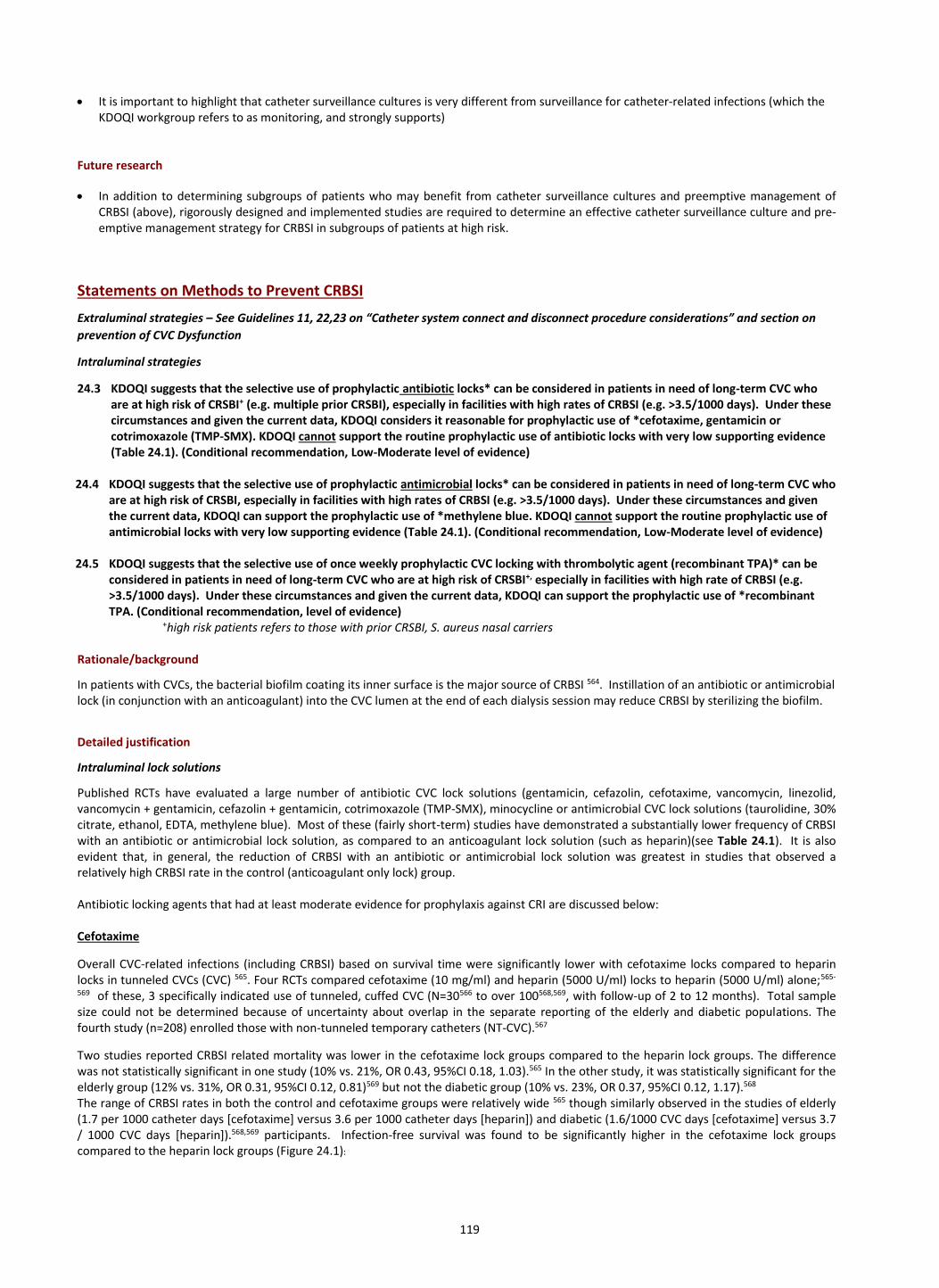
119
• It is important to highlight that catheter surveillance cultures is very different from surveillance for catheter-related infections (which the KDOQI workgroup refers to as monitoring, and strongly supports)
Future research
• In addition to determining subgroups of patients who may benefit from catheter surveillance cultures and preemptive management of CRBSI (above), rigorously designed and implemented studies are required to determine an effective catheter surveillance culture and pre-emptive management strategy for CRBSI in subgroups of patients at high risk.
Statements on Methods to Prevent CRBSI
Extraluminal strategies – See Guidelines 11, 22,23 on “Catheter system connect and disconnect procedure considerations” and section on
prevention of CVC Dysfunction
Intraluminal strategies
24.3 KDOQI suggests that the selective use of prophylactic antibiotic locks* can be considered in patients in need of long-term CVC who are at high risk of CRSBI+ (e.g. multiple prior CRSBI), especially in facilities with high rates of CRBSI (e.g. >3.5/1000 days). Under these circumstances and given the current data, KDOQI considers it reasonable for prophylactic use of *cefotaxime, gentamicin or cotrimoxazole (TMP-SMX). KDOQI cannot support the routine prophylactic use of antibiotic locks with very low supporting evidence (Table 24.1). (Conditional recommendation, Low-Moderate level of evidence)
24.4 KDOQI suggests that the selective use of prophylactic antimicrobial locks* can be considered in patients in need of long-term CVC who are at high risk of CRSBI, especially in facilities with high rates of CRBSI (e.g. >3.5/1000 days). Under these circumstances and given the current data, KDOQI can support the prophylactic use of *methylene blue. KDOQI cannot support the routine prophylactic use of antimicrobial locks with very low supporting evidence (Table 24.1). (Conditional recommendation, Low-Moderate level of evidence)
24.5 KDOQI suggests that the selective use of once weekly prophylactic CVC locking with thrombolytic agent (recombinant TPA)* can be
considered in patients in need of long-term CVC who are at high risk of CRSBI+, especially in facilities with high rate of CRBSI (e.g. >3.5/1000 days). Under these circumstances and given the current data, KDOQI can support the prophylactic use of *recombinant TPA. (Conditional recommendation, level of evidence)
+high risk patients refers to those with prior CRSBI, S. aureus nasal carriers Rationale/background
In patients with CVCs, the bacterial biofilm coating its inner surface is the major source of CRBSI 564. Instillation of an antibiotic or antimicrobial lock (in conjunction with an anticoagulant) into the CVC lumen at the end of each dialysis session may reduce CRBSI by sterilizing the biofilm.
Detailed justification
Intraluminal lock solutions
Published RCTs have evaluated a large number of antibiotic CVC lock solutions (gentamicin, cefazolin, cefotaxime, vancomycin, linezolid, vancomycin + gentamicin, cefazolin + gentamicin, cotrimoxazole (TMP-SMX), minocycline or antimicrobial CVC lock solutions (taurolidine, 30% citrate, ethanol, EDTA, methylene blue). Most of these (fairly short-term) studies have demonstrated a substantially lower frequency of CRBSI with an antibiotic or antimicrobial lock solution, as compared to an anticoagulant lock solution (such as heparin)(see Table 24.1). It is also evident that, in general, the reduction of CRBSI with an antibiotic or antimicrobial lock solution was greatest in studies that observed a relatively high CRBSI rate in the control (anticoagulant only lock) group. Antibiotic locking agents that had at least moderate evidence for prophylaxis against CRI are discussed below: Cefotaxime
Overall CVC-related infections (including CRBSI) based on survival time were significantly lower with cefotaxime locks compared to heparin locks in tunneled CVCs (CVC) 565. Four RCTs compared cefotaxime (10 mg/ml) and heparin (5000 U/ml) locks to heparin (5000 U/ml) alone;565-
569 of these, 3 specifically indicated use of tunneled, cuffed CVC (N=30566 to over 100568,569, with follow-up of 2 to 12 months). Total sample size could not be determined because of uncertainty about overlap in the separate reporting of the elderly and diabetic populations. The fourth study (n=208) enrolled those with non-tunneled temporary catheters (NT-CVC).567
Two studies reported CRBSI related mortality was lower in the cefotaxime lock groups compared to the heparin lock groups. The difference was not statistically significant in one study (10% vs. 21%, OR 0.43, 95%CI 0.18, 1.03).565 In the other study, it was statistically significant for the elderly group (12% vs. 31%, OR 0.31, 95%CI 0.12, 0.81)569 but not the diabetic group (10% vs. 23%, OR 0.37, 95%CI 0.12, 1.17).568 The range of CRBSI rates in both the control and cefotaxime groups were relatively wide 565 though similarly observed in the studies of elderly (1.7 per 1000 catheter days [cefotaxime] versus 3.6 per 1000 catheter days [heparin]) and diabetic (1.6/1000 CVC days [cefotaxime] versus 3.7 / 1000 CVC days [heparin]).568,569 participants. Infection-free survival was found to be significantly higher in the cefotaxime lock groups compared to the heparin lock groups (Figure 24.1):
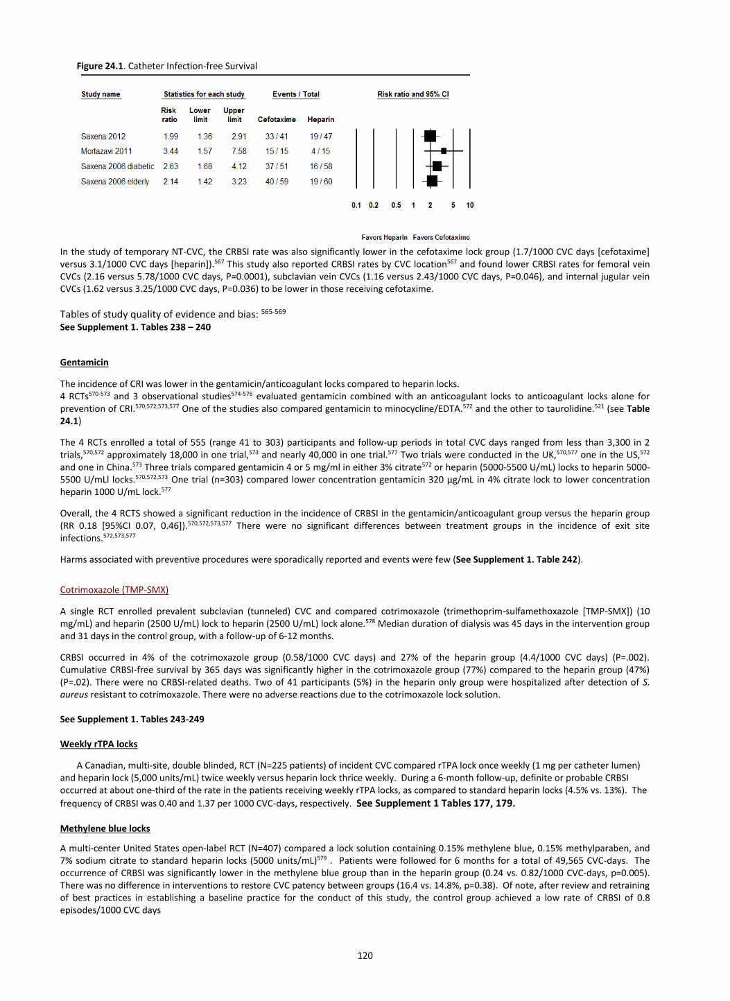
120
Figure 24.1. Catheter Infection-free Survival
In the study of temporary NT-CVC, the CRBSI rate was also significantly lower in the cefotaxime lock group (1.7/1000 CVC days [cefotaxime] versus 3.1/1000 CVC days [heparin]).567 This study also reported CRBSI rates by CVC location567 and found lower CRBSI rates for femoral vein CVCs (2.16 versus 5.78/1000 CVC days, P=0.0001), subclavian vein CVCs (1.16 versus 2.43/1000 CVC days, P=0.046), and internal jugular vein CVCs (1.62 versus 3.25/1000 CVC days, P=0.036) to be lower in those receiving cefotaxime.
Tables of study quality of evidence and bias: 565-569 See Supplement 1. Tables 238 – 240
Gentamicin
The incidence of CRI was lower in the gentamicin/anticoagulant locks compared to heparin locks. 4 RCTs570-573 and 3 observational studies574-576 evaluated gentamicin combined with an anticoagulant locks to anticoagulant locks alone for prevention of CRI.570,572,573,577 One of the studies also compared gentamicin to minocycline/EDTA.572 and the other to taurolidine.521 (see Table 24.1)
The 4 RCTs enrolled a total of 555 (range 41 to 303) participants and follow-up periods in total CVC days ranged from less than 3,300 in 2 trials,570,572 approximately 18,000 in one trial,573 and nearly 40,000 in one trial.577 Two trials were conducted in the UK,570,577 one in the US,572 and one in China.573 Three trials compared gentamicin 4 or 5 mg/ml in either 3% citrate572 or heparin (5000-5500 U/mL) locks to heparin 5000-5500 U/mLl locks.570,572,573 One trial (n=303) compared lower concentration gentamicin 320 µg/mL in 4% citrate lock to lower concentration heparin 1000 U/mL lock.577
Overall, the 4 RCTS showed a significant reduction in the incidence of CRBSI in the gentamicin/anticoagulant group versus the heparin group (RR 0.18 [95%CI 0.07, 0.46]).570,572,573,577 There were no significant differences between treatment groups in the incidence of exit site infections.572,573,577
Harms associated with preventive procedures were sporadically reported and events were few (See Supplement 1. Table 242).
Cotrimoxazole (TMP-SMX)
A single RCT enrolled prevalent subclavian (tunneled) CVC and compared cotrimoxazole (trimethoprim-sulfamethoxazole [TMP-SMX]) (10 mg/mL) and heparin (2500 U/mL) lock to heparin (2500 U/mL) lock alone.578 Median duration of dialysis was 45 days in the intervention group and 31 days in the control group, with a follow-up of 6-12 months.
CRBSI occurred in 4% of the cotrimoxazole group (0.58/1000 CVC days) and 27% of the heparin group (4.4/1000 CVC days) (P=.002). Cumulative CRBSI-free survival by 365 days was significantly higher in the cotrimoxazole group (77%) compared to the heparin group (47%) (P=.02). There were no CRBSI-related deaths. Two of 41 participants (5%) in the heparin only group were hospitalized after detection of S. aureus resistant to cotrimoxazole. There were no adverse reactions due to the cotrimoxazole lock solution. See Supplement 1. Tables 243-249 Weekly rTPA locks
A Canadian, multi-site, double blinded, RCT (N=225 patients) of incident CVC compared rTPA lock once weekly (1 mg per catheter lumen) and heparin lock (5,000 units/mL) twice weekly versus heparin lock thrice weekly. During a 6-month follow-up, definite or probable CRBSI occurred at about one-third of the rate in the patients receiving weekly rTPA locks, as compared to standard heparin locks (4.5% vs. 13%). The
frequency of CRBSI was 0.40 and 1.37 per 1000 CVC-days, respectively. See Supplement 1 Tables 177, 179. Methylene blue locks
A multi-center United States open-label RCT (N=407) compared a lock solution containing 0.15% methylene blue, 0.15% methylparaben, and 7% sodium citrate to standard heparin locks (5000 units/mL)579 . Patients were followed for 6 months for a total of 49,565 CVC-days. The occurrence of CRBSI was significantly lower in the methylene blue group than in the heparin group (0.24 vs. 0.82/1000 CVC-days, p=0.005). There was no difference in interventions to restore CVC patency between groups (16.4 vs. 14.8%, p=0.38). Of note, after review and retraining of best practices in establishing a baseline practice for the conduct of this study, the control group achieved a low rate of CRBSI of 0.8 episodes/1000 CVC days

121
Table 24.1 Intraluminal Strategies: Effect of antibiotic and antimicrobial catheter lock solutions on catheter-related bloodstream infections
(CRBSI): summary of randomized clinical trials
Agents studied Number
of patients
Relative risk [95% CI] risk
of Anticipated incidence of CRBSI (95% CI)
Quality of evidence
Locking solutions that can be used selectively for CRBSI prophylaxis
Cefotaxime (3 RCTs) 580-582
227 RR 0.42 [0.25 to 0.72]
Without antibiotic: 67.5%
With cefotaxime 23.6%
Moderate
Gentamicin 570-572,583 (4 RCTs)
555 RR 0.18 [0.07 to 0.46]
Without antibiotic 22.1%
With gentamicin: 4.0% (1.5 to 10.2)
Moderate
Weekly rTPA 494 (1 RCT)
225 RR 0.30 [0.11 to 0.85]
Heparin: 13%
Weekly rTPA: 5%
Moderate
Cotrimoxazole 584 (1 RCT)
87 RR 0.16 [0.04 to 0.69]
Without antibiotic: 4.4 per 1000 days
With antibiotic: 0.58 per 1000 days
Low
Methylene blue 585 (1 RCT)
407 RR 0.29 [0.12 to 0.70]
Heparin: 0.82 per 1000 days
Methylene blue 0.24 per 1000 days
Low
No significant effect or Very Low Evidence – cannot be recommended (below)
Taurolidine/Citrate 520,586 183
RR 0.49 [0.20 to 1.24]
Heparin: 21.5%
Taurolidine/citrate 10.5% (4.3 to 26.7)
Low
Vancomycin or linezolid 587 (1 RCT)
152 Without antibiotic: 6.7 per 1000 days
With vancomycin: 1.2 per 1000 days With linezolid: 0 per 1000 days
Very low
Vancomycin + gentamicin 588 (1 RCT)
86 RR 0.18 Without antibiotic: 4.0 per 1000 days
With antibiotic: 0.7 per 1000 days
Very low
Cefazolin 589,590 (2 RCTs)
159 RR 0.58 [0.32 to 1.04]
Without antibiotic: 29.3%
With cefazolin: 17.0% (9.4 to 30.5)
Very low
Cefazolin + gentamicin 591 (1 RCT)
120 RR 0.14 Without antibiotic: 3.12 per 1000 days
With antibiotic: 0.44 per 1000 days
Very low
Minocycline 592 (1 RCT)
204 RR 0.32 [0.14 to 0.71]
Without antibiotic: 4.3 per 1000 days
With antibiotic:
1.1 per 1000 days
Not reviewed by ERT
Antimicrobial solutions
EDTA 593 (1 RCT)
117 RR 0.34 [0.04 to 3.24]
Heparin: 1.08 per 1000 days
EDTA: 0.14 per 1000 days
Very low
Ethanol/heparin 594 (1 RCT)
49
RR 0.17 [0.02 to 0.63]
Heparin: 13%
Ethanol/heparin: 4%
Very low
Ethanol/citrate 595 (1 RCT)
40 Heparin: 5%
Ethanol/citrate: 0%
Very low
Hypertonic saline 531 (1 RCT)
59 Heparin: 10%
Hypertonic saline: 15%
Very low
30% citrate 515 (1 RCT)
291 RR 0.13-0.36 Heparin: 4.1 per 1000 days
30% citrate 1.1 per 1000 days
Not reviewed by ERT
46.7% citrate 513 (1 RCT)
232 46.7% citrate: 0.7 per 1000 days
Heparin: 0.7 per 1000 days
Not reviewed by ERT
Special Discussions Several RCTs have found that the use of prophylactic antibiotic or antimicrobial locks reduces the frequency of CRBSI, as compared to conventional anticoagulant locks alone. From that perspective, this approach seems attractive. However, there are several concerns. First, an aliquot of the intraluminal CVC lock solution invariably leaks into the systemic circulation and may cause toxicity. For example, concentrated gentamicin locks resulted in ototoxicity in a subset of patients in one study 596. Similarly, 30% citrate may cause symptomatic hypocalcemia, as manifested by paresthesia in 15% of patients in one study 513. More importantly, the Work Group committee members were concerned about the potential of long-term prophylactic antibiotic locks to lead to antibiotic-resistant infections. There are conflicting reports about the potential of prophylactic gentamicin locks to select for infection with resistant bacteria during prolonged use. Landry et al 597 documented this possibility, but 2 other large studies did not observe such a complication 571,576.
The considered use of antibiotic locks is limited to high risk patients as defined by the high risk groups studied in the trials reviewed by the ERT i.e. patients with prior CRBSI and S. aureus nasal carriage, other potential “high risk” patients have not been defined. The facility threshold for use of an intraluminal prophylactic strategy has not been validated and was discussed at length by the Work Group. The Work Group came to a consensus of a baseline CRBSI rate of >=3.5/1000 CVC days based on prior literature, potential resource implications and the finding that high rates of CRBSI can be reduced first with retraining and use of best CVC care practices and infection control 566,579,598
122
Implementation considerations
The potential concern about selection for antibiotic-resistant infections makes the Work Group committee reluctant to recommend antibiotic lock solutions across the board. Thus, our recommendation is to use prophylactic antibiotic locks in those scenarios where the benefit outweighs the potential risk, namely patients at high risk for CRBSI or units with high rates of CRBSI. These would include patients at high risk of CRSBI (e.g. prior CRSBI, S. aureus nasal carriers) or facilities with high rate of CRBSI (e.g. >3.5/1000 days). Use of non-antibiotic antimicrobial CVC solutions may be a superior option, due to the low likelihood of selecting for antibiotic-resistant infections.
Of note, the RCT using methylene blue highlighted that review, retraining and implementation of best CVC care practices can achieve a low baseline CRBSI (0.8 episodes/1000 CVC days)599, emphasizing that use of antibiotic lock prophylaxis should only be used in facilities with very high baseline risk of CRBSI even after the implementation of best CVC care and infection control practices. Future research
• Determine and validate patients at high risk of CRBSI, despite confirmed excellent CVC care and infection control practices
• Effect of risk stratification of patients at high risk of CRBSI and their prophylactic management of CRBSI, including strategies that involve exit and intraluminal antimicrobial prophylactic care
• It is unclear whether prophylactic strategies should be targeted to patients, facilities or both at high risk of CRBSI; considerations of the effect of widespread prophylactic use of antibiotics and the potential emergence of antibiotic resistant organisms impacting not only at the patient level, but facility level, must be considered and rigorously studied.
123
GUIDELINE 25. TREATMENT OF CVC-RELATED INFECTION
Statement: Management of the patient with a catheter-related infection
25.1 KDOQI considers it reasonable and necessary to obtain appropriate cultures prior to initiating empiric antibiotics for the treatment of
suspected catheter-related infection, with a change in antibiotics according to culture sensitivities* (Expert Opinion)
Statement: Management of the catheter in a patient with a catheter-related infection
25.2 KDOQI considers it reasonable to have an individualized approach to the management of an infected catheter based on the patient’s
health, dialysis and vascular access circumstances. Options include CVC exchange via guidewire, CVC removal and reinsertion, CVC
salvage and concurrent antibiotic lock (particularly if the CVC is deemed to be the patient’s final access)*. (Expert Opinion)
* see detailed justification for further detailed guidance
Rationale and Detailed Justification
CVC-related infections and bacteremia is a significant cause of morbidity and mortality for hemodialysis patients. CVC-related infections
include exit site infections, infections of the tunnel track, and bacteremias. Bacteremias are the most significant complication because they
have the potential to lead to life-threatening sepsis. The common clinical features of CVC infection include fever or chills, hemodynamic
instability, catheter dysfunction, hypothermia, nausea and vomiting, and generalized malaise559,600,601. Treatment of the 3 main types of CVC-
related infection - exit-site infection, tunnel infections, and CVC-related bacteremia – requires careful consideration on how to: 1) treat the
patient and 2) manage the CVC.
Please see Guideline 24 for definitions of CVC-related infections. Treatment of each type of CVC-related infection must consider local infection
control practices and its influence of organism patterns, cultures and sensitivities. Properly collected cultures must be obtained before
initiating empiric antibiotic treatment for all types of CVC-related infections in order to properly treat the infection and avoid antibiotic
resistance.
Exit site infections.
If drainage is present from the exit site, cultures should be obtained before initiation of antibiotics559. Empiric antibiotic treatment should
cover Gram-positive organisms and further modified once culture and sensitivity results are finalized559. Duration of treatment for exit site
infections typically range between 7 to 14 days559.
CVC management of exit site infections: typically does not require removal; however, this depends on the infecting organism and the response
to antibiotic therapy.
Tunnel infections.
When CVC tunnel infections are present, cultures from drainage from the tunnel or exit site and blood cultures from the CVC need to be
obtained559. Following the attainment of the appropriate cultures, empiric antibiotics should be initiated to target both Gram-positive and
Gram-negative organisms559. Antibiotics should be modified once culture and sensitivity results are available. The typical treatment duration
for CVC tunnel infections is 10-14 days in the absence concurrent bacteremia559. If CVC-related bacteremia is also present, the duration of
treatment should be dictated by the management strategy for the CVC-related bacteremia559.
CVC management of tunnel infections: If tunnel infection is not effectively treated with antibiotics, consider CVC exchange with a new
subcutaneous tunnel to preserve the venous access site.602 If not possible, the CVC should be removed and a new CVC placed at a new entry
site559.
Catheter-related bloodstream infection (CRSBI) or bacteremia.
Patients with suspected CRBSI should have blood cultures obtained from the CVC and peripheral source (dialysis circuit +/- peripheral veins –
see Guideline 24). Broad-spectrum antibiotics that treat both Gram-positive and Gram-negative organisms should be initiated immediately559.
Due to the high prevalence of methicillin-resistance Staphylococcus aureus (MRSA), empiric therapy should include coverage for MRSA310, but
should also be guided by the local infection rates, antibiotic sensitivities, and dialysis center policies. Once the final organism and sensitivities
are identified, patients should receive long-term antibiotic treatment, according to the CDC or Infectious Disease Society of America
Guidelines, for 4-6 weeks for uncomplicated Staphylococcus aureus, 7-14 days for Gram negative bacilli or enterococcus, and a minimum of 14
days with Candida species311. See Figure 25.1 Algorithm for Treatment of CVC infections.
CVC Management (concurrent with systemic antibiotics) of CRBSI:
There are 4 main options that involve removing the CVC or retaining the CVC, as follows:
Removing the CVC: 1) CVC removal with CVC exchange over a guidewire at the same site; 2) CVC removal with new CVC replaced at a new site
(+/- “CVC free” duration [whereby the patient has a period when there is no CVC in-situ] with insertion of a temporary CVC as needed for
dialysis). Several situations necessitate the latter option of immediate CVC removal with delayed CVC placement310,559: (i) clinically and
hemodynamically unstable patients, (ii) persistent fever after 48 to 72 following initiation of systemic antibiotics, (iii) persistent bacteremia
after 48-72 hours after initiating antibiotics. (iv) metastatic complications, (v) fungal infections, and (vi) presence of a tunnel-site infection
At the time of CVC removal, evaluation for the presence of fibrin sheath (presumably with infected biofilm) and fibrin sheath disruption can be
performed (see Guideline 27).
124
Retaining the CVC: 3) retain CVC and use antibiotic CVC lock; 4) retain CVC without use of antibiotic CVC lock. Antibiotic locks with concurrent
systemic antibiotics may be an alternative treatment strategy to preserve the CVC. While there are no RCTs evaluating the role of antibiotic
CVC locks in the treatment of CRBSI, there have been several observation studies demonstrating eradication of bacteremia with antibiotic
locks in conjunction with systemic antibiotics compared to CVC exchange or removal in conjunction with systemic antibiotics.559,603,604
While the best management of CVC-related infection is to avoid them altogether, it is very unlikely that CVC can be completely eliminated, as
80% of patients initiate hemodialysis with a CVC504. Thus, there are several core interventions recommended by the CDC that KDOQI endorses
that can be useful to decrease infection rates and complications in patients requiring dialysis with a CVC, and are covered in Guidelines 24 and
25). These core interventions include559: (1) Infection surveillance and feedback; (2) Hand hygiene observations; (3) CVC/vascular access care
observations; (4) Staff education and competency; (5) Patient education and engagement; (6) CVC reduction efforts; (7) chlorohexidine for skin
antisepsis; (8) CVC hub disinfection; and (9) antimicrobial ointment.
Multidisciplinary team surveillance and management of CVC-related infections has been demonstrated to reduce infections and improve
outcomes in hemodialysis patients who require a CVC for dialysis access305,316
Future research
• Clinical sequelae of disrupting a fibrin sheath in patients with CRBSI requires further study
• Prospective study comparing treatment of CRBSI using antibiotic locks in conjunction with systemic antibiotics versus CVC exchange or removal in conjunction with systemic antibiotics
125
GUIDELINE 26. OTHER VASCULAR ACCESS-RELATED COMPLICATIONS
Treatment of other AV-access or Catheter related complications
Statement for intervention of asymptomatic central venous stenosis
26.1 KDOQI considers it reasonable that if asymptomatic central venous stenosis is identified and/or associated with the prior or current presence of a CVC it should not be treated (Expert Opinion).
Rationale/Background
Stenoses or occlusions in the major intra-thoracic veins (i.e. central veins – internal jugular, subclavian, brachiocephalic, superior vena cava) can compromise AV-access function and lead to ineffective dialysis. These lesions can also lead to marked venous hypertension due to the increased flow associated with the AVF with sequelae ranging from mild to severe symptoms as characterized by varicosities, arm edema, dermatosclerosis, ulceration, and superior vena cava syndrome (See Table 26.1; and Figure 26.1). Female patients can occasionally present with ipsilateral breast edema despite a normal or only moderately swollen extremity. These stenotic lesions can lead to clinical signs and symptoms such as prolonged bleeding after removal of the dialysis cannulas and/or elevated venous pressures during routine monitoring with high AV-access recirculation. Central vein stenoses (CVS) or occlusions have been reported to occur in up to 5 – 50% of cases223,605-608 and represent the “Achilles heel” of maintaining or creating a functional AV-access. They can preclude the placement of an ipsilateral AV- access and likely account for the leading etiology of complex or “tertiary” AV-access problems.
The central vein lesions are likely caused by an initial injury to the vascular endothelium that precipitates a local inflammatory response that leads to fibrosis. A variety of factors have shown to compound or exacerbate the various steps of this process including deep venous thromboses, intravascular CVC, cardiac rhythm devices (e.g. pacemakers) and hemodialysis AV-accesses themselves. Peripherally inserted central catheters (i.e. PICC lines) have been reported to cause a CVS or occlusion in up to 7% of cases while subclavian catheters have been reported to cause these lesions in up to 50% of cases.260 Even appropriately positioned (i.e. internal jugular insertion) CVC can lead to CVS and/or occlusions within a relatively brief period of time. The true incidence of CVC induced CVS/occlusion is unknown. If CVS is incidentally identified on angiographic assessment without clinical symptoms, it is unknown what the natural history of the stenosis is over time. However, overall, CVS/occlusions may represent the worst of the catheter-related complications, underscoring the importance of limiting their use.
Indeed, angioplasty for asymptomatic stenosis is associated with a more rapid progression to symptomatic stenosis609,610 and should therefore be avoided unless there is a clinical indication to perform an angioplasty. In patients with symptomatic central venous stenosis/occlusion, primary patency with angioplasty/angioplasty with stenting has a very poor primary patency611. Although stent grafts have demonstrated improved patency in peripheral stenosis, outcomes for central lesions are uncertain based on small retrospective studies.
Prior KDOQI guidelines recommended treatment of CVS with PTA but without clarity regarding if clinical symptoms were also present as an indication for intervention. Stent placement was recommended for 1) acute elastic recoil of >50% or 2) stenosis recurrence within three months. Elastic recoil itself has been poorly defined. Detailed Justification In two retrospective studies that assessed central vein patency in patients with asymptomatic versus symptomatic CVS, both studies recommended against intervention for asymptomatic stenosis. The first study excluded patients with CVCs but found that treatment of asymptomatic CVS greater than 50% in the setting of hemodialysis access maintenance procedures was associated with more rapid stenosis progression and escalation of lesions, compared with a non-treatment approach 609. In the second study, primary central vein patency at 12, 24 and 36 months in asymptomatic/pauci-symptomatic patients and symptomatic patients with CVS were 77±6 versus 55±9, 71±7 versus 35±9 and 67±7 versus 18±9%, respectively (P=0.002).610
Angioplasty and/or angioplasty and stenting for CVS are associated with relatively poor patency. In one retrospective study, primary patency of 76% was equivalent for angioplasty or angioplasty with stenting at 30 days, with 12-month rates of 29% for angioplasty and 21% for stenting.611 However, in another retrospective study in hemodialysis patients without CVCs, primary patency (time from intervention to next intervention) was 24.5 months in the angioplasty group and 13.4 months in the stent group.612 Special discussions
The Work Group discussed the signs and symptoms that may require confirmatory diagnosis and intervention, including:
• Ipsilateral facial, neck, breast or extremity swelling (without other cause)
• Repeated thrombosis of an upper arm access in the absence of other causes within six months
• Pain in the extremity related to venous obstruction
• Neurological symptoms in the absence of other etiologies
• Venous pressure is highly variable and dependent on many patient and hemodialysis factors and their interactions. Therefore, intervention should not be based solely on venous pressure but other signs and symptoms should be present as well.
Future research
• Understanding the natural history of CVS
• Prospective studies on intervention of symptomatic CVS
.
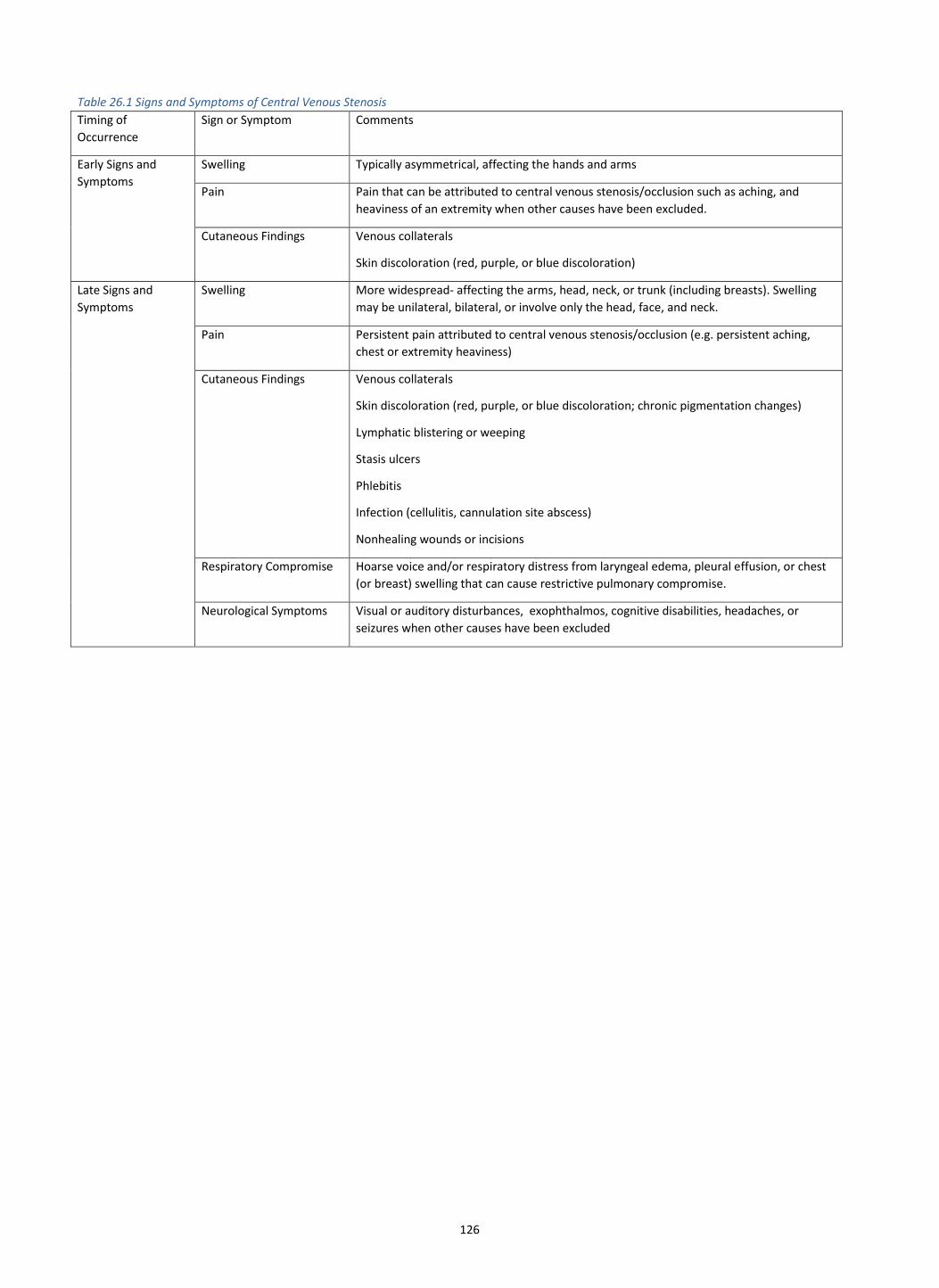
126
Table 26.1 Signs and Symptoms of Central Venous Stenosis
Timing of
Occurrence
Sign or Symptom Comments
Early Signs and
Symptoms
Swelling Typically asymmetrical, affecting the hands and arms
Pain Pain that can be attributed to central venous stenosis/occlusion such as aching, and
heaviness of an extremity when other causes have been excluded.
Cutaneous Findings Venous collaterals
Skin discoloration (red, purple, or blue discoloration)
Late Signs and
Symptoms
Swelling More widespread- affecting the arms, head, neck, or trunk (including breasts). Swelling
may be unilateral, bilateral, or involve only the head, face, and neck.
Pain Persistent pain attributed to central venous stenosis/occlusion (e.g. persistent aching,
chest or extremity heaviness)
Cutaneous Findings Venous collaterals
Skin discoloration (red, purple, or blue discoloration; chronic pigmentation changes)
Lymphatic blistering or weeping
Stasis ulcers
Phlebitis
Infection (cellulitis, cannulation site abscess)
Nonhealing wounds or incisions
Respiratory Compromise Hoarse voice and/or respiratory distress from laryngeal edema, pleural effusion, or chest
(or breast) swelling that can cause restrictive pulmonary compromise.
Neurological Symptoms Visual or auditory disturbances, exophthalmos, cognitive disabilities, headaches, or
seizures when other causes have been excluded
127
Statement for Investigation and treatment of symptomatic central venous stenosis
26.2 Same as guidelines for “Treatment of Access flow dysfunction and Clinically Significant Stenosis” (See Guideline 15)
Rationale/Background – as above for asymptomatic CVS Detailed Justification
Close routine clinical monitoring will detect signs and symptoms suggestive of clinically significant CVS and venous hypertension (see Guidelines 11, 13, 14). The culprit lesion can usually be confirmed with catheter-based and CT venography, given the limitations of ultrasound in the thoracic cavity. CT venography offers the advantage of being non-invasive and the potential to image all four extremities at the same time although the timing of the contrast injection and image acquisition can be challenging. Patients with mild symptoms can improve over time with the development of collaterals and it is not uncommon to see patients with a functional AV-access and no arm edema despite a CVS/occlusion.613 Intervention is not indicated for asymptomatic lesions or those associated with minimal symptoms.610 (See KDOQI Guideline 13)
The treatment indications for CVS/occlusions include persistent moderate-severe clinical signs and symptoms (See Guideline 13), and/or related ineffective dialysis. Of note, it is relatively common for patients to develop some arm edema after AV-access construction, likely related to the operative trauma and mild venous hypertension. This usually resolves in the first 2 – 6 weeks postoperatively with the resolution of the inflammation from the surgical trauma and the development of venous collaterals.
The endovascular approach with balloon angioplasty is the first line of treatment for these symptomatic CVS/occlusions. Intraluminal stenting is reserved for angioplasty failures. While the early technical success rates for the endovascular treatment are excellent and can exceed 90%,260, the longer-term 6- and 12-month primary patency is poor at 50% and 25%, respectively.614-616 Notably, Yan et al.617 reported that balloon angioplasty of the central vein lesions had little impact on the AV-access flow although it was effective in relieving the symptoms. Intraluminal stenting should be reserved for recurrent stenosis, given the uncertainty of outcomes.260,616 Indeed, stents should be used with caution (or avoided altogether) in the region of the thoracic outlet due to the potential for extrinsic compression and stent fracture from the overlying structures. Placing stents over pacer wires can complicate their removal. Thus, pacer wires can be removed (and the pacer re-sited) prior to the placement of an intraluminal stent; however, it may be simpler to create another AV-access on the contralateral extremity (if possible), particularly given that the sclerotic lesions associated with the pacer wires tend to be refractory to balloon angioplasty. Covered stents afford some theoretical appeal because the intimal hyperplasia does not develop within the covered segment although it can develop at the proximal and distal ends of the stent. Furthermore, the covered component of the stent can inadvertently cover or “jail” important collaterals or the major central veins (e.g. internal jugular). Caution must be exercised when using intraluminal stents. A careful tiered approach for de novo and recurrent lesions progressing from balloon angioplasty to bare metal stents followed by covered stents should be considered in the context of the patients Life-Plan and their vascular access contingency and successions plans.616 There are a variety of open surgical options for patients with CVS/occlusions associated with their vascular access. However, these are considered secondary or tertiary options and are largely dictated by the anatomy in terms of available patent inflow and outflow veins. Potential options including axillary-jugular bypass, axillary-axillary bypass, axillary-femoral bypass, axillary-atrial bypass or the jugular vein turndown procedure.618,619 Alternatively, associated venous hypertension may be reduced by limiting the flow through the AV-access using some variant of a “banding” or “flow limiting procedure” (see Guideline 19). The Hemoaccess Reliable Outflow (HeRO) Vascular Access Device (Hemosphere, Inc., Minneapolis, Minn.) can be used as a hybrid alternative (i.e. combination endovascular/open surgical approach) access once the occlusion is bypassed.620 The HeRO graft is a 6 mm (i.d.) PTFE graft that is coupled to a 19 Fr (o.d.) central venous catheter. Although typically used as a new AV-access configuration, the HeRO graft can be used in combination with an existing AVF or AVG to provide the “central vein runoff” provided that the delivery sheath for the catheter can be passed through the lesion.620,621

128
Statement for management of CVC fibrin sheath associated with clinical problems
26.3 KDOQI considers it reasonable that when a catheter fibrin sheath is associated with adverse clinical manifestations (CVC dysfunction and/or infection), to perform a catheter exchange with or without balloon disruption of the fibrin sheath (Expert Opinion).
Rationale/Background Fibrin sheaths are a known cause of CVC dysfunction and infection that cannot be treated with thrombolytic therapy alone. CVC exchange alone or exchange with balloon disruption likely results in disruption of the fibrin sheath.622,623 The fibrin sheath is composed of smooth muscle cells and vascularized connective tissue that originates at the venotomy site and grows along the CVC.548,624 Once growth extends to the tip of the CVC, the sheath acts as a valve interrupting aspiration of blood from the CVC but not return of blood through the CVC. As thrombolytics dissolve acute clot (<14 days) and not tissue, they are ineffective against fibrin sheaths. Also, fibrin sheaths can harbor bacteria. A prior typical management strategy for suspected CRBSI is to remove the CVC, wait 48-72 hours and reinsert a new CVC. The presence of an infected fibrin sheath and its consequences was often not considered in the setting of CRBSI. Disrupting the fibrin sheath may eliminate CRBSI and has been shown not to increase bacteremia rates551; thus its disruption may assist in eliminating a source of recurrent infection.
The prior 2006 NKF-KDOQI guidelines stated that a fibrin sheath causing CVC malfunction can be treated with CVC exchange with or without balloon disruption. No statement was made in the prior guidelines regarding disrupting fibrin sheaths to prevent or treat bacteremia.
Detailed Justification
In a randomized prospective pilot study, 47 long-term HD patients with secondary, refractory CVC malfunction underwent guidewire exchange to replace their CVCs. Fibrin sheaths were present in 33 (70%) of the 47 patients. In 18 patients who were randomly assigned to disruption, the median time to repeat dysfunction was 373 days compared with 97.5 days in patients who did not undergo disruption (P = 0.22), and the median time to repeat CVC exchange was 411 and 198 days, respectively (P = 0.17). Although significance was not obtained due to the small sample size, the study highlighted a high incidence of fibrin sheaths and a trend towards improved function with balloon disruption.547 Another study found the incidence of fibrin sheath formation for dysfunctional CVCs to be 76%.625 In an animal study assessing if fibrin sheath presence results in enhanced catheter-related infection and persistent bacteremia, the fibrin sheath group exhibited a 50% infection rate vs. 0% in the no fibrin group (P <0.01).626 There is no comparable published study in humans. Special discussions
There is very limited literature on fibrin sheaths and their impact in HD patients. The current data are comprised of small series, mostly retrospective in nature. Thoughts regarding bacteremia are anecdotal. The Work Group acknowledges that our suggestion of considering balloon disruption is based on limited data. Implementation considerations
Must balance an increase in intervention rate and potential complications with balloon angioplasty compared to simple CVC exchange. Future research
• Randomized study comparing thrombolytics versus catheter exchange versus catheter exchange with fibrin sheath disruption.
• Association of fibrin sheath incidence with CRBSI and impact of disruption and antibiotics versus antibiotics alone on CVC malfunction and infection
129
RESEARCH RECOMMENDATIONS
Future Research for Hemodialysis Vascular Access and Related Topics
Effective and appropriate application of the individualized ESKD Life-Plan strategy described in these guidelines requires a broader perspective than
focusing solely on the creation of a specific vascular access or the monitoring and management of an existing vascular access. The Life-Plan
approach requires consideration of the patient’s current and future goals and preferences, as well as their current and future medical condition. To
appropriately consider all of these aspects will require additional knowledge on patient preferences, more accurate tools to predict the timing of
the need for dialysis, and better data on the expected outcomes associated with various approaches to dialysis access creation and preservation. To
truly individualize the choices, each of these research areas will need to address potential differences across patient subgroups to allow evidence-
based decision-making for specific patients.
Timing of the need for dialysis
Planning for initial dialysis access in CKD requires some knowledge of the urgency of the need for dialysis. The Work Group agrees that assessment
for vascular access should occur if the patient has a ≥50% risk of needing RRT within 2 years and/or has an eGFR ≤15 mL/min/1.73m2; however, this
is based on Expert Opinion only. Tools to predict risk of ESKD, using eGFR and/or other commonly obtained variables, have been developed
recently,627-630 including the 4-variable Kidney Failure Risk Index (KFRI).631 The current limited evidence supporting the validity of these prediction
tools is based on a single evaluation of each participant’s status, which may change quickly. Future studies will require repeated assessments to
determine whether a patient has crossed a threshold where dialysis access evaluation or creation is deemed appropriate. Only recently has a
“dynamic prediction” tool been published.630 While this tool uses updated information, it does not take full advantage of the patient’s history and
previous experiences.
While numerous prediction tools have been developed, few have been validated in other populations, and even fewer have been subjected to an
evaluation of their impact.632 The impact of utilizing a risk prediction tool and associated practices on vascular access specifically has, to our
knowledge, not been evaluated for any specific tool. Much work remains in how best to implement vascular access-related clinical decisions based
on the projected probability of kidney failure for a specified time horizon or the estimated time to kidney failure. Again, the best approach may
differ based on a specific patient’s characteristics and preferences, which must be addressed in these evaluation studies.
The other piece of the puzzle for optimizing the timing of access creation, of course, is the time required from creation to use. Additional research is
needed on appropriate time for cannulation and how to objectively and reliably determine “readiness for cannulation” in traditional and newer
vascular access types, and methods to aid in maturation. [See other sections]
Expected outcomes associated with various approaches to vascular access creation and preservation
National programs, including the Centers for Medicare and Medicaid Services’ breakthrough initiative, “Fistula First,” have aggressively campaigned
for greater use of AVFs and set goals of at least 66% AVF use and less than 10% CVC use among prevalent hemodialysis patients.633 Despite these
efforts, more than 80% of ESKD patients initiate hemodialysis with a CVC and CVC use remains common among prevalent hemodialysis patients.42 135 Numerous studies have raised serious questions about this “one-size-fits-all” approach to vascular access, finding substantial heterogeneity
across patients in the benefit derived from immediate AVF creation.634-636 The Work Group hypothesizes that a more patient-specific approach will
result in more appropriate vascular access choices.18,637
Multiple factors must be considered to apply the Life-Plan strategy to vascular access creation decision-making, including the urgency of the need
for a functioning AV-access versus the expected time required for maturation, the infection risk associated with CVC use during maturation versus
the expected long-term benefit of an AV-access, the probability of maturation failure of an AVF versus the potential long-term benefit over an AVG,
and the potential need for future accesses. Although data exist to provide useful estimates based on patient characteristics for some of these, such
as predicting the probability of maturation,638 there remain significant gaps in our knowledge. Additional research is needed to provide estimates
for each of these variables across various patient and vessel characteristics to inform patient-centered, evidence-based decision-making algorithms
In the meantime, KDOQI has provided an app (website link here) that can be used to facilitate dialysis access decision-making, based on evidence
gained through the UCLA RAND process. This tool requires further validation and research into the impact on its implementation.
Patient preferences
In addition to the clinical outcomes associated with choosing any specific access, numerous studies have found that vascular access is a major
concern of patients faced with ESKD.639-641 Issues such as physical disfigurement from an AVF and the pain and fear of needle cannulation are of
great concern to patients, but have been overlooked by initiatives such as “Fistula First.”642 Research has uncovered substantial heterogeneity in
utilities assigned to various factors across patients by age, sex, and country.639 Additional research is recommended to identify characteristics about
processes and health outcomes associated with vascular access that are most relevant to patients’ decisions.
Specific research recommendations pertaining to individual guidelines
Please see the accompanying Table of Potential Future Research for Hemodialysis Vascular Access and Related Topics.
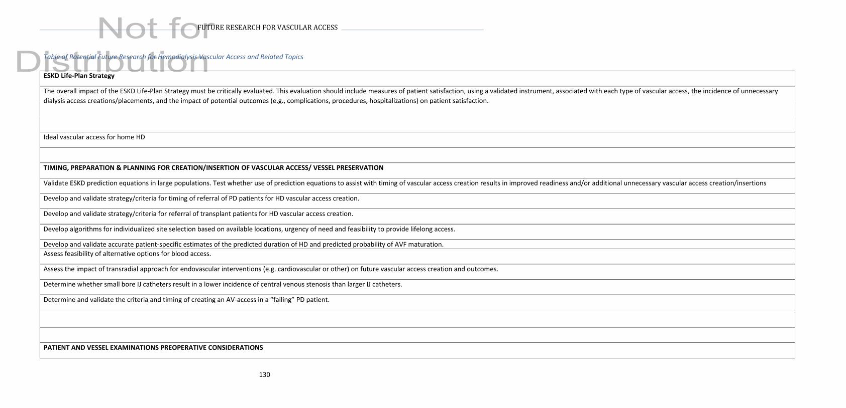
130
FUTURE RESEARCH FOR VASCULAR ACCESS
Table of Potential Future Research for Hemodialysis Vascular Access and Related Topics
ESKD Life-Plan Strategy
The overall impact of the ESKD Life-Plan Strategy must be critically evaluated. This evaluation should include measures of patient satisfaction, using a validated instrument, associated with each type of vascular access, the incidence of unnecessary
dialysis access creations/placements, and the impact of potential outcomes (e.g., complications, procedures, hospitalizations) on patient satisfaction.
Ideal vascular access for home HD
TIMING, PREPARATION & PLANNING FOR CREATION/INSERTION OF VASCULAR ACCESS/ VESSEL PRESERVATION
Validate ESKD prediction equations in large populations. Test whether use of prediction equations to assist with timing of vascular access creation results in improved readiness and/or additional unnecessary vascular access creation/insertions
Develop and validate strategy/criteria for timing of referral of PD patients for HD vascular access creation.
Develop and validate strategy/criteria for referral of transplant patients for HD vascular access creation.
Develop algorithms for individualized site selection based on available locations, urgency of need and feasibility to provide lifelong access.
Develop and validate accurate patient-specific estimates of the predicted duration of HD and predicted probability of AVF maturation.
Assess feasibility of alternative options for blood access.
Assess the impact of transradial approach for endovascular interventions (e.g. cardiovascular or other) on future vascular access creation and outcomes.
Determine whether small bore IJ catheters result in a lower incidence of central venous stenosis than larger IJ catheters.
Determine and validate the criteria and timing of creating an AV-access in a “failing” PD patient.
PATIENT AND VESSEL EXAMINATIONS PREOPERATIVE CONSIDERATIONS

131
FUTURE RESEARCH FOR VASCULAR ACCESS
Determine the optimal training methods for preoperative clinical examination to assess patients and their vessels to determine the most suitable type and location of their vascular access.
Studies are needed to examine the influence of radial approach angiograms on future vascular access outcomes
Develop and validate approaches to reduce potential damage to central and peripheral vessels by providers throughout the health care system.
Research and develop tools for patient education and vessel preservation
VASCULAR ACCESS TYPES: INCIDENT AND PREVELANT PATIENTS
Develop and test risk prediction models to assist patient-specific decision-making regarding choice of vascular access. As it is unlikely that randomized trials will be performed to compare different initial or subsequent access types, we must rely on
high-quality observational data to inform these algorithms. Specifically, data on expected probability of maturation, likely length of time until cannulation, incidence of various complications, and likely length of AV-access survival will be needed,
ideally for numerous strata of patient characteristics (e.g., age, sex, race, life expectancy, number and severity of comorbid conditions). For specific areas where existing data are lacking, provide conflicting results, or are susceptible to substantial bias,
new analyses and studies will be required.
Develop, validate and implement the standardized definitions for maturation, patency, and specific complications to provide consistency across studies and allow development of decision-making tools.
VASCULAR ACCESS LOCATIONS
Determine whether large arteries (>2 mm) with higher baseline flows are associated with greater risk of flow-associated problems.
To identify criteria for upper arm as first AV-access choice
Evaluate the impact of real time US evaluation of operating surgeon on location identification.
AV-ACCESS CREATION
The preponderance of stenosis in specific anatomic locations suggest local hemodynamics play a significant role in its development. The availability of better imaging techniques and computational fluid dynamics (CFD) models provide an opportunity
to model hemodynamics and shear stress patterns. New CFD models, specific for AV-access, should be developed.
Studies of hemodynamics (e.g., flow, shear stress) as a causative factor of AV-access outcomes, including outflow vein and juxta-anastomotic stenosis, are needed.
Evaluate a variety of anastomotic configurations and techniques, including the end-to-side, side-to-side configuration, the ‘piggyback’ straight-line onlay technique (i.e., pSLOT), and the Radial Artery Deviation And Reimplantation (RADAR) technique.
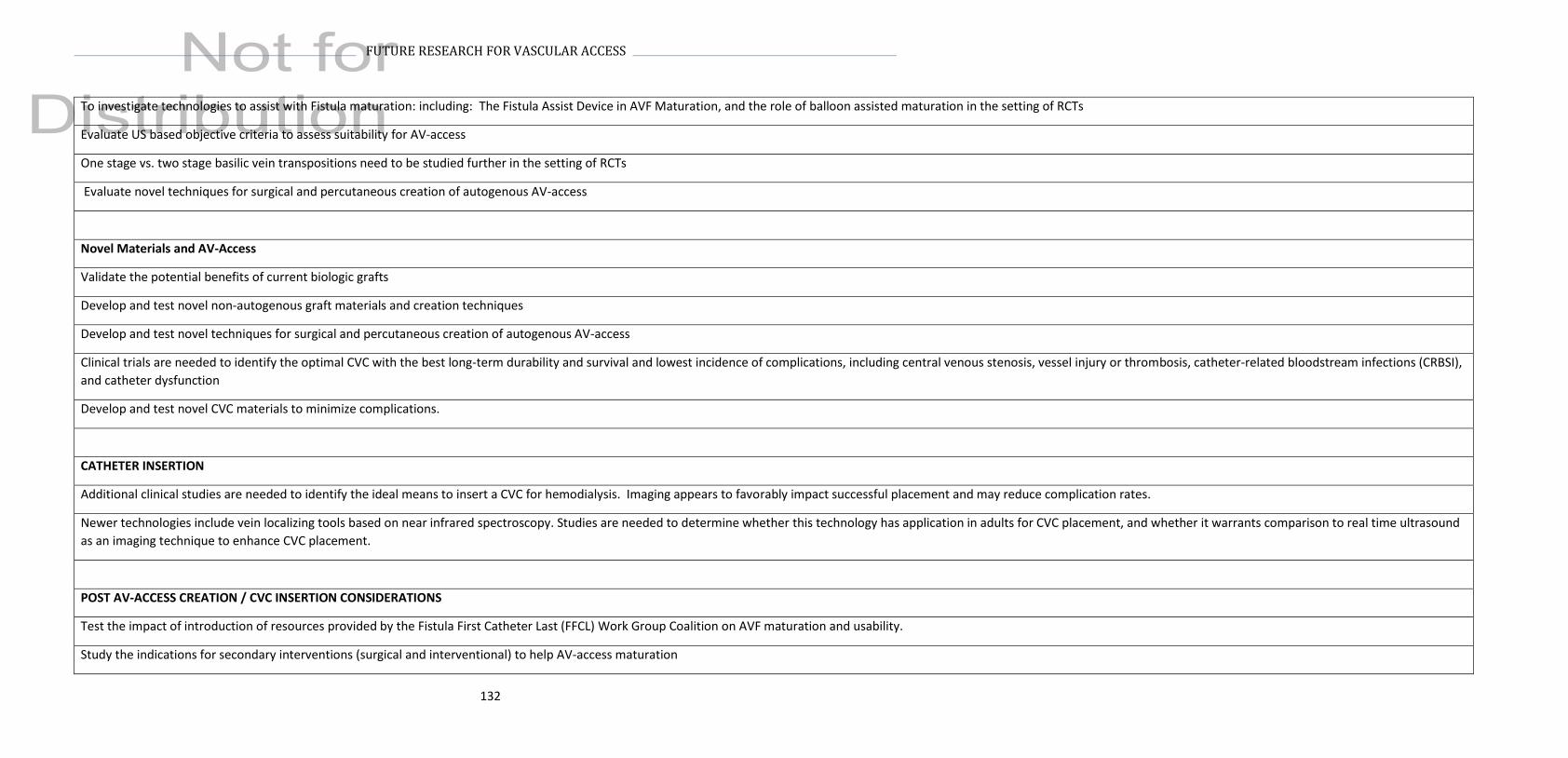
132
FUTURE RESEARCH FOR VASCULAR ACCESS
To investigate technologies to assist with Fistula maturation: including: The Fistula Assist Device in AVF Maturation, and the role of balloon assisted maturation in the setting of RCTs
Evaluate US based objective criteria to assess suitability for AV-access
One stage vs. two stage basilic vein transpositions need to be studied further in the setting of RCTs
Evaluate novel techniques for surgical and percutaneous creation of autogenous AV-access
Novel Materials and AV-Access
Validate the potential benefits of current biologic grafts
Develop and test novel non-autogenous graft materials and creation techniques
Develop and test novel techniques for surgical and percutaneous creation of autogenous AV-access
Clinical trials are needed to identify the optimal CVC with the best long-term durability and survival and lowest incidence of complications, including central venous stenosis, vessel injury or thrombosis, catheter-related bloodstream infections (CRBSI),
and catheter dysfunction
Develop and test novel CVC materials to minimize complications.
CATHETER INSERTION
Additional clinical studies are needed to identify the ideal means to insert a CVC for hemodialysis. Imaging appears to favorably impact successful placement and may reduce complication rates.
Newer technologies include vein localizing tools based on near infrared spectroscopy. Studies are needed to determine whether this technology has application in adults for CVC placement, and whether it warrants comparison to real time ultrasound
as an imaging technique to enhance CVC placement.
POST AV-ACCESS CREATION / CVC INSERTION CONSIDERATIONS
Test the impact of introduction of resources provided by the Fistula First Catheter Last (FFCL) Work Group Coalition on AVF maturation and usability.
Study the indications for secondary interventions (surgical and interventional) to help AV-access maturation

133
FUTURE RESEARCH FOR VASCULAR ACCESS
VASCULAR ACCESS USE
Test the safety, efficacy, and impact on health care resources and patient outcomes of ultrasound-guided cannulation in operating dialysis units.
Studies are need to identify best practices for mechanics of cannulation, including needle type and size, angle of insertion, and graduated flow rates.
Test effectiveness of simulation models and other techniques for improving cannulation success, reducing complications and improving patient satisfaction
Define “expert cannulator” and determine how such expert cannulators can maintain their expertise and be best utilized to improve overall cannulation success within a unit and for individual patients.
Rigorous studies are needed to identify obstacles to achieving complication-free cannulation and strategies to mitigate such obstacles
RCT to assess standard needles vs. plastic cannulas in preserving AV-access patency and reducing complications
Impact of needle size and pump speed on long-term AV-access patency
Impact of manual compression versus mechanical clamp use post needle withdrawal on access patency/stenosis- (AV-access flow dysfunction)
Evaluate outcomes associated with alternative cannulation devices.
AV-ACCESS FLOW DYSFUNCTION
“AV –Access flow dysfunction” refers to clinically significant abnormalities in AV-access (AVF or AVG) flow or patency due to underlying stenosis, thrombosis r related pathology
Determine which indicators from a clinical monitoring exam are most strongly predictive of clinically significant stenosis.
Impact of annual mandatory physical examination skill assessment for all stakeholders on recognizing early AV-access flow dysfunction (Similar to BLS for cardiac arrest)
Adequately powered randomized controlled trials are needed to determine which surveillance protocols and thresholds for intervention improve overall AV-access patency. Studies should identify indications and relative benefits/harms of earlier (pre-
emptive) interventions.
Clinical studies are needed to determine the most appropriate indications for angiographic versus surgical intervention.
Define and validate specific outcome metrics for interventions for dysfunctional and thrombosed AV-access
Determine outcomes with surgically corrected occluded accesses that are followed by a completion angiogram/imaging +/- further corrective procedure. How does this strategy compare with historical surgical correction without completion imaging
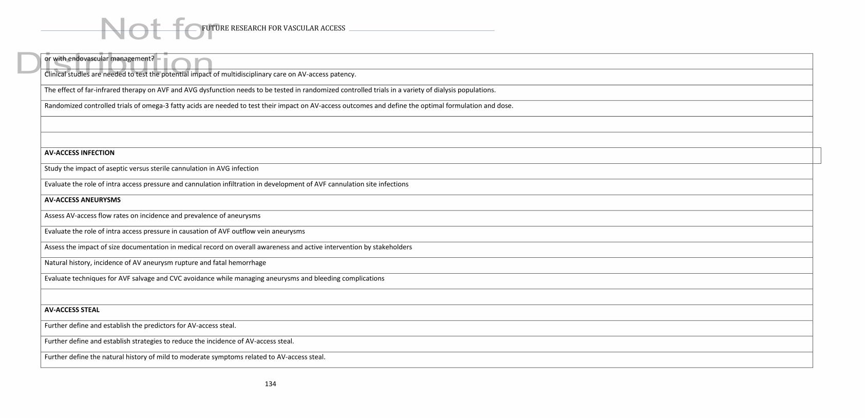
134
FUTURE RESEARCH FOR VASCULAR ACCESS
or with endovascular management?
Clinical studies are needed to test the potential impact of multidisciplinary care on AV-access patency.
The effect of far-infrared therapy on AVF and AVG dysfunction needs to be tested in randomized controlled trials in a variety of dialysis populations.
Randomized controlled trials of omega-3 fatty acids are needed to test their impact on AV-access outcomes and define the optimal formulation and dose.
AV-ACCESS INFECTION
Study the impact of aseptic versus sterile cannulation in AVG infection
Evaluate the role of intra access pressure and cannulation infiltration in development of AVF cannulation site infections
AV-ACCESS ANEURYSMS
Assess AV-access flow rates on incidence and prevalence of aneurysms
Evaluate the role of intra access pressure in causation of AVF outflow vein aneurysms
Assess the impact of size documentation in medical record on overall awareness and active intervention by stakeholders
Natural history, incidence of AV aneurysm rupture and fatal hemorrhage
Evaluate techniques for AVF salvage and CVC avoidance while managing aneurysms and bleeding complications
AV-ACCESS STEAL
Further define and establish the predictors for AV-access steal.
Further define and establish strategies to reduce the incidence of AV-access steal.
Further define the natural history of mild to moderate symptoms related to AV-access steal.
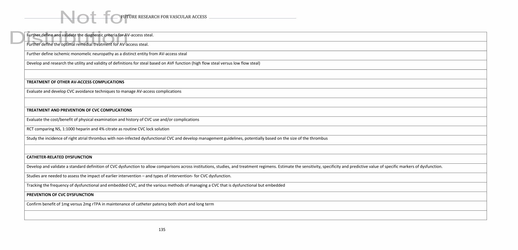
135
FUTURE RESEARCH FOR VASCULAR ACCESS
Further define and validate the diagnostic criteria for AV-access steal.
Further define the optimal remedial treatment for AV-access steal.
Further define ischemic monomelic neuropathy as a distinct entity from AV-access steal
Develop and research the utility and validity of definitions for steal based on AVF function (high flow steal versus low flow steal)
TREATMENT OF OTHER AV-ACCESS COMPLICATIONS
Evaluate and develop CVC avoidance techniques to manage AV-access complications
TREATMENT AND PREVENTION OF CVC COMPLICATIONS
Evaluate the cost/benefit of physical examination and history of CVC use and/or complications
RCT comparing NS, 1:1000 heparin and 4% citrate as routine CVC lock solution
Study the incidence of right atrial thrombus with non-infected dysfunctional CVC and develop management guidelines, potentially based on the size of the thrombus
CATHETER-RELATED DYSFUNCTION
Develop and validate a standard definition of CVC dysfunction to allow comparisons across institutions, studies, and treatment regimens. Estimate the sensitivity, specificity and predictive value of specific markers of dysfunction.
Studies are needed to assess the impact of earlier intervention – and types of intervention- for CVC dysfunction.
Tracking the frequency of dysfunctional and embedded CVC, and the various methods of managing a CVC that is dysfunctional but embedded
PREVENTION OF CVC DYSFUNCTION
Confirm benefit of 1mg versus 2mg rTPA in maintenance of catheter patency both short and long term

136
FUTURE RESEARCH FOR VASCULAR ACCESS
CATHETER-RELATED INFECTION
Further develop and validate criteria for catheter-related bloodstream infection (CRBSI) in hemodialysis patients
Studies are needed to validate diagnostic criterion for exit site and tunnel infections.
RCT to assess impact of taking a shower on CRBSI with healed CVC exit site
PREVENTION OF CVC INFECTION
Rigorously designed and implemented studies are required to determine an effective surveillance and pre-emptive management strategy for CRBSI in subgroups of patients at high risk.
Determine predictors of CRBSI in patients receiving standard infection control practices.
Evaluate the impact on incidence of CRBSI of strategies that involve exit and intraluminal antimicrobial prophylactic care Studies should be performed at both the patient and facility level and evaluate the potential emergence of antibiotic resistant
organisms.
RCT to assess impact of taking a shower on CRBSI with healed CVC exit site
OTHER VASCULAR ACCESS-RELATED COMPLICATIONS
The natural history and predictors of CVS needs to be understood in greater detail.
Effective interventions for symptomatic CVS need to be developed and validated.
Randomized controlled trials for treatment of catheters with a fibrin sheath are needed to compare effectiveness of thrombolytics, catheter exchange, and catheter exchange with fibrin sheath disruption on outcomes of patency and infection.
The association of fibrin sheath incidence with CBRSI needs to be evaluated.
The effectiveness of fibrin sheath disruption plus antibiotics compared to antibiotics alone needs to be evaluated in terms of CVC dysfunction and incidence of infection.
Characterizing catheter related central vein thrombosis vs. atrial thrombosis and its management principles
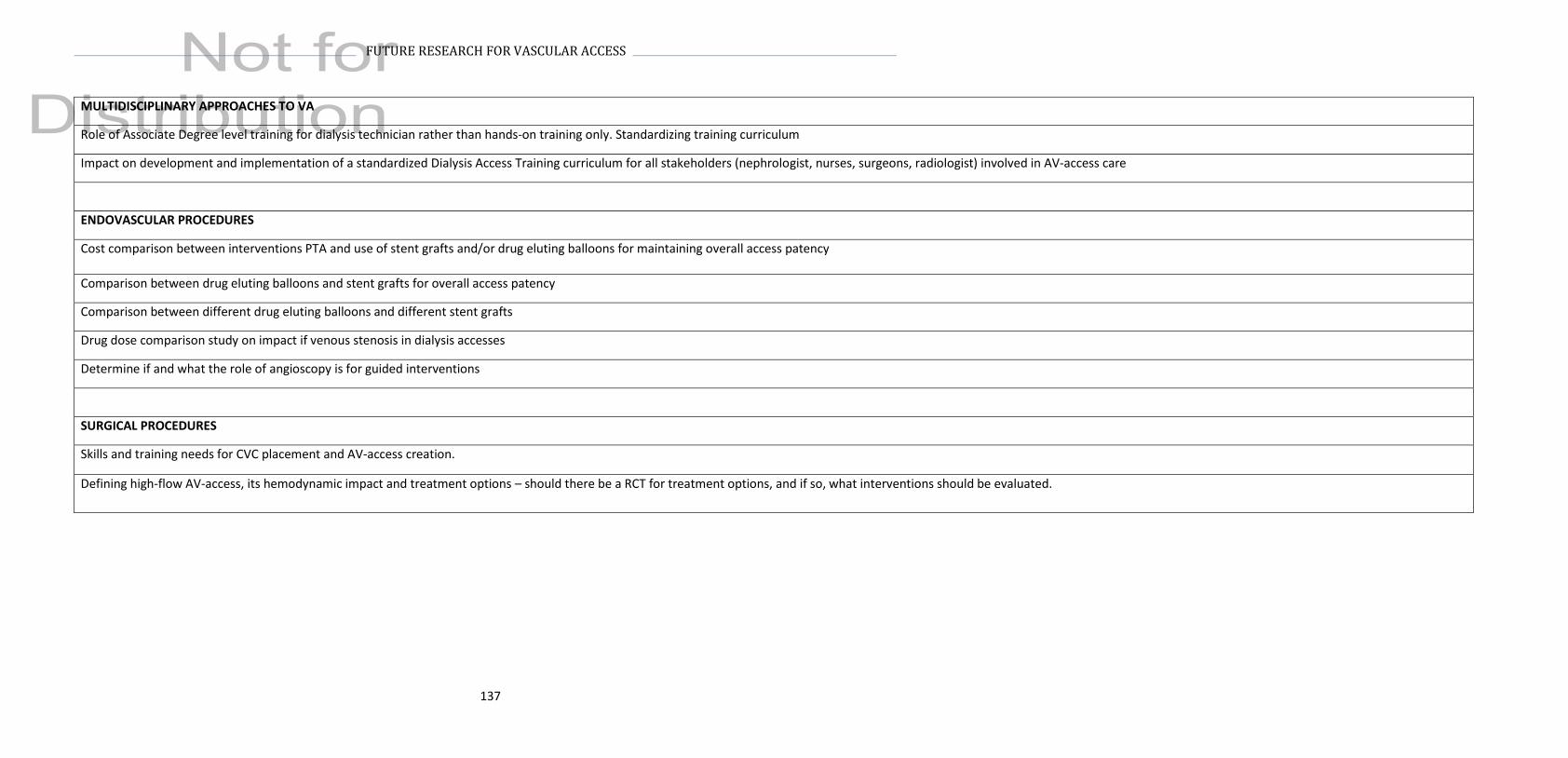
137
FUTURE RESEARCH FOR VASCULAR ACCESS
MULTIDISCIPLINARY APPROACHES TO VA
Role of Associate Degree level training for dialysis technician rather than hands-on training only. Standardizing training curriculum
Impact on development and implementation of a standardized Dialysis Access Training curriculum for all stakeholders (nephrologist, nurses, surgeons, radiologist) involved in AV-access care
ENDOVASCULAR PROCEDURES
Cost comparison between interventions PTA and use of stent grafts and/or drug eluting balloons for maintaining overall access patency
Comparison between drug eluting balloons and stent grafts for overall access patency
Comparison between different drug eluting balloons and different stent grafts
Drug dose comparison study on impact if venous stenosis in dialysis accesses
Determine if and what the role of angioscopy is for guided interventions
SURGICAL PROCEDURES
Skills and training needs for CVC placement and AV-access creation.
Defining high-flow AV-access, its hemodynamic impact and treatment options – should there be a RCT for treatment options, and if so, what interventions should be evaluated.
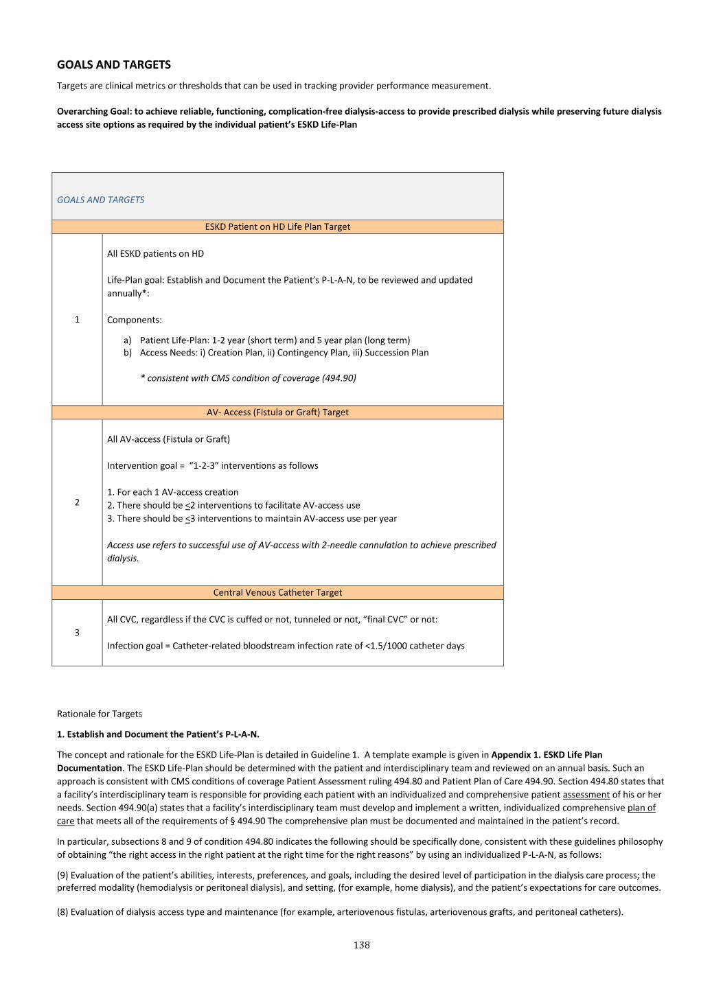
138
GOALS AND TARGETS
Targets are clinical metrics or thresholds that can be used in tracking provider performance measurement.
Overarching Goal: to achieve reliable, functioning, complication-free dialysis-access to provide prescribed dialysis while preserving future dialysis
access site options as required by the individual patient’s ESKD Life-Plan
GOALS AND TARGETS
ESKD Patient on HD Life Plan Target
1
All ESKD patients on HD
Life-Plan goal: Establish and Document the Patient’s P-L-A-N, to be reviewed and updated
annually*:
Components:
a) Patient Life-Plan: 1-2 year (short term) and 5 year plan (long term) b) Access Needs: i) Creation Plan, ii) Contingency Plan, iii) Succession Plan
* consistent with CMS condition of coverage (494.90)
AV- Access (Fistula or Graft) Target
2
All AV-access (Fistula or Graft)
Intervention goal = “1-2-3” interventions as follows
1. For each 1 AV-access creation
2. There should be <2 interventions to facilitate AV-access use
3. There should be <3 interventions to maintain AV-access use per year
Access use refers to successful use of AV-access with 2-needle cannulation to achieve prescribed
dialysis.
Central Venous Catheter Target
3
All CVC, regardless if the CVC is cuffed or not, tunneled or not, “final CVC” or not:
Infection goal = Catheter-related bloodstream infection rate of <1.5/1000 catheter days
Rationale for Targets
1. Establish and Document the Patient’s P-L-A-N.
The concept and rationale for the ESKD Life-Plan is detailed in Guideline 1. A template example is given in Appendix 1. ESKD Life Plan
Documentation. The ESKD Life-Plan should be determined with the patient and interdisciplinary team and reviewed on an annual basis. Such an
approach is consistent with CMS conditions of coverage Patient Assessment ruling 494.80 and Patient Plan of Care 494.90. Section 494.80 states that
a facility’s interdisciplinary team is responsible for providing each patient with an individualized and comprehensive patient assessment of his or her
needs. Section 494.90(a) states that a facility’s interdisciplinary team must develop and implement a written, individualized comprehensive plan of
care that meets all of the requirements of § 494.90 The comprehensive plan must be documented and maintained in the patient’s record.
In particular, subsections 8 and 9 of condition 494.80 indicates the following should be specifically done, consistent with these guidelines philosophy
of obtaining “the right access in the right patient at the right time for the right reasons” by using an individualized P-L-A-N, as follows:
(9) Evaluation of the patient’s abilities, interests, preferences, and goals, including the desired level of participation in the dialysis care process; the preferred modality (hemodialysis or peritoneal dialysis), and setting, (for example, home dialysis), and the patient’s expectations for care outcomes. (8) Evaluation of dialysis access type and maintenance (for example, arteriovenous fistulas, arteriovenous grafts, and peritoneal catheters).

139
Furthermore, subsection 5 of condition 494.80 indicates: (5) Vascular access. The interdisciplinary team must provide vascular access monitoring and appropriate, timely referrals to achieve and sustain
vascular access. The hemodialysis patient must be evaluated for the appropriate vascular access type, taking into consideration co-morbid
conditions, other risk factors, and whether the patient is a potential candidate for AVF placement. The patient’s vascular access must be monitored
to prevent access failure, including monitoring of AVG and AVF for symptoms of stenosis.
In terms of implementing the ESKD Life-Plan, the conditions of coverage stipulate: Standard: Implementation of the patient plan of care.
(1) The patient’s plan of care must—
(i) Be completed by the interdisciplinary team, including the patient if the patient desires; and
(ii) Be signed by team members, including the patient or the patient’s designee; or, if the patient chooses not to sign the plan of care, this choice
must be documented on the plan of care, along with the reason the signature was not provided.
The above specifications in the CMS conditions of coverage are in alignment with the current KDOQI vascular access guidelines 2018.
2. Intervention goal for AV-Access
AV-access interventions are often necessary to facilitate an AV-access in order for it to be usable for dialysis and/or to maintain its patency and use.
However, unnecessary interventions can lead to reductions in patient quality (e.g. discomfort/pain, inconvenience) and increased costs to the
healthcare system. Currently, there is no guidance regarding intervention thresholds over which a patient and care team should consider alternate
vascular access options, but should be included as part of the vascular access contingency plan (i.e. how many interventions should a patient tolerate
before abandoning the current AV-access). The only exception to limiting repeated interventions beyond the suggested thresholds is if the AV-access
is the patient’s “destination” vascular access i.e. no other AV-access options are feasible, and heroic attempts may be required to continue its
prolonged use.
The thresholds for interventions to facilitate and maintain its use are based on available data from the current literature (Table GT1. Interventions).
Furthermore, greater interventions to either facilitate or maintain an AVF have not demonstrated superior survival compared with AV-accesses with
fewer interventions.
3. Catheter related bloodstream infection goal for Central Venous Catheters
The two most significant complications of CVC use are CVC dysfunction (Guidelines 22, 23) and CVC related infection, especially catheter-related
bloodstream infections (CRBSI) (Guidelines 24-26). While CVC dysfunction may be frequently inconveniencing to the patient due to necessary
medical or interventional treatment, it is infrequently life-threatening. On the other hand, CRBSI pose significant risks to patient morbidity and
mortality. While the KDOQI 2018’s “P-L-A-N” permits an individualized approach to vascular access choice and use of CVC under specific
circumstances, it is critical that should a CVC be the appropriate vascular access for a patient, that processes are followed to limit the risk of catheter
related infections, particularly CRBSI. (Guidelines 11, 21, 24, 25). Evidence clearly demonstrates that prophylactic measures can significantly reduce
infection rates, and national data demonstrates that a target CRBSI rate of 1.5 or fewer infections/1000 catheter days is possible.
Such data is derived from a national database, CROWNWEB that is reported to the National Healthcare Safety Network (NHSN). Each dialysis facility tracks hemodialysis related bloodstream infections that is submitted on a monthly basis and reviewed by the NHSN and Centers for Disease Control and Prevention (CDC is the measure steward) and reported via the National Quality Forum (NQF). The most updated report (NQF #1460) on bloodstream infections (BSI) in hemodialysis units reported “in facilities all using a uniform method of measuring and reporting BSI has facility-specific rates that ranges 0-30.8 BSI per 100-patient years”, which is equivalent to 0.84/1000 CVC days. The pooled mean BSI for CVC among facilities reporting was 4.2 per 100 patient months, equivalent to 1.38/1000 CVC days. Thus a targeted rate of <1.5/1000 CVC days is reasonable. Prophylactic measures have been demonstrated to have risk reductions for CRBSI of 22-60% (see Guideline 25)643-645, and should be used to achieve this target.
Comparison to prior KDOQI 2006 Targets
Due to changes in patient demographics and practice patterns, many of the prior KDOQI 2006 targets are no longer clinically relevant and require re-
evaluation or the targets have been achieved or have become standard of care (e.g. establishing a CQI process). Over the past decade, we have
gained great insight due to clinical experiences and research. For example, we now know that targeting thrombosis for fistulas does not result in
improvement in access survival/longevity and that intervention may be counterproductive. Understanding the role of patient, vessel and surgical
factors for superior access survival and assessing the patient for eligibility for different access types and what is “right” for that individual patient is a
priority in these guidelines.
Overarching Goal is to provide functional, complication-free dialysis-access while preserving future dialysis access site options as required by the
individual patient’s ESKD Life-Plan. Hence, the current targets differ from the prior guidelines in focusing on the major components that might
interfere with achieving this overarching goal, and by doing so also encompass many of the prior guideline targets.
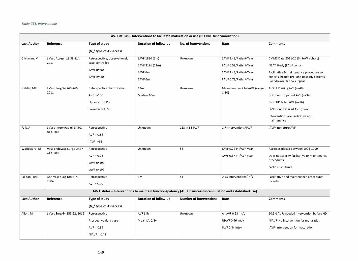
140
Table GT1. Interventions
AV- Fistulas – Interventions to facilitate maturation or use (BEFORE first cannulation)
Last Author Reference Type of study
(N)/ type of AV-access
Duration of follow-up No. of interventions Rate Comments
Glickman, M J Vasc Access, 18:S8-S14,
2017
Retrospective, observational,
case-controlled
SAVF n= 60
EAVF n= 60
SAVF 183d (6m)
EAVF 319d (11m)
SAVF 6m
EAVF 6m
Unknown SAVF 3.43/Patient-Year
EAVF 0.59/Patient-Year
SAVF 3.43/Patient-Year
EAVF 0.78/Patient-Year
CMMS Data 2011-2013 (SAVF cohort)
NEAT Study (EAVF cohort)
Facilitative & maintenance procedure so
cohorts include pre- and post-HD patients.
E=endovascular; S=surgical
Nehler, MR J Vasc Surg 54:760-766,
2011
Retrospective chart review
AVF n=150
Upper arm 54%
Lower arm 46%
12m
Median 10m
Unknown Mean number 2 Int/AVF (range,
1-10)
A-On HD using AVF (n=48)
B-Not on HD patent AVF (n=34)
C-On HD failed AVF (n=26)
D-Not on HD failed AVF (n=42)
Interventions are facilitative and
maintenance
Falk, A J Vasc Interv Radiol 17:807-
813, 2006
Retrospective
AVF n=154
IAVF n=65
Unknown 113 in 65 IAVF 1.7 interventions/IAVF IAVF=immature AVF
Woodward, RS Vasc Endovasc Surg 39:437-
443, 2005
Retrospective
AVF n=398
cAVF n=199
sAVF n=199
Unknown 53 cAVF 0.22 Int/AVF-year
sAVF 0.37 Int/AVF-year
Accesses placed between 1996-1999
Does not specify facilitative or maintenance
procedures
c=clips; s=sutures
Fujitani, RM Ann Vasc Surg 18:66-73,
2004
Retrospective
AVF n=100
3 y 51 0.53 interventions/Pt/Y Facilitative and maintenance procedures
included
AV- Fistulas – Interventions to maintain function/patency (AFTER successful cannulation and established use)
Last Author Reference Type of study
(N)/ type of AV-access
Duration of follow-up Number of interventions Rate Comments
Allon, M J Vasc Surg 64:155-62, 2016 Retrospective
Prospective data base
AVF n=289
NIAVF n=143
AVF 6.5y
Mean f/u 2.3y
Unknown All AVF 0.63 Int/y
NIAVF 0.46 Int/y
IAVF 0.84 Int/y
50.5% AVFs needed intervention before HD
NIAVF=No intervention for maturation
IAVF=Intervention for maturation

141
IAVF n=146
Roy-Chauduhry,
P
Clin J Am Soc Nephrol
6:575-581, 2011
Retrospective
Prospective data bases
AVF n=173
Median 672d Unknown 0IAVF(96) 0.76 Int/y
1IAVF(54) 1.37 Int/y
2IAVF(23) 3.51 Int/y
Data base UC & UAB 2005-2007
f/u from time of cannulation so only
maintenance procedures. Of 173 pt 77
needed facilitative interventions
Falk, A J Vasc Interv Radiol 17:807-
813, 2006
Retrospective
AVF n=154
MAVF n=63
Mean 317d 209 3.3 Int/AVF
1.75 Int/AVF year
MAVF=mature AVF
Murphy, B J Am Soc Nephrol 16: 201-
209, 2005
Prospective
AVF n=157
12 months Unknown AVF surgery 1.39/PY
AVF angiogram 0.81/PY
AVF angioplasty 0.43/PY
Data from Southern Alberta
Transplant Program Database (ALTRAbase)
PY=patient year
AV- Grafts – Interventions to facilitate maturation or use (BEFORE first cannulation)
Last Author Reference Type of study
(N)/ type of AV-access
Duration of follow-up Number of interventions Rate Comments
Woodward, RS Vasc Endovasc Surg
39:437-443, 2005 Retrospective
AVG n=745
cAVG n=401
sAVG n=344
635 cAVG 0.86/AVG-year
sAVG 1.73/AVG-year
Accesses placed between 1998-1999
Does not specify facilitative or maintenance
C=clips; s=sutures
AV- Grafts – Interventions to maintain function/patency (AFTER successful cannulation and established use)
Allon, M J Vasc Surg 64:155-62,
2016
Retrospective
Prospective data base
AVG n=310
AVG 6.5y
Mean f/u 2.01y
Unknown All AVG 1.58 Int/y
No Int AVG 1.48 Int/y
Int AVG 2.2 Int/y
17.7% AVGs needed intervention before HD
Murphy, B J Am Soc Nephrol 16: 201-
209, 2005 Prospective
AVG n=33
12 months Unknown AVG surgery 1.70/PY
AVG angiogram 1.33/PY
AVG angioplasty 0.94/PY
Data from Southern Alberta
Transplant Program Database (ALTRAbase)
PY=patient year
Fujitani, RM Ann Vasc Surg 18:66-73,
2004
Retrospective
AVG n=131
3y 170 0.92 Interventions/Pt/Y PY=patient year; Facilitative and
maintenance procedures included

142
Biographical and Disclosure Information
Guideline Development Work Group
Ken Abreo, MD, FASDIN
Dr. Kenneth Abreo is Professor of Medicine, Chief of the Division of Nephrology and Vice Chairman of the Department of Medicine at LSU Health
Shreveport School of Medicine. After completing his Nephrology Fellowship training at the University of California-Davis in Sacramento and the
University of Wisconsin in Madison he joined the Medicine faculty at the University of Kentucky in Lexington from 1982 to 1985 and Louisiana State
University Health Sciences Center in Shreveport in 1985. His early research interests were focused on clinical and bench research on aluminum
toxicity causing bone disease and anemia in renal failure patients. In the past decade he has focused on training Nephrology Fellows in
Interventional Nephrology. His current research interests are centered on improving vascular and peritoneal access in dialysis patients. He has
played an active role in the American Society of Diagnostic and Interventional Nephrology, most recently as President. He serves on the Editorial
Board of the Journal of Vascular Access. He has been involved in national and international scientific and educational programs with ASDIN, ASN,
NKF, ISN, ANZIN, ERA-EDTA, AVATAR and VAS.
Michael Allon, MD
Dr. Allon is Professor of Medicine at University of Alabama at Birmingham, where he serves as the Associate Director for Clinical Affairs and the
Medical Director of Dialysis Operations in the Division of Nephrology. He has a longstanding clinical research interest in dialysis, and in particular in
vascular access for hemodialysis. He was the P.I. for several clinical dialysis trials sponsored by NIH, including HEMO, DAC, and HFM. He has
published over 100 peer-reviewed manuscripts on various aspects of vascular access. Finally, he has been an invited speaker at numerous
nephrology meetings on various topics related to vascular access.
Arif Asif MD, MHCM
Dr. Arif Asif is the Chairman of the Department of Medicine at Jersey Shore Medical Center and Professor of Medicine at Seton Hall-Hackensack
Meridian School of Health, Neptune, New Jersey. Dr. Asif completed his residency in Internal Medicine at Mercy Hospital and Medical Center,
University of Illinois at Chicago and Fellowship in Nephrology and Hypertension at the University of Miami, Miami; Florida. He also received a
Masters of Health Care Management and business administration from Harvard University, Boston; Massachusetts. He is a nationally and
internationally recognized expert in hemodialysis vascular access and has organized and chaired multiple scientific meetings on hemodialysis
access. Dr. Asif has been involved in vascular access research since 2000. He has had numerous funded research projects, received many teaching
awards and developed multiple training programs in the area of dialysis vascular access. He has edited four books and authored over 200 articles
and book chapters. Among his many professional editorial responsibilities, he is the section editor of the Journal of Vascular Access and the
Associate Editor of the American Journal of Kidney Diseases. He is the Past President of the American Society of Diagnostic and Interventional
Nephrology Society and currently serves as the Chair of its Clinical Practice Committee.
Brad Astor, PhD
Dr. Brad Astor is Associate Professor in the Departments of Medicine and Population Health Sciences at the University of Wisconsin School of
Medicine and Public Health. He serves as the Director of Research for the Division of Nephrology. Dr. Astor was a medical device reviewer at the
United States Food and Drug Administration prior to training as an epidemiologist at The Johns Hopkins Bloomberg School of Public Health. He was
a faculty member in the Departments of Epidemiology and Medicine at Johns Hopkins until 2011 when he joined the University of Wisconsin. His
research covers several different aspects of kidney disease and its interaction with cardiovascular disease. Other specific areas of interest include
predictors of outcomes in kidney transplant recipients and hemodialysis vascular access complications. This research has been pursued in the
general population (Atherosclerosis Risk in Communities [ARIC] Study, Multi-Ethnic Study of Atherosclerosis [MESA]) as well as in clinical trials
(African American Study of Kidney Disease and Hypertension [AASK]) and observational studies of dialysis patients (Choices for Healthy Outcomes in
Caring for ESKD [CHOICE]) and kidney transplant recipients (Wisconsin Allograft Recipient Database [WisARD]).
Marc Glickman, MD
Marc Glickman, MD is a retired vascular surgeon who has written over 100 articles on Vascular Access surgery. He has written several chapters in
books on vascular access surgery as well. He was previous President of the Vascular Access Society of the Americas, part of the clinical faculty of
the Eastern Virginia Medical School. He has been PI for many vascular access graft studies that are currently in use today. He has lectured
extensively nationally and internationally on Vascular Access surgery.
Janet Graham, MHScN, CNeph(C)
Ms. Graham’s career has focused her career on the care of patients with chronic kidney disease over almost three decades. Currently Janet is the
Clinical Director of Nephrology at The Ottawa Hospital (TOH) and Regional Director of Nephrology Champlain LHIN Ontario Renal Network (ORN),
and has recently completed a secondment as Clinical Education and Quality Improvement at the ORN leading the development and implementation
of both clinical education and QI initiatives at a Provincial level. Janet’s career has spanned the entire care continuum in renal care holding a

143
number of leadership positions at TOH. She holds a Masters of Health Science degree in Nursing and has conducted clinical research in the area of
dialysis access having several publications in this area.
Thomas S. Huber, MD, PhD
Dr. Thomas S. Huber is Professor and Chief of the Division of Vascular Surgery at the University of Florida College of Medicine. He has a long-
standing interest in hemodialysis access and was one of the Principal Investigators for the NIH/NIDDK Hemodialysis Fistula Maturation Study. He
has served on the editorial boards of the Journal of Vascular Surgery and the Yearbook of Surgery while co-editing Mastery of Vascular and
Endovascular Surgery. He currently serves on the Vascular Surgery Board of the American Board of Surgery.
Timmy Lee, MD
Dr. Timmy Lee is Professor of Medicine in the Department of Medicine and Division of Nephrology with a secondary appointment in the
Department of Biomedical Engineering at the University of Alabama at Birmingham. Dr. Lee received his medical degree at the Louisiana State
University Health Sciences Center in Shreveport and completed his Internal medicine residency and Nephrology fellowship at the University of
Alabama at Birmingham, Nephrology research fellowship at the University of Alabama at Birmingham, and Masters of Science in Public Health at
the University of Alabama at Birmingham. Dr. Lee currently serves as the Associate Director of Interventional Nephrology, Associate Nephrology
Fellowship Director, Associate Director of the Nephrology Research Training Center, Associate Section Chief and Director of the Hemodialysis
Program at the Birmingham Veterans Affairs Medical Center. Dr. Lee has been involved in hemodialysis vascular access research since 2002 and has
over 50 publications in vascular access research. He has active research programs in clinical trials and large epidemiologic studies in dialysis
vascular access and a laboratory-based translational research program in dialysis vascular access studying human vascular access blood vessel tissue
biorepositories and mechanisms of arteriovenous fistula dysfunction using animal models (mouse, rat, and pig). The goal of his research program is
to develop novel therapeutics to prevent and treat complications of hemodialysis vascular access dysfunction and improve the delivery and
processes of care for patients requiring a hemodialysis vascular access. Dr. Lee’s research program is currently funded by the National Institutes of
Health and Veterans Affairs Medical Center. Dr. Lee serves a counselor in the American Society of Diagnostic and Interventional Nephrology.
Charmaine Lok, MD, MSc (Chair)
Dr. Charmaine Lok is a Professor of Medicine, Faculty of Medicine, at the University of Toronto and Senior Scientist at the Toronto General Hospital
Research Institute. She is also associated with the Department of Health Research Methods, Evidence, and Impact (HEI), Faculty of Health Sciences,
McMaster University. Dr. Lok is the Medical Director of both the chronic kidney diseases and hemodialysis programs at the University Health
Network - Toronto General Hospital, Toronto, Canada. Her research focus is on chronic kidney disease (CKD) and end-stage kidney disease (ESKD)
outcomes. She has a special interest in improving dialysis access outcomes and reducing adverse cardiovascular events in CKD/ESKD. She is active in
raising awareness of CKD and ESKD and its importance in population health. Dr. Lok is involved in a variety of local and international scientific and
educational programs including CIHR, KFOC, DOPPS, NKF, ASN, VASA, ASDIN and Kidney CARE Network International.
Louise Moist, MD
Dr. Moist is a nephrologist at Victoria Hospital, Ontario, Canada; and Professor of Medicine, Epidemiology and Biostatistics, Associate Chair of the
Division of Nephrology, and Scientist in the Program of Experimental Medicine at Western University. Her clinical and research interests are focused
on chronic kidney disease with a specific expertise in the vascular access for hemodialysis patient. Dr. Moist has a number of leadership roles
including The Ontario Renal Network Physician Lead for Dialysis Access, integrating change to improve vascular access practice and reduce catheter
use. Dr Moist has chaired and been involved in several guideline groups including CSN anemia guidelines, timing of dialysis start and vascular
access. Dr. Moist is active in education, clinical and research with over 150 peer reviewed academic papers and numerous research grants. Dr Moist
has received the University of Western Ontario award for Excellence for Teaching and for Excellence in Research.
Dheeraj Rajan, MD
Dr. Rajan completed his Diagnostic Radiology residency at Wayne State University in 1999 and Fellowship in Vascular and
Interventional Radiology at the University of Pennsylvania in 2000. Over his career, he has published 100 manuscripts primarily focused
on arterial and dialysis interventions, multiple book chapters and one textbook titled “Essentials of Percutaneous Dialysis
Interventions”. He is a Professor of Medicine and the Division Chief of VIR at the University of Toronto. Dr. Rajan also sits on various
committees for the Society of Interventional Radiology, Cardiac Care Network of Ontario and the Ontario Renal Network. He formerly
was an Associate Editor for the Journal of Vascular and Interventional Radiology and reviews for multiple medical journals. Dr. Rajan’s
current focus is on the percutaneous creation of dialysis fistulas where he was one of the pioneers for the procedure itself. He also
serves as an advisor to multiple start-up and established companies with devices focused on hemodialysis interventions.
Cindy Roberts, RN, CNN
Ms. Roberts is a nephrology and vascular access nurse caring for patients with chronic kidney and end stage renal disease for 28 years. She works
with Renal Research Institute, partnering with the University of North Carolina at Chapel Hill Nephrology and Hypertension division and UNC
144
Kidney Center. Cynthia specializes in developing patient care models, which emphasizes collaboration of care with dedicated vascular access
surgery and interventional clinic systems and nephrology integrated care management. She has multiple publications in ANNA Nursing Journal and
Nephrology News and Issues where she served as editor of the vascular access quarterly column.
Surendra Shenoy, MD
Dr. Surendra Shenoy is a Professor of Surgery and the Director of the Living Donor Transplantation Program at the Washington University School of
Medicine in Saint Louis, Missouri, USA. His clinical and research interests include liver, kidney and living donor transplantation, liver, and dialysis
access surgery. He serves on the Board of Directors and is the immediate past president of the Vascular Access Society of Americas (VASA). He is on
the Editorial Board of the Journal of Vascular Access and serves as the VASA Editor for the Americas. Dr. Shenoy was the Co-chair for the working
group that was tasked to describe appropriate end-points for dialysis vascular access trials established by the Kidney Health Initiative (KHI) of the
American Society of Nephrology. He has been actively involved in several clinical research projects related to abdominal organ transplantation and
vascular access for hemodialysis. He has published several innovative surgical techniques in the field of transplantation and vascular access.
Tushar Vachharajani, MD
Dr. Tushar Vachharajani is an Adjunct Professor in the Department of Medicine at the University of North Carolina, Chapel Hill and Chief of
Nephrology and Medical Director of the Hemodialysis Program at the Salisbury Veterans Affairs Health Care System, North Carolina. Dr.
Vachharajani is an interventional nephrologist with international eminence who holds several leadership positions with International Society of
Nephrology. He has published over 70 peer reviewed scientific papers, several book chapters and is an Associate Editor for the Journal of Vascular
Access (ASDIN). He served as a Co-Lead on the Access Monitoring Work Group of Fistula First Breakthrough Initiative and as a Chair for the Medical
Review Board of ESKD Network 6. He is passionate about training and improving global awareness of dialysis access care through various national
and international professional organizations including ISN, ASN, NKF, ASDIN, AVATAR, ISHD and Kidney Education Foundation. Besides serving as a
Co-Chair for the Interventional Nephrology Workshop at the World Congress of Nephrology in 2017 and 2019, he has helped organize vascular
access symposiums, given scientific presentations and conducted hands-on workshop in 15 different countries. Dr. Vachharajani has received the
Gerald Beathard Award for Excellence in Teaching and Scholarly Activity from ASDIN.
Rudolph Valentini, MD
Dr. Valentini is a clinical professor of Pediatrics at Wayne State University School of Medicine and the Chief Medical Officer for the Children’s
Hospital of Michigan (CHM). He served as the Director of Dialysis Services at CHM for 10 years before taking on his role as CMO. He has authored
or co-authored a number of articles and book chapters on vascular access in the pediatric hemodialysis patient. This clinical passion has led to a
near elimination of central venous catheters for delivery of chronic hemodialysis at his center. He has been an invited speaker at regional and
national meetings to speak on the topic of the ideal vascular access choice for the pediatric hemodialysis patient. He was the American Society of
Pediatric Nephrology (ASPN) representative and participant on the Center for Medicaid and Medicare Services (CMS) Technical Expert Panel on
Dialysis Vascular Access in 2015. He has also served as the pediatric representative on the Medical Review Committee for the Midwest Kidney
Network (formerly Network 11) and was a long-standing member for the Midwest Pediatric Nephrology Consortium review committee.
Alexander Yevzlin, MD
Dr. Yevzlin graduated magna cum laude from Dartmouth College. He did his residency in Internal Medicine at the University of Michigan and
fellowship in Nephrology at Northwestern. Dr. Yevzlin is currently Professor of Medicine and director of Interventional Nephrology at the University
of Michigan. Dr. Yevzlin has presented and published over 150 abstracts, invited lectures, and manuscripts. He is an internationally
recognized leader in the field of Interventional Nephrology, having edited the first three textbooks on the subject, and is a past President of the
American Society of Diagnostic and Interventional Nephrology. In addition to his academic contributions, Dr. Yevzlin has been involved the
invention, design, and reduction to practice of multiple medical devices in his role as chief medical officer, chief science officer, and founder of
multiple start-up biotech companies.
Evidence Review Team
Michelle Brasure, PhD, MSPH, MLIS is Program Manager and Medical Librarian for the Minnesota Evidence-based Practice Center. She has
extensive experience leading multidisciplinary systematic review teams on a variety of topics. Dr. Brasure reported no relevant financial
relationships.
Nancy Greer, PhD is a senior research associate in the Minneapolis Center for Chronic Disease Outcomes Research and Program Manager for the
Minneapolis VA Evidence-Synthesis Program. She has extensive experience in leading multidisciplinary teams in conducting evidence synthesis
reports across a wide range of topics. Dr. Greer reported no relevant financial relationships.
Areef Ishani, MD, MS, is the Chief of the Primary Care Service Line at the Minneapolis VA Health Care System and a Professor of Medicine at the
University of Minnesota. His primary research interests are in CKD, acute kidney injury and end stage kidney disease. Dr Ishani reported no relevant
financial relationships.
145
Roderick MacDonald, MS is a senior research assistant in the Minneapolis VA Center for Chronic Disease Outcomes Research. He has nearly 20
years of experience in conducting systematic reviews across a wide range of topics. Mr. MacDonald reported no relevant financial relationships.
Victoria Nelson, MSc, is a research fellow in the Minnesota Evidence-based Practice Center. Her primary research interests are research
methodology, evidence synthesis, health psychology, and behavior change. Ms Nelson reported no relevant financial relationships.
Carin Olson, MD, MS, is a systematic reviewer, medical editor and writer, and physician, working with the Minnesota Evidence-based Practice
Center at the University of Minnesota. Her interests include research methodology (especially relating to meta-analysis and publication bias),
research ethics, injury prevention, and cardiopulmonary resuscitation. Dr. Olson reported no relevant financial relationships.
Yelena Slinin, MD, MS, is a nephrologist at Kaiser Permanente in California. Her primary research interests are optimal medical care delivery and
outcomes of patient with kidney disease, evidence-based medicine, and critical literature appraisal. Dr Slinin reported no relevant financial
relationships.
Timothy J. Wilt, MD, MPH, is a Professor of Medicine at the University of Minnesota and Core Investigator in the Center for Chronic Disease
Outcomes Research at the Veterans Affairs Medical Center in Minneapolis, Minnesota. He has a research agenda which involves conducting clinical
trials, systematic reviews and meta-analysis to evaluate the effects of health care interventions on outcomes in adults with chronic diseases. Dr Wilt
reported no relevant financial relationships.
References
References
1. Lee T, Mokrzycki M, Moist L, Maya I, Vazquez M, Lok CE. Standardized Definitions for Hemodialysis Vascular Access. Semin Dial. 2011;24(5):515-524.
2. Lee T, Barker J, Allon M. Needle infiltration of arteriovenous fistulae in hemodialysis: risk factors and consequences. Am J Kidney Dis. 2006;47(6):1020-1026.
3. Beathard GA, Lok CE, Glickman MH, et al. Definitions and End Points for Interventional Studies for Arteriovenous Dialysis Access. Clin J Am Soc Nephrol. 2018;13(3):501-512.
4. Beathard GA, Arnold P, Jackson J, Litchfield T, Physician Operators Forum of RMSL. Aggressive treatment of early fistula failure. Kidney Int. 2003;64(4):1487-1494.
5. KDIGO 2012 Clinical Practice Guideline for the Evaluation and Management of Chronic Kidney Disease: Summary of Recommendation Statements. Kidney International Supplements. 2013;3(1):5-14.
6. United States Renal Data System. 2018 USRDS annual data report: Epidemiology of kidney disease in the United States. Bethesda, MD: National Institutes of Health, National Institute of Diabetes and Digestive and Kidney Diseases;2018.
7. Alonso-Coello P, Oxman AD, Moberg J, et al. GRADE Evidence to Decision (EtD) frameworks: a systematic and transparent approach to making well informed healthcare choices. 2: Clinical practice guidelines. BMJ. 2016;353:i2089.
8. Alonso-Coello P, Schunemann HJ, Moberg J, et al. GRADE Evidence to Decision (EtD) frameworks: a systematic and transparent approach to making well informed healthcare choices. 1: Introduction. BMJ. 2016;353:i2016.
9. Viswanathan M, Ansari MT, Berkman ND, et al. AHRQ Methods for Effective Health Care Assessing the Risk of Bias of Individual Studies in Systematic Reviews of Health Care Interventions. Methods Guide for Effectiveness and
Comparative Effectiveness Reviews. Rockville (MD): Agency for Healthcare Research and Quality (US); 2008. 10. Guyatt G, Oxman AD, Akl EA, et al. GRADE guidelines: 1. Introduction-GRADE evidence profiles and summary of findings tables.
Journal of clinical epidemiology. 2011;64(4):383-394. 11. KDOQI Clinical Practice Guidelines for Vascular Access. American Journal of Kidney Diseases. 2006;48:S176-S247. 12. Uhlig K, Macleod A, Craig J, et al. Grading evidence and recommendations for clinical practice guidelines in nephrology. A position
statement from Kidney Disease: Improving Global Outcomes (KDIGO). Kidney Int. 2006;70(12):2058-2065. 13. Lok CE, Davidson I. Optimal choice of dialysis access for chronic kidney disease patients: developing a life plan for dialysis access.
Seminars in nephrology. 2012;32(6):530-537. 14. Woo KaL, CE. New Insights into Dialysis Vascular Access: What is the Optimal Vascular Access Type and Timing of Access Creation in
CKD and Dialysis Patients? CJASN. 2016;In press. 15. First F. Fistula First. 2012. Accessed May 4 2012. 16. Woo K, Goldman DP, Romley JA. Early Failure of Dialysis Access among the Elderly in the Era of Fistula First. Clin J Am Soc Nephrol.
2015;10(10):1791-1798.
146
17. Brown RS, Patibandla BK, Goldfarb-Rumyantzev AS. The Survival Benefit of "Fistula First, Catheter Last" in Hemodialysis Is Primarily Due to Patient Factors. Journal of the American Society of Nephrology : JASN. 2017;28(2):645-652.
18. Gomes A, Schmidt R, Wish J. Re-envisioning Fistula First in a Patient-Centered Culture. Clin J Am Soc Nephrol. 2013. 19. Disbrow DE, Cull DL, Carsten CG, 3rd, Yang SK, Johnson BL, Keahey GP. Comparison of arteriovenous fistulas and arteriovenous grafts
in patients with favorable vascular anatomy and equivalent access to health care: is a reappraisal of the Fistula First Initiative indicated? J Am Coll Surg. 2013;216(4):679-685; discussion 685-676.
20. Schinstock CA, Albright RC, Williams AW, et al. Outcomes of arteriovenous fistula creation after the Fistula First Initiative. Clin J Am Soc Nephrol. 2011;6(8):1996-2002.
21. DeSilva RN, Patibandla BK, Vin Y, et al. Fistula first is not always the best strategy for the elderly. Journal of the American Society of Nephrology : JASN. 2013;24(8):1297-1304.
22. Woo K, Lok CE. New Insights into Dialysis Vascular Access: What Is the Optimal Vascular Access Type and Timing of Access Creation in CKD and Dialysis Patients? Clin J Am Soc Nephrol. 2016;11(8):1487-1494.
23. Kalloo S, Blake PG, Wish J. A Patient-Centered Approach to Hemodialysis Vascular Access in the Era of Fistula First. Semin Dial. 2016. 24. Vachharajani TJ, Moossavi S, Jordan JR, Vachharajani V, Freedman BI, Burkart JM. Re-evaluating the Fistula First Initiative in
Octogenarians on Hemodialysis. Clin J Am Soc Nephrol. 2011;6(7):1663-1667. 25. Drew DA, Lok CE. Strategies for planning the optimal dialysis access for an individual patient. Current opinion in nephrology and
hypertension. 2014;23(3):314-320. 26. Allon M, Lockhart ME, Lilly RZ, et al. Effect of preoperative sonographic mapping on vascular access outcomes in hemodialysis
patients. Kidney Int. 2001;60(5):2013-2020. 27. Schwab SJ. Vascular access for hemodialysis. Kidney Int. 1999;55:2078-2090. 28. Lok CE, Sontrop JM, Tomlinson G, et al. Cumulative patency of contemporary fistulas versus grafts (2000-2010). Clin J Am Soc Nephrol.
2013;8(5):810-818. 29. Dember LM, Beck GJ, Allon M, et al. Effect of clopidogrel on early failure of arteriovenous fistulas for hemodialysis: a randomized
controlled trial. Jama. 2008;299(18):2164-2171. 30. Lacson E, Jr., Lazarus JM, Himmelfarb J, Ikizler TA, Hakim RM. Balancing Fistula First with Catheters Last. Am J Kidney Dis.
2007;50(3):379-395. 31. Lok CE. Fistula first initiative: advantages and pitfalls. Clin J Am Soc Nephrol. 2007;2(5):1043-1053. 32. Ravani P, Palmer SC, Oliver MJ, et al. Associations between hemodialysis access type and clinical outcomes: a systematic review.
Journal of the American Society of Nephrology : JASN. 2013;24(3):465-473. 33. Quinn RR, Oliver MJ, Devoe D, et al. The Effect of Predialysis Fistula Attempt on Risk of All-Cause and Access-Related Death. Journal of
the American Society of Nephrology : JASN. 2017;28(2):613-620. 34. Quinn RR, Ravani P. Fistula-first and catheter-last: fading certainties and growing doubts. Nephrol Dial Transplant. 2014;29(4):727-
730. 35. Alhassan SU, Adamu B, Abdu A, Aji SA. Outcome and complications of permanent hemodialysis vascular access in Nigerians: A single
centre experience. Annals of African Medicine. 2013;12(2):127. 36. Arhuidese IJ, Obeid T, Hicks C, et al. Vascular access modifies the protective effect of obesity on survival in hemodialysis patients.
Surgery. 2015;158(6):1628-1634. 37. Bray BD, Boyd J, Daly C, et al. Vascular access type and risk of mortality in a national prospective cohort of haemodialysis patients.
Qjm. 2012;105(11):1097-1103. 38. Charlton-Ouw KM, Nosrati N, Miller CC, 3rd, Coogan SM, Safi HJ, Azizzadeh A. Outcomes of arteriovenous fistulae compared with
heparin-bonded and conventional grafts for hemodialysis access. Journal of Vascular Access. 2012;13(2):163-167. 39. Clark WF, Na Y, Rosansky SJ, et al. Association between estimated glomerular filtration rate at initiation of dialysis and mortality.
CMAJ Canadian Medical Association Journal. 2011;183(1):47-53. 40. DeSilva RN, Sandhu GS, Garg J, Goldfarb-Rumyantzev AS. Association between initial type of hemodialysis access used in the elderly
and mortality. Hemodialysis International. 2012;16(2):233-241. 41. Di Iorio BR, Bellizzi V, Cillo N, et al. Vascular access for hemodialysis: the impact on morbidity and mortality. Jn, J. 2004;17(1):19-25. 42. Foley RN, Chen SC, Collins AJ. Hemodialysis access at initiation in the United States, 2005 to 2007: still "catheter first". Hemodialysis
international International Symposium on Home Hemodialysis. 2009;13(4):533-542. 43. Gonzalez E, Kashuk JL, Moore EE, Linas S, Sauaia A. Two-stage brachial-basilic transposition fistula provides superior patency rates
for dialysis access in a safety-net population. Surgery. 2010;148(4):687-693; discussion 693-684. 44. Gruss E, Portoles J, Tato A, et al. Clinical and economic repercussions of the use of tunneled haemodialysis catheters in a health area.
Nefrologia. 2009;29(2):123-129. 45. Jorna T, Methven S, Ravanan R, Weale AR, Mouton R. 30-day mortality after haemodialysis vascular access surgery: a retrospective
observational study. Journal of Vascular Access. 2016;17(3):215-219. 46. Kasza J, Wolfe R, McDonald SP, Marshall MR, Polkinghorne KR. Dialysis modality, vascular access and mortality in end-stage kidney
disease: A bi-national registry-based cohort study. Nephrology. 2016;21(10):878-886. 47. Krzanowski M, Janda K, Chowaniec E, Sulowicz W. Hemodialysis vascular access infection and mortality in maintenance hemodialysis
patients. Przegl Lek. 2011;68(12):1157-1161. 48. Lacson E, Jr., Wang W, Hakim RM, Teng M, Lazarus JM. Associates of mortality and hospitalization in hemodialysis: potentially
actionable laboratory variables and vascular access. American Journal of Kidney Diseases. 2009;53(1):79-90. 49. Lacson E, Jr., Wang W, Lazarus JM, Hakim RM. Change in vascular access and mortality in maintenance hemodialysis patients. Am J
Kidney Dis. 2009;54(5):912-921. 50. Lacson E, Jr., Wang W, Lazarus JM, Hakim RM. Change in vascular access and hospitalization risk in long-term hemodialysis patients.
Clinical Journal of The American Society of Nephrology: CJASN. 2010;5(11):1996-2003. 51. Leake AE, Yuo TH, Wu T, et al. Arteriovenous grafts are associated with earlier catheter removal and fewer catheter days in the
United States Renal Data System population. Journal of Vascular Surgery. 2015;62(1):123-127. 52. Lioupis C, Mistry H, Rix T, Chandak P, Tyrrell M, Valenti D. Comparison among transposed brachiobasilic, brachiobrachial
arteriovenous fistulas and FlixeneTM vascular graft. Journal of Vascular Access. 2011;12(1):36-44. 53. Malas MB, Canner JK, Hicks CW, et al. Trends in incident hemodialysis access and mortality. JAMA Surg. 2015;150(5):441-448. 54. Moist LM, Trpeski L, Na Y, Lok CE. Increased hemodialysis catheter use in Canada and associated mortality risk: data from the
Canadian Organ Replacement Registry 2001-2004. Clinical journal of the American Society of Nephrology : CJASN. 2008;3(6):1726-1732.
55. Ng YY, Hung YN, Wu SC, Ko PJ. Characteristics and 3-year mortality and infection rates among incident hemodialysis patients with a permanent catheter undergoing a first vascular access conversion. Clin Exp Nephrol. 2014;18(2):329-338.
147
56. Ong S, Barker-Finkel J, Allon M. Long-term outcomes of arteriovenous thigh grafts in hemodialysis patients: A comparison with tunneled dialysis catheters. Clinical Journal of the American Society of Nephrology. 2013;8(5):804-809.
57. Park HS, Kim WJ, Kim YK, et al. Comparison of Outcomes with Arteriovenous Fistula and Arteriovenous Graft for Vascular Access in Hemodialysis: A Prospective Cohort Study. American journal of nephrology. 2016;43(2):120-128.
58. Portoles J, Lopez-Gomez JM, Gruss E, Aljama P, Group MARS. Course of vascular access and relationship with treatment of anemia. Clinical Journal of The American Society of Nephrology: CJASN. 2007;2(6):1163-1169.
59. Praga M, Merello JI, Palomares I, et al. Type of vascular access and survival among very elderly hemodialysis patients. Nephron. 2013;124(1-2):47-53.
60. Wasse H, Speckman RA, McClellan WM. Arteriovenous fistula use is associated with lower cardiovascular mortality compared with catheter use among ESRD patients. Seminars in Dialysis. 2008;21(5):483-489.
61. Woo K, Yao J, Selevan D, Hye RJ. Influence of vascular access type on sex and ethnicity-related mortality in hemodialysis-dependent patients. Perm. 2012;16(2):4-9.
62. Wystrychowski G, Kitzler TM, Thijssen S, Usvyat L, Kotanko P, Levin NW. Impact of switch of vascular access type on key clinical and laboratory parameters in chronic haemodialysis patients. Nephrology Dialysis Transplantation. 2009;24(7):2194-2200.
63. Xue H, Ix JH, Wang W, et al. Hemodialysis access usage patterns in the incident dialysis year and associated catheter-related complications. Am J Kidney Dis. 2013;61(1):123-130.
64. Zhang JC, Al-Jaishi AA, Na Y, de Sa E, Moist LM. Association between vascular access type and patient mortality among elderly patients on hemodialysis in Canada. Hemodialysis International. 2014;18(3):616-624.
65. Ravani P, Quinn R, Oliver M, et al. Examining the Association between Hemodialysis Access Type and Mortality: The Role of Access Complications. Clin J Am Soc Nephrol. 2017;12(6):955-964.
66. Grubbs V, Wasse H, Vittinghoff E, Grimes BA, Johansen KL. Health status as a potential mediator of the association between hemodialysis vascular access and mortality. Nephrol Dial Transplant. 2014;29(4):892-898.
67. Quinn R, Ravani P. ACCESS HD pilot: A randomised feasibility trial Comparing Catheters with fistulas in Elderly patientS Starting haemodialysis. BMJ open. 2016;6(11):e013081.
68. Schwab SJ, Harrington JT, Singh A, et al. Vascular access for hemodialysis. Kidney Int. 1999;55(5):2078-2090. 69. Lee T, Barker J, Allon M. Comparison of survival of upper arm arteriovenous fistulas and grafts after failed forearm fistula. J Am Soc
Nephrol. 2007;18(6):1936-1941. 70. Snyder DC, Clericuzio CP, Stringer A, May W. Comparison of outcomes of arteriovenous grafts and fistulas at a single Veterans' Affairs
medical center. Am J Surg. 2008;196(5):641-646. 71. Harms JC, Rangarajan S, Young CJ, Barker-Finkel J, Allon M. Outcomes of arteriovenous fistulas and grafts with or without
intervention before successful use. J Vasc Surg. 2016;64(1):155-162. 72. Rooijens PP, Burgmans JP, Yo TI, et al. Autogenous radial-cephalic or prosthetic brachial-antecubital forearm loop AVF in patients
with compromised vessels? A randomized, multicenter study of the patency of primary hemodialysis access. J Vasc Surg. 2005;42(3):481-486; discussions 487.
73. Keuter XH, De Smet AA, Kessels AG, van der Sande FM, Welten RJ, Tordoir JH. A randomized multicenter study of the outcome of brachial-basilic arteriovenous fistula and prosthetic brachial-antecubital forearm loop as vascular access for hemodialysis. Journal of Vascular Surgery. 2008;47(2):395-401.
74. Woo K, Ulloa J, Allon M, et al. Establishing patient-specific criteria for selecting the optimal upper extremity vascular access procedure. Journal of vascular surgery : official publication, the Society for Vascular Surgery [and] International Society for Cardiovascular Surgery, North American Chapter. 2017;65(4):1089-1103 e1081.
75. Al-Jaishi AA, Oliver MJ, Thomas SM, et al. Patency rates of the arteriovenous fistula for hemodialysis: a systematic review and meta-analysis. Am J Kidney Dis. 2014;63(3):464-478.
76. Baracco R, Mattoo T, Jain A, Kapur G, Valentini RP. Reducing central venous catheters in chronic hemodialysis--a commitment to arteriovenous fistula creation in children. Pediatr Nephrol. 2014;29(10):2013-2020.
77. Ma A, Shroff R, Hothi D, et al. A comparison of arteriovenous fistulas and central venous lines for long-term chronic haemodialysis. Pediatr Nephrol. 2013;28(2):321-326.
78. Sheth RD, Brandt ML, Brewer ED, Nuchtern JG, Kale AS, Goldstein SL. Permanent hemodialysis vascular access survival in children and adolescents with end-stage renal disease. Kidney Int. 2002;62(5):1864-1869.
79. Oguzkurt L, Tercan F, Torun D, Yildirim T, Zumrutdal A, Kizilkilic O. Impact of short-term hemodialysis catheters on the central veins: a catheter venographic study. Eur J Radiol. 2004;52(3):293-299.
80. Foster BJ, Mitsnefes MM, Dahhou M, Zhang X, Laskin BL. Changes in Excess Mortality from End Stage Renal Disease in the United States from 1995 to 2013. Clin J Am Soc Nephrol. 2018;13(1):91-99.
81. Pisoni RL, Zepel L, Fluck R, et al. International Differences in the Location and Use of Arteriovenous Accesses Created for Hemodialysis: Results From the Dialysis Outcomes and Practice Patterns Study (DOPPS). Am J Kidney Dis. 2017.
82. Koksoy C, Demirci RK, Balci D, Solak T, Kose SK. Brachiobasilic versus brachiocephalic arteriovenous fistula: a prospective randomized study. Journal of Vascular Surgery. 2009;49(1):171-177.e175.
83. Diskin CJ, Stokes TJ, Dansby LM, Radcliff L, Carter TB, Lazenby A. The first fistula: influence of location on catheter use and the influence of catheter use on maturation. International urology and nephrology. 2015;47(9):1571-1575.
84. Field M, MacNamara K, Bailey G, Jaipersad A, Morgan RH, Pherwani AD. Primary patency rates of AV fistulas and the effect of patient variables. Journal of Vascular Access. 2008;9(1):45-50.
85. Masengu A, Maxwell AP, Hanko JB. Investigating clinical predictors of arteriovenous fistula functional patency in a European cohort. Clin Kidney J. 2016;9(1):142-147.
86. Masengu A, McDaid J, Maxwell AP, Hanko JB. Preoperative radial artery volume flow is predictive of arteriovenous fistula outcomes. Journal of Vascular Surgery. 2016;63(2):429-435.
87. Maya ID, O'Neal JC, Young CJ, Barker-Finkel J, Allon M. Outcomes of brachiocephalic fistulas, transposed brachiobasilic fistulas, and upper arm grafts. Clin J Am Soc Nephrol. 2009;4(1):86-92.
88. Mestres G, Fontsere N, Garcia-Madrid C, Campelos P, Maduell F, Riambau V. Intra-operative factors predicting 1-month arteriovenous fistula thrombosis. Journal of Vascular Access. 2012;13(2):193-197.
89. Roozbeh J, Serati AR, Malekhoseini SA. Arteriovenous fistula thrombosis in patients on regular hemodialysis: a report of 171 patients. Arch Iran Med. 2006;9(1):26-32.
90. Shingarev R, Barker-Finkel J, Allon M. Association of hemodialysis central venous catheter use with ipsilateral arteriovenous vascular access survival. Am J Kidney Dis. 2012;60(6):983-989.
91. Wilmink T, Hollingworth L, Powers S, Allen C, Dasgupta I. Natural History of Common Autologous Arteriovenous Fistulae: Consequences for Planning of Dialysis. European Journal of Vascular & Endovascular Surgery. 2016;51(1):134-140.
148
92. Farber A, Tan TW, Hu B, et al. The effect of location and configuration on forearm and upper arm hemodialysis arteriovenous grafts. Journal of Vascular Surgery. 2015;62(5):1258-1265.
93. Dixon BS, Beck GJ, Vazquez MA, et al. Effect of dipyridamole plus aspirin on hemodialysis graft patency. New England Journal of Medicine. 2009;360(21):2191-2201.
94. Engstrom BI, Horvath JJ, Stewart JK, et al. Tunneled internal jugular hemodialysis catheters: impact of laterality and tip position on catheter dysfunction and infection rates. J Vasc Interv Radiol. 2013;24(9):1295-1302.
95. Shenoy S. Surgical anatomy of upper arm: what is needed for AVF planning. The journal of vascular access. 2009;10(4):223-232. 96. Johnson D CP, Healy JC, et.al. . Gray’s anatomic basis for clinical practice. 40th Edition ed: Elsevier Churchill Livingstone; 2008. 97. Parienti JJ, Thirion M, Megarbane B, et al. Femoral vs jugular venous catheterization and risk of nosocomial events in adults requiring
acute renal replacement therapy: a randomized controlled trial. JAMA : the journal of the American Medical Association. 2008;299(20):2413-2422.
98. Marik PE, Flemmer M, Harrison W. The risk of catheter-related bloodstream infection with femoral venous catheters as compared to subclavian and internal jugular venous catheters: a systematic review of the literature and meta-analysis. Critical care medicine. 2012;40(8):2479-2485.
99. Ge X, Cavallazzi R, Li C, Pan SM, Wang YW, Wang FL. Central venous access sites for the prevention of venous thrombosis, stenosis and infection. Cochrane Database Syst Rev. 2012(3):Cd004084.
100. Kordzadeh A, Chung J, Panayiotopoulos YP. Cephalic vein and radial artery diameter in formation of radiocephalic arteriovenous fistula: a systematic review. The journal of vascular access. 2015;16(6):506-511.
101. Baker LD, Jr., Johnson JM, Goldfarb D. Expanded polytetrafluoroethylene (PTFE) subcutaneous arteriovenous conduit: an improved vascular access for chronic hemodialysis. Trans Am Soc Artif Intern Organs. 1976;22:382-387.
102. Barron PT, Wellington JL, Lorimer JW, Cole CW, Moher D. A comparison between expanded polytetrafluoroethylene and plasma tetrafluoroethylene grafts for hemodialysis access. Can J Surg. 1993;36(2):184-186.
103. Helling TS, Nelson PW, Shelton L. A prospective evaluation of plasma-TFE and expanded PTFE grafts for routine and early use as vascular access during hemodialysis. Ann Surg. 1992;216(5):596-599.
104. Glickman MH, Stokes GK, Ross JR, et al. Multicenter evaluation of a polytetrafluoroethylene vascular access graft as compared with the expanded polytetrafluoroethylene vascular access graft in hemodialysis applications. J Vasc Surg. 2001;34(3):465-472.
105. Ravari H, Kazemzade GH, Modaghegh MH, Khashayar P. Patency rate and complications of polytetrafluoroethylene grafts compared with polyurethane grafts for hemodialysis access. Ups J Med Sci. 2010;115(4):245-248.
106. Hurlbert SN, Mattos MA, Henretta JP, et al. Long-term patency rates, complications and cost-effectiveness of polytetrafluoroethylene (PTFE) grafts for hemodialysis access: a prospective study that compares Impra versus Gore-tex grafts. Cardiovasc Surg. 1998;6(6):652-656.
107. Kaufman JL, Garb JL, Berman JA, Rhee SW, Norris MA, Friedmann P. A prospective comparison of two expanded polytetrafluoroethylene grafts for linear forearm hemodialysis access: does the manufacturer matter? J Am Coll Surg. 1997;185(1):74-79.
108. Lenz BJ, Veldenz HC, Dennis JW, Khansarinia S, Atteberry LR. A three-year follow-up on standard versus thin wall ePTFE grafts for hemodialysis. J Vasc Surg. 1998;28(3):464-470.
109. Tordoir JH, Hofstra L, Leunissen KM, Kitslaar PJ. Early experience with stretch polytetrafluoroethylene grafts for haemodialysis access surgery: results of a prospective randomised study. Eur J Vasc Endovasc Surg. 1995;9(3):305-309.
110. Dammers R, Planken RN, Pouls KP, et al. Evaluation of 4-mm to 7-mm versus 6-mm prosthetic brachial-antecubital forearm loop access for hemodialysis: results of a randomized multicenter clinical trial. J Vasc Surg. 2003;37(1):143-148.
111. Polo JR, Ligero JM, Diaz-Cartelle J, Garcia-Pajares R, Cervera T, Reparaz L. Randomized comparison of 6-mm straight grafts versus 6- to 8-mm tapered grafts for brachial-axillary dialysis access. J Vasc Surg. 2004;40(2):319-324.
112. Ko PJ, Liu YH, Hung YN, Hsieh HC. Patency rates of cuffed and noncuffed extended polytetrafluoroethylene grafts in dialysis access: a prospective, randomized study. World J Surg. 2009;33(4):846-851.
113. Sorom AJ, Hughes CB, McCarthy JT, et al. Prospective, randomized evaluation of a cuffed expanded polytetrafluoroethylene graft for hemodialysis vascular access. Surgery. 2002;132(2):135-140.
114. Shemesh D, Goldin I, Hijazi J, et al. A prospective randomized study of heparin-bonded graft (Propaten) versus standard graft in prosthetic arteriovenous access. Journal of Vascular Surgery. 2015;62(1):115-122.
115. Ko PJ, Hsieh HC, Chu JJ, Lin PJ, Liu YH. Patency rates and complications of Exxcel yarn-wrapped polytetrafluoroethylene grafts versus Gore-tex stretch polytetrafluoroethylene grafts: a prospective study. Surg Today. 2004;34(5):409-412.
116. Aitken E, Thomson P, Bainbridge L, Kasthuri R, Mohr B, Kingsmore D. A randomized controlled trial and cost-effectiveness analysis of early cannulation arteriovenous grafts versus tunneled central venous catheters in patients requiring urgent vascular access for hemodialysis. J Vasc Surg. 2017;65(3):766-774.
117. Al SJ, Houston G, Inston N. Early cannulation grafts for haemodialysis: a systematic review. J Vasc Access. 2015;%2015 Jun 12. 118. Nassar GM, Glickman MH, McLafferty RB, et al. A comparison between the HeRO graft and conventional arteriovenous grafts in
hemodialysis patients. Seminars in Dialysis. 2014;27(3):310-318. 119. Kennealey PT, Elias N, Hertl M, et al. A prospective, randomized comparison of bovine carotid artery and expanded
polytetrafluoroethylene for permanent hemodialysis vascular access. Journal of Vascular Surgery. 2011;53(6):1640-1648. 120. Mousavi SR, Moatamedi MR, Me AM. Comparing frozen saphenous vein with Gore-tex in vascular access for chronic hemodialysis.
Hemodial Int. 2011;15(4):559-562. 121. Huber TS, Hirneise CM, Lee WA, Flynn TC, Seeger JM. Outcome after autogenous brachial-axillary translocated superficial
femoropopliteal vein hemodialysis access. J Vasc Surg. 2004;40(2):311-318. 122. Illig KA, Orloff M, Lyden SP, Green RM. Transposed saphenous vein arteriovenous fistula revisited: new technology for an old idea.
Cardiovasc Surg. 2002;10(3):212-215. 123. Robbin ML, Chamberlain NE, Lockhart ME, et al. Hemodialysis arteriovenous fistula maturity: US evaluation. Radiology.
2002;225(1):59-64. 124. Hertzberg BS, Kliewer MA, DeLong DM, et al. Sonographic assessment of lower limb vein diameters: implications for the diagnosis
and characterization of deep venous thrombosis. AJR Am J Roentgenol. 1997;168(5):1253-1257. 125. Rajan DK, Ebner A, Desai SB, Rios JM, Cohn WE. Percutaneous creation of an arteriovenous fistula for hemodialysis access. J Vasc
Interv Radiol. 2015;26(4):484-490. 126. Gage SM, Lawson JH. Bioengineered hemodialysis access grafts. J Vasc Access. 2017;18(Suppl. 1):56-63. 127. Power A, Hill P, Singh SK, Ashby D, Taube D, Duncan N. Comparison of Tesio and LifeCath twin permanent hemodialysis catheters: the
VyTes randomized trial. Journal of Vascular Access. 2014;15(2):108-115.
149
128. Van Der Meersch H, De Bacquer D, Vandecasteele SJ, et al. Hemodialysis catheter design and catheter performance: a randomized controlled trial.[Erratum appears in Am J Kidney Dis. 2015 May;65(5):810]. American Journal of Kidney Diseases. 2014;64(6):902-908.
129. Hwang HS, Kang SH, Choi SR, Sun IO, Park HS, Kim Y. Comparison of the palindrome vs. step-tip tunneled hemodialysis catheter: a prospective randomized trial. Seminars in Dialysis. 2012;25(5):587-591.
130. O'Dwyer H, Fotheringham T, O'Kelly P, et al. A prospective comparison of two types of tunneled hemodialysis catheters: the Ash Split versus the PermCath. Cardiovascular & Interventional Radiology. 2005;28(1):23-29.
131. Trerotola SO, Kraus M, Shah H, et al. Randomized comparison of split tip versus step tip high-flow hemodialysis catheters. Kidney International. 2002;62(1):282-289.
132. Al-Balas A, Lee T, Young CJ, Barker-Finkel J, Allon M. Predictors of Initiation for Predialysis Arteriovenous Fistula. Clin J Am Soc Nephrol. 2016;11(10):1802-1808.
133. Shechter SM, Skandari MR, Zalunardo N. Timing of arteriovenous fistula creation in patients With CKD: a decision analysis. Am J Kidney Dis. 2014;63(1):95-103.
134. Hod T, Patibandla BK, Vin Y, Brown RS, Goldfarb-Rumyantzev AS. Arteriovenous fistula placement in the elderly: when is the optimal time? Journal of the American Society of Nephrology : JASN. 2015;26(2):448-456.
135. USRDS. U.S. Renal Data System. USRDS 2017 Annual Data Report: Atlas of Chronic Kidney Disease and End-Stage Renal Disease in the United States, National Institutes of Health. Chapter 3: Vascular Access. Bethesda, MD,: National Institute of Diabetes and Digestive and Kidney Diseases; 2017.
136. Al-Jaishi AA, Jain AK, Garg AX, Zhang JC, Moist LM. Hemodialysis Vascular Access Creation in Patients Switching From Peritoneal Dialysis to Hemodialysis: A Population-Based Retrospective Cohort. Am J Kidney Dis. 2016;67(5):813-816.
137. McGill RL, Ruthazer R, Meyer KB, Miskulin DC, Weiner DE. Peripherally Inserted Central Catheters and Hemodialysis Outcomes. Clin J Am Soc Nephrol. 2016;11(8):1434-1440.
138. Shingarev R, Allon M. Peripherally inserted central catheters and other intravascular devices: how safe are they for hemodialysis patients? Am J Kidney Dis. 2012;60(4):510-513.
139. El Ters M, Schears GJ, Taler SJ, et al. Association between prior peripherally inserted central catheters and lack of functioning arteriovenous fistulas: a case-control study in hemodialysis patients. Am J Kidney Dis. 2012;60(4):601-608.
140. Gonsalves CF, Eschelman DJ, Sullivan KL, DuBois N, Bonn J. Incidence of central vein stenosis and occlusion following upper extremity PICC and port placement. Cardiovasc Intervent Radiol. 2003;26(2):123-127.
141. Itkin M, Mondshein JI, Stavropoulos SW, Shlansky-Goldberg RD, Soulen MC, Trerotola SO. Peripherally inserted central catheter thrombosis--reverse tapered versus nontapered catheters: a randomized controlled study. J Vasc Interv Radiol. 2014;25(1):85-91.e81.
142. McGill RL, Tsukahara T, Bhardwaj R, Kapetanos AT, Marcus RJ. Inpatient venous access practices: PICC culture and the kidney patient. J Vasc Access. 2015;16(3):206-210.
143. Schillinger F, Schillinger D, Montagnac R, Milcent T. Post catheterisation vein stenosis in haemodialysis: comparative angiographic study of 50 subclavian and 50 internal jugular accesses. Nephrol Dial Transplant. 1991;6(10):722-724.
144. Winters JP, Callas PW, Cushman M, Repp AB, Zakai NA. Central venous catheters and upper extremity deep vein thrombosis in medical inpatients: the Medical Inpatients and Thrombosis (MITH) Study. Journal of thrombosis and haemostasis : JTH. 2015;13(12):2155-2160.
145. Sakhuja R, Keebler M, Lai TS, McLaughlin Gavin C, Thakur R, Bhatt DL. Meta-analysis of mortality in dialysis patients with an implantable cardioverter defibrillator. Am J Cardiol. 2009;103(5):735-741.
146. Saad TF, Hentschel DM, Koplan B, et al. Cardiovascular implantable electronic device leads in CKD and ESRD patients: review and recommendations for practice. Semin Dial. 2013;26(1):114-123.
147. Dhamija RK, Tan H, Philbin E, et al. Subcutaneous implantable cardioverter defibrillator for dialysis patients: a strategy to reduce central vein stenoses and infections. Am J Kidney Dis. 2015;66(1):154-158.
148. Da Costa A, Axiotis A, Romeyer-Bouchard C, et al. Transcatheter leadless cardiac pacing: The new alternative solution. International journal of cardiology. 2017;227:122-126.
149. Rashid M, Kwok CS, Pancholy S, et al. Radial Artery Occlusion After Transradial Interventions: A Systematic Review and Meta-Analysis. Journal of the American Heart Association. 2016;5(1).
150. Vassalotti JA, Falk A, Teodorescu V, Uribarri J. The multidisciplinary approach to hemodialysis vascular access at the Mount Sinai Hospital. The Mount Sinai journal of medicine, New York. 2004;71(2):94-102.
151. Vassalotti JA, Jennings WC, Beathard GA, et al. Fistula first breakthrough initiative: targeting catheter last in fistula first. Semin Dial. 2012;25(3):303-310.
152. Kimmel PL, Peterson RA, Weihs KL, et al. Aspects of quality of life in hemodialysis patients. J Am Soc Nephrol. 1995;6(5):1418-1426. 153. Allon M, Imrey PB, Cheung AK, et al. Relationships Between Clinical Processes and Arteriovenous Fistula Cannulation and Maturation:
A Multicenter Prospective Cohort Study. Am J Kidney Dis. 2018;71(5):677-689. 154. Cavanaugh KL, Wingard RL, Hakim RM, Elasy TA, Ikizler TA. Patient dialysis knowledge is associated with permanent arteriovenous
access use in chronic hemodialysis. Clin J Am Soc Nephrol. 2009;4(5):950-956. 155. Fenton A, Sayar Z, Dodds A, Dasgupta I. Multidisciplinary care improves outcome of patients with stage 5 chronic kidney disease.
Nephron Clinical practice. 2010;115(4):c283-288. 156. Hemmelgarn BR, Manns BJ, Zhang J, et al. Association between multidisciplinary care and survival for elderly patients with chronic
kidney disease. J Am Soc Nephrol. 2007;18(3):993-999. 157. Lacson E, Jr., Wang W, DeVries C, et al. Effects of a nationwide predialysis educational program on modality choice, vascular access,
and patient outcomes. Am J Kidney Dis. 2011;58(2):235-242. 158. Lei CC, Lee PH, Hsu YC, et al. Educational intervention in CKD retards disease progression and reduces medical costs for patients with
stage 5 CKD. Renal Failure. 2013;35(1):9-16. 159. Wei SY, Chang YY, Mau LW, et al. Chronic kidney disease care program improves quality of pre-end-stage renal disease care and
reduces medical costs. Nephrology. 2010;15(1):108-115. 160. Wilson SM, Robertson JA, Chen G, et al. The IMPACT (Incident Management of Patients, Actions Centered on Treatment) program: a
quality improvement approach for caring for patients initiating long-term hemodialysis. American Journal of Kidney Diseases. 2012;60(3):435-443.
161. Wingard RL, Chan KE, Lazarus JM, Hakim RM. The "right" of passage: surviving the first year of dialysis. Clinical Journal of The American Society of Nephrology: CJASN. 2009;4 Suppl 1:S114-120.
150
162. Wingard RL, Pupim LB, Krishnan M, Shintani A, Ikizler TA, Hakim RM. Early intervention improves mortality and hospitalization rates in incident hemodialysis patients: RightStart program. Clinical Journal of The American Society of Nephrology: CJASN. 2007;2(6):1170-1175.
163. Polkinghorne KR, Seneviratne M, Kerr PG. Effect of a vascular access nurse coordinator to reduce central venous catheter use in incident hemodialysis patients: a quality improvement report. Am J Kidney Dis. 2009;53(1):99-106.
164. Wu IW, Wang SY, Hsu KH, et al. Multidisciplinary predialysis education decreases the incidence of dialysis and reduces mortality--a controlled cohort study based on the NKF/DOQI guidelines. Nephrol Dial Transplant. 2009;24(11):3426-3433.
165. Pisoni RL, Zepel L, Port FK, Robinson BM. Trends in US Vascular Access Use, Patient Preferences, and Related Practices: An Update From the US DOPPS Practice Monitor With International Comparisons. Am J Kidney Dis. 2015;65(6):905-915.
166. Lok CE, Foley R. Vascular Access Morbidity and Mortality: Trends of the Last Decade. Clinical Journal of the American Society of Nephrology. 2013;8(7):1213-1219.
167. Kiaii M, MacRae JM. A dedicated vascular access program can improve arteriovenous fistula rates without increasing catheters. The journal of vascular access. 2008;9(4):254-259.
168. Mbamalu G, Whiteman K. Vascular access team collaboration to decrease catheter rates in patients on hemodialysis: utilization of Kotter's change process. Nephrol Nurs J. 2014;41(3):283-287; quiz 288.
169. Gill S, Quinn R, Oliver M, et al. Multi-Disciplinary Vascular Access Care and Access Outcomes in People Starting Hemodialysis Therapy. Clin J Am Soc Nephrol. 2017;12(12):1991-1999.
170. Dwyer A, Shelton P, Brier M, Aronoff G. A vascular access coordinator improves the prevalent fistula rate. Semin Dial. 2012;25(2):239-243.
171. Kalman PG, Pope M, Bhola C, Richardson R, Sniderman KW. A practical approach to vascular access for hemodialysis and predictors of success. Journal of vascular surgery : official publication, the Society for Vascular Surgery [and] International Society for Cardiovascular Surgery, North American Chapter. 1999;30(4):727-733.
172. Mokrzycki MH, Zhang M, Golestaneh L, Laut J, Rosenberg SO. An interventional controlled trial comparing 2 management models for the treatment of tunneled cuffed catheter bacteremia: a collaborative team model versus usual physician-managed care. Am J Kidney Dis. 2006;48(4):587-595.
173. Inrig JK, Sun JL, Yang Q, Briley LP, Szczech LA. Mortality by dialysis modality among patients who have end-stage renal disease and are awaiting renal transplantation. Clin J Am Soc Nephrol. 2006;1(4):774-779.
174. Ramanathan V, Chiu EJ, Thomas JT, Khan A, Dolson GM, Darouiche RO. Healthcare costs associated with hemodialysis catheter-related infections: a single-center experience. Infection control and hospital epidemiology. 2007;28(5):606-609.
175. Ferring M, Claridge M, Smith SA, Wilmink T. Routine preoperative vascular ultrasound improves patency and use of arteriovenous fistulas for hemodialysis: a randomized trial. Clin J Am Soc Nephrol. 2010;5(12):2236-2244.
176. Nursal TZ, Oguzkurt L, Tercan F, et al. Is routine preoperative ultrasonographic mapping for arteriovenous fistula creation necessary in patients with favorable physical examination findings? Results of a randomized controlled trial. World J Surg. 2006;30(6):1100-1107.
177. Yaghoubian A, Lewis RJ, de Virgilio C. Can the National Kidney Foundation guidelines for first-time arteriovenous fistula creation be met in underserved end-stage renal disease patients? Ann Vasc Surg. 2008;22(1):5-10.
178. Smith GE, Barnes R, Chetter IC. Randomized clinical trial of selective versus routine preoperative duplex ultrasound imaging before arteriovenous fistula surgery. The British journal of surgery. 2014;101(5):469-474.
179. Silva MB, Jr., Hobson RW, 2nd, Pappas PJ, et al. A strategy for increasing use of autogenous hemodialysis access procedures: impact of preoperative noninvasive evaluation. Journal of vascular surgery : official publication, the Society for Vascular Surgery [and] International Society for Cardiovascular Surgery, North American Chapter. 1998;27(2):302-307; discussion 307-308.
180. Mendes RR, Farber MA, Marston WA, Dinwiddie LC, Keagy BA, Burnham SJ. Prediction of wrist arteriovenous fistula maturation with preoperative vein mapping with ultrasonography. J Vasc Surg. 2002;36(3):460-463.
181. Feldman HI, Joffe M, Rosas SE, Burns JE, Knauss J, Brayman K. Predictors of successful arteriovenous fistula maturation. Am J Kidney Dis. 2003;42(5):1000-1012.
182. Lockhart ME, Robbin ML, Fineberg NS, Wells CG, Allon M. Cephalic vein measurement before forearm fistula creation: does use of a tourniquet to meet the venous diameter threshold increase the number of usable fistulas? J Ultrasound Med. 2006;25(12):1541-1545.
183. Dageforde LA, Harms KA, Feurer ID, Shaffer D. Increased minimum vein diameter on preoperative mapping with duplex ultrasound is associated with arteriovenous fistula maturation and secondary patency. Journal of vascular surgery : official publication, the Society for Vascular Surgery [and] International Society for Cardiovascular Surgery, North American Chapter. 2015;61(1):170-176.
184. Malovrh M. Native arteriovenous fistula: preoperative evaluation. Am J Kidney Dis. 2002;39(6):1218-1225. 185. Lee C. Anesthesia for vascular access surgery in renal failure. In: Wilson S, ed. Vascular access surgery. Chicago: Year Book Medical
Publishers; 1988:153-173. 186. Mouquet C, Bitker MO, Bailliart O, et al. Anesthesia for creation of a forearm fistula in patients with endstage renal failure.
Anesthesiology. 1989;70(6):909-914. 187. Sahin L, Gul R, Mizrak A, et al. Ultrasound-guided infraclavicular brachial plexus block enhances postoperative blood flow in
arteriovenous fistulas. Journal of Vascular Surgery. 2011;54(3):749-753. 188. Rooijens PP, Tordoir JH, Stijnen T, Burgmans JP, Smet de AA, Yo TI. Radiocephalic wrist arteriovenous fistula for hemodialysis: meta-
analysis indicates a high primary failure rate. European journal of vascular and endovascular surgery : the official journal of the European Society for Vascular Surgery. 2004;28(6):583-589.
189. Schenk WG, 3rd. Improving dialysis access: regional anesthesia improves arteriovenous fistula prevalence. American Surgeon. 2010;76(9):938-942.
190. Shemesh D, Olsha O, Orkin D, et al. Sympathectomy-like effects of brachial plexus block in arteriovenous access surgery. Ultrasound in medicine & biology. 2006;32(6):817-822.
191. Malinzak EB, Gan TJ. Regional anesthesia for vascular access surgery. Anesth Analg. 2009;109(3):976-980. 192. Marhofer P, Harrop-Griffiths W, Willschke H, Kirchmair L. Fifteen years of ultrasound guidance in regional anaesthesia: Part 2—
Recent developments in block techniques. BJA: British Journal of Anaesthesia. 2010;104(6):673-683. 193. Aitken E, Jackson A, Kearns R, et al. Effect of regional versus local anaesthesia on outcome after arteriovenous fistula creation: a
randomised controlled trial. The Lancet. 2016;388(10049):1067-1074. 194. Lo Monte AI, Damiano G, Mularo A, et al. Comparison between local and regional anesthesia in arteriovenous fistula creation. Journal
of Vascular Access. 2011;12(4):331-335. 195. Meena S, Arya V, Sen I, Minz M, Prakash M. Ultrasound-guided supraclavicular brachial plexus anaesthesia improves arteriovenous
fistula flow characteristics in end-stage renal disease patients. Southern African Journal of Anaesthesia and Analgesia. 2015;21(5):12-15.
151
196. Yildirim V, Doganci S, Yanarates O, et al. Does preemptive stellate ganglion blockage increase the patency of radiocephalic arteriovenous fistula? Scand Cardiovasc J. 2006;40(6):380-384.
197. Neal JM, Gerancher JC, Hebl JR, et al. Upper extremity regional anesthesia: essentials of our current understanding, 2008. Regional anesthesia and pain medicine. 2009;34(2):134-170.
198. Robbin ML, Greene T, Cheung AK, et al. Arteriovenous Fistula Development in the First 6 Weeks after Creation. Radiology. 2016;279(2):620-629.
199. Ojha M, Cobbold RS, Johnston KW. Influence of angle on wall shear stress distribution for an end-to-side anastomosis. J Vasc Surg. 1994;19(6):1067-1073.
200. Staalsen NH, Ulrich M, Winther J, Pedersen EM, How T, Nygaard H. The anastomosis angle does change the flow fields at vascular end-to-side anastomoses in vivo. J Vasc Surg. 1995;21(3):460-471.
201. Hofstra L, Bergmans DC, Leunissen KM, et al. Anastomotic intimal hyperplasia in prosthetic arteriovenous fistulas for hemodialysis is associated with initial high flow velocity and not with mismatch in elastic properties. J Am Soc Nephrol. 1995;6(6):1625-1633.
202. Badero OJ, Salifu MO, Wasse H, Work J. Frequency of swing-segment stenosis in referred dialysis patients with angiographically documented lesions. Am J Kidney Dis. 2008;51(1):93-98.
203. Ene-Iordache B, Cattaneo L, Dubini G, Remuzzi A. Effect of anastomosis angle on the localization of disturbed flow in 'side-to-end' fistulae for haemodialysis access. Nephrol Dial Transplant. 2013;28(4):997-1005.
204. Bharat A, Jaenicke M, Shenoy S. A novel technique of vascular anastomosis to prevent juxta-anastomotic stenosis following arteriovenous fistula creation. Journal of Vascular Surgery. 2012;55(1):274-280.
205. Mozaffar M, Fallah M, Lotfollahzadeh S, et al. Comparison of efficacy of side to side versus end to side arteriovenous fistulae formation in chronic renal failure as a permanent hemodialysis access. Nephrourol Mon. 2013;5(3):827-830.
206. Sadaghianloo N, Declemy S, Jean-Baptiste E, et al. Radial artery deviation and reimplantation inhibits venous juxta-anastomotic stenosis and increases primary patency of radial-cephalic fistulas for hemodialysis. Journal of Vascular Surgery. 2016;64(3):698-706.e691.
207. Walker SR. U Clips for arteriovenous anastomosis: a pilot, randomized study. ANZ J Surg. 2012;82(9):630-632. 208. Zeebregts CJ, van den Dungen JJ, van Det RJ, Verhoeven EL, Geelkerken RH, van Schilfgaarde R. Randomized clinical trial of
continuous sutures or non-penetrating clips for radiocephalic arteriovenous fistula. British Journal of Surgery. 2004;91(11):1438-1442.
209. Shenoy S. Surgical technique determines the outcome of the Brescia/Cimino AVF. J Vasc Access. 2017;18(Suppl. 1):1-4. 210. Ene-Iordache B, Mosconi L, Remuzzi G, Remuzzi A. Computational fluid dynamics of a vascular access case for hemodialysis. J
Biomech Eng. 2001;123(3):284-292. 211. Darcy M, Vachharajani N, Zhang T, et al. Long-term outcome of upper extremity arteriovenous fistula using pSLOT: single-center
longitudinal follow-up using a protocol-based approach. J Vasc Access. 2017;18(6):515-521. 212. Bronder CM, Cull DL, Kuper SG, et al. Fistula elevation procedure: experience with 295 consecutive cases during a 7-year period. J Am
Coll Surg. 2008;206(5):1076-1081. 213. Bourquelot P, Tawakol JB, Gaudric J, et al. Lipectomy as a new approach to secondary procedure superficialization of direct
autogenous forearm radial-cephalic arteriovenous accesses for hemodialysis. J Vasc Surg. 2009;50(2):369-374, 374.e361. 214. Inkollu S, Wellen J, Beller Z, Zhang T, Vachharajani N, Shenoy S. Successful use of minimal incision superficialization technique for
arteriovenous fistula maturation. J Vasc Surg. 2016;%2015 Dec 8. 215. Galt S, Crawford M, Blebea J, Ladenheim E, Browne B. The efficacy and durability of the Venous Window Needle Guide implanted on
uncannulatable arteriovenous fistulas. J Vasc Surg. 2016;64(3):708-714. 216. Miles Maliska C, 3rd, Jennings W, Mallios A. When Arteriovenous Fistulas Are Too Deep: Options in Obese Individuals. J Am Coll Surg.
2015;221(6):1067-1072. 217. Barnard KJ, Taubman KE, Jennings WC. Accessible autogenous vascular access for hemodialysis in obese individuals using lipectomy.
Am J Surg. 2010;200(6):798-802. 218. Stoikes N, Nezakatgoo N, Fischer P, Bahr M, Magnotti L. Salvage of inaccessible arteriovenous fistulas in obese patients: a review of
132 brachiocephalic fistulas. Am Surg. 2009;75(8):705-709. 219. Ene-Iordache B, Remuzzi A. Disturbed flow in radial-cephalic arteriovenous fistulae for haemodialysis: low and oscillating shear
stress locates the sites of stenosis. Nephrol Dial Transplant. 2012;27(1):358-368. 220. Hull JE, Balakin BV, Kellerman BM, Wrolstad DK. Computational fluid dynamic evaluation of the side-to-side anastomosis for
arteriovenous fistula. Journal of vascular surgery : official publication, the Society for Vascular Surgery [and] International Society for Cardiovascular Surgery, North American Chapter. 2013;58(1):187-193 e181.
221. Miller PE, Carlton D, Deierhoi MH, Redden DT, Allon M. Natural history of arteriovenous grafts in hemodialysis patients. Am J Kidney Dis. 2000;36(1):68-74.
222. Miller CD, Robbin ML, Barker J, Allon M. Comparison of arteriovenous grafts in the thigh and upper extremities in hemodialysis patients. J Am Soc Nephrol. 2003;14(11):2942-2947.
223. Beathard GA. Percutaneous transvenous angioplasty in the treatment of vascular access stenosis. Kidney Int. 1992;42(6):1390-1397. 224. Lilly RZ, Carlton D, Barker J, et al. Predictors of arteriovenous graft patency after radiologic intervention in hemodialysis patients. Am
J Kidney Dis. 2001;37(5):945-953. 225. Maya ID, Oser R, Saddekni S, Barker J, Allon M. Vascular access stenosis: comparison of arteriovenous grafts and fistulas. Am J Kidney
Dis. 2004;44(5):859-865. 226. Conte MS, Nugent HM, Gaccione P, Guleria I, Roy-Chaudhury P, Lawson JH. Multicenter phase I/II trial of the safety of allogeneic
endothelial cell implants after the creation of arteriovenous access for hemodialysis use: the V-HEALTH study. Journal of Vascular Surgery. 2009;50(6):1359-1368.e1351.
227. Conte MS, Nugent HM, Gaccione P, Roy-Chaudhury P, Lawson JH. Influence of diabetes and perivascular allogeneic endothelial cell implants on arteriovenous fistula remodeling. Journal of Vascular Surgery. 2011;54(5):1383-1389.
228. Hye RJ, Peden EK, O'Connor TP, et al. Human type I pancreatic elastase treatment of arteriovenous fistulas in patients with chronic kidney disease. Journal of Vascular Surgery. 2014;60(2):454-461.e451.
229. Peden EK, Leeser DB, Dixon BS, et al. A multi-center, dose-escalation study of human type I pancreatic elastase (PRT-201) administered after arteriovenous fistula creation. Journal of Vascular Access. 2013;14(2):143-151.
230. Dwivedi AJ, Roy-Chaudhury P, Peden EK, et al. Application of human type I pancreatic elastase (PRT-201) to the venous anastomosis of arteriovenous grafts in patients with chronic kidney disease. Journal of Vascular Access. 2014;15(5):376-384.
231. Nikam M, Chemla ES, Evans J, et al. Prospective controlled pilot study of arteriovenous fistula placement using the novel Optiflow device. J Vasc Surg. 2015;61(4):1020-1025.
152
232. Chemla E, Tavakoli A, Nikam M, et al. Arteriovenous fistula creation using the Optiflow vascular anastomotic connector: the OPEN (Optiflow PatEncy and MaturatioN) study. J Vasc Access. 2014;15(1):38-44.
233. Syed FA, Smolock CJ, Duran C, et al. Comparison of outcomes of one-stage basilic vein transpositions and two-stage basilic vein transpositions. Ann Vasc Surg. 2012;26(6):852-857.
234. Cooper J, Power AH, DeRose G, Forbes TL, Dubois L. Similar failure and patency rates when comparing one- and two-stage basilic vein transposition. J Vasc Surg. 2015;61(3):809-816.
235. De Marco Garcia LP, Davila-Santini LR, Feng Q, Calderin J, Krishnasastry KV, Panetta TF. Primary balloon angioplasty plus balloon angioplasty maturation to upgrade small-caliber veins (<3 mm) for arteriovenous fistulas. Journal of vascular surgery : official publication, the Society for Vascular Surgery [and] International Society for Cardiovascular Surgery, North American Chapter. 2010;52(1):139-144.
236. Dember LM, Beck GJ, Allon M, et al. Effect of Clopidogrel on Early Failure of Arteriovenous Fistulas for Hemodialysis: A Randomized Controlled Trial. JAMA : the journal of the American Medical Association. 2008;299(18):2164-2171.
237. Baguneid MS, Goldner S, Fulford PE, Hamilton G, Walker MG, Seifalian AM. A comparison of para-anastomotic compliance profiles after vascular anastomosis: nonpenetrating clips versus standard sutures. J Vasc Surg. 2001;33(4):812-820.
238. Kirsch WM, Zhu YH, Hardesty RA, Chapolini R. A new method for microvascular anastomosis: report of experimental and clinical research. The American surgeon. 1992;58(12):722-727.
239. Ross JR. Creation of native arteriovenous fistulas with interrupted anastomoses using a self-closing clip device - one clinic's experience. J Vasc Access. 2002;3(4):140-146.
240. Aitken E, Jeans E, Aitken M, Kingsmore D. A randomized controlled trial of interrupted versus continuous suturing techniques for radiocephalic fistulas. J Vasc Surg. 2015;62(6):1575-1582.
241. Nguyen KP, Teruya T, Alabi O, et al. Comparison of Nonpenetrating Titanium Clips versus Continuous Polypropylene Suture in Dialysis Access Creation. Ann Vasc Surg. 2016;32:15-19.
242. Shenoy S, Miller A, Petersen F, et al. A multicenter study of permanent hemodialysis access patency: beneficial effect of clipped vascular anastomotic technique. J Vasc Surg. 2003;38(2):229-235.
243. Berdat PA, Lavanchy JL, Schonhoff F, Pavlovic M, Pfammatter JP, Carrel TP. Growth potential of U-clip interrupted versus polypropylene running suture anastomosis in congenital cardiac surgery: intermediate term results. Interactive cardiovascular and thoracic surgery. 2009;9(4):565-570.
244. Yevzlin AS, Song GU, Sanchez RJ, Becker YT. Fluoroscopically guided vs modified traditional placement of tunneled hemodialysis catheters: clinical outcomes and cost analysis. Journal of Vascular Access. 2007;8(4):245-251.
245. Maecken T, Marcon C, Bomas S, Zenz M, Grau T. Relationship of the internal jugular vein to the common carotid artery: implications for ultrasound-guided vascular access. European journal of anaesthesiology. 2011;28(5):351-355.
246. Troianos CA, Kuwik RJ, Pasqual JR, Lim AJ, Odasso DP. Internal jugular vein and carotid artery anatomic relation as determined by ultrasonography. Anesthesiology. 1996;85(1):43-48.
247. Prabhu MV, Juneja D, Gopal PB, et al. Ultrasound-guided femoral dialysis access placement: a single-center randomized trial. Clinical Journal of The American Society of Nephrology: CJASN. 2010;5(2):235-239.
248. Hourmozdi JJ, Markin A, Johnson B, Fleming PR, Miller JB. Routine Chest Radiography Is Not Necessary After Ultrasound-Guided Right Internal Jugular Vein Catheterization. Crit Care Med. 2016;44(9):e804-808.
249. Pikwer A, Baath L, Davidson B, Perstoft I, Akeson J. The incidence and risk of central venous catheter malpositioning: a prospective cohort study in 1619 patients. Anaesthesia and intensive care. 2008;36(1):30-37.
250. Nazarian GK, Bjarnason H, Dietz CA, Jr., Bernadas CA, Hunter DW. Changes in tunneled catheter tip position when a patient is upright. J Vasc Interv Radiol. 1997;8(3):437-441.
251. Clark E, Barsuk JH, Karpinski J, McQuillan R. Achieving Procedural Competence during Nephrology Fellowship Training: Current Requirements and Educational Research. Clin J Am Soc Nephrol. 2016;11(12):2244-2249.
252. christiemed.com CMH. Vein Viewer. 2018. 253. Phipps K, Modic A, O'Riordan MA, Walsh M. A randomized trial of the Vein Viewer versus standard technique for placement of
peripherally inserted central catheters (PICCs) in neonates. Journal of perinatology : official journal of the California Perinatal Association. 2012;32(7):498-501.
254. Zibari GB, Rohr MS, Landreneau MD, et al. Complications from permanent hemodialysis vascular access. Surgery. 1988;104(4):681-686.
255. MacRae JM, Dipchand C, Oliver M, et al. Arteriovenous Access: Infection, Neuropathy, and Other Complications. Can J Kidney Health Dis. 2016;3:2054358116669127.
256. Ryan SV, Calligaro KD, Scharff J, Dougherty MJ. Management of infected prosthetic dialysis arteriovenous grafts. J Vasc Surg. 2004;39(1):73-78.
257. Leon C, Asif A. Arteriovenous Access and Hand Pain: The Distal Hypoperfusion Ischemic Syndrome. Clinical Journal of the American Society of Nephrology. 2007;2(1):175-183.
258. Padberg FT, Jr., Calligaro KD, Sidawy AN. Complications of arteriovenous hemodialysis access: recognition and management. J Vasc Surg. 2008;48(5 Suppl):55S-80S.
259. Suding PN, Wilson SE. Strategies for management of ischemic steal syndrome. Semin Vasc Surg. 2007;20(3):184-188. 260. Agarwal AK. Central vein stenosis. Am J Kidney Dis. 2013;61(6):1001-1015. 261. Asif A, Leon C, Orozco-Vargas LC, et al. Accuracy of physical examination in the detection of arteriovenous fistula stenosis. Clin J Am
Soc Nephrol. 2007;2(6):1191-1194. 262. Ferring M, Henderson J, Wilmink T. Accuracy of early postoperative clinical and ultrasound examination of arteriovenous fistulae to
predict dialysis use. J Vasc Access. 2014;15(4):291-297. 263. Maldonado-Carceles AB, Garcia-Medina J, Torres-Cantero AM. Performance of physical examination versus ultrasonography to detect
stenosis in haemodialysis arteriovenous fistula. J Vasc Access. 2017;18(1):30-34. 264. Wong V, Ward R, Taylor J, Selvakumar S, How TV, Bakran A. Factors associated with early failure of arteriovenous fistulae for
haemodialysis access. Eur J Vasc Endovasc Surg. 1996;12(2):207-213. 265. Zhu YL, Ding H, Fan PL, Gu QL, Teng J, Wang WP. Predicting the maturity of haemodialysis arteriovenous fistulas with colour Doppler
ultrasound: a single-centre study from China. Clin Radiol. 2016;71(6):576-582. 266. Fontsere N, Mestres G, Yugueros X, et al. Effect of a postoperative exercise program on arteriovenous fistula maturation: A
randomized controlled trial. Hemodialysis International. 2016;20(2):306-314. 267. Salimi F, Majd Nassiri G, Moradi M, et al. Assessment of effects of upper extremity exercise with arm tourniquet on maturity of
arteriovenous fistula in hemodialysis patients. Journal of Vascular Access. 2013;14(3):239-244.
153
268. Bhomi KK, Shrestha S, Bhattachan CL. Role of systemic anticoagulation in patients undergoing vascular access surgery. Nepal Med Coll J. 2008;10(4):222-224.
269. Chen L, Ling YS, Lin CH, Guan TJ. Combined use of heparin and anisodamine reduces the risk of early thrombosis in native arteriovenous fistula. Vascular. 2013;21(6):369-374.
270. D'Ayala M, Smith RM, Martone C, Briggs W, Deitch JS, Wise L. The effect of systemic anticoagulation in patients undergoing angioaccess surgery. Annals of vascular surgery. 2008;22(1):11-15.
271. Wang BR, Rowe VL, Ham SW, et al. A prospective clinical evaluation of the effects of intraoperative systemic anticoagulation in patients undergoing arteriovenous fistula surgery. American Surgeon. 2010;76(10):1112-1114.
272. Ghorbani A, Aalamshah M, Shahbazian H, Ehsanpour A, Aref A. Randomized controlled trial of clopidogrel to prevent primary arteriovenous fistula failure in hemodialysis patients. Indian Journal of Nephrology. 2009;19(2):57-61.
273. Field M, McGrogan D, Marie Y, et al. Randomized clinical trial of the use of glyceryl trinitrate patches to aid arteriovenous fistula maturation. British Journal of Surgery. 2016;103(10):1269-1275.
274. Wasse H, Huang R, Long Q, et al. Very high-dose cholecalciferol and arteriovenous fistula maturation in ESRD: a randomized, double-blind, placebo-controlled pilot study. Journal of Vascular Access. 2014;15(2):88-94.
275. Abacilar AF, Atalay H, Dogan OF. Oral prostacycline analog and clopidogrel combination provides early maturation and long-term survival after arteriovenous fistula creation: A randomized controlled study. Indian Journal of Nephrology. 2015;25(3):136-142.
276. Tessitore N, Mansueto G, Lipari G, et al. Endovascular versus surgical preemptive repair of forearm arteriovenous fistula juxta-anastomotic stenosis: analysis of data collected prospectively from 1999 to 2004. Clin J Am Soc Nephrol. 2006;1(3):448-454.
277. Ito Y, Sato T, Okada R, et al. Comparison of clinical effectiveness between surgical and endovascular treatment for thrombotic obstruction in hemodialysis access. The journal of vascular access. 2011;12(1):63-66.
278. Lee T, Tindni A, Roy-Chaudhury P. Improved cumulative survival in fistulas requiring surgical interventions to promote fistula maturation compared with endovascular interventions. Semin Dial. 2013;26(1):85-89.
279. Lee T, Ullah A, Allon M, et al. Decreased cumulative access survival in arteriovenous fistulas requiring interventions to promote maturation. Clin J Am Soc Nephrol. 2011;6(3):575-581.
280. Long B, Brichart N, Lermusiaux P, et al. Management of perianastomotic stenosis of direct wrist autogenous radial-cephalic arteriovenous accesses for dialysis. Journal of vascular surgery. 2011;53(1):108-114.
281. Miller GA, Goel N, Khariton A, et al. Aggressive approach to salvage non-maturing arteriovenous fistulae: a retrospective study with follow-up. J Vasc Access. 2009;10(3):183-191.
282. Miller GA, Hwang W, Preddie D, Khariton A, Savransky Y. Percutaneous salvage of thrombosed immature arteriovenous fistulas. Semin Dial. 2011;24(1):107-114.
283. Weijmer MC, Vervloet MG, ter Wee PM. Compared to tunnelled cuffed haemodialysis catheters, temporary untunnelled catheters are associated with more complications already within 2 weeks of use. Nephrol Dial Transplant. 2004;19(3):670-677.
284. Vats HS. Complications of catheters: tunneled and nontunneled. Advances in chronic kidney disease. 2012;19(3):188-194. 285. Coryell L, Lott JP, Stavropoulos SW, et al. The case for primary placement of tunneled hemodialysis catheters in acute kidney injury. J
Vasc Interv Radiol. 2009;20(12):1578-1581; quiz 1582. 286. Sequeira A, Sachdeva B, Abreo K. Uncommon complications of long-term hemodialysis catheters: adhesion, migration, and
perforation by the catheter tip. Semin Dial. 2010;23(1):100-104. 287. Liu T, Hanna N, Summers D. Retained central venous haemodialysis access catheters. Nephrol Dial Transplant. 2007;22(3):960-961;
author reply 961. 288. Miller DL, O'Grady NP, Society of Interventional R. Guidelines for the prevention of intravascular catheter-related infections:
recommendations relevant to interventional radiology for venous catheter placement and maintenance. Journal of vascular and interventional radiology : JVIR. 2012;23(8):997-1007.
289. O'Grady NP, Alexander M, Burns LA, et al. Summary of recommendations: Guidelines for the Prevention of Intravascular Catheter-related Infections. Clin Infect Dis. 2011;52(9):1087-1099.
290. Carrillo RG, Garisto JD, Salman L, Merrill D, Asif A. A novel technique for tethered dialysis catheter removal using the laser sheath. Semin Dial. 2009;22(6):688-691.
291. Vellanki VS, Watson D, Rajan DK, Bhola CB, Lok CE. The stuck catheter: a hazardous twist to the meaning of permanent catheters. The journal of vascular access. 2015;16(4):289-293.
292. Timsit JF, Sebille V, Farkas JC, et al. Effect of subcutaneous tunneling on internal jugular catheter-related sepsis in critically ill patients: a prospective randomized multicenter study. Jama. 1996;276(17):1416-1420.
293. Nalesso F. GF, Muraro E., Brendolan A., Ronco C. Fistula Cannulation with a Novel Fistula Cannula: A Review of Cannulation Devices and Procedures. Blood purification. 2018;45:278-283.
294. Harwood LE, Wilson BM, Oudshoorn A. Improving vascular access outcomes: attributes of arteriovenous fistula cannulation success. Clin Kidney J. 2016;9(2):303-309.
295. MacRae JM, Ahmed SB, Atkar R, Hemmelgarn BR. A randomized trial comparing buttonhole with rope ladder needling in conventional hemodialysis patients. Clin J Am Soc Nephrol. 2012;7(10):1632-1638.
296. Macrae JM, Ahmed SB, Hemmelgarn BR, Alberta Kidney Disease N. Arteriovenous fistula survival and needling technique: long-term results from a randomized buttonhole trial. American Journal of Kidney Diseases. 2014;63(4):636-642.
297. Chow J, Rayment G, San Miguel S, Gilbert M. A randomised controlled trial of buttonhole cannulation for the prevention of fistula access complications. Journal of renal care. 2011;37(2):85-93.
298. Struthers J, Allan A, Peel RK, Lambie SH. Buttonhole needling of ateriovenous fistulae: a randomized controlled trial. ASAIO Journal. 2010;56(4):319-322.
299. Vaux E, King J, Lloyd S, et al. Effect of buttonhole cannulation with a polycarbonate PEG on in-center hemodialysis fistula outcomes: a randomized controlled trial. Am J Kidney Dis. 2013;62(1):81-88.
300. Toma S, Shinzato T, Fukui H, et al. A timesaving method to create a fixed puncture route for the buttonhole technique. Nephrol Dial Transplant. 2003;18(10):2118-2121.
301. Wong B, Muneer M, Wiebe N, et al. Buttonhole versus rope-ladder cannulation of arteriovenous fistulas for hemodialysis: a systematic review. Am J Kidney Dis. 2014;64(6):918-936.
302. Muir CA, Kotwal SS, Hawley CM, et al. Buttonhole cannulation and clinical outcomes in a home hemodialysis cohort and systematic review. Clin J Am Soc Nephrol. 2014;9(1):110-119.
303. Doss S, Schiller B, Moran J. Buttonhole cannulation--an unexpected outcome. Nephrol Nurs J. 2008;35(4):417-419. 304. Grudzinski A, Mendelssohn D, Pierratos A, Nesrallah G. A systematic review of buttonhole cannulation practices and outcomes. Semin
Dial. 2013;26(4):465-475.
154
305. Battistella M, Bhola C, Lok CE. Long-term follow-up of the Hemodialysis Infection Prevention with Polysporin Ointment (HIPPO) Study: a quality improvement report. Am J Kidney Dis. 2011;57(3):432-441.
306. Schuman E, Alexander P, Ronfeld A. Use of bovine carotid artery for buttonhole technique hemodialysis. The Journal of Vascular Access. 2018;19(1):89-91.
307. Robbin ML, Greene T, Allon M, et al. Prediction of Arteriovenous Fistula Clinical Maturation from Postoperative Ultrasound Measurements: Findings from the Hemodialysis Fistula Maturation Study. J Am Soc Nephrol. 2018;29(11):2735-2744.
308. Rayner HC, Pisoni RL, Gillespie BW, et al. Creation, cannulation and survival of arteriovenous fistulae: data from the Dialysis Outcomes and Practice Patterns Study. Kidney Int. 2003;63(1):323-330.
309. Saran R, Dykstra DM, Pisoni RL, et al. Timing of first cannulation and vascular access failure in haemodialysis: an analysis of practice patterns at dialysis facilities in the DOPPS. Nephrol Dial Transplant. 2004;19(9):2334-2340.
310. Lok CE, Mokrzycki MH. Prevention and management of catheter-related infection in hemodialysis patients. Kidney Int. 2011;79(6):587-598.
311. Mermel LA, Allon M, Bouza E, et al. Clinical practice guidelines for the diagnosis and management of intravascular catheter-related infection: 2009 Update by the Infectious Diseases Society of America. Clin Infect Dis. 2009;49(1):1-45.
312. Schwab SJ. Assessing the adequacy of vascular access and its relationship to patient outcome. Am J Kidney Dis. 1994;24(2):316-320. 313. Tordoir J, Canaud B, Haage P, et al. EBPG on Vascular Access. Nephrol Dial Transplant. 2007;22 Suppl 2:ii88-117. 314. Lee DH, Jung KY, Choi YH. Use of maximal sterile barrier precautions and/or antimicrobial-coated catheters to reduce the risk of
central venous catheter-related bloodstream infection. Infection control and hospital epidemiology. 2008;29(10):947-950. 315. Raad, II, Hohn DC, Gilbreath BJ, et al. Prevention of central venous catheter-related infections by using maximal sterile barrier
precautions during insertion. Infect Control Hosp Epidemiol. 1994;15(4 Pt 1):231-238. 316. Mokrzycki MH, Zhang M, Cohen H, Golestaneh L, Laut JM, Rosenberg SO. Tunnelled haemodialysis catheter bacteraemia: risk factors
for bacteraemia recurrence, infectious complications and mortality. Nephrology Dialysis Transplantation. 2006;21(4):1024-1031. 317. Beathard GA. Catheter management protocol for catheter-related bacteremia prophylaxis. Semin Dial. 2003;16(5):403-405. 318. Rosenblum A, Wang W, Ball LK, Latham C, Maddux FW, Lacson E, Jr. Hemodialysis catheter care strategies: a cluster-randomized
quality improvement initiative. American Journal of Kidney Diseases. 2014;63(2):259-267. 319. McCann M, Fitzpatrick F, Mellotte G, Clarke M. Is 2% chlorhexidine gluconate in 70% isopropyl alcohol more effective at preventing
central venous catheter-related infections than routinely used chlorhexidine gluconate solutions: A pilot multicenter randomized trial (ISRCTN2657745)? American Journal of Infection Control. 2016.
320. Onder AM, Chandar J, Billings A, et al. Chlorhexidine-based antiseptic solutions effectively reduce catheter-related bacteremia. Pediatric Nephrology. 2009;24(9):1741.
321. Chaiyakunapruk N, Veenstra DL, Lipsky BA, Saint S. Chlorhexidine compared with povidone-iodine solution for vascular catheter-site care: a meta-analysis. Annals of internal medicine. 2002;136(11):792-801.
322. Winterbottom A, Bekker HL, Conner M, Mooney A. Choosing dialysis modality: decision making in a chronic illness context. Health expectations : an international journal of public participation in health care and health policy. 2014;17(5):710-723.
323. Ishizuka M, Nagata H, Takagi K, Kubota K. Comparison of 0.05% chlorhexidine and 10% povidone-iodine as cutaneous disinfectant for prevention of central venous catheter-related bloodstream infection: a comparative study. European surgical research Europaische chirurgische Forschung Recherches chirurgicales europeennes. 2009;43(3):286-290.
324. Lok CE, Stanley KE, Hux JE, Richardson R, Tobe SW, Conly J. Hemodialysis infection prevention with polysporin ointment. J Am Soc Nephrol. 2003;14(1):169-179.
325. Levin A, Mason AJ, Jindal KK, Fong IW, Goldstein MB. Prevention of hemodialysis subclavian vein catheter infections by topical povidone-iodine. Kidney Int. 1991;40(5):934-938.
326. Sesso R, Barbosa D, Leme IL, et al. Staphylococcus aureus prophylaxis in hemodialysis patients using central venous catheter: effect of mupirocin ointment. Journal of the American Society of Nephrology : JASN. 1998;9(6):1085-1092.
327. Johnson DW, van Eps C, Mudge DW, et al. Randomized, controlled trial of topical exit-site application of honey (Medihoney) versus mupirocin for the prevention of catheter-associated infections in hemodialysis patients. J Am Soc Nephrol. 2005;16(5):1456-1462.
328. Johnson DW, van Eps C, Mudge DW, et al. Randomized, controlled trial of topical exit-site application of honey (Medihoney) versus mupirocin for the prevention of catheter-associated infections in hemodialysis patients. J Am Soc Nephrol. 2005;16(5):1456-1462.
329. Maki DG, Stolz SS, Wheeler S, Mermel LA. A prospective, randomized trial of gauze and two polyurethane dressings for site care of pulmonary artery catheters: implications for catheter management. Crit Care Med. 1994;22(11):1729-1737.
330. Hoffmann KK, Weber DJ, Samsa GP, Rutala WA. Transparent polyurethane film as an intravenous catheter dressing. A meta-analysis of the infection risks. JAMA : the journal of the American Medical Association. 1992;267(15):2072-2076.
331. Le Corre I, Delorme M, Cournoyer S. A prospective, randomized trial comparing a transparent dressing and a dry gauze on the exit site of long term central venous catheters of hemodialysis patients. The journal of vascular access. 2003;4(2):56-61.
332. de Barros L AV, Bettencourt A, Diccini S, Fram D, Belasco A, et al. . Evaluation of two types of dressings used on central venous catheters for hemodialysis. Acta Paulista de Enfermagem. 2009;22(Especial - Nefrologia):481–486.
333. Hadaway L. Technology of Flushing Vascular Access Devices. Journal of Infusion Nursing. 2006;29(3):137-145. 334. Dutka P, Brickel H. A practical review of the kidney dialysis outcomes quality initiative (KDOQI) guidelines for hemodialysis catheters
and their potential impact on patient care. Nephrol Nurs J. 2010;37(5):531-535; quiz 536. 335. Manns B, Tonelli M, Yilmaz S, et al. Establishment and maintenance of vascular access in incident hemodialysis patients: a prospective
cost analysis. J Am Soc Nephrol. 2005;16(1):201-209. 336. Viecelli AK, Tong A, O'Lone E, et al. Report of the Standardized Outcomes in Nephrology-Hemodialysis (SONG-HD) Consensus
Workshop on Establishing a Core Outcome Measure for Hemodialysis Vascular Access. Am J Kidney Dis. 2018;71(5):690-700. 337. Tonelli M, James M, Wiebe N, Jindal K, Hemmelgarn B, Alberta Kidney Disease N. Ultrasound monitoring to detect access stenosis in
hemodialysis patients: a systematic review. Am J Kidney Dis. 2008;51(4):630-640. 338. Polkinghorne KR, Lau KK, Saunder A, Atkins RC, Kerr PG. Does monthly native arteriovenous fistula blood-flow surveillance detect
significant stenosis--a randomized controlled trial. Nephrol Dial Transplant. 2006;21(9):2498-2506. 339. Tessitore N, Lipari G, Poli A, et al. Can blood flow surveillance and pre-emptive repair of subclinical stenosis prolong the useful life of
arteriovenous fistulae? A randomized controlled study. Nephrol Dial Transplant. 2004;19(9):2325-2333. 340. Tessitore N, Bedogna V, Poli A, et al. Should current criteria for detecting and repairing arteriovenous fistula stenosis be
reconsidered? Interim analysis of a randomized controlled trial. Nephrology Dialysis Transplantation. 2014;29(1):179-187. 341. Sands JJ. Vascular access monitoring improves outcomes. Blood purification. 2005;23(1):45-49. 342. Moist LM, Churchill DN, House AA, et al. Regular monitoring of access flow compared with monitoring of venous pressure fails to
improve graft survival. J Am Soc Nephrol. 2003;14(10):2645-2653.

155
343. Angle JF, Shilling AT, Schenk WG, et al. Utility of percutaneous intervention in the management of tunneled hemodialysis catheters. Cardiovascular & Interventional Radiology. 2003;26(1):9-18.
344. Paulson WD, Ram SJ, Work J. Use of vascular access blood flow to evaluate vascular access. Am J Kidney Dis. 2001;38(4):916. 345. Coentrao L, Faria B, Pestana M. Physical examination of dysfunctional arteriovenous fistulae by non-interventionalists: a skill worth
teaching. Nephrol Dial Transplant. 2012;27(5):1993-1996. 346. Leon C, Asif A. Physical examination of arteriovenous fistulae by a renal fellow: does it compare favorably to an experienced
interventionalist? Semin Dial. 2008;21(6):557-560. 347. Leon C, Orozco-Vargas LC, Krishnamurthy G, et al. Accuracy of physical examination in the detection of arteriovenous graft stenosis.
Semin Dial. 2008;21(1):85-88. 348. Frinak S, Zasuwa G, Dunfee T, Besarab A, Yee J. Dynamic venous access pressure ratio test for hemodialysis access monitoring. Am J
Kidney Dis. 2002;40(4):760-768. 349. Tonelli M, Hirsch DJ, Chan CT, et al. Factors associated with access blood flow in native vessel arteriovenous fistulae. Nephrol Dial
Transplant. 2004;19(10):2559-2563. 350. Polkinghorne KR, Atkins RC, Kerr PG. Determinants of native arteriovenous fistula blood flow. Nephrology (Carlton). 2004;9(4):205-
211. 351. Robbin ML, Oser RF, Lee JY, Heudebert GR, Mennemeyer ST, Allon M. Randomized comparison of ultrasound surveillance and clinical
monitoring on arteriovenous graft outcomes. Kidney Int. 2006;69(4):730-735. 352. Bacchini G, Cappello A, La Milia V, Andrulli S, Locatelli F. Color doppler ultrasonography imaging to guide transluminal angioplasty of
venous stenosis. Kidney Int. 2000;58(4):1810-1813. 353. Paulson WD, Moist L, Lok CE. Vascular access surveillance: an ongoing controversy. Kidney Int. 2012;81(2):132-142. 354. Paulson WD, Ram SJ, Birk CG, Zapczynski M, Martin SR, Work J. Accuracy of decrease in blood flow in predicting hemodialysis graft
thrombosis. Am J Kidney Dis. 2000;35(6):1089-1095. 355. Polkinghorne KR, Atkins RC, Kerr PG. Native arteriovenous fistula blood flow and resistance during hemodialysis. Am J Kidney Dis.
2003;41(1):132-139. 356. Tessitore N, Bedogna V, Melilli E, et al. In search of an optimal bedside screening program for arteriovenous fistula stenosis. Clin J Am
Soc Nephrol. 2011;6(4):819-826. 357. Roy-Chaudhury P, Sukhatme VP, Cheung AK. Hemodialysis vascular access dysfunction: a cellular and molecular viewpoint. Journal of
the American Society of Nephrology : JASN. 2006;17(4):1112-1127. 358. Allon M, Robbin ML. Hemodialysis vascular access monitoring: current concepts. Hemodialysis international International Symposium
on Home Hemodialysis. 2009;13(2):153-162. 359. Schuman E, Ronfeld A, Barclay C, Heinl P. Comparison of clinical assessment with ultrasound flow for hemodialysis access
surveillance. Arch Surg. 2007;142(12):1129-1133. 360. Krivitski NM, MacGibbon D, Gleed RD, Dobson A. Accuracy of dilution techniques for access flow measurement during hemodialysis.
Am J Kidney Dis. 1998;31(3):502-508. 361. Chan KE, Pflederer TA, Steele DJ, et al. Access survival amongst hemodialysis patients referred for preventive angiography and
percutaneous transluminal angioplasty. Clin J Am Soc Nephrol. 2011;6(11):2669-2680. 362. Schwarz C, Mitterbauer C, Boczula M, et al. Flow monitoring: performance characteristics of ultrasound dilution versus color Doppler
ultrasound compared with fistulography. Am J Kidney Dis. 2003;42(3):539-545. 363. Tessitore N, Bedogna V, Gammaro L, et al. Diagnostic accuracy of ultrasound dilution access blood flow measurement in detecting
stenosis and predicting thrombosis in native forearm arteriovenous fistulae for hemodialysis. Am J Kidney Dis. 2003;42(2):331-341. 364. Dember LM, Holmberg EF, Kaufman JS. Randomized controlled trial of prophylactic repair of hemodialysis arteriovenous graft
stenosis. Kidney Int. 2004;66(1):390-398. 365. Lumsden AB, MacDonald MJ, Kikeri D, Cotsonis GA, Harker LA, Martin LG. Prophylactic balloon angioplasty fails to prolong the
patency of expanded polytetrafluoroethylene arteriovenous grafts: results of a prospective randomized study. Journal of vascular surgery : official publication, the Society for Vascular Surgery [and] International Society for Cardiovascular Surgery, North American Chapter. 1997;26(3):382-390; discussion 390-382.
366. Ram SJ, Work J, Caldito GC, Eason JM, Pervez A, Paulson WD. A randomized controlled trial of blood flow and stenosis surveillance of hemodialysis grafts. Kidney Int. 2003;64(1):272-280.
367. Malik J, Slavikova M, Svobodova J, Tuka V. Regular ultrasonographic screening significantly prolongs patency of PTFE grafts. Kidney Int. 2005;67(4):1554-1558.
368. Tessitore N, Mansueto G, Bedogna V, et al. A prospective controlled trial on effect of percutaneous transluminal angioplasty on functioning arteriovenous fistulae survival. J Am Soc Nephrol. 2003;14(6):1623-1627.
369. Campos RP, Chula DC, Perreto S, Riella MC, do Nascimento MM. Accuracy of physical examination and intra-access pressure in the detection of stenosis in hemodialysis arteriovenous fistula. Semin Dial. 2008;21(3):269-273.
370. Aragoncillo I, Amezquita Y, Caldes S, et al. The impact of access blood flow surveillance on reduction of thrombosis in native arteriovenous fistula: a randomized clinical trial. J Vasc Access. 2016;17(1):13-19.
371. Beathard GA. Physical Examination of the Dialysis Vascular Access. Semin Dial. 1998;11(4):231-236. 372. Beathard GA. An algorithm for the physical examination of early fistula failure. Semin Dial. 2005;18(4):331-335. 373. Allon M, Robbin ML. Increasing arteriovenous fistulas in hemodialysis patients: problems and solutions. Kidney Int. 2002;62(4):1109-
1124. 374. Allon M. Current management of vascular access. Clin J Am Soc Nephrol. 2007;2(4):786-800. 375. Lin CC, Chang CF, Lai MY, Chen TW, Lee PC, Yang WC. Far-infrared therapy: a novel treatment to improve access blood flow and
unassisted patency of arteriovenous fistula in hemodialysis patients. Journal of the American Society of Nephrology. 2007;18(3):985-992.
376. Lin CC, Yang WC, Chen MC, Liu WS, Yang CY, Lee PC. Effect of far infrared therapy on arteriovenous fistula maturation: an open-label randomized controlled trial. American Journal of Kidney Diseases. 2013;62(2):304-311.
377. Lin CC, Chung MY, Yang WC, Lin SJ, Lee PC. Length polymorphisms of heme oxygenase-1 determine the effect of far-infrared therapy on the function of arteriovenous fistula in hemodialysis patients: a novel physicogenomic study. Nephrology Dialysis Transplantation. 2013;28(5):1284-1293.
378. Lai CC, Fang HC, Mar GY, Liou JC, Tseng CJ, Liu CP. Post-angioplasty far infrared radiation therapy improves 1-year angioplasty-free hemodialysis access patency of recurrent obstructive lesions. European Journal of Vascular & Endovascular Surgery. 2013;46(6):726-732.
379. Irish AB, Viecelli AK, Hawley CM, et al. Effect of Fish Oil Supplementation and Aspirin Use on Arteriovenous Fistula Failure in Patients Requiring Hemodialysis: A Randomized Clinical Trial. JAMA Intern Med. 2017;177(2):184-193.
156
380. Herrington W, Emberson J, Staplin N, et al. The effect of lowering LDL cholesterol on vascular access patency: post hoc analysis of the Study of Heart and Renal Protection. Clinical Journal of The American Society of Nephrology: CJASN. 2014;9(5):914-919.
381. Lok CE, Moist L, Hemmelgarn BR, et al. Effect of fish oil supplementation on graft patency and cardiovascular events among patients with new synthetic arteriovenous hemodialysis grafts: a randomized controlled trial. Jama. 2012;307(17):1809-1816.
382. Bowden RG, Wilson RL, Gentile M, Ounpraseuth S, Moore P, Leutholtz BC. Effects of omega-3 fatty acid supplementation on vascular access thrombosis in polytetrafluorethylene grafts. J Ren Nutr. 2007;17(2):126-131.
383. Dixon BS, Beck GJ, Dember LM, et al. Use of aspirin associates with longer primary patency of hemodialysis grafts. Journal of the American Society of Nephrology. 2011;22(4):773-781.
384. Kaufman JS, O'Connor TZ, Zhang JH, et al. Randomized controlled trial of clopidogrel plus aspirin to prevent hemodialysis access graft thrombosis. Journal of the American Society of Nephrology : JASN. 2003;14(9):2313-2321.
385. Crowther MA, Clase CM, Margetts PJ, et al. Low-intensity warfarin is ineffective for the prevention of PTFE graft failure in patients on hemodialysis: a randomized controlled trial. Journal of the American Society of Nephrology : JASN. 2002;13(9):2331-2337.
386. Riella MC, Roy-Chaudhury P. Vascular access in haemodialysis: strengthening the Achilles' heel. Nature reviews Nephrology. 2013;9(6):348-357.
387. Roy-Chaudhury P, Wang Y, Krishnamoorthy M, et al. Cellular phenotypes in human stenotic lesions from haemodialysis vascular access. Nephrol Dial Transplant. 2009;24(9):2786-2791.
388. Nassar GM, Rhee E, Khan AJ, Nguyen B, Achkar K, Beathard G. Percutaneous thrombectomy of AVF: immediate success and long-term patency rates. Semin Dial. 2015;28(2):E15-22.
389. Collins AJ, Foley RN, Herzog C, et al. US Renal Data System 2012 Annual Data Report. Am J Kidney Dis. 2013;61(1 Suppl 1):A7, e1-476. 390. Collins AJ, Foley RN, Herzog C, et al. United States Renal Data System 2008 Annual Data Report. Am J Kidney Dis. 2009;53(1 Suppl):S1-
374. 391. Kuo TH, Tseng CT, Lin WH, et al. Association Between Vascular Access Dysfunction and Subsequent Major Adverse Cardiovascular
Events in Patients on Hemodialysis: A Population-Based Nested Case-Control Study. Medicine. 2015;94(26):e1032. 392. Duque JC, Tabbara M, Martinez L, Cardona J, Vazquez-Padron RI, Salman LH. Dialysis Arteriovenous Fistula Failure and Angioplasty:
Intimal Hyperplasia and Other Causes of Access Failure. Am J Kidney Dis. 2017;69(1):147-151. 393. Quencer KB, Kidd J, Kinney T. Preprocedure Evaluation of a Dysfunctional Dialysis Access. Techniques in vascular and interventional
radiology. 2017;20(1):20-30. 394. Nassar GM, Beathard G, Rhee E, Khan AJ, Nguyen B. Management of transposed arteriovenous fistula swing point stenosis at the
basilic vein angle of transposition by stent grafts. J Vasc Access. 2017:0. 395. Kakkos SK, Kouri AK, Tsolakis IA, Haddad GK, Lampropoulos GC, Karnabatidis D. Surgical and endovascular revision of brachio-
basilic vein fistula. J Vasc Access. 2016;17 Suppl 1:S6-11. 396. Ravani P, Quinn RR, Oliver MJ, et al. Pre-emptive correction for haemodialysis arteriovenous access stenosis. Cochrane Database Syst
Rev. 2016(1):CD010709. 397. Leontiev O, Shlansky-Goldberg RD, Stavropoulos SW, et al. Should all inflow stenoses be treated in failing autogenous hemodialysis
fistulae? J Vasc Interv Radiol. 2014;25(4):542-547. 398. Li B, Li Q, Chen C, Guan Y, Liu S. Diagnostic accuracy of computer tomography angiography and magnetic resonance angiography in
the stenosis detection of autologuous hemodialysis access: a meta-analysis. PLoS One. 2013;8(10):e78409. 399. Kariya S, Tanigawa N, Kojima H, et al. Efficacy of carbon dioxide for diagnosis and intervention in patients with failing hemodialysis
access. Acta Radiol. 2010;51(9):994-1001. 400. Ross JR, Franga DL, Gallichio M, Patel AJ, Ouriel K. Role of intravascular ultrasound imaging during endovascular interventions of
failing hemodialysis access grafts. J Vasc Surg. 2017;65(5):1383-1389. 401. Bode AS, Planken RN, Merkx MA, et al. Feasibility of non-contrast-enhanced magnetic resonance angiography for imaging upper
extremity vasculature prior to vascular access creation. Eur J Vasc Endovasc Surg. 2012;43(1):88-94. 402. Yevzlin AS, Chan MR, Becker YT, Roy-Chaudhury P, Lee T, Becker BN. "Venopathy" at work: recasting neointimal hyperplasia in a new
light. Transl Res. 2010;156(4):216-225. 403. Lee T, Roy-Chaudhury P. Advances and new frontiers in the pathophysiology of venous neointimal hyperplasia and dialysis access
stenosis. Advances in chronic kidney disease. 2009;16(5):329-338. 404. Woods JD, Turenne MN, Strawderman RL, et al. Vascular access survival among incident hemodialysis patients in the United States.
Am J Kidney Dis. 1997;30(1):50-57. 405. Chang CJ, Ko PJ, Hsu LA, et al. Highly increased cell proliferation activity in the restenotic hemodialysis vascular access after
percutaneous transluminal angioplasty: implication in prevention of restenosis. Am J Kidney Dis. 2004;43(1):74-84. 406. Katsanos K, Karnabatidis D, Kitrou P, Spiliopoulos S, Christeas N, Siablis D. Paclitaxel-coated balloon angioplasty vs. plain balloon
dilation for the treatment of failing dialysis access: 6-month interim results from a prospective randomized controlled trial. J Endovasc Ther. 2012;19(2):263-272.
407. Kitrou PM, Spiliopoulos S, Katsanos K, Papachristou E, Siablis D, Karnabatidis D. Paclitaxel-coated versus plain balloon angioplasty for dysfunctional arteriovenous fistulae: one-year results of a prospective randomized controlled trial. J Vasc Interv Radiol. 2015;26(3):348-354.
408. Lai CC, Fang HC, Tseng CJ, Liu CP, Mar GY. Percutaneous angioplasty using a paclitaxel-coated balloon improves target lesion restenosis on inflow lesions of autogenous radiocephalic fistulas: a pilot study. J Vasc Interv Radiol. 2014;25(4):535-541.
409. Aftab SA, Tay KH, Irani FG, et al. Randomized clinical trial of cutting balloon angioplasty versus high-pressure balloon angioplasty in hemodialysis arteriovenous fistula stenoses resistant to conventional balloon angioplasty. Journal of Vascular & Interventional Radiology. 2014;25(2):190-198.
410. Rasuli P, Chennur VS, Connolly MJ, et al. Randomized Trial Comparing the Primary Patency following Cutting Versus High-Pressure Balloon Angioplasty for Treatment of de Novo Venous Stenoses in Hemodialysis Arteriovenous Fistulae. Journal of Vascular & Interventional Radiology. 2015;26(12):1840-1846.e1841.
411. Forauer AR, Hoffer EK, Homa K. Dialysis access venous stenoses: treatment with balloon angioplasty--1- versus 3-minute inflation times. Radiology. 2008;249(1):375-381.
412. Elramah M, Boujelbane L, Yevzlin AS, Wakeen M, Astor BC, Chan MR. Dialysis access venous stenosis: treatment with balloon angioplasty 30-second vs. 1-minute inflation times. Hemodialysis international International Symposium on Home Hemodialysis. 2015;19(1):108-114.
413. Haskal ZJ, Trerotola S, Dolmatch B, et al. Stent graft versus balloon angioplasty for failing dialysis-access grafts. New England Journal of Medicine. 2010;362(6):494-503.
414. Vesely T, DaVanzo W, Behrend T, Dwyer A, Aruny J. Balloon angioplasty versus Viabahn stent graft for treatment of failing or thrombosed prosthetic hemodialysis grafts. J Vasc Surg. 2016;64(5):1400-1410.e1401.
157
415. Vesely T, DaVanzo W, Behrend T, Dwyer A, Aruny J. Balloon angioplasty versus Viabahn stent graft for treatment of failing or thrombosed prosthetic hemodialysis grafts. Journal of Vascular Surgery. 2016;11.
416. Haskal ZJ, Saad TF, Hoggard JG, et al. Prospective, Randomized, Concurrently-Controlled Study of a Stent Graft versus Balloon Angioplasty for Treatment of Arteriovenous Access Graft Stenosis: 2-Year Results of the RENOVA Study. J Vasc Interv Radiol. 2016;27(8):1105-1114.e1103.
417. Shemesh D, Goldin I, Zaghal I, Berlowitz D, Raveh D, Olsha O. Angioplasty with stent graft versus bare stent for recurrent cephalic arch stenosis in autogenous arteriovenous access for hemodialysis: a prospective randomized clinical trial. Journal of Vascular Surgery. 2008;48(6):1524-1531, 1531.e1521-1522.
418. Rajan DK, Falk A. A Randomized Prospective Study Comparing Outcomes of Angioplasty versus VIABAHN Stent-Graft Placement for Cephalic Arch Stenosis in Dysfunctional Hemodialysis Accesses. Journal of Vascular & Interventional Radiology. 2015;26(9):1355-1361.
419. Falk A, Maya ID, Yevzlin AS. A Prospective, Randomized Study of an Expanded Polytetrafluoroethylene Stent Graft versus Balloon Angioplasty for In-Stent Restenosis in Arteriovenous Grafts and Fistulae: Two-Year Results of the RESCUE Study. Journal of Vascular and Interventional Radiology. 2016;27(10):1465-1476.
420. Beathard GA, Welch BR, Maidment HJ. Mechanical thrombolysis for the treatment of thrombosed hemodialysis access grafts. Radiology. 1996;200(3):711-716.
421. Schon D, Mishler R. Pharmacomechanical thrombolysis of natural vein fistulas: reduced dose of TPA and long-term follow-up. Semin Dial. 2003;16(3):272-275.
422. Tordoir JH, Bode AS, Peppelenbosch N, Van Der Sande FM, de Haan MW. Surgical or endovascular repair of thrombosed dialysis vascular access: is there any evidence? J Vasc Surg. 2009;50(4):953-956.
423. Forsythe RO, Chemla ES. Surgical Options in the Problematic Arteriovenous Haemodialysis Access. Cardiovasc Intervent Radiol. 2015;38(6):1405-1415.
424. Belli S, Yabanoglu H, Aydogan C, Parlakgumus A, Yildirim S, Haberal M. Surgical interventions for late complications of arteriovenous fistulas. Int Surg. 2014;99(4):467-474.
425. Romann A, Beaulieu MC, Rheaume P, Clement J, Sidhu R, Kiaii M. Risk factors associated with arteriovenous fistula failure after first radiologic intervention. J Vasc Access. 2016;17(2):167-174.
426. Sigala F, Sassen R, Kontis E, Kiefhaber LD, Forster R, Mickley V. Surgical treatment of cephalic arch stenosis by central transposition of the cephalic vein. J Vasc Access. 2014;15(4):272-277.
427. Davies MG, Hicks TD, Haidar GM, El-Sayed HF. Outcomes of intervention for cephalic arch stenosis in brachiocephalic arteriovenous fistulas. J Vasc Surg. 2017;66(5):1504-1510.
428. Vasanthamohan L, Gopee-Ramanan P, Athreya S. The Management of Cephalic Arch Stenosis in Arteriovenous Fistulas for Hemodialysis: A Systematic Review. Cardiovasc Intervent Radiol. 2015;%2015 Jul 29.
429. Quencer KB, Friedman T. Declotting the Thrombosed Access. Techniques in vascular and interventional radiology. 2017;20(1):38-47. 430. Salmela B, Hartman J, Peltonen S, Alback A, Lassila R. Thrombophilia and arteriovenous fistula survival in ESRD. Clin J Am Soc
Nephrol. 2013;8(6):962-968. 431. Green LD, Lee DS, Kucey DS. A metaanalysis comparing surgical thrombectomy, mechanical thrombectomy, and pharmacomechanical
thrombolysis for thrombosed dialysis grafts. J Vasc Surg. 2002;36(5):939-945. 432. Kuhan G, Antoniou GA, Nikam M, et al. A meta-analysis of randomized trials comparing surgery versus endovascular therapy for
thrombosed arteriovenous fistulas and grafts in hemodialysis. Cardiovasc Intervent Radiol. 2013;36(3):699-705. 433. Troisi N, Chisci E, Frosini P, et al. Hybrid simultaneous treatment of thrombosed prosthetic grafts for hemodialysis. J Vasc Access.
2014;15(5):396-400. 434. Bachleda P, Janeckova J, Xinopulos P, Smakal O. New hybrid procedures in treating occluded arteriovenous hemodialysis grafts.
Biomedical papers of the Medical Faculty of the University Palacky, Olomouc, Czechoslovakia. 2016;160(1):149-152. 435. Cull DL, Washer JD, Carsten CG, Keahey G, Johnson B. Description and outcomes of a simple surgical technique to treat thrombosed
autogenous accesses. J Vasc Surg. 2012;56(3):861-865. 436. Rabin I, Shani M, Mursi J, et al. Effect of timing of thrombectomy on survival of thrombosed arteriovenous hemodialysis grafts. Vasc
Endovascular Surg. 2013;47(5):342-345. 437. Sadaghianloo N, Jean-Baptiste E, Gaid H, et al. Early surgical thrombectomy improves salvage of thrombosed vascular accesses. J Vasc
Surg. 2014;59(5):1377-1384.e1371-1372. 438. Malka KT, Flahive J, Csizinscky A, et al. Results of repeated percutaneous interventions on failing arteriovenous fistulas and grafts and
factors affecting outcomes. J Vasc Surg. 2016;%2015 Nov 17. 439. Nikam MD, Ritchie J, Jayanti A, et al. Acute arteriovenous access failure: long-term outcomes of endovascular salvage and assessment
of co-variates affecting patency. Nephron. 2015;129(4):241-246. 440. Simoni E, Blitz L, Lookstein R. Outcomes of AngioJet(R) thrombectomy in hemodialysis vascular access grafts and fistulas: PEARL I
Registry. J Vasc Access. 2013;14(1):72-76. 441. Rosenberg JE, Yevzlin AS, Chan MR, Valliant AM, Astor BC. Prediction of Arteriovenous Fistula Dysfunction: Can it be Taught? Semin
Dial. 2015;28(5):544-547. 442. Gallieni M, Martini A, Mezzina N. Dialysis access: an increasingly important clinical issue. Int J Artif Organs. 2009;32(12):851-856. 443. Patel PR, Kallen AJ, Arduino MJ. Epidemiology, surveillance, and prevention of bloodstream infections in hemodialysis patients. Am J
Kidney Dis. 2010;56(3):566-577. 444. Anderson JE, Chang AS, Anstadt MP. Polytetrafluoroethylene hemoaccess site infections. Asaio J. 2000;46(6):S18-21. 445. Bachleda P, Kalinova L, Utikal P, Kolar M, Hricova K, Stosova T. Infected prosthetic dialysis arteriovenous grafts: a single dialysis
center study. Surg Infect (Larchmt ). 2012;13(6):366-370. 446. Solid C, Foley R. Vascular access use and access infections from dialysis claims data. Paper presented at: Kidney Week 2012; October
30–November 4, 2012; San Diego, CA. 447. Christensen LD, Skadborg MB, Mortensen AH, et al. Bacteriology of the Buttonhole Cannulation Tract in Hemodialysis Patients: A
Prospective Cohort Study. Am J Kidney Dis. 2018. 448. Al-Jaishi AA, Liu AR, Lok CE, Zhang JC, Moist LM. Complications of the Arteriovenous Fistula: A Systematic Review. J Am Soc Nephrol.
2017;28(6):1839-1850. 449. Zhang J, Burr RA, Sheth HS, Piraino B. Organism-specific bacteremia by hemodialysis access. Clin Nephrol. 2016;86(9):141-146. 450. Tabbara MR, O'Hara PJ, Hertzer NR, Krajewski LP, Beven EG. Surgical management of infected PTFE hemodialysis grafts: analysis of a
15-year experience. Ann Vasc Surg. 1995;9(4):378-384. 451. Akoh JA, Patel N. Infection of hemodialysis arteriovenous grafts. J Vasc Access. 2010;11(2):155-158.

158
452. Lafrance JP, Rahme E, Lelorier J, Iqbal S. Vascular access-related infections: definitions, incidence rates, and risk factors. Am J Kidney Dis. 2008;52(5):982-993.
453. Harish A, Allon M. Arteriovenous graft infection: a comparison of thigh and upper extremity grafts. Clin J Am Soc Nephrol. 2011;6(7):1739-1743.
454. Minga TE, Flanagan KH, Allon M. Clinical consequences of infected arteriovenous grafts in hemodialysis patients. Am J Kidney Dis. 2001;38(5):975-978.
455. Lew SQ, Kaveh K. Dialysis access related infections. Asaio J. 2000;46(6):S6-12. 456. Schneider JR, White GW, Dejesus EF. Pasteurella multocida-infected expanded polytetrafluoroethylene hemodialysis access graft. Ann
Vasc Surg. 2012;26(8):1128.e1115-1127. 457. Onorato IM, Axelrod JL, Lorch JA, Brensilver JM, Bokkenheuser V. Fungal infections of dialysis fistulae. Annals of internal medicine.
1979;91(1):50-52. 458. Legout L, D'Elia PV, Sarraz-Bournet B, et al. Diagnosis and management of prosthetic vascular graft infections. Medecine et maladies
infectieuses. 2012;42(3):102-109. 459. Ayus JC, Sheikh-Hamad D. Silent infection in clotted hemodialysis access grafts. J Am Soc Nephrol. 1998;9(7):1314-1317. 460. Cull JD, Cull DL, Taylor SM, et al. Prosthetic thigh arteriovenous access: outcome with SVS/AAVS reporting standards. J Vasc Surg.
2004;39(2):381-386. 461. Antoniou GA, Lazarides MK, Georgiadis GS, Sfyroeras GS, Nikolopoulos ES, Giannoukas AD. Lower-extremity arteriovenous access for
haemodialysis: a systematic review. Eur J Vasc Endovasc Surg. 2009;38(3):365-372. 462. Balaz P, Bjorck M. True aneurysm in autologous hemodialysis fistulae: definitions, classification and indications for treatment. J Vasc
Access. 2015;%2015 May 30. 463. Inston N, Mistry H, Gilbert J, et al. Aneurysms in vascular access: state of the art and future developments. J Vasc Access.
2017;18(6):464-472. 464. Rajput A, Rajan DK, Simons ME, et al. Venous aneurysms in autogenous hemodialysis fistulas: is there an association with venous
outflow stenosis. J Vasc Access. 2013;14(2):126-130. 465. Valenti D, Mistry H, Stephenson M. A novel classification system for autogenous arteriovenous fistula aneurysms in renal access
patients. Vasc Endovascular Surg. 2014;48(7-8):491-496. 466. Kordzadeh A, D'Espiney Barbara RM, Ahmad AS, Hanif MA, Panayiotopoulos YP. Donor artery aneurysm formation following the
ligation of haemodialysis arteriovenous fistula: a systematic review and case reports. J Vasc Access. 2015;16(1):5-12. 467. Jose MD, Marshall MR, Read G, et al. Fatal Dialysis Vascular Access Hemorrhage. Am J Kidney Dis. 2017;70(4):570-575. 468. Lazarides MK, Georgiadis GS, Argyriou C. Aneurysm formation and infection in AV prosthesis. J Vasc Access. 2014;15 Suppl 7:S120-
124. 469. Hedin U, Engstrom J, Roy J. Endovascular treatment of true and false aneurysms in hemodialysis access. The Journal of cardiovascular
surgery. 2015;56(4):599-605. 470. Woo K, Cook PR, Garg J, Hye RJ, Canty TG. Midterm results of a novel technique to salvage autogenous dialysis access in aneurysmal
arteriovenous fistulas. J Vasc Surg. 2010;51(4):921-925, 925. 471. Vo T, Tumbaga G, Aka P, Behseresht J, Hsu J, Tayarrah M. Staple aneurysmorrhaphy to salvage autogenous arteriovenous fistulas with
aneurysm-related complications. J Vasc Surg. 2015;%2014 Oct 24. 472. Rokosny S, Balaz P, Wohlfahrt P, Palous D, Janousek L. Reinforced aneurysmorrhaphy for true aneurysmal haemodialysis vascular
access. Eur J Vasc Endovasc Surg. 2014;47(4):444-450. 473. Fotiadis N, Shawyer A, Namagondlu G, Iyer A, Matson M, Yaqoob MM. Endovascular repair of symptomatic hemodialysis access graft
pseudoaneurysms. J Vasc Access. 2014;15(1):5-11. 474. Zink JN, Netzley R, Erzurum V, Wright D. Complications of endovascular grafts in the treatment of pseudoaneurysms and stenoses in
arteriovenous access. J Vasc Surg. 2013;57(1):144-148. 475. Sidawy AN, Gray R, Besarab A, et al. Recommended standards for reports dealing with arteriovenous hemodialysis accesses. J Vasc
Surg. 2002;35(3):603-610. 476. Chemla E, Raynaud A, Carreres T, et al. Preoperative assessment of the efficacy of distal radial artery ligation in treatment of steal
syndrome complicating access for hemodialysis. Ann Vasc Surg. 1999;13(6):618-621. 477. Miller GA, Khariton K, Kardos SV, Koh E, Goel N, Khariton A. Flow interruption of the distal radial artery: treatment for finger
ischemia in a matured radiocephalic AVF. J Vasc Access. 2008;9(1):58-63. 478. Ferrante L, Faggioli G, Pini R, D'Amico R, Mauro R, Stella A. Plication for the treatment of a radio-cephalic fistula with ulnar artery
steal. Int J Artif Organs. 2016;39(2):90-93. 479. Cordova E, Pettorini L, Scrivano J, et al. Preoperative duplex examination in patients with dialysis access-related hand ischemia:
indication for distal radial artery ligation. J Vasc Access. 2015;%2015 Jan 27. 480. Hye RJ, Wolf YG. Ischemic monomelic neuropathy: an under-recognized complication of hemodialysis access. Ann Vasc Surg.
1994;8(6):578-582. 481. Wilbourn AJ, Furlan AJ, Hulley W, Ruschhaupt W. Ischemic monomelic neuropathy. Neurology. 1983;33(4):447-451. 482. Thermann F, Kornhuber M. Ischemic monomelic neuropathy: a rare but important complication after hemodialysis access placement-
-a review. J Vasc Access. 2011;12(2):113-119. 483. Coscione AI, Gale-Grant O, Maytham GD. Early access ligation resolves presumed ischaemic monomelic neuropathy in a patient with
recurrence of central venous occlusion. J Vasc Access. 2015;16(4):344-346. 484. Sasaki Y, Shiozaki K, Ozaki K, et al. [Seroma formation associated with expanded polytetrafluoroethylene graft used for dialysis
access: a case report]. Hinyokika kiyo Acta urologica Japonica. 2014;60(10):489-491. 485. Dauria DM, Dyk P, Garvin P. Incidence and management of seroma after arteriovenous graft placement. J Am Coll Surg.
2006;203(4):506-511. 486. Sequeira A, Tan TW. Complications of a High-flow Access and Its Management. Semin Dial. 2015;28(5):533-543. 487. Chuang MK, Chang CH, Chan CY. The Effect of Haemodialysis Access Types on Cardiac Performance and Morbidities in Patients with
Symptomatic Heart Disease. PLoS One. 2016;11(2):e0148278. 488. Basile C, Lomonte C, Vernaglione L, Casucci F, Antonelli M, Losurdo N. The relationship between the flow of arteriovenous fistula and
cardiac output in haemodialysis patients. Nephrol Dial Transplant. 2007;. 489. Kanno T, Kamijo Y, Hashimoto K, Kanno Y. Outcomes of blood flow suppression methods of treating high flow access in hemodialysis
patients with arteriovenous fistula. J Vasc Access. 2015;16 Suppl 10:S28-33. 490. Gkotsis G, Jennings WC, Malik J, Mallios A, Taubman K. Treatment of High Flow Arteriovenous Fistulas after Successful Renal
Transplant Using a Simple Precision Banding Technique. Ann Vasc Surg. 2016;31:85-90.
159
491. Shintaku S, Kawanishi H, Moriishi M, Banshodani M, Ago R, Tsuchiya S. Modified MILLER banding procedure for managing high-flow access and dialysis-associated steal syndrome. J Vasc Access. 2015;%2015 Jan 17.
492. Vaes RH, van Loon M, Vaes SM, Cuypers P, Tordoir JH, Scheltinga MR. One-year efficacy of the RUDI technique for flow reduction in high-flow autologous brachial artery-based hemodialysis vascular access. J Vasc Access. 2015;16 Suppl 9:S96-101.
493. Aitken E, Kerr D, Geddes C, Berry C, Kingsmore D. Cardiovascular changes occurring with occlusion of a mature arteriovenous fistula. J Vasc Access. 2015;16(6):459-466.
494. Hemmelgarn BR, Moist LM, Lok CE, et al. Prevention of dialysis catheter malfunction with recombinant tissue plasminogen activator. N Engl J Med. 2011;364(4):303-312.
495. Han X, Yang X, Huang B, Yuan L, Cao Y. Low-dose versus high-dose heparin locks for hemodialysis catheters: a systematic review and meta-analysis. Clin Nephrol. 2016;86(7):1-8.
496. Wang Y, Ivany JN, Perkovic V, Gallagher MP, Woodward M, Jardine MJ. Anticoagulants and antiplatelet agents for preventing central venous haemodialysis catheter malfunction in patients with end‐stage kidney disease. Cochrane Database of Systematic Reviews. 2016(4).
497. Carson RC, Kiaii M, MacRae JM. Urea clearance in dysfunctional catheters is improved by reversing the line position despite increased access recirculation. Am J Kidney Dis. 2005;45(5):883-890.
498. Moist LM, Hemmelgarn BR, Lok CE. Relationship between blood flow in central venous catheters and hemodialysis adequacy. Clin J Am Soc Nephrol. 2006;1(5):965-971.
499. Niyyar VD, Chan MR. Interventional nephrology: Catheter dysfunction--prevention and troubleshooting. Clin J Am Soc Nephrol. 2013;8(7):1234-1243.
500. Mokrzycki MH, Lok CE. Traditional and non-traditional strategies to optimize catheter function: go with more flow. Kidney Int. 2010;78(12):1218-1231.
501. Saran R, Bragg-Gresham JL, Rayner HC, et al. Nonadherence in hemodialysis: associations with mortality, hospitalization, and practice patterns in the DOPPS. Kidney Int. 2003;64(1):254-262.
502. Griffiths RI, Newsome BB, Leung G, Block GA, Herbert RJ, Danese MD. Impact of hemodialysis catheter dysfunction on dialysis and other medical services: an observational cohort study. International journal of nephrology. 2012;2012:673954.
503. Lee T, Barker J, Allon M. Tunneled catheters in hemodialysis patients: reasons and subsequent outcomes. Am J Kidney Dis. 2005;46(3):501-508.
504. Saran R, Li Y, Robinson B, et al. US Renal Data System 2015 Annual Data Report: Epidemiology of Kidney Disease in the United States. Am J Kidney Dis. 2016;67(3 Suppl 1):Svii, S1-305.
505. MacRae JM, Ahmed A, Johnson N, Levin A, Kiaii M. Central vein stenosis: a common problem in patients on hemodialysis. Asaio J. 2005;51(1):77-81.
506. Yuo TH, Chaer RA, Dillavou ED, Leers SA, Makaroun MS. Patients started on hemodialysis with tunneled dialysis catheter have similar survival after arteriovenous fistula and arteriovenous graft creation. J Vasc Surg. 2015;62(6):1590-1597 e1592.
507. Lee T, Thamer M, Zhang Q, Zhang Y, Allon M. Vascular Access Type and Clinical Outcomes among Elderly Patients on Hemodialysis. Clin J Am Soc Nephrol. 2017;12(11):1823-1830.
508. Moist LM, Lok CE. Incident Dialysis Access in Patients With End-Stage Kidney Disease: What Needs to Be Improved. Seminars in nephrology. 2017;37(2):151-158.
509. Manns BJ, Scott-Douglas N, Tonelli M, et al. An economic evaluation of rt-PA locking solution in dialysis catheters. Journal of the American Society of Nephrology : JASN. 2014;25(12):2887-2895.
510. Ponikvar R, Buturovic-Ponikvar J. Temporary hemodialysis catheters as a long-term vascular access in chronic hemodialysis patients. Ther Apher Dial. 2005;9(3):250-253.
511. Bonkain F, Racape J, Goncalvez I, et al. Prevention of tunneled cuffed hemodialysis catheter-related dysfunction and bacteremia by a neutral-valve closed-system connector: a single-center randomized controlled trial. American Journal of Kidney Diseases. 2013;61(3):459-465.
512. Brunelli SM, Van Wyck DB, Njord L, Ziebol RJ, Lynch LE, Killion DP. Cluster-Randomized Trial of Devices to Prevent Catheter-Related Bloodstream Infection. J Am Soc Nephrol. 2018;29(4):1336-1343.
513. Power A, Duncan N, Singh SK, et al. Sodium citrate versus heparin catheter locks for cuffed central venous catheters: A single-center randomized controlled trial. Am J Kidney Dis. 2009;53:1034-1041.
514. Hendrickx L, Kuypers D, Evenepoel P, Maes B, Messiaen T, Vanrenterghem Y. A comparative prospective study on the use of low concentrate citrate lock versus heparin lock in permanent dialysis catheters. International Journal of Artificial Organs. 2001;24(4):208-211.
515. Weijmer MC, Van den Dorpel A, Van de Ven PJG, et al. Randomized, clinical trial comparison of trisodium citrate 30% and heparin as catheter-locking solution in hemodialysis patients. J Am Soc Nephrol. 2005;16:2769-2777.
516. Hryszko T, Brzosko S, Mysliwiec M. Low concentration of heparin used for permanent catheters canal locking is effective and diminishes the risk of bleeding. International Urology & Nephrology. 2013;45(3):825-829.
517. Renaud CJ, Lim EK, Tho SJ, Yeoh LY. Effect of ultra-low dose and standard heparin locks on early tunnelled dialysis catheter outcomes in low-risk dialysis patients. Nephrology. 2015;20(2):85-90.
518. Hemmelgarn BR, Moist LM, Lok CE, et al. Prevention of dialysis catheter malfunction with recombinant tissue plasminogen activator. New England Journal of Medicine. 2011;364(4):303-312.
519. Betjes MG, van Agteren M. Prevention of dialysis catheter-related sepsis with a citrate-taurolidine-containing lock solution. Nephrology Dialysis Transplantation. 2004;19(6):1546-1551.
520. Solomon LR, Cheesbrough JS, Ebah L, et al. A randomized double-blind controlled trial of taurolidine-citrate catheter locks for the prevention of bacteremia in patients treated with hemodialysis. American Journal of Kidney Diseases. 2010;55(6):1060-1068.
521. Filiopoulos V, Hadjiyannakos D, Koutis I, et al. Approaches to prolong the use of uncuffed hemodialysis catheters: results of a randomized trial. American journal of nephrology. 2011;33(3):260-268.
522. Solomon LR, Cheesbrough JS, Bhargava R, et al. Observational study of need for thrombolytic therapy and incidence of bacteremia using taurolidine-citrate-heparin, taurolidine-citrate and heparin catheter locks in patients treated with hemodialysis. Semin Dial. 2012;25(2):233-238.
523. Malo J, Jolicoeur C, Theriault F, Lachaine J, Senecal L. Comparison between standard heparin and tinzaparin for haemodialysis catheter lock. ASAIO Journal. 2010;56(1):42-47.
524. Abdul-Rahman IS, Al-Howaish AK. Warfarin versus aspirin in preventing tunneled hemodialysis catheter thrombosis: A prospective randomized study. Hong Kong Journal of Nephrology. 2007;9(1):23-30.
525. Herrington WG, Nye HJ, Haynes RJ, Winearls CG, Vaux EC. Does prophylactic anticoagulation reduce the risk of femoral tunneled dialysis catheter-related complications. J Vasc Access. 2013;14(2):135-142.
160
526. Mozafar M, Samsami M, Sobhiyeh MR, Jabbehdari S, Fallah Zavareh M. Effectiveness of aspirin on double lumen permanent catheter efficacy in ESRD. Nephrourol Mon. 2013;5(2):762-765.
527. Wilkieson TJ, Ingram AJ, Crowther MA, et al. Low-intensity adjusted-dose warfarin for the prevention of hemodialysis catheter failure: a randomized, controlled trial. Clinical Journal of The American Society of Nephrology: CJASN. 2011;6(5):1018-1024.
528. Coli L, Donati G, Cianciolo G, et al. Anticoagulation therapy for the prevention of hemodialysis tunneled cuffed catheters (TCC) thrombosis. Journal of Vascular Access. 2006;7(3):118-122.
529. Lopez-Briz E, Ruiz Garcia V, Cabello JB, Bort-Marti S, Carbonell Sanchis R, Burls A. Heparin versus 0.9% sodium chloride intermittent flushing for prevention of occlusion in central venous catheters in adults. Cochrane Database Syst Rev. 2014(10):CD008462.
530. Schallom ME, Prentice D, Sona C, Micek ST, Skrupky LP. Heparin or 0.9% sodium chloride to maintain central venous catheter patency: a randomized trial. Crit Care Med. 2012;40(6):1820-1826.
531. Oguzhan N, Pala C, Sipahioglu M. Locking tunneled hemodialysis catheters with hypertonic saline (26% NaCl) and heparin to prevent catheter-related bloodstream infections and thrombosis: a randomized, prospective trial. Ren Fail. 2012;34:181-188.
532. Zhong L, Wang HL, Xu B, et al. Normal saline versus heparin for patency of central venous catheters in adult patients - a systematic review and meta-analysis. Crit Care. 2017;21(1):5.
533. Godwin JD, Chen JT. Thoracic venous anatomy. AJR Am J Roentgenol. 1986;147(4):674-684. 534. Chasen MH, Charnsangavej C. Venous chest anatomy: clinical implications. Eur J Radiol. 1998;27(1):2-14. 535. Haygood TM, Malhotra K, Ng C, Chasen B, McEnery KW, Chasen M. Migration of central lines from the superior vena cava to the
azygous vein. Clin Radiol. 2012;67(1):49-54. 536. Skandalos I, Hatzibaloglou A, Evagelou I, et al. Deviations of placement/function of permanent central vein catheters for
hemodialysis. Int J Artif Organs. 2005;28(6):583-590. 537. Beathard GA. Catheter thrombosis. Semin Dial. 2001;14(6):441-445. 538. Besarab A, Pandey R. Catheter management in hemodialysis patients: delivering adequate flow. Clin J Am Soc Nephrol. 2011;6(1):227-
234. 539. Pollo V, Dionizio D, Bucuvic EM, Castro JH, Ponce D. Alteplase vs. urokinase for occluded hemodialysis catheter: A randomized trial.
Hemodialysis International. 2016;20(3):378-384. 540. Tumlin J, Goldman J, Spiegel DM, et al. A phase III, randomized, double-blind, placebo-controlled study of tenecteplase for
improvement of hemodialysis catheter function: TROPICS 3. Clinical Journal of The American Society of Nephrology: CJASN. 2010;5(4):631-636.
541. Yaseen O, El-Masri MM, El Nekidy WS, et al. Comparison of alteplase (tissue plasminogen activator) high-dose vs. low-dose protocol in restoring hemodialysis catheter function: the ALTE-DOSE study. Hemodialysis International. 2013;17(3):434-440.
542. Donati G, Coli L, Cianciolo G, et al. Thrombosis of tunneled-cuffed hemodialysis catheters: treatment with high-dose urokinase lock therapy. Artif Organs. 2012;36(1):21-28.
543. Meeus G, Kuypers DR, Claes K, Evenepoel P, Maes B, Vanrenterghem Y. A prospective, randomized, double-blind crossover study on the use of 5% citrate lock versus 10% citrate lock in permanent hemodialysis catheters. Blood Purif. 2005;23(2):101-105.
544. Renaud CJ, Leong CR, Bin HW, Wong JCL. Effect of brachial plexus block-driven vascular access planning on primary distal arteriovenous fistula recruitment and outcomes. Journal of Vascular Surgery. 2015;62(5):1266-1272.
545. Vercaigne LM, Zacharias J, Bernstein KN. Alteplase for blood flow restoration in hemodialysis catheters: a multicenter, randomized, prospective study comparing "dwell" versus "push" administration. Clinical nephrology. 2012;78(4):287-296.
546. Cassidy FP, Jr., Zajko AB, Bron KM, Reilly JJ, Jr., Peitzman AB, Steed DL. Noninfectious complications of long-term central venous catheters: radiologic evaluation and management. AJR Am J Roentgenol. 1987;149(4):671-675.
547. Oliver MJ, Mendelssohn DC, Quinn RR, et al. Catheter patency and function after catheter sheath disruption: a pilot study. Clinical Journal of The American Society of Nephrology: CJASN. 2007;2(6):1201-1206.
548. Xiang DZ, Verbeken EK, Van Lommel AT, Stas M, De Wever I. Composition and formation of the sleeve enveloping a central venous catheter. Journal of vascular surgery : official publication, the Society for Vascular Surgery [and] International Society for Cardiovascular Surgery, North American Chapter. 1998;28(2):260-271.
549. Brismar B, Hardstedt C, Jacobson S. Diagnosis of thrombosis by catheter phlebography after prolonged central venous catheterization. Ann Surg. 1981;194(6):779-783.
550. Savader SJ, Ehrman KO, Porter DJ, Haikal LC, Oteham AC. Treatment of hemodialysis catheter-associated fibrin sheaths by rt-PA infusion: critical analysis of 124 procedures. J Vasc Interv Radiol. 2001;12(6):711-715.
551. Valliant AM, Chaudhry MK, Yevzlin AS, Astor B, Chan MR. Tunneled dialysis catheter exchange with fibrin sheath disruption is not associated with increased rate of bacteremia. Journal of Vascular Access. 2015;16(1):52-56.
552. Lund GB, Trerotola SO, Scheel PF, Jr., et al. Outcome of tunneled hemodialysis catheters placed by radiologists. Radiology. 1996;198(2):467-472.
553. Wong JK, Sadler DJ, McCarthy M, Saliken JC, So CB, Gray RR. Analysis of early failure of tunneled hemodialysis catheters. AJR Am J Roentgenol. 2002;179(2):357-363.
554. Wang J, Nguyen TA, Chin AI, Ross JL. Treatment of tunneled dialysis catheter malfunction: revision versus exchange. J Vasc Access. 2016;17(4):328-332.
555. Duszak R, Jr., Haskal ZJ, Thomas-Hawkins C, et al. Replacement of failing tunneled hemodialysis catheters through pre-existing subcutaneous tunnels: a comparison of catheter function and infection rates for de novo placements and over-the-wire exchanges. Journal of vascular and interventional radiology : JVIR. 1998;9(2):321-327.
556. Foley RN, Guo H, Snyder JJ, Gilbertson DT, Collins AJ. Septicemia in the United States dialysis population, 1991 to 1999. J Am Soc Nephrol. 2004;15(4):1038-1045.
557. Hemmelgarn BR, Moist L, Pilkey RM, et al. Prevention of catheter lumen occlusion with rT-PA versus heparin (Pre-CLOT): study protocol of a randomized trial [ISRCTN35253449]. BMC Nephrol. 2006;7(1):8.
558. Ravani P, Gillespie BW, Quinn RR, et al. Temporal risk profile for infectious and noninfectious complications of hemodialysis access. Journal of the American Society of Nephrology : JASN. 2013;24(10):1668-1677.
559. Miller LM, Clark E, Dipchand C, et al. Hemodialysis Tunneled Catheter-Related Infections. Can J Kidney Health Dis. 2016;3:2054358116669129.
560. Axon RN, Engemann JJ, Butcher J, Lockamy K, Kaye KS. Control of Nosocomial Acquisition of Vancomycin-Resistant Enterococcus Through Active Surveillance of Hemodialysis Patients. Infection Control & Hospital Epidemiology. 2004;25(5):436-438.
561. Hannah EL, Stevenson KB, Lowder CA, et al. Outbreak of Hemodialysis Vascular Access Site Infections Related to Malfunctioning Permanent Tunneled Catheters: Making the Case for Active Infection Surveillance. Infection Control & Hospital Epidemiology. 2002;23(9):538-541.
161
562. Patel PR, Yi SH, Booth S, et al. Bloodstream infection rates in outpatient hemodialysis facilities participating in a collaborative prevention effort: a quality improvement report. Am J Kidney Dis. 2013;62(2):322-330.
563. Branas P, Morales E, Rios F, et al. Usefulness of endoluminal catheter colonization surveillance cultures to reduce catheter-related bloodstream infections in hemodialysis. American Journal of Infection Control. 2014;42(11):1182-1187.
564. Donlan RM. Biofilm formation: a clinically relevant microbiologic process. Clin Infect Dis. 2001;33:1387-1392. 565. Saxena AK, Panhotra BR, Al-hafiz AA, Sundaram DS, Abu-Oyun B, Al Mulhim K. Cefotaxime-heparin lock prophylaxis against
hemodialysis catheter-related sepsis among Staphylococcus aureus nasal carriers. Saudi J Kidney Dis Transpl. 2012;23(4):743-754. 566. Mortazavi M, Alsaeidi S, Sobhani R, et al. Successful prevention of tunneled, central catheter infection by antibiotic lock therapy using
cefotaxime. J. 2011;16(3):303-309. 567. Saxena AK, Panhotra BR. The impact of catheter-restricted filling with cefotaxime and heparin on the lifespan of temporary
hemodialysis catheters: a case controlled study. Jn, J. 2005;18(6):755-763. 568. Saxena AK, Panhotra BR, Sundaram DS, et al. Tunneled catheters' outcome optimization among diabetics on dialysis through
antibiotic-lock placement. Kidney Int. 2006;70(9):1629-1635. 569. Saxena AK, Panhotra BR, Sundaram DS, Morsy MN, Al-Ghamdi AM. Enhancing the survival of tunneled haemodialysis catheters using
an antibiotic lock in the elderly: a randomised, double-blind clinical trial. Nephrology. 2006;11(4):299-305. 570. McIntyre CW, Hulme LJ, Taal M, Fluck RJ. Locking of tunneled hemodialysis catheters with gentamicin and heparin. Kidney Int.
2004;66(2):801-805. 571. Moran JE, Sun S, Khababa I, Pedan A, Doss S, Schiller B. A randomized trial comparing gentamicin-citrate and heparin locks for central
venous catheters in maintenance hemodialysis patients. Am J Kidney Dis. 2012;59:102-107. 572. Nori US, Manoharan A, Yee J, Besarab A. Comparison of low-dose gentamicin with minocycline as catheter lock solutions in the
prevention of catheter-related bacteremia. Am J Kidney Dis. 2006;48(4):596-605. 573. Zhang P, Yuan J, Tan H, Lv R, Chen J. Successful prevention of cuffed hemodialysis catheter-related infection using an antibiotic lock
technique by strictly catheter-restricted antibiotic lock solution method. Blood purification. 2009;27(2):206-211. 574. Chow KM, Poon YL, Lam MP, Poon KL, Szeto CC, Li PK. Antibiotic lock solutions for the prevention of catheter-related bacteraemia in
haemodialysis patients. Hong Kong Med. 2010;16(4):269-274. 575. Landry DL, Braden GL, Gobeille SL, Haessler SD, Vaidya CK, Sweet SJ. Emergence of gentamicin-resistant bacteremia in hemodialysis
patients receiving gentamicin lock catheter prophylaxis. Clin J Am Soc Nephrol. 2010;5(10):1799-1804. 576. Moore CL, Besarab A, Ajluni M, et al. Comparative effectiveness of two catheter locking solutions to reduce catheter-related
bloodstream infection in hemodialysis patients. Clinical Journal of The American Society of Nephrology: CJASN. 2014;9(7):1232-1239. 577. Moran J, Sun S, Khababa I, Pedan A, Doss S, Schiller B. A randomized trial comparing gentamicin/citrate and heparin locks for central
venous catheters in maintenance hemodialysis patients. Am J Kidney Dis. 2012;59(1):102-107. 578. Moghaddas A, Abbasi MR, Gharekhani A, et al. Prevention of hemodialysis catheter-related blood stream infections using a
cotrimoxazole-lock technique. Future Microbiol. 2015;10(2):169-178. 579. Maki DG, Ash SR, Winger RK, Lavin P. A novel antimicrobial and antithrombotic lock solution for hemodialysis catheters: A multi-
center, controlled, randomized trial. Critical care medicine. 580. Saxena AK, Panhotra BR, Sundaram DS, Naguib M, Morsy F, Al-Ghamdi AMA. Enhancing the survival of tunneled hemodialysis
catheters using an antibiotic lock in the elderly: a randomised, double-blind clinical trial. Nephrology. 2006;11:299-305. 581. Saxena A, Panhotra B, Al-hafiz A, Sundaram D, Abu-Oyun B, Al Mulhim K. Cefotaxime-heparin lock prophylaxis against hemodialysis
catheter-related sepsis among Staphylococcus aureus nasal carriers. Saudi J Kidney Dis Transpl. 2012;23:743-754. 582. Mortazavi M, Alsaeidi S, Sobhani R. Successful prevention of tunneled, central catheter infection by antibiotic lock therapy using
cefotaxime. J Res Med Sci. 2011;16:303-309. 583. Zhang P, Yuan J, Tan, Lv R, Chen J. Successful prevention of cuffed hemodialysis catheter-related infection using an antibiotic lock
technique by strictly catheter-restricted antibiotic lock solution method. Blood Purif. 2009;27:206-211. 584. Moghaddas A, Abbas iM, Gharekhani A. Prevention of hemodialysis catheter-related blood stream infections using a cotrimoxazole-
lock technique. Future Microbiol. 2015;10:169-178. 585. Maki DG, Ash SR, Winger RK, Lavin P. A novel antimicrobial and antithrombotic lock solution for hemodialysis catheters: A multi-
center, controlled, randomized trial. Crit Care Med. 2011;39:613-620. 586. Betjes MGH, van Agteren M. Prevention of dialysis catheter-related sepsis with a citrate-taurolidine-containing lock solution. Nephrol
Dial Transplant. 2004;19:1546-1551. 587. Sofroniadou S, Revela I, Smirloglou D. Linezolid versus vancomycin antibiotic lock solution for the prevention of nontunneled
catheter-related blood stream infections in hemodialysis patients: a prospective randomized study. Semin Dial. 2012;25:344-350. 588. Al-Hwiesh A. Tunneled catheter-antibiotic lock therapy for prevention of dialysis catheter-related infections: a single center
experience. . Saudi J Kidney Dis Transpl. 2008;19:593-602. 589. Ghazimoghaddam B, Ghazimoghaddam K, Besharat S, Rajabli M. The role of cefazolin in prophylaxis of central vein catheter-related
infection in dialysis patients. . Pak J Med Sci. 2011;27:1073-1075. 590. Huddam B, Azak A, Kocak G, Ortabozkoyun L, Duranay M. The efficacy of prophylactic antibiotics administration prior to insertion of
tunneled catheter in hemodialysis patients. Renal Failure. 2012;34(8):998-1001. 591. Kim SH, Song KI, J.W. C, et al. Prevention of uncuffed hemodialysis catheter-related bacteremia using an antibiotic lock technique: A
prospective randomized clinical trial. Kidney Int. 2006;69:161-164. 592. Campos R, do Nascimento M, Chula D, Rielaa M. Minocycline-EDTA lock solution prevents catheter-related bacteremia in
hemodialysis. J Am Soc Nephrol. 2011;22:1939-1945. 593. Kanaa M, Wright M, Akbani H, Laboi P, Bhandar iS, Sandoe J. Cathasept Line Lock and Microbial Colonization of Tunneled
Hemodialysis Catheters: A Multicenter Randomized Controlled Trial. Am J Kidney Dis. 2015;66:1015-1023. 594. Broom J, Krishnasamy R, Hawley C, Playford E, Johnson D. A randomised controlled trial of Heparin versus EthAnol Lock THerapY for
the prevention of Catheter Associated infecTion in Haemodialysis patients--the HEALTHY-CATH trial. BMC Nephrol. 2012;13:146. 595. Vercaigne L, Allan D, Armstrong S, Zacharias J, Miller L. An ethanol/sodium citrate locking solution compared to heparin to prevent
hemodialysis catheter-related infections: A randomized pilot study. J Vasc Access. 2016;17:55-62. 596. Dogra GK, Herson H, Hutchison B, et al. Prevention of tunneled hemodialysis catheter-related infections using catheter-restricted
filling with gentamicin and citrate: a randomized controlled study. J Am Soc Nephrol. 2002;13:2133-2139. 597. Landry DL, Braden GL, Gobeille SL, Haessler SD, Vaidya CK, Sweet JS. Emergence of gentamicin-resistant bacteremia in hemodialysis
patients receiving gentamicin lock catheter prophylaxis. Clin J Am Soc Nephrol. 2010;5:1799-1804. 598. Beathard GA, Urbanes A. Infection associated with tunneled hemodialysis catheters. Semin Dial. 2008;21(6):528-538. 599. Maki DG, Ash SR, Winger RK, Lavin P, Investigators AT. A novel antimicrobial and antithrombotic lock solution for hemodialysis
catheters: a multi-center, controlled, randomized trial. Critical care medicine. 2011;39(4):613-620.
162
600. Krishnasami Z, Carlton D, Bimbo L, et al. Management of hemodialysis catheter-related bacteremia with an adjunctive antibiotic lock solution. Kidney Int. 2002;61(3):1136-1142.
601. Poole K. Efflux-mediated multiresistance in Gram-negative bacteria. Clinical Microbiology and Infection. 2004;10(1):12-26. 602. Beathard GA. Management of bacteremia associated with tunneled-cuffed hemodialysis catheters. Journal of the American Society of
Nephrology : JASN. 1999;10(5):1045-1049. 603. Capdevila JA, Segarra A, Planes AM, et al. Successful treatment of haemodialysis catheter-related sepsis without catheter removal.
Nephrol Dial Transplant. 1993;8(3):231-234. 604. Allon M. Dialysis catheter-related bacteremia: treatment and prophylaxis. Am J Kidney Dis. 2004;44(5):779-791. 605. Vanherweghem JL, Yassine T, Goldman M, et al. Subclavian vein thrombosis: a frequent complication of subclavian vein cannulation
for hemodialysis. Clin Nephrol. 1986;26(5):235-238. 606. Schwab SJ, Quarles LD, Middleton JP, Cohan RH, Saeed M, Dennis VW. Hemodialysis-associated subclavian vein stenosis. Kidney Int.
1988;33(6):1156-1159. 607. Haage P, Vorwerk D, Wildberger JE, Piroth W, Schurmann K, Gunther RW. Percutaneous treatment of thrombosed primary
arteriovenous hemodialysis access fistulae. Kidney Int. 2000;57(3):1169-1175. 608. Toomay S, Rectenwald J, Vazquez MA. How Can the Complications of Central Vein Catheters Be Reduced?: Central Venous Stenosis in
Hemodialysis Patients. Semin Dial. 2016;29(3):201-203. 609. Levit RD, Cohen RM, Kwak A, et al. Asymptomatic central venous stenosis in hemodialysis patients. Radiology. 2006;238(3):1051-
1056. 610. Renaud CJ, Francois M, Nony A, Fodil-Cherif M, Turmel-Rodrigues L. Comparative outcomes of treated symptomatic versus non-
treated asymptomatic high-grade central vein stenoses in the outflow of predominantly dialysis fistulas. Nephrol Dial Transplant. 2012;27(4):1631-1638.
611. Bakken AM, Protack CD, Saad WE, Lee DE, Waldman DL, Davies MG. Long-term outcomes of primary angioplasty and primary stenting of central venous stenosis in hemodialysis patients. J Vasc Surg. 2007;45(4):776-783.
612. Ozyer U, Harman A, Yildirim E, Aytekin C, Karakayali F, Boyvat F. Long-term results of angioplasty and stent placement for treatment of central venous obstruction in 126 hemodialysis patients: a 10-year single-center experience. AJR Am J Roentgenol. 2009;193(6):1672-1679.
613. Shi Y, Zhu M, Cheng J, Zhang J, Ni Z. Venous stenosis in chronic dialysis patients with a well-functioning arteriovenous fistula. Vascular. 2016;24(1):25-30.
614. Massara M, De Caridi G, Alberti A, Volpe P, Spinelli F. Symptomatic superior vena cava syndrome in hemodialysis patients: mid-term results of primary stenting. Seminars in vascular surgery. 2016;29(4):186-191.
615. Horikawa M, Quencer KB. Central Venous Interventions. Techniques in vascular and interventional radiology. 2017;20(1):48-57. 616. Krishna VN, Eason JB, Allon M. Central Venous Occlusion in the Hemodialysis Patient. Am J Kidney Dis. 2016;68(5):803-807. 617. Yan Y, Sudheendra D, Dagli MS, et al. Effect of central venous angioplasty on hemodialysis access circuit flow: prospective study of 25
symptomatic patients. J Vasc Interv Radiol. 2015;26(7):984-991. 618. Acri I, Carmignani A, Vazzana G, et al. Ipsilateral jugular to distal subclavian vein transposition to relieve central venous hypertension
in rescue vascular access surgery: a surgical report of 3 cases. Ann Thorac Cardiovasc Surg. 2013;19(1):55-59. 619. Grimm JC, Beaulieu RJ, Sultan IS, Malas MB, Reifsnyder T. Efficacy of axillary-to-femoral vein bypass in relieving venous hypertension
in dialysis patients with symptomatic central vein occlusion. J Vasc Surg. 2014;59(6):1651-1656. 620. Allan BJ, Prescott AT, Tabbara M, Bornak A, Goldstein LJ. Modified use of the Hemodialysis Reliable Outflow (HeRO) graft for salvage
of threatened dialysis access. J Vasc Surg. 2012;56(4):1127-1129. 621. Davis KL, Gurley JC, Davenport DL, Xenos ES. The use of HeRo catheter in catheter-dependent dialysis patients with superior vena
cava occlusion. J Vasc Access. 2016;%2016 Jan;%20. 622. Janne d'Othee B, Tham JC, Sheiman RG. Restoration of patency in failing tunneled hemodialysis catheters: a comparison of catheter
exchange, exchange and balloon disruption of the fibrin sheath, and femoral stripping. Journal of vascular and interventional radiology : JVIR. 2006;17(6):1011-1015.
623. Merport M, Murphy TP, Egglin TK, Dubel GJ. Fibrin sheath stripping versus catheter exchange for the treatment of failed tunneled hemodialysis catheters: randomized clinical trial. J Vasc Interv Radiol. 2000;11(9):1115-1120.
624. Forauer AR, Theoharis CG, Dasika NL. Jugular vein catheter placement: histologic features and development of catheter-related (fibrin) sheaths in a swine model. Radiology. 2006;240(2):427-434.
625. Alomari AI, Falk A. The natural history of tunneled hemodialysis catheters removed or exchanged: a single-institution experience. J Vasc Interv Radiol. 2007;18(2):227-235.
626. Mehall JR, Saltzman DA, Jackson RJ, Smith SD. Fibrin sheath enhances central venous catheter infection. Critical care medicine. 2002;30(4):908-912.
627. Tangri N, Kitsios GD, Inker LA, et al. Risk prediction models for patients with chronic kidney disease: a systematic review. Annals of internal medicine. 2013;158(8):596-603.
628. Tangri N, Grams ME, Levey AS, et al. Multinational Assessment of Accuracy of Equations for Predicting Risk of Kidney Failure: A Meta-analysis. JAMA : the journal of the American Medical Association. 2016;315(2):164-174.
629. Drawz PE, Goswami P, Azem R, Babineau DC, Rahman M. A simple tool to predict end-stage renal disease within 1 year in elderly adults with advanced chronic kidney disease. Journal of the American Geriatrics Society. 2013;61(5):762-768.
630. Tangri N, Inker LA, Hiebert B, et al. A Dynamic Predictive Model for Progression of CKD. Am J Kidney Dis. 2017;69(4):514-520. 631. Tangri N, Stevens LA, Griffith J, et al. A predictive model for progression of chronic kidney disease to kidney failure. JAMA : the journal
of the American Medical Association. 2011;305(15):1553-1559. 632. Zoccali C. Moderator's view: Predictive models: a prelude to precision nephrology. Nephrol Dial Transplant. 2017;32(5):756-758. 633. Lynch JR, Wasse H, Armistead NC, McClellan WM. Achieving the goal of the Fistula First breakthrough initiative for prevalent
maintenance hemodialysis patients. Am J Kidney Dis. 2011;57(1):78-89. 634. Xue H, Lacson E, Jr., Wang W, Curhan GC, Brunelli SM. Choice of vascular access among incident hemodialysis patients: a decision and
cost-utility analysis. Clin J Am Soc Nephrol. 2010;5(12):2289-2296. 635. Drew DA, Lok CE, Cohen JT, Wagner M, Tangri N, Weiner DE. Vascular access choice in incident hemodialysis patients: a decision
analysis. J Am Soc Nephrol. 2015;26(1):183-191. 636. Hiremath S, Knoll G, Weinstein MC. Should the arteriovenous fistula be created before starting dialysis?: a decision analytic approach.
PloS one. 2011;6(12):e28453. 637. Tamura MK, Tan JC, O'Hare AM. Optimizing renal replacement therapy in older adults: a framework for making individualized
decisions. Kidney Int. 2012;82(3):261-269.

163
638. Lok CE, Allon M, Moist L, Oliver MJ, Shah H, Zimmerman D. Risk equation determining unsuccessful cannulation events and failure to maturation in arteriovenous fistulas (REDUCE FTM I). J Am Soc Nephrol. 2006;17(11):3204-3212.
639. Fissell RB, Fuller DS, Morgenstern H, et al. Hemodialysis patient preference for type of vascular access: variation and predictors across countries in the DOPPS. The journal of vascular access. 2013;14(3):264-272.
640. Keating PT, Walsh M, Ribic CM, Brimble KS. The impact of patient preference on dialysis modality and hemodialysis vascular access. BMC nephrology. 2014;15:38.
641. Xi W, Harwood L, Diamant MJ, et al. Patient attitudes towards the arteriovenous fistula: a qualitative study on vascular access decision making. Nephrol Dial Transplant. 2011;26(10):3302-3308.
642. Casey JR, Hanson CS, Winkelmayer WC, et al. Patients' perspectives on hemodialysis vascular access: a systematic review of qualitative studies. Am J Kidney Dis. 2014;64(6):937-953.
643. James MT, Conley J, Tonelli M, Manns BJ, MacRae J, Hemmelgarn BR. Meta-analysis: antibiotics for prophylaxis against hemodialysis catheter-related infections. Annals of internal medicine. 2008;148(8):596-605.
644. Arechabala MC, Catoni MI, Claro JC, et al. Antimicrobial lock solutions for preventing catheter-related infections in haemodialysis. The Cochrane database of systematic reviews. 2018;4:CD010597.
645. Hymes JL, Mooney A, Van Zandt C, Lynch L, Ziebol R, Killion D. Dialysis Catheter-Related Bloodstream Infections: A Cluster-Randomized Trial of the ClearGuard HD Antimicrobial Barrier Cap. Am J Kidney Dis. 2017;69(2):220-227.