Embed Size (px)
Citation preview
American Journal of Pathology, Vol. 127, No. 3, June 1987Copyright ( American Association of Pathologists
Keratin Polypeptides in MalignantEpithelial Liver Tumors
Differential Diagnostic and Histogenetic Aspects
HANS PETER FISCHER, MD,MICHAEL ALTMANNSBERGER, MD,
KLAUS WEBER, PhD, and MARY OSBORN, PhD
Five monoclonal antibodies recognizing different ker-atin polypeptides in immunoblotting or different epi-thelial cell types in complex tissues were studied fortheir suitability as reagents for the differential diag-nosis of primary and secondary malignant epithelialliver tumors. The broad specifity keratin antibodieslu-S and KL-1 stained all epithelial liver neoplasms. Incontrast the antibodies CK-7 (Ker-7-specific), CK-2(Ker-18-specific) und KA-4 (Ker-19-specific in liver)allow these neoplasms to be divided into three groups:1) Hepatocellular carcinomas were CK-2-positive andCK-7-negative. 2) Cholangiocellular carcinomas, liver
MALIGNANT epithelial tumors of the liver can besubdivided into liver carcinomas and metastases. Thedistinction between the two groups is often difficultby means ofconventional histologic staining. In addi-tion, when a metastasis is confirmed, the location ofthe primary tumor has to be determined. Detection ofthe primary tumor usually involves the use of costlyand invasive methods; however, in many cases, eventhen, the primary tumor cannot be found.
Intermediate filament (IF) typing yields informa-tion about the histogenetic origin oftumor, 1-3 becausetumors continue to express the major IF type charac-teristic ofthe cell of origin. Epithelial tissues as well ascarcinomas express the keratin IF type, and in man 19individual keratins can be characterized by their dif-fering molecular weights and isoelectric points ontwo-dimensional gels. Normal epithelia from varioustissues contain characteristically different, sometimesoverlapping keratin subsets. Adenocarcinomas andtheir metastases in general conserve the keratin subsetpresent in the epithelial cell type from which theyoriginate.4'5 In contrast, the polypeptide expression of
From the Department ofPathology, University of Giessen, andthe Max Planck Institutefor Biophysical Chemistry, Gottingen,Federal Republic ofGermany
metastases of extrahepatic bile duct carcinomas, livermetastases ofa ductal carcinoma ofbreast, and a follic-ular thyroid carcinoma were stained positively byCK-2, CK-7, and KA-4. In 1 of 6 hepatocellular carci-nomas neoplastic hepatocytes were focally labeled byKA-4. In a focal nodular hyperplasia ofthe liver modi-fied hepatocytes were decorated not only by CK-2 butalso by CK-7 and KA-4. 3) Liver metastases of colorec-tal adenocarcinomas and of a carcinoid tumor werestained positively by CK-2 and KA-4 but not by CK-7.(Am J Pathol 1987, 127:530-537)
squamous cell carcinomas may differ from that seenin normal keratinocytes.6 The development ofa set ofkeratin antibodies each member of which recognizesonly one (or a few) of the 19 keratin polypeptidesallows the spectrum ofkeratin polypeptides present innormal epithelia and in carcinomas to be investigatedand catalogued by immunohistologic techniques,rather than by two-dimensional gel electrophore-SiS.77-1 Recently, using such a set of monoclonal anti-bodies specific for either keratin 7, or 8, or 18, or 19 tocharacterize gastrointestinal tumors, we showed thatkeratin 7 and keratin 19 antibodies appeared to beuseful in differential diagnosis.'2 Here we documentwith a similar set of keratin monoclonal antibodiesthe usefulness of this approach in the differentialdiagnosis of primary and metastatic carcinomas inthe liver.
Accepted for publication January 29, 1987.Address reprint requests to Prof. Dr. M. Altmannsberger,
Zentrum fur Pathologie der Universitat Giessen, Lang-hansstrasse 10, 6300 Giessen, FRG.
530
KERATIN POLYPEPTIDES IN LIVER TUMORS 531
Materials and MethodsBiopsies from 19 tumors were obtained during sur-
gery, and 6 other tumors were obtained from autopsymaterial. One part ofeach tumor was fixed in formal-dehyde, embedded in paraffin, and stained with he-matoxylin and eosin (H&E) for histologic classifica-tion. The other part was frozen in liquid nitrogen andstored at -70 C.
Immunohistochemical Techniques
Immunologic reactions were detected either withthe alkaline phosphatase-anti-alkaline phosphatasetechnique (APAAP) or with the avidin-biotinmethod (ABC). Cryostat sections approximately 4-7,u thick were prepared, dried for 15 minutes at 37 C,and fixed in acetone for 15 minutes. Specimens werefurther fixed at -10 C in chloroform for 15 minutesand then washed with Tris-buffered saline (TBS, 0.05M Tris, 0.10 M NaCl, pH 7.6). Sections were incu-bated with the first antibody for 30 minutes at 20 C.After washing with TBS the appropriate second anti-body was added. For the APAAP technique rabbitanti-mouse IgG followed by the APAAP complex(Dako, Klostrup, Denmark) was used. The APAAPcomplex was visualized with fast red. For the ABCmethod biotinylated rabbit-anti-mouse IgG was
added, followed by the avidin-biotinlylated peroxi-dase complex (Vector laboratories, Burlingame,Calif). Sections were counterstained with Meyer'sacid hemalum.
Antibodies
The mouse monoclonal antibodies used were asfollows: K/-i, a broad-specifity monoclonal keratinantibody raised against keratin polypeptides of55-57,000 daltons'3 (Dianova, Hamburg, FRG);lu-5, a broad-specificity monoclonal keratin anti-body,'6 obtained from Dr. C. Staehli, Central Re-search Division, Hoffmann La Roche, Basle, Switzer-land; CK-7, a monoclonal antibody specific forkeratin polypeptide 710 (Amersham International;Boehringer GmbH, Mannheim, FRG); CK-2, amonoclonal antibody specific for keratin polypeptide187,8 (CK-2 or CK-4, which has the same specificity,can be purchased from a number of different com-mercial sources); KA-4, a monoclonal keratin anti-body with high specificity for keratin polypeptide 1914(a recent study has shown that KA-4 reacts not onlywith keratin 19 but also less strongly with keratins 14,15, and 1615; this antibody was a gift from Dr. R.Nagle, of the University of Arizona). Monoclonalantibodies were used as hybridoma supernatants,with the exception of KA-4, which was used as a
1: 300 dilution of ascites fluid.
Results
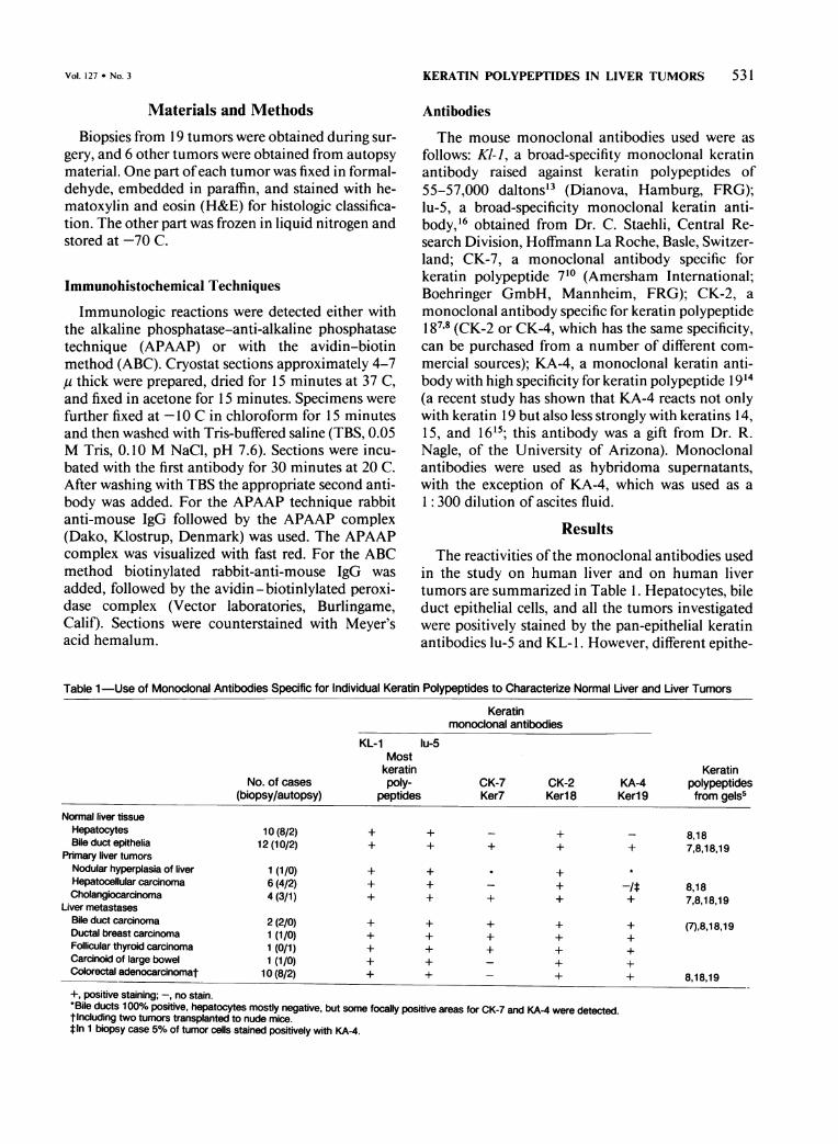
The reactivities of the monoclonal antibodies usedin the study on human liver and on human livertumors are summarized in Table 1. Hepatocytes, bileduct epithelial cells, and all the tumors investigatedwere positively stained by the pan-epithelial keratinantibodies lu-5 and KL- 1. However, different epithe-
Table 1 -Use of Monoclonal Antibodies Specific for Individual Keratin Polypeptides to Characterize Normal Liver and Liver Tumors
Keratinmonoclonal antibodies
KL-1 lu-5
No. of cases(biopsy/autopsy)
Normal liver tissueHepatocytesBile duct epithelia
Primary liver tumorsNodular hyperplasia of liverHepatocellular carcinomaCholangiocarcinoma
Liver metastasesBile duct carcinomaDuctal breast carcinomaFollicular thyroid carcinomaCarcinoid of large bowelColorectal adenocarcinomat
10 (8/2)12 (10/2)
1(1/0)
6 (4/2)4 (3/1)
2 (2/0)1(1/0)
1 (0/1)1(1/0)
10 (8/2)
Mostkeratinpoly-
peptides
+ +
+ +
+
+
+
+
+ +
+ +
KeratinCK-7 CK-2 KA-4 polypeptidesKer7 Ker 8 Ker 9 from gels5
+
+
- 8,18+ 7,8,18,19
-+
+
8,187,8,18,19
(7),8,18,19
8,18,19
+, positive staining; -, no stain.*Bile ducts 100% positive, hepatocytes mostly negative, but some focally positive areas for CK-7 and KA-4 were detected.tincluding two tumors transplanted to nude mice.t In 1 biopsy case 5% of tumor cells stained positively with KA-4.
Vol. 127 * No. 3

532 FISCHER ET AL
lial cell types in normal liver, as well as differenttumor types, could be distinguished when monoclo-nal antibodies specific for particular keratin polypep-tides were used. In normal liver, hepatocytes weredecorated by antibody CK-2 specific for keratin 18,while bile duct epithelia stained positively with theCK-2 antibody, with the CK-7 antibody specific forkeratin 7, and with the KA-4 antibody, which recog-nizes keratin 19 with high specificity. When primaryliver tumors were studied, 5 of 6 hepatocellular carci-nomas (HCCs) were stained only by the CK-2 anti-body specific for keratin 18 and not by the CK-7 anti-body specific for keratin 7 or the KA-4 antibody,specific for keratin 19 (Figure 1A). In one of the 6HCC specimens the antibody KA-4 yielded a positivefocal staining in some tumor areas. Four specimens ofcholangiocellular carcinoma ofthe liver were stronglypositively stained by the antibodies CK-7 (Figure 1 B),CK-2 (Figure 1C), and KA-4 (Figure 1 D). Thus, theCK-7 antibody appears able to distinguish hepatocel-lular carcinomas which are not stained from cholan-giocellular carcinomas which are stained.
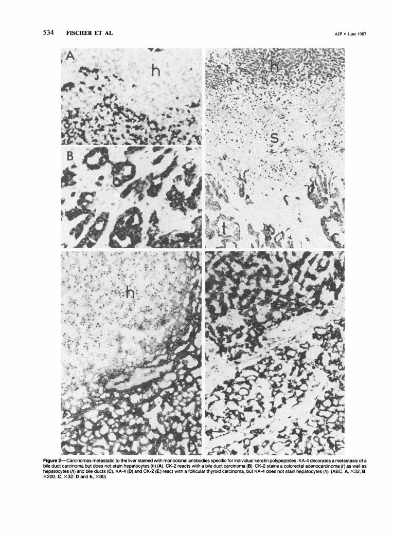
Metastases to the liver from gastrointestinal carci-nomas of known origin, as well as metastases to theliver of single examples of a carcinoid of the largebowel and of ductal breast carcinoma were studiedwith the keratin monoclonal antibody panel. Alltumors were positive with the KA-4 and CK-2 anti-bodies. KA-4- and CK-2-positive staining of tumorcells in a liver metastasis from a bile duct carcinoma isshown in Figure 2A and B, respectively, and CK-2-positive staining ofa metastasis from a colorectal ade-nocarcinoma is shown in Figure 2C. KA-4 and CK-2positivity of the tumor cells of a liver metastasis froma follicular thyroid carcinomas is shown in Figure 2Dand E, respectively. The CK-7 antibody stainedtumor cells in the liver metastases from the bile ductcarcinoma, from the ductal breast carcinoma, andfrom the follicular thyroid carcinoma. However,CK-7 antibody did not stain tumor cells in the livermetastases of the carcinoid from large bowel, or inliver metastases from eight colorectal adenocarci-nomas. Two colorectal adenocarcinomas were alsotested after they had been transplanted onto nudemice for 15 weeks, and the tumor cells were still nega-tive when tested with the CK-7 antibody. The keratincontent of the metastatic tumors is summarized inTable 1. Note that the liver metastasis ofa carcinoid ofthe large bowel showed a keratin spectrum similar tothat seen with the colorectal adenocarcinomas, asjudged by our immunohistochemical assays.We also examined one focal nodular hyperplasia of
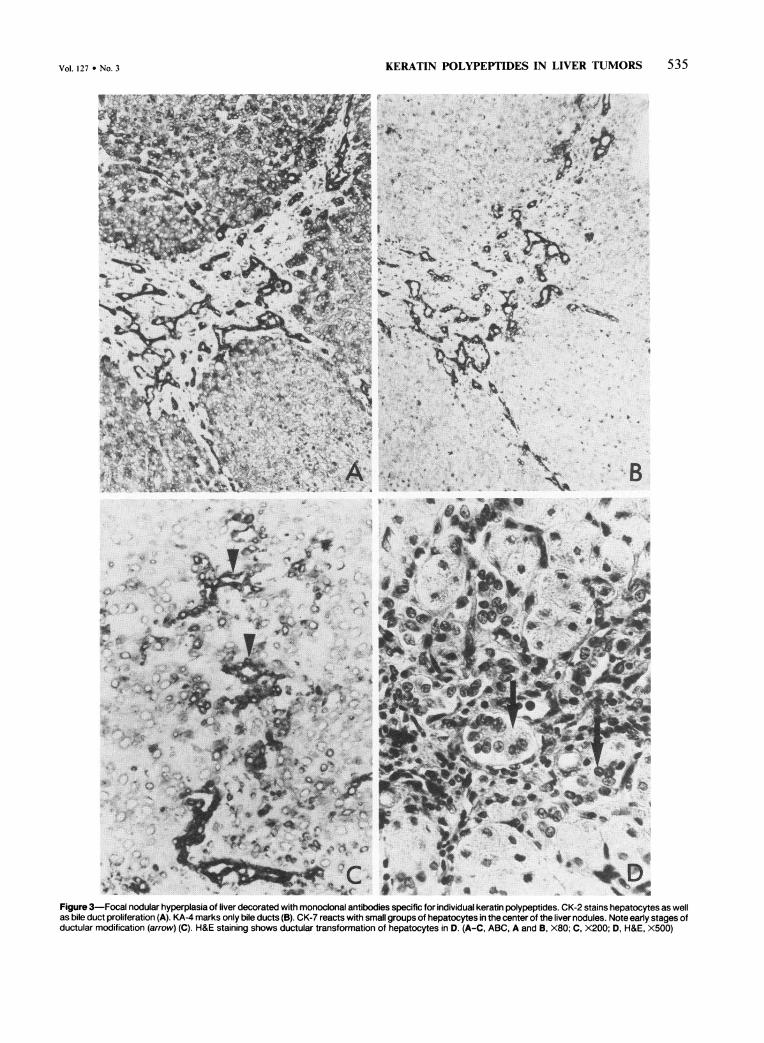
the liver. Nearly all hepatocytes of the liver noduleswere positively stained by the CK-2 antibody (Figure
3A) and not by the KA-4 (Figure 3B) and CK-7 anti-bodies. Unexpectedly, however, a few hepatocytes inthis material were, in addition, positively stained bythe antibodies CK-7 (Figure 3C), and KA-4, antibod-ies which in normal liver never stain hepatocytes (Fig-ure 2D and Table 1). Hepatocytes positive withthe CK-7 antibody were mostly found in small groupslocated at the periphery ofthe nodules. Ductular pro-liferations, mostly separate from larger bile ducts,could by detected in the center ofthe liver nodules, byhematoxylin-eosin staining (Figure 3D). SingleKA-4- and CK-7-positive cells or small ductular pro-liferations were often found in the center of the nod-ules (Figure 3C). The CK-7 and KA-4 antibodies dec-orated these apparently abnormal hepatocytes lessstrongly than bile duct epithelia.
Table 1 summarizes the results obtained by im-munohistochemical methods on epithelial cells innormal liver, on primary liver tumors, and on tumorsof known origin metastatic to the liver. Note theagreement of the results obtained by immunohisto-chemical techniques with the keratin polypeptidecontent of tumors of the same type determined byMoll and Franke by two-dimensional gel electropho-resis.5
DiscussionWhen malignant epithelial liver tumors are classi-
fied, three differential diagnostic questions have to beanswered: 1) the discrimination between primary andsecondary liver tumors; 2) the differential diagnosis ofprimary liver carcinomas; 3) the determination oftheprimary tumor location when liver metastases arefound. In this study we have shown that the immuno-histologic detection of single keratin polypeptides bymeans ofmonoclonal antibodies can help solve thesediagnostic problems.The antibodies KL- 1 and lu-5 are broad-specificity
keratin antibodies recognizing the different keratinpolypeptides present in a wide variety of normalepithelia and carcinomas.'3"16 Both antibodies posi-tively stained all the tumors listed in Table 1 as well ashepatocytes and bile duct epithelia in normal liver.Thus, although both antibodies are ery useful re-agents with which to confirm the epithelial nature ofthe neoplasms, and although positive staining withsuch antibodies excludes metastases of sarcoma ormelanoma, they have no diagnostic relevance when itcomes to the subdivision of malignant epithelial livertumors. In contrast, the CK-7, CK-2, and KA-4 anti-bodies allowed primary and metastatic liver neo-plasms to be divided into three groups. In the firstgroup hepatocytes and hepatocellular carcinomas
AJP * June 1987

KERATIN POLYPEPTIDES IN LIVER TUMORS 533
i '1, .I.
_r_.t
4P4:, 00: lb*..^RC^''..qks # .iEw
,1 F s,P,, iE .. ,,. P^ s , .:1bl,. .. ..| -.}'1i.; .... ¢;
_.J: : . 1*:1,
:. e a^.f:o ... :...:.... -:
.- * 1 FF . .o , _.n< z,,, .. r
-
-1i eeSe
_w_@ ^ _'- _ .} _ . _
I.
.r.....:: 4
;.....
Figure 1-Primary liver carcinomas stained with monoclonal antibodies specific for keratin polypeptides. CK-7 specific for keratin 7 does not stain tumor cells inhepatocellular carcinoma (t) but stains bile ducts (arrows) (A). CK-7 (B), CK-2 specific for keratin 18 (C), and KA-4 (D) with high specificity for keratin 19 staincholangiocarcinoma positively. (APAAP, A, X200; B-D, X320)
i * tkV
S!
S-.4'
l vll
*,r.
s 14l
0..*
...i. .k.
4
'A
Vol. 127 * No. 3
I.41, 7tA:.....
W:.::.:.:.H:.
:.&WAPk...^......1
0
- ..- d_b di%
534 FISCHER ET AL AJP * June 1987
; * * t- ,r ,~4. ,~ *,, . ;
a- **-.
. .;* *'*.*,.** Sr
Figure 2-Carcinomas metastatic to the liver stained with monoclonal antibodies specific for individual keratin polypeptides. KA-4 decorates a metastasis of abile duct carcinoma but does not stain hepatocytes (h) (A). CK-2 reacts with a bile duct carcinoma (B). CK-2 stains a colorectal adenocarcinoma (t) as well ashepatocytes (h) and bile ducts (C). KA-4 (D) and CK-2 (E) react with a follicular thyroid carcinoma, but KA-4 does not stain hepatocytes (h). (ABC, A, X32; B,X200; C, X32; D and E, X80)
III
I
Vol. 127 * No. 3 KLKAI'IN rFUxLrYrH uIN LIVLi Iu1VIU' J..J
Figure 3-Focal nodular hyperplasia of liver decorated with monoclonal antibodies specific for individual keratin polypeptides. CK-2 stains hepatocytes as wellas bile duct proliferation (A). KA-4 marks only bile ducts (B). CK-7 reacts with small groups of hepatocytes in the center of the liver nodules. Note early stages ofductular modification (arrow) (C). H&E staining shows ductular transformation of hepatocytes in D. (A-C, ABC, A and B, X80; C, X200; D, H&E, X500)
1VIM" A'lrlrT "d-%Y '%JlrbVWrlr"VQ IXT Ir Irt7VD 9rl TX49'%DQ r% 11 r%
536 FISCHER ET AL AJP * June 1987
were positive with the CK-2 antibody, negative withthe CK-7 antibody, and, with the exception of onehepatocellular carcinoma where focal staining withthe KA-4 antibody was seen, negative with KA-4. Thekeratin complement found by immunohistologicmethods is consistent with the keratin 8 and 18 posi-tively determined by two-dimensional gels.4'5"7 In thesecond group, bile duct epithelial cells and cholangio-cellular carcinomas stained positively with the CK-7,CK-2, and KA-4 antibodies, ie, were positive for ker-atin 7, 18, and 19. Again, for cholangiocellular carci-nomas, the keratin complement determined by im-munohistochemical methods is consistent with thekeratin 7, 8, 18, and 19 positively determined by gelelectrophoresis. Liver metastases from extrahepaticbile duct carcinomas were also positive with theCK-2, CK-7, and KA-4 antibodies, as were ductalcarcinomas ofpancreas.'2 In addition, the majority ofadenocarcinomas of endometrium and ovary as wellas adenocarcinomas ofthe lung have a similar keratincomplement from gel electrophoresis.5 The liver me-tastasis of a ductal breast carcinoma contained thesame spectrum of keratin subtypes corresponding tothe keratin polypeptides found in most ductal andlobular breast carcinomas.5 8 Thus, a differentialdiagnosis of cholangiocellular carcinomas and thesemetastatic liver tumors is not possible by keratin typ-ing, because all these tumor types appear to expressthe same keratin polypeptide spectrum. However,tumors in the third group, ie, the liver metastases ofall10 colorectal adenocarcinomas we examined and ofthe carcinoid tumor of large bowel reacted with theCK-2 and KA-4 antibodies but were not decorated bythe CK-7 antibody. The same keratin polypeptidesare found in normal epithelial of small and largebowel.5 The negative reaction with the CK-7 antibodyand the positive staining with the KA-4 antibodyallows colorectal carcinomas to be distinguished fromall other tumors in our study. Here, also, however, ithas to be noted that keratin polypeptide 7 is expressedheterogeneously in a few ductular breast carcinomasand is absent in some adenocarcinomas of ovary andendometrium.5
Differences in keratin polypeptide content betweenhepatocellular carcinoma and cholangiocarcinomahave been noted not only in this but also in otherstudies.5" 2" 9 Although the results have usually beeninterpreted as implying an origin ofcholangiocellularcarcinoma from bile duct epithelial cells, the situationmay be more complex. One of our HCCs containedsmall areas focally labeled by the KA-4 antibody. Thenodular hyperplasia of liver likewise showed someCK-7- and KA-4-positive hepatocytes, and in someinstances ductular transformation was also noted. In
this situation it seems that hepatocytes can expressadditional keratin polypeptides normally seen only inbile duct epithelia. Possibly the focal expression ofthekeratin polypeptide reacting with the KA-4 antibodyin HCC is a first step of differentiation or metaplastictransformation which later can result in cholangio-carcinoma. Ductular differentiation of hepatocyteshas been observed in other instances. In fetal liver, forexample small bile ducts develop from primitive hep-atocytes.20'2' In liver dystrophia, as well as in focalhyperplasia, ductular structures can be observed inconnection with hepatocytes which are far from pre-formed bile ducts.22'23Our results show that the transition from hepato-
cytes to bile duct epithelia is accompanied by a dis-tinct program of keratin polypeptide expression.Thus, the relatively simple combination of keratinpolypeptides characteristic of hepatocytes and mosthepatocellular carcinomas, ie, 8 and 18, appears to befollowed by the additional expression of keratin 19,which we observed focally in one hepatocellular carci-noma. In nodular hyperplasia some modified hepato-cytes begin to synthesize polypeptide 7, and thisseems to be the last step before duct formation charac-terized by expression of 7, 8, 18, and 19. Thus far, thepolypeptide combination 7, 8, 18 without expressionof 19 has not been observed.Some investigators assume that cholangiocellular
carcinomas as well as some cholangiocarcinomas aremore or less metaplastic HCCs.2226 The 1 HCCthat showed focal reactivity with KA-4 might supportthis hypothesis. Then, if this point of view is furtherextended, cholangiocellular carcinoma could origi-nate also from hepatocytes.Although the number oftumors investigated is still
rather small, there appears to be a high diagnosticvalue to immunohistochemical staining of livertumors with antibodies specific for keratins 7, 8, and19.Our conclusions are as follows: 1) These antibodies
allow the differential diagnosis of cholangiocarci-nomas and most hepatocellular carcinomas. 2) Dis-crimination between extrahepatic and intrahepaticbile duct carcinomas as well as of the majority ofadenocarcinoma metastases, eg, of breast, pancreas,or lung carcinomas, is not possible. 3) The keratinpattern characteristic of primary liver tumors and ofliver metastases of breast, pancreas, or lung carci-nomas differs from that of colorectal adenocarci-nomas.
References1. Osborn M, Altmannsberger M, Debus E, Weber K:
Conventional and monoclonal antibodies to interme-
Vol. 127 * No. 3 KERATIN POLYPEPrIDES IN LIVER TUMORS 537
diate filament proteins in human tumor diagnosis,Cancer Cells: I. The transformed phenotype. ColdSpring Harbor Laboratory, 1984, pp 191-200
2. Osborn M, Altmannsberger M, Debus E, Weber K:Differentiation of the major human tumor groupsusing conventional and monoclonal antibodies specificfor individual intermediate filament proteins. NewYork Acad Sci USA 1985, 455:649-668
3. Qunilan RA, Schiller DL, Hatzfeld M, Achtstaetter T,Moll R, Jorcano JL, Magin TM, Franke WW: Patternsofexpression and organization ofcytokeratin interme-diate filaments. Ann NY Acad Sci 1985, 455:282-306
4. Moll R, Franke WW, Schiller DL, Geiger B, Krepler R:The catalogue ofhuman cytokeratin polypeptides: Pat-terns of expression of specific cytokeratins in normalepithelia, tumors and cultured cells. Cell 1982, 31:11 -24
5. Moll R, Franke WW: Cytochemical cell typing ofmeta-static tumors according to their cytoskeletal proteins,Biochemistry and Molecular Genetics of Cancer Me-tastasis. Edited by LA Liotta, AS Rabson. The Hague,Nijhoff Martinus 1986, pp 101- 114
6. Cooper D, Schermer A, Sun T-T: Classification ofhuman epithelial and their neoplasms using monoclo-nal antibodies to keratins: Strategies, applications andlimitations. Lab Invest 1986, 52:243-256
7. Debus E, Weber K, Osborn M: Monoclonal cytokera-tin antibodies that distinguish simple from stratifiedsquamous epithelia: Characterization on human tis-sues. EMBO J 1982, 2:1641-1647
8. Debus E, Moll R, Franke WW, Weber K, Osborn M:Immunohistological distinction ofhuman carcinomasby cytokeratin typing with monoclonal antibodies. AmJPathol 1984, 114:121-130
9. Lane EB: Monoclonal antibodies provide specific in-tramolecular markers for the study ofepithelial tonofi-lament organization. J Cell Biol 1982, 92:665-673
10. Toelle HG, Weber K, Osborn M: Microinjection ofmonoclonal antibodies specific for one intermediatefilament protein in cells containing multiple keratinsallow insight into composition of particular 10 nmfilaments. Eur J Cell Biol 1985, 38:234-240
11. Tseng SCG, Jarvinen MJ, Nelson WG, Huang JW,Woodcoock MJ, Sun TT: Correlation of specific kera-tins with different type of epithelial differentiation:Monoclonal antibody studies. Cell 1982, 30:361 -372
12. Osborn M, van Lessen G, Weber K, Kloeppel G, Alt-mannsberger M: Differential diagnosis ofgastrointesti-nal tumors using monoclonal antibodies specific forindividual keratin polypeptides. Lab Invest 55:497-504
13. Viac J, Reano A, Brochier J, Staquet MJ, Thivolet J:
Reactivity pattern of a monoclonal anti-keratin anti-body (KL 1). J Invest Dermatol 1983, 81:351-354
14. Nagle RB, Lucas DO, McDaniel KM, Clark VA,Schmalzel GM: New evidence linking mammary andextramammary Paget cells to a common cell pheno-type. (Abstr) Am J Clin Pathol 1985, 83:431-438a
15. Nagle RB, Moll R, Weidauer H, Nemetschek H,Franke WW: Different patterns of cytokeratin expres-sion in the normal epithelia of the upper respiratorytract. Differentiation 1985, 30:130-140
16. Von Overbeck J, Stahli C, Gudat F, Carmann H, Lau-tenschlager C, Durmuller U, Takacs B, Miggiano V,Staehelin T, Heitz PU: Immunohistochemical charac-terization of an anti-epithelial monoclonal antibody(mAB lu-5). Virchows Arch [Pathol Anat] 1985,407:1-12
17. Wu YL, Parker LM, Binder NE, Beckett JH, SinardJH, Griffiths CT, Rheinwald JG: The mesothelial kera-tins: A new family of cytoskeletal proteins identified incultured mesothelial cells and nonkeratinizing epithe-lia. Cell 1983, 31:670-693
18. Altmannsberger M, Dirk T, Droese M, Weber K, Os-born M: Keratin polypeptide distribution in benignand malignant breast tumors: Subdivision of ductalcarcinomas using monoclonal antibodies. VirchowsArch [Cell Pathol] 1986, 51:265-275
19. Denk H, Krepler R, Lackinger E, Artlieb U, FrankeWW: Biochemical and immunocytochemical analysisof the intermediate filament cytoskeleton in humanhepatocellular carcinomas and in hepatic neoplasticnodules of mice. Lab Invest 1982, 46:584-596
20. Enzan H, Ohkita T, Fujita H, Iijima S: Light- and elec-tronmicroscopic studies on the development of peri-portal bile ducts of the human embryos. Acta PatholJpn 1974, 24:427-447
21. Wegmann R, Corcos V, Caroli J: Histoenzymologiedes ductules biliares chez l'embryon humain normal etau cours des cirrhoses humaines. Arch Mal Appr Dig1965, 54:215-228
22. Altmann HW: Pathology of human liver tumors, Pri-mary Liver Tumors. Edited by H Remmer, HM Bolt, PBannasch, H Popper. Lancaster, MTP Press Ltd., 1978,pp 53-71
23. Popper H, Schaffner F: Die Leber: Struktur und Funk-tion. Stuttgart, Thieme, 1961
24. Altmann HW: Neubildung der Leber. Verh DtschKrebs Ges 1984, 5:423-435
25. Lapis K, Johannessen JV: Pathology of primary livercancer. J Toxicol Environmental Health 1979, 5:315 -355
26. Peters RL: Pathology of hepatocellular carcinoma,Hepatocellular Carcinoma. Edited by K Okuda, RLPeters. New York, John Wiley & Sons, 1976, pp107-168


![YDJC Induces Epithelial-Mesenchymal Transition via ...downloads.hindawi.com/journals/jo/2019/3542537.pdf · atin results in loss of keratin []. Interestingly, a recent study reported](https://img.pdfslide.net/doc/110x75/5f28cbc3adce09548c3a0a96/ydjc-induces-epithelial-mesenchymal-transition-via-atin-results-in-loss-of-keratin.jpg)