Embed Size (px)
Citation preview
J. Vet. Anat. Vol 5 No 2, (2012) 15 - 3115
Kinematics of some elephant limb bones Melaku Tefera
12 15 18 21 24 27 30 33 36LV
40
45
50
55
60
65
70
75
80
85
TG
Fig (2): Linear fit for live weight (LW) and thoracic girth (TG) and RMA analysis. (95 % confidence) LV (kg) = (0.659 x TG) (cm) – 17.467 RMA Regression Slope a: 0.65974 Intercept b: -17.467 Std. err. a: 0.056768 Std. err. b: 11.189 Chi squared: 0 r: 0.84335 R2: 0.71125 t statistic: 9.8012 p(uncorrel): 4.505E-12 Permutat. p: 0.0001 p(a=1): 5.282E-07 95% bootstrapped confidence intervals: a: [0.5001; 0.8068] b: [-25.38; -8.735]
Kinematics and Comparative Anatomy of Some Limb Bones of the African Elephant (Loxodonta africana) and Large Domestic Animals Melaku Tefera College of Veterinary Medicine, Haramaya University. P.O. Box 144 Haramaya Campus. Ethiopia. 251-0914722459, [email protected] With 3 plates &2 figures Received May, accepted for publication December, 2011 Abstract Elephants are the largest extant ter-restrial animals and the archetype of ‘graviportal’ animals, with large body size and a pattern of pentadactyl limbs. The fundamental structures are homologous in all tetrapods but in the course of evolution these structures have been modified in the elephant. Osteometric parameters show that the relationship of the length of the femur to the circumfer-ence is 2.5, 2.75 and 2.8 in ele-phant, horse and cattle respectively. Similarly humerus length to circum-ference is 2.3 in the three species showing isometric scaling. There is a positive allometric scaling be-tween bone weight and bone length; the ratio of femur length to weight is 205g/cm, 72g/cm and 64g/cm in el-ephants, horses and cattle. The ratio of weight of the humerus to length or weights of the humerus plus femur to their combined length is a good estimate of the body
weight in kg= ( ). We have
observed three gaits in the ele-phant: slow, fast walk, and trot. Ei-ther one or a maximum of two con-trolateral legs are lifted from ground, but never two ipislateral limbs. The propulsive force originates from the retractor muscles of the hind legs, elephants moving by extension of the forelegs rather than flexion. The head’s conical structure makes it aerodynamically efficient, serving as nose cone. The joint Articulatio at-lanto occipitalis is less movable than the horse or cattle. The main mech-anism by which an elephant over-comes the effect of heavy weight is by having high density bones. The articular surfaces of the bones are less developed in elephant com-pared to horse or cattle, resulting in poor angular movements with less ground shock waves. The pes is like a cushion filled with a fat layer that serves as a shock absorber. The skull is spongy and the arrangement

J. Vet. Anat. Vol 5 No 2, (2012) 15 - 3116
Kinematics of some elephant limb bones Melaku Tefera
of trabeculae makes the skull lighter in weight. Key words biomechanics, bone allometry, comparative anatomy, elephant, gait, osteology, skeleton, aerody-namics. Introduction The Order Proboscidea includes the animals with elongated trunks that function both as nose and as a pre-hensile organ to grasp and manipu-late objects in the environment. Modern elephants are the heaviest land animals, no other terrestrial animal weighs half as much (McMahon, 1975). There are two genera of the family Elephantidae: Elephas and Loxodonta. The Asian elephant (Elephas maximus) is also known as the Indian elephant, and is found in Asia. The elephants of the genus Loxodonta, known collec-tively as African elephants, are cur-rently found in 37 countries in Africa (Blanc, 2008; Elephant encyclope-dia). African elephants have tradi-tionally been classified as a single species. However genetic study has shown that to comprise three dis-tinct subspecies, namely the savan-na elephant (Loxodonta africana africana and Loxodonta africana. knochenhaueri), the forest elephant
(Loxodonta africana cyclotis) (Rohland, et al., 2010), all other species and genera of elephantidae like Loxodonta adaurora (the pre-sumed ancestor of the modern Afri-can elephants) are extinct (Eggert, et al., 2002). Elephant anatomy is poorly under-stood (Hutchinson, et al., 2006) and access to specimens is severely difficult. The pattern of limb called pentadactyl is an example of ho-mologous structure found in all classes of tetrapods. Suggesting that they have originated from a common ancestor but in the course of evolution these fundamental structures have changed. The paw of the dog, the hoof of the horse, the manus and pes of the elephant and foot of the human all share some common features of structure, or-ganization and function. Each of these organisms’ foot structures function as the load transmission platform which is essential to bal-ance, standing and locomotion strategies (such as walking, trotting, galloping and running). Elephants as the largest extant terrestrial ani-mals and as the archetype of ‘gravi-portal’ animals, having large body size with columnar, robust limbs (Coombs, 1978); provide insight into the biomechanical and physiological constraints that extremely large body size imposes.
Severe scaling constraint on func-tional capacity may result as organ-isms evolve to large size. It has long been recognized that body size is a critical factor influencing mechanical support of animals. Specifically, the ability of muscle to generate force or bones to resist force depends on tissue cross-sectional area which decreases in proportion to an ani-mal’s weight with increased size. The scaling of bone and muscle ge-ometry in mammals suggests that force on the skeleton increases with increasing body size. McMahon, (1973) has proposed a scaling model of elastic similarity which ar-gues that the linear dimensions of animals do not increase in the same proportion. Instead bones increase in proportion to their diameter so that animals become distorted in shape and relatively more stout as they increase in size. Yet mammali-an limb bones scale close to isome-try in proportion (Biewner, 1983). Others (Russell, 1985) do not sup-port this hypothesis instead arguing that large animals must compensate for geometric scaling of their bones by reducing the forces acting on the bones of their skeleton. The most effective way of achieving this re-duction is to reduce the bending force. Another mechanism is to re-duce ground force exerted on the limb during the support phase of locomotion by reducing the ground
contact time (Alexander, et al., 1979). Our understanding of ele-phant locomotion is impaired by a lack of data. Hence the objective of this research is to study the anatomy of the long bones of both limbs of the elephant and compare its morphology with some domestic animals, to see if there is an isometric or allometric relationship between the osteomet-ric parameters, describe the skull bone and investigate the biome-chanics of the elephant gait. Materials and Methods Osteology The elephant (Loxodota africana africana) bones were property of Haramaya University, Ethiopia. Five bones were acquired, two femora, a humerus and two halves of the face bone. The elephant was in the uni-versity zoo and died in the year 1960. The condition of the bones was fair with slight erosions of the periosteum, because the bones were stored in outdoors in shed. The femur and humerus of cattle and horse were property of Ha-ramaya University College of Veter-inary Medicine, Anatomy Laborato-ry; they were two years old since slaughtering of the animals. The bones were weighed using a digital balance. The length of bones was measured using a tape measure
J. Vet. Anat. Vol 5 No 2, (2012) 15 - 3117
Kinematics of some elephant limb bones Melaku Tefera
of trabeculae makes the skull lighter in weight. Key words biomechanics, bone allometry, comparative anatomy, elephant, gait, osteology, skeleton, aerody-namics. Introduction The Order Proboscidea includes the animals with elongated trunks that function both as nose and as a pre-hensile organ to grasp and manipu-late objects in the environment. Modern elephants are the heaviest land animals, no other terrestrial animal weighs half as much (McMahon, 1975). There are two genera of the family Elephantidae: Elephas and Loxodonta. The Asian elephant (Elephas maximus) is also known as the Indian elephant, and is found in Asia. The elephants of the genus Loxodonta, known collec-tively as African elephants, are cur-rently found in 37 countries in Africa (Blanc, 2008; Elephant encyclope-dia). African elephants have tradi-tionally been classified as a single species. However genetic study has shown that to comprise three dis-tinct subspecies, namely the savan-na elephant (Loxodonta africana africana and Loxodonta africana. knochenhaueri), the forest elephant
(Loxodonta africana cyclotis) (Rohland, et al., 2010), all other species and genera of elephantidae like Loxodonta adaurora (the pre-sumed ancestor of the modern Afri-can elephants) are extinct (Eggert, et al., 2002). Elephant anatomy is poorly under-stood (Hutchinson, et al., 2006) and access to specimens is severely difficult. The pattern of limb called pentadactyl is an example of ho-mologous structure found in all classes of tetrapods. Suggesting that they have originated from a common ancestor but in the course of evolution these fundamental structures have changed. The paw of the dog, the hoof of the horse, the manus and pes of the elephant and foot of the human all share some common features of structure, or-ganization and function. Each of these organisms’ foot structures function as the load transmission platform which is essential to bal-ance, standing and locomotion strategies (such as walking, trotting, galloping and running). Elephants as the largest extant terrestrial ani-mals and as the archetype of ‘gravi-portal’ animals, having large body size with columnar, robust limbs (Coombs, 1978); provide insight into the biomechanical and physiological constraints that extremely large body size imposes.
Severe scaling constraint on func-tional capacity may result as organ-isms evolve to large size. It has long been recognized that body size is a critical factor influencing mechanical support of animals. Specifically, the ability of muscle to generate force or bones to resist force depends on tissue cross-sectional area which decreases in proportion to an ani-mal’s weight with increased size. The scaling of bone and muscle ge-ometry in mammals suggests that force on the skeleton increases with increasing body size. McMahon, (1973) has proposed a scaling model of elastic similarity which ar-gues that the linear dimensions of animals do not increase in the same proportion. Instead bones increase in proportion to their diameter so that animals become distorted in shape and relatively more stout as they increase in size. Yet mammali-an limb bones scale close to isome-try in proportion (Biewner, 1983). Others (Russell, 1985) do not sup-port this hypothesis instead arguing that large animals must compensate for geometric scaling of their bones by reducing the forces acting on the bones of their skeleton. The most effective way of achieving this re-duction is to reduce the bending force. Another mechanism is to re-duce ground force exerted on the limb during the support phase of locomotion by reducing the ground
contact time (Alexander, et al., 1979). Our understanding of ele-phant locomotion is impaired by a lack of data. Hence the objective of this research is to study the anatomy of the long bones of both limbs of the elephant and compare its morphology with some domestic animals, to see if there is an isometric or allometric relationship between the osteomet-ric parameters, describe the skull bone and investigate the biome-chanics of the elephant gait. Materials and Methods Osteology The elephant (Loxodota africana africana) bones were property of Haramaya University, Ethiopia. Five bones were acquired, two femora, a humerus and two halves of the face bone. The elephant was in the uni-versity zoo and died in the year 1960. The condition of the bones was fair with slight erosions of the periosteum, because the bones were stored in outdoors in shed. The femur and humerus of cattle and horse were property of Ha-ramaya University College of Veter-inary Medicine, Anatomy Laborato-ry; they were two years old since slaughtering of the animals. The bones were weighed using a digital balance. The length of bones was measured using a tape measure
J. Vet. Anat. Vol 5 No 2, (2012) 15 - 3118
Kinematics of some elephant limb bones Melaku Tefera
between the two longest distances on the proximal and distal extremi-ties. The circumference of each bone was measured in centimeters mid way on the diaphysis. Photographs were taken using a Canon IXUS 750 camera. Since the photographs were taken from differ-ent distances a ruler of 22 cm was placed beside the specimen during the photographing in order to scale the size of the structure. All descrip-tions are according to Nomina Ana-tomica Veterinaria, 2005. Biomechanical techniques To study the gait of the elephant, video analysis was used as means of identifying the movement patterns through distance and angular measurements. The distance varia-bles describe the stride length and the distances between individual limb placements. For this purpose the following video films were up-loaded from the internet all ac-cessed 29 January 2011. a. Associated Press.Baby elephant
runs with herd. http:// www.youtube.com/watch?v=pTHtIf2YuRs. February17. 2010. Run time 1:20
b. BBC. Elephant mating, fighting & pregnancy - BBC Animals. http:// www.youtube.com/watch?v=ODy3CiS7H4o. Added February 17, 2009. Run time 4:02
c. Britannica.com. African-Eleph-ants in their habitat. http:// www.5min.com/Video/ . Run time 2:43
d. Indigo film television. The African Elephant. http://www.5min.com/Video/The-African-Elephant-516911223. Run time 4:17
e. Animal Planet Video. Mutual of Omaha's Wild Kingdom: An Ele-phant Oasishttp:// animal. dis-covery.com/videos/elephants breaking boundaries an elephant oasis.html. Added: Apr 7, 2009. Runtime: 2:20
f. Animal Planet Video. Mutual of Omaha's Wild Kingdom: Saddest Elephant Ever? http:// ani-mal.discovery.com/videos/elephants breaking-boundaries-saddest-elephant ever.html. Added: Apr 7, 2009. Runtime: 02:03
g. Animal Planet Video. http://animal.discovery.com/videos/the lost elephants of-timbuktu-secrets revealed.html. Added April. 19, 2008. Runtime: 02:56
h. Animal Planet Video. Mutual of Omaha's Wild Kingdom: Ele-phants form bonds http:// ani-mal.discovery.com/videos/mutual-of-omahas-wild-kingdom-bonds-for-life.html.Added Mar 12, 2008. Runtime 03:00
i. Animal Planet Video. Planet's Best: African Elephant. http:// an-imal.discovery.com/videos/planet
s-best-african-elephant.html. Added May 4, 2009.Runtime 2:22
j. Animal Planet Video, http:// ani-mal.discovery.com/videos/planets best okavango delta ele-phants.html. Added May 4, 2009. Runtime: 02:32
k. Elephant swimming. http:// www.youtube.com/watch?v=ywXYfLFapLY envymexxx. Added December 4 2006. Run time 2:12
Swimming elephant - Les éléphants nageurs. http://www.youtube.com/watch?v=XWt_0lXvd8g&feature=relatedhttp://www.youtube.com/user/cousteaucontent . Added October 31 2008. Run time 2:40 Statistical Analysis As there were only few specimens from individual animals of each spe-cies no statistical analysis was done. Ratios and percentages were calculated. Results The bones used in this study belong to an elephant which died 50 years ago. Due to lack of data, the age of the animal could not be determined. However, absence of the epiphysal plates (Cartilegio epiphysialis) point out that the animal was adult. And from the combined length of the humerus and femur which was 205
cm, the height at shoulder was es-timated to be between 250-320 cm and the body weight in kilogram was estimated with allometric formula derived from data depicted on Table 1, to be 2500 kg. The skull appeared not massive for such animal from the size of tusk buds it was confirmed that the ele-phant was an adult young female. The morphology of the bones has some similarities and variation. The basic structures are present in all three species with some disparity indicating differences in proportions and muscle and tendon insertion structures. The cranial and caudal views (Faci-es cranialis et caudalis) of the femur (Os femoris) are depicted on Plate 1. On the proximal extremity, the head (caput ossis femoris) projects medially and the neck (collum ossis femoris) is longer and less demar-cated in the elephant while the tro-chanter major (trochanter major) and lesser trochanters (trochanter minor) are small and rudimentary in the elephant. Trochanter tertius is well developed in the horse .The shaft (Corpus ossis femoris) is somewhat curved medially in the elephant to help stabilizing the head in the acetablum (Fossa acetabuli). On the distal extremity the medial and lateral condyles (Condylus me-
J. Vet. Anat. Vol 5 No 2, (2012) 15 - 3119
Kinematics of some elephant limb bones Melaku Tefera
between the two longest distances on the proximal and distal extremi-ties. The circumference of each bone was measured in centimeters mid way on the diaphysis. Photographs were taken using a Canon IXUS 750 camera. Since the photographs were taken from differ-ent distances a ruler of 22 cm was placed beside the specimen during the photographing in order to scale the size of the structure. All descrip-tions are according to Nomina Ana-tomica Veterinaria, 2005. Biomechanical techniques To study the gait of the elephant, video analysis was used as means of identifying the movement patterns through distance and angular measurements. The distance varia-bles describe the stride length and the distances between individual limb placements. For this purpose the following video films were up-loaded from the internet all ac-cessed 29 January 2011. a. Associated Press.Baby elephant
runs with herd. http:// www.youtube.com/watch?v=pTHtIf2YuRs. February17. 2010. Run time 1:20
b. BBC. Elephant mating, fighting & pregnancy - BBC Animals. http:// www.youtube.com/watch?v=ODy3CiS7H4o. Added February 17, 2009. Run time 4:02
c. Britannica.com. African-Eleph-ants in their habitat. http:// www.5min.com/Video/ . Run time 2:43
d. Indigo film television. The African Elephant. http://www.5min.com/Video/The-African-Elephant-516911223. Run time 4:17
e. Animal Planet Video. Mutual of Omaha's Wild Kingdom: An Ele-phant Oasishttp:// animal. dis-covery.com/videos/elephants breaking boundaries an elephant oasis.html. Added: Apr 7, 2009. Runtime: 2:20
f. Animal Planet Video. Mutual of Omaha's Wild Kingdom: Saddest Elephant Ever? http:// ani-mal.discovery.com/videos/elephants breaking-boundaries-saddest-elephant ever.html. Added: Apr 7, 2009. Runtime: 02:03
g. Animal Planet Video. http://animal.discovery.com/videos/the lost elephants of-timbuktu-secrets revealed.html. Added April. 19, 2008. Runtime: 02:56
h. Animal Planet Video. Mutual of Omaha's Wild Kingdom: Ele-phants form bonds http:// ani-mal.discovery.com/videos/mutual-of-omahas-wild-kingdom-bonds-for-life.html.Added Mar 12, 2008. Runtime 03:00
i. Animal Planet Video. Planet's Best: African Elephant. http:// an-imal.discovery.com/videos/planet
s-best-african-elephant.html. Added May 4, 2009.Runtime 2:22
j. Animal Planet Video, http:// ani-mal.discovery.com/videos/planets best okavango delta ele-phants.html. Added May 4, 2009. Runtime: 02:32
k. Elephant swimming. http:// www.youtube.com/watch?v=ywXYfLFapLY envymexxx. Added December 4 2006. Run time 2:12
Swimming elephant - Les éléphants nageurs. http://www.youtube.com/watch?v=XWt_0lXvd8g&feature=relatedhttp://www.youtube.com/user/cousteaucontent . Added October 31 2008. Run time 2:40 Statistical Analysis As there were only few specimens from individual animals of each spe-cies no statistical analysis was done. Ratios and percentages were calculated. Results The bones used in this study belong to an elephant which died 50 years ago. Due to lack of data, the age of the animal could not be determined. However, absence of the epiphysal plates (Cartilegio epiphysialis) point out that the animal was adult. And from the combined length of the humerus and femur which was 205
cm, the height at shoulder was es-timated to be between 250-320 cm and the body weight in kilogram was estimated with allometric formula derived from data depicted on Table 1, to be 2500 kg. The skull appeared not massive for such animal from the size of tusk buds it was confirmed that the ele-phant was an adult young female. The morphology of the bones has some similarities and variation. The basic structures are present in all three species with some disparity indicating differences in proportions and muscle and tendon insertion structures. The cranial and caudal views (Faci-es cranialis et caudalis) of the femur (Os femoris) are depicted on Plate 1. On the proximal extremity, the head (caput ossis femoris) projects medially and the neck (collum ossis femoris) is longer and less demar-cated in the elephant while the tro-chanter major (trochanter major) and lesser trochanters (trochanter minor) are small and rudimentary in the elephant. Trochanter tertius is well developed in the horse .The shaft (Corpus ossis femoris) is somewhat curved medially in the elephant to help stabilizing the head in the acetablum (Fossa acetabuli). On the distal extremity the medial and lateral condyles (Condylus me-
J. Vet. Anat. Vol 5 No 2, (2012) 15 - 3120
Kinematics of some elephant limb bones Melaku Tefera
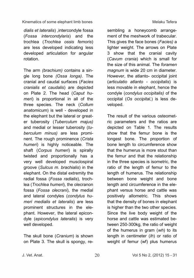
dialis et lateralis) ,intercondyle fossa (Fossa intercondylaris) and the trochlea (Trochlea ossis femoris) are less developed indicating less developed articulation for angular rotation. The arm (brachium) contains a sin-gle long bone (Ossa longa). The cranial and caudal surfaces (Facies cranialis et caudalis) are depicted on Plate 2. The head (Caput hu-meri) is proportional in all of the three species. The neck (Collum anatomicum) is well - developed in the elephant but the lateral or great-er tuberosity (Tuberculum majus) and medial or lesser tuberosity (tu-berculum minus) are less promi-nent. The rough prominence (Crista humeri) is highly noticeable. The shaft (Corpus humeri) is spirally twisted and proportionally has a very well developed musclospiral groove (Sulcus m. brachialis) in the elephant. On the distal extremity the radial fossa (Fossa radialis), troch-lea (Trochlea humeri), the olecranon fossa (Fossa olecrani), the medial and lateral condyles (condylus hu-meri medialis et lateralis) are less prominent structures in the ele-phant. However, the lateral epicon-dyle (epicondylus lateralis) is very well developed. The skull bone (Cranium) is shown on Plate 3. The skull is spongy, re-
sembling a honeycomb arrange-ment of the meshwork of trabecular. This gives the face bones (Facies) a lighter weight. The arrows on Plate 3 show that the cranial cavity (Cavum crania) which is small for the size of this animal. The foramen magnum is wide 20 cm in diameter. However, the atlanto- occipital joint (articulatio atlanto - occipitalis) is less movable in elephant, hence the condyle (condylus occipitalis) of the occipital (Os occipital,) is less de-veloped. The result of the various osteomet-ric parameters and the ratios are depicted on Table 1. The results show that the femur bone is the longest bone. The proportion of bone length to circumference show that the humerus is more stout than the femur and that the relationship in the three species is isometric, the ratio of the length of femur to the length of humerus. The relationship between bone weight and bone length and circumference in the ele-phant versus horse and cattle was positively allometric. This shows that the density of bones in elephant is higher than the two other species. Since the live body weight of the horse and cattle was estimated be-tween 250-300kg, the ratio of weight of the humerus in gram (wh) to its length in centimeter (lh) or ratio of weight of femur (wf) plus humerus
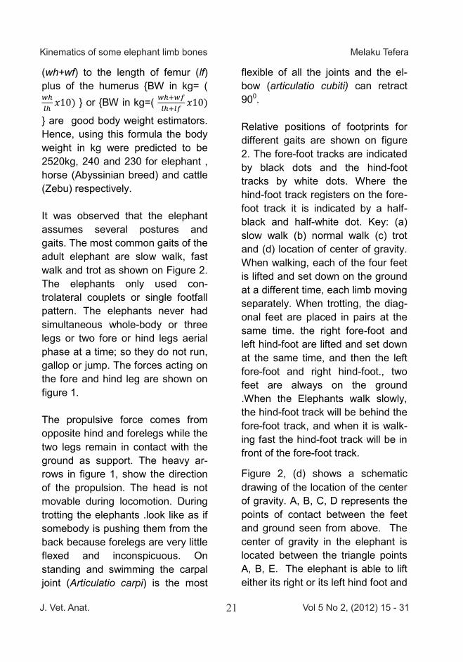
(wh+wf) to the length of femur (lf) plus of the humerus {BW in kg= ( } or {BW in kg=( } are good body weight estimators. Hence, using this formula the body weight in kg were predicted to be 2520kg, 240 and 230 for elephant , horse (Abyssinian breed) and cattle (Zebu) respectively. It was observed that the elephant assumes several postures and gaits. The most common gaits of the adult elephant are slow walk, fast walk and trot as shown on Figure 2. The elephants only used con-trolateral couplets or single footfall pattern. The elephants never had simultaneous whole-body or three legs or two fore or hind legs aerial phase at a time; so they do not run, gallop or jump. The forces acting on the fore and hind leg are shown on figure 1. The propulsive force comes from opposite hind and forelegs while the two legs remain in contact with the ground as support. The heavy ar-rows in figure 1, show the direction of the propulsion. The head is not movable during locomotion. During trotting the elephants .look like as if somebody is pushing them from the back because forelegs are very little flexed and inconspicuous. On standing and swimming the carpal joint (Articulatio carpi) is the most
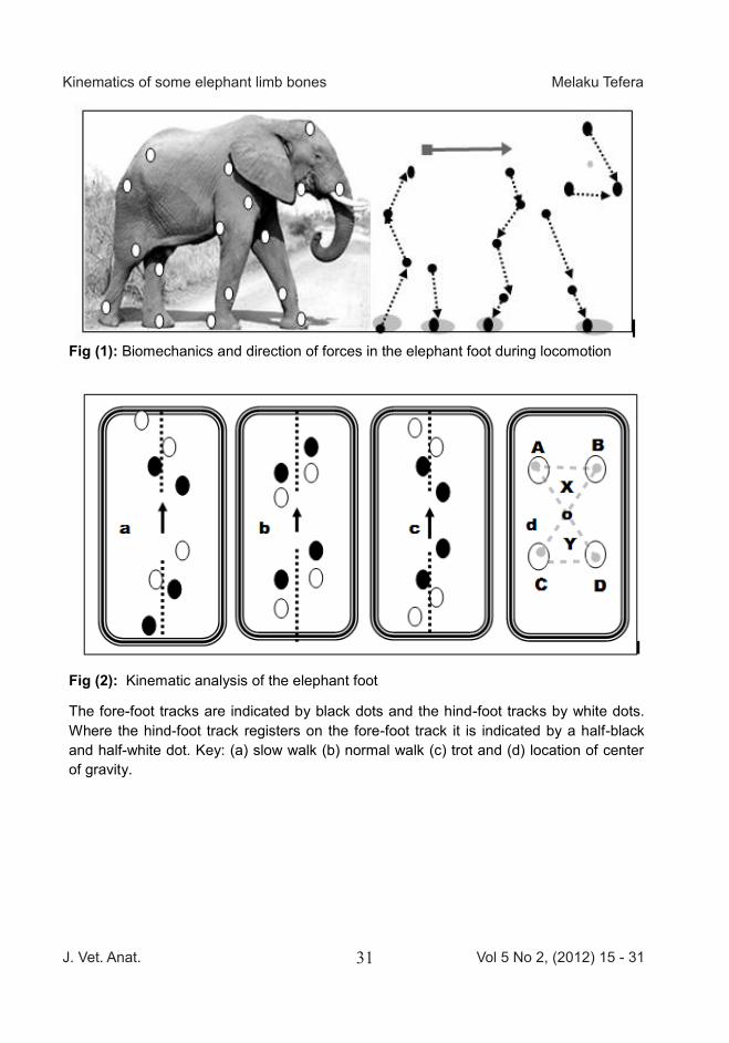
flexible of all the joints and the el-bow (articulatio cubiti) can retract 900. Relative positions of footprints for different gaits are shown on figure 2. The fore-foot tracks are indicated by black dots and the hind-foot tracks by white dots. Where the hind-foot track registers on the fore-foot track it is indicated by a half-black and half-white dot. Key: (a) slow walk (b) normal walk (c) trot and (d) location of center of gravity. When walking, each of the four feet is lifted and set down on the ground at a different time, each limb moving separately. When trotting, the diag-onal feet are placed in pairs at the same time. the right fore-foot and left hind-foot are lifted and set down at the same time, and then the left fore-foot and right hind-foot., two feet are always on the ground .When the Elephants walk slowly, the hind-foot track will be behind the fore-foot track, and when it is walk-ing fast the hind-foot track will be in front of the fore-foot track.
Figure 2, (d) shows a schematic drawing of the location of the center of gravity. A, B, C, D represents the points of contact between the feet and ground seen from above. The center of gravity in the elephant is located between the triangle points A, B, E. The elephant is able to lift either its right or its left hind foot and
J. Vet. Anat. Vol 5 No 2, (2012) 15 - 3121
Kinematics of some elephant limb bones Melaku Tefera
dialis et lateralis) ,intercondyle fossa (Fossa intercondylaris) and the trochlea (Trochlea ossis femoris) are less developed indicating less developed articulation for angular rotation. The arm (brachium) contains a sin-gle long bone (Ossa longa). The cranial and caudal surfaces (Facies cranialis et caudalis) are depicted on Plate 2. The head (Caput hu-meri) is proportional in all of the three species. The neck (Collum anatomicum) is well - developed in the elephant but the lateral or great-er tuberosity (Tuberculum majus) and medial or lesser tuberosity (tu-berculum minus) are less promi-nent. The rough prominence (Crista humeri) is highly noticeable. The shaft (Corpus humeri) is spirally twisted and proportionally has a very well developed musclospiral groove (Sulcus m. brachialis) in the elephant. On the distal extremity the radial fossa (Fossa radialis), troch-lea (Trochlea humeri), the olecranon fossa (Fossa olecrani), the medial and lateral condyles (condylus hu-meri medialis et lateralis) are less prominent structures in the ele-phant. However, the lateral epicon-dyle (epicondylus lateralis) is very well developed. The skull bone (Cranium) is shown on Plate 3. The skull is spongy, re-
sembling a honeycomb arrange-ment of the meshwork of trabecular. This gives the face bones (Facies) a lighter weight. The arrows on Plate 3 show that the cranial cavity (Cavum crania) which is small for the size of this animal. The foramen magnum is wide 20 cm in diameter. However, the atlanto- occipital joint (articulatio atlanto - occipitalis) is less movable in elephant, hence the condyle (condylus occipitalis) of the occipital (Os occipital,) is less de-veloped. The result of the various osteomet-ric parameters and the ratios are depicted on Table 1. The results show that the femur bone is the longest bone. The proportion of bone length to circumference show that the humerus is more stout than the femur and that the relationship in the three species is isometric, the ratio of the length of femur to the length of humerus. The relationship between bone weight and bone length and circumference in the ele-phant versus horse and cattle was positively allometric. This shows that the density of bones in elephant is higher than the two other species. Since the live body weight of the horse and cattle was estimated be-tween 250-300kg, the ratio of weight of the humerus in gram (wh) to its length in centimeter (lh) or ratio of weight of femur (wf) plus humerus
(wh+wf) to the length of femur (lf) plus of the humerus {BW in kg= ( } or {BW in kg=( } are good body weight estimators. Hence, using this formula the body weight in kg were predicted to be 2520kg, 240 and 230 for elephant , horse (Abyssinian breed) and cattle (Zebu) respectively. It was observed that the elephant assumes several postures and gaits. The most common gaits of the adult elephant are slow walk, fast walk and trot as shown on Figure 2. The elephants only used con-trolateral couplets or single footfall pattern. The elephants never had simultaneous whole-body or three legs or two fore or hind legs aerial phase at a time; so they do not run, gallop or jump. The forces acting on the fore and hind leg are shown on figure 1. The propulsive force comes from opposite hind and forelegs while the two legs remain in contact with the ground as support. The heavy ar-rows in figure 1, show the direction of the propulsion. The head is not movable during locomotion. During trotting the elephants .look like as if somebody is pushing them from the back because forelegs are very little flexed and inconspicuous. On standing and swimming the carpal joint (Articulatio carpi) is the most
flexible of all the joints and the el-bow (articulatio cubiti) can retract 900. Relative positions of footprints for different gaits are shown on figure 2. The fore-foot tracks are indicated by black dots and the hind-foot tracks by white dots. Where the hind-foot track registers on the fore-foot track it is indicated by a half-black and half-white dot. Key: (a) slow walk (b) normal walk (c) trot and (d) location of center of gravity. When walking, each of the four feet is lifted and set down on the ground at a different time, each limb moving separately. When trotting, the diag-onal feet are placed in pairs at the same time. the right fore-foot and left hind-foot are lifted and set down at the same time, and then the left fore-foot and right hind-foot., two feet are always on the ground .When the Elephants walk slowly, the hind-foot track will be behind the fore-foot track, and when it is walk-ing fast the hind-foot track will be in front of the fore-foot track.
Figure 2, (d) shows a schematic drawing of the location of the center of gravity. A, B, C, D represents the points of contact between the feet and ground seen from above. The center of gravity in the elephant is located between the triangle points A, B, E. The elephant is able to lift either its right or its left hind foot and
J. Vet. Anat. Vol 5 No 2, (2012) 15 - 3122
Kinematics of some elephant limb bones Melaku Tefera
the trunk is supported by the feet A,B,C or A,B,D. When the center of gravity is shifted to C, D, E the ele-phant can lift either the left or the right fore foot. In trotting the center of gravity remains point “O” so that the elephant is able to support the body on two opposite legs A and D or B and C.
Discussion
The limbs of elephants reveal many peculiarities both in structure and in kinematic patterns. In this study the linear measurements of the bones have shown an isometric scale in the three species studied; ele-phants, horses and cattle. Allometric scaling between bone circumfer-ence and length gave a good esti-mate of the body weights which is about 10 times the ratio (Weight in grams divided by length). Similar results were observed in mammals by Christiansen (2002) and Alexan-der (2009). The weight of bones could vary from time to time and ac-cording to the condition of preserva-tion, accordingly there is limitation of the allometric equation. In the pre-sent study, the weight of the femur was 21.5 while the humerus was 24 kg. In most mammalian species the femur is considered the longest and the heaviest bone. In this study we could not confirm if the lower weight of the femur is inherent to elephants or due to preservation condition of
the bone. It might have been de-structed by saprophytic bacteria. The larger circumference of the forelimb sole might be related to the assumption that also in graviportal elephants the majority of the body weight rests on the forelimbs, as occurs in cursorial, quadrupedal mammals (Alexander, 1979), for this reason, the humerus could be heav-ier than the femur. As an alternative, isometric scales could be used to predict a body weight (Anderson, and Hall-Martin, 1985; Bonnan, 2007). Despite the fact that the center of gravity in ele-phants is forward biased, the head is lighter. This is due to the skull bones architecture which is mesh-work of trabeculae giving them a spongy structure. (Vandermerwe et al., 1995).Male African elephants possess a distinctive head shape compared with females: the head of the male is more massive than that of a female. This is one reason why we classified our skull specimen to be for a female elephant. It is generally recognized that if bones of animals are geometrically similar, their length increases in di-rect proportion to their diameter. Stress acting on them should in-crease with increasing size (Ren, et al., 2008). This is because the strength of a bone, or its ability to
withstand stress compression is proportional to its crossectional area where as the forces acting on bone are proportional to some multiple of the body weight (Alexander et al., 2009; Biewner,1989). Contrary to the study by McMahon, (1989), in this study a decrease in proportion of the bone length to circumference or stoutness in elephants was not observed. An increase in bone density was detected and could be interpreted as a mechanism of adaptation of the elephant’s appendicle skeleton to its heavy weight. In terrestrial vertebrates, the mass-es of most appendicular bones scale with significant positive allom-etry. These include the pectoral and pelvic girdles, humerus, radius & ulna, and metacarpus. Total hind limb mass and the masses of indi-vidual hind limb bones (femur, tibia, and metatarsus) scale isometrically (Anderson and Hall-Martin, 1985). Metapodial mass correlates more poorly with body mass than the gir-dles or any of the long bones. The mid-shaft circumferences of the humerus and femur are closely re-lated to body weight in living terres-trial vertebrates. Because these el-ements are frequently preserved in subfossil and fossil vertebrate skele-tal materials, the relationship can be used to estimate body weight in ex-
tinct vertebrates (Anderson and Hall-Martin, 1985). Limb bones of Loxodonta are somewhat slender: their isometric rather than allometric scaling would predict their size. The articular sur-faces of the distal extremities and proximal extremity of the elephant femur and humerus were less de-veloped this is in agreement with the results of (Christiansen, 2002) that bone joints do not flex greatly in the elephant. Distal ends of bones were nearly as massive as proximal ends which would not conserve en-ergy in the non-propulsive phase of movement (Weissengruber, et al., 2006). The skeleton is comparative-ly inflexible and characterised by vertically oriented legs and a ridged nearly horizontal spine offering sup-port for a heavy body (Vandermer-we, et al., 1995). The upper and lower parts of the limb align vertical-ly with each other when the limb is extended and thus a mass of the animal is carried on the legs that are like columns or pillars. Elephants do not run and there is no free flight phases in which all feet are off the ground at the same time. The max-imum rearward and foreword exten-sion of moving legs are shorter dur-ing a walk then fast walk and great-er in trot (Hutchinson, et al., 2006). All angular velocities decrease with increasing size (Alexande, et al., 1977).

J. Vet. Anat. Vol 5 No 2, (2012) 15 - 3123
Kinematics of some elephant limb bones Melaku Tefera
the trunk is supported by the feet A,B,C or A,B,D. When the center of gravity is shifted to C, D, E the ele-phant can lift either the left or the right fore foot. In trotting the center of gravity remains point “O” so that the elephant is able to support the body on two opposite legs A and D or B and C.
Discussion
The limbs of elephants reveal many peculiarities both in structure and in kinematic patterns. In this study the linear measurements of the bones have shown an isometric scale in the three species studied; ele-phants, horses and cattle. Allometric scaling between bone circumfer-ence and length gave a good esti-mate of the body weights which is about 10 times the ratio (Weight in grams divided by length). Similar results were observed in mammals by Christiansen (2002) and Alexan-der (2009). The weight of bones could vary from time to time and ac-cording to the condition of preserva-tion, accordingly there is limitation of the allometric equation. In the pre-sent study, the weight of the femur was 21.5 while the humerus was 24 kg. In most mammalian species the femur is considered the longest and the heaviest bone. In this study we could not confirm if the lower weight of the femur is inherent to elephants or due to preservation condition of
the bone. It might have been de-structed by saprophytic bacteria. The larger circumference of the forelimb sole might be related to the assumption that also in graviportal elephants the majority of the body weight rests on the forelimbs, as occurs in cursorial, quadrupedal mammals (Alexander, 1979), for this reason, the humerus could be heav-ier than the femur. As an alternative, isometric scales could be used to predict a body weight (Anderson, and Hall-Martin, 1985; Bonnan, 2007). Despite the fact that the center of gravity in ele-phants is forward biased, the head is lighter. This is due to the skull bones architecture which is mesh-work of trabeculae giving them a spongy structure. (Vandermerwe et al., 1995).Male African elephants possess a distinctive head shape compared with females: the head of the male is more massive than that of a female. This is one reason why we classified our skull specimen to be for a female elephant. It is generally recognized that if bones of animals are geometrically similar, their length increases in di-rect proportion to their diameter. Stress acting on them should in-crease with increasing size (Ren, et al., 2008). This is because the strength of a bone, or its ability to
withstand stress compression is proportional to its crossectional area where as the forces acting on bone are proportional to some multiple of the body weight (Alexander et al., 2009; Biewner,1989). Contrary to the study by McMahon, (1989), in this study a decrease in proportion of the bone length to circumference or stoutness in elephants was not observed. An increase in bone density was detected and could be interpreted as a mechanism of adaptation of the elephant’s appendicle skeleton to its heavy weight. In terrestrial vertebrates, the mass-es of most appendicular bones scale with significant positive allom-etry. These include the pectoral and pelvic girdles, humerus, radius & ulna, and metacarpus. Total hind limb mass and the masses of indi-vidual hind limb bones (femur, tibia, and metatarsus) scale isometrically (Anderson and Hall-Martin, 1985). Metapodial mass correlates more poorly with body mass than the gir-dles or any of the long bones. The mid-shaft circumferences of the humerus and femur are closely re-lated to body weight in living terres-trial vertebrates. Because these el-ements are frequently preserved in subfossil and fossil vertebrate skele-tal materials, the relationship can be used to estimate body weight in ex-
tinct vertebrates (Anderson and Hall-Martin, 1985). Limb bones of Loxodonta are somewhat slender: their isometric rather than allometric scaling would predict their size. The articular sur-faces of the distal extremities and proximal extremity of the elephant femur and humerus were less de-veloped this is in agreement with the results of (Christiansen, 2002) that bone joints do not flex greatly in the elephant. Distal ends of bones were nearly as massive as proximal ends which would not conserve en-ergy in the non-propulsive phase of movement (Weissengruber, et al., 2006). The skeleton is comparative-ly inflexible and characterised by vertically oriented legs and a ridged nearly horizontal spine offering sup-port for a heavy body (Vandermer-we, et al., 1995). The upper and lower parts of the limb align vertical-ly with each other when the limb is extended and thus a mass of the animal is carried on the legs that are like columns or pillars. Elephants do not run and there is no free flight phases in which all feet are off the ground at the same time. The max-imum rearward and foreword exten-sion of moving legs are shorter dur-ing a walk then fast walk and great-er in trot (Hutchinson, et al., 2006). All angular velocities decrease with increasing size (Alexande, et al., 1977).
J. Vet. Anat. Vol 5 No 2, (2012) 15 - 3124
Kinematics of some elephant limb bones Melaku Tefera
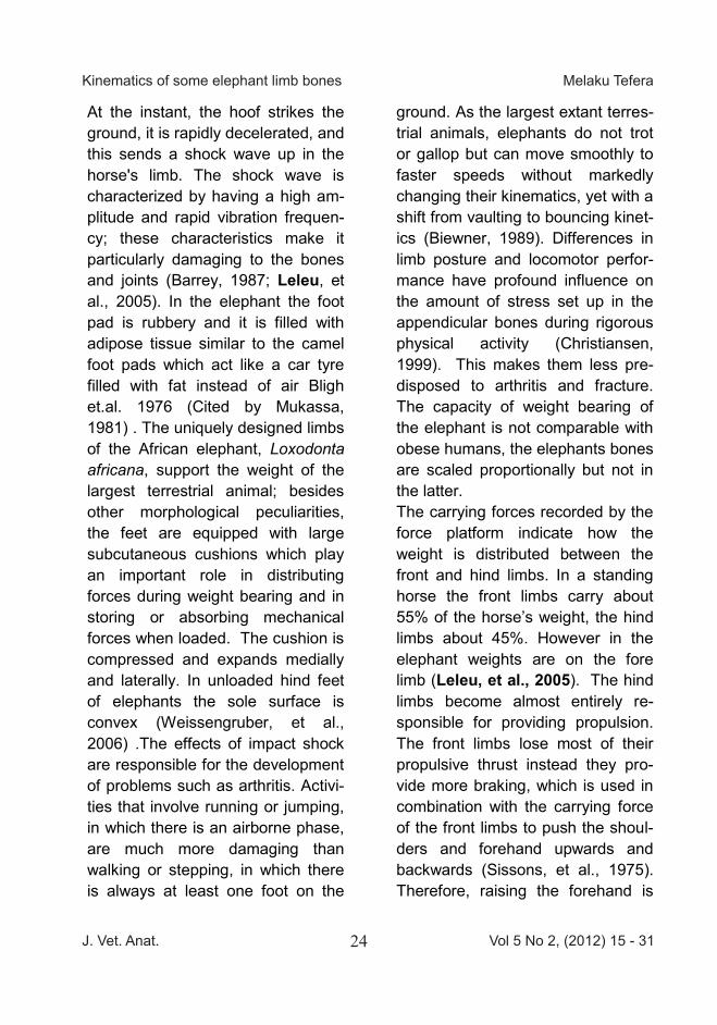
At the instant, the hoof strikes the ground, it is rapidly decelerated, and this sends a shock wave up in the horse's limb. The shock wave is characterized by having a high am-plitude and rapid vibration frequen-cy; these characteristics make it particularly damaging to the bones and joints (Barrey, 1987; Leleu, et al., 2005). In the elephant the foot pad is rubbery and it is filled with adipose tissue similar to the camel foot pads which act like a car tyre filled with fat instead of air Bligh et.al. 1976 (Cited by Mukassa, 1981) . The uniquely designed limbs of the African elephant, Loxodonta africana, support the weight of the largest terrestrial animal; besides other morphological peculiarities, the feet are equipped with large subcutaneous cushions which play an important role in distributing forces during weight bearing and in storing or absorbing mechanical forces when loaded. The cushion is compressed and expands medially and laterally. In unloaded hind feet of elephants the sole surface is convex (Weissengruber, et al., 2006) .The effects of impact shock are responsible for the development of problems such as arthritis. Activi-ties that involve running or jumping, in which there is an airborne phase, are much more damaging than walking or stepping, in which there is always at least one foot on the
ground. As the largest extant terres-trial animals, elephants do not trot or gallop but can move smoothly to faster speeds without markedly changing their kinematics, yet with a shift from vaulting to bouncing kinet-ics (Biewner, 1989). Differences in limb posture and locomotor perfor-mance have profound influence on the amount of stress set up in the appendicular bones during rigorous physical activity (Christiansen, 1999). This makes them less pre-disposed to arthritis and fracture. The capacity of weight bearing of the elephant is not comparable with obese humans, the elephants bones are scaled proportionally but not in the latter. The carrying forces recorded by the force platform indicate how the weight is distributed between the front and hind limbs. In a standing horse the front limbs carry about 55% of the horse’s weight, the hind limbs about 45%. However in the elephant weights are on the fore limb (Leleu, et al., 2005). The hind limbs become almost entirely re-sponsible for providing propulsion. The front limbs lose most of their propulsive thrust instead they pro-vide more braking, which is used in combination with the carrying force of the front limbs to push the shoul-ders and forehand upwards and backwards (Sissons, et al., 1975). Therefore, raising the forehand is
much more than simply a result of lowering the hindquarters, it is an active process brought about by the action of the front limbs. Cattle are kept in confinement, most studies focus on lameness thus locomotion and kinematic studies of domestic animals have been almost solely focused on horses (Fredricson, et al. 1989).The natural gaits of cattle are walking, trotting or galloping (Raven, 1989). Normally they move relatively slow walking pace, they trot when they have to move fast and this movement might change into gallop only for short distance. The head is mobile and the limbs are angular. In all the tree species the propulsion force comes from the hind limb. However, in elephants the head moves very little dorsoventrally or laterally. The elephant should turn its body to see things behind. The shape of the head is conical and it serves as cone nose making the elephant aerodynamically efficient, including during swimming, when the elephants moves the trunk stays straight down used as front splitter. In conclusion understanding the role of the feet of a variety of different organisms in a wide range of body types, foot shapes, arrangement of structures, loading conditions and other variables is important to the understanding of biomechanics and
predisposing factors to lameness, arthritis and fractures. Comparing bone morphology and allometry can be a tool in archeological and foren-sic research, and help to solve prob-lems of Shoe and prosthetic engi-neering. References Alexander, R. M. Jayes, A. S.
Maloiy, G. M. O and Wathuta, E. M. (2009): Al-lometry of the limb bones of mammals from shrews (Sorex) to elephant (Lox-odonta). Journal of Zoolo-gy.189 (3) 305–314. DOI: 10.1111/j.1469-7998.1979.tb03964.x.
Alexander, R. Maloiy, G.M.O. Hunter, B. Jayes, A.S. and Nturibi, J. (1979): Mechani-cal stresses in fast locomo-tion of buffalo (Syncerus caffer) and elephant (Lox-odonta africana). J. Zool. 189: 135–144.
Alexander, R.M. Langman, V.A. and Jayes, A.S. (1977): Fast lo-comotion of some African ungulates. J. Zool. 183, 291-300.
Anderson, J.F and Hall-Martin, A. (1985): Long-bone circum-ference and weight in mam-mals, birds and dinosaurs. Journal of Zoology. 207 (1):
J. Vet. Anat. Vol 5 No 2, (2012) 15 - 3125
Kinematics of some elephant limb bones Melaku Tefera
At the instant, the hoof strikes the ground, it is rapidly decelerated, and this sends a shock wave up in the horse's limb. The shock wave is characterized by having a high am-plitude and rapid vibration frequen-cy; these characteristics make it particularly damaging to the bones and joints (Barrey, 1987; Leleu, et al., 2005). In the elephant the foot pad is rubbery and it is filled with adipose tissue similar to the camel foot pads which act like a car tyre filled with fat instead of air Bligh et.al. 1976 (Cited by Mukassa, 1981) . The uniquely designed limbs of the African elephant, Loxodonta africana, support the weight of the largest terrestrial animal; besides other morphological peculiarities, the feet are equipped with large subcutaneous cushions which play an important role in distributing forces during weight bearing and in storing or absorbing mechanical forces when loaded. The cushion is compressed and expands medially and laterally. In unloaded hind feet of elephants the sole surface is convex (Weissengruber, et al., 2006) .The effects of impact shock are responsible for the development of problems such as arthritis. Activi-ties that involve running or jumping, in which there is an airborne phase, are much more damaging than walking or stepping, in which there is always at least one foot on the
ground. As the largest extant terres-trial animals, elephants do not trot or gallop but can move smoothly to faster speeds without markedly changing their kinematics, yet with a shift from vaulting to bouncing kinet-ics (Biewner, 1989). Differences in limb posture and locomotor perfor-mance have profound influence on the amount of stress set up in the appendicular bones during rigorous physical activity (Christiansen, 1999). This makes them less pre-disposed to arthritis and fracture. The capacity of weight bearing of the elephant is not comparable with obese humans, the elephants bones are scaled proportionally but not in the latter. The carrying forces recorded by the force platform indicate how the weight is distributed between the front and hind limbs. In a standing horse the front limbs carry about 55% of the horse’s weight, the hind limbs about 45%. However in the elephant weights are on the fore limb (Leleu, et al., 2005). The hind limbs become almost entirely re-sponsible for providing propulsion. The front limbs lose most of their propulsive thrust instead they pro-vide more braking, which is used in combination with the carrying force of the front limbs to push the shoul-ders and forehand upwards and backwards (Sissons, et al., 1975). Therefore, raising the forehand is
much more than simply a result of lowering the hindquarters, it is an active process brought about by the action of the front limbs. Cattle are kept in confinement, most studies focus on lameness thus locomotion and kinematic studies of domestic animals have been almost solely focused on horses (Fredricson, et al. 1989).The natural gaits of cattle are walking, trotting or galloping (Raven, 1989). Normally they move relatively slow walking pace, they trot when they have to move fast and this movement might change into gallop only for short distance. The head is mobile and the limbs are angular. In all the tree species the propulsion force comes from the hind limb. However, in elephants the head moves very little dorsoventrally or laterally. The elephant should turn its body to see things behind. The shape of the head is conical and it serves as cone nose making the elephant aerodynamically efficient, including during swimming, when the elephants moves the trunk stays straight down used as front splitter. In conclusion understanding the role of the feet of a variety of different organisms in a wide range of body types, foot shapes, arrangement of structures, loading conditions and other variables is important to the understanding of biomechanics and
predisposing factors to lameness, arthritis and fractures. Comparing bone morphology and allometry can be a tool in archeological and foren-sic research, and help to solve prob-lems of Shoe and prosthetic engi-neering. References Alexander, R. M. Jayes, A. S.
Maloiy, G. M. O and Wathuta, E. M. (2009): Al-lometry of the limb bones of mammals from shrews (Sorex) to elephant (Lox-odonta). Journal of Zoolo-gy.189 (3) 305–314. DOI: 10.1111/j.1469-7998.1979.tb03964.x.
Alexander, R. Maloiy, G.M.O. Hunter, B. Jayes, A.S. and Nturibi, J. (1979): Mechani-cal stresses in fast locomo-tion of buffalo (Syncerus caffer) and elephant (Lox-odonta africana). J. Zool. 189: 135–144.
Alexander, R.M. Langman, V.A. and Jayes, A.S. (1977): Fast lo-comotion of some African ungulates. J. Zool. 183, 291-300.
Anderson, J.F and Hall-Martin, A. (1985): Long-bone circum-ference and weight in mam-mals, birds and dinosaurs. Journal of Zoology. 207 (1):
J. Vet. Anat. Vol 5 No 2, (2012) 15 - 3126
Kinematics of some elephant limb bones Melaku Tefera
53–61. DOI: 10.1111/j.1469-7998.1985.tb04915.x.
Barrey, E. (1987): Foot Biomechan- ics in the Normal Horse: A study of the Hoof Force Dis-tribution in the Forelimb with a New Measuring Method. http://w4.ub.uni konstanz. de/cpa/article/viewFile/2355/2225
Biewner, A.A. (1983): Allometry of quadripedal locomotion: The scaling of duty factor, bone curvature and limb orienta-tion to body size. J. exp. Bi-ol.105:47-171.
Biewner, A.A.(1989): Scaling sup port in mammalian limb. Sci-ence News Series. 245: (4913) 45-48.
Blanc, J. (2008): Loxodonta Africa na. In: IUCN 2010. IUCN Red List of Threatened Spe-cies. Version 2010.4. <www.iucnredlist.org>. Downloaded on 31 January 2011.
Bonnan, M.F. (2007): Linear and ge ometric morphometric analy-sis of long bone scaling pat-terns in Jurassic neosauro-pod dinosaurs: their func-tional and paleobiological implications. Anat Rec (Ho-boken). 290(9):1089-111
Christiansen, P. (1999): Scaling of the limb long bones to body mass in terrestrial mammals
Journal of Morphology. 239 :(20) 167–190. DOI: 10.1002/ (SICI)1097-4687(199902)239:2<167::AID-JMOR5>3.0.CO;2-8
Christiansen, P. (2002): Mass alo- metry of the appendicular skeleton in terrestrial mam-mals. Journal of Morphology. 251: (2) 195–209. DOI: 10. 1002 / J. mor.1083.
Coombs, W.P. (1978): Theoretical aspects of cursorial adapta-tions in dinosaurs. Quarterly Rev. BioI. 53(4): 393-418.
Eggert, L.S. Rasner, C.A. and Woodruff, D.S. (2002): The evolution and phylogeogra-phy of the African elephant inferred from mitochondrial DNA sequence and nuclear microsatellite markers. Proc. R. Soc. Lond. B 269, 1993–2006. DOI 10.1098/ rspb. 2002. 2070.
Fredricson, I. Drevemo, S. Dalin, G. Hjertén, G. Björne, K. and Rynde, R. (1983): Treadmill for equine locomotion analy-sis. Equine Vet. J. 15(2), 111- 115.
Elephant encyclopedia. http://www.upali.ch/elephant_encyclopedia.html www.elephantmagazine.org.
Hildebrand, M. (1984): Rotations of the leg segments of three fast-running cursors and an
elephant. J. Mammal. 65: 718-720.
Hutchinson, J.R, D. Schwerda, D. J. Famini, R. H. I. Dale, M. S. Fischer and R. Kram, (2006): The locomotor kine-matics of Asian and African elephants: changes with speed and size. The Journal of Experimental Biology. 209: 3812-3827
Hutchinson, J.R. Schwerda, D. Famini, D. Dale, R.H.I. Fischer, M and Kram, R. (2006): The locomotor kine-matics of African and Asian elephants: changes with speed and size. Journal of Experimental Biology. 209: 3812-3827.
Leleu C. Cotrel C.and Barrey, E. (2005): Relationships be-tween biomechanical varia-bles and race performance in French Standard bred trot-ters. Livestock Production Science. 92 I (1) Pages 39-46.
McMahon, T.A. (1973): Size and shape in biology. Elastic cri-teria impose limits on biolog-ical proportions, and conse-quently on metabolic rates. Science. 179(4079): 1201-1204.
McMahon, T.A. (1975): Using body size to understand the struc-tural design of animals:
Quadrupedal locomotion. J. App. Physiol. 39(4): 619-627.
Mukasa-Mugerewa, E. (1981): The camel (Camelus dromedar-ies): a bibliographical review. ILCA. Monograph. Addis Ababa, Ethiopia. pp71
Nomina Anatomica Veterinaria. (2005): 5th ed. International Committee on Veterinary Gross Anatomical Nomen-clature (I.C.V.G.A.N.). Knox-ville, TN. USA. Toussaint E. Raven T.E. 1989. Cattle Foot Care and Claw Trim-ming. Farming Press, Ips-wich, UK
Ren, L. Butler, M. Miller , C. Paxton, H. Schwerda , D. Fischer, M.S and Hutchinson, J.R. (2008): The movements of limb segments and joints during locomotion in African and Asian elephants. J Exp Biol. 211(17):2735-51.
Rohland, N. Reich, D. Mallick , S. Meyer, M. Green R.E. (2010): Genomic DNA Se-quences from Mastodon and Woolly Mammoth Reveal Deep Speciation of Forest and Savanna Elephants. PLoS Biol 8(12): e1000564. doi: 10. 1371/journal.pbio.1000564
Russell, D.A. (1985): Scaling of the
J. Vet. Anat. Vol 5 No 2, (2012) 15 - 3127
Kinematics of some elephant limb bones Melaku Tefera
53–61. DOI: 10.1111/j.1469-7998.1985.tb04915.x.
Barrey, E. (1987): Foot Biomechan- ics in the Normal Horse: A study of the Hoof Force Dis-tribution in the Forelimb with a New Measuring Method. http://w4.ub.uni konstanz. de/cpa/article/viewFile/2355/2225
Biewner, A.A. (1983): Allometry of quadripedal locomotion: The scaling of duty factor, bone curvature and limb orienta-tion to body size. J. exp. Bi-ol.105:47-171.
Biewner, A.A.(1989): Scaling sup port in mammalian limb. Sci-ence News Series. 245: (4913) 45-48.
Blanc, J. (2008): Loxodonta Africa na. In: IUCN 2010. IUCN Red List of Threatened Spe-cies. Version 2010.4. <www.iucnredlist.org>. Downloaded on 31 January 2011.
Bonnan, M.F. (2007): Linear and ge ometric morphometric analy-sis of long bone scaling pat-terns in Jurassic neosauro-pod dinosaurs: their func-tional and paleobiological implications. Anat Rec (Ho-boken). 290(9):1089-111
Christiansen, P. (1999): Scaling of the limb long bones to body mass in terrestrial mammals
Journal of Morphology. 239 :(20) 167–190. DOI: 10.1002/ (SICI)1097-4687(199902)239:2<167::AID-JMOR5>3.0.CO;2-8
Christiansen, P. (2002): Mass alo- metry of the appendicular skeleton in terrestrial mam-mals. Journal of Morphology. 251: (2) 195–209. DOI: 10. 1002 / J. mor.1083.
Coombs, W.P. (1978): Theoretical aspects of cursorial adapta-tions in dinosaurs. Quarterly Rev. BioI. 53(4): 393-418.
Eggert, L.S. Rasner, C.A. and Woodruff, D.S. (2002): The evolution and phylogeogra-phy of the African elephant inferred from mitochondrial DNA sequence and nuclear microsatellite markers. Proc. R. Soc. Lond. B 269, 1993–2006. DOI 10.1098/ rspb. 2002. 2070.
Fredricson, I. Drevemo, S. Dalin, G. Hjertén, G. Björne, K. and Rynde, R. (1983): Treadmill for equine locomotion analy-sis. Equine Vet. J. 15(2), 111- 115.
Elephant encyclopedia. http://www.upali.ch/elephant_encyclopedia.html www.elephantmagazine.org.
Hildebrand, M. (1984): Rotations of the leg segments of three fast-running cursors and an
elephant. J. Mammal. 65: 718-720.
Hutchinson, J.R, D. Schwerda, D. J. Famini, R. H. I. Dale, M. S. Fischer and R. Kram, (2006): The locomotor kine-matics of Asian and African elephants: changes with speed and size. The Journal of Experimental Biology. 209: 3812-3827
Hutchinson, J.R. Schwerda, D. Famini, D. Dale, R.H.I. Fischer, M and Kram, R. (2006): The locomotor kine-matics of African and Asian elephants: changes with speed and size. Journal of Experimental Biology. 209: 3812-3827.
Leleu C. Cotrel C.and Barrey, E. (2005): Relationships be-tween biomechanical varia-bles and race performance in French Standard bred trot-ters. Livestock Production Science. 92 I (1) Pages 39-46.
McMahon, T.A. (1973): Size and shape in biology. Elastic cri-teria impose limits on biolog-ical proportions, and conse-quently on metabolic rates. Science. 179(4079): 1201-1204.
McMahon, T.A. (1975): Using body size to understand the struc-tural design of animals:
Quadrupedal locomotion. J. App. Physiol. 39(4): 619-627.
Mukasa-Mugerewa, E. (1981): The camel (Camelus dromedar-ies): a bibliographical review. ILCA. Monograph. Addis Ababa, Ethiopia. pp71
Nomina Anatomica Veterinaria. (2005): 5th ed. International Committee on Veterinary Gross Anatomical Nomen-clature (I.C.V.G.A.N.). Knox-ville, TN. USA. Toussaint E. Raven T.E. 1989. Cattle Foot Care and Claw Trim-ming. Farming Press, Ips-wich, UK
Ren, L. Butler, M. Miller , C. Paxton, H. Schwerda , D. Fischer, M.S and Hutchinson, J.R. (2008): The movements of limb segments and joints during locomotion in African and Asian elephants. J Exp Biol. 211(17):2735-51.
Rohland, N. Reich, D. Mallick , S. Meyer, M. Green R.E. (2010): Genomic DNA Se-quences from Mastodon and Woolly Mammoth Reveal Deep Speciation of Forest and Savanna Elephants. PLoS Biol 8(12): e1000564. doi: 10. 1371/journal.pbio.1000564
Russell, D.A. (1985): Scaling of the
J. Vet. Anat. Vol 5 No 2, (2012) 15 - 3128
Kinematics of some elephant limb bones Melaku Tefera
limb long bones to body mass in terrestrial mammals Journal of Zoology. 207: (1) 53–61.
Sissons, S. Grossman, J and Getty, R. (1975): The anatomy of domestic animals. 5th eds. Saunders Company USA
Vandermerwe, N.J. Bezuidenhout, A.J and Seegers, C.D. (1995): The skull and man-dible of African elephant
(Loxodonta africana). Jour-nal of Veterinary Research. 62:245-260
Weissengruber, G. E. Egger, G. F. Hutchinson, J. R. Groe-newald, H. B. Elsässer, L.
Famini, D and Forstenpoint-ner, G. (2006): The structure of the cushions in the feet of African Elephants (Loxodon-ta Africana). Journal of Anatomy.209: (6)781-792
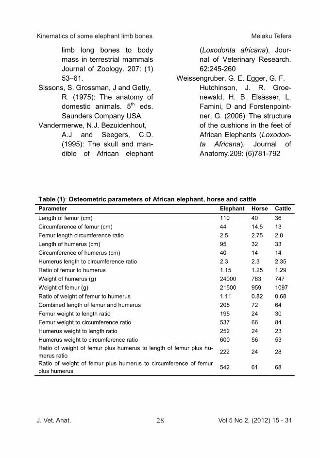
Table (1): Osteometric parameters of African elephant, horse and cattle Parameter Elephant Horse Cattle Length of femur (cm) 110 40 36 Circumference of femur (cm) 44 14.5 13 Femur length circumference ratio 2.5 2.75 2.8 Length of humerus (cm) 95 32 33 Circumference of humerus (cm) 40 14 14 Humerus length to circumference ratio 2.3 2.3 2.35 Ratio of femur to humerus 1.15 1.25 1.29 Weight of humerus (g) 24000 783 747 Weight of femur (g) 21500 959 1097 Ratio of weight of femur to humerus 1.11 0.82 0.68 Combined length of femur and humerus 205 72 64 Femur weight to length ratio 195 24 30 Femur weight to circumference ratio 537 66 84 Humerus weight to length ratio 252 24 23 Humerus weight to circumference ratio 600 56 53 Ratio of weight of femur plus humerus to length of femur plus hu-merus ratio
222 24 28
Ratio of weight of femur plus humerus to circumference of femur plus humerus 542 61 68
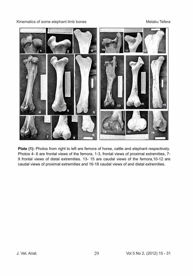
Plate (1): Photos from right to left are femora of horse, cattle and elephant respectively. Photos 4- 6 are frontal views of the femora, 1-3, frontal views of proximal extremities, 7-9 frontal views of distal extremities. 13- 15 are caudal views of the femora,10-12 are caudal views of proximal extremities and 16-18 caudal views of and distal extremities.
J. Vet. Anat. Vol 5 No 2, (2012) 15 - 3129
Kinematics of some elephant limb bones Melaku Tefera
limb long bones to body mass in terrestrial mammals Journal of Zoology. 207: (1) 53–61.
Sissons, S. Grossman, J and Getty, R. (1975): The anatomy of domestic animals. 5th eds. Saunders Company USA
Vandermerwe, N.J. Bezuidenhout, A.J and Seegers, C.D. (1995): The skull and man-dible of African elephant
(Loxodonta africana). Jour-nal of Veterinary Research. 62:245-260
Weissengruber, G. E. Egger, G. F. Hutchinson, J. R. Groe-newald, H. B. Elsässer, L.
Famini, D and Forstenpoint-ner, G. (2006): The structure of the cushions in the feet of African Elephants (Loxodon-ta Africana). Journal of Anatomy.209: (6)781-792
Table (1): Osteometric parameters of African elephant, horse and cattle Parameter Elephant Horse Cattle Length of femur (cm) 110 40 36 Circumference of femur (cm) 44 14.5 13 Femur length circumference ratio 2.5 2.75 2.8 Length of humerus (cm) 95 32 33 Circumference of humerus (cm) 40 14 14 Humerus length to circumference ratio 2.3 2.3 2.35 Ratio of femur to humerus 1.15 1.25 1.29 Weight of humerus (g) 24000 783 747 Weight of femur (g) 21500 959 1097 Ratio of weight of femur to humerus 1.11 0.82 0.68 Combined length of femur and humerus 205 72 64 Femur weight to length ratio 195 24 30 Femur weight to circumference ratio 537 66 84 Humerus weight to length ratio 252 24 23 Humerus weight to circumference ratio 600 56 53 Ratio of weight of femur plus humerus to length of femur plus hu-merus ratio
222 24 28
Ratio of weight of femur plus humerus to circumference of femur plus humerus 542 61 68
Plate (1): Photos from right to left are femora of horse, cattle and elephant respectively. Photos 4- 6 are frontal views of the femora, 1-3, frontal views of proximal extremities, 7-9 frontal views of distal extremities. 13- 15 are caudal views of the femora,10-12 are caudal views of proximal extremities and 16-18 caudal views of and distal extremities.
J. Vet. Anat. Vol 5 No 2, (2012) 15 - 3130
Kinematics of some elephant limb bones Melaku Tefera
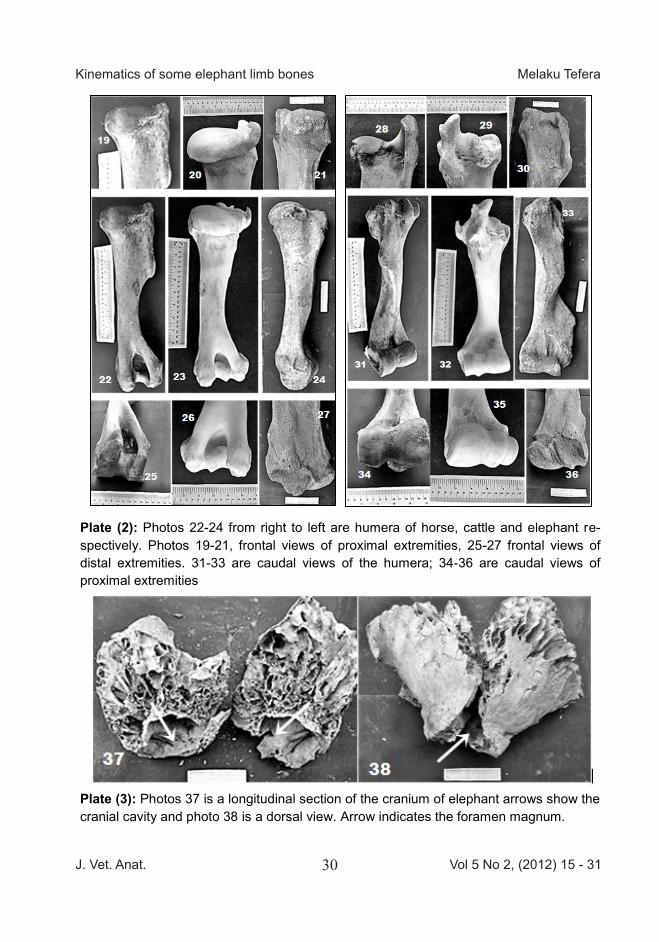
Plate (2): Photos 22-24 from right to left are humera of horse, cattle and elephant re-spectively. Photos 19-21, frontal views of proximal extremities, 25-27 frontal views of distal extremities. 31-33 are caudal views of the humera; 34-36 are caudal views of proximal extremities
Plate (3): Photos 37 is a longitudinal section of the cranium of elephant arrows show the cranial cavity and photo 38 is a dorsal view. Arrow indicates the foramen magnum.
Fig (1): Biomechanics and direction of forces in the elephant foot during locomotion
Fig (2): Kinematic analysis of the elephant foot
The fore-foot tracks are indicated by black dots and the hind-foot tracks by white dots. Where the hind-foot track registers on the fore-foot track it is indicated by a half-black and half-white dot. Key: (a) slow walk (b) normal walk (c) trot and (d) location of center of gravity.
J. Vet. Anat. Vol 5 No 2, (2012) 15 - 3131
Kinematics of some elephant limb bones Melaku Tefera
Plate (2): Photos 22-24 from right to left are humera of horse, cattle and elephant re-spectively. Photos 19-21, frontal views of proximal extremities, 25-27 frontal views of distal extremities. 31-33 are caudal views of the humera; 34-36 are caudal views of proximal extremities
Plate (3): Photos 37 is a longitudinal section of the cranium of elephant arrows show the cranial cavity and photo 38 is a dorsal view. Arrow indicates the foramen magnum.
Fig (1): Biomechanics and direction of forces in the elephant foot during locomotion
Fig (2): Kinematic analysis of the elephant foot
The fore-foot tracks are indicated by black dots and the hind-foot tracks by white dots. Where the hind-foot track registers on the fore-foot track it is indicated by a half-black and half-white dot. Key: (a) slow walk (b) normal walk (c) trot and (d) location of center of gravity.

Morphometric Studies on the Spinal Cord Seg-ments of the Domestic Rabbit (Oryctolagus cunicu-lus) Farag,F.M., Elayat,M.A., Wally,Y.R. and ElKarmoty,A.F. Faculty of Veterinary Medicine, Cairo University, Egypt With 12 figures, 3 tables Received January, accepted for publication September 2012 Abstract Twelve adult angora and chinchillas rabbits of different ages, sex and weights were used. After the rou-tine preparation and dissection of the specimens the spinal cord ex-posed for morphometric studies by using Venire Caliber and magnifying lens. The measurements taken comprised; the total length of the spinal cord, the dorsal, ventral root attachment and inter root lengths – segment lengths, the transverse and dorsoventral diameters lengths, the cervical and lumbar enlargements, as well as the conus medullaris. Key words Rabbit, Spinal cord, Morphometry Introduction The anatomical studies of the spinal cord received the attention of many anatomists. In this respect Mansour (1980), Abu-Zaid (1982), Abd El-ghany (1995) gave valuable studies on the anatomy of the spinal cord in donkey, buffalo, and goat respec-tively. Gabr (1982) provided devel-
opmental studies on the spinal cord of the rabbit however, the present investigation aimed to extend the knowledge on the morphometric records of the spinal cord of the rabbit via the quantitative measure-ments. Material and Methods The present study was conducted on twelve adult angora and chinchil-las rabbits of different sex. The animals were prepared, scari-fied and bled through the common carotid arteries. The blood vessels were thoroughly washed by worm normal saline solution then injected by an amount of 150-180 cc forma-lin (10%). The cadavers were then preserved in 10% formalin solution for a duration ranged between 10-15 days before they manually dis-sected. Measurements were achie-ved by the aid of a magnifying lens and Venire caliper. The obtained values are recorded and tabulated. The Nomenclature used in this study was adopted according to the Nomina Anatomica Vetrinaria N.A.V. (2005).
Animals of this issue
African bush elephant (Loxodonta Africana)
Kingdom: Animalia, Phylum: Chordata, Subphylum: Vertebrata, Class: Mammalia, Superorder: Afrotheria, Order: Proboscidea, Family: Elephantidae, Genus:
Loxodonta & Elephas
Elephants are large mammals of the family Elephantidae and the order Proboscidea. They are represented by three extant species: the African bush elephant (Loxodonta africana), the African forest elephant (L. cyclotis) and the Asian elephant (Elephas maximus). The two African species were traditionally considered to be two different subspecies, in the same species. These three species are scattered throughout sub-Saharan Africa and South to Southeast Asia. They are the only surviving proboscideans, although several extinct species have been identified, including the elephants' close relatives, the mammoths. Elephants are the largest living terrestrial animals. Male African bush elephants can reach a height of 3.20–4 m (10.5–13.1 ft) and a weight of 4,700–6,048 kg (10,362–13,334 lb). The animals have several distinctive features, including a long proboscis or trunk that they use for numerous purposes, particularly for grasping objects. The ear flaps are particularly large and help to control the temperature of their massive bodies. Their incisors grow into large tusks, which serve as tools for digging and moving, as well as weapons for fighting. The African species have larger ears and concave backs while the Asian elephant has smaller ears and a convex back. (Source: Wikipedia)