Embed Size (px)
Citation preview
Met. Mater. Int., Vol. 18, No. 3 (2012), pp. 433~438doi: 10.1007/s12540-012-3008-0
Kinetic Models of Controllable Pore Growth of Anodic AluminumOxide Membrane
Yan Huang, Hong-yan Zeng*, Ce Zhao, Ye-qing Qu, and Pin Zhang
College of Chemical Engineering, University of Xiangtan, Hunan 411105, China
(received date: 14 June 2010 / accepted date: 31 May 2011)
An anodized Al2O3 (AAO) membrane with apertures about 72 nm in diameter was prepared by two-stepanodic oxidation. The appearance and pore arrangement of the AAO membrane were characterized byenergy dispersive x-ray spectroscopy and scanning electron microscopy. It was confirmed that the poreswith high pore aspect ratio were parallel, well-ordered, and uniform. The kinetics of pores growth in theAAO membrane was derived, and the kinetic models showed that pores stopped developing when thepressure (σ) trended to equal the surface tension at the end of anodic oxidation. During pore expansion,the effects of the oxalic acid concentration and expansion time on the pore size were investigated, and thekinetic behaviors were explained with two kinetic models derived in this study. They showed that the poresize increased with extended time (r=G·t+G’), but decreased with increased concentration (r = −K·lnc-K’)through the derived mathematic formula. Also, the values of G, G’, K, and K’ were derived from ourexperimental data.
Key words: porous materials, alumina, anodization, electrochemistry, scanning electron microscopy(SEM)
1. INTRODUCTION
The process of aluminum anodization has received grow-ing attention because anodic porous alumina templates arewidely used for fabrication of various nanostructured materi-als. In the last decade, nanoporous anodic aluminum oxide(AAO) has become the object of much attention in variousfields of research. Anodic oxidation of aluminum in variousacid solutions causes a layer of aluminum oxide to form onthe metal surface. This layer is characterized by parallelpores with orientations perpendicular to the sheet surface,and the pore aspect ratio (pore diameter vs. pore length) canreach values of 1:1 000 and more [1-5].
Porous anodic aluminum oxide is predominantly used asan important material in the fabrication of nanostructures. Anumber of studies have been performed in which the nanop-orous structure is used as a template in the production ofnanowires or nanotubes from various materials, such ascarbon, metals, or polymers [6-10]. In addition, the porousmembrane is used as a filter [11,12]. The adhesion of sub-stances to the pore walls has also been used in some bio-sensor applications [13,14]. Another approach is to usethe filtration capacities of nanoporous aluminum oxide for
purification of DNA or whole cells from blood [15,16].Furthermore, alumina is already a well-known material inorthopedic surgery and dentistry. Here, it is normally usedwith smoothed surfaces and has already proven biocompat-ible [17].
There has been a great deal of work investigating thestructure, nature, and properties of AAOs in recent decades[18-23]. Takhistov and Biosens [24] studied mass-transfer atthe nanoporous substrate by electrochemical impedancespectroscopy. Patermarakis and Moussoutzanis [25] devel-oped a kinetic model obtained at different bath temperaturesand current densities of Al anodization. Sullivan and Wood[26] presented a model of self-organizing pore growth whichwas based on the electric field distribution at the pore tips.Together with other models proposed by the previousresearchers [27], Singaraju et al. [28,29] developed a modelfor porous AAO formation under constant current conditionsand a model under constant voltage anodization. Parkhutikand Shershulsky [30] explained the dependence of pore diam-eters and pore distances on applied voltage and electrolytecomposition. However, little work on investigating thekinetics of pore growth (the specific mathematic relation ofpore size vs. electrolyte concentration and pore size vs.expansion time) was reported. In our work, kinetics modelswere obtained by investigating the pore growth process inan AAO membrane and the specific mathematic relation ofpore size to expansion time and electrolyte concentration.
*Corresponding author: [email protected],[email protected]©KIM and Springer, Published 20 June 2012
434 Yan Huang et al.
2. EXPERIMENTAL PROCEDURES
2.1. Preparation of AAO membraneA nanoporous aluminum membrane was prepared by
anodization of aluminum. Aluminum sheets (99.999%, sizeof 100 mm × 50 mm × 0.5 mm) were annealed at 500 °C for2 h in a high-purity N2 atmosphere and then cooled in a fur-nace. To remove surface oil from the specimens, they wererinsed with absolute acetone and ethanol, respectively, andthen dried after twice washing with deionized water. Thesamples were immersed in 4 wt% H2SO4 and 7 wt% H2CrO4
mixed solution (by volume of 1:1) at 70 °C for 2 min toremove the natural oxide membrane, and then immersed in10 wt% NaOH at 20 °C for 15 s to neutralize acid substanceresidues on the surface of the Al sheets. The samples weredried after twice washing with deionized water, immersed inabsolute ethanol at 25 °C for 3 min, and subsequently putinto 60 wt% HCLO4 ethanol solution (HClO4: C2H5OH: H2O=4:4:2, v/v/v) at 30 °C to carry out electrochemical polishingat voltage of 2 to 3 V for 2 to 3 min. The polished sampleswere immersed in a 0.3 M oxalic acid solution under a cur-rent density of 4.0 mA/cm2 for 2 h (first step of anodic oxida-tion). To remove the first oxide membrane, the samples wereimmersed in a mixed solution with a 1:1 volume ratio of 6wt% H3PO4 and 1.8 wt% H2CrO4 at 40 °C for 15 h and thendried after washing with deionized water. The samples wereimmersed in oxalic acid solutions with various concentra-tions of oxalic acid at 40 °C under a constant voltage of 40.0 Vfor 9 h (second step of anodic oxidation). Finally, the sampleswere immersed in 5% H3PO4 (v/v) solution at 30 °C to makethe pores broaden for a period of time, dried after washingwith deionized water, and then annealed with a muffle at900 °C for 1 h. Thus, the AAO membrane was obtained.
2.2. Characterization of AAO membraneScanning electron micrography (SEM) was carried out
with a JEOL JSM-6700F instrument and energy dispersivex-ray spectroscopy (EDS) analysis was performed with aNoran SystemSix instrument. EDS was used to determinethe elemental composition of the AAO membrane. TheSEM photographs were analyzed by the software ImagePro. following the procedure reported by Xing [31]. Thedistribution of pore diameters could be obtained with theanalysis.
3. RESULTS AND DISCUSSION
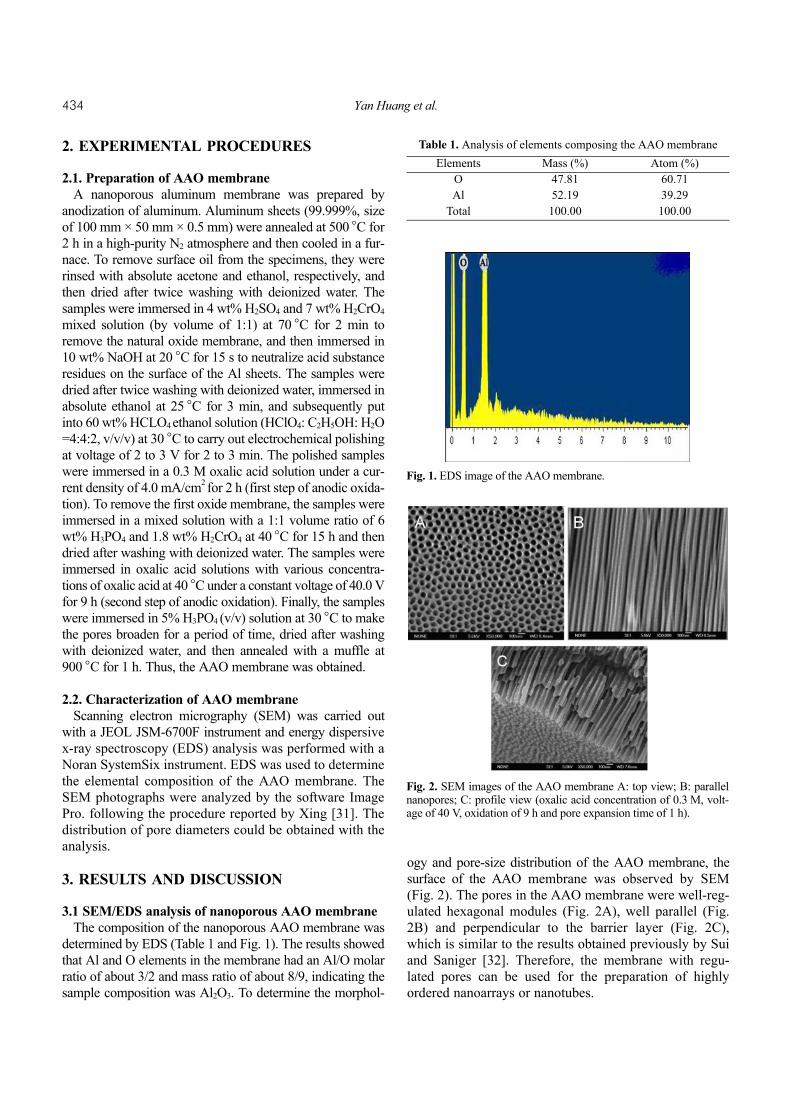
3.1 SEM/EDS analysis of nanoporous AAO membraneThe composition of the nanoporous AAO membrane was
determined by EDS (Table 1 and Fig. 1). The results showedthat Al and O elements in the membrane had an Al/O molarratio of about 3/2 and mass ratio of about 8/9, indicating thesample composition was Al2O3. To determine the morphol-
ogy and pore-size distribution of the AAO membrane, thesurface of the AAO membrane was observed by SEM(Fig. 2). The pores in the AAO membrane were well-reg-ulated hexagonal modules (Fig. 2A), well parallel (Fig.2B) and perpendicular to the barrier layer (Fig. 2C),which is similar to the results obtained previously by Suiand Saniger [32]. Therefore, the membrane with regu-lated pores can be used for the preparation of highlyordered nanoarrays or nanotubes.
Table 1. Analysis of elements composing the AAO membraneElements Mass (%) Atom (%)
O 47.81 60.71Al 52.19 39.29
Total 100.00 100.00
Fig. 1. EDS image of the AAO membrane.
Fig. 2. SEM images of the AAO membrane A: top view; B: parallelnanopores; C: profile view (oxalic acid concentration of 0.3 M, volt-age of 40 V, oxidation of 9 h and pore expansion time of 1 h).
Kinetic Models of Controllable Pore Growth of Anodic Aluminum Oxide Membrane 435
3.2. Growth kinetics of AAO membrane3.2.1. Growth process of pore in the AAO membrane
Under a high electric field, no electron but ions were con-ducted in the aluminum sheet, Therefore, the relation betweenthe applied voltage and current density may be expressed asin [27] as
J=A·exp(B ), (1)
where J is the current density (mA/cm2), A and B areparameters dependent on temperature which is determinedby material physical property, U is the applied voltage (V),and d is the thickness of the barrier layer (nm).
In this process, the change of the electric charge distribu-tion was much faster than that of the geometric size of theAAO membrane pores, so the process could be consideredto be a state of quasi-static electric field. The pores on theAAO membrane grew lengthways, while they developedalong the aperture under force of the electric field parallel tothe aperture. The transverse electric field intensity is denotedas Ex (Ex<<E, E was total electric field intensity). The theoryof electromagnetic field is given as
, (2)
where ω is the density of the electrostatic field, and ε is thedielectric constant of the membrane. Assuming that there isa small volume-element where the depth is l and the area isdS=2πrdr, its electric field energy is
dA= ·2πrdr·l= ·rdr·l . (3)
Therefore, the electrostatic force (F) along the aperture is
F= = ·r·l , (4)
and the pressure (σ) of the electrostatic field per depth is
σ = = ·r. (5)
The value of the pressure increased linearly with increasein the square of Ex and the instant pore diameter, while Ex
decreased with the increase of the pore diameter. The rate ofEx decrease was higher than that of the pore diameterincrease, so that σ decreased with increase in the pore diam-eter. The pores stopped growing, and the pore size and thesurface tension were dependent when the value of σ wasequal to the surface tension of the membrane. Hence, a largenumber of rounded pores with a similar diameter were
formed (Figs. 4B and 7B), which was confirmed in the SEMand AFM images (Figs. 2A, 3Band 6B).
3.2.2. Kinetics of pore diameter to electrolyte concentrationThe growth of the pores in the AAO membrane was not
only related to the applied voltage, but also with electrolyteconcentration and the time of pore expansion [33]. To exam-ine the change of the pore diameter in various oxalic acidconcentrations, an AAO membrane sample was prepared at40 °C and 40 V after 9 h oxidation, and the pores in themembrane were un-expanded (Fig. 3). The pore size distri-bution curves for various concentrations of oxalic acid elec-trolyte are shown in Fig. 4. It was found that the pore diameterdecreased with increasing oxalic acid concentrations. The
Ud----
ωεEX
2
8π--------=
εEX2
8π-------- εEX
2
4--------
∂A∂r------ εEX
2
4--------
EL--- εEX
2
4--------
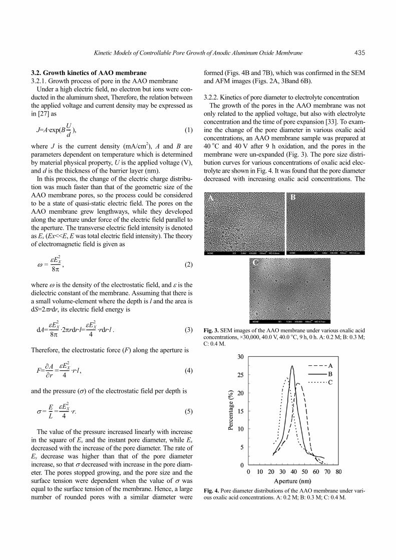
Fig. 3. SEM images of the AAO membrane under various oxalic acidconcentrations, ×30,000, 40.0 V, 40.0 °C, 9 h, 0 h. A: 0.2 M; B: 0.3 M;C: 0.4 M.
Fig. 4. Pore diameter distributions of the AAO membrane under vari-ous oxalic acid concentrations. A: 0.2 M; B: 0.3 M; C: 0.4 M.
436 Yan Huang et al.
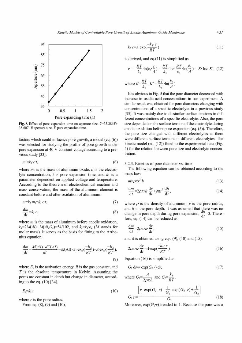
AAO membrane in 0.3 M oxalic acid electrolyte showed thenarrowest pore size distribution of about 39 nm (Fig. 4B),but the AAO membrane in 0.2 and 0.4 M oxalic acid electro-lyte solutions had broader pore size distributions of about 45nm (Fig. 4A) and 35 nm (Fig. 4C), respectively. The resultswere in agreement with SEM data (Fig. 5) calculated fromSEM images (Fig. 3). It was likely because the current den-sity was enhanced with increased oxalic acid concentrations,and the higher the current density, the smaller the aperture atthe same voltage. When the concentration was 0.2 M, thepores of the AAO membrane were the largest but disordered(Fig. 3A). At 0.3 M, the pores grew smaller but were orderedand uniform (Fig. 3B). At 0.4 M, the aperture was the small-est and the pores were disordered, while there appeared to besome mergence between pores in the AAO membrane (Fig.3C). The results showed that the pores were the most orderedand uniform at the concentration of 0.3 M. In addition, theeffect of oxalic acid concentrations on aperture size wasinvestigated at five concentrations, i.e. 0.1, 0.2, 0.3, 0.4, and0.5 M. The data were plotted lnX (X was the oxalic acid con-centration) versus aperture size to give a straight line with ahigh correlation coefficient (R2=0.9973) (Fig. 5).
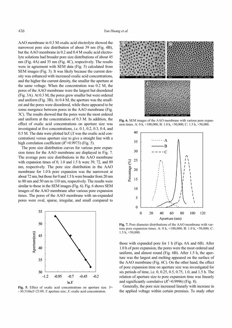
The pore size distribution curves for various pore expan-sion times for the AAO membrane are displayed in Fig. 7.The average pore size distributions in the AAO membranewith expansion times of 0, 1.0 and 1.5 h were 39, 72, and 89nm, respectively. The pore size distribution in the AAOmembrane for 1.0 h pore expansion was the narrowest atabout 72 nm, but those for 0 and 1.5 h were broader from 20 nmto 80 nm and 50 nm to 110 nm, respectively. The results weresimilar to those in the SEM images (Fig. 6). Fig. 6 shows SEMimages of the AAO membrane after various pore expansiontimes. The pores of the AAO membrane with un-expandedpores were oval, sparse, irregular, and small compared to
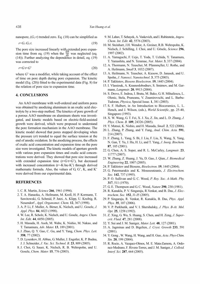
those with expanded pore for 1 h (Figs. 6A and 6B). After1.0 h of pore expansion, the pores were the most ordered anduniform, and almost round (Fig. 6B). After 1.5 h, the aper-ture was the largest and melting appeared on the surface ofthe AAO membrane (Fig. 6C). On the other hand, the effectof pore expansion time on aperture size was investigated forsix periods of time, i.e. 0, 0.25, 0.5, 0.75, 1.0, and 1.5 h. Therelation of aperture size to pore expansion time was linearlyand significantly correlative (R2=0.9996) (Fig. 8).
Generally, the pore size increased linearly with increase inthe applied voltage within certain premises. To study other
Fig. 5. Effect of oxalic acid concentrations on aperture size. Y=−30.316lnX+23.09, Y: aperture size; X: oxalic acid concentration.
Fig. 6. SEM images of the AAO membrane with various pore expan-sion times. A: 0 h, ×100,000; B: 1.0 h, ×50,000; C: 1.5 h, ×50,000.
Fig. 7. Pore diameter distributions of the AAO membrane with var-ious pore expansion times. A: 0 h, ×100,000; B: 1.0 h, ×50,000; C:1.5 h, ×50,000.
Kinetic Models of Controllable Pore Growth of Anodic Aluminum Oxide Membrane 437
factors which could influence pore growth, a model (eq. (6))was selected for studying the profile of pore growth underpore expansion at 40 V constant voltage according to a pre-vious study [33]:
m1=k1·c·t, (6)
where m1 is the mass of aluminum oxide, c is the electro-lyte concentration, t is pore expansion time, and k1 is aparameter dependent on applied voltage and temperature.According to the theorem of electrochemical reaction andmass conservation, the mass of the aluminum element isconstant before and after oxidation of aluminum:
m=k2·m1=k3·c·t, (7)
=k3·c, (8)
where m is the mass of aluminum before anodic oxidation,k2=2M(Al): M(Al2O3)=54/102, and k3=k1·k2 (M stands formolar mass). It serves as the basis for fitting to the Arrhe-nius equation:
= =M(Al) ·A1·exp( )=A·exp( ),
(9)
where Ea is the activation energy, R is the gas constant, andT is the absolute temperature in Kelvin. Assuming thepores are constant in depth but change in diameter, accord-ing to the eq. (10) [34],
Ea=k4·r (10)
where r is the pore radius.From eq. (8), (9) and (10),
k3·c=A·exp( ) (11)
is derived, and eq.(11) is simplified as
r = − ·ln(k3· )=− ·lnc- ·ln( )=−K· lnc-K’, (12)
where K= , K’ = ·ln( ).
It is obvious in Fig. 5 that the pore diameter decreased withincrease in oxalic acid concentrations in our experiment. Asimilar result was obtained for pore diameters changing withconcentrations of a specific electrolyte in a previous study[35]. It was mainly due to dissimilar surface tensions in dif-ferent concentrations of a specific electrolyte. Also, the poresize depended on the surface tension of the electrolyte duringanodic oxidation before pore expansion (eq. (5)). Therefore,the pore size changed with different electrolytes as therewere different surface tensions in different electrolytes. Thekinetic model (eq. (12)) fitted to the experimental data (Fig.5) for the relation between pore size and electrolyte concen-tration.
3.2.3. Kinetics of pore diameter vs. timeThe following equation can be obtained according to the
mass law:
m=ρπr2·h (13)
=2ρπrh· +ρπr2· , (14)
where ρ is the density of aluminum, r is the pore radius,and h is the pore depth. It was assumed that there was nochange in pore depth during pore expansion, =0. There-fore, eq. (14) can be reduced as
=2ρπrh· , (15)
and it is obtained using eqs. (9), (10) and (15).
2ρπrh· =A·exp( ) (16)
Equation (16) is simplified as
G1·dt=r·exp(G2·r)·dr, (17)
where G1= and G2= .
G1·t = (18)
Moreover, exp(G2·r) trended to 1. Because the pore was a
dmdt-------
dmdt------- M Al( ) dC Al( )⋅
dt------------------------------------ Ea–
RT-------- Ea–
RT--------
k4– r⋅RT
-------------
RTk4------- c
A--- RT
k4------- RT
k4------- k3
A----
RTk4------- RT
k4------- k3
A----
dmdt------- dr
dt----- dh
dt------
dhdt------
dmdt------- dr
dt-----
drdt----- k4– r⋅
RT-------------
A2ρπh------------- k4
RT-------
r exp G2 r⋅( )⋅ 1G2------ exp G2 r⋅( )⋅– 1
G2------+
G2---------------------------------------------------------------------------------------
Fig. 8. Effect of pore expansion time on aperture size. Y=33.286T+38.607, Y: aperture size; T: pore expansion time.
438 Yan Huang et al.
nanopore, (G2·r) trended zero. Eq. (18) can be simplified as
r=G1·G2·t. (19)
The pore size increased linearly with extended pore expan-sion time from eq. (19) when the was neglected (eq.(14)). Further analyzing the dependence in detail, eq. (19)was corrected to
r=G·t+G’ (20)
where G’ was a modifier, while taking account of the effectof time on pore depth during pore expansion. The kineticmodel (Eq. (20)) fitted to the experimental data (Fig. 8) forthe relation of pore size to expansion time.
4. CONCLUSIONS
An AAO membrane with well-ordered and uniform poreswas obtained by anodizing aluminum in an oxalic acid elec-trolyte by a two-step method. The kinetics of pore growth ofa porous AAO membrane on aluminum sheets was investi-gated, and kinetic models based on electric-field-assistedgrowth were derived, which were proposed to understandthe pore formation mechanism in the AAO membrane. Thekinetic model showed that pores stopped developing whenthe pressure (σ) trended to equal the surface tension at theend of anodic oxidation. In the anodizing process, the effectsof oxalic acid concentration and expansion time on the poresize were investigated. The kinetic models of aperture growthwith various pore expansion times and oxalic acid concen-trations were derived. They showed that pore size increasedwith extended expansion time (r=G·t+G’), but decreasedwith increased concentration (r=-K·lnc-K’) through derivedmathematic formula. Also, the values of G, G’, K, and K’were derived from our experimental data.
REFERENCES
1. C. R. Martin, Science 266, 1961 (1994).2. T. A. Hanaoka, A. Heilmann, M. Kroll, H. P. Kormann, T.
Sawitowski, G. Schmid, P. Jutzi, A. Klipp, U. Kreibig, R.Neuendorf , Appl. Organomet. Chem. 12, 367 (1998).
3. A. P. Li, F. Muller, A. Birner, K. Nielsch, and U. Gosele, J.Appl. Phys. 84, 6023 (1998).
4. W. Lee, R. Scholz, K. Nielsch, and U. Gosele, Angew. Chem.Int. Edit. 44, 6050 (2005).
5. H. Masuda, H. Asoh, M. Wabe, K. Nishio, M. Nakao, andT. Tamamura, Adv. Mater. 13, 189 (2001).
6. J. Zhao, Q. Y. Gao, C. Gu, and Y. Yang, Chem. Phys. Lett.358, 77 (2002).
7. D. Lysenkov, H. Abbas, G. Muller, J. Engstler, K. P. Budna,J. J. Schneider, J. Vac. Sci. Technol. B. 23, 809 (2005).
8. J. Choi, G. Sauer, K. Nielsch, R. B. Wehrspohn, and U.Gosele, Chem. Mater. 15, 776 (2003).
9. M. Lahav, T. Sehayek, A. Vaskevich, and I. Rubinstein, Angew.Chem. Int. Edit. 42, 5576 (2003).
10. M. Steinhart, J.H. Wendor, A. Greiner, R.B. Wehrspohn, K.Nielsch, J. Schilling, J. Choi, and U. Gösele, Science 296,1997 (2002).
11. A. Yamaguchi, F. Uejo, T. Yoda, T. Uchida, Y. Tanamura,T. Yamashita, and N. Teramae, Nat. Mater. 3, 337 (2004).
12. A. Thormann, N. Teuscher, M. Pfannmçller, U. Rothe, andA. Heilmann, Small 3, 1032 (2007).
13. A. Heilmann, N. Teuscher, A. Kiesow, D. Janasek, and U.Spohn, J. Nanosci. Nanotechnol. 3, 375 (2003).
14. P. Takhistov, Biosens Bioelectron. 19, 1445 (2004).15. I. Vlassiouk, A. Krasnoslobodtsev, S. Smirnov, and M. Ger-
mann, Langmuir. 20, 9913 (2004).16. S. Dreve, E. Indrea, I. Bratu, M. Bako, G. H. Mihailescu, L.
Olenic, Stela, Pruneanu, V. Znamirovschi, and L. Barbu-Tudoran, Physica, Special Issue. 1, 381 (2003).
17. S. F. Hulbert, in An Introduction to Bioceramics, L. L.Hench, and J. Wilson, (eds.), World Scientific, pp. 25-40,Singapore (1993).
18. X. W. Wang, G. T. Fei, X. J. Xu, Z. Jin, and L. D. Zhang, J.Phys. Chem. B. 109, 24326 (2005).
19. Y. Matsui, K. Nishio, and H. Masuda, Small. 2, 522 (2006).20. L. Zhang, P. Zhang, and Y. Fang, Anal. Chim. Acta. 591,
214 (2007).21. F. Zhang, L. Yang, S. Bi, J. Liu, F. Liu, X. Wang, X. Yang,
N. Gan, T. Yu, J. Hu, H. Li, and T. Yang, J. Inorg. Biochem.87, 105 (2001).
22. G. Chen, A. S. Soper, and R. L. McCarley, Langmuir. 23,11777 (2007).
23. W. Zhang, Z. Huang, J. Yu, D. Gao, J. Qian, J. BiomedicalEngineering 22, 1007 (2005).
24. P. Takhistov and Biosens, Bioelectron. 19, 1445 (2004).25. G. Patermarakis and K. Moussoutzanis, J. Electrochem.
Soc. 142, 737 (1995).26. P. O. Sullivan and G. C. Wood, P. Roy. Soc. A-Math. Phy.
317, 511 (1970).27. G. E. Thompson and G. C. Wood, Nature 290, 230 (1981). 28. R. Kanakla, P. V. Singaraju, R.Venkat, and B. Das, J. Elec-
trochem. Soc. 152, J1-J5 (2005).29. P. Singaraju, R. Venkat, R. Kanakla, B. Das, Phys. Appl.
Phys. 35, 107 (2006).30. V. P. Parkhutik, and V. I. Shershulsky, J. Phys. B-At. Mol.
Opt. 25, 1258 (1992). 31. Z. Xing, G. Wu, S. Huang, S. Chen, and H. Zeng, J. Super-
crit. Fluid. 47, 281 (2008). 32. Y. Sui and J. M. Saniger, Mater. Lett. 48, 127 (2001).33. A. Jagminas and D. Bigelien, J. Cryst. Growth 233, 591
(2001).34. Y. Gong , W. Wang, H. Wang, and H. Guo, Acta. Phys-Chim.
Sin. 20, 199 (2004).35. R. Rocio, A. Vazquez-Olmos, M. E. Mata-Zamora, A. Ordo-
nez-Medrano, F. Rivera-Torres, and J. M. Saniger, J. Colloid.Interf. Sci. 287, 664 (2005).
dhdt------


![Controllable Sliding Bearings and Controllable Lubrication ... · Review Controllable Sliding Bearings and Controllable ... or evolutionary [5], but it does not change the fact that](https://img.pdfslide.net/doc/110x75/5fc50df11ca4e1756528a85b/controllable-sliding-bearings-and-controllable-lubrication-review-controllable.jpg)