Embed Size (px)
Citation preview
King’s Research Portal
DOI:10.1016/j.ydbio.2016.02.009
Document VersionPublisher's PDF, also known as Version of record
Link to publication record in King's Research Portal
Citation for published version (APA):Xavier, G. M., Seppala, M., Barrell, W., Birjandi, A. A., Geoghegan, F., & Cobourne, M. T. (2016). Hedgehogreceptor function during craniofacial development. Developmental Biology.https://doi.org/10.1016/j.ydbio.2016.02.009
Citing this paperPlease note that where the full-text provided on King's Research Portal is the Author Accepted Manuscript or Post-Print version this maydiffer from the final Published version. If citing, it is advised that you check and use the publisher's definitive version for pagination,volume/issue, and date of publication details. And where the final published version is provided on the Research Portal, if citing you areagain advised to check the publisher's website for any subsequent corrections.
General rightsCopyright and moral rights for the publications made accessible in the Research Portal are retained by the authors and/or other copyrightowners and it is a condition of accessing publications that users recognize and abide by the legal requirements associated with these rights.
•Users may download and print one copy of any publication from the Research Portal for the purpose of private study or research.•You may not further distribute the material or use it for any profit-making activity or commercial gain•You may freely distribute the URL identifying the publication in the Research Portal
Take down policyIf you believe that this document breaches copyright please contact [email protected] providing details, and we will remove access tothe work immediately and investigate your claim.
Download date: 01. Jun. 2020

Developmental Biology ∎ (∎∎∎∎) ∎∎∎–∎∎∎
Contents lists available at ScienceDirect
Developmental Biology
http://d0012-16
n CorrDental I
E-m
Pleasdoi.o
journal homepage: www.elsevier.com/locate/developmentalbiology
Hedgehog receptor function during craniofacial development
Guilherme M. Xavier a,b, Maisa Seppala a,b, William Barrell a, Anahid A. Birjandi a,Finn Geoghegan a, Martyn T. Cobourne a,b,n
a Department of Craniofacial Development and Stem Cell Biology, King’s College London Dental Institute, Floor 27, Guy’s Hospital, London SE1 9RT, UKb Department of Orthodontics, King’s College London Dental Institute, Floor 27, Guy’s Hospital, London SE1 9RT, UK
a r t i c l e i n f o
Article history:Received 30 July 2015Received in revised form9 February 2016Accepted 10 February 2016
Keywords:Sonic hedgehogCraniofacial developmentDiseaseHoloprosencephalyDispatchedScube2Patched1Patched2Gas1CdonBocLrp2/megalinGpr161SmoothenedEvC1/2
x.doi.org/10.1016/j.ydbio.2016.02.00906/& 2016 The Authors. Published by Elsevier
esponding author at: Department of Orthodonstitute, Floor 27, Guy’s Hospital, London SE1ail address: [email protected] (M.T.
e cite this article as: Xavier, G.M., etrg/10.1016/j.ydbio.2016.02.009i
a b s t r a c t
The Hedgehog signalling pathway plays a fundamental role in orchestrating normal craniofacial devel-opment in vertebrates. In particular, Sonic hedgehog (Shh) is produced in three key domains during theearly formation of the head; neuroectoderm of the ventral forebrain, facial ectoderm and the pharyngealendoderm; with signal transduction evident in both ectodermal and mesenchymal tissue compartments.Shh signalling from the prechordal plate and ventral midline of the diencephalon is required for ap-propriate division of the eyefield and forebrain, with mutation in a number of pathway componentsassociated with Holoprosencephaly, a clinically heterogeneous developmental defect characterized by afailure of the early forebrain vesicle to divide into distinct halves. In addition, signalling from thepharyngeal endoderm and facial ectoderm plays an essential role during development of the face, in-fluencing cranial neural crest cells that migrate into the early facial processes. In recent years, thecomplexity of Shh signalling has been highlighted by the identification of multiple novel proteins that areinvolved in regulating both the release and reception of this protein. Here, we review the contributions ofShh signalling during early craniofacial development, focusing on Hedgehog receptor function and de-scribing the consequences of disruption for inherited anomalies of this region in both mouse models andhuman populations.& 2016 The Authors. Published by Elsevier Inc. This is an open access article under the CC BY-NC-ND
license (http://creativecommons.org/licenses/by-nc-nd/4.0/).
1. Introduction
The Hedgehog signalling pathway plays a key role in normalvertebrate development (Lee et al., 2016; Briscoe and Therond, 2013;Ingham and McMahon, 2001) and maintenance of appropriatepostnatal tissue homoeostasis (Petrova and Joyner, 2014). In mam-mals, there are three Hedgehog-family members, Sonic hedgehog(Shh), Indian hedgehog (Ihh) and Desert hedgehog (Dhh); andamongst these, Shh has a predominant role in orchestrating devel-opmental processes. The complexity of vertebrate Shh signalling hasbeen highlighted by the identification of multiple novel proteins thatare involved in mediating pathway transduction. Here, we review thecontribution of Shh signalling during early craniofacial development,focusing on Hedgehog receptor function and the consequences ofdisruption for anomalies affecting this region in both vertebratemodels and human populations.
Inc. This is an open access article u
ntics, King’s College London9RT, UK.Cobourne).
al., Hedgehog receptor func
2. Shh signal reception and transduction: an overview
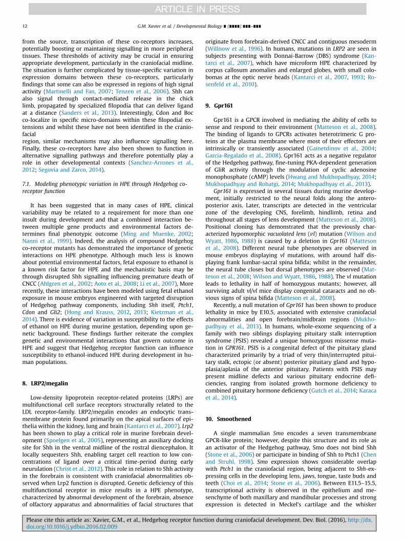
Shh is released from the surface of signalling cells as a dual-lipidated protein modified by the addition of cholesterol andpalmitate groups at the C and N-terminal regions, respectively(Pepinsky et al., 1998; Porter et al., 1995). Release from the en-virons of the cell is achieved through the combined activity ofDispatched (Disp), a multipass sterol-sensing domain protein(Caspary et al., 2002; Kawakami et al., 2002; Ma et al., 2002) andScube2 (Signal peptide CUB EGF-like domain-containing protein),a secreted glycoprotein; both of which interact with Shh throughits cholesterol moiety (Creanga et al., 2012; Tukachinsky et al.,2012) (Fig. 1). Once released, Shh can signal within embryonictissues at both short and long-range (Gritli-Linde et al., 2001).
In recent years, the primary cilium has been identified as a keymultifunctional organelle involved in the essential regulation ofnumerous signalling pathways, including vertebrate Hedgehog(Berbari et al., 2009; Huangfu and Anderson, 2005; Pedersen andRosenbaum, 2008) (Fig. 1). Indeed, ciliopathies are now recognizedas a significant group of disorders that arise from defective func-tion of these organelles (Badano et al., 2006), which often involve
nder the CC BY-NC-ND license (http://creativecommons.org/licenses/by-nc-nd/4.0/).
tion during craniofacial development. Dev. Biol. (2016), http://dx.
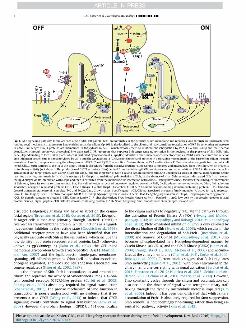
Fig. 1. Shh signalling pathway. In the absence of Shh (OFF, left panel) Ptch1 predominates in the primary cilium membrane and represses Smo through an uncharacterized(but indirect) mechanism that prevents Smo enrichment at the cilium. Gpr161 is also localized to the cilium and may contribute to activation of PKA by generating an increasein cAMP. Full length Gli2/3 proteins are maintained in the cytosol by SuFu, which exposes them to multiple phosphorylation by PKA, CKIα and GSK3β and their partialdegradation (through proteolytic processing) into truncated Gli3R repressors that suppress Shh target gene transcription in the nucleus. In the presence of Shh (ON, rightpanel) ligand binding to Ptch1 takes place, which is facilitated by formation of a Lrp2/Boc/Cdon/Gas1 multi-molecular co-receptor complex. Ptch1 exits the cilium and relief ofSmo inhibition occurs. Smo is phosphorylated by CK1α and the GPCR kinase-2 (GRK2) (not shown) and enriches in a signalling microdomain at the base of the cilium throughformation of an EvC complex involving the ciliary proteins EFCAB7 and IQCE. This results in Smo inhibition of PKA and facilitates Kif7-mediated anterograde transport of a fulllength Gli2/3-SuFu complex to the tip of the cilium, where it dissociates from the negative regulator Sufu. Gpr161 is removed and internalized from the cilium, which preventsits inhibitory activity (not shown). The production of Gli2/3 activators (GliA) derived from the full-length Gli proteins occurs, and accumulation of GliA in the nucleus enablesactivation of Shh target genes, such as Ptch1, Gli1 and Hhip1, and the inhibition of Gas1, Cdo and Boc. In secreting cells, Shh undergoes a series of internal modifications beforereaching an active, multimeric form. Hhat is necessary for the post-translational palmitoylation of Shh; in the absence of Hhat, Shh secretion is decreased. Shh first traversesthe lipid bilayer via its interaction with Disp1 and then is extracted from the membrane via interaction with Scube2. Exactly how Scube2 facilitates the subsequent movementof Shh away from its source remains unclear. Boc, Boc cell adhesion associated oncogene regulated protein; cAMP, Cyclic adenosine monophosphate; Cdon, Cell adhesionassociated, oncogene regulated protein; CK1α, Casein Kinase 1 alpha; Disp1, Dispatched 1; EFCAB7, EF-hand calcium-binding domain-containing protein7; EvC, Ellis-vanCreveld transmembrane protein complex (EvC and EvC2); Gas1, Growth-arrest specific gene 1; Gli, Glioma-associated oncogene family member (A, active form; R, repressorform; FL, full length); Gpr161, orphan rhodopsin GPCR 161; GSK3β, Glycogen synthase kinase 3 beta; Hhat, Hedgehog acyltransferase; Hhip1, Hedgehog-interacting protein 1;IQCE, IQ-domain containing protein E; Kif7, Kinesin family 7; P, phosphorylation; PKA, Protein Kinase A; Ptch1, Patched 1; Lrp2, low-density lipoprotein receptor-relatedprotein; Scube2, Signal peptide CUB EGF-like domain-containing protein 2; Shh, Sonic hedgehog; Smo, Smoothened; Sufu, Suppressor-of-fused.
G.M. Xavier et al. / Developmental Biology ∎ (∎∎∎∎) ∎∎∎–∎∎∎2
disrupted Hedgehog signalling and have an affect on the cranio-facial region (Brugmann et al., 2010; Cortes et al., 2015). Receptionat target cells is mediated primarily through Patched1 (Ptch1), atwelve-pass transmembrane protein, which functions as a ligand-independent inhibitor in the resting state (Goodrich et al., 1996).Additional receptor proteins have also been identified that canphysically associate with Shh at the cell surface, which include thelow-density lipoprotein receptor-related protein, Lrp2 (otherwiseknown as gp330/megalin) (Saito et al., 1994), the GPI-linkedmembrane glycoprotein Growth arrest-specific1 (Gas1) (Martinelliand Fan, 2007) and the Ig/fibronectin single-pass membrane-spanning cell adhesion proteins Cdon (cell adhesion associated,oncogene regulated) and Boc (Boc cell adhesion associated, on-cogene regulated) (Kang et al., 2002, 1997).
In the absence of Shh, Ptch1 accumulates in and around thecilium and represses the activity of Smoothened (Smo), a G-pro-tein coupled receptor (GPCR)-like protein (Corbit et al., 2005;Rohatgi et al., 2007) absolutely required for signal transduction(Zhang et al., 2001). The precise mechanism of Smo function intransduction is poorly understood, with no evidence that it re-presents a true GPCR (Wang et al., 2013) or indeed, that GPCRsignalling events contribute to signal transduction (Low et al.,2008). However, the orphan rhodopsin GPCR Gpr161 does localize
Please cite this article as: Xavier, G.M., et al., Hedgehog receptor funcdoi.org/10.1016/j.ydbio.2016.02.009i
to the cilium and acts to negatively regulate the pathway throughthe activation of Protein Kinase A (PKA) (Hwang and Mukho-padhyay, 2014; Mukhopadhyay and Rohatgi, 2014; Mukhopadhyayet al., 2013). Ptch1-mediated inhibition of Smo is only relieved bythe direct binding of Shh (Stone et al., 2006); which results in theinternalization and degradation of Shh-Ptch1 (Incardona et al.,2000) and removal of Gpr161 (Mukhopadhyay et al., 2013). Smobecomes phosphorylated in a Hedgehog-dependent manner byCasein Kinase 1α (CK1α) and the GPCR Kinase (GRK2) (Chen et al.,2004; Evangelista et al., 2008; Meloni et al., 2006) and accumu-lates at the ciliary membrane (Chen et al., 2011; Corbit et al., 2005;Rohatgi et al., 2009). Current models suggest that Ptch1 regulatesSmo indirectly (Taipale et al., 2002), with Smo enrichment in theciliary membrane correlating with signal activation (Barakat et al.,2013; Firestone et al., 2012; Nedelcu et al., 2013; Ocbina and An-derson, 2008; Ocbina et al., 2011; Rohatgi et al., 2009). However,Smo constantly cycles through the cilium and accumulation canalso occur in the absence of signal when retrograde ciliary traf-ficking through the dynein2 microtubule motor is impaired (Kimet al., 2009). Indeed, it has been demonstrated that whilst ciliaryaccumulation of Ptch1 is absolutely required for Smo suppression,Smo removal is not; seemingly fine-tuning, rather than being es-sential for pathway activity (Kim et al., 2015).
tion during craniofacial development. Dev. Biol. (2016), http://dx.
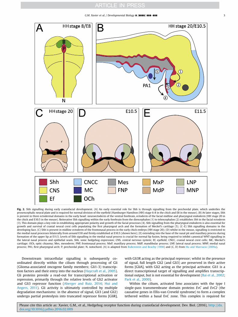
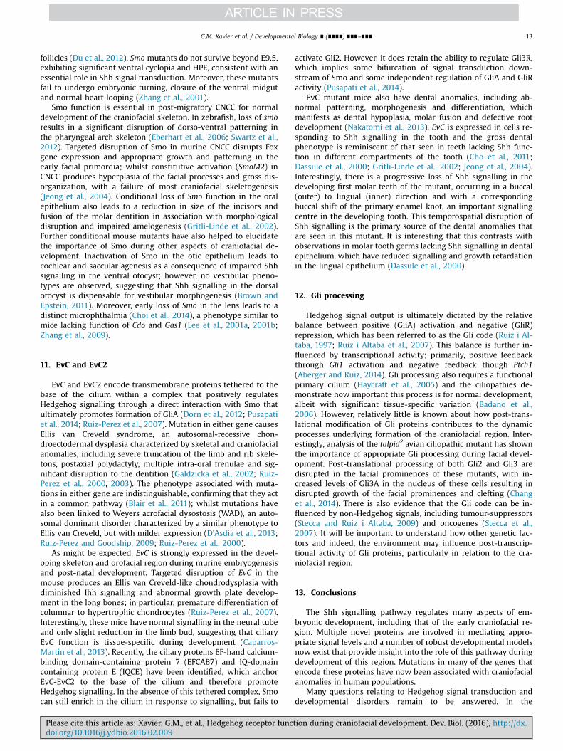
Fig. 2. Shh signalling during early craniofacial development. (A) An early essential role for Shh is through signalling from the prechordal plate, which underlies theprosencephalic neural plate and is required for normal division of the eyefield (Hamburger Hamilton (HH) stage 6-8 in the chick and E8 in the mouse). (B) At later stages, Shhis present in three ectodermal domains in the early head: neuroectoderm of the ventral forebrain, ectoderm of the facial midline and pharyngeal endoderm (HH stage 20 inthe chick and E10.5 in the mouse). Reiterative Shh signalling within the early forebrain from the diencephalon (1) to telencephalon (2) establishes Shh in the facial ectoderm(3). This domain plays a key role in establishing appropriate polarity and growth of the facial processes (4). Shh signalling from the pharyngeal endoderm is also essential forgrowth and survival of cranial neural crest cells populating the first pharyngeal arch and the formation of Meckel's cartilage (5). (C–E) Shh signalling domains in thedeveloping face. (C) Shh is present in midline ectoderm of the frontonasal process in the early chick embryo (HH stage 20); (D) whilst in the mouse, signalling is restricted tothe medial nasal processes bilaterally from around E10 and firmly established at E10.5 (shown here); (E) extending into the base of the nasal pit and maxillary process duringformation of the upper lip at E11.5. Levels of Shh signalling in the medial nasal process is crucial for normal lip fusion, being required to inhibit canonical WNT signalling inthe lateral nasal process and epithelial seam. Shh, sonic hedgehog expression; CNS, central nervous system; Ef, eyefield; CNCC, cranial neural crest cells; MC, Meckel'scartilage; OCh, optic chiasma; Mes, mesoderm; FNP, frontonasal process; MxP, maxillary process; MdP, mandibular process; LNP, lateral nasal process; MNP, medial nasalprocess; PA1, first pharyngeal arch; P, prechordal plate; N, notochord. (A) is adapted from Rubenstein and Beachy (1998) and (C, D) from Hu and Marcucio (2009a).
G.M. Xavier et al. / Developmental Biology ∎ (∎∎∎∎) ∎∎∎–∎∎∎ 3
Downstream intracellular signalling is subsequently co-ordinated directly within the cilium through processing of Gli(Glioma-associated oncogene family members; Gli1-3) transcrip-tion factors and their entry into the nucleus (Haycraft et al., 2005).Gli proteins provide a read-out for transcriptional activation orrepression, primarily through the relative levels of Gli2 activatorand Gli3 repressor function (Aberger and Ruiz, 2014; Hui andAngers, 2011). Gli activity is ultimately controlled by multipledegradation mechanisms: in the absence of signal, Gli3 (and Gli2)undergo partial proteolysis into truncated repressor forms [GliR],
Please cite this article as: Xavier, G.M., et al., Hedgehog receptor funcdoi.org/10.1016/j.ydbio.2016.02.009i
with Gli3R acting as the principal repressor; whilst in the presenceof signal, full length Gli2 (and Gli3) are preserved in their activeforms [GliA], with Gli2 acting as the principal activator. Gli1 is adirect transcriptional target of signalling and amplifies transcrip-tional output, but is not essential for development (Bai et al., 2002;Park et al., 2000).
Within the cilium, activated Smo associates with the type Isingle-pass transmembrane domain proteins EvC and EvC2 (thecausative genes in Ellis-van Creveld syndrome) to form a complextethered within a basal EvC zone. This complex is required for
tion during craniofacial development. Dev. Biol. (2016), http://dx.
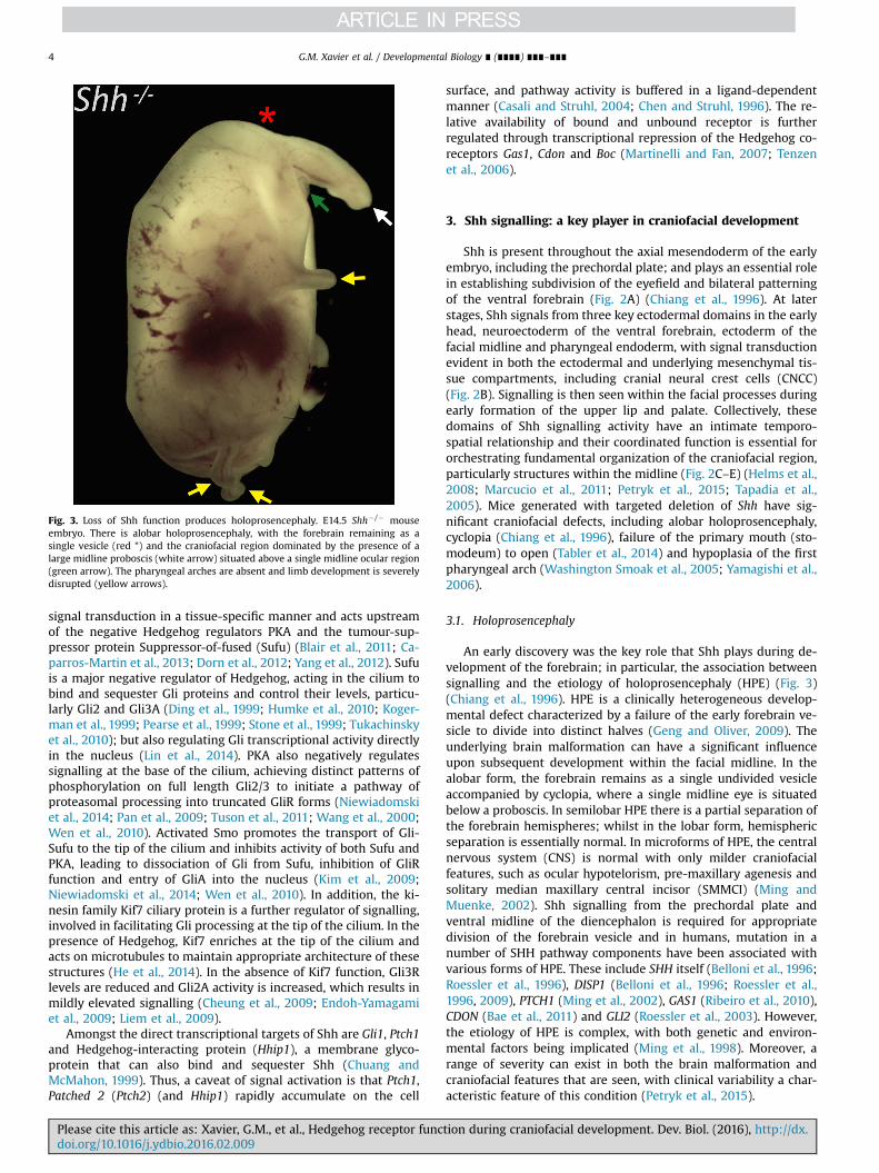
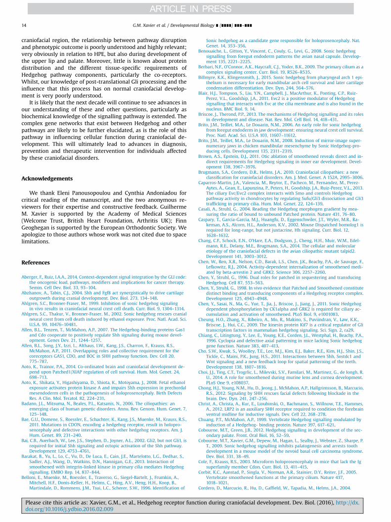
Fig. 3. Loss of Shh function produces holoprosencephaly. E14.5 Shh� /� mouseembryo. There is alobar holoprosencephaly, with the forebrain remaining as asingle vesicle (red *) and the craniofacial region dominated by the presence of alarge midline proboscis (white arrow) situated above a single midline ocular region(green arrow). The pharyngeal arches are absent and limb development is severelydisrupted (yellow arrows).
G.M. Xavier et al. / Developmental Biology ∎ (∎∎∎∎) ∎∎∎–∎∎∎4
signal transduction in a tissue-specific manner and acts upstreamof the negative Hedgehog regulators PKA and the tumour-sup-pressor protein Suppressor-of-fused (Sufu) (Blair et al., 2011; Ca-parros-Martin et al., 2013; Dorn et al., 2012; Yang et al., 2012). Sufuis a major negative regulator of Hedgehog, acting in the cilium tobind and sequester Gli proteins and control their levels, particu-larly Gli2 and Gli3A (Ding et al., 1999; Humke et al., 2010; Koger-man et al., 1999; Pearse et al., 1999; Stone et al., 1999; Tukachinskyet al., 2010); but also regulating Gli transcriptional activity directlyin the nucleus (Lin et al., 2014). PKA also negatively regulatessignalling at the base of the cilium, achieving distinct patterns ofphosphorylation on full length Gli2/3 to initiate a pathway ofproteasomal processing into truncated GliR forms (Niewiadomskiet al., 2014; Pan et al., 2009; Tuson et al., 2011; Wang et al., 2000;Wen et al., 2010). Activated Smo promotes the transport of Gli-Sufu to the tip of the cilium and inhibits activity of both Sufu andPKA, leading to dissociation of Gli from Sufu, inhibition of GliRfunction and entry of GliA into the nucleus (Kim et al., 2009;Niewiadomski et al., 2014; Wen et al., 2010). In addition, the ki-nesin family Kif7 ciliary protein is a further regulator of signalling,involved in facilitating Gli processing at the tip of the cilium. In thepresence of Hedgehog, Kif7 enriches at the tip of the cilium andacts on microtubules to maintain appropriate architecture of thesestructures (He et al., 2014). In the absence of Kif7 function, Gli3Rlevels are reduced and Gli2A activity is increased, which results inmildly elevated signalling (Cheung et al., 2009; Endoh-Yamagamiet al., 2009; Liem et al., 2009).
Amongst the direct transcriptional targets of Shh are Gli1, Ptch1and Hedgehog-interacting protein (Hhip1), a membrane glyco-protein that can also bind and sequester Shh (Chuang andMcMahon, 1999). Thus, a caveat of signal activation is that Ptch1,Patched 2 (Ptch2) (and Hhip1) rapidly accumulate on the cell
Please cite this article as: Xavier, G.M., et al., Hedgehog receptor funcdoi.org/10.1016/j.ydbio.2016.02.009i
surface, and pathway activity is buffered in a ligand-dependentmanner (Casali and Struhl, 2004; Chen and Struhl, 1996). The re-lative availability of bound and unbound receptor is furtherregulated through transcriptional repression of the Hedgehog co-receptors Gas1, Cdon and Boc (Martinelli and Fan, 2007; Tenzenet al., 2006).
3. Shh signalling: a key player in craniofacial development
Shh is present throughout the axial mesendoderm of the earlyembryo, including the prechordal plate; and plays an essential rolein establishing subdivision of the eyefield and bilateral patterningof the ventral forebrain (Fig. 2A) (Chiang et al., 1996). At laterstages, Shh signals from three key ectodermal domains in the earlyhead, neuroectoderm of the ventral forebrain, ectoderm of thefacial midline and pharyngeal endoderm, with signal transductionevident in both the ectodermal and underlying mesenchymal tis-sue compartments, including cranial neural crest cells (CNCC)(Fig. 2B). Signalling is then seen within the facial processes duringearly formation of the upper lip and palate. Collectively, thesedomains of Shh signalling activity have an intimate temporo-spatial relationship and their coordinated function is essential fororchestrating fundamental organization of the craniofacial region,particularly structures within the midline (Fig. 2C–E) (Helms et al.,2008; Marcucio et al., 2011; Petryk et al., 2015; Tapadia et al.,2005). Mice generated with targeted deletion of Shh have sig-nificant craniofacial defects, including alobar holoprosencephaly,cyclopia (Chiang et al., 1996), failure of the primary mouth (sto-modeum) to open (Tabler et al., 2014) and hypoplasia of the firstpharyngeal arch (Washington Smoak et al., 2005; Yamagishi et al.,2006).
3.1. Holoprosencephaly
An early discovery was the key role that Shh plays during de-velopment of the forebrain; in particular, the association betweensignalling and the etiology of holoprosencephaly (HPE) (Fig. 3)(Chiang et al., 1996). HPE is a clinically heterogeneous develop-mental defect characterized by a failure of the early forebrain ve-sicle to divide into distinct halves (Geng and Oliver, 2009). Theunderlying brain malformation can have a significant influenceupon subsequent development within the facial midline. In thealobar form, the forebrain remains as a single undivided vesicleaccompanied by cyclopia, where a single midline eye is situatedbelow a proboscis. In semilobar HPE there is a partial separation ofthe forebrain hemispheres; whilst in the lobar form, hemisphericseparation is essentially normal. In microforms of HPE, the centralnervous system (CNS) is normal with only milder craniofacialfeatures, such as ocular hypotelorism, pre-maxillary agenesis andsolitary median maxillary central incisor (SMMCI) (Ming andMuenke, 2002). Shh signalling from the prechordal plate andventral midline of the diencephalon is required for appropriatedivision of the forebrain vesicle and in humans, mutation in anumber of SHH pathway components have been associated withvarious forms of HPE. These include SHH itself (Belloni et al., 1996;Roessler et al., 1996), DISP1 (Belloni et al., 1996; Roessler et al.,1996, 2009), PTCH1 (Ming et al., 2002), GAS1 (Ribeiro et al., 2010),CDON (Bae et al., 2011) and GLI2 (Roessler et al., 2003). However,the etiology of HPE is complex, with both genetic and environ-mental factors being implicated (Ming et al., 1998). Moreover, arange of severity can exist in both the brain malformation andcraniofacial features that are seen, with clinical variability a char-acteristic feature of this condition (Petryk et al., 2015).
tion during craniofacial development. Dev. Biol. (2016), http://dx.

G.M. Xavier et al. / Developmental Biology ∎ (∎∎∎∎) ∎∎∎–∎∎∎ 5
3.2. Cranial neural crest cells
There is good evidence that Hedgehog transduction is im-portant for the survival of CNCC, with function-blocking experi-ments in the chick resulting in premature death of this cell po-pulation (Ahlgren and Bronner-Fraser, 1999). Survival in the firstpharyngeal arch is promoted by Shh acting from ventral foregutendoderm (see below) at least partially through the inhibition ofCdon, which acts as a dependence receptor to promote apoptoticactivity in this cell population (Delloye-Bourgeois et al., 2013). Inthe zebrafish, Hedgehog signalling from the ventral brain pri-mordium has an early indirect role in orchestrating craniofacialdevelopment, establishing the stomodeum, which subsequentlyinteracts in a Hedgehog-independent manner with CNCC to gen-erate the pterygoid process of the quadrate in the upper jaw andthe anterior neurocranium (Eberhart et al., 2006). Hedgehog sig-nalling also becomes established in oral ectoderm of the zebrafish,acting directly to specify the movement of distinct CNCC popula-tions forming the main cartilaginous elements of the anteriorneurocranium and regulating their differentiation into cartilage(Wada et al., 2005). In addition, Hedgehog signalling to CNCC inthe pharyngeal arches is further required to maintain shh ex-pression in the pharyngeal endoderm of zebrafish (Swartz et al.,2012)
Fig. 4. Shh pathway expression domains in the murine craniofacial region. (A–C) Sagittain the ventral prosencephalon; (B) By E10.5, transcripts are localized to the ventral diencRathke’s pouch (*); (C) At E11.5, the domains within the diencephalon and telencephalonin ectoderm of the early tongue and the developing incisor tooth germs. (D–I) Frontal sectoderm of the early medial nasal processes (arrowed); (E, F) At E10.5, Shh is expressed isuperficially; however, a gradient of signalling activity exists in the medial nasal processthis region (G). (H, I) At E11.5, Shh is expressed in ectoderm at the base of the nasal pits anoptic recess; p, prosencephalon; pe, pharyngeal endoderm; t, tongue; te, telencephalon;process; np, nasal pit.
Please cite this article as: Xavier, G.M., et al., Hedgehog receptor funcdoi.org/10.1016/j.ydbio.2016.02.009i
Loss of Hedgehog signalling in murine CNCC through Wnt1Cre-mediated targeted disruption of Smo (Wnt1Cre; Smo) function hasalso demonstrated the importance of normal Shh responsivenessin these cells during craniofacial development. A number of CNCC-derived skeletal and non-skeletal components are absent in thesemice, which have facial truncation due to reduced growth anddevelopment of the pharyngeal arches secondary to increasedapoptosis and decreased cell proliferation (Jeong et al., 2004).Moreover, the catastrophic loss of architecture seen in pharyngealarch I of Shh mutant embryos may also compromise the domainsof CNCC populations within this region (Washington Smoak et al.,2005; Yamagishi et al., 2006). Interestingly, CNCC migration ap-pears to be normal in the absence of Hedgehog-responsiveness inCNCC (Jeong et al., 2004), consistent with findings in zebrafish smoand disp1 mutants (Eberhart et al., 2006; Schwend and Ahlgren,2009); however, zebrafish cdon does play a role in mediating themigration of trunk NCC through the regulation of N-cadherin lo-calization (Powell et al., 2015).
More recently, it has been shown that mice with loss-of-func-tion mutation in the Fuzzy ciliogenesis regulator have high-archedpalate, which is a common feature amongst human ciliopathies.Interestingly, whilst there is dysregulated Gli processing and re-duced Hedgehog signalling in these mice, the phenotype arisesthrough upregulated Fgf8 expression early in development and
l sections through the developing craniofacial midline. (A) At E9.5, Shh is expressedephalon, telencephalon and pharyngeal endoderm, but are absent from ectoderm ofare well-established and separated by the optic recess, with expression also presentections through the early facial region. (D) At E10.0, Shh is expressed bilaterally inn ectoderm of the medial nasal processes in deeper regions of the face but not moreat this stage, running from medial to lateral, as demonstrated by Ptch1 expression ind in the maxillary processes during formation of the upper lip. di, diencephalon; or,tg, tooth germ; lnp, lateral nasal process; mnp, medial nasal process; mxp, maxillary
tion during craniofacial development. Dev. Biol. (2016), http://dx.

G.M. Xavier et al. / Developmental Biology ∎ (∎∎∎∎) ∎∎∎–∎∎∎6
increased numbers of CNCC in the maxillary process. A similarphenotype is seen in oral-facial-digital syndrome (Ofd1) mutantmice, which also have defective ciliogenesis, suggesting shareddevelopmental mechanisms between the ciliopathies and Fgf-re-lated craniofacial syndromes (Tabler et al., 2013).
3.3. Facial development
Shh expression in surface ectoderm of the facial processes isdynamic and reflects the capacity of this pathway to organize anddirectly influence formation of this region (Helms et al., 1997; Huand Helms, 1999) (Fig. 4).
In the chick (see Fig. 2C), Shh is initially expressed in midlineectoderm of the frontonasal process (FNP), forming a boundarywith Fgf8-expressing cells situated more dorsally, in a so-calledfrontonasal ectodermal zone (or FEZ), which acts as a signallingcentre to control growth and polarity within the upper jaw(Abzhanov and Tabin, 2004; Hu and Marcucio, 2009a, 2009b; Huet al., 2003). A FEZ is also present in mice (see Fig. 2D), but thedomains of Shh are restricted to bilateral regions of ectodermwithin the early medial nasal processes (MNP), later extendinginto the base of the MNP and nasal pit, and appearing in themaxillary processes as the upper lip is formed (see Fig. 2E). Thesedomains subtly direct appropriate growth and cell survival withinthese regions during the establishment of early facial form (Huet al., 2015). Interestingly, all the primary Shh signalling domainswithin the early craniofacial midline are reciprocal and closelycoordinated, particularly those present at very early stages be-tween the developing forebrain and facial region (Marcucio et al.,2011) (Fig. 4A–C). Early expression of Shh is seen in the ventralprosencephalon and diencephalon, subsequently inducing a do-main in the basal telencephalon, anterior to the optic recess; andin the chick, establishing competency of the FEZ to express Shh(Hu and Marcucio, 2009a, 2009b; Marcucio et al., 2005). Shh sig-nalling between the brain and face therefore follows a very specifictemporo-spatial pattern, influencing outgrowth and patterning ofthe upper midface (Chong et al., 2012). Indeed, this signalling ac-tivity in the embryonic midface has been shown to have a pre-dictive relationship for shape variation within the upper jaw of thechick. This dose-response is non-linear and consistent with Shhacting through a morphogenetic gradient (Young et al., 2010).
Collectively, these findings suggest a basis for the wide range ofseverity that is seen in conditions such as HPE, particularly in re-lation to the midline facial anomalies. Variation in the temporalactivity of this pathway providing some context to the phenotypicspectrum that is often observed (Cordero et al., 2004).
3.4. Oro-facial clefting
Shh is expressed in regionally-restricted domains of the earlyfacial processes (Fig. 4D–I) and later in the palatal shelves, which isconsistent with a role in development of the upper lip (Kurosaka,2015) and secondary palate (Cobourne and Green, 2012). Phar-macological antagonism of Hedgehog signalling in the mouseembryo between E7-9 produces cleft lip and palate (CLP), whilstbetween E9-10 it causes isolated cleft palate (CP) (Heyne et al.,2015; Lipinski et al., 2010).
There is evidence that Shh interacts with Bone morphogeneticprotein (Bmp), Fibroblast growth factor (Fgf) and Wingless-typeMMTV integration site (Wnt) pathways in a dynamic and rapidly-changing manner during development of the murine upper lip(Kurosaka et al., 2014; Metzis et al., 2013; Thomason et al., 2008).Around E10.5–11, a gradient of Hedgehog signal activity extendsfrom the facial midline across the MNP’s (Fig. 4E–G), with loss ofPtch1 in CNCC resulting in increased transduction and reduced Fgfsignalling in the facial prominences, associated with defective
Please cite this article as: Xavier, G.M., et al., Hedgehog receptor funcdoi.org/10.1016/j.ydbio.2016.02.009i
nasal pit invagination and cleft lip (Metzis et al., 2013), and con-sistent with associations between PTCH1 mutations and CLP inhuman populations (Letra et al., 2010; Mansilla et al., 2006; Sasakiet al., 2009). At E11, Shh signal activity within the MNP repressescanonical Wnt signal transduction in regions cranial to the lamb-doid region and epithelial seam. This facilitates Wnt-mediatedinduction of p63 and Irf6 in this region, and appropriate removal ofthe epithelial seam during upper lip formation (Kurosaka et al.,2014). Interestingly, at E11.5 Shh becomes strongly expressed inectoderm of the caudal MNP and adjacent maxillary process -regions of expression that are downregulated and lost, respec-tively in the p63 mutant (Thomason et al., 2008).
Shh is also expressed on the oral surface of the murine sec-ondary palate from around E12 in the mouse, in a series of eightstripes that correspond to the future rugae palatinae (Pantalacciet al., 2008; Welsh and O'Brien, 2009). Shh signalling is requiredfrom epithelium to mesenchyme for appropriate proliferation inthe mesenchymal compartment of the palatal shelves and dis-ruption produces CP (Dassule et al., 2000; Jeong et al., 2004; Lanand Jiang, 2009), although with ablation of Shh-responsiveness inthe epithelium alone, the palate remains intact (Gritli-Linde et al.,2002). Shh lies downstream of Wnt signalling in the oral epithe-lium (Lin et al., 2011) and participates in reciprocal induction ofFgf10 in the mesenchyme, mediated though Fgfr2b function in theepithelium, with loss of both these Fgf signalling components alsoassociated with CP (Rice et al., 2004). Further associations betweenperturbed Shh signalling and oro-facial clefting are seen in Cdonand Gas1 mutant mice, which exhibit philtral dysgenesis and CP,respectively in a 129sv/C57BL/6 background (Cole and Krauss,2003; Seppala et al., 2007); whilst Gas1; Bocmutant mice have CLPassociated with increased cell death and reduced proliferation inthe facial midline (Seppala et al., 2014).
The eight rugae palatinae appear sequentially from within agrowth zone situated in the mid-palate, anterior to the first-forming ruga 8. Subsequent growth of the palatal shelves andappearance of the rugae is closely co-ordinated, with ruga 2 ap-pearing next, followed by rugae 3, 1 and then 4–7 (Economouet al., 2012; Pantalacci et al., 2008; Welsh and O'Brien, 2009). It hasbeen suggested that the regular spacing between rugae is estab-lished through an activator-inhibitor mechanism, with Fgf andWnt signalling acting as the activator component and Shh as aninhibitor (Economou et al., 2012).
3.5. Pharyngeal endoderm
Shh is also strongly expressed within the pharyngeal endoderm(see Fig. 4B, C), directly regulating normal morphogenetic move-ment of the pharyngeal arches in zebrafish (Swartz et al., 2012). Inthe chick, it is required for cell survival and proliferation withinthe mandibular primordium (Brito et al., 2006; Couly et al., 2002;Haworth et al., 2007a) and induction of the mesethmoid cartilagein the most rostral-zone (Benouaiche et al., 2008). In the mouse,conditional inactivation of Shh in the pharyngeal endoderm leadsto micrognathia secondary to increased mesenchymal cell death inpharyngeal arch I (Billmyre and Klingensmith, 2015). These micealso have significant pattern defects within the first arch, con-sistent with observations that Fgf8 is downstream of Shh in thisregion (Haworth et al., 2007a). A failure of Meckel’s cartilage dif-ferentiation also occurs in the absence of Shh signalling from theendoderm (Billmyre and Klingensmith, 2015), whilst a source ofectopic Shh in mandibular arch ectoderm or mesenchyme caninduce an ectopic or supernumerary Meckel’s cartilage in thechick, which can produce mirror-image supernumerary jaws(Brito et al., 2008; Haworth et al., 2007a). Interestingly, Gas1� /�;Shhþ /� compound mutant mice have ectopic duplications of theproximal mandible that include the molar dentition, a finding
tion during craniofacial development. Dev. Biol. (2016), http://dx.

G.M. Xavier et al. / Developmental Biology ∎ (∎∎∎∎) ∎∎∎–∎∎∎ 7
seemingly difficult to reconcile with reduced Hedgehog signallingin this region (Seppala et al., 2007). In addition, transgenic miceoverexpressing Shh in the oral epithelium through a Keratin14promoter have essentially normal morphology of the mandible,apart from some diminution of the coronoid process (Cobourneet al., 2009).
4. Hedgehog receptor function in craniofacial development
In recent years, progress has been made in further under-standing the role of multiple proteins that interact with Shh dur-ing normal signal transduction. In particular, these proteins areintimately involved in both the production and reception of Shhduring development. Here, we focus on the function of these di-verse proteins within the context of Shh signalling during cra-niofacial development and the consequences of disrupted functionfor normal development of this region (Table 1).
5. Hedgehog production
5.1. Dispatched
There are two Disp homologues in mouse (Disp1 and Disp2), butgenetic studies support the view that only Disp1 is involved insignalling, with mutants failing to survive beyond embryonic day(E) 9.5 and presenting with abnormal morphology of the forebrain(indicative of a loss of ventral midline fate) and delayed cardiacmorphogenesis (Caspary et al., 2002; Kawakami et al., 2002; Maet al., 2002). Disp1 activity is required in Shh-producing cells forparacrine signalling through cholesterol-modified Shh (Tian et al.,2005). Indeed, during facial development attenuating Disp1 ac-tivity using both hypomorphic and missense alleles produces aloss of midline facial structures, with a dose-dependent geneticinteraction existing between Shh and Disp1. The severity of thefacial anomalies is reflected in the amount of disruption seenwithin the premaxilla, and both truncation and fusion in themandibular region, with the most severe embryos lacking a pre-maxilla and the mandibular incisor dentition (Tian et al., 2005).Inactivation of zebrafish disp1 using the chameleon (con/disp1)mutant results in a significant, but incomplete reduction inHedgehog signalling. These mutants demonstrate a requirementfor disp1 in patterning post-migratory CNCC, specifically in phar-yngeal arch I and regulating the chondrogenic markers dlx2a andsox9a. In addition, patterning of the jaw joints is severely dis-rupted, through loss of bapx1 and gdf5 (Schwend and Ahlgren,2009).
Significantly, truncating mutations of DISP1 have been identi-fied in two independent human families, where affected in-dividuals demonstrated clinical features of microform HPE(Roessler et al., 2009). DISP1 interacts with human SHH via itscholesterol anchor, and this interaction is necessary for appro-priate SHH secretion. However, DISP1 alone is not sufficient torelease SHH from cells, this process also requires SCUBE2 functionin order to overcome the insolubility conferred by SHH cholesterolmodification (Creanga et al., 2012; Tukachinsky et al., 2012).
5.2. Scube2
The Scube gene family consists of three independent evolu-tionarily conserved members (Scube1-3) (Haworth et al., 2007b;Hollway et al., 2006; Woods and Talbot, 2005; Wu et al., 2004,2011; Xavier et al., 2013, 2009; Yang et al., 2002). These genesencode secreted and cell surface-associated proteins that share adomain organization of at least five recognizable motifs, including
Please cite this article as: Xavier, G.M., et al., Hedgehog receptor funcdoi.org/10.1016/j.ydbio.2016.02.009i
multiple EGF (epidermal growth factor-like) and N-linked glyco-sylation sites, and a C-terminal CUB domain (Grimmond et al.,2000). Zebrafish scube2 plays a non-cell-autonomous role inmediating long-range Hedgehog signalling, with mutants havingmorphological traits consistent with reduced pathway activity,such as a curled tail, U-shaped somite boundaries and significantly,weak cyclopia (Hollway et al., 2006; Johnson et al., 2012; Kawa-kami et al., 2005; Woods and Talbot, 2005). A more severe loss ofHedgehog target gene expression is observed when all Scube fa-mily members are knocked down; however, defects in Hedgehog-sensitive cell types are also evident when scube1 or scube3 are lostin combination with scube2. These findings suggest that scube2 isthe most important family member in modulating Hedgehog sig-nalling (Johnson et al., 2012), an observation consistent with thegrossly normal craniofacial development of murine Scube3 mu-tants (Xavier et al., 2013). However, mammalian Scube2 has re-cently been shown to be a potent modulator of Ihh signalling,enhancing Ihh-stimulated osteoblast differentiation during en-dochondral bone formation (Lin et al., 2015).
6. Hedgehog reception
6.1. Patched1 and patched2
Ptch1 is first detected in the developing mouse embryo at E7.5with transcripts present in the ventral neural tube and later, thesomites, limb buds and developing craniofacial region (Goodrichet al., 1996; Hahn et al., 1996). In humans, protein-truncatingPTCH1 mutations are responsible for the autosomal dominantBasal Cell Nevus Syndrome (BCNS; also known as the Nevoid BasalCell Carcinoma Syndrome (NBCCS) or Gorlin–Goltz syndrome)(Wicking et al., 1997), which is characterized primarily by multiplerecurrent basal cell carcinomas, recurrant odontogenic keratocystsof the jaws, palmar/plantar pits, ectopic calcification of the falxcerebri and less commonly, CLP and tooth agenesis (Kimonis et al.,1997, 2004, 2013).
Homozygous Ptch1 mutant mice die between E9.0 and 10.5,with gross phenotypic changes evident at E8. The neural tube failsto close completely and there is overgrowth of the head folds,hindbrain and spinal cord. Embryonic lethality is thought to besecondary to abnormal cardiac development, which has hamperedanalysis of any potential craniofacial phenotype (Goodrich et al.,1997). However, conditional inactivation of Ptch1 in CNCC(Wnt1Cre; Ptch1c/c) has established a direct role in the pathogen-esis of craniofacial anomalies. Loss of Ptch1 function in CNCCcauses mid-facial expansion and an early defect in the nasal pit,which culminates in cleft lip (Metzis et al., 2013). Interestingly, thewider mid-facial morphology observed in these mutants has beenexplained by changes in cell packing, specifically a more looselypacked cellular network within the mesenchyme. Cleft lip occursthrough a failure of coordinated fusion between medial nasalprocess, lateral nasal process and maxilla. In Wnt1Cre; Ptch1c/c
embryos, these prominences fail to meet; which has been corre-lated to defective nasal pit invagination, Ptch1 required in a non-cell-autonomous manner for maintenance of cell shape in the in-vaginating nasal pit epithelium (Metzis et al., 2013). Unfortunately,even conditional disruption of Ptch1 in CNCC leads to lethality atE12.0; after primary, but before secondary palate development(Ferguson, 1988; Metzis et al., 2013). It is likely that abnormalitiesof the secondary palate, such as high-arched and CP would also beobserved in these mice, if embryos survived until later inembryogenesis.
Ptch1 is also a key molecule involved in the co-ordination ofbrain and facial development during mouse embryogenesis. Thecapability of the C-terminal domain of Ptch1 to regulate Caspase-9
tion during craniofacial development. Dev. Biol. (2016), http://dx.
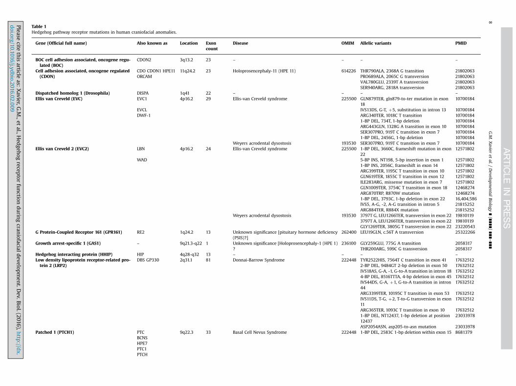
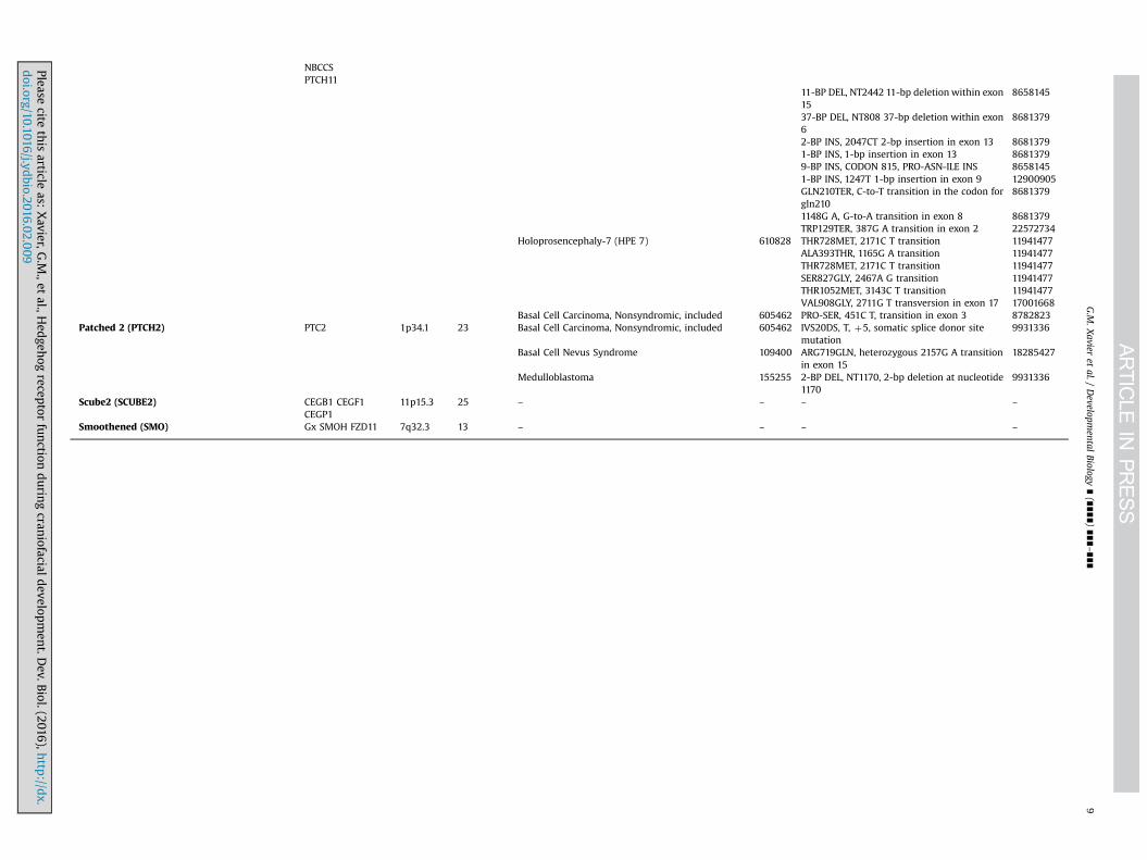
Table 1Hedgehog pathway receptor mutations in human craniofacial anomalies.
Gene (Official full name) Also known as Location Exoncount
Disease OMIM Allelic variants PMID
BOC cell adhesion associated, oncogene regu-lated (BOC)
CDON2 3q13.2 23 – – – –
Cell adhesion associated, oncogene regulated(CDON)
CDO CDON1 HPE11 11q24.2 23 Holoprosencephaly-11 (HPE 11) 614226 THR790ALA, 2368A G transition 21802063ORCAM PRO689ALA, 2065C G transversion 21802063
VAL780GLU, 2339T A transversion 21802063SER940ARG, 2818A transversion 21802063
Dispatched homolog 1 (Drosophila) DISPA 1q41 22 – – – –
Ellis van Creveld (EVC) EVC1 4p16.2 29 Ellis-van Creveld syndrome 225500 GLN879TER, gln879-to-ter mutation in exon18
10700184
EVCL IVS13DS, G-T, þ5, substitution in intron 13 10700184DWF-1 ARG340TER, 1018C T transition 10700184
1-BP DEL, 734T, 1-bp deletion 10700184ARG443GLN, 1328G A transition in exon 10 10700184SER307PRO, 919T C transition in exon 7 107001841-BP DEL, 2456G, 1-bp deletion 10700184
Weyers acrodental dysostosis 193530 SER307PRO, 919T C transition in exon 7 10700184Ellis van Creveld 2 (EVC2) LBN 4p16.2 24 Ellis-van Creveld syndrome 225500 1-BP DEL, 3660C, frameshift mutation in exon
2212571802
WAD 5-BP INS, NT198, 5-bp insertion in exon 1 125718021-BP INS, 2056C, frameshift in exon 14 12571802ARG399TER, 1195C T transition in exon 10 12571802GLN619TER, 1855C T transition in exon 12 12571802ILE283ARG, missense mutation in exon 7 12571802GLN1009TER, 3754C T transition in exon 18 12468274ARG870TRP, R870W mutation 124682741-BP DEL, 3793C, 1-bp deletion in exon 22 16,404,586IVS5, A-G, -2, A-G transition in intron 5 21815252ARG884TER, R884X mutation 21815252
Weyers acrodental dysostosis 193530 3797T G, LEU1266TER, transversion in exon 22 198101193797T A, LEU1266TER, transversion in exon 22 19810119GLY1269TER, 3805G T transversion in exon 22 23220543
G Protein-Coupled Receptor 161 (GPR161) RE2 1q24.2 13 Unknown significance [pituitary hormone deficiency(PSIS)?]
262400 LEU19GLN, c.56T A transversion 25322266
Growth arrest-specific 1 (GAS1) – 9q21.3-q22 1 Unknown significance [Holoprosencephaly-1 (HPE 1)?
236100 GLY259GLU, 775G A transition 2058317THR200ARG, 599C G transversion 2058317
Hedgehog interacting protein (HHIP) HIP 4q28-q32 13 – – – –
Low density lipoprotein receptor-related pro-tein 2 (LRP2)
DBS GP330 2q31.1 81 Donnai-Barrow Syndrome 222448 TYR2522HIS, 7564T C transition in exon 41 176325122-BP DEL, 9484GT 2-bp deletion in exon 50 17632512IVS18AS, G-A, -1, G-to-A transition in intron 18 176325124-BP DEL, 8516TTTA, 4-bp deletion in exon 45 17632512IVS44DS, G-A, þ1, G-to-A transition in intron44
17632512
ARG3399TER, 10195C T transition in exon 53 17632512IVS11DS, T-G, þ2, T-to-G transversion in exon11
17632512
ARG365TER, 1093C T transition in exon 10 176325121-BP DEL, NT12437, 1-bp deletion at position12437
23033978
ASP2054ASN, asp205-to-asn mutation 23033978Patched 1 (PTCH1) PTC 9q22.3 33 Basal Cell Nevus Syndrome 222448 1-BP DEL, 2583C 1-bp deletion within exon 15 8681379
BCNSHPE7PTC1PTCH
G.M
.Xavier
etal./
Developm
entalBiology
∎(∎∎∎∎)
∎∎∎–∎∎∎
8
Pleasecite
this
articleas:
Xavier,G
.M.,et
al.,Hed
gehog
receptor
function
durin
gcran
iofacialdevelop
men
t.Dev.B
iol.(2016),http
://dx.
doi.org/10.1016/j.yd
bio.2016.02.009i
NBCCSPTCH11
11-BP DEL, NT2442 11-bp deletion within exon15
8658145
37-BP DEL, NT808 37-bp deletion within exon6
8681379
2-BP INS, 2047CT 2-bp insertion in exon 13 86813791-BP INS, 1-bp insertion in exon 13 86813799-BP INS, CODON 815, PRO-ASN-ILE INS 86581451-BP INS, 1247T 1-bp insertion in exon 9 12900905GLN210TER, C-to-T transition in the codon forgln210
8681379
1148G A, G-to-A transition in exon 8 8681379TRP129TER, 387G A transition in exon 2 22572734
Holoprosencephaly-7 (HPE 7) 610828 THR728MET, 2171C T transition 11941477ALA393THR, 1165G A transition 11941477THR728MET, 2171C T transition 11941477SER827GLY, 2467A G transition 11941477THR1052MET, 3143C T transition 11941477VAL908GLY, 2711G T transversion in exon 17 17001668
Basal Cell Carcinoma, Nonsyndromic, included 605462 PRO-SER, 451C T, transition in exon 3 8782823Patched 2 (PTCH2) PTC2 1p34.1 23 Basal Cell Carcinoma, Nonsyndromic, included 605462 IVS20DS, T, þ5, somatic splice donor site
mutation9931336
Basal Cell Nevus Syndrome 109400 ARG719GLN, heterozygous 2157G A transitionin exon 15
18285427
Medulloblastoma 155255 2-BP DEL, NT1170, 2-bp deletion at nucleotide1170
9931336
Scube2 (SCUBE2) CEGB1 CEGF1CEGP1
11p15.3 25 – – – –
Smoothened (SMO) Gx SMOH FZD11 7q32.3 13 – – – –
G.M
.Xavier
etal./
Developm
entalBiology
∎(∎∎∎∎)
∎∎∎–∎∎∎
9
Pleasecite
this
articleas:
Xavier,G
.M.,et
al.,Hed
gehog
receptor
function
durin
gcran
iofacialdevelop
men
t.Dev.B
iol.(2016),http
://dx.
doi.org/10.1016/j.yd
bio.2016.02.009i

G.M. Xavier et al. / Developmental Biology ∎ (∎∎∎∎) ∎∎∎–∎∎∎10
(Casp9)-associated mitochondrial cell death has a seemingly directeffect on size of the forebrain and the adjacent nasal processes(Aoto and Trainor, 2014). Interestingly, the X-linked inhibitoryapoptosis protein (XIAP), which is observed in primary cilia in aHedgehog-dependent manner, acts as an inhibitor of Ptch1-in-duced cell death. Orchestration of Ptch1-induced apoptosis byXIAP in primary cilia protects Ptch1 from translocation into mi-tochondria, providing an important survival mechanism in thisorganelle. This mechanism may also help co-ordinate brain andfacial development, with these observations supporting the notionthat mitochondrial dysfunction may be a developmental risk fac-tor associated in the pathogenesis of HPE (Aoto and Trainor, 2014).
N-ethyl-N-nitrosourea (ENU)-induced mutagenesis provides anunbiased forward genetic approach for identifying novel allelesimportant for embryogenesis. In a recent screen for recessivemouse mutations affecting craniofacial morphology, a single nu-cleotide change was identified at the 3′-end of Ptch1 exon 13.These mice (Ptch1DL, termed DL: Dogface-Like) display abnormalskull and snout morphology and craniosynostosis of the lambdoidsuture. Skeletal defects related to the pathology of BCNS were alsopresent, including the scapula, ribcage, secondary palate, cranialbase and cranial vault. The general overgrowth, rhabdomyosarco-mas and medulloblastomas observed in Ptch1 heterozygous mice(Goodrich et al., 1997; Hahn et al., 1998) are not seen in Ptch1DL
mice. This mutation therefore represents a hypomorphic allele ofPtch1 with the potential to further assess the role of Hedgehogsignalling in multiple developmental events, particularly thoseregulating facial shape (Feng et al., 2013). An additional ENUscreen in mice has produced further novel alleles critically re-quired for early craniofacial development, including the wiggablemutant, so-called because of the excessive leaf-like laminae orfolia in the brain, which resemble a wig (Sandell et al., 2011).Genome sequencing revealed a T to A nucleotide change in intron15 of Ptch1 in this mutant, which created a new splice acceptorsite, resulting in a premature stop codon in exon 16 and generationof a truncated protein. Ptch1wiggable mice die in utero at aroundE12.0 as a result of various defects, including open neural tube andhypertelorism of the face, consistent with a gain-of-function inHedgehog signalling (Kurosaka et al., 2014; Sandell et al., 2011). Inan effort to rescue the phenotype observed in Ptch1wiggable mice,compound mutants were generated lacking function of theHedgehog acyltransferase (Hhat), responsible for modifyingHedgehog proteins through the addition of palmitic acid and es-sential for appropriate long-range signalling. Hhatcreface mutantshave a phenotype consistent with a loss of Shh signalling, in-cluding HPE, acrania and agnathia (Dennis et al., 2012). Interest-ingly, Hhatcreface; Ptch1wiggable mice have cleft lip and fissure of thepremaxillary bone at E16.5, demonstrating an important role forHhat and Ptch1 in regulating Shh signalling in the FNP during lipdevelopment, specifically through the restriction of canonical Wntsignal activity in the lambdoidal region (Kurosaka et al., 2014).
In addition to engineered and induced Ptch1 mutants, sponta-neous mutants have also been reported. A recessive Ptch1 mousemutation, mesenchymal dysplasia (mes) causes excess skin, in-creased body weight and mild preaxial polydactyly, with thesemice also having a shortened face, wide-set eyes and dome head(Makino et al., 2001). Ptch1D11 is another spontaneous mutationcaused by an aberrant recombination event during production of aPtch1 null allele. The PtchD11 locus presumably results in a weakPtch1 allele, with homozygous animals being sterile, but otherwisenormal (Oro and Higgins, 2003).
In humans, PTCH1 maps to chromosome 9q22.3 and is widelyassumed to be a tumour-suppressor gene (Hahn et al., 1996).Mutation analysis of PTCH1 in both familial and sporadic HPE caseshas revealed four different missense mutations in five unrelatedindividuals. These findings demonstrate that mutations in
Please cite this article as: Xavier, G.M., et al., Hedgehog receptor funcdoi.org/10.1016/j.ydbio.2016.02.009i
different components of the Shh pathway that lead to a commoneffect on Shh signalling can each result in the same phenotype:decreased Shh activity causing HPE and increased activity causingtumours (Goodrich et al., 1999; Ming et al., 2002).
Vertebrates have an additional Patched receptor Ptch2, which isa structural homologue of Ptch1 (Motoyama et al., 1998). Ptch2mutant mice are viable, fertile and do not display obvious devel-opmental defects, although males develop skin lesions associatedwith alopecia and ulceration, with progressing age (Nieuwenhuiset al., 2006). However, Ptch2 is a target of Hedgehog signalling andparticipates in ligand-dependent feedback inhibition, which inconjunction with Ptch1 (and Hhip1) is an important antagonist ofpathway activity, in the neural tube at least (Holtz et al., 2013).
7. Gas1, Cdo and Boc
Gas1, Cdon and Boc are a diverse group of proteins that act asco-receptors within the context of Hedgehog signalling, retainingthe ability to interact directly with Shh (Lee et al., 2001a, 2001b;Martinelli and Fan, 2007; McLellan et al., 2008; Okada et al., 2006;Tenzen et al., 2006) and form high-affinity individual complexeswith Ptch1 on the surface of receiving cells (Bae et al., 2011; Izziet al., 2011). Boc and Cdon are also able to bind Gas1, althoughthese interactions are unlikely to be tripartite (Bae et al., 2011; Izziet al., 2011); whilst, Cdon and Boc can complex with each otherthrough their extra- and intra-cellular domains (Kang et al., 2002).Collectively, these co-receptors are essential for vertebratehedgehog signalling; binding of Shh to Ptch1 alone is not sufficientfor pathway activation (Izzi et al., 2011) and mice with collectivetargeted disruption of Gas1, Cdon and Boc lack all Shh transductionexcept for some very early rudimentary activity (Allen et al., 2011).A prevailing view is that these individual tissue-specific receptorcomplexes bind Shh, which leads to de-repression of Smo andactivation of the pathway, consistent with findings of both re-dundancy and specific requirements for these co-receptors indifferent developmental contexts.
The role of these co-receptors during early craniofacial devel-opment has been investigated extensively through the generationof single and compound mouse mutants, which demonstrate avariable and background-dependent severity of phenotype (Fig. 5).Loss of Cdon can produce a semilobar-type HPE characterized bycebocephalic face (single nostril, ocular hypotelorism and max-illary hypoplasia) or a less severe microform HPE with philtraldysgenesis and maxillary incisor agenesis (Cole and Krauss, 2003;Hong and Krauss, 2012; Zhang et al., 2006). Loss of Gas1 results ina more consistent microform HPE associated with maxillary in-cisor fusion and CP (Seppala et al., 2007), whilst Boc mutants lackcraniofacial abnormalities but do have misguided commissuralaxon guidance, cerebellum reduction and reduced ipsilateral ret-inal ganglion cells (Izzi et al., 2011; Okada et al., 2006; Sanchez-Arrones et al., 2013). As might be expected, Gas1; Cdon compoundmutants have a more severe semilobar HPE associated with asingle external nares, fusion of the nasal processes and absence ofmaxillary and mandibular skeletal elements (Allen et al., 2007;Seppala et al., 2014). In addition, the generation of Cdon; Boc miceon a background associated with only microform HPE in the ab-sence of Cdon alone, results in a lobar HPE with much more severecraniofacial anomalies (Zhang et al., 2011). More recently, thecraniofacial region of Gas1; Boc mutants have been analyzed, de-monstrating an allele dosage-dependent phenotype. In particular,Gas1; Boc mice have lobar HPE and disruption of the corpus cal-losum, CLP and maxillary incisor agenesis (Seppala et al., 2014).Significantly, loss-of-function mutations in GAS1 and CDON haveboth been associated with HPE in humans (Bae et al., 2011; Ribeiroet al., 2010).
tion during craniofacial development. Dev. Biol. (2016), http://dx.

Fig. 5. Craniofacial skeletal defects in mice lacking function of Cdon and Gas1. Comparison of E17.5 wild type and mutant skulls differentially stained for bone (alizarin red)and cartilage (alcian blue). (A, C, E, G) Norma lateralis; (B, D, F, H) Norma basalis. The Cdon� /� skull is grossly comparable to wild type in size and overall morphology. Thepalate is intact, but there is only a single maxillary incisor tooth (pale blue arrow). The Gas1� /� skull is reduced in size when compared to the wild type and has a number ofdefects, including single maxillary incisor (pale blue arrow), cleft palate (yellow arrows), fenestration of the neurocranial base (dark green arrowhead), diminutive and poorlyformed ectotympanic (orange arrow) and absence of the hypoglossal canals within the exoccipital (white arrow). In the Gas1� /�; Cdon� /� skull there is a further reductionin size and gross disruption of the skull with maxillary-mandibular stenosis (yellow arrowhead) and a marked open bite occlusion (red *). In addition, the maxillary incisorsfail to form (pale blue arrow), there is stenosis within the premaxilla (red arrow), the nasal cavity is ossified (light green arrow), the pterygoid plates are absent (light pinkarrow) and the exoccipital-basioccipital bones are fused (dark blue arrow). In addition, the pars canalicularis and pars cochlearis are severely disrupted (dark pink arrow)with the ectotympanic fused to the lamina obturans (orange arrow). etm, ectotympanic; hc, hypoglossal canal; mxi, maxillary incisors; ppmx, palatal process of the maxilla;pppmx, palatal process of the premaxilla; pppl, palatal process of the palatine.
G.M. Xavier et al. / Developmental Biology ∎ (∎∎∎∎) ∎∎∎–∎∎∎ 11
An obvious potential influence on the variation that is seen inthese loss-of-function co-receptor mouse models is redundancy;however, phenotype may also be influenced by subtle variation inmodulating signal activity. Gas1, Cdo and Boc are all negatively
Please cite this article as: Xavier, G.M., et al., Hedgehog receptor funcdoi.org/10.1016/j.ydbio.2016.02.009i
regulated by Hedgehog and as signal levels increase, transcriptionis progressively reduced (Martinelli and Fan, 2007; Tenzen et al.,2006). A gradient of transcriptional regulation therefore existswithin the Shh target field, and as signalling reduces at distance
tion during craniofacial development. Dev. Biol. (2016), http://dx.
G.M. Xavier et al. / Developmental Biology ∎ (∎∎∎∎) ∎∎∎–∎∎∎12
from the source, transcription of these co-receptors increases,potentially boosting or maintaining signalling in more peripheraltissues. These thresholds of activity may be crucial in ensuringappropriate development, particularly in the craniofacial midline.The situation is further complicated by tissue-specific variation inexpression domains between these co-receptors, particularlyfindings that some can also be expressed in regions of high signalactivity (Martinelli and Fan, 2007; Tenzen et al., 2006). Shh canalso signal through contact-mediated release in the chicklimb, propagated by specialized filopodia that can deliver ligandat a distance (Sanders et al., 2013). Interestingly, Cdon and Bocco-localize in specific micro-domains within these filopodial ex-tensions and whilst these have not been identified in the cranio-facialregion, similar mechanisms may also influence signalling here.Finally, these co-receptors have also been shown to function inalternative signalling pathways and therefore potentially play arole in other developmental contexts (Sanchez-Arrones et al.,2012; Segovia and Zarco, 2014).
7.1. Modeling phenotypic variation in HPE through Hedgehog co-receptor function
It has been suggested that in many cases of HPE, clinicalvariability may be related to a requirement for more than oneinsult during development and that a combined interaction be-tween multiple gene products and environmental factors de-termines final phenotypic outcome (Ming and Muenke, 2002;Nanni et al., 1999). Indeed, the analysis of compound Hedgehogco-receptor mutants has demonstrated the importance of geneticinteractions on HPE phenotype. Although much less is knownabout potential environmental factors, fetal exposure to ethanol isa known risk factor for HPE and the mechanistic basis may bethrough disrupted Shh signalling influencing premature death ofCNCC (Ahlgren et al., 2002; Aoto et al., 2008; Li et al., 2007), Morerecently, these interactions have been modeled using fetal ethanolexposure in mouse embryos engineered with targeted disruptionof Hedgehog pathway components, including Shh itself, Ptch1,Cdon and Gli2; (Hong and Krauss, 2012, 2013; Kietzman et al.,2014). There is evidence of variation in susceptibility to the effectsof ethanol on HPE during murine gestation, depending upon ge-netic background. These findings further reiterate the complexgenetic and environmental interactions that govern outcome inHPE and suggest that Hedgehog receptor function can influencesusceptibility to ethanol-induced HPE during development in hu-man populations.
8. LRP2/megalin
Low-density lipoprotein receptor-related proteins (LRPs) aremultifunctional cell surface receptors structurally related to theLDL receptor-family. LRP2/megalin encodes an endocytic trans-membrane protein found primarily on the apical surfaces of epi-thelia within the kidney, lung and brain (Kantarci et al., 2007). Lrp2has been shown to play a critical role in murine forebrain devel-opment (Spoelgen et al., 2005), representing an auxiliary dockingsite for Shh in the ventral midline of the rostral diencephalon. Itlocally sequesters Shh, enabling target cell reaction to low con-centrations of ligand over a critical time-period during earlyneurulation (Christ et al., 2012). This role in relation to Shh activityin the forebrain is consistent with craniofacial abnormalities ob-served when Lrp2 function is disrupted. Genetic deficiency of thismultifunctional receptor in mice results in a HPE phenotype,characterized by abnormal development of the forebrain, absenceof olfactory apparatus and abnormalities of facial structures that
Please cite this article as: Xavier, G.M., et al., Hedgehog receptor funcdoi.org/10.1016/j.ydbio.2016.02.009i
originate from forebrain-derived CNCC and contiguous mesoderm(Willnow et al., 1996). In humans, mutations in LRP2 are seen insubjects presenting with Donnai-Barrow (DBS) syndrome (Kan-tarci et al., 2007), which have microform HPE characterized bycorpus callosum anomalies and enlarged globes, with small colo-bomas at the optic nerve heads (Kantarci et al., 2007, 1993; Ro-senfeld et al., 2010).
9. Gpr161
Gpr161 is a GPCR involved in mediating the ability of cells tosense and respond to their environment (Matteson et al., 2008).The binding of ligands to GPCRs activates heterotrimeric G pro-teins at the plasma membrane where most of their effectors areintrinsically or transiently associated (Gainetdinov et al., 2004;Garcia-Regalado et al., 2008). Gpr161 acts as a negative regulatorof the Hedgehog pathway, fine-tuning PKA-dependent generationof GliR activity through the modulation of cyclic adenosinemonophosphate (cAMP) levels (Hwang and Mukhopadhyay, 2014;Mukhopadhyay and Rohatgi, 2014; Mukhopadhyay et al., 2013).
Gpr161 is expressed in several tissues during murine develop-ment, initially restricted to the neural folds along the antero-posterior axis. Later, transcripts are detected in the ventricularzone of the developing CNS, forelimb, hindlimb, retina andthroughout all stages of lens development (Matteson et al., 2008).Positional cloning has demonstrated that the previously char-acterized hypomorphic vacuolated lens (vl) mutation (Wilson andWyatt, 1986, 1988) is caused by a deletion in Gpr161 (Mattesonet al., 2008). Different neural tube phenotypes are observed inmouse embryos displaying vl mutations, with around half dis-playing frank lumbar-sacral spina bifida; whilst in the remainder,the neural tube closes but dorsal phenotypes are observed (Mat-teson et al., 2008; Wilson and Wyatt, 1986, 1988). The vl mutationleads to lethality in half of homozygous mutants; however, allsurviving adult vl/vl mice display congenital cataracts and no ob-vious signs of spina bifida (Matteson et al., 2008).
Recently, a null mutation of Gpr161 has been shown to producelethality in mice by E10.5, associated with extensive craniofacialabnormalities and open forebrain/midbrain regions (Mukho-padhyay et al., 2013). In humans, whole-exome sequencing of afamily with two siblings displaying pituitary stalk interruptionsyndrome (PSIS) revealed a unique homozygous missense muta-tion in GPR161. PSIS is a congenital defect of the pituitary glandcharacterized primarily by a triad of very thin/interrupted pitui-tary stalk, ectopic (or absent) posterior pituitary gland and hypo-plasia/aplasia of the anterior pituitary. Patients with PSIS maypresent midline defects and various pituitary endocrine defi-ciencies, ranging from isolated growth hormone deficiency tocombined pituitary hormone deficiency (Gutch et al., 2014; Karacaet al., 2014).
10. Smoothened
A single mammalian Smo encodes a seven transmembraneGPCR-like protein; however, despite this structure and its role asan activator of the Hedgehog pathway, Smo does not bind Shh(Stone et al., 2006) or participate in binding of Shh to Ptch1 (Chenand Struhl, 1998). Smo expression shows considerable overlapwith Ptch1 in the craniofacial region, being adjacent to Shh-ex-pressing cells in the developing lens, jaws, tongue, taste buds andteeth (Choi et al., 2014; Stone et al., 2006). Between E11.5–15.5,transcriptional activity is observed in the epithelium and me-senchyme of both maxillary and mandibular processes and strongexpression is detected in Meckel's cartilage and the whisker
tion during craniofacial development. Dev. Biol. (2016), http://dx.
G.M. Xavier et al. / Developmental Biology ∎ (∎∎∎∎) ∎∎∎–∎∎∎ 13
follicles (Du et al., 2012). Smo mutants do not survive beyond E9.5,exhibiting significant ventral cyclopia and HPE, consistent with anessential role in Shh signal transduction. Moreover, these mutantsfail to undergo embryonic turning, closure of the ventral midgutand normal heart looping (Zhang et al., 2001).
Smo function is essential in post-migratory CNCC for normaldevelopment of the craniofacial skeleton. In zebrafish, loss of smoresults in a significant disruption of dorso-ventral patterning inthe pharyngeal arch skeleton (Eberhart et al., 2006; Swartz et al.,2012). Targeted disruption of Smo in murine CNCC disrupts Foxgene expression and appropriate growth and patterning in theearly facial primordia; whilst constitutive activation (SmoM2) inCNCC produces hyperplasia of the facial processes and gross dis-organization, with a failure of most craniofacial skeletogenesis(Jeong et al., 2004). Conditional loss of Smo function in the oralepithelium also leads to a reduction in size of the incisors andfusion of the molar dentition in association with morphologicaldisruption and impaired amelogenesis (Gritli-Linde et al., 2002).Further conditional mouse mutants have also helped to elucidatethe importance of Smo during other aspects of craniofacial de-velopment. Inactivation of Smo in the otic epithelium leads tocochlear and saccular agenesis as a consequence of impaired Shhsignalling in the ventral otocyst; however, no vestibular pheno-types are observed, suggesting that Shh signalling in the dorsalotocyst is dispensable for vestibular morphogenesis (Brown andEpstein, 2011). Moreover, early loss of Smo in the lens leads to adistinct microphthalmia (Choi et al., 2014), a phenotype similar tomice lacking function of Cdo and Gas1 (Lee et al., 2001a, 2001b;Zhang et al., 2009).
11. EvC and EvC2
EvC and EvC2 encode transmembrane proteins tethered to thebase of the cilium within a complex that positively regulatesHedgehog signalling through a direct interaction with Smo thatultimately promotes formation of GliA (Dorn et al., 2012; Pusapatiet al., 2014; Ruiz-Perez et al., 2007). Mutation in either gene causesEllis van Creveld syndrome, an autosomal-recessive chon-droectodermal dysplasia characterized by skeletal and craniofacialanomalies, including severe truncation of the limb and rib skele-tons, postaxial polydactyly, multiple intra-oral frenulae and sig-nificant disruption to the dentition (Galdzicka et al., 2002; Ruiz-Perez et al., 2000, 2003). The phenotype associated with muta-tions in either gene are indistinguishable, confirming that they actin a common pathway (Blair et al., 2011); whilst mutations havealso been linked to Weyers acrofacial dysostosis (WAD), an auto-somal dominant disorder characterized by a similar phenotype toEllis van Creveld, but with milder expression (D'Asdia et al., 2013;Ruiz-Perez and Goodship, 2009; Ruiz-Perez et al., 2000).
As might be expected, EvC is strongly expressed in the devel-oping skeleton and orofacial region during murine embryogenesisand post-natal development. Targeted disruption of EvC in themouse produces an Ellis van Creveld-like chondrodysplasia withdiminished Ihh signalling and abnormal growth plate develop-ment in the long bones; in particular, premature differentiation ofcolumnar to hypertrophic chondrocytes (Ruiz-Perez et al., 2007).Interestingly, these mice have normal signalling in the neural tubeand only slight reduction in the limb bud, suggesting that ciliaryEvC function is tissue-specific during development (Caparros-Martin et al., 2013). Recently, the ciliary proteins EF-hand calcium-binding domain-containing protein 7 (EFCAB7) and IQ-domaincontaining protein E (IQCE) have been identified, which anchorEvC-EvC2 to the base of the cilium and therefore promoteHedgehog signalling. In the absence of this tethered complex, Smocan still enrich in the cilium in response to signalling, but fails to
Please cite this article as: Xavier, G.M., et al., Hedgehog receptor funcdoi.org/10.1016/j.ydbio.2016.02.009i
activate Gli2. However, it does retain the ability to regulate Gli3R,which implies some bifurcation of signal transduction down-stream of Smo and some independent regulation of GliA and GliRactivity (Pusapati et al., 2014).
EvC mutant mice also have dental anomalies, including ab-normal patterning, morphogenesis and differentiation, whichmanifests as dental hypoplasia, molar fusion and defective rootdevelopment (Nakatomi et al., 2013). EvC is expressed in cells re-sponding to Shh signalling in the tooth and the gross dentalphenotype is reminiscent of that seen in teeth lacking Shh func-tion in different compartments of the tooth (Cho et al., 2011;Dassule et al., 2000; Gritli-Linde et al., 2002; Jeong et al., 2004).Interestingly, there is a progressive loss of Shh signalling in thedeveloping first molar teeth of the mutant, occurring in a buccal(outer) to lingual (inner) direction and with a correspondingbuccal shift of the primary enamel knot, an important signallingcentre in the developing tooth. This temporospatial disruption ofShh signalling is the primary source of the dental anomalies thatare seen in this mutant. It is interesting that this contrasts withobservations in molar tooth germs lacking Shh signalling in dentalepithelium, which have reduced signalling and growth retardationin the lingual epithelium (Dassule et al., 2000).
12. Gli processing
Hedgehog signal output is ultimately dictated by the relativebalance between positive (GliA) activation and negative (GliR)repression, which has been referred to as the Gli code (Ruiz i Al-taba, 1997; Ruiz i Altaba et al., 2007). This balance is further in-fluenced by transcriptional activity; primarily, positive feedbackthrough Gli1 activation and negative feedback though Ptch1(Aberger and Ruiz, 2014). Gli processing also requires a functionalprimary cilium (Haycraft et al., 2005) and the ciliopathies de-monstrate how important this process is for normal development,albeit with significant tissue-specific variation (Badano et al.,2006). However, relatively little is known about how post-trans-lational modification of Gli proteins contributes to the dynamicprocesses underlying formation of the craniofacial region. Inter-estingly, analysis of the talpid2 avian ciliopathic mutant has shownthe importance of appropriate Gli processing during facial devel-opment. Post-translational processing of both Gli2 and Gli3 aredisrupted in the facial prominences of these mutants, with in-creased levels of Gli3A in the nucleus of these cells resulting indisrupted growth of the facial prominences and clefting (Changet al., 2014). There is also evidence that the Gli code can be in-fluenced by non-Hedgehog signals, including tumour-suppressors(Stecca and Ruiz i Altaba, 2009) and oncogenes (Stecca et al.,2007). It will be important to understand how other genetic fac-tors and indeed, the environment may influence post-transcrip-tional activity of Gli proteins, particularly in relation to the cra-niofacial region.
13. Conclusions
The Shh signalling pathway regulates many aspects of em-bryonic development, including that of the early craniofacial re-gion. Multiple novel proteins are involved in mediating appro-priate signal levels and a number of robust developmental modelsnow exist that provide insight into the role of this pathway duringdevelopment of this region. Mutations in many of the genes thatencode these proteins have now been associated with craniofacialanomalies in human populations.
Many questions relating to Hedgehog signal transduction anddevelopmental disorders remain to be answered. In the
tion during craniofacial development. Dev. Biol. (2016), http://dx.
G.M. Xavier et al. / Developmental Biology ∎ (∎∎∎∎) ∎∎∎–∎∎∎14
craniofacial region, the relationship between pathway disruptionand phenotypic outcome is poorly understood and highly relevant;very obviously in relation to HPE, but also during development ofthe upper lip and palate. Moreover, little is known about proteindistribution and the different tissue-specific requirements ofHedgehog pathway components, particularly the co-receptors.Whilst, our knowledge of post-translational Gli processing and theinfluence that this process has on normal craniofacial develop-ment is very poorly understood.
It is likely that the next decade will continue to see advances inour understanding of these and other questions, particularly asbiochemical knowledge of the signalling pathway is extended. Thecomplex gene networks that exist between Hedgehog and otherpathways are likely to be further elucidated, as is the role of thispathway in influencing cellular function during craniofacial de-velopment. This will ultimately lead to advances in diagnosis,prevention and therapeutic intervention for individuals affectedby these craniofacial disorders.
Acknowledgements
We thank Eleni Panousopoulou and Cynthia Andoniadou forcritical reading of the manuscript, and the two anonymous re-viewers for their expertise and constructive feedback. GuilhermeM. Xavier is supported by the Academy of Medical Sciences(Welcome Trust, British Heart Foundation, Arthritis UK); FinnGeoghegan is supported by the European Orthodontic Society. Weapologize to those authors whose work was not cited due to spacelimitations.
References
Aberger, F., Ruiz, I.A.A., 2014. Context-dependent signal integration by the GLI code:the oncogenic load, pathways, modifiers and implications for cancer therapy.Semin. Cell Dev. Biol. 33, 93–104.
Abzhanov, A., Tabin, C.J., 2004. Shh and Fgf8 act synergistically to drive cartilageoutgrowth during cranial development. Dev. Biol. 273, 134–148.
Ahlgren, S.C., Bronner-Fraser, M., 1999. Inhibition of sonic hedgehog signalingin vivo results in craniofacial neural crest cell death. Curr. Biol. 9, 1304–1314.
Ahlgren, S.C., Thakur, V., Bronner-Fraser, M., 2002. Sonic hedgehog rescues cranialneural crest from cell death induced by ethanol exposure. Proc. Natl. Acad. Sci.U.S.A. 99, 10476–10481.
Allen, B.L., Tenzen, T., McMahon, A.P., 2007. The Hedgehog-binding proteins Gas1and Cdo cooperate to positively regulate Shh signaling during mouse devel-opment. Genes Dev. 21, 1244–1257.
Allen, B.L., Song, J.Y., Izzi, L., Althaus, I.W., Kang, J.S., Charron, F., Krauss, R.S.,McMahon, A.P., 2011. Overlapping roles and collective requirement for thecoreceptors GAS1, CDO, and BOC in SHH pathway function. Dev. Cell 20,775–787.
Aoto, K., Trainor, P.A., 2014. Co-ordinated brain and craniofacial development de-pend upon Patched1/XIAP regulation of cell survival. Hum. Mol. Genet. 24,698–713.
Aoto, K., Shikata, Y., Higashiyama, D., Shiota, K., Motoyama, J., 2008. Fetal ethanolexposure activates protein kinase A and impairs Shh expression in prechordalmesendoderm cells in the pathogenesis of holoprosencephaly. Birth DefectsRes. A Clin. Mol. Teratol. 82, 224–231.
Badano, J.L., Mitsuma, N., Beales, P.L., Katsanis, N., 2006. The ciliopathies: anemerging class of human genetic disorders. Annu. Rev. Genom. Hum. Genet. 7,125–148.
Bae, G.U., Domene, S., Roessler, E., Schachter, K., Kang, J.S., Muenke, M., Krauss, R.S.,2011. Mutations in CDON, encoding a hedgehog receptor, result in holopro-sencephaly and defective interactions with other hedgehog receptors. Am. J.Hum. Genet. 89, 231–240.
Bai, C.B., Auerbach, W., Lee, J.S., Stephen, D., Joyner, A.L., 2002. Gli2, but not Gli1, isrequired for initial Shh signaling and ectopic activation of the Shh pathway.Development 129, 4753–4761.
Barakat, B., Yu, L., Lo, C., Vu, D., De Luca, E., Cain, J.E., Martelotto, L.G., Dedhar, S.,Sadler, A.J., Wang, D., Watkins, D.N., Hannigan, G.E., 2013. Interaction ofsmoothened with integrin-linked kinase in primary cilia mediates Hedgehogsignalling. EMBO Rep. 14, 837–844.
Belloni, E., Muenke, M., Roessler, E., Traverso, G., Siegel-Bartelt, J., Frumkin, A.,Mitchell, H.F., Donis-Keller, H., Helms, C., Hing, A.V., Heng, H.H., Koop, B.,Martindale, D., Rommens, J.M., Tsui, L.C., Scherer, S.W., 1996. Identification of
Please cite this article as: Xavier, G.M., et al., Hedgehog receptor funcdoi.org/10.1016/j.ydbio.2016.02.009i
Sonic hedgehog as a candidate gene responsible for holoprosencephaly. Nat.Genet. 14, 353–356.
Benouaiche, L., Gitton, Y., Vincent, C., Couly, G., Levi, G., 2008. Sonic hedgehogsignalling from foregut endoderm patterns the avian nasal capsule. Develop-ment 135, 2221–2225.
Berbari, N.F., O'Connor, A.K., Haycraft, C.J., Yoder, B.K., 2009. The primary cilium as acomplex signaling center. Curr. Biol. 19, R526–R535.
Billmyre, K.K., Klingensmith, J., 2015. Sonic hedgehog from pharyngeal arch 1 epi-thelium is necessary for early mandibular arch cell survival and later cartilagecondensation differentiation. Dev. Dyn. 244, 564–576.
Blair, H.J., Tompson, S., Liu, Y.N., Campbell, J., MacArthur, K., Ponting, C.P., Ruiz-Perez, V.L., Goodship, J.A., 2011. Evc2 is a positive modulator of Hedgehogsignalling that interacts with Evc at the cilia membrane and is also found in thenucleus. BMC Biol. 9, 14.
Briscoe, J., Therond, P.P., 2013. The mechanisms of Hedgehog signalling and its rolesin development and disease. Nat. Rev. Mol. Cell Biol. 14, 418–431.
Brito, J.M., Teillet, M.A., Le Douarin, N.M., 2006. An early role for sonic hedgehogfrom foregut endoderm in jaw development: ensuring neural crest cell survival.Proc. Natl. Acad. Sci. U.S.A 103, 11607–11612.
Brito, J.M., Teillet, M.A., Le Douarin, N.M., 2008. Induction of mirror-image super-numerary jaws in chicken mandibular mesenchyme by Sonic Hedgehog-pro-ducing cells. Development 135, 2311–2319.
Brown, A.S., Epstein, D.J., 2011. Otic ablation of smoothened reveals direct and in-direct requirements for Hedgehog signaling in inner ear development. Devel-opment 138, 3967–3976.
Brugmann, S.A., Cordero, D.R., Helms, J.A., 2010. Craniofacial ciliopathies: a newclassification for craniofacial disorders. Am. J. Med. Genet. A 152A, 2995–3006.
Caparros-Martin, J.A., Valencia, M., Reytor, E., Pacheco, M., Fernandez, M., Perez-Aytes, A., Gean, E., Lapunzina, P., Peters, H., Goodship, J.A., Ruiz-Perez, V.L., 2013.The ciliary Evc/Evc2 complex interacts with Smo and controls Hedgehogpathway activity in chondrocytes by regulating Sufu/Gli3 dissociation and Gli3trafficking in primary cilia. Hum. Mol. Genet. 22, 124–139.
Casali, A., Struhl, G., 2004. Reading the Hedgehog morphogen gradient by mea-suring the ratio of bound to unbound Patched protein. Nature 431, 76–80.
Caspary, T., Garcia-Garcia, M.J., Huangfu, D., Eggenschwiler, J.T., Wyler, M.R., Ra-keman, A.S., Alcorn, H.L., Anderson, K.V., 2002. Mouse Dispatched homolog1 isrequired for long-range, but not juxtacrine, Hh signaling. Curr. Biol. 12,1628–1632.
Chang, C.F., Schock, E.N., O'Hare, E.A., Dodgson, J., Cheng, H.H., Muir, W.M., Edel-mann, R.E., Delany, M.E., Brugmann, S.A., 2014. The cellular and molecularetiology of the craniofacial defects in the avian ciliopathic mutant talpid2.Development 141, 3003–3012.
Chen, W., Ren, X.R., Nelson, C.D., Barak, L.S., Chen, J.K., Beachy, P.A., de Sauvage, F.,Lefkowitz, R.J., 2004. Activity-dependent internalization of smoothened medi-ated by beta-arrestin 2 and GRK2. Science 306, 2257–2260.
Chen, Y., Struhl, G., 1996. Dual roles for patched in sequestering and transducingHedgehog. Cell 87, 553–563.
Chen, Y., Struhl, G., 1998. In vivo evidence that Patched and Smoothened constitutedistinct binding and transducing components of a Hedgehog receptor complex.Development 125, 4943–4948.
Chen, Y., Sasai, N., Ma, G., Yue, T., Jia, J., Briscoe, J., Jiang, J., 2011. Sonic Hedgehogdependent phosphorylation by CK1alpha and GRK2 is required for ciliary ac-cumulation and activation of smoothened. PLoS Biol. 9, e1001083.
Cheung, H.O., Zhang, X., Ribeiro, A., Mo, R., Makino, S., Puviindran, V., Law, K.K.,Briscoe, J., Hui, C.C., 2009. The kinesin protein Kif7 is a critical regulator of Glitranscription factors in mammalian hedgehog signaling. Sci. Sign. 2, ra29.
Chiang, C., Litingtung, Y., Lee, E., Young, K.E., Corden, J.L., Westphal, H., Beachy, P.A.,1996. Cyclopia and defective axial patterning in mice lacking Sonic hedgehoggene function. Nature 383, 407–413.
Cho, S.W., Kwak, S., Woolley, T.E., Lee, M.J., Kim, E.J., Baker, R.E., Kim, H.J., Shin, J.S.,Tickle, C., Maini, P.K., Jung, H.S., 2011. Interactions between Shh, Sostdc1 andWnt signaling and a new feedback loop for spatial patterning of the teeth.Development 138, 1807–1816.
Choi, J.J., Ting, C.T., Trogrlic, L., Milevski, S.V., Familari, M., Martinez, G., de Iongh, R.U., 2014. A role for smoothened during murine lens and cornea development.PLoS One 9, e108037.
Chong, H.J., Young, N.M., Hu, D., Jeong, J., McMahon, A.P., Hallgrimsson, B., Marcucio,R.S., 2012. Signaling by SHH rescues facial defects following blockade in thebrain. Dev. Dyn. 241, 247–256.
Christ, A., Christa, A., Kur, E., Lioubinski, O., Bachmann, S., Willnow, T.E., Hammes,A., 2012. LRP2 is an auxiliary SHH receptor required to condition the forebrainventral midline for inductive signals. Dev. Cell 22, 268–278.
Chuang, P.T., McMahon, A.P., 1999. Vertebrate Hedgehog signalling modulated byinduction of a Hedgehog- binding protein. Nature 397, 617–621.
Cobourne, M.T., Green, J.B., 2012. Hedgehog signalling in development of the sec-ondary palate. Front. Oral Biol. 16, 52–59.
Cobourne, M.T., Xavier, G.M., Depew, M., Hagan, L., Sealby, J., Webster, Z., Sharpe, P.T., 2009. Sonic hedgehog signalling inhibits palatogenesis and arrests toothdevelopment in a mouse model of the nevoid basal cell carcinoma syndrome.Dev. Biol. 331, 38–49.
Cole, F., Krauss, R.S., 2003. Microform holoprosencephaly in mice that lack the Igsuperfamily member Cdon. Curr. Biol. 13, 411–415.
Corbit, K.C., Aanstad, P., Singla, V., Norman, A.R., Stainier, D.Y., Reiter, J.F., 2005.Vertebrate smoothened functions at the primary cilium. Nature 437,1018–1021.
Cordero, D., Marcucio, R., Hu, D., Gaffield, W., Tapadia, M., Helms, J.A., 2004.
tion during craniofacial development. Dev. Biol. (2016), http://dx.

G.M. Xavier et al. / Developmental Biology ∎ (∎∎∎∎) ∎∎∎–∎∎∎ 15
Temporal perturbations in sonic hedgehog signaling elicit the spectrum ofholoprosencephaly phenotypes. J. Clin. Investig. 114, 485–494.
Cortes, C.R., Metzis, V., Wicking, C., 2015. Unmasking the ciliopathies: craniofacialdefects and the primary cilium. Wiley Interdisc. Rev. Dev. Biol. 4, 637–653.
Couly, G., Creuzet, S., Bennaceur, S., Vincent, C., Le Douarin, N.M., 2002. Interactionsbetween Hox-negative cephalic neural crest cells and the foregut endoderm inpatterning the facial skeleton in the vertebrate head. Development 129,1061–1073.
Creanga, A., Glenn, T.D., Mann, R.K., Saunders, A.M., Talbot, W.S., Beachy, P.A., 2012.Scube/You activity mediates release of dually lipid-modified Hedgehog signal insoluble form. Genes Dev. 26, 1312–1325.
D'Asdia, M.C., Torrente, I., Consoli, F., Ferese, R., Magliozzi, M., Bernardini, L., Guida,V., Digilio, M.C., Marino, B., Dallapiccola, B., De Luca, A., 2013. Novel and re-current EVC and EVC2 mutations in Ellis-van Creveld syndrome and Weyersacrofacial dyostosis. Eur. J. Med. Genet. 56, 80–87.
Dassule, H.R., Lewis, P., Bei, M., Maas, R., McMahon, A.P., 2000. Sonic hedgehogregulates growth and morphogenesis of the tooth. Development 127,4775–4785.
Delloye-Bourgeois, C., Gibert, B., Rama, N., Delcros, J.G., Gadot, N., Scoazec, J.Y.,Krauss, R., Bernet, A., Mehlen, P., 2013. Sonic Hedgehog promotes tumor cellsurvival by inhibiting CDON pro-apoptotic activity. PLoS Biol. 11, e1001623.
Dennis, J.F., Kurosaka, H., Iulianella, A., Pace, J., Thomas, N., Beckham, S., Williams, T.,Trainor, P.A., 2012. Mutations in Hedgehog acyltransferase (Hhat) perturbHedgehog signaling, resulting in severe acrania-holoprosencephaly-agnathiacraniofacial defects. PLoS Genet. 8, e1002927.
Ding, Q., Fukami, S., Meng, X., Nishizaki, Y., Zhang, X., Sasaki, H., Dlugosz, A., Na-kafuku, M., Hui, C., 1999. Mouse suppressor of fused is a negative regulator ofsonic hedgehog signaling and alters the subcellular distribution of Gli1. Curr.Biol. 9, 1119–1122.
Dorn, K.V., Hughes, C.E., Rohatgi, R., 2012. A Smoothened-Evc2 complex transducesthe Hedgehog signal at primary cilia. Dev. Cell 23, 823–835.
Du, J., Fan, Z., Ma, X., Wu, Y., Liu, S., Gao, Y., Shen, Y., Fan, M., Wang, S., 2012. Ex-pression of smoothened in mouse embryonic maxillofacial development. Bio-tech. Histochem. 87, 187–194.
Eberhart, J.K., Swartz, M.E., Crump, J.G., Kimmel, C.B., 2006. Early Hedgehog sig-naling from neural to oral epithelium organizes anterior craniofacial develop-ment. Development 133, 1069–1077.
Economou, A.D., Ohazama, A., Porntaveetus, T., Sharpe, P.T., Kondo, S., Basson, M.A.,Gritli-Linde, A., Cobourne, M.T., Green, J.B., 2012. Periodic stripe formation by aTuring mechanism operating at growth zones in the mammalian palate. Nat.Genet. 44, 348–351.
Endoh-Yamagami, S., Evangelista, M., Wilson, D., Wen, X., Theunissen, J.W., Pham-luong, K., Davis, M., Scales, S.J., Solloway, M.J., de Sauvage, F.J., Peterson, A.S.,2009. The mammalian Cos2 homolog Kif7 plays an essential role in modulatingHh signal transduction during development. Curr. Biol. 19, 1320–1326.
Evangelista, M., Lim, T.Y., Lee, J., Parker, L., Ashique, A., Peterson, A.S., Ye, W., Davis,D.P., de Sauvage, F.J., 2008. Kinome siRNA screen identifies regulators of cilio-genesis and hedgehog signal transduction. Sci. Sign. 1, ra7.
Feng, W., Choi, I., Clouthier, D.E., Niswander, L., Williams, T., 2013. The Ptch1(DL)mouse: a new model to study lambdoid craniosynostosis and basal cell nevussyndrome-associated skeletal defects. Genesis 51, 677–689.
Ferguson, M.W., 1988. Palate development. Development 103 (Suppl), S41–S60.Firestone, A.J., Weinger, J.S., Maldonado, M., Barlan, K., Langston, L.D., O'Donnell, M.,
Gelfand, V.I., Kapoor, T.M., Chen, J.K., 2012. Small-molecule inhibitors of theAAAþ ATPase motor cytoplasmic dynein. Nature 484, 125–129.
Gainetdinov, R.R., Premont, R.T., Bohn, L.M., Lefkowitz, R.J., Caron, M.G., 2004. De-sensitization of G protein-coupled receptors and neuronal functions. Annu. Rev.Neurosci. 27, 107–144.
Galdzicka, M., Patnala, S., Hirshman, M.G., Cai, J.F., Nitowsky, H., Egeland, J.A., Ginns,E.I., 2002. A new gene, EVC2, is mutated in Ellis-van Creveld syndrome. Mol.Genet. Metab. 77, 291–295.
Garcia-Regalado, A., Guzman-Hernandez, M.L., Ramirez-Rangel, I., Robles-Molina,E., Balla, T., Vazquez-Prado, J., Reyes-Cruz, G., 2008. G protein-coupled receptor-promoted trafficking of Gbeta1gamma2 leads to AKT activation at endosomesvia a mechanism mediated by Gbeta1gamma2-Rab11a interaction. Mol. Biol.Cell 19, 4188–4200.
Geng, X., Oliver, G., 2009. Pathogenesis of holoprosencephaly. J. Clin. Investig. 119,1403–1413.
Goodrich, L.V., Milenkovic, L., Higgins, K.M., Scott, M.P., 1997. Altered neural cellfates and medulloblastoma in mouse patched mutants. Science 277, 1109–1113.
Goodrich, L.V., Jung, D., Higgins, K.M., Scott, M.P., 1999. Overexpression of ptc1inhibits induction of Shh target genes and prevents normal patterning in theneural tube. Dev. Biol. 211, 323–334.
Goodrich, L.V., Johnson, R.L., Milenkovic, L., McMahon, J.A., Scott, M.P., 1996. Con-servation of the hedgehog/patched signaling pathway from flies to mice: in-duction of a mouse patched gene by Hedgehog. Genes Dev. 10, 301–312.
Grimmond, S., Larder, R., Van Hateren, N., Siggers, P., Hulsebos, T.J., Arkell, R.,Greenfield, A., 2000. Cloning, mapping, and expression analysis of a gene en-coding a novel mammalian EGF-related protein (SCUBE1). Genomics 70, 74–81.
Gritli-Linde, A., Lewis, P., McMahon, A.P., Linde, A., 2001. The whereabouts of amorphogen: direct evidence for short- and graded long-range activity ofhedgehog signaling peptides. Dev. Biol. 236, 364–386.
Gritli-Linde, A., Bei, M., Maas, R., Zhang, X.M., Linde, A., McMahon, A.P., 2002. Shhsignaling within the dental epithelium is necessary for cell proliferation,growth and polarization. Development 129, 5323–5337.
Gutch, M., Kumar, S., Razi, S.M., Saran, S., Gupta, K.K., 2014. Pituitary stalk
Please cite this article as: Xavier, G.M., et al., Hedgehog receptor funcdoi.org/10.1016/j.ydbio.2016.02.009i
interruption syndrome: case report of three cases with review of literature. J.Pediatr. Neurosci. 9, 188–191.
Hahn, H., Wojnowski, L., Zimmer, A.M., Hall, J., Miller, G., Zimmer, A., 1998. Rhab-domyosarcomas and radiation hypersensitivity in a mouse model of Gorlinsyndrome. Nat. Med. 4, 619–622.
Hahn, H., Christiansen, J., Wicking, C., Zaphiropoulos, P.G., Chidambaram, A., Ger-rard, B., Vorechovsky, I., Bale, A.E., Toftgard, R., Dean, M., Wainwright, B., 1996.A mammalian patched homolog is expressed in target tissues of sonic hedge-hog and maps to a region associated with developmental abnormalities. J. Biol.Chem. 271, 12125–12128.
Haworth, K.E., Wilson, J.M., Grevellec, A., Cobourne, M.T., Healy, C., Helms, J.A.,Sharpe, P.T., Tucker, A.S., 2007a. Sonic hedgehog in the pharyngeal endodermcontrols arch pattern via regulation of Fgf8 in head ectoderm. Dev. Biol. 303,244–258.
Haworth, K., Smith, F., Zoupa, M., Seppala, M., Sharpe, P.T., Cobourne, M.T., 2007b.Expression of the Scube3 epidermal growth factor-related gene during earlyembryonic development in the mouse. Gene Expr. Patterns 7, 630–634.
Haycraft, C.J., Banizs, B., Aydin-Son, Y., Zhang, Q., Michaud, E.J., Yoder, B.K., 2005.Gli2 and Gli3 localize to cilia and require the intraflagellar transport proteinpolaris for processing and function. PLoS Genet. 1, e53.
He, M., Subramanian, R., Bangs, F., Omelchenko, T., Liem Jr., K.F., Kapoor, T.M., An-derson, K.V., 2014. The kinesin-4 protein Kif7 regulates mammalian Hedgehogsignalling by organizing the cilium tip compartment. Nat. Cell Biol. 16,663–672.
Helms, J.A., Brugmann, S., Cordero, D.R., 2008. SHH and other genes in the Holo-prosencephaly Malformation Sequence. In: Epstein, Charles J., Erickson, RobertP., Wynshaw-Boris, Anthony (Eds.), Inborn Errors of Development. The Mole-cular Basis of Clinical Disorders of Morphogenesis, Second Edition OxfordUniversity Press, United Kingdom, pp. 291–300.
Helms, J.A., Kim, C.H., Hu, D., Minkoff, R., Thaller, C., Eichele, G., 1997. Sonichedgehog participates in craniofacial morphogenesis and is down-regulated byteratogenic doses of retinoic acid. Dev. Biol. 187, 25–35.
Heyne, G.W., Melberg, C.G., Doroodchi, P., Parins, K.F., Kietzman, H.W., Everson, J.L.,Ansen-Wilson, L.J., Lipinski, R.J., 2015. Definition of critical periods for Hedge-hog pathway antagonist-induced holoprosencephaly, cleft lip, and cleft palate.PLoS One 10, e0120517.
Hollway, G.E., Maule, J., Gautier, P., Evans, T.M., Keenan, D.G., Lohs, C., Fischer, D.,Wicking, C., Currie, P.D., 2006. Scube2 mediates Hedgehog signalling in thezebrafish embryo. Dev. Biol. 294, 104–118.
Holtz, A.M., Peterson, K.A., Nishi, Y., Morin, S., Song, J.Y., Charron, F., McMahon, A.P.,Allen, B.L., 2013. Essential role for ligand-dependent feedback antagonism ofvertebrate hedgehog signaling by PTCH1, PTCH2 and HHIP1 during neuralpatterning. Development 140, 3423–3434.
Hong, M., Krauss, R.S., 2012. Cdon mutation and fetal ethanol exposure synergize toproduce midline signaling defects and holoprosencephaly spectrum disordersin mice. PLoS Genet. 8, e1002999.
Hong, M., Krauss, R.S., 2013. Rescue of holoprosencephaly in fetal alcohol-exposedCdon mutant mice by reduced gene dosage of Ptch1. PLoS One 8, e79269.
Hu, D., Helms, J.A., 1999. The role of sonic hedgehog in normal and abnormalcraniofacial morphogenesis. Development 126, 4873–4884.
Hu, D., Marcucio, R.S., 2009a. Unique organization of the frontonasal ectodermalzone in birds and mammals. Dev. Biol. 325, 200–210.
Hu, D., Marcucio, R.S., 2009b. A SHH-responsive signaling center in the forebrainregulates craniofacial morphogenesis via the facial ectoderm. Development136, 107–116.
Hu, D., Marcucio, R.S., Helms, J.A., 2003. A zone of frontonasal ectoderm regulatespatterning and growth in the face. Development 130, 1749–1758.
Hu, D., Young, N.M., Li, X., Xu, Y., Hallgrimsson, B., Marcucio, R.S., 2015. A dynamicShh expression pattern, regulated by SHH and BMP signaling, coordinates fu-sion of primordia in the amniote face. Development 142, 567–574.
Huangfu, D., Anderson, K.V., 2005. Cilia and Hedgehog responsiveness in themouse. Proc. Natl. Acad. Sci. U.S.A 102, 11325–11330.
Hui, C.C., Angers, S., 2011. Gli proteins in development and disease. Annu. Rev. CellDev. Biol. 27, 513–537.
Humke, E.W., Dorn, K.V., Milenkovic, L., Scott, M.P., Rohatgi, R., 2010. The output ofHedgehog signaling is controlled by the dynamic association between Sup-pressor of Fused and the Gli proteins. Genes Dev. 24, 670–682.
Hwang, S.H., Mukhopadhyay, S., 2014. G-protein-coupled receptors and localizedsignaling in the primary cilium during ventral neural tube patterning. BirthDefects Res. A Clin. Mol. Teratol. 103, 12–19.
Incardona, J.P., Lee, J.H., Robertson, C.P., Enga, K., Kapur, R.P., Roelink, H., 2000.Receptor-mediated endocytosis of soluble and membrane-tethered Sonichedgehog by Patched-1. Proc. Natl. Acad. Sci. U.S.A 97, 12044–12049.
Ingham, P.W., McMahon, A.P., 2001. Hedgehog signaling in animal development:paradigms and principles. Genes Dev. 15, 3059–3087.
Izzi, L., Levesque, M., Morin, S., Laniel, D., Wilkes, B.C., Mille, F., Krauss, R.S.,McMahon, A.P., Allen, B.L., Charron, F., 2011. Boc and Gas1 each form distinctShh receptor complexes with Ptch1 and are required for Shh-mediated cellproliferation. Dev. Cell 20, 788–801.
Jeong, J., Mao, J., Tenzen, T., Kottmann, A.H., McMahon, A.P., 2004. Hedgehog sig-naling in the neural crest cells regulates the patterning and growth of facialprimordia. Genes Dev. 18, 937–951.
Johnson, J.L., Hall, T.E., Dyson, J.M., Sonntag, C., Ayers, K., Berger, S., Gautier, P.,Mitchell, C., Hollway, G.E., Currie, P.D., 2012. Scube activity is necessary forHedgehog signal transduction in vivo. Dev. Biol. 368, 193–202.
Kang, J.S., Mulieri, P.J., Hu, Y., Taliana, L., Krauss, R.S., 2002. BOC, an Ig superfamily
tion during craniofacial development. Dev. Biol. (2016), http://dx.
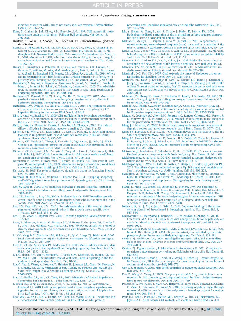
G.M. Xavier et al. / Developmental Biology ∎ (∎∎∎∎) ∎∎∎–∎∎∎16
member, associates with CDO to positively regulate myogenic differentiation.EMBO J. 21, 114–124.
Kang, S., Graham Jr., J.M., Olney, A.H., Biesecker, L.G., 1997. GLI3 frameshift muta-tions cause autosomal dominant Pallister-Hall syndrome. Nat. Genet. 15,266–268.
Kantarci, S., Donnai, D., Noonan, K.M., Pober, B.R., 1993. Donnai-Barrow Syndrome.GeneReviews.
Kantarci, S., Al-Gazali, L., Hill, R.S., Donnai, D., Black, G.C., Bieth, E., Chassaing, N.,Lacombe, D., Devriendt, K., Teebi, A., Loscertales, M., Robson, C., Liu, T., Ma-cLaughlin, D.T., Noonan, K.M., Russell, M.K., Walsh, C.A., Donahoe, P.K., Pober, B.R., 2007. Mutations in LRP2, which encodes the multiligand receptor megalin,cause Donnai-Barrow and facio-oculo-acoustico-renal syndromes. Nat. Genet.39, 957–959.
Karaca, E., Buyukkaya, R., Pehlivan, D., Charng, W.L., Yaykasli, K.O., Bayram, Y.,Gambin, T., Withers, M., Atik, M.M., Arslanoglu, I., Bolu, S., Erdin, S., Buyukkaya,A., Yaykasli, E., Jhangiani, S.N., Muzny, D.M., Gibbs, R.A., Lupski, J.R., 2014. Wholeexome sequencing identifies homozygous GPR161 mutation in a family withpituitary stalk interruption syndrome. J. Clin. Endocrinol. Metab., jc20141984
Kawakami, A., Nojima, Y., Toyoda, A., Takahoko, M., Satoh, M., Tanaka, H., Wada, H.,Masai, I., Terasaki, H., Sakaki, Y., Takeda, H., Okamoto, H., 2005. The zebrafish-secreted matrix protein you/scube2 is implicated in long-range regulation ofhedgehog signaling. Curr. Biol. 15, 480–488.
Kawakami, T., Kawcak, T., Li, Y.J., Zhang, W., Hu, Y., Chuang, P.T., 2002. Mouse dis-patched mutants fail to distribute hedgehog proteins and are defective inhedgehog signaling. Development 129, 5753–5765.
Kietzman, H.W., Everson, J.L., Sulik, K.K., Lipinski, R.J., 2014. The teratogenic effectsof prenatal ethanol exposure are exacerbated by Sonic Hedgehog or GLI2haploinsufficiency in the mouse. PLoS One 9, e89448.
Kim, J., Kato, M., Beachy, P.A., 2009. Gli2 trafficking links Hedgehog-dependentactivation of Smoothened in the primary cilium to transcriptional activation inthe nucleus. Proc. Natl. Acad. Sci. U.S.A 106, 21666–21671.
Kim, J., Hsia, E.Y., Brigui, A., Plessis, A., Beachy, P.A., Zheng, X., 2015. The role ofciliary trafficking in Hedgehog receptor signaling. Sci. Sign. 8, ra55.
Kimonis, V.E., Mehta, S.G., Digiovanna, J.J., Bale, S.J., Pastakia, B., 2004. Radiologicalfeatures in 82 patients with nevoid basal cell carcinoma (NBCC or Gorlin)syndrome. Genet. Med. 6, 495–502.
Kimonis, V.E., Singh, K.E., Zhong, R., Pastakia, B., Digiovanna, J.J., Bale, S.J., 2013.Clinical and radiological features in young individuals with nevoid basal cellcarcinoma syndrome. Genet. Med. 15, 79–83.
Kimonis, V.E., Goldstein, A.M., Pastakia, B., Yang, M.L., Kase, R., DiGiovanna, J.J., Bale,A.E., Bale, S.J., 1997. Clinical manifestations in 105 persons with nevoid basalcell carcinoma syndrome. Am. J. Med. Genet. 69, 299–308.
Kogerman, P., Grimm, T., Kogerman, L., Krause, D., Unden, A.B., Sandstedt, B., Toft-gard, R., Zaphiropoulos, P.G., 1999. Mammalian suppressor-of-fused modulatesnuclear-cytoplasmic shuttling of Gli-1. Nat. Cell Biol. 1, 312–319.
Kurosaka, H., 2015. The roles of Hedgehog signaling in upper lip formation. Biomed.Res. Int. 2015, 901041.
Kurosaka, H., Iulianella, A., Williams, T., Trainor, P.A., 2014. Disrupting hedgehogand WNT signaling interactions promotes cleft lip pathogenesis. J. Clin. Investig.124, 1660–1671.
Lan, Y., Jiang, R., 2009. Sonic hedgehog signaling regulates reciprocal epithelial-mesenchymal interactions controlling palatal outgrowth. Development 136,1387–1396.
Lee, C.S., Buttitta, L., Fan, C.M., 2001a. Evidence that the WNT-inducible growtharrest-specific gene 1 encodes an antagonist of sonic hedgehog signaling in thesomite. Proc. Natl. Acad. Sci. U.S.A 98, 11347–11352.
Lee, C.S., May, N.R., Fan, C.M., 2001b. Transdifferentiation of the ventral retinalpigmented epithelium to neural retina in the growth arrest specific gene1 mutant. Dev. Biol. 236, 17–29.
Lee, R.T.H., Zhao, Z., Ingham, P.W., 2016. Hedgehog signaling. Development 143,367–372.
Letra, A., Menezes, R., Govil, M., Fonseca, R.F., McHenry, T., Granjeiro, J.M., Castilla, E.E., Orioli, I.M., Marazita, M.L., Vieira, A.R., 2010. Follow-up association studies ofchromosome region 9q and nonsyndromic cleft lip/palate. Am. J. Med. Genet. A152A, 1701–1710.
Li, Y.X., Yang, H.T., Zdanowicz, M., Sicklick, J.K., Qi, Y., Camp, T.J., Diehl, A.M., 2007.Fetal alcohol exposure impairs Hedgehog cholesterol modification and signal-ing. Lab. Inv. 87, 231–240.
Liem Jr., K.F., He, M., Ocbina, P.J., Anderson, K.V., 2009. Mouse Kif7/Costal2 is a cilia-associated protein that regulates Sonic hedgehog signaling. Proc. Natl. Acad. Sci.U.S.A 106, 13377–13382.
Lin, C., Fisher, A.V., Yin, Y., Maruyama, T., Veith, G.M., Dhandha, M., Huang, G.J., Hsu,W., Ma, L., 2011. The inductive role of Wnt-beta-Catenin signaling in the for-mation of oral apparatus. Dev. Biol. 356, 40–50.
Lin, C., Yao, E., Wang, K., Nozawa, Y., Shimizu, H., Johnson, J.R., Chen, J.N., Krogan, N.J., Chuang, P.T., 2014. Regulation of Sufu activity by p66beta and Mycbp pro-vides new insight into vertebrate Hedgehog signaling. Genes Dev. 28,2547–2563.
Lin, Y.C., Roffler, S.R., Yan, Y.T., Yang, R.B., 2015. Disruption of Scube2 impairs en-dochondral bone formation. J. Bone Min. Res. 30, 1255–1267.
Lipinski, R.J., Song, C., Sulik, K.K., Everson, J.L., Gipp, J.J., Yan, D., Bushman, W.,Rowland, I.J., 2010. Cleft lip and palate results from Hedgehog signaling an-tagonism in the mouse: phenotypic characterization and clinical implications.Birth Defects Res. A Clin. Mol. Teratol. 88, 232–240.
Low, W.C., Wang, C., Pan, Y., Huang, X.Y., Chen, J.K., Wang, B., 2008. The decouplingof Smoothened from Galphai proteins has little effect on Gli3 protein
Please cite this article as: Xavier, G.M., et al., Hedgehog receptor funcdoi.org/10.1016/j.ydbio.2016.02.009i
processing and Hedgehog-regulated chick neural tube patterning. Dev. Biol.321, 188–196.
Ma, Y., Erkner, A., Gong, R., Yao, S., Taipale, J., Basler, K., Beachy, P.A., 2002.Hedgehog-mediated patterning of the mammalian embryo requires transpor-ter-like function of dispatched. Cell 111, 63–75.
Makino, S., Masuya, H., Ishijima, J., Yada, Y., Shiroishi, T., 2001. A spontaneousmouse mutation, mesenchymal dysplasia (mes), is caused by a deletion of themost C-terminal cytoplasmic domain of patched (ptc). Dev. Biol. 239, 95–106.
Mansilla, M.A., Cooper, M.E., Goldstein, T., Castilla, E.E., Lopez Camelo, J.S., Marazita,M.L., Murray, J.C., 2006. Contributions of PTCH gene variants to isolated cleft lipand palate. Cleft Palate Craniofacial J. 43, 21–29.
Marcucio, R.S., Cordero, D.R., Hu, D., Helms, J.A., 2005. Molecular interactions co-ordinating the development of the forebrain and face. Dev. Biol. 284, 48–61.
Marcucio, R.S., Young, N.M., Hu, D., Hallgrimsson, B., 2011. Mechanisms that un-derlie co-variation of the brain and face. Genesis 49, 177–189.
Martinelli, D.C., Fan, C.M., 2007. Gas1 extends the range of Hedgehog action byfacilitating its signaling. Genes Dev. 21, 1231–1243.
Matteson, P.G., Desai, J., Korstanje, R., Lazar, G., Borsuk, T.E., Rollins, J., Kadambi, S.,Joseph, J., Rahman, T., Wink, J., Benayed, R., Paigen, B., Millonig, J.H., 2008. Theorphan G protein-coupled receptor, Gpr161, encodes the vacuolated lens locusand controls neurulation and lens development. Proc. Natl. Acad. Sci. U.S.A 105,2088–2093.
McLellan, J.S., Zheng, X., Hauk, G., Ghirlando, R., Beachy, P.A., Leahy, D.J., 2008. Themode of Hedgehog binding to Ihog homologues is not conserved across dif-ferent phyla. Nature 455, 979–983.
Meloni, A.R., Fralish, G.B., Kelly, P., Salahpour, A., Chen, J.K., Wechsler-Reya, R.J.,Lefkowitz, R.J., Caron, M.G., 2006. Smoothened signal transduction is promotedby G protein-coupled receptor kinase 2. Mol. Cell Biol. 26, 7550–7560.
Metzis, V., Courtney, A.D., Kerr, M.C., Ferguson, C., Rondon Galeano, M.C., Parton, R.G., Wainwright, B.J., Wicking, C., 2013. Patched1 is required in neural crest cellsfor the prevention of orofacial clefts. Hum. Mol. Genet. 22, 5026–5035.
Ming, J.E., Muenke, M., 2002. Multiple hits during early embryonic development:digenic diseases and holoprosencephaly. Am. J. Hum. Genet. 71, 1017–1032.
Ming, J.E., Roessler, E., Muenke, M., 1998. Human developmental disorders and theSonic hedgehog pathway. Mol. Med. Today 4, 343–349.
Ming, J.E., Kaupas, M.E., Roessler, E., Brunner, H.G., Golabi, M., Tekin, M., Stratton, R.F., Sujansky, E., Bale, S.J., Muenke, M., 2002. Mutations in PATCHED-1, the re-ceptor for SONIC HEDGEHOG, are associated with holoprosencephaly. Hum.Genet. 110, 297–301.
Motoyama, J., Takabatake, T., Takeshima, K., Hui, C., 1998. Ptch2, a second mousePatched gene is co-expressed with Sonic hedgehog. Nat. Genet. 18, 104–106.
Mukhopadhyay, S., Rohatgi, R., 2014. G-protein-coupled receptors, Hedgehog sig-naling and primary cilia. Semin. Cell Dev. Biol. 33, 63–72.
Mukhopadhyay, S., Wen, X., Ratti, N., Loktev, A., Rangell, L., Scales, S.J., Jackson, P.K.,2013. The ciliary G-protein-coupled receptor Gpr161 negatively regulates theSonic hedgehog pathway via cAMP signaling. Cell 152, 210–223.
Nakatomi, M., Hovorakova, M., Gritli-Linde, A., Blair, H.J., MacArthur, K., Peterka, M.,Lesot, H., Peterkova, R., Ruiz-Perez, V.L., Goodship, J.A., Peters, H., 2013. Evcregulates a symmetrical response to Shh signaling in molar development. J.Dent. Res. 92, 222–228.
Nanni, L., Ming, J.E., Bocian, M., Steinhaus, K., Bianchi, D.W., Die-Smulders, C.,Giannotti, A., Imaizumi, K., Jones, K.L., Campo, M.D., Martin, R.A., Meinecke, P.,Pierpont, M.E., Robin, N.H., Young, I.D., Roessler, E., Muenke, M., 1999. Themutational spectrum of the sonic hedgehog gene in holoprosencephaly: SHHmutations cause a significant proportion of autosomal dominant holopro-sencephaly. Hum. Mol. Genet. 8, 2479–2488.
Nedelcu, D., Liu, J., Xu, Y., Jao, C., Salic, A., 2013. Oxysterol binding to the extra-cellular domain of Smoothened in Hedgehog signaling. Nat. Chem. Biol. 9,557–564.
Nieuwenhuis, E., Motoyama, J., Barnfield, P.C., Yoshikawa, Y., Zhang, X., Mo, R.,Crackower, M.A., Hui, C.C., 2006. Mice with a targeted mutation of patched2 areviable but develop alopecia and epidermal hyperplasia. Mol. Cell Biol. 26,6609–6622.
Niewiadomski, P., Kong, J.H., Ahrends, R., Ma, Y., Humke, E.W., Khan, S., Teruel, M.N.,Novitch, B.G., Rohatgi, R., 2014. Gli protein activity is controlled by multisitephosphorylation in vertebrate Hedgehog signaling. Cell Rep. 6, 168–181.
Ocbina, P.J., Anderson, K.V., 2008. Intraflagellar transport, cilia, and mammalianHedgehog signaling: analysis in mouse embryonic fibroblasts. Dev. Dyn. 237,2030–2038.
Ocbina, P.J., Eggenschwiler, J.T., Moskowitz, I., Anderson, K.V., 2011. Complex in-teractions between genes controlling trafficking in primary cilia. Nat. Genet. 43,547–553.
Okada, A., Charron, F., Morin, S., Shin, D.S., Wong, K., Fabre, P.J., Tessier-Lavigne, M.,McConnell, S.K., 2006. Boc is a receptor for sonic hedgehog in the guidance ofcommissural axons. Nature 444, 369–373.
Oro, A.E., Higgins, K., 2003. Hair cycle regulation of Hedgehog signal reception. Dev.Biol. 255, 238–248.
Pan, Y., Wang, C., Wang, B., 2009. Phosphorylation of Gli2 by protein kinase A isrequired for Gli2 processing and degradation and the Sonic Hedgehog-regu-lated mouse development. Dev. Biol. 326, 177–189.
Pantalacci, S., Prochazka, J., Martin, A., Rothova, M., Lambert, A., Bernard, L., Charles,C., Viriot, L., Peterkova, R., Laudet, V., 2008. Patterning of palatal rugae throughsequential addition reveals an anterior/posterior boundary in palatal develop-ment. BMC Dev. Biol. 8, 116.
Park, H.L., Bai, C., Platt, K.A., Matise, M.P., Beeghly, A., Hui, C.C., Nakashima, M.,Joyner, A.L., 2000. Mouse Gli1 mutants are viable but have defects in SHH
tion during craniofacial development. Dev. Biol. (2016), http://dx.

G.M. Xavier et al. / Developmental Biology ∎ (∎∎∎∎) ∎∎∎–∎∎∎ 17
signaling in combination with a Gli2 mutation. Development 127, 1593–1605.Pearse 2nd, R.V., Collier, L.S., Scott, M.P., Tabin, C.J., 1999. Vertebrate homologs of
Drosophila suppressor of fused interact with the gli family of transcriptionalregulators. Dev. Biol. 212, 323–336.
Pedersen, L.B., Rosenbaum, J.L., 2008. Intraflagellar transport (IFT) role in ciliaryassembly, resorption and signalling. Curr. Top. Dev. Biol. 85, 23–61.
Pepinsky, R.B., Zeng, C., Wen, D., Rayhorn, P., Baker, D.P., Williams, K.P., Bixler, S.A.,Ambrose, C.M., Garber, E.A., Miatkowski, K., Taylor, F.R., Wang, E.A., Galdes, A.,1998. Identification of a palmitic acid-modified form of human Sonic hedgehog.J. Biol. Chem. 273, 14037–14045.
Petrova, R., Joyner, A.L., 2014. Roles for Hedgehog signaling in adult organ home-ostasis and repair. Development 141, 3445–3457.
Petryk, A., Graf, D., Marcucio, R., 2015. Holoprosencephaly: signaling interactionsbetween the brain and the face, the environment and the genes, and thephenotypic variability in animal models and humans. Wiley Interdiscip. Rev.Dev. Biol. 4, 17–32.
Porter, J.A., von Kessler, D.P., Ekker, S.C., Young, K.E., Lee, J.J., Moses, K., Beachy, P.A.,1995. The product of hedgehog autoproteolytic cleavage active in local andlong-range signalling. Nature 374, 363–366.
Powell, D.R., Williams, J.S., Hernandez-Lagunas, L., Salcedo, E., O'Brien, J.H., BrukArtinger, K., 2015. Cdon promotes neural crest migration by regulating N-cad-herin localization. Dev. Biol. 407, 289–299.
Pusapati, G.V., Hughes, C.E., Dorn, K.V., Zhang, D., Sugianto, P., Aravind, L., Rohatgi,R., 2014. EFCAB7 and IQCE regulate hedgehog signaling by tethering the EVC-EVC2 complex to the base of primary cilia. Dev. Cell 28, 483–496.
Ribeiro, L.A., Quiezi, R.G., Nascimento, A., Bertolacini, C.P., Richieri-Costa, A., 2010.Holoprosencephaly and holoprosencephaly-like phenotype and GAS1 DNA se-quence changes: report of four Brazilian patients. Am. J. Med. Genet. 152A,1688–1694.
Rice, R., Spencer-Dene, B., Connor, E.C., Gritli-Linde, A., McMahon, A.P., Dickson, C.,Thesleff, I., Rice, D.P., 2004. Disruption of Fgf10/Fgfr2b-coordinated epithelial-mesenchymal interactions causes cleft palate. J. Clin. Investig. 113, 1692–1700.
Roessler, E., Belloni, E., Gaudenz, K., Jay, P., Berta, P., Scherer, S.W., Tsui, L.C., Muenke,M., 1996. Mutations in the human Sonic Hedgehog gene cause holoprosence-phaly. Nat. Genet. 14, 357–360.
Roessler, E., Ma, Y., Ouspenskaia, M.V., Lacbawan, F., Bendavid, C., Dubourg, C.,Beachy, P.A., Muenke, M., 2009. Truncating loss-of-function mutations of DISP1contribute to holoprosencephaly-like microform features in humans. Hum.Genet. 125, 393–400.
Roessler, E., Du, Y.Z., Mullor, J.L., Casas, E., Allen, W.P., Gillessen-Kaesbach, G.,Roeder, E.R., Ming, J.E., Ruiz i Altaba, A., Muenke, M., 2003. Loss-of-functionmutations in the human GLI2 gene are associated with pituitary anomalies andholoprosencephaly-like features. Proc. Natl. Acad. Sci. U.S.A 100, 13424–13429.
Rohatgi, R., Milenkovic, L., Scott, M.P., 2007. Patched1 regulates hedgehog signalingat the primary cilium. Science 317, 372–376.
Rohatgi, R., Milenkovic, L., Corcoran, R.B., Scott, M.P., 2009. Hedgehog signaltransduction by Smoothened: pharmacologic evidence for a 2-step activationprocess. Proc. Natl. Acad. Sci. U.S.A 106, 3196–3201.
Rosenfeld, J.A., Ballif, B.C., Martin, D.M., Aylsworth, A.S., Bejjani, B.A., Torchia, B.S.,Shaffer, L.G., 2010. Clinical characterization of individuals with deletions ofgenes in holoprosencephaly pathways by aCGH refines the phenotypic spec-trum of HPE. Hum. Genet. 127, 421–440.
Rubenstein, J.L., Beachy, P.A., 1998. Patterning of the embryonic forebrain. Curr.Opin. Neurobiol. 8, 18–26.
Ruiz i Altaba, A., 1997. Catching a Gli-mpse of Hedgehog. Cell 90, 193–196.Ruiz i Altaba, A., Mas, C., Stecca, B., 2007. The Gli code: an information nexus
regulating cell fate, stemness and cancer. Trends Cell Biol. 17, 438–447.Ruiz-Perez, V.L., Goodship, J.A., 2009. Ellis-van Creveld syndrome and Weyers ac-
rodental dysostosis are caused by cilia-mediated diminished response tohedgehog ligands. Am. J. Med. Genet. C Semin. Med. Genet. 151C, 341–351.
Ruiz-Perez, V.L., Blair, H.J., Rodriguez-Andres, M.E., Blanco, M.J., Wilson, A., Liu, Y.N.,Miles, C., Peters, H., Goodship, J.A., 2007. Evc is a positive mediator of Ihh-regulated bone growth that localises at the base of chondrocyte cilia. Devel-opment 134, 2903–2912.
Ruiz-Perez, V.L., Tompson, S.W., Blair, H.J., Espinoza-Valdez, C., Lapunzina, P., Silva,E.O., Hamel, B., Gibbs, J.L., Young, I.D., Wright, M.J., Goodship, J.A., 2003. Mu-tations in two nonhomologous genes in a head-to-head configuration causeEllis-van Creveld syndrome. Am. J. Hum. Genet. 72, 728–732.
Ruiz-Perez, V.L., Ide, S.E., Strom, T.M., Lorenz, B., Wilson, D., Woods, K., King, L.,Francomano, C., Freisinger, P., Spranger, S., Marino, B., Dallapiccola, B., Wright,M., Meitinger, T., Polymeropoulos, M.H., Goodship, J., 2000. Mutations in a newgene in Ellis-van Creveld syndrome and Weyers acrodental dysostosis. Nat.Genet. 24, 283–286.
Saito, A., Pietromonaco, S., Loo, A.K., Farquhar, M.G., 1994. Complete cloning andsequencing of rat gp330/"megalin," a distinctive member of the low densitylipoprotein receptor gene family. Proc. Natl. Acad. Sci. U.S.A. 91, 9725–9729.
Sanchez-Arrones, L., Cardozo, M., Nieto-Lopez, F., Bovolenta, P., 2012. Cdon and Boc:two transmembrane proteins implicated in cell-cell communication. Int. J.Biochem. Cell Biol. 44, 698–702.
Sanchez-Arrones, L., Nieto-Lopez, F., Sanchez-Camacho, C., Carreres, M.I., Herrera,E., Okada, A., Bovolenta, P., 2013. Shh/Boc signaling is required for sustainedgeneration of ipsilateral projecting ganglion cells in the mouse retina. J. Neu-rosci. 33, 8596–8607.
Sandell, L.L., Iulianella, A., Melton, K.R., Lynn, M., Walker, M., Inman, K.E., Bhatt, S.,Leroux-Berger, M., Crawford, M., Jones, N.C., Dennis, J.F., Trainor, P.A., 2011. Aphenotype-driven ENU mutagenesis screen identifies novel alleles with
Please cite this article as: Xavier, G.M., et al., Hedgehog receptor funcdoi.org/10.1016/j.ydbio.2016.02.009i
functional roles in early mouse craniofacial development. Genesis 49, 342–359.Sanders, T.A., Llagostera, E., Barna, M., 2013. Specialized filopodia direct long-range
transport of SHH during vertebrate tissue patterning. Nature 497, 628–632.Sasaki, R., Saito, K., Watanabe, Y., Takayama, Y., Fujii, K., Agawa, K., Miyashita, T.,
Ando, T., Akizuki, T., 2009. Nevoid basal cell carcinoma syndrome with cleft lipand palate associated with the novel PTCH gene mutations. J. Hum. Genet. 54,398–402.
Schwend, T., Ahlgren, S.C., 2009. Zebrafish con/disp1 reveals multiple spatio-temporal requirements for Hedgehog-signaling in craniofacial development.BMC Dev. Biol. 9, 59.
Segovia, J., Zarco, N., 2014. Gas1 is a pleiotropic regulator of cellular functions: fromembryonic development to molecular actions in cancer gene therapy. Mini Rev.Med. Chem. 14, 1139–1147.
Seppala, M., Xavier, G.M., Fan, C.M., Cobourne, M.T., 2014. Boc modifies the spec-trum of holoprosencephaly in the absence of Gas1 function. Biol. Open 3,728–740.
Seppala, M., Depew, M.J., Martinelli, D.C., Fan, C.M., Sharpe, P.T., Cobourne, M.T.,2007. Gas1 is a modifier for holoprosencephaly and genetically interacts withsonic hedgehog. J. Clin Investig. 117, 1575–1584.
Spoelgen, R., Hammes, A., Anzenberger, U., Zechner, D., Andersen, O.M., Jerchow, B.,Willnow, T.E., 2005. LRP2/megalin is required for patterning of the ventraltelencephalon. Development 132, 405–414.
Stecca, B., Ruiz i Altaba, A., 2009. A GLI1-p53 inhibitory loop controls neural stemcell and tumour cell numbers. EMBO J. 28, 663–676.
Stecca, B., Mas, C., Clement, V., Zbinden, M., Correa, R., Piguet, V., Beermann, F., Ruiz,I.A.A., 2007. Melanomas require HEDGEHOG-GLI signaling regulated by inter-actions between GLI1 and the RAS-MEK/AKT pathways. Proc. Natl. Acad. Sci. U.S.A 104, 5895–5900.
Stone, D.M., Murone, M., Luoh, S., Ye, W., Armanini, M.P., Gurney, A., Phillips, H.,Brush, J., Goddard, A., de Sauvage, F.J., Rosenthal, A., 1999. Characterization ofthe human suppressor of fused, a negative regulator of the zinc-finger tran-scription factor Gli. J. Cell Sci. 112 (Pt 23), 4437–4448.
Stone, D.M., Hynes, M., Armanini, M., Swanson, T.A., Gu, Q., Johnson, R.L., Scott, M.P.,Pennica, D., Goddard, A., Phillips, H., Noll, M., Hooper, J.E., de Sauvage, F., Ro-senthal, A., 2006. The tumour-suppressor gene patched encodes a candidatereceptor for Sonic hedgehog. Nature 384, 129–134.
Swartz, M.E., Nguyen, V., McCarthy, N.Q., Eberhart, J.K., 2012. Hh signaling regulatespatterning and morphogenesis of the pharyngeal arch-derived skeleton. Dev.Biol. 369, 65–75.
Tabler, J.M., Bolger, T.G., Wallingford, J., Liu, K.J., 2014. Hedgehog activity controlsopening of the primary mouth. Dev. Biol. 396, 1–7.
Tabler, J.M., Barrell, W.B., Szabo-Rogers, H.L., Healy, C., Yeung, Y., Perdiguero, E.G.,Schulz, C., Yannakoudakis, B.Z., Mesbahi, A., Wlodarczyk, B., Geissmann, F.,Finnell, R.H., Wallingford, J.B., Liu, K.J., 2013. Fuz mutant mice reveal sharedmechanisms between ciliopathies and FGF-related syndromes. Dev. Cell 25,623–635.
Taipale, J., Cooper, M.K., Maiti, T., Beachy, P.A., 2002. Patched acts catalytically tosuppress the activity of Smoothened. Nature 418, 892–897.
Tapadia, M.D., Cordero, D.R., Helms, J.A., 2005. It's all in your head: new insightsinto craniofacial development and deformation. J. Anat. 207, 461–477.
Tenzen, T., Allen, B.L., Cole, F., Kang, J.S., Krauss, R.S., McMahon, A.P., 2006. The cellsurface membrane proteins Cdo and Boc are components and targets of theHedgehog signaling pathway and feedback network in mice. Dev. Cell 10,647–656.
Thomason, H.A., Dixon, M.J., Dixon, J., 2008. Facial clefting in Tp63 deficient miceresults from altered Bmp4, Fgf8 and Shh signaling. Dev. Biol. 321, 273–282.
Tian, H., Jeong, J., Harfe, B.D., Tabin, C.J., McMahon, A.P., 2005. Mouse Disp1 is re-quired in sonic hedgehog-expressing cells for paracrine activity of the choles-terol-modified ligand. Development 132, 133–142.
Tukachinsky, H., Lopez, L.V., Salic, A., 2010. A mechanism for vertebrate Hedgehogsignaling: recruitment to cilia and dissociation of SuFu-Gli protein complexes. J.Cell Biol. 191, 415–428.
Tukachinsky, H., Kuzmickas, R.P., Jao, C.Y., Liu, J., Salic, A., 2012. Dispatched andscube mediate the efficient secretion of the cholesterol-modified hedgehogligand. Cell Rep. 2, 308–320.
Tuson, M., He, M., Anderson, K.V., 2011. Protein kinase A acts at the basal body ofthe primary cilium to prevent Gli2 activation and ventralization of the mouseneural tube. Development 138, 4921–4930.
Wada, N., Javidan, Y., Nelson, S., Carney, T.J., Kelsh, R.N., Schilling, T.F., 2005.Hedgehog signaling is required for cranial neural crest morphogenesis andchondrogenesis at the midline in the zebrafish skull. Development 132,3977–3988.
Wang, B., Fallon, J.F., Beachy, P.A., 2000. Hedgehog-regulated processing of Gli3produces an anterior/posterior repressor gradient in the developing vertebratelimb. Cell 100, 423–434.
Wang, C., Wu, H., Katritch, V., Han, G.W., Huang, X.P., Liu, W., Siu, F.Y., Roth, B.L.,Cherezov, V., Stevens, R.C., 2013. Structure of the human smoothened receptorbound to an antitumour agent. Nature 497, 338–343.
Washington Smoak, I., Byrd, N.A., Abu-Issa, R., Goddeeris, M.M., Anderson, R.,Morris, J., Yamamura, K., Klingensmith, J., Meyers, E.N., 2005. Sonic hedgehog isrequired for cardiac outflow tract and neural crest cell development. Dev. Biol.283, 357–372.
Welsh, I.C., O'Brien, T.P., 2009. Signaling integration in the rugae growth zone di-rects sequential SHH signaling center formation during the rostral outgrowth ofthe palate. Dev. Biol. 336, 53–67.
Wen, X., Lai, C.K., Evangelista, M., Hongo, J.A., de Sauvage, F.J., Scales, S.J., 2010.
tion during craniofacial development. Dev. Biol. (2016), http://dx.
G.M. Xavier et al. / Developmental Biology ∎ (∎∎∎∎) ∎∎∎–∎∎∎18
Kinetics of hedgehog-dependent full-length Gli3 accumulation in primary ciliaand subsequent degradation. Mol. Cell Biol. 30, 1910–1922.
Wicking, C., Shanley, S., Smyth, I., Gillies, S., Negus, K., Graham, S., Suthers, G.,Haites, N., Edwards, M., Wainwright, B., Chenevix-Trench, G., 1997. Most germ-line mutations in the nevoid basal cell carcinoma syndrome lead to a pre-mature termination of the PATCHED protein, and no genotype-phenotypecorrelations are evident. Am. J. Hum. Genet. 60, 21–26.
Willnow, T.E., Hilpert, J., Armstrong, S.A., Rohlmann, A., Hammer, R.E., Burns, D.K.,Herz, J., 1996. Defective forebrain development in mice lacking gp330/megalin.Proc. Natl. Acad. Sci. U.S.A 93, 8460–8464.
Wilson, D.B., Wyatt, D.P., 1986. Pathogenesis of neural dysraphism in the mousemutant vacuolated lens (vl). J. Neuropathol. Exp. Neurol. 45, 43–55.
Wilson, D.B., Wyatt, D.P., 1988. Closure of the posterior neuropore in the vl mutantmouse. Anat. Embryol. 178, 559–563.
Woods, I.G., Talbot, W.S., 2005. The you gene encodes an EGF-CUB protein essentialfor Hedgehog signaling in zebrafish. PLoS Biol. 3, e66.
Wu, B.T., Su, Y.H., Tsai, M.T., Wasserman, S.M., Topper, J.N., Yang, R.B., 2004. A novelsecreted, cell-surface glycoprotein containing multiple epidermal growth fac-tor-like repeats and one CUB domain is highly expressed in primary osteoblastsand bones. J. Biol. Chem. 279, 37485–37490.
Wu, Y.Y., Peck, K., Chang, Y.L., Pan, S.H., Cheng, Y.F., Lin, J.C., Yang, R.B., Hong, T.M.,Yang, P.C., 2011. SCUBE3 is an endogenous TGF-beta receptor ligand and reg-ulates the epithelial-mesenchymal transition in lung cancer. Oncogene 30,3682–3693.
Xavier, G.M., Sharpe, P.T., Cobourne, M.T., 2009. Scube1 is expressed during facialdevelopment in the mouse. J. Exp. Zool. B Mol. Dev. Evol. 312B, 518–524.
Please cite this article as: Xavier, G.M., et al., Hedgehog receptor funcdoi.org/10.1016/j.ydbio.2016.02.009i
Xavier, G.M., Panousopoulos, L., Cobourne, M.T., 2013. Scube3 is expressed inmultiple tissues during development but is dispensable for embryonic survivalin the mouse. PLoS One 8, e55274.
Yamagishi, C., Yamagishi, H., Maeda, J., Tsuchihashi, T., Ivey, K., Hu, T., Srivastava, D.,2006. Sonic hedgehog is essential for first pharyngeal arch development. Pe-diatr. Res. 59, 349–354.
Yang, C., Chen, W., Chen, Y., Jiang, J., 2012. Smoothened transduces Hedgehog signalby forming a complex with Evc/Evc2. Cell Res. 22, 1593–1604.
Yang, R.B., Ng, C.K., Wasserman, S.M., Colman, S.D., Shenoy, S., Mehraban, F., Ko-muves, L.G., Tomlinson, J.E., Topper, J.N., 2002. Identification of a novel family ofcell-surface proteins expressed in human vascular endothelium. J. Biol. Chem.277, 46364–46373.
Young, N.M., Chong, H.J., Hu, D., Hallgrimsson, B., Marcucio, R.S., 2010. Quantitativeanalyses link modulation of sonic hedgehog signaling to continuous variationin facial growth and shape. Development 137, 3405–3409.
Zhang, W., Kang, J.S., Cole, F., Yi, M.J., Krauss, R.S., 2006. Cdo functions at multiplepoints in the Sonic Hedgehog pathway, and Cdo-deficient mice accuratelymodel human holoprosencephaly. Dev. Cell 10, 657–665.
Zhang, W., Hong, M., Bae, G.U., Kang, J.S., Krauss, R.S., 2011. Boc modifies the ho-loprosencephaly spectrum of Cdo mutant mice. Dis. Model Mech. 4, 368–380.
Zhang, W., Mulieri, P.J., Gaio, U., Bae, G.U., Krauss, R.S., Kang, J.S., 2009. Ocular ab-normalities in mice lacking the immunoglobulin superfamily member Cdo.FEBS J. 276, 5998–6010.
Zhang, X.M., Ramalho-Santos, M., McMahon, A.P., 2001. Smoothened mutants re-veal redundant roles for Shh and Ihh signaling including regulation of L/Rsymmetry by the mouse node. Cell 106, 781–792.
tion during craniofacial development. Dev. Biol. (2016), http://dx.


![URINARY EXCRETION OF ALBUMIN - nephro-necker.org · urinary excretion of albumin ... tojo and endou [12], ... 105, 1353-1361 2000. renal albumin handling in megalin knock out mice](https://img.pdfslide.net/doc/110x75/5c4a0c7693f3c317653c31ff/urinary-excretion-of-albumin-nephro-urinary-excretion-of-albumin-tojo.jpg)