Embed Size (px)
Citation preview
King’s Research Portal
DOI:10.1016/j.carbpol.2017.02.077
Document VersionPeer reviewed version
Link to publication record in King's Research Portal
Citation for published version (APA):Grundy, M. M. L., Quint, J., Rieder, A., Ballance, S., Dreiss, C. A., Butterworth, P. J., & Ellis, P. R. (2017).Impact of hydrothermal and mechanical processing on dissolution kinetics and rheology of oat -glucan.Carbohydrate polymers, 166, 387-397. DOI: 10.1016/j.carbpol.2017.02.077
Citing this paperPlease note that where the full-text provided on King's Research Portal is the Author Accepted Manuscript or Post-Print version this maydiffer from the final Published version. If citing, it is advised that you check and use the publisher's definitive version for pagination,volume/issue, and date of publication details. And where the final published version is provided on the Research Portal, if citing you areagain advised to check the publisher's website for any subsequent corrections.
General rightsCopyright and moral rights for the publications made accessible in the Research Portal are retained by the authors and/or other copyrightowners and it is a condition of accessing publications that users recognize and abide by the legal requirements associated with these rights.
•Users may download and print one copy of any publication from the Research Portal for the purpose of private study or research.•You may not further distribute the material or use it for any profit-making activity or commercial gain•You may freely distribute the URL identifying the publication in the Research Portal
Take down policyIf you believe that this document breaches copyright please contact [email protected] providing details, and we will remove access tothe work immediately and investigate your claim.
Download date: 22. Jun. 2018
Accepted Manuscript
Title: Impact of hydrothermal and mechanical processing ondissolution kinetics and rheology of oat �-glucan
Authors: Myriam Grundy, Janina Quint, Anne Rieder, SimonBallance, Cecile A. Dreiss, Peter J. Butterworth, Peter R. Ellis
PII: S0144-8617(17)30207-2DOI: http://dx.doi.org/doi:10.1016/j.carbpol.2017.02.077Reference: CARP 12056
To appear in:
Received date: 21-11-2016Revised date: 7-2-2017Accepted date: 20-2-2017
Please cite this article as: Grundy, Myriam., Quint, Janina., Rieder, Anne., Ballance,Simon., Dreiss, Cecile A., Butterworth, Peter J., & Ellis, Peter R., Impact ofhydrothermal and mechanical processing on dissolution kinetics and rheology of oat�-glucan.Carbohydrate Polymers http://dx.doi.org/10.1016/j.carbpol.2017.02.077
This is a PDF file of an unedited manuscript that has been accepted for publication.As a service to our customers we are providing this early version of the manuscript.The manuscript will undergo copyediting, typesetting, and review of the resulting proofbefore it is published in its final form. Please note that during the production processerrors may be discovered which could affect the content, and all legal disclaimers thatapply to the journal pertain.
1
Impact of hydrothermal and mechanical processing on dissolution kinetics
and rheology of oat β-glucan
Myriam M.-L. Grundya, b,†, Janina Quintc, Anne Riederd, Simon Ballanced, Cécile A.
Dreisse, Peter J. Butterwortha, and Peter R. Ellisa,*
a Biopolymers Group, Diabetes and Nutritional Sciences Division, King’s College London,
Franklin-Wilkins Building, 150 Stamford Street, London SE1 9NH, UK
b Institute of Food Research, Norwich Research Park, Colney, Norwich NR4 7UA, UK
c Department of Nutritional Sciences, University of Vienna, Althanstraße 14 (UZA II), 1090
Vienna, Austria
d Nofima, Norwegian Institute for Food, Fisheries and Aquaculture Research, PB 210, N-
1431 Ås, Norway
e Institute of Pharmaceutical Science, King’s College London, Franklin-Wilkins Building,
150 Stamford Street, London SE1 9NH, UK
* Corresponding author at: King’s College London, Diabetes and Nutritional Sciences
Division, Franklin-Wilkins Building, 150 Stamford Street, London SE1 9NH, UK. Tel.: +44
0 207 848 4238; fax: +44 0 207 848 4171.
E-mail addresses: [email protected] (M.M.-L. Grundy),
[email protected] (J. Quint), [email protected] (A. Reider),
[email protected] (S. Balance), [email protected] (C. Dreiss),
[email protected] (P.J. Butterworth), [email protected] (P.R. Ellis).
† Present/permanent address: Myriam M.-L. Grundy, Institute of Food Research, Norwich
Research Park, Colney, Norwich NR4 7UA, UK.
2
Highlights
β-Glucan release from oat cell walls during incubation was not complete.
Processing of oats affects the rate and extent of β-glucan release and solution
rheology.
The rheology of β-glucan solutions varied depending on oat composition and
presence of particulates.
Variations in β-glucan solubility have important implications for physiological
activity.
3
ABST RA CT
Oat mixed-linkage β-glucan has been shown to lower fasting blood cholesterol concentrations
due notably to an increase in digesta viscosity in the proximal gut. To exert its action, the
polysaccharide has to be released from the food matrix and hydrated. The dissolution kinetics
of β-glucan from three oat materials, varying in their structure, composition and degree of
processing, was investigated by incubating the oats at 37ºC over multiple time points (up to
72 h). The samples were analysed for β-glucan content, weight-average molecular weight and
rheological behaviour. Regardless of the materials studied and the processing applied, the
solubilisation of β-glucan was not complete. Mechanical and hydrothermal processing led to
differences in the viscosity flow curves of the recovered solutions, with the presence of
particulates having a marked effect. This study revealed that the structure and processing
methods applied to oat materials resulted in varied and complex rheological properties,
especially when particulates are present.
Keywords: Oat β-glucan, Solubility, Oat structure, Viscosity flow behaviour, Molecular
weight
4
1. Introduction
The common oat grain (Avena sativa L.) is consumed by humans mainly as breakfast
cereals, comprising whole grain flour or flakes, which can be eaten either as porridge after
heating in water/milk or in the form of ready-to-eat cereals, such as muesli and granola
(Webster, 2011). Oat flour is often used as an ingredient in bread, muffins, granola bars,
biscuits and snack bars. Typical commercial products vary in the size and shape of the oat
particles they contain. An important component of oats is β-glucan, which is composed of a
mixed-linkage linear polymer of (1→3)(1→4)-β-D-glucan. This polymer is a water-soluble
dietary fibre that is considered to have nutritional benefits, such as lowering plasma
cholesterol concentrations.
β-Glucan is located in the endosperm cell walls of oats, with particularly rich
concentrations found in the sub-aleurone layers, i.e., the endosperm tissue located adjacent to
the aleurone layer (Miller & Fulcher, 2011). Commercial oat bran can contain significant
concentrations of β-glucan because the milled bran comprises large amounts of adhering
endosperm, including sub-aleurone tissue. The β-glucan content of oat varies depending on
genotype and environmental conditions during growth and ranges from ~2.2 to 7.8%
(Lazaridou, Biliaderis & Izydorczyk, 2007). It is a polydisperse polysaccharide with reported
values of average molecular weight (MW) between ~0.1 and 2.5 million g/mol (Åman,
Rimsten & Andersson, 2004; Andersson & Börjesdotter, 2011; Beer, Wood & Weisz, 1997;
Doublier & Wood, 1995; Johansson, Virkki, Maunu, Lehto, Ekholm & Varo, 2000). This
variation in MW, together with the structural modifications resulting from the domestic and
commercial processing of oats, has a direct impact on some of the properties of the β-glucan
(Beer, Wood, Weisz & Fillion, 1997; Tosh et al., 2010). For instance, manipulating the MW
of β-glucan and the particle size of oat particles led to materials with different solubility and
viscosity (Wang & Ellis, 2014).
5
Oat β-glucan has similar solution properties to other types of soluble dietary fibre, such
as guar galactomannan, which exist in solution as fluctuating ‘random coils’ of glycan chains
(Ellis, Wang, Rayment, Ren & Ross-Murphy, 2001; Morris, 1992). The solution properties of
these conformationally-disordered polysaccharides, specifically their capacity to generate
viscosity, are dependent largely on the number (i.e. concentration) and size (i.e. MW) of the
polymer chains that become completely hydrated. Thus, the rate and extent of dissolution as
well as the concentration and molecular size of the polysaccharide, are strongly linked to
their physiological activity (Judd & Ellis, 2005; Wang & Ellis, 2014). As the polymer
concentration increases, individual polymer coils interpenetrate to form an entangled
network, resulting in an increase in viscosity (Ellis, Wang, Rayment, Ren & Ross-Murphy,
2001).
The nutritional value and health benefits of oat are now well established, particularly in
relation to its β-glucan content and positive effects on lipid metabolism and potential risk-
reduction of cardiometabolic diseases (Welch, 2011; Wolever et al., 2010; Wood, 2007).
Moreover, it has been previously reported that oat β-glucan attenuates blood cholesterol and
lipid concentrations due notably to its capacity of generating highly viscous solutions in the
proximal gut (Othman, Moghadasian & Jones, 2011; Wolever et al., 2010). As explained
above, this property relies on the MW and concentration of the β-glucan present in solution.
However, the details of the mode of action of this polysaccharide and its behaviour in the
gastrointestinal tract are not fully understood. In particular, it is still unknown how much and
how quickly β-glucan is released from the cell walls of the oat tissue matrix. Depending on
the method used, oat β-glucan can be difficult to extract and the quantity of polymer that
solubilises relies on various parameters such as pre-treatment, particle size and the
temperature of extraction (Wood, Siddiqui & Paton, 1978; Zhang, Liang, Pei, Gao & Zhang,
2009).
6
The purpose of the current study was to investigate the effects of differences in structure
and particle size of selected oat flours and flakes on the dissolution kinetics and solution
rheology of β-glucan during aqueous incubation. We have characterised these complex oat
materials by analysing their chemical composition, MW of β-glucan and rheological
properties. In addition, we have determined, using a new assay, the temporal release of β-
glucan (i.e. dissolved polymer) from raw, milled and hydrothermally processed oat materials.
The flow behaviour of the released β-glucan was compared with purer forms of the polymer
and also guar gum, an endospermic flour of a leguminous seed containing galactomannan,
that has been well-characterised. Finally, macro- and microscopic examination of the material
before and after incubation was performed to provide some additional insight of the physical
changes in the oat materials.
2. Materials and Methods
2.1. Materials
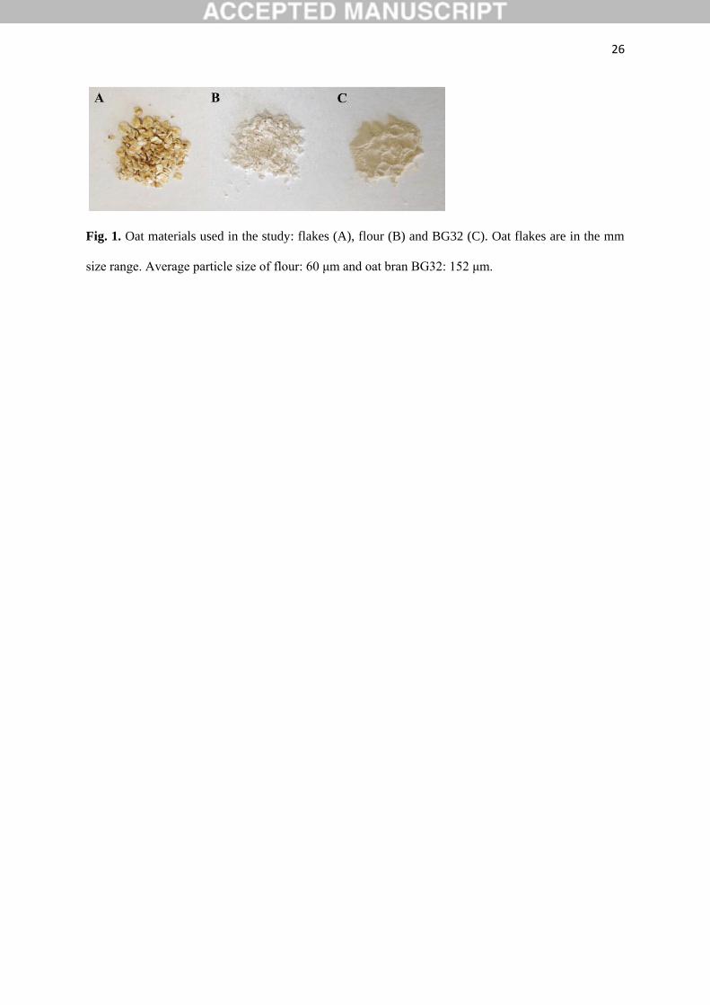
Oat flakes from the Belinda variety were obtained from Lantmännen Cerealia, Moss,
Norway. Oat flour was produced at Nofima from the Belinda oats by milling the flakes on a
laboratory hammer mill (Retsch, Model ZM100, Retsch GmbH, Haan, Germany) with a
0.5 mm mesh (Fig. 1). Extracted oat β-glucan of high MW (BG1) was a generous gift from
Dr Susan Tosh at Agricultural and Agri-Food Canada. Swedish Oat Fiber (Swedish Oat Fiber
AB, Bua, Sweden) provided oat flour enriched with bran (BG32) and medium MW β-glucan
(BG2). Commercial, food grade guar gum flour (Meyprogat, M150) was generously provided
by Dr Graham Sworn (Danisco, Paris, France). Lichenase (EC 3.2.1.73) was purchased from
Megazyme (Bray, Wicklow, Ireland) and thermostable Bacillus licheniformis α-amylase
(Thermamyl® 120) was obtained from Sigma-Aldrich Chemical Co. (Poole, UK). Phosphate
7
buffer (20 mM, pH 6.5) was prepared by dissolving NaH2PO4 and NaN3 (0.02%) in deionised
water followed by adjustment of pH with 0.1 M NaOH.
2.2. Physical and chemical characterisation of materials
The average particle size of the flours (Fig. 1) was measured using a Malvern laser
diffraction particle sizer 2000 equipped with a dispersant unit (Hydro 2000G) filled with
water (Malvern Instruments Ltd.). Each oat material and the guar gum (the positive control)
were analysed for protein (Kjeldhal method with protein N factor of 5.7), lipid (Soxhlet;
hexane), starch (AOAC 996.11), non-starch polysaccharides (Englyst, Quigley & Hudson,
1994) and β-glucan (AOAC 995.16) content. Moisture (oven-dried at 102°C) and ash
(combustion at 525°C) contents were also determined. β-Glucans were extracted from the
original material using a method previously described (Rieder, Holtekjølen, Sahlstrøm &
Moldestad, 2012) and MW of the extracted β-glucans was analysed with the calcofluor
method as described below. β-Glucanase activity in the oat materials (original and incubated
samples) was determined using the Megazyme kit assay employing the use of the substrate
azo-barley glucan (Megazyme, Product Code: K-MBGL). Duplicate measurements were
made for each analysis.
2.3. Quantification of β-glucan release
Raw oat material (flakes, flour or BG32) was added to 12 mL of phosphate buffer to
obtain a β-glucan concentration of either 0.5 or 1.0% (w/v). Hydrothermally processed
(cooked) oat samples of 0.5 or 1.0 % (w/v) β-glucan were obtained by adding deionised water
(16% of final weight) to the oats, and by placing the samples into a boiling water bath. After
10 min of cooking, 12 mL of phosphate buffer were added to each sample.
8
The samples were then incubated at 37°C on a rotator for periods of 0, 5, 10, 15, 30 or 60
min, 2, 5, 8, 24, 48 or 72 h, using one sample per time point. It is still unknown how long the
cell walls of plant foods (dietary fibre), such as in oat tissue, remain in the digestive tract
before being degraded by bacterial fermentation and, if not totally fermented, excreted in the
faeces. Therefore, 72 h was chosen as an end point that represents the maximum time that
oats might reside in the gastrointestinal tract. After centrifugation at 1800 g for 10 min,
depending on the viscosity, 0.1 to 0.5 mL of the supernatant was collected and the β-glucan
precipitated in ethanol (two steps: first 95% w/v, then 50% w/v). The extracted β-glucan
samples were analysed using an enzymic method based on a cereal mixed-linkage β-glucan
kit from Megazyme. The released β-glucan (i.e. solubilised fraction) was expressed as a
percentage of total β-glucan originally present in the sample. Each measurement was
performed in duplicate. For presentational purposes in the Results section, the experimental
data were fitted by non-linear regression using Sigma Plot (version 13 Systat© Software
Inc.).
2.4. Calcofluor weight-average molecular weight measurements
Calcofluor and cereal β-glucan form a distinct fluorescent complex, which enables the
determination of β-glucan MW distributions in the presence of other macromolecules (Wood,
2011; Wood & Fulcher, 1978). In this study, an aqueous size-exclusion chromatographic
(SEC) separation of β-glucan with HPLC and an on-line and post-column addition of
calcofluor was employed to measure the β-glucan MW in the original oat materials (after
extraction as described above) and in the supernatants of the incubated (1, 2, 5 or 72 h) raw or
cooked oat samples. Aliquots of the incubated samples were diluted either ten-fold or two-
fold in phosphate buffer, depending on β-glucan content. The solution was mixed thoroughly,
centrifuged at 1,800 g for 10 min, and filtered (0.8 µm syringe filter, Millipore) before
9
injection of 50 µL into the HPLC system as previously described (Rieder et al., 2015).
Briefly, the system consisted of two pumps (UltiMate 3000 pump and degasser module,
Thermo Scientific), a Spectaphysics AS3500 auto injector, a guard-column (Tosoh PWXL),
two serially connected columns (Tosoh TSK-gel G5000 PWXL and G6000PWXL,
maintained at 40ºC) and a fluorescence detector (Shimadzu RF-10A, Shimadzu Europa,
Duisburg, Germany). The eluent (50 mM Na2SO4) was delivered at a flow rate of 0.5
mL/min. Calcofluor (Megazyme) solution (25 mg/L in 0.1 M
tris(hydroxymethyl)aminomethane) was delivered post-column through a T-valve at a flow
rate of 0.25 mL/min. Fluorescence detection of the formed calcofluor/glucan complexes
occurred at λex = 415 nm and λem = 445 nm. A calibration curve for the MW of β-glucan
was constructed with in-house β-glucan standards and standards purchased from Megazyme
with peak MW from 100 to 1080 x 103 g/mol. A proprietary third order polynomial
regression (PSS Poly 3) was fitted to the retention time plotted against the peak MW using
PSS WinGPC Unichrome software (PSS, Polymer Standard Service, Mainz, Germany). If not
otherwise stated, MW values reported in this study are calcofluor weight-average MW
calculated from the measured MW distributions by using PSS WinGPC Unichrome software.
Each measurement was performed in duplicate.
It should be noted that outside a MW range of 10-500 x 103 g/mol, the post column
calcofluor method yields only relative/apparent molecular weight values (Rieder, Ballance &
Knutsen, 2015). Above a MW of 500 x 103 g/mol, the calcofluor method results in an
increasing underestimation of the MW compared to other methods such as SEC-RI, but has
the advantage of being able to determine β-glucan MW in crude extracts.
2.5. Weight-average molecular weight determination using SEC-MALLS-VISC-RI
10
The MW distribution of guar galactomannan was determined in aqueous solutions at
concentrations of 1mg/mL as previously described (Rieder, Ballance & Knutsen, 2015) and is
reported as weight-average molecular weight.
2.6. Rheological behaviour
Raw and cooked oat samples incubated for 1, 2, 5 and 72 h, as described above (section
2.3.), were centrifuged (1,800 g for 10 min) and the supernatant collected for rheological
measurements. Also, solutions of purified polymers, guar gum and β-glucan (BG1 and BG2),
were used as controls. To ensure total hydration of the polysaccharides, the solutions were
prepared by slowly sprinkling the polymer into a rapidly swirling vortex of phosphate buffer
and the mixture left to warm up at 80ºC for 2 h, followed by cooling to room temperature
overnight. The rheological measurements were carried out on the control and oat samples
using a dynamic strain-controlled rheometer (Physica MCR 301, Anton Paar, Stuttgart,
Germany) equipped with a double gap geometry (DG 26.7) and a temperature-controlling
Peltier unit (C-PTD 200). Viscosity flow curves were obtained in duplicate at 25°C after 2
min temperature equilibration with the operating shear rate ranging from 0.01 to 1000 s−1
with seven measurement points per decade. The measurement point duration ranged from 100
to 1 s during the forward ramp and the backward ramp. The cooked oat samples were
recovered after the rheological measurement and treated with thermostable amylase (0.5
mL/g of starch, at 90ºC for 2 h) or lichenase (0.035 mL/mL of β-glucan solution, at 50ºC for
1 h) and the flow behaviour measured a second time. The apparent zero-shear viscosity was
estimated by fitting the data to the Cross model (Cross, 1965):
η = η∞ + [η0x − η∞]/[1 + (𝑎γ)𝑝] (1)
where η0x and η∞ are viscosities at zero and infinite shear rate, a and γ are a relaxation time
and shear rate, respectively, and p is an exponent.
11
2.7. Microstructural characterisation
Raw and cooked particles of oat flour and BG32 were collected at baseline or after 72 h
of incubation, mounted and immediately examined with a Leica DMR light microscope
(Leica Microsystems Ltd, Lysaker, Norway). Images were captured with a Leica DC3000
CCD camera.
2.8. Statistical analysis
The data for β-glucan dissolution were analysed using SPSS version 17.0. For all tests,
the significance level was set at P < 0.05 (2 tailed) and all data are expressed as means of
duplicates. The differences between materials and/or treatments were assessed by one-way
analysis of variance (ANOVA) followed by Tukey’s post-hoc test.
3. Results and discussion
3.1. Characterisation of the studied materials
The purified β-glucan samples contained 87.6 and 82.8% (wet basis) of the polymer for
BG1 and BG2, respectively (Table 1). The β-glucan content of oat flakes/flour compared
with BG32 was markedly different with samples containing ~4.5% and 34.8% of β-glucan,
respectively. The starch content also differed greatly between the purified β-glucan and the
oats, from 60.3% for the oat flakes and flour to 2.4% for BG2. However, the average MW of
the β-glucan in BG32 and oat flakes and flour were in a narrow range (~1080 – 1120 x 103
g/mol), but much higher than the purified β-glucan samples, BG1 and BG2.
3.2. Quantification of β-glucan release
The release of β-glucan from three oat materials, in both raw and cooked states, was
investigated by incubating the samples at 37ºC in phosphate buffer for various times, up to
12
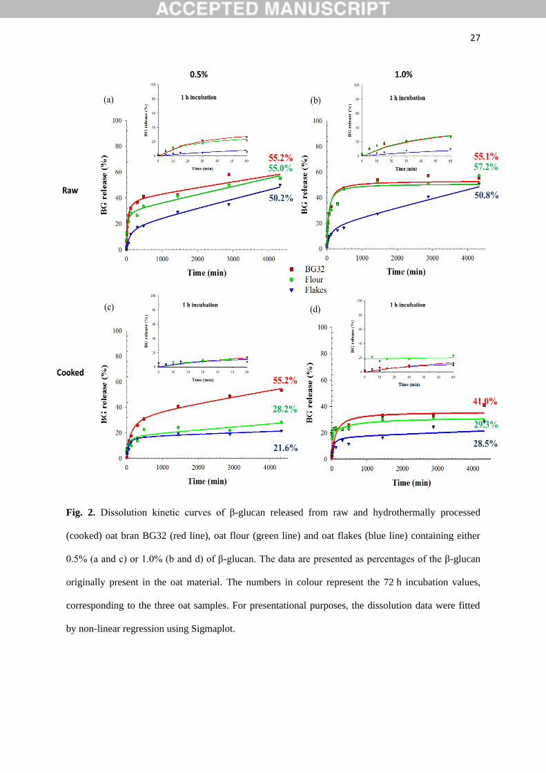
72 h. The rate of β-glucan dissolution varied between the raw oat flakes and the two raw
flours (P < 0.001) with a more gradual release of the polymer from the flakes (Fig. 2). This
can be explained by the differences in particle size (Fig. 1) and therefore surface area to
volume ratio. Large particles (mm size range), like the ones from the raw flakes, have a
greater proportion of intact cell walls and a longer incubation time is required for the polymer
to leach out from the cell walls, especially from walls that are not in close contact with bulk
water. Regardless of the oat samples and initial β-glucan concentration, the final values for β-
glucan release (72 h incubation) were remarkably close, and were in the range of 50-57%.
These results are in agreement with a previous study that showed that after more than two
hours of digestion, the solubility of β-glucan from unprocessed oat bran was not complete,
and only ~39% of the polymer was released from the oat tissue (Tosh et al., 2010). In the
present study, 2 h of incubation led to a β-glucan release of ~33, 30 and 12% for BG32, flour
and flakes, respectively. The work by Tosh and colleagues also revealed that more processed
forms of β-glucan sources showed higher levels of polymer solubility (~67 to 100%), and the
amount released increased with decreasing MW. This observation is compatible with data
obtained from a study of hydration kinetics performed on the leguminous cell wall
polysaccharide guar galactomannan, which showed an inverse relationship between MW and
dissolution rate of the polymer (Wang, Ellis & Ross-Murphy, 2003). A more recent study
also showed incomplete solubility of β-glucan from cereals, including barley, that underwent
different forms of processing (Comino, Collins, Lahnstein & Gidley, 2016).
The hydrothermally treated (cooked) oat flour and flakes (Fig 2c and 2d) showed much
lower amounts (P < 0.001) of β-glucan released after 72 h of incubation (28.8 and 25.1% for
flour and flakes, respectively) compared with the raw samples (56.3 and 50.5% for flour and
flakes, respectively). The presence of starch could explain this phenomenon, since starch and
water-soluble polysaccharides are highly likely to compete for the available free water
13
(Webster, 2011). At the beginning of the hydrothermal process, the starch located on the
fractured surfaces of the milled oat particles would have hydrated and swollen by absorbing
water. The gelatinisation of the readily available starch on the fractured surface, and in the
underlying contiguous cell layers (i.e. the encapsulated starch), are likely to have hindered the
release and solubility of the β-glucan. Indeed, it is well known that the physical structure of
starches, including oat starch, undergoes rapid and considerable changes when heated at
95°C, such as swelling and co-leaching of mainly amylose and also some amylopectin (Autio
& Eliasson, 2009).
The texture of the oat flakes/flour is also affected by the method of cooking preparation
and the resulting starch gelatinisation (Webster, 2011). In general, adding boiling water to the
oat flakes will give a grainy texture, while adding cold water, mixing thoroughly and then
gradually boiling the flakes (as done in the present study) generates a smoother texture. The
preparation method is therefore not without consequences for the release and solubility of β-
glucan as revealed by an early study (Yiu, Wood & Weisz, 1987).
3.3. Molecular weight measurements
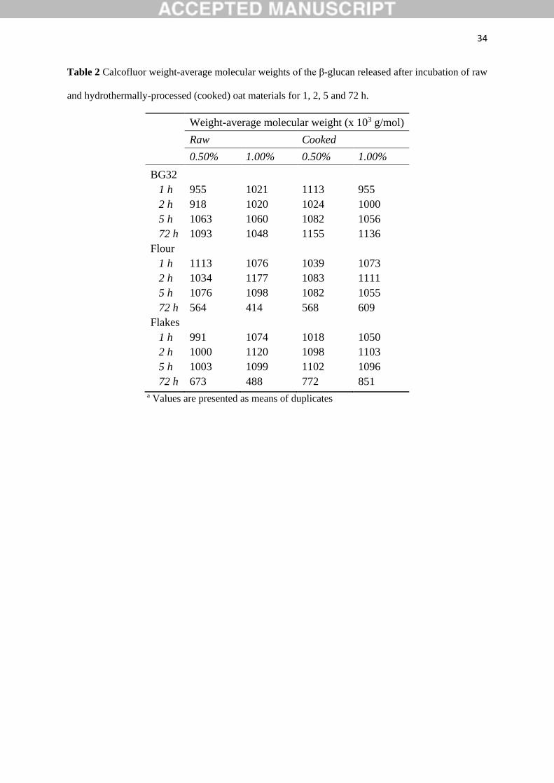
The results in Table 2 indicate that the MWs of the β-glucan released from the raw and
hydrothermally treated oat samples incubated for 1-5 h were similar to the values found in the
original non-hydrated oat materials, i.e., ~1100 x 103 g/mol (see Table 1). Therefore, the β-
glucan chains of high MW hydrated relatively rapidly to form polymer solutions in the early
stages of incubation without any significant changes in the first 5 hours. Prolonged incubation
(72 h), however, led to a significant reduction in MW. This is likely to be due to hydrolysis
(depolymerisation) of the β-glucan by endogenous β-glucanase as detected by the presence of
β-glucanase activity using the Megazyme assay (data not shown). The enzyme may have
been released therefore at a later stage of incubation because of its likely entrapment within
14
the oat tissue. The cooking method used in the present study did not succeed in fully
inhibiting the β-glucanase activity and longer cooking time with stirring may have been more
effective, as previously reported (Yiu, 1986; Yiu, Wood & Weisz, 1987). Indeed, this method
may have permitted starch gelatinisation, but some of the structural integrity of the oat
matrix, including the cell walls as well as the native starch structure, appeared to be preserved
(see Microscopy results section). The decrease in MW after 72 h, relative to earlier
incubation times, was also more noticeable for the flour than the flakes, suggesting perhaps
that β-glucanase activity is preserved in the relatively undamaged cells in the inner regions of
oat tissue particles, as in the case of the flakes with large particle size. In contrast, the MW of
β-glucan in the BG32 sample remained constant throughout the whole incubation time, since
this particular flour, enriched in bran, had undergone a processing step used to inactivate β-
glucanase.
3.4. Rheological behaviour
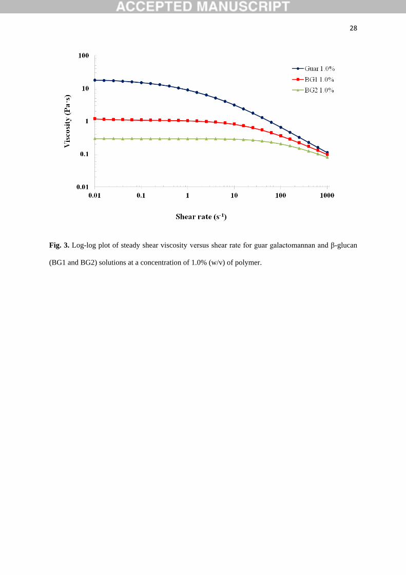
As previously reported, the fully hydrated polysaccharide solutions (1%, w/v) of guar
galactomannan (Rayment, Ross-Murphy & Ellis, 1995) and the β-glucan samples BG1 and
BG2 (Doublier & Wood, 1995; Ren, Ellis, Ross-Murphy, Wang & Wood, 2003) displayed
shear-thinning (pseudoplastic) behaviour with a Newtonian plateau at low shear rates and a
shear rate dependence at higher shear rates (Fig. 3). Such solution behaviour is characteristic
of semi-flexible polysaccharides and typically described by the entanglement model (Ellis,
Wang, Rayment, Ren, Ross-Murphy, 2001). The guar galactomannan solution showed the
highest viscosity values over the range of shear rates measured, followed by lower viscosities
(in descending order) for the BG1 and BG2 solutions. These profiles and zero-shear viscosity
values (Table S1 of the supplementary material: 18.52, 1.12 and 0.30 Pa·s for guar
15
galactomannan, BG1 and BG2, respectively) are consistent with the MW values reported in
Table 1.
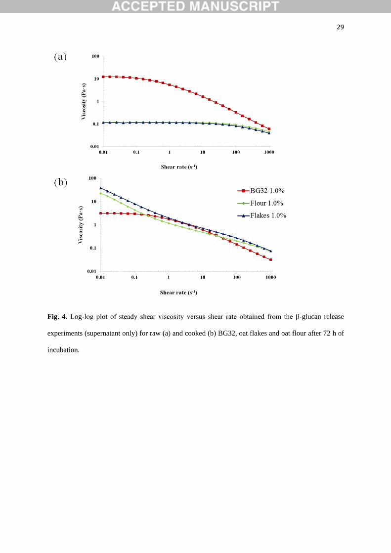
The viscosity profiles of the supernatant of the incubated solutions of raw and cooked oat
BG32, flakes and flour showed a varied and more complex rheological behaviour than the
profiles obtained for the purified polysaccharide solutions (Fig. 4). Thus, despite containing
similar amounts of β-glucan at the end of the 72 h incubation period (Fig. 2), large
differences were observed in the flow curves of raw BG32, which exhibited the highest
viscosities (two orders of magnitude for the Newtonian region), compared with the raw oat
flakes and flour (Fig. 4a). Moreover, the zero-shear viscosity values (Supplementary
Material; Table S1) show >100-fold difference between these samples. The markedly lower
values for the oat flake and flour dispersions after 72 h of incubation are presumably related
to the lower MW of the β-glucan contained in these samples, as explained above (Table 2).
The flow curves of the 72 h incubated solutions containing either raw or hydrothermally
processed BG32 showed a similar pattern, namely, a Newtonian plateau followed by a shear-
thinning region, typical of an entangled polymer solution, although the viscosity values were
lower overall after thermal treatment (Fig. 4b). This reduction in viscosity is likely to be due
to the smaller proportion of solubilised β-glucan in the 1.0% polymer solution post-cooking
compared with the raw samples, as shown by the release experiments (Fig. 2b and d). Factors
such as denatured protein and gelatinised starch located on the periphery of the BG32
particles, where the cells are likely to be fractured, may potentially hinder β-glucan release
from the cell wall matrix (see Microscopy section below). Furthermore, the cell wall content
and structural interactions between the β-glucan and the other cell wall polysaccharides,
specifically cellulose and arabinoxylans, are known to vary between different parts of the oat
grain, i.e., endosperm versus aleurone layers (Miller & Fulcher, 2011; Wang & Ellis, 2014).
16
Different thermal and mechanical processing conditions are known to affect the properties of
oat materials (Yiu, 1986), including the behaviour of the cell wall matrix (Wang & Ellis,
2014). Thus, physical changes during processing are likely to impact on the release and
dissolution of the β-glucan during incubation of the BG32, especially if there are alterations
in the structure and properties of cell walls that hinder the interaction of β-glucan with the
aqueous phase.
As well as significantly inhibiting the release of β-glucan from the oat flakes and flour
(Fig. 2), cooking also had marked effects on the rheology of the corresponding samples of
incubated solutions. These effects relative to the rheological behaviour of the raw samples,
included a substantial increase in viscosity, and the disappearance of the Newtonian plateau
at the lowest shear rates of the flakes and flour solutions (Fig. 4). The loss of the Newtonian
region and the appearance of a ‘power-law’ behaviour at low shear rates could be attributed
to the presence of residual particulates in the samples, in particular starch. This hypothesis is
supported by the rheological data for the BG32 sample, which has a substantially lower
starch content than the flakes and flour. Thus, solutions of both raw and cooked BG32
displayed similar rheological behaviour typical of an entangled polysaccharide network (Fig.
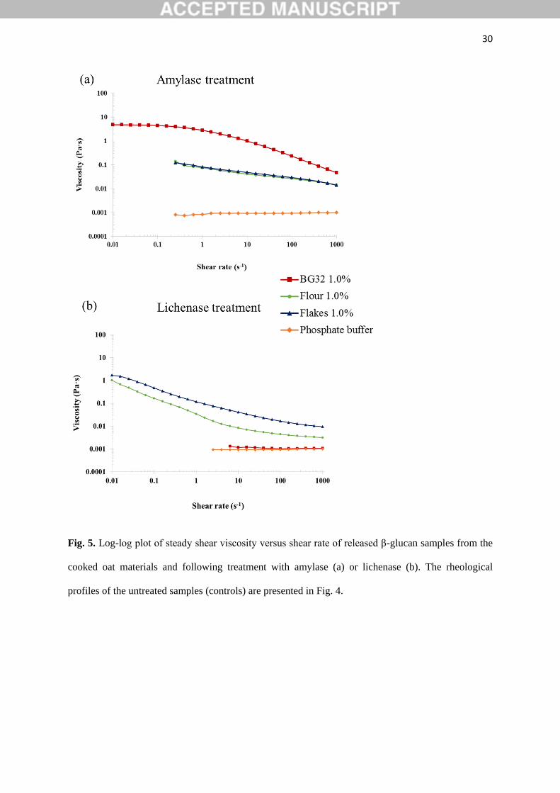
4). Treatment with amylase or lichenase also allowed us to distinguish between the effects of
starch and β-glucan on the solution rheology of oat BG32, flakes and flour by monitoring the
changes in viscosity-shear rate profiles (Fig. 5). Amylase addition to the incubated BG32
solution had virtually no effect on viscosity values and shear-thinning behaviour, indicating
that the starch in this sample did not contribute to solution rheology (Fig. 5a). Instead, the
addition of lichenase (which depolymerises β-glucan) induced a major drop in the viscosity
of BG32 (Fig 5b), which is consistent with the results from a recent study (Gamel, Abdel-
Aal, Ames, Duss & Tosh, 2014). This behaviour provides convincing evidence that the β-
glucan content in BG32 is the major contributor to the rheological behaviour of the incubated
17
BG32 solutions. However, in the case of the oat flake and flour solutions, the enzyme
treatments induced some decrease in the viscosity compared with the original cooked samples
(Figs. 4 and 5). Treatment with amylase had the greatest impact on the rheological properties,
with a decrease of several orders of magnitude in the viscosity. This result again confirms the
major role of starch in contributing to the rheological behaviour of the incubated oat flakes
and flour. Comparison of the solution rheology of flakes and flour (Fig. 4) with the viscosity
curves of pure β-glucan (Fig. 3), suggests that the more complex rheological behaviour and
higher viscosities of the former samples is attributed mainly to the starch. Other components
that may contribute in a minor way to the rheology include proteins and insoluble
particulates, such as β-glucan aggregates and micro-fragments of cells and cell walls that
remain suspended in the flake/flour solutions.
The effect of these components, including insoluble particulates, on the relative viscosity
becomes apparent only when the oat flour and flakes are hydrothermally processed, since
starch granules can then swell and gelatinise (Yiu, Wood & Weisz, 1987) and proteins can be
denatured (Turgeon & Rioux, 2011). The contribution of gelatinised starch to solution
rheology in our experiments may have originated from leached polymer (mostly amylose)
and/or amylopectin-rich ghosts (Debet & Gidley, 2007). The flake and flour solutions
displayed a similar pattern of behaviour to those of model systems of soluble polysaccharides
and particulates of different shapes and sizes, namely microcrystalline cellulose and native
starch (Rayment, Ross-Murphy & Ellis, 1995, 2000). Thus, in a guar galactomannan/rice
starch system, with increasing filler (starch) concentrations, an increase in viscosity and
greater rate-dependence at low shear rates (i.e. power-law behaviour) were reported
(Rayment, Ross-Murphy & Ellis, 1995).
18
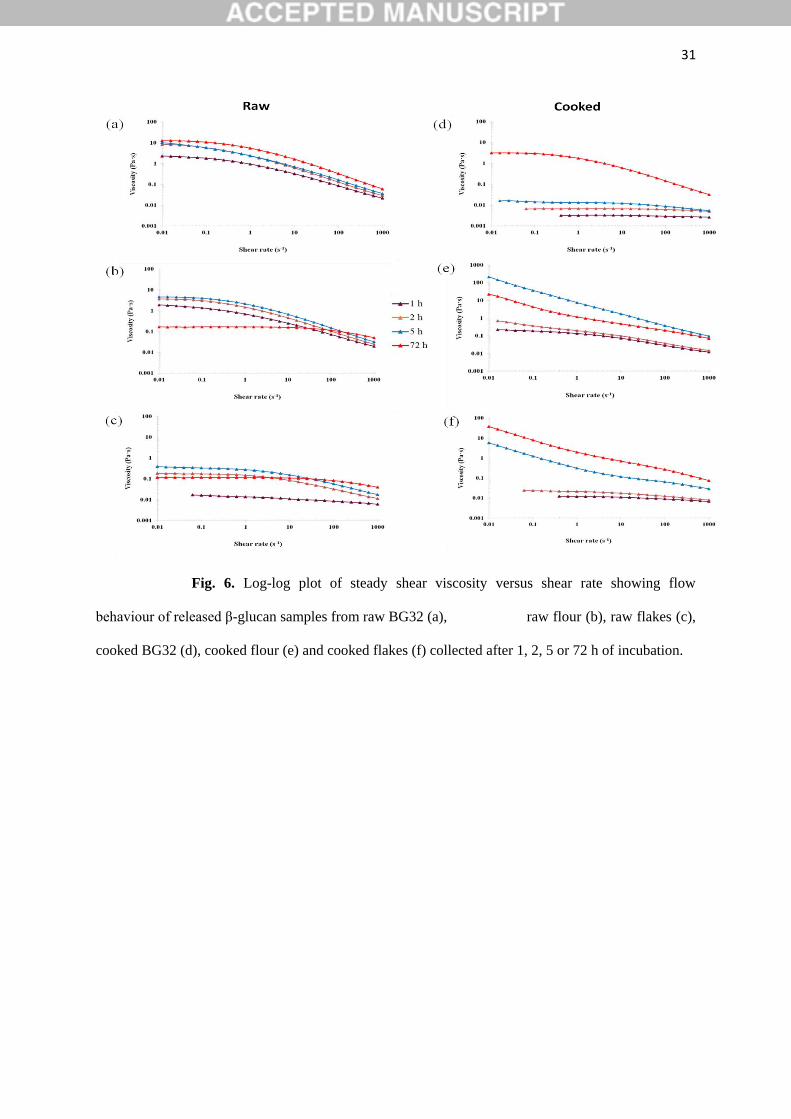
The flow behaviour of the oat solutions incubated for different periods of time (1, 2, 5 or
72 h) are shown in Fig. 6. The trend of the viscosity curves over the range of shear rates
measured followed an expected pattern, based on what we observed for the β-glucan release
data (Fig. 2), in that viscosity levels increased with increasing incubation time. This
relationship is particularly clear-cut for the raw and hydrothermally processed BG32, and for
the flakes and flour at early incubation times (1-5 h). However, at the longest incubation
times the relationship between viscosity and incubation time is much less clear, as the
viscosity curves for some of the 72 h samples were significantly lower than the values
obtained at earlier times. As explained in Section 3.3, the retention of β-glucanase activity,
which would hydrolyse the β-glucan, occurred in some of the flake and flour samples.
Moreover, the viscosity profiles of the solutions produced from the incubation of raw and
cooked oat flakes were markedly lower than the corresponding flour samples milled from the
flakes, apart from the 72 h solution (Fig. 6). This suggests that the particle size, and therefore
the surface area to volume ratio, of the oat materials had a significant impact on the rate and
extent of β-glucan release from the cell walls and the accompanying release of intra-cellular
macronutrients (e.g. starch). The kinetics of nutrient release (bioaccessibility) will be highly
dependent on the relative proportions of fractured cells in the flour produced by milling,
which has important physiological consequences, as recently observed in other plant
ingredients and foods (Edwards et al., 2015; Edwards, Warren, Milligan, Butterworth & Ellis,
2014; Grundy, Wilde, Butterworth, Gray & Ellis, 2015).
Therefore, as previously demonstrated (Gamel, Abdel-Aal, Wood, Ames & Tosh, 2012),
both the structure of the oat matrix and its composition have an impact on the rheological
profile of the ‘solubilised’ material. In the current rheological studies, the properties of the
supernatants obtained from incubated and centrifuged oat suspensions were investigated to
study the dissolution kinetics of the oat β-glucan (i.e. polymer release into the aqueous
19
phase). Our in vitro data suggest that from a physiological perspective, specifically the flow
properties of digesta in the gastrointestinal tract, the contribution of solubilised starch and
insoluble particulates (filler) to the solution rheology of dissolved β-glucan from ingested
oats, may play an important role. Digesta samples containing high concentrations of starch
would be digested in vivo by α-amylase, hence the contribution of starch to the rheology of
the system is expected to decrease during transit in the proximal gut. The contribution of
other components such as cell walls (dietary fibre), which are not digested, may be significant
until the cell wall polysaccharides are fermented in the large intestine
3.5. Macro- and microstructural characteristics of recovered particles (pellet)
Photographs of the sediments of the raw and cooked oats, examined after 72 h incubation
and centrifugation to remove the supernatant, are presented in the supplementary material
(Fig. S1). The images showed that insoluble particulates could easily be distinguished in all
of these samples, and not just in the processed macro-particles of oat flakes; i.e. mm-size
range. However, much of the particulate structure in the oat samples appears to have been
lost post-cooking. This loss in structure was more noticeable in the oat flour, suggesting that
one would expect to see an increased release and solubilisation of components, such as starch
and β-glucan. Nevertheless, evidence to show increased β-glucan release was not provided by
the hydration experiments (Fig. 2d); indeed, β-glucan release into solution was found to be
hindered, probably by the presence of gelatinised starch (see Section 3.2). The images of the
flour and flakes also show an increase in volume post-cooking mainly because of water
uptake and gelatinisation of the starch, which is a major component of these oat samples
(Table 1).
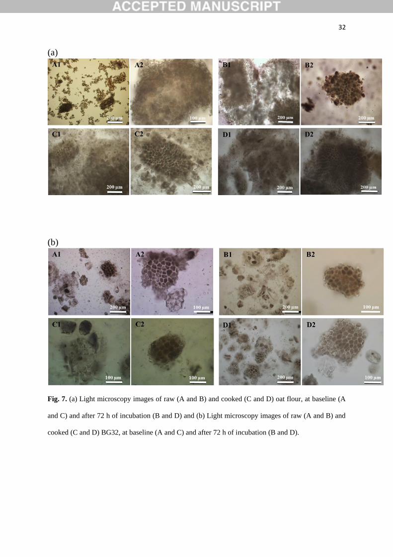
Microscopic examination of the two raw flours (oat flour and BG32) revealed the
presence of numerous oat tissue particles with seemingly minimal alterations in their
20
structure after 72 h incubation, relative to the pre-incubated baseline (0 h) samples (Fig. 7 a
and b). Thus, some of these intact oat particles, which varied in size (~10-200 µm) and
contained starch-rich cells of endosperm tissue and β-glucan-rich cell walls, seemed
relatively well preserved during incubation, apart from evidence of some swelling and
leaching of cell contents. However, the BG32 samples, which are particularly rich in bran,
showed incubated particles that appeared even more physically intact than the oat flour,
highlighting the robust structure of the tissue. Marked differences in structural characteristics
were observed between raw and hydrothermally processed tissues of oat flour. These
differences are visible in Fig. 7a (A1 and C1), which show that cooking disrupted the cellular
tissue of flour and led to leaching of cellular contents. This leached material formed a swollen
network of gelatinised starch together with β-glucan released from the cell walls.
4. Conclusions
The novel incubation assay presented in the current work has provided a simple and
reproducible method to evaluate the effects of mechanical and hydrothermal processing of
oats on oat β-glucan solubility and subsequent solution viscosity. The milling of oat flakes to
produce oat flour of smaller particle size increased the rate and extent of release and
dissolution of β-glucan from the cell walls of the oat tissue. We have provided evidence that
cooking has a significant impact on dissolution kinetics of cell wall β-glucan and its
rheological behaviour in solution. Thus, for example, the rate and extent of β-glucan
dissolution was severely inhibited by the cooking process in the oat flakes and flour, possibly
related to physical hindrance by gelatinised starch. The solutions obtained from cooked flour
and flakes displayed complex rheological properties by showing a significant increase in the
viscosity profiles, and also a loss of the Newtonian plateau (i.e. power-law behaviour at low
shear rates) compared to the raw samples. This behaviour can probably be explained by the
contribution of insoluble particulates (e.g. cell fragments and swollen starch granules) and
21
leached amylose. This study also demonstrated that β-glucans from oats, in particular flour,
are susceptible to degradation by β-glucanase during incubation, thereby attenuating
viscosity, but this occurs only after very prolonged periods of time (72 h).
Therefore, mechanical and hydrothermal processing of oats will strongly influence the
release of cell wall β-glucan and intra-cellular nutrients such as starch. This has important
implications for the physiological effects of oat β-glucan on gut function, including the rate
of gastric emptying, nutrient digestion and absorption, and on subsequent postprandial
metabolism (e.g. lipaemia). Variations in β-glucan action, resulting from changes in
processing conditions applied to oat products, will significantly impact on a range of related
health effects, in particular the cholesterol-lowering property of β-glucan. Our future
experiments will focus on investigating the effects of oat structure and β-glucan dissolution
properties on the digestion of lipids and other macronutrients under simulated physiological
conditions. In addition, the contribution of insoluble particles to the rheological behaviour of
β-glucan during simulated digestion warrants investigation to assess their physiological
relevance.
Acknowledgements
This work was funded by the BBSRC DRINC project BB/L025272/1 and the Research
Council of Norway (Project No. 225347/F40) with funds from the Norwegian Research Levy
on Agricultural Products. The authors thank Dr Simon Penson (Campden BRI , Chipping
Campden, UK) for useful discussions and help with the nutritional analysis, and Hanne Zobel
(Nofima, Ås, Norway) for her skilful technical assistance.
Appendix A. Supplementary data
Supplementary data associated with this article can be found, in the online version, at http:
22
References
Åman, P., Rimsten, L., & Andersson, R. (2004). Molecular weight distribution of β-glucan in
oat-based foods. Cereal Chemistry, 81(3), 356-360.
Andersson, A. A. M., & Börjesdotter, D. (2011). Effects of environment and variety on
content and molecular weight of β-glucan in oats. Journal of Cereal Science, 54(1), 122-128.
Autio, K., & Eliasson, A.-C. (2009). Oat starch. In J. N. BeMiller & R. L. Whistler (Eds.).
Starch: Chemistry and Technology. Burlington: Elsevier Science.
Beer, M. U., Wood, P. J., & Weisz, J. (1997). Molecular weight distribution and
(1→3)(1→4)-β-D-glucan content of consecutive extracts of various oat and barley cultivars.
Cereal Chemistry, 74(4), 476-480.
Beer, M. U., Wood, P. J., Weisz, J., & Fillion, N. (1997). Effect of cooking and storage on
the amount and molecular weight of (1→3)(1→4)-β-D-glucan extracted from oat products by
an in vitro digestion system. Cereal Chemistry, 74(6), 705-709.
Comino, P., Collins, H., Lahnstein, J., & Gidley, M. J. (2016). Effects of diverse food
processing conditions on the structure and solubility of wheat, barley and rye endosperm
dietary fibre. Journal of Food Engineering, 169, 228-237.
Cross, M.M. (1965). Rheology of non-Newtonian fluids: a new flow equation for
psudoplastic systems. Journal of Colloid Science 20(5), 417-437.
Debet, M. R., & Gidley, M. J. (2007). Why do gelatinized starch granules not dissolve
completely? Roles for amylose, protein, and lipid in granule "ghost" integrity. Journal of
Agricultural and Food Chemistry, 55(12), 4752-4760.
Doublier, J.-L., & Wood, P. J. (1995). Rheological properties of aqueous solutions of
(1→3)(1→4)-β-D-glucan from oats (Avena sativa L.). Cereal Chemistry, 72(4), 335-340.
Edwards, C. H., Grundy, M. M.-L., Grassby, T., Vasilopoulou, D., Frost, G. S., Butterworth,
P. J., Berry, S. E. E., Sanderson, J., & Ellis, P. R. (2015). Manipulation of starch
bioaccessibility in wheat endosperm to regulate starch digestion, postprandial glycemia,
insulinemia, and gut hormone responses: a randomized controlled trial in healthy ileostomy
participants. American Journal of Clinical Nutrition, 102(4), 791-800.
Edwards, C. H., Warren, F. J., Milligan, P. J., Butterworth, P. J., & Ellis, P. R. (2014). A
novel method for classifying starch digestion by modelling the amylolysis of plant foods
using first-order enzyme kinetic principles. Food & Function, 5(11), 2751-2578.
Ellis, P. R., Wang, Q., Rayment, P., Ren, Y. L., & Ross-Murphy, S. B. (2001). Guar gum:
agricultural and botanical aspects, physicochemical and nutritional properties, and its use in
the development of functional foods. In S. S. Cho & M. L. Dreker (Eds.). Food Science and
Technology (Vol. 113, pp. 613-657). New York: Marcel Dekker, Inc.
Englyst, H. N., Quigley, M. E., & Hudson, G. J. (1994). Determination of dietary fibre as
non-starch polysaccharides with gas-liquid chromatographic, high-performance liquid
chromatographic or spectrophotometric measurement of constituent sugars. Analyst, 119(7),
1497-1509.
23
Gamel, T. H., Abdel-Aal, E.-S. M., Ames, N. P., Duss, R., & Tosh, S. M. (2014). Enzymatic
extraction of beta-glucan from oat bran cereals and oat crackers and optimization of viscosity
measurement. Journal of Cereal Science, 59(1), 33-40.
Gamel, T. H., Abdel-Aal, E.-S. M., Wood, P. J., Ames, N. P., & Tosh, S. M. (2012).
Application of the Rapid Visco Analyzer (RVA) as an effective rheological tool for
measurement of β-glucan viscosity. Cereal Chemistry Journal, 89(1), 52-58.
Grundy, M. M.-L., Wilde, P. J., Butterworth, P. J., Gray, R., & Ellis, P. R. (2015). Impact of
cell wall encapsulation of almonds on in vitro duodenal lipolysis. Food Chemistry, 185, 405-
412.
Johansson, L., Virkki, L., Maunu, S., Lehto, M., Ekholm, P., & Varo, P. (2000). Structural
characterization of water soluble β-glucan of oat bran. Carbohydrate Polymers, 42(2), 143-
148.
Judd, P. A., & Ellis, P. R. (2005). Plant polysaccharides in the prevention and treatment of
diabetes mellitus. Traditional medicines for modern times: CRC Press.
Lazaridou, A., Biliaderis, C. G., & Izydorczyk, M. S. (2007). Cereal beta-glucans: structures,
physical properties, and physiological functions. Functional food carbohydrates, 1-72.
Miller, S. S., & Fulcher, R. G. (2011). Microstructure and chemistry of the oat kernel. In F.
H. Webster & P. J. Wood (Eds.). Oats: chemistry and technology (pp. 77-94). St Paul:
American Association of Cereal Chemists, Inc (AACC).
Morris, E. R. (1992). Physico-chemical properties of food polysaccharides. In T. F.
Schweizer & C. A. Edwards (Eds.). Dietary fibre - a component of food: nutritional function
in health and disease (pp. 103-117). London: Springer-Verlag.
Othman, R. A., Moghadasian, M. H., & Jones, P. J. (2011). Cholesterol-lowering effects of
oat beta-glucan. Nutrition Reviews, 69(6), 299-309.
Rayment, P., Ross-Murphy, S. B., & Ellis, P. R. (1995). Rheological properties of guar
galactomannan and rice starch mixtures— I. Steady shear measurements. Carbohydrate
Polymers, 28(2), 121-130.
Rayment, P., Ross-Murphy, S. B., & Ellis, P. R. (2000). Effect of size and shape of
particulate inclusions on the rheology of guar galactomannan solutions. Carbohydrate
Polymers, 43(1), 1-9.
Ren, Y., Ellis, P. R., Ross-Murphy, S. B., Wang, Q., & Wood, P. J. (2003). Dilute and semi-
dilute solution properties of (1→3), (1→4)-β-D-glucan, the endosperm cell wall
polysaccharide of oats (Avena sativa L.). Carbohydrate Polymers, 53(4), 401-408.
Rieder, A., Ballance, S., & Knutsen, S. H. (2015). Viscosity based quantification of
endogenous beta-glucanase activity in flour. Carbohydrate Polymers, 115, 104-111.
Rieder, A., Holtekjølen, A. K., Sahlstrøm, S., & Moldestad, A. (2012). Effect of barley and
oat flour types and sourdoughs on dough rheology and bread quality of composite wheat
bread. Journal of Cereal Science, 55(1), 44-52.

24
Rieder, A., Knutsen, S. H., Ulset, A. S., Christensen, B. E., Andersson, R., Mikkelson, A.,
Tuomainen, P., Maina, N., & Ballance, S. (2015). Inter-laboratory evaluation of SEC-post-
column calcofluor for determination of the weight-average molar mass of cereal beta-glucan.
Carbohydrate Polymers, 124, 254-264.
Tosh, S. M., Brummer, Y., Miller, S. S., Regand, A., Defelice, C., Duss, R., Wolever, T. M.,
& Wood, P. J. (2010). Processing affects the physicochemical properties of beta-glucan in oat
bran cereal. Journal of Agricultural and Food Chemistry, 58(13), 7723-7730.
Turgeon, S. L., & Rioux, L.-E. (2011). Food matrix impact on macronutrients nutritional
properties. Food Hydrocolloids, 25(8), 1915-1924.
Wang, Q., & Ellis, P. R. (2014). Oat β-glucan: physico-chemical characteristics in relation to
its blood-glucose and cholesterol-lowering properties. British Journal of Nutrition, 112(Suppl
2), S4-S13.
Wang, Q., Ellis, P. R., & Ross-Murphy, S. B. (2003). Dissolution kinetics of guar gum
powders - II. Effects of concentration and molecular weight. Carbohydrate Polymers, 53(1),
75-83.
Webster, F. H. (2011). Oat utilization: past, present and future. In F. H. Webster & P. J.
Wood (Eds.). Oats: chemistry and technology (pp. 347–361). St Paul: American Association
of Cereal Chemists, Inc (AACC).
Welch, R. W. (2011). Nutrient composition and nutritional quality of oats and comparisons
with other cereals. In F. H. Webster & P. J. Wood (Eds.). Oats: chemistry and technology
(pp. 95-107). St Paul: American Association of Cereal Chemists, Inc (AACC).
Wolever, T. M. S., Tosh, S. M., Gibbs, A. L., Brand-Miller, J., Duncan, A. M., Hart, V.,
Lamarche, B., Thomson, B. A., Duss, R., & Wood, P. J. (2010). Physicochemical properties
of oat β-glucan influence its ability to reduce serum LDL cholesterol in humans: a
randomized clinical trial. American Journal of Clinical Nutrition, 92(4), 723-732.
Wood, P. J. (2007). Cereal β-glucans in diet and health. Journal of Cereal Science, 46(3),
230-238.
Wood, P. J. (2011). Oat β-glucan: Properties and function. In F. H. Webster & P. J. Wood
(Eds.). Oats: chemistry and technology (Vol. 2, pp. 219-254). St Paul: American Association
of Cereal Chemists, Inc (AACC).
Wood, P. J., & Fulcher, R. G. (1978). Interaction of some dyes with cereal beta-glucans.
Cereal Chemistry, 55(6), 952-966.
Wood, P. J., Siddiqui, I. R., & Paton, D. (1978). Extraction of high-viscosity gums from oats.
Cereal Chemistry, 55, 1038-1049.
Yiu, S. H. (1986). Effects of processing and cooking on the structural and microchemical
composition of oats. Food Structure, 5, 219-225.
Yiu, S. H., Wood, P. J., & Weisz, J. (1987). Effects of cooking on starch and β-glucan of
rolled oats. Cereal Chemistry, 64(6), 373-379.
25
Zhang, M., Liang, Y., Pei, Y., Gao, W., & Zhang, Z. (2009). Effect of process on
physicochemical properties of oat bran soluble dietary fiber. Journal of Food Science, 74(8),
C628-636.
26
Fig. 1. Oat materials used in the study: flakes (A), flour (B) and BG32 (C). Oat flakes are in the mm
size range. Average particle size of flour: 60 μm and oat bran BG32: 152 μm.
27
Fig. 2. Dissolution kinetic curves of β-glucan released from raw and hydrothermally processed
(cooked) oat bran BG32 (red line), oat flour (green line) and oat flakes (blue line) containing either
0.5% (a and c) or 1.0% (b and d) of β-glucan. The data are presented as percentages of the β-glucan
originally present in the oat material. The numbers in colour represent the 72 h incubation values,
corresponding to the three oat samples. For presentational purposes, the dissolution data were fitted
by non-linear regression using Sigmaplot.
28
Fig. 3. Log-log plot of steady shear viscosity versus shear rate for guar galactomannan and β-glucan
(BG1 and BG2) solutions at a concentration of 1.0% (w/v) of polymer.
29
Fig. 4. Log-log plot of steady shear viscosity versus shear rate obtained from the β-glucan release
experiments (supernatant only) for raw (a) and cooked (b) BG32, oat flakes and oat flour after 72 h of
incubation.
30
Fig. 5. Log-log plot of steady shear viscosity versus shear rate of released β-glucan samples from the
cooked oat materials and following treatment with amylase (a) or lichenase (b). The rheological
profiles of the untreated samples (controls) are presented in Fig. 4.
31
Fig. 6. Log-log plot of steady shear viscosity versus shear rate showing flow
behaviour of released β-glucan samples from raw BG32 (a), raw flour (b), raw flakes (c),
cooked BG32 (d), cooked flour (e) and cooked flakes (f) collected after 1, 2, 5 or 72 h of incubation.
32
(a)
(b)
Fig. 7. (a) Light microscopy images of raw (A and B) and cooked (C and D) oat flour, at baseline (A
and C) and after 72 h of incubation (B and D) and (b) Light microscopy images of raw (A and B) and
cooked (C and D) BG32, at baseline (A and C) and after 72 h of incubation (B and D).

33
Table 1 Chemical composition of the guar gum and oat materialsa, and weight-average molecular
weights of the extracted and purified polysaccharides, guar galactomannan and oat β-glucan.
Guar gum BG1b BG2b BG32 Oat flake
Oat flour
Protein (N x 5.7) (%) 4.4 - - 18.9 11.5
Crude lipid (%) 1.0 - - 3.5 9.6
Starch (%) 0.8 4.2 2.2 7.5 60.3
Non-starch polysaccharides (%) 81.2 76.9 74.6 47.6 6.8
Cellulose (%) - 0.8 0.7 2.4 0.6
Arabinoxylan (%) - 6.1 1.5 12.8 1.8
Beta-glucan (%) - 82.8 87.6 34.8 4.5
Galactomannan (%) 78.2 - - - -
Moisture (%) 11.3 9.8 14.3 8.3 10.4
Ash (%) 0.6 1.7 2.8 3.0 1.7
Weight-average molecular
weight (x 103 g/mol) 2500c 730d 470d 1080d 1120d
a The data are expressed on a wet weight basis. Values are presented as means of duplicates.
b BG1: high MW β-glucan; BG2: medium MW β-glucan.
c Weight-average molecular weight values were determined by SEC-MALLS-VISC-RI.
d Weight-average molecular weight values were determined by the calcofluor method.
-: Not present or in trace amounts only.
34
Table 2 Calcofluor weight-average molecular weights of the β-glucan released after incubation of raw
and hydrothermally-processed (cooked) oat materials for 1, 2, 5 and 72 h.
Weight-average molecular weight (x 103 g/mol)
Raw Cooked
0.50% 1.00% 0.50% 1.00%
BG32
1 h 955 1021 1113 955
2 h 918 1020 1024 1000
5 h 1063 1060 1082 1056
72 h 1093 1048 1155 1136
Flour
1 h 1113 1076 1039 1073
2 h 1034 1177 1083 1111
5 h 1076 1098 1082 1055
72 h 564 414 568 609
Flakes
1 h 991 1074 1018 1050
2 h 1000 1120 1098 1103
5 h 1003 1099 1102 1096
72 h 673 488 772 851
a Values are presented as means of duplicates