Embed Size (px)
Citation preview
Kingdom of Bahrain
Arabian Gulf University
College of Medicine and Medical Sciences
Hematology
- Anemia:
It is a reduction in number of RBCs or Hb concentration > 2 SD below the mean
for a corresponding age and sex.
HbF: has a higher affinity to oxygen but it reduces and disappears by 6 months of
age. Thus infants with SCD will not experience crisis until that age.
80% of children in developing countries will experience anemia sometime during
their childhood.
Reticulocyte count: represents immature RBCs in the circulation (normally 1%).
Increased count indicates active hemolysis while decreased count with anemia
indicates bone marrow failure in producing new RBCs.
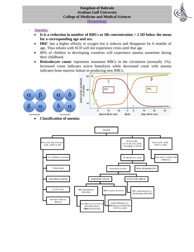
Classification of anemia:
Iron-deficiency anemia:
It is microcytic, hypochromic and is the most common blood disease during
infancy and childhood.
Causes:
Nutritional deficiency: iron stores are depleted by 6 months of age thus
iron-fortified cereals must be added at that time to prevent anemia.
Notice that cow’s milk is deficient in iron.
Increased demand (especially in adolescent females): due to blood
loss during menstruation or pregnancy.
Blood loss: PUD (in developed countries); hookworms (developing
countries).
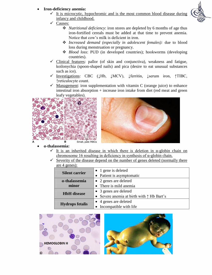
Clinical features: pallor (of skin and conjunctiva), weakness and fatigue,
koilonychia (spoon-shaped nails) and pica (desire to eat unusual substances
such as ice).
Investigations: CBC (↓Hb, ↓MCV), ↓ferritin, ↓serum iron, ↑TIBC,
↑reticulocyte count.
Management: iron supplementation with vitamin C (orange juice) to enhance
intestinal iron absorption + increase iron intake from diet (red meat and green
leafy vegetables).
α-thalassemia:
It is an inherited disease in which there is deletion in α-globin chain on
chromosome 16 resulting in deficiency in synthesis of α-globin chain.
Severity of the disease depend on the number of genes deleted (normally there
are 4 genes):
Silent carrier 1 gene is deleted
Patient is asymptomatic
α-thalassemia
minor
2 genes are deleted
There is mild anemia
HbH disease 3 genes are deleted
Severe anemia at birth with ↑ Hb Bart’s
Hydrops fetalis 4 genes are deleted
Incompatible with life
β-thalassemia:
β-thalassemia major Β-thalassemia minor (trait)
There is total absence of β-globin
chains (genes are present on
chromosome 11)
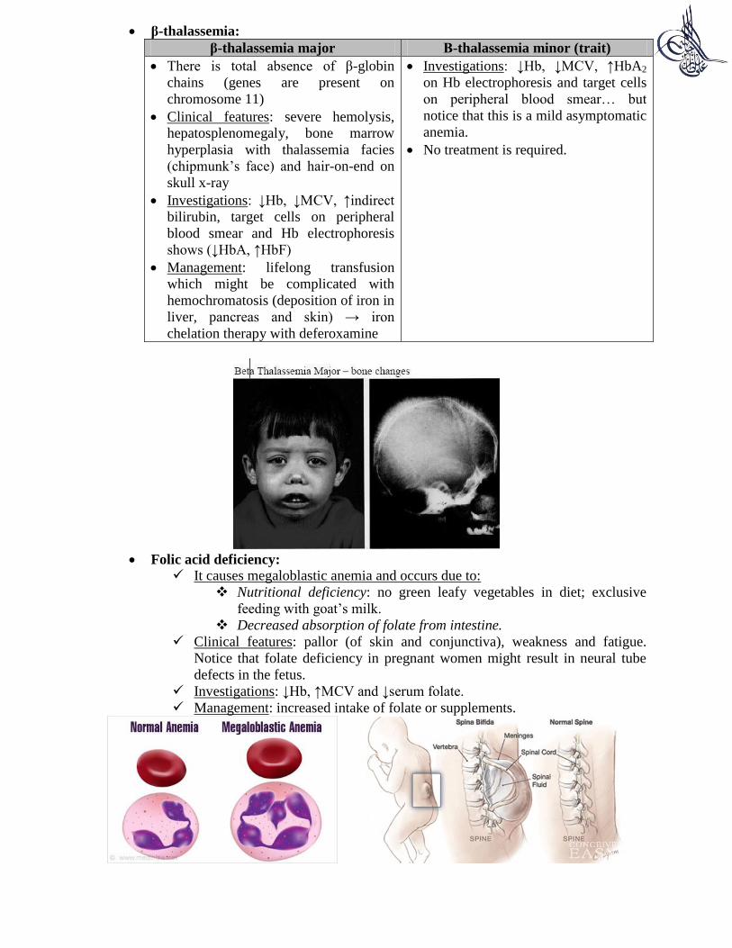
Clinical features: severe hemolysis,
hepatosplenomegaly, bone marrow
hyperplasia with thalassemia facies
(chipmunk’s face) and hair-on-end on
skull x-ray
Investigations: ↓Hb, ↓MCV, ↑indirect
bilirubin, target cells on peripheral
blood smear and Hb electrophoresis
shows (↓HbA, ↑HbF)
Management: lifelong transfusion
which might be complicated with
hemochromatosis (deposition of iron in
liver, pancreas and skin) → iron
chelation therapy with deferoxamine
Investigations: ↓Hb, ↓MCV, ↑HbA2
on Hb electrophoresis and target cells
on peripheral blood smear… but
notice that this is a mild asymptomatic
anemia.
No treatment is required.
Folic acid deficiency:
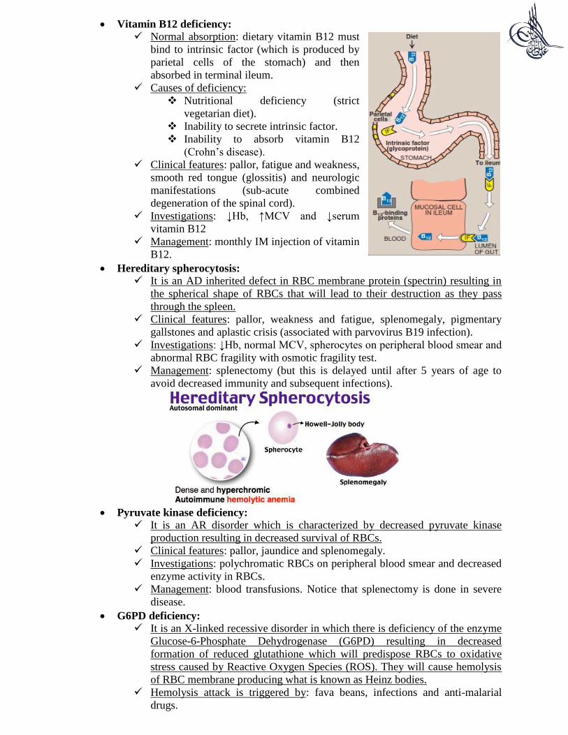
It causes megaloblastic anemia and occurs due to:
Nutritional deficiency: no green leafy vegetables in diet; exclusive
feeding with goat’s milk.
Decreased absorption of folate from intestine.
Clinical features: pallor (of skin and conjunctiva), weakness and fatigue.
Notice that folate deficiency in pregnant women might result in neural tube
defects in the fetus.
Investigations: ↓Hb, ↑MCV and ↓serum folate.
Management: increased intake of folate or supplements.
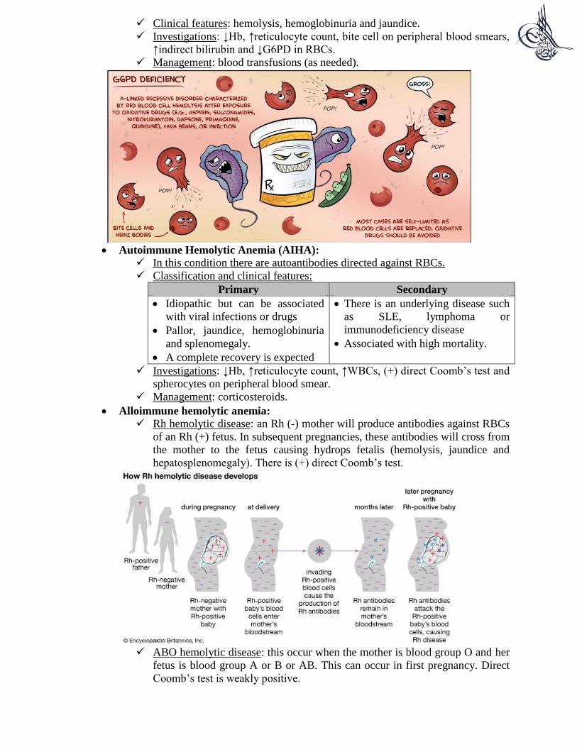
Vitamin B12 deficiency:
Normal absorption: dietary vitamin B12 must
bind to intrinsic factor (which is produced by
parietal cells of the stomach) and then
absorbed in terminal ileum.
Causes of deficiency:
Nutritional deficiency (strict
vegetarian diet).
Inability to secrete intrinsic factor.
Inability to absorb vitamin B12
(Crohn’s disease).
Clinical features: pallor, fatigue and weakness,
smooth red tongue (glossitis) and neurologic
manifestations (sub-acute combined
degeneration of the spinal cord).
Investigations: ↓Hb, ↑MCV and ↓serum
vitamin B12
Management: monthly IM injection of vitamin
B12.
Hereditary spherocytosis:
It is an AD inherited defect in RBC membrane protein (spectrin) resulting in
the spherical shape of RBCs that will lead to their destruction as they pass
through the spleen.
Clinical features: pallor, weakness and fatigue, splenomegaly, pigmentary
gallstones and aplastic crisis (associated with parvovirus B19 infection).
Investigations: ↓Hb, normal MCV, spherocytes on peripheral blood smear and
abnormal RBC fragility with osmotic fragility test.
Management: splenectomy (but this is delayed until after 5 years of age to
avoid decreased immunity and subsequent infections).
Pyruvate kinase deficiency:
It is an AR disorder which is characterized by decreased pyruvate kinase
production resulting in decreased survival of RBCs.
Clinical features: pallor, jaundice and splenomegaly.
Investigations: polychromatic RBCs on peripheral blood smear and decreased
enzyme activity in RBCs.
Management: blood transfusions. Notice that splenectomy is done in severe
disease.
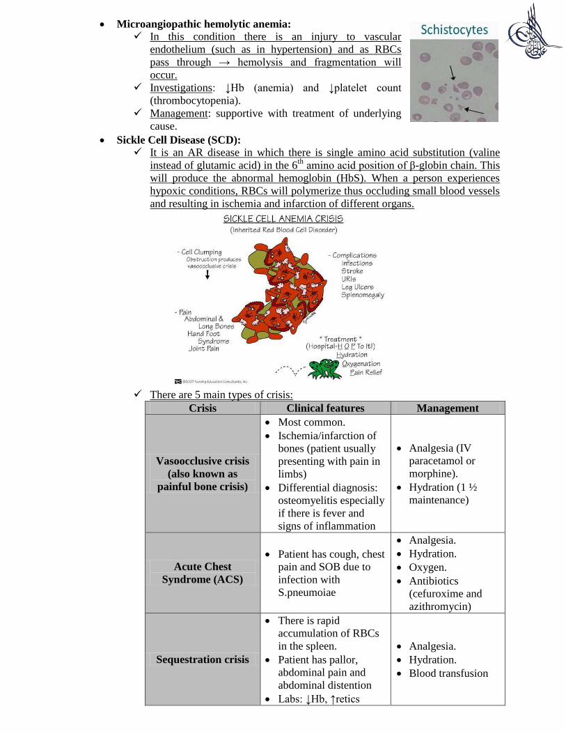
G6PD deficiency:
It is an X-linked recessive disorder in which there is deficiency of the enzyme
Glucose-6-Phosphate Dehydrogenase (G6PD) resulting in decreased
formation of reduced glutathione which will predispose RBCs to oxidative
stress caused by Reactive Oxygen Species (ROS). They will cause hemolysis
of RBC membrane producing what is known as Heinz bodies.
Hemolysis attack is triggered by: fava beans, infections and anti-malarial
drugs.
Clinical features: hemolysis, hemoglobinuria and jaundice.
Investigations: ↓Hb, ↑reticulocyte count, bite cell on peripheral blood smears,
↑indirect bilirubin and ↓G6PD in RBCs.
Management: blood transfusions (as needed).
Autoimmune Hemolytic Anemia (AIHA):
In this condition there are autoantibodies directed against RBCs.
Classification and clinical features:
Primary Secondary
Idiopathic but can be associated
with viral infections or drugs
Pallor, jaundice, hemoglobinuria
and splenomegaly.
A complete recovery is expected
There is an underlying disease such
as SLE, lymphoma or
immunodeficiency disease
Associated with high mortality.
Investigations: ↓Hb, ↑reticulocyte count, ↑WBCs, (+) direct Coomb’s test and
spherocytes on peripheral blood smear.
Management: corticosteroids.
Alloimmune hemolytic anemia:
Rh hemolytic disease: an Rh (-) mother will produce antibodies against RBCs
of an Rh (+) fetus. In subsequent pregnancies, these antibodies will cross from
the mother to the fetus causing hydrops fetalis (hemolysis, jaundice and
hepatosplenomegaly). There is (+) direct Coomb’s test.
ABO hemolytic disease: this occur when the mother is blood group O and her
fetus is blood group A or B or AB. This can occur in first pregnancy. Direct
Coomb’s test is weakly positive.
Microangiopathic hemolytic anemia:
In this condition there is an injury to vascular
endothelium (such as in hypertension) and as RBCs
pass through → hemolysis and fragmentation will
occur.
Investigations: ↓Hb (anemia) and ↓platelet count
(thrombocytopenia).
Management: supportive with treatment of underlying
cause.
Sickle Cell Disease (SCD):
It is an AR disease in which there is single amino acid substitution (valine
instead of glutamic acid) in the 6th
amino acid position of β-globin chain. This
will produce the abnormal hemoglobin (HbS). When a person experiences
hypoxic conditions, RBCs will polymerize thus occluding small blood vessels
and resulting in ischemia and infarction of different organs.
There are 5 main types of crisis:
Crisis Clinical features Management
Vasoocclusive crisis
(also known as
painful bone crisis)
Most common.
Ischemia/infarction of
bones (patient usually
presenting with pain in
limbs)
Differential diagnosis:
osteomyelitis especially
if there is fever and
signs of inflammation
Analgesia (IV
paracetamol or
morphine).
Hydration (1 ½
maintenance)
Acute Chest
Syndrome (ACS)
Patient has cough, chest
pain and SOB due to
infection with
S.pneumoiae
Analgesia.
Hydration.
Oxygen.
Antibiotics
(cefuroxime and
azithromycin)
Sequestration crisis
There is rapid
accumulation of RBCs
in the spleen.
Patient has pallor,
abdominal pain and
abdominal distention
Labs: ↓Hb, ↑retics
Analgesia.
Hydration.
Blood transfusion
Anaplastic crisis
Bone marrow failure
mainly due to infection
with parvovirus B19
Labs: ↓Hb, ↓retics
Analgesia.
Hydration.
Blood transfusion
Hemolytic crisis
Rapid hemolysis often
occurring in patients
with other hemolytic
disease (such as G6PD
deficiency)
Labs: ↓Hb, ↑retics,
↑bilirubin
Analgesia
Hydration
Blood transfusion
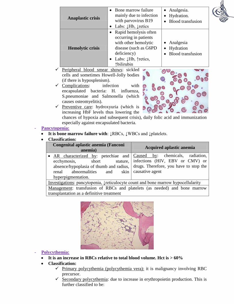
Peripheral blood smear shows: sickled
cells and sometimes Howell-Jolly bodies
(if there is hyposplenism).
Complications: infection with
encapsulated bacteria: H. influenza,
S.pneumoniae and Salmonella (which
causes osteomyelitis).
Preventive care: hydroxyuria (which is
increasing HbF levels thus lowering the
chances of hypoxia and subsequent crisis), daily folic acid and immunization
especially against encapsulated bacteria.
- Pancytopenia:
It is bone marrow failure with: ↓RBCs, ↓WBCs and ↓platelets.
Classification:
Congenital aplastic anemia (Fanconi
anemia) Acquired aplastic anemia
AR characterized by: petechiae and
ecchymosis, short stature,
absence/hypoplasia of thumb and radius,
renal abnormalities and skin
hyperpigmentation.
Caused by: chemicals, radiation,
infections (HIV, EBV or CMV) or
drugs. Therefore, you have to stop the
causative agent
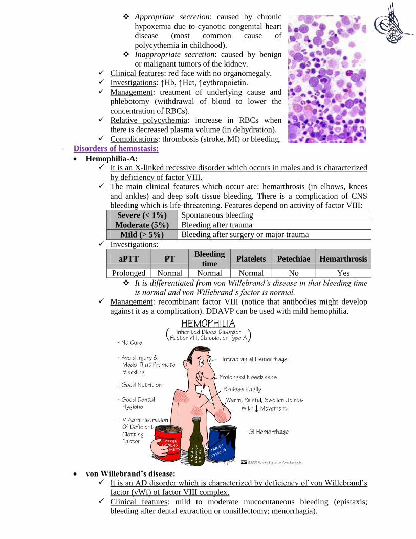
Investigations: pancytopenia, ↓reticulocyte count and bone marrow hypocellularity
Management: transfusion of RBCs and platelets (as needed) and bone marrow
transplantation as a definitive treatment
- Polycythemia:
It is an increase in RBCs relative to total blood volume. Hct is > 60%
Classification:
Primary polycythemia (polycythemia vera): it is malignancy involving RBC
precursor.
Secondary polycythemia: due to increase in erythropoietin production. This is
further classified to be:
Appropriate secretion: caused by chronic
hypoxemia due to cyanotic congenital heart
disease (most common cause of
polycythemia in childhood).
Inappropriate secretion: caused by benign
or malignant tumors of the kidney.
Clinical features: red face with no organomegaly.
Investigations: ↑Hb, ↑Hct, ↑eythropoietin.
Management: treatment of underlying cause and
phlebotomy (withdrawal of blood to lower the
concentration of RBCs).
Relative polycythemia: increase in RBCs when
there is decreased plasma volume (in dehydration).
Complications: thrombosis (stroke, MI) or bleeding.
- Disorders of hemostasis:
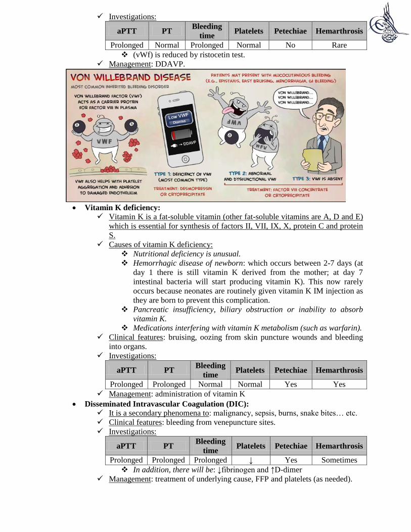
Hemophilia-A:
It is an X-linked recessive disorder which occurs in males and is characterized
by deficiency of factor VIII.
The main clinical features which occur are: hemarthrosis (in elbows, knees
and ankles) and deep soft tissue bleeding. There is a complication of CNS
bleeding which is life-threatening. Features depend on activity of factor VIII:
Severe (< 1%) Spontaneous bleeding
Moderate (5%) Bleeding after trauma
Mild (> 5%) Bleeding after surgery or major trauma
Investigations:
aPTT PT Bleeding
time Platelets Petechiae Hemarthrosis
Prolonged Normal Normal Normal No Yes
It is differentiated from von Willebrand’s disease in that bleeding time
is normal and von Willebrand’s factor is normal.
Management: recombinant factor VIII (notice that antibodies might develop
against it as a complication). DDAVP can be used with mild hemophilia.
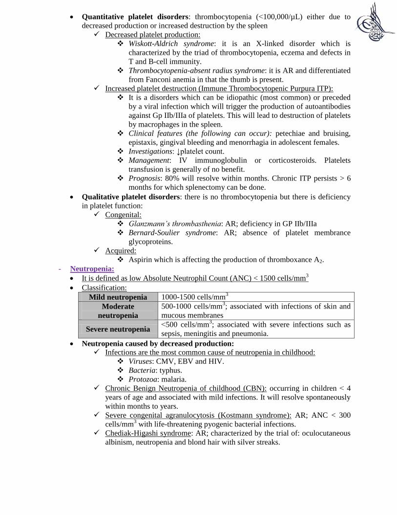
von Willebrand’s disease:
It is an AD disorder which is characterized by deficiency of von Willebrand’s
factor (vWf) of factor VIII complex.
Clinical features: mild to moderate mucocutaneous bleeding (epistaxis;
bleeding after dental extraction or tonsillectomy; menorrhagia).
Investigations:
aPTT PT Bleeding
time Platelets Petechiae Hemarthrosis
Prolonged Normal Prolonged Normal No Rare
(vWf) is reduced by ristocetin test.
Management: DDAVP.
Vitamin K deficiency:
Vitamin K is a fat-soluble vitamin (other fat-soluble vitamins are A, D and E)
which is essential for synthesis of factors II, VII, IX, X, protein C and protein
S.
Causes of vitamin K deficiency:
Nutritional deficiency is unusual.
Hemorrhagic disease of newborn: which occurs between 2-7 days (at
day 1 there is still vitamin K derived from the mother; at day 7
intestinal bacteria will start producing vitamin K). This now rarely
occurs because neonates are routinely given vitamin K IM injection as
they are born to prevent this complication.
Pancreatic insufficiency, biliary obstruction or inability to absorb
vitamin K.
Medications interfering with vitamin K metabolism (such as warfarin).
Clinical features: bruising, oozing from skin puncture wounds and bleeding
into organs.
Investigations:
aPTT PT Bleeding
time Platelets Petechiae Hemarthrosis
Prolonged Prolonged Normal Normal Yes Yes
Management: administration of vitamin K
Disseminated Intravascular Coagulation (DIC):
It is a secondary phenomena to: malignancy, sepsis, burns, snake bites… etc.
Clinical features: bleeding from venepuncture sites.
Investigations:
aPTT PT Bleeding
time Platelets Petechiae Hemarthrosis
Prolonged Prolonged Prolonged ↓ Yes Sometimes
In addition, there will be: ↓fibrinogen and ↑D-dimer
Management: treatment of underlying cause, FFP and platelets (as needed).
Quantitative platelet disorders: thrombocytopenia (>100,000/µL) either due to
decreased production or increased destruction by the spleen
Decreased platelet production:
Wiskott-Aldrich syndrome: it is an X-linked disorder which is
characterized by the triad of thrombocytopenia, eczema and defects in
T and B-cell immunity.
Thrombocytopenia-absent radius syndrome: it is AR and differentiated
from Fanconi anemia in that the thumb is present.
Increased platelet destruction (Immune Thrombocytopenic Purpura ITP):
It is a disorders which can be idiopathic (most common) or preceded
by a viral infection which will trigger the production of autoantibodies
against Gp IIb/IIIa of platelets. This will lead to destruction of platelets
by macrophages in the spleen.
Clinical features (the following can occur): petechiae and bruising,
epistaxis, gingival bleeding and menorrhagia in adolescent females.
Investigations: ↓platelet count.
Management: IV immunoglobulin or corticosteroids. Platelets
transfusion is generally of no benefit.
Prognosis: 80% will resolve within months. Chronic ITP persists > 6
months for which splenectomy can be done.
Qualitative platelet disorders: there is no thrombocytopenia but there is deficiency
in platelet function:
Congenital:
Glanzmann’s thrombasthenia: AR; deficiency in GP IIb/IIIa
Bernard-Soulier syndrome: AR; absence of platelet membrance
glycoproteins.
Acquired:
Aspirin which is affecting the production of thromboxance A2.
- Neutropenia:
It is defined as low Absolute Neutrophil Count (ANC) < 1500 cells/mm3
Classification:
Mild neutropenia 1000-1500 cells/mm3
Moderate
neutropenia
500-1000 cells/mm3; associated with infections of skin and
mucous membranes
Severe neutropenia >500 cells/mm
3; associated with severe infections such as
sepsis, meningitis and pneumonia.
Neutropenia caused by decreased production:
Infections are the most common cause of neutropenia in childhood:
Viruses: CMV, EBV and HIV.
Bacteria: typhus.
Protozoa: malaria.
Chronic Benign Neutropenia of childhood (CBN): occurring in children > 4
years of age and associated with mild infections. It will resolve spontaneously
within months to years.
Severe congenital agranulocytosis (Kostmann syndrome): AR; ANC < 300
cells/mm3 with life-threatening pyogenic bacterial infections.
Chediak-Higashi syndrome: AR; characterized by the trial of: oculocutaneous
albinism, neutropenia and blond hair with silver streaks.


![Arabian Gulf Food Recipes[MyebookShelf].pdf](https://img.pdfslide.net/doc/110x75/55cf9340550346f57b9d2bb1/arabian-gulf-food-recipesmyebookshelfpdf.jpg)