Embed Size (px)
Citation preview

619
INTEG. AND COMP. BIOL., 42:619–632 (2002)
Ultrastructure, Biology, and Phylogenetic Relationships of Kinorhyncha1
BIRGER NEUHAUS2,* AND ROBERT P. HIGGINS†*Museum fur Naturkunde, Zentralinstitut der Humboldt-Universitat zu Berlin, Institut fur Systematische Zoologie,
Invalidenstr. 43, D-10115 Berlin, Germany†2 Pond Lane, Asheville, North Carolina 28804
SYNOPSIS. The article summarizes current knowledge mainly about the (functional) morphology and ul-trastructure, but also about the biology, development, and evolution of the Kinorhyncha. The Kinorhynchaare microscopic, bilaterally symmetrical, exclusively free-living, benthic, marine animals and ecologicallypart of the meiofauna. They occur throughout the world from the intertidal to the deep sea, generally insediments but sometimes associated with plants or other animals. From adult stages 141 species are known,but 38 species have been described from juvenile stages. The trunk is arranged into 11 segments as evidencedby cuticular plates, sensory spots, setae or spines, nervous system, musculature, and subcuticular glands.The ultrastructure of several organ systems and the postembryonic development are known for very fewspecies. Almost no data are available about the embryology and only a single gene has been sequenced fora single species. The phylogenetic relationships within Kinorhyncha are unresolved. Priapulida, Loricifera,and Kinorhyncha are grouped together as Scalidophora, but arguments are found for every possible sister-group relationship within this taxon. The recently published Ecdysozoa hypothesis suggests a closer rela-tionship of the Scalidophora, Nematoda, Nematomorpha, Tardigrada, Onychophora, and Arthropoda.
INTRODUCTION
Since the last compilation on the ultrastructure(Kristensen and Higgins, 1991), numerous studieshave provided different views of morphology and phy-logenetic relationships both within Kinorhyncha andof Kinorhyncha to other Bilateria (Nebelsick, 1992a,b, 1993; Adrianov et al., 1993; Adrianov and Malak-hov, 1994, 1996, 1999a; Neuhaus, 1994, 1995, 1997;Neuhaus et al., 1996, 1997b; Wallace et al., 1996;GaOrdonez et al., 2000; Muller and Schmidt-Rhaesa,2002). In addition, the traditional view of a taxon Ar-ticulata consisting of Annelida, Tardigrada, Onycho-phora, and Arthropoda (see Schmidt-Rhaesa et al.,1998) has been recently challenged by the Ecdysozoahypothesis uniting all moulting animals such as Tar-digrada, Onychophora, Arthropoda, Nematoda, Nem-atomorpha, Kinorhyncha, and Priapulida (Aguinaldo etal., 1997). Therefore, this review summarizes the newinformation and provides additional data on these top-ics and other aspects of kinorhynch biology.
The Kinorhyncha is a group of exclusively free-liv-ing, marine, meiofaunal, benthic animals up to 1.03mm long. They occur throughout the world, generallyin sediments but sometimes associated with algae,sponges, or other invertebrates usually themselvesclosely associated with sediment. They are found fromintertidal (Zelinka, 1928) to abyssal zones at least asdeep as 5,300 m (Meadows et al., 1994) interstitiallyin usually the upper few millimeters (mud) or centi-meters (sand) of the sediment layer. At depths of 8–30 m along the coast of the North Adriatic, Kinorhyn-cha occupies the top, well oxygenated, soft silt sedi-
1 From the Symposium Lesser-Known Protostome Taxa; Evolu-tion, Development, and Ecology presented at the Annual Meeting ofthe Society for Integrative and Comparative Biology, 3–7 January2001, at Chicago, Illinois.
2 E-mail: [email protected]
ment layer (Vidakovic, 1984). In the Pacific deep seaaround 5,200 m depth, the animals inhabit the upper,oxygenated centimeter of the sediment where the num-ber of microorganisms is highest and decreases expo-nentially with increasing sediment depth (Meadows etal., 1994). At a deep-sea location in Japan (depth1,450 m), kinorhynchs occur only temporarily belowthe oxygenated sediment layer; the animals react pos-itively to seasonal food supply and migrate some 5mm into upper layers (Shimanaga et al., 2000). Mostspecies have been described from muddy substrates,but kinorhynchs also inhabit sandy biotopes such asthe mid-tide level of exposed beaches at depths of 10–60 cm (Cateria gerlachi, C. styx, and Echinoderes ny-bakkeni: Higgins, 1968, 1986). Echinoderes coulliwithstands salinities reaching from 12–42‰ for atleast a short time in intertidal flats of North Carolina(Horn, 1978). In an 18-mo study, Rao and Satapathy(1996) found that unidentified species of Echinoderesand Pycnophyes survive salinities of 5.7–20.3‰, pHof 7.64–9.50 and temperatures up to 33.58C in a la-goon in India.
Kinorhynchs may reach densities of 45 animals per10 cm2 in shallow waters of the Antarctic and densitiesof 1–10 animals per 10 cm2 in the deep sea (Dinet,1979; Vanhove et al., 1995). Most studies concentrateon the upper 100 m of the continental shelf, and norecord of a kinorhynch from below 500 m depth iden-tified to species level has been published yet. Our un-published observations from both the Atlantic and thePacific Ocean suggest a high diversity of Kinorhynchain the deep sea, nearly all species being new to sci-ence. However, such species can be assigned to knowngenera from the continental shelf.
The Kinorhyncha is divided into the two sub-groups, Cyclorhagida and Homalorhagida, containing
by guest on Novem
ber 24, 2010icb.oxfordjournals.org
Dow
nloaded from
620 B. NEUHAUS AND R. P. HIGGINS
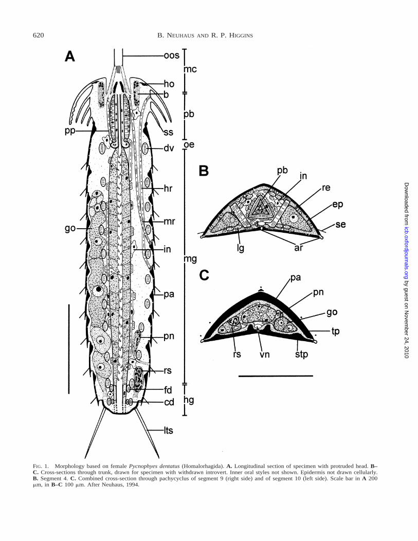
FIG. 1. Morphology based on female Pycnophyes dentatus (Homalorhagida). A. Longitudinal section of specimen with protruded head. B–C. Cross-sections through trunk, drawn for specimen with withdrawn introvert. Inner oral styles not shown. Epidermis not drawn cellularly.B. Segment 4. C. Combined cross-section through pachycyclus of segment 9 (right side) and of segment 10 (left side). Scale bar in A 200mm, in B–C 100 mm. After Neuhaus, 1994.
by guest on Novem
ber 24, 2010icb.oxfordjournals.org
Dow
nloaded from
621KINORHYNCHA
141 species described from adult life history stagesand 38 species described from juvenile stages, allgrouped in 15 genera (Bauer-Nebelsick, 1996; Pardoset al., 1998; Adrianov and Malakhov, 1999a). Zelinka(1928) and others introduced new genus names forspecies described from juvenile stages of species ofknown genera, although these scientists were aware ofthe life-history relationships. Higgins (1983) exten-sively synonomized species described from juveniles.The species names based on descriptions of juvenilestages are available and valid in the sense of the In-ternational Code of Zoological Nomenclature, so cur-rently 179 species are recognized. No formal classifi-cation is presented in this review, because hierarchicalassignments in classification systems cannot be definedon an objective basis. Several classification schemesare summarized and discussed by Adrianov and Ma-lakhov (1996).
EXTERNAL GROSS ANATOMY
The kinorhynch body is divided into 3 regions: head(5introvert), very short neck, and trunk (Figs. 1A,2A). Introvert and neck have often been considered asa ‘‘segment’’ comparable to the 11 trunk segments(Kristensen and Higgins, 1991). Trunk segmentation isapparent externally with cuticular plates and spinesand in the internal organs such as longitudinal anddorsoventral muscles, central nervous system, glands,and sensory structures (Zelinka, 1928; Kristensen andHiggins, 1991; Neuhaus, 1994; Adrianov and Malak-hov, 1994). However, these segmental characters donot appear in the head and neck region at all, andextension of the term ‘‘segmentation’’ to the neck andhead is therefore not justified.
The head bears 5–7 rings of posteriorly directed,sensory-locomotory spinose appendages (5scalids)(Figs. 1A, 2A, 3A). The 54–93 scalids are arrangedregularly in circles of 10–20; succeeding circles showa staggered arrangement. Scalids of the first 6 ringsconsist of two elements, a broad basal part and a nar-rower spinose distal part (Brown, 1989; Higgins, 1990;Nebelsick, 1993; Neuhaus, 1995).
The neck possesses up to 16 cuticular plates (5pla-cids) (Fig. 3A) which may be barely distinguishablefrom the first trunk segment (Higgins, 1990). Theseplates act solely or in combination with specializedtrunk plates (Semnoderes, Sphenoderes) to close offthe withdrawn head (Zelinka, 1928; Higgins, 1969a,1983).
In cross-section, the trunk may appear round, broad-ly oval (Fig. 2B, C), subtriangular, triangular (Fig. 1B,C), or narrowly oval (laterally compressed). The cu-ticle is organized into one to several plates, beyondtrunk segment 2 or 3 in a single dorsal (5tergal) plate,and two ventral (5sternal) plates (Zelinka, 1928; Hig-gins, 1968, 1969b). Borders between cuticular dorsaland ventral plates may not be recognizable in the SEMin species with a thin cuticle such as Zelinkaderes flor-idensis (Fig. 3B; Higgins, 1990). In those species witha thick cuticle (e.g., species of Echinoderes, Pycno-
phyes, and Kinorhynchus), a distinct ball-and-socketarticulation usually exists at the anterior margin of theplates of most segments. The cuticle of the anteriormargin of each segment is thickened towards the in-terior of the animal in order to form a pachycyclus forthe attachment of longitudinal muscles (Figs. 1A, C,2A, 4A; Zelinka, 1928; Kristensen and Higgins, 1991;Adrianov and Malakhov, 1994).
INTEGUMENT
A cuticle ensheathes the entire animal includingforegut, hindgut, tubules of the protonephridial ne-phropore, and tubes of the sensory spots (Neuhaus,1993). The cuticle of each sternal and tergal plate iscomposed of a chitinous basal layer which appears tobe finely granular at the TEM level (Fig. 4A–C), anda monolamellar, membrane-like epicuticle (Neuhaus etal., 1996). Thin fibrillar material may occur below thebasal layer in some species especially in flexible areas.Stronger fibres connect the sternal and tergal plateslaterally as well as anteriorly and posteriorly in manyspecies (Fig. 4A–C; Kristensen and Higgins, 1991;Adrianov and Malakhov, 1994). In the matrix embed-ding the thin and stronger fibres, chitin may be local-ized too (Fig. 4B, C; Neuhaus et al., 1996). To dem-onstrate chitin in a specific cuticular layer (Fig. 4B,C), the lectin-gold labelling technique was used; it pre-viously detected chitin in the body cuticle of Lorici-fera, Priapulida, and juvenile Nematomorpha, and inthe pharyngeal cuticle of nematodes (Saldarriaga et al.,1995; Neuhaus et al., 1996, 1997a, b; Lemburg, 1998).
With increasing age of the adult individual, the fine-granular layer of the cuticle as well as the pachycycliincrease in thickness and ball-and-socket articulationsbecome more prominent and turn increasingly yellow-brown (tanning?) in species of Echinoderes and es-pecially of Pycnophyes and Kinorhynchus. The epi-dermis is cellular and does not possess any locomotorycilia (Kristensen and Higgins, 1991; Adrianov and Ma-lakhov, 1994).
At least two types of integumental gland cells seemto exist in several species of Echinoderes, Pycnophyes,and Kinorhynchus, and they are arranged in species-specific patterns. These gland cells open to the outsidevia a convoluted duct system or through a single pore(Nebelsick, 1992b; Adrianov and Malakhov, 1994;GaOrdonez et al., 2000). A sensory cell with or with-out a modified cilium may be associated with a glandcell. A gland cell is often associated with a sensoryspot (Fig. 3C), a cuspidate spine, a seta (Fig. 3C), oran adhesive spine in segment 4 of male Pycnophyesand Kinorhynchus (Kristensen and Higgins, 1991; Ad-rianov and Malakhov, 1994).
NERVOUS SYSTEM AND SENSE ORGANS
The central nervous system consists of a circumen-teric brain (Figs. 1A, 2A, D) and several longitudinalnerve cords in the trunk (Figs. 1B, C, 2B, C) whichare connected generally by two commissures per trunksegment (Kristensen and Higgins, 1991; Nebelsick,
by guest on Novem
ber 24, 2010icb.oxfordjournals.org
Dow
nloaded from

622 B. NEUHAUS AND R. P. HIGGINS
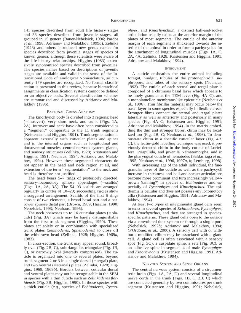
FIG. 2. Morphology based on female Zelinkaderes floridensis (Cyclorhagida). A. Longitudinal section through entire specimen with withdrawnhead. B, C. Cross-sections through trunk in segment 5 (B) and in segment 9 (C), respectively. Epidermis not drawn cellularly in A–C. D.Longitudinal section through mouth cone, brain, and anterior pharynx. Nervous system and epithelia not drawn cellularly. Note innervationof pharynx through mouth cone. Arrow heads mark extracellular matrix separating inner and outer mouth cone epithelium. Numbers refer toinner oral styles of rings 1–4. Arrows point to subapical opening of oral styles. Scale bar in A–C 100 mm, in D 20 mm. After Neuhaus, 1994.
by guest on Novem
ber 24, 2010icb.oxfordjournals.org
Dow
nloaded from

623KINORHYNCHA
FIG. 2. Continued.
1993). The brain is organized into a 10-lobed forebrainwith numerous perikarya, a midbrain with few peri-karya but abundant neuropile, and a hindbrain alsowith numerous perikarya (Fig. 2D; Kristensen andHiggins, 1991; Adrianov and Malakhov, 1994; Neu-haus, 1994). In Echinoderes capitatus, 10 nerve cordsextend from the forebrain and innervate the introvert,neck, and trunk. The nerves fuse to two subdorsal, twosublateral, and one midventral nerve strand from trunk
segment 2 on (Nebelsick, 1993). The perikarya of theunpaired ventral nerve cord (Fig. 4D) are distributedover almost the entire length of a trunk segment. InZelinkaderes floridensis, 12 longitudinal nerve cordsseem to exist (Fig. 2B, C; Neuhaus, 1994), whereas 8nerves have been reported for Pycnophyes greenlan-dicus and P. kielensis (Kristensen and Higgins, 1991;Adrianov and Malakhov, 1994) and 7 for P. dentatus(Fig. 1B; Neuhaus, 1994).
by guest on Novem
ber 24, 2010icb.oxfordjournals.org
Dow
nloaded from

624 B. NEUHAUS AND R. P. HIGGINS
FIG. 3. SEM. A. Head of Antygomonas sp. with far extended mouth cone. B. Ventral view of last trunk segments of female Zelinkaderesfloridensis. Notice regular spinose and cuspidate spines, gonopores, and lack of obvious border between ventral and dorsal cuticular plates. C.Sensory spot and seta of Pycnophyes greenlandicus. Notice gland pore and sensory pore. D. Ventral view of posterior trunk segments of P.greenlandicus with cuticular hairs surrounding male gonopores. Scale bar in A, D 50 mm, in B 20 mm, and in C 10 mm.
The posterior midbrain sends 9 nerve cords frontallyto the outer oral styles. From there the nerves turncaudally to the basal mouth cone nerve ring (Z. flori-densis, E. capitatus) or to the nerve ring in the outerpharyngeal epithelium (P. kielensis, P. dentatus). Thenerves continue over the anterior pharyngeal muscle
bulb into the inner pharyngeal epithelium (Fig. 2D)until they end in the esophageal epithelium (Nebelsick,1993; Adrianov and Malakhov, 1994; Neuhaus, 1994).The alimentary canal is not innervated via the ventralnervous system as has been claimed earlier (Kristensenand Higgins, 1991).
by guest on Novem
ber 24, 2010icb.oxfordjournals.org
Dow
nloaded from

625KINORHYNCHA
No nerve cells have been traced to innervate thelongitudinal and circular muscle cells of the midgut.Therefore, it is assumed that this musculature acts au-tonomously; however, the muscles may be triggered tosome degree by presumably sensorimotor cells in themidgut epithelium (Neuhaus, 1994).
Scalids, in addition to their function in locomotion,may also be mechano- and chemoreceptive. A scalidcontains up to 10 monociliary sensory cells whichcommunicate with the exterior by way of a distal pore(Adrianov and Malakhov, 1994). Spinoscalids maycontain a cell with piles of membranes and a porouscuticle. Possibly, this cell releases some secretions(Brown, 1989; Kristensen and Higgins, 1991).
A sensory cell with or without cilium is associatedwith each cuticular spine, seta, and gland cell of thetrunk. All species of Kinorhyncha show a species-spe-cific pattern of sensory spots on the trunk. On the sur-face of the cuticle, each round to oval sensory spotexhibits numerous cuticular micropapillae around oneor two pores (Fig. 3C) through which a cilium mayjut out (Kristensen and Higgins, 1991; Adrianov andMalakhov, 1994; Neuhaus, 1995); below, one to fewmonociliary sensory cells occur; their cilia are sur-rounded by a circle of 9 microvilli containing electron-dense fibrils. At least for E. capitatus, an additionalsheath cell has been reported (Nebelsick, 1992a).Gland cells seem to be associated with sensory spotsin Pycnophyes and Kinorhynchus (Fig. 3C; Kristensenand Higgins, 1991; Adrianov and Malakhov, 1994).The sensory spots of juvenile stages of P. kielensisappear as papillae which elevate slightly above the sur-face of the trunk cuticle. Fewer cuticular micropapillaethan in the adult are also characteristic of these juve-nile organs (Neuhaus, 1993; Adrianov and Malakhov,1994). Adult Kinorhynchus yushini possess sensoryspots with a reduced number of cuticular micropapillaeand have been named ‘‘flosculi’’ by Adrianov and Ma-lakhov (1994) although a detailed reconstruction at theTEM level for comparison with the flosculi of Lori-cifera (Kristensen, 1991) and Priapulida (Lemburg,1995) is missing; Priapulida and/or Loricifera are cer-tainly most closely related to Kinorhyncha (see Phy-logenetic relationships below).
In Pycnophyes dentatus, P. kielensis, P. greenlan-dicus, and Kinorhynchus phyllotropis, one pair of ce-phalic sensory organs is located at the base of the first-ring head scalids (Fig. 1A). Each organ consists of oneenveloping cell and one receptor cell. The latter pos-sesses a single, modified cilium which extends into thelumen of the sensory organ. The cilium swells andbranches into multiple processes (Neuhaus, 1997). Pig-mented eye spots with lenses have been reported forseveral species of Echinoderes, but pigmentation dis-appears during fixation of specimens (Zelinka, 1928).However, TEM results are missing entirely.
MUSCULATURE
The segmental cuticular plates are moved by sets ofsegmentally arranged dorsal and ventral longitudinal
muscles, oblique muscles (only in Cyclorhagida), anddorsoventral muscles (Fig. 4E; Muller and Schmidt-Rhaesa, 2002). In Zelinkaderes floridensis, the longi-tudinal muscles seem to be distributed regularly in theperiphery of a cross-section (Fig. 2B, C), whereasthese muscles concentrate in bundles in species ofEchinoderes, Pycnophyes, and Kinorhynchus (Fig. 1B,C; Kristensen and Higgins, 1991; Adrianov and Ma-lakhov, 1994; Neuhaus, 1994). Mouth cone retractormuscle cells extend between the mouth cone and pos-terior trunk segments (Figs. 1A, 2A). Head retractormuscle cells stretch between the base of head scalidsand posterior trunk segments (Figs. 1A, 2A, 4E); theypass the periphery of the brain via extremely elongat-ed, epidermal cells containing intermediate filaments(5tanycytes) (Kristensen and Higgins, 1991; Neuhaus,1994). Circular muscles occur in association with theplacids of the neck region (Fig. 4E; Muller andSchmidt-Rhaesa, 2002) and in the mouth cone (Kris-tensen and Higgins, 1991; Adrianov and Malakhov,1994; Neuhaus, 1994). One pair of small trapezoidalmuscles is reported for each middorsal spine of An-tygomonas sp. (Fig. 4E; Muller and Schmidt-Rhaesa,2002).
All muscle cells connecting to the cuticle attach viadesmosomes, epidermal intermediate filaments, hemi-desmosomes and thin filaments into the cuticle (Fig.4A). The longitudinal and dorsoventral muscles appearcross-striated with isolated z-elements. This arrange-ment allows supercontraction of the muscle cells byletting the thick filaments pass the z-elements (Nyholmand Nyholm, 1976). Dorsoventral, longitudinal, atleast some circular and dilatator muscles send cell pro-cesses towards nerve cells (Figs. 1B, 2B, C, 4D; Neu-haus, 1994) and not vice versa as found in most in-vertebrate groups.
Kinorhynch locomotion is facilitated by alternativeaction of dorsoventral muscles and head and mouthcone retractor muscles. Contraction of dorsoventralmuscles diminishes the space between dorsal and ven-tral cuticular trunk plates, increases the pressure in thesmall but liquid-filled body cavity, and last but notleast protrudes the head. Slight contraction of the dor-sal and ventral longitudinal muscles and the stiff cu-ticle overlapping the intersegmental cuticle from an-terior (Fig. 4A, B) may prevent the flexible cuticlebetween subsequent segments from bulging outwardby the increased hydrostatic pressure. Consequently,the abundant head scalids move forward, plow back-ward through the water and interstices surrounding theanimal, and therefore propel the animal forward. Atthe minute size of a kinorhynch and its very quickmovement of the introvert, the water has to be imag-ined more as a viscous mass than as a thin fluid. Fi-nally, the head and mouth cone retractors, respectively,withdraw the head and mouth cone back into the trunk,and the cycle can start again.
DIGESTIVE SYSTEM
The alimentary canal is divided into a cuticular fore-gut (with the mouth cone, pharyngeal crown in many
by guest on Novem
ber 24, 2010icb.oxfordjournals.org
Dow
nloaded from

626 B. NEUHAUS AND R. P. HIGGINS
by guest on Novem
ber 24, 2010icb.oxfordjournals.org
Dow
nloaded from

627KINORHYNCHA
←
FIG. 4. A–D. TEM longitudinal sections (A, B) and cross-sections (C, D). Originals B. Neuhaus. E, F. Confocal laser scanning microscopeimages of muscles labelled with phalloidin. Courtesy of M. Muller, Osnabruck. A. Echinoderes sp., two subsequent segments with dorsallongitudinal muscles attaching to pachycyclus anteriorly and posteriorly. B. Pycnophyes dentatus, dorsal cuticular plate and intersegmentalcuticle. C. Paracentrophyes praedictus, midventral connection (arrowhead) between the sternal plates of a trunk segment. Black dots in B andC are gold particles conjugated to the lectin wheat germ agglutinin (WGA). WGA binds specifically to N-acetyl-D-glucosamine and its polymerssuch as chitin. Control incubations (not shown) reveal that labelling results from binding to chitin. D. Zelinkaderes floridensis, midventralconnection (arrowhead) between sternal plates and ventral longitudinal nerve cord. Asterisks mark cell processes of dorsoventral muscle cellsto nerve cells. E, F. Antygomonas sp. E. Lateral view of the muscle system. F. Muscle net surrounding the midgut. Scale bar in A–C 1 mm,in D 2 mm, and in E–F 25 mm.
species, pharyngeal bulb, and esophagus), the non-cil-iated midgut with microvilli, and a cuticularized hind-gut (Figs. 1A, 2A). The digestive system is cellularthroughout (Kristensen and Higgins, 1991; Adrianovand Malakhov, 1994; Neuhaus, 1994).
The mouth cone bears one ring of 9 outer oral styles(Fig. 3A) and 3–4 rings of 5 inner oral styles (Fig.2D; Brown, 1989; Nebelsick, 1993; Neuhaus, 1994).Both outer and inner oral styles possess monociliarysensory cells which contact the outside via terminalpores. One or 2 circular muscle cells allow constrictionof the anterior mouth cone lumen. Each outer oral styleof at least Zelinkaderes floridensis can be manipulatedwith the help of 2 longitudinal muscle cells (Fig. 2D).Each basal inner oral style is connected with a circularmuscle cell which allows limited movement of styles(Nebelsick, 1993; Neuhaus, 1994). The anterior mouthcone of Pycnophyes kielensis, P. dentatus, and Kinor-hynchus phyllotropis possesses longitudinal cuticularrods which probably serve as filtering system for bac-teria (Brown, 1989; Neuhaus, 1994). Species such asZ. floridensis do not exhibit a cuticular weir and seemto feed on diatoms (Neuhaus, 1994). Echinoderesseems to feed on diatoms in three ways: algae are se-lectively sucked in by the pharynx or collected be-tween the head scalids and then ingested by the phar-ynx or stripped off the lateroterminal spines betweenthe head scalids and taken up afterwards (Adrianovand Malakhov, 1994).
In order to facilitate feeding, the sucking pharynxmay be protruded by a set of pharynx protractor mus-cle cells while the head is pushed out of the trunk. Theprotractors extend from the basal mouth cone to thecaudal end of the pharynx (Figs. 1A, 2A). The pha-ryngeal lumen is triradiate in species of Pycnophyesand Kinorhynchus (Fig. 1B), but round (Fig. 2B), oval,or 9-lobed in the Cyclorhagida. Muscular sphinctersmay exist anteriorly or posteriorly in the pharyngealbulb. Two layers constitute the pharynx of the Kino-rhyncha: an outer layer of circular and radial muscu-lature surrounds the inner epithelium (Fig. 2D; Kris-tensen and Higgins, 1991; Adrianov and Malakhov,1994; Neuhaus, 1994). Circular and radial muscle cellsare arranged like a roll of coins with a central hole(5the pharyngeal lumen) and alternate in the pharynxof Z. floridensis (Fig. 2D), whereas circular musclecells occur only in the periphery of the muscle bulbof Pycnophyes kielensis and P. dentatus. Monociliaryreceptor cells and gland cells are embedded into the
pharyngeal epithelium. An additional set of 10, usuallymonociliary gland cells opens anteriorly and outsideof the pharyngeal bulb into the mouth cone lumen(Neuhaus, 1994).
The existence of ‘‘salivary glands’’ or ‘‘pancreaticglands’’ has not been confirmed by TEM investiga-tions (Neuhaus, 1994). The midgut is composed morethan 95% of heavily interdigitated epithelial cellswhich probably have an absorptive function. Based onTEM observations, Adrianov et al. (1993) report epi-thelial cells of the midgut of P. kielensis to containbacteria as well as osmiophilic granules possibly filledwith sulfur; therefore, the authors claim the bacteria torepresent endosymbiontic, chemolithoautotroph sulfuroxidizer. However, neither have the bacteria been test-ed to possess the enzymes for sulfur oxidizing, norhave the electron-dense granules been checked for in-clusion of sulfur, nor has the actual concentration ofH2S been measured in the exact sediment layer fromwhich the kinorhynchs originate. Therefore, it cannotbe excluded that the ‘‘endosymbiontic bacteria’’ rep-resent ingested and partly digested bacteria.
Few gland cells and at least 2–5 monociliary recep-tor cells are interspersed with the epithelial cells. Thereceptor cells may be sensorimotor in function andmay influence the net of longitudinal and circular mus-cle cells surrounding the midgut (Fig. 4F; Muller andSchmidt-Rhaesa, 2002) in order to facilitate peristalsis.The receptor cells may even assist in defecation, sincesome of them are in very close contact with the dila-tator muscle cells of the hindgut (Neuhaus, 1994). Themidgut lumen may branch irregularly (Z. floridensis)or be triradiate (Echinoderes aquilonius, species ofPycnophyes: Fig. 1C).
The lumen of the hindgut may be triradiate (E. aqui-lonius) or a transverse broad slit (Z. floridensis, Ki-norhynchus phyllotropis, P. kielensis, and P. dentatus)(Kristensen and Higgins, 1991; Neuhaus, 1994). Sevendilatator muscle cells attach to the rectal epithelium(Figs. 1A, 2A, partly seen in 4E). During defecation,these muscles probably dilate the rectal lumen againstthe body pressure. Adrianov and Malakhov (1994: Fig.86 A, B) report a ‘‘peritrophic membrane’’ in the an-terior hindgut. However from their TEM images, theauthors of this review conclude that the ‘‘peritrophicmembrane’’ in fact represents the anteriormost part ofthe hindgut cuticle which at this point lacks the basalfine-granular cuticular layer that exists more posteri-orly (cf. Neuhaus, 1994).
by guest on Novem
ber 24, 2010icb.oxfordjournals.org
Dow
nloaded from

628 B. NEUHAUS AND R. P. HIGGINS
Apicomplexa parasitize the gut of Kinorhynchus yu-shini and Z. floridensis (Adrianov et al., 1993; Neu-haus, 1994).
REPRODUCTIVE SYSTEM
Sexes are separate, and asexual reproduction hasnever been demonstrated. Only males of some speciesshow primary external sexual dimorphic characters(Higgins, 1974; Adrianov and Malakhov, 1999b; Neu-haus, 1999). In Paracentrophyes praedictus, P. quad-ridentatus and Neocentrophyes intermedius, two pairsof regular spines of the last trunk segments are prob-ably transformed to penile spines. Two pairs of penilespines together with penial bristles (Fig. 3D) are as-sumed to have evolved de novo ventrally in species ofPycnophyes and Kinorhynchus. In Echinoderes, onepair of regular spines of the last trunk segment is trans-formed to penile spines, whereas two pairs may haveevolved de novo laterally. Due to differences in posi-tion and morphology, penile spines are suspected tohave been modified for reproduction several times in-dependently (Neuhaus, 1999).
Secondary external dimorphic characters are foundin the last two trunk segments. Middorsal, lateral orlaterodorsal spines appear flexible in males of manyCyclorhagida, Paracentrophyes praedictus, P. quad-ridentatus and Neocentrophyes intermedius (Zelinka,1928; Higgins, 1969b, 1983) but are rigid or missingin females. Male Pycnophyes and Kinorhynchus showone pair of weakly cuticularized (adhesive?) tubuleson the 2nd trunk segment. Male and female of thesame species may exhibit a different arrangement andnumber of sensory organs or tubules. Female gono-pores (Fig. 3B) are more strongly sclerotized in Cy-clorhagida than in Homalorhagida (Fig. 3D) but oftenmore difficult to observe in males of both groups (Hig-gins, 1983, 1990).
One pair of saccate gonads lies between the dorso-ventral muscles and the epidermis (Figs. 1A, 2A; Hig-gins, 1974). Spermatogonia at the anterior end of themale gonad differentiate to mature sperm cells in thecaudal part of the gonad via spermatocytes and sper-matids (Nyholm and Nyholm, 1983; Adrianov andMalakhov, 1999b). Mature spermatozoa are cigar-shaped and measure up to ¼ of the body length. InPycnophyes flaveolatus, P. communis, P. kielensis andKinorhynchus phyllotropis, a central elongated nucleusis surrounded by 3 types of vesicles. Opposite to thevesicles, tubes with a central electron-dense filamentrun along the entire lengh of each spermatozoon. Anacrosomal structure is missing. One type of vesiclederives from mitochondria and may serve as a storageorganelle of the female, the zygote, or for movementsof the spermatozoon. The latter hypothesis is support-ed by the observations that a middle piece is lackingand that locomotory activity is limited to the anteriorthird of the cell. Whereas in P. flaveolatus and P. com-munis a short cilium with a 9 3 2 1 2 axoneme seemsto originate at the posterior end of the mature sperm(Nyholm and Nyholm, 1983), Adrianov and Malakhov
(1999b) report for P. kielensis that the cilium starts atthe anterior end accompanying the elongated spermcell over its entire length.
In females, a pair of receptacula seminales joins thegonoducts from dorsally close to the genital apertureat the border of the 12th and 13th segment (Figs. 1A,2A). Duct cells exhibit numerous microvilli anteriorlybut a thin cuticular lining posteriorly. Epithelial cellsenvelop the receptacula and seem to surround the go-nads in some species, whereas extracellular matrixalone covers the gonads in other species (Neuhaus,1999). Ultrastructural observations are limited to Ze-linkaderes floridensis, Echinoderes aquilonius, P. flav-eolatus, P. communis, P. kielensis, and K. phyllotropis(Brown, 1983; Nyholm and Nyholm, 1983; Kristensenand Higgins, 1991; Adrianov and Malakhov, 1994;own unpublished observations).
A single, immense oocyte with numerous yolk ves-icles and an enormous nucleus and nucleolus developsin the centre of each ovary. Smaller cells frontal andcaudal of the giant oocyte are interpreted as immaturegerm cells which are probably resorbed during matu-ration of the gonad in P. flaveolatus and P. communis(see Nyholm and Nyholm, 1983). Sperm cells in thereceptaculum seminis differ considerably from spermin what has been called spermatophore (see below) andtestis. In the receptaculum, 20–30 irregularly shapedsperm cells exhibit condensed or uncondensed, poly-morphic nuclei, vesicles are smaller and fewer, andmembrane-bound tubes each with a central electron-dense filament occur in the periphery of the cells. Ny-holm (1977) considered these dramatic changes in theanatomy of sperm cells in P. communis ‘‘morpholog-ical hermaphroditism.’’ However, wall cells of the re-ceptaculum may have been misinterpreted as sper-matogonia which were assumed to differentiate to thesperm cells in the lumen of the receptaculum (Kristen-sen and Higgins, 1991); the authors of this review fol-low Kristensen and Higgins’s interpretation.
Fertilization is supposed to be internal, but data arequite scarce (Higgins, 1974; Needham, 1989). Penilespines of all species of Echinoderes, Pycnophyes, andKinorhynchus appear to be rigid. These spines containepidermal cells and ciliary sensory cells but no duct;muscles do not attach to them (Adrianov and Malak-hov, 1999b; Neuhaus, 1999). Possibly, penile spinesare inserted into the female gonopores by contractionof the trunk’s dorsoventral muscles, which increasesthe internal body pressure. In this way, the spines areassumed to keep the genital apertures open and to an-chor males and females together. However, the flexiblespines of Paracentrophyes praedictus, P. quadriden-tatus and Neocentrophyes intermedius may be exclu-sively sensory during copulation (Neuhaus, 1999).
Females of Pycnophyes and Kinorhynchus are oftenfound with a brownish, mucous mass at their posteriorend. The mass is usually covered by detritus and seemsto contain one or two spherical bodies filled with about140 intertwined spermatozoa and about the same num-ber of spermatids. Therefore, this mass has been in-
by guest on Novem
ber 24, 2010icb.oxfordjournals.org
Dow
nloaded from

629KINORHYNCHA
terpreted as a spermatophore (Brown, 1983; Kristen-sen and Higgins, 1991).
Copulation has been observed only once, in P. kie-lensis by the first author. Here, the ventral posteriorends of male and female are directed towards eachother with the heads of the animals facing oppositedirections. A brownish mucous mass surrounds theposterior ends. From this observation, it is concludedthat the spermatophore indeed originates from the sub-stance secreted during copulation (Neuhaus, 1999).
EXCRETORY SYSTEM
The excretory system consists of one pair of pro-tonephridia located dorsolaterally to the gut in trunksegments 8–9 (Figs. 1A, 2A). The protonephridia openlaterally on trunk segment 9 through pores in the bodycuticle (Horn, 1978; Neuhaus, 1988; Kristensen andHay-Schmidt, 1989). In Echinoderes aquilonius, a pro-tonephridium consists of 3 terminal cells each with 2cilia, a single, long, non-ciliated canal cell, and a ne-phridiopore cell with many microvilli. Long microvillifilled with thin fibres constitute the common filtrationarea of all terminal cells (Kristensen and Hay-Schmidt,1989). In Pycnophyes kielensis, a protonephridium iscomposed of 22 terminal cells each with two cilia, twobiciliary canal cells, and a single, non-ciliated nephri-diopore cell. The peripheral cytoplasmic walls of allterminal cells contain narrow, longitudinal openingsand build up the compound filter with a common fil-tration area. The first juvenile stage of P. kielensis hasa protonephridium with only three terminal cells, onecanal cell, and one nephridiopore cell. Both in juvenileand adult P. kielensis, two or three accessory cellseach with a modified cilium penetrate into the nephri-dopore cell. Presumably, such cells are sensory infunction (Neuhaus, 1988). Only 11 terminal cells, twocanal cells, and one nephridiopore cell comprise theprotonephridia of adult P. greenlandicus (Kristensenand Higgins, 1991).
BODY CAVITY
The spacious body cavity between the digestive sys-tem and the epidermis described by previous authors(Zelinka, 1928) represents an artifact of inappropriatefixation. The inner organs lie closely together, but flu-id-filled gaps do exist especially in the anterior trunksegments. Numerous polymorphous amoebocytes oc-cur in the interstices between the inner organs. Theamoebocytes show electron-dense vesicles (Kristensenand Higgins, 1991; Adrianov and Malakhov, 1994;Neuhaus, 1994). Whereas Adrianov and Malakhov(1994) suspect the electron-dense material in the bodycavity to represent respiratory pigments, Neuhaus(1994) assumes such pigments in the osmiophilic ves-icles of the amoebocytes. Extracellular matrix sur-rounds each inner organ and probably allows smoothgliding of the organs against each other while the an-imal moves or feeds. Surprisingly, Adrianov and Ma-lakhov (1999a, p. 237) name the muscle net of themidgut ‘‘an incomplete myoepithelial lining’’ and the
amoebocytes ‘‘coelomocytes.’’ Such terminology isusually applied to taxa with a coelom or with a re-duced coelom (e.g., Schmidt-Rhaesa et al., 1998).However, the Russian authors do not indicate in theEnglish text or in the English figure legends that theyassume the body cavity of kinorhynchs to represent areduced coelom.
DEVELOPMENT
The life history of kinorhynchs includes a series ofsix juvenile stages, which has been documented forZelinkaderes floridensis, Echinoderes bookhouti, Par-acentrophyes praedictus, Pycnophyes kielensis, P.dentatus, P. beaufortensis, and Kinorhynchus phyllo-tropis (Higgins, 1974, 1990; Brown, 1985; Neuhaus,1993, 1995). In Z. floridensis and Antygomonas oreas,moulting has been observed in the adult (Higgins,1990; Bauer-Nebelsick, 1996). Juveniles emerge fromthe egg with 9 of the adult 11 trunk segments well-defined. The remaining two segments are introducedin a subcaudal growth zone at various life history stag-es in different species. Also, the number of head scal-ids increases during ontogeny. Each scalid appears asa spinose anlage (5protoscalid) and differentiates inthe next stage. Four rings of scalids develop in a su-bfrontal growth zone between the 1st and 2nd ringscalids of the 1st juvenile stage so that these 2nd ringscalids later become the 6th ring scalids (Neuhaus,1995). The cuticle of juvenile stages is thin and weaklysclerotized, and sternal and tergal plates are barely rec-ognizable in the light microscope (Higgins, 1974,1990; Neuhaus, 1993, 1995; Adrianov and Malakhov,1994).
PHYLOGENETIC RELATIONSHIPS
The phylogenetic relationships within the Kinorhyn-cha are far from being resolved. Characters substan-tiating the monophyly of Kinorhyncha are (1) the ex-ternal and internal segmentation of the trunk (cuticularplates, paired lateral und unpaired middorsal spines,sensory spots, gland cells, nervous system, muscula-ture); (2) the mouth cone as a ring-like epidermal foldaround the pharynx with inner and outer oral styles,circular musculature, and basal nerve ring; and (3) the7 dilatators of the hindgut (Neuhaus, 1994). The latterpublication also lists the characters in the ground pat-tern of the Kinorhyncha based on a phylogenetic anal-ysis sensu Hennig (1966). Very similar characters havebeen considered to belong to the ‘‘morphological pro-totype’’ or ‘‘Ur-Kinorhyncha’’ by Adrianov and Ma-lakhov (1996, p. 26; 1999a, p. 240). In addition, theseauthors ‘‘suggest evolutionary lines of primitive (ple-siomorphic) and advanced (apomorphic) states of themain morphological characteristics’’ (Adrianov andMalakhov, 1996, p. 26; 1999a, p. 240). However, someof their evaluations of character state polarity are notat all clear.
Prior assumptions of radiation in Kinorhyncha byneoteny are dubious. Cateria styx, C. gerlachi, Para-centrophyes praedictus, P. quadridentatus, and Neo-
by guest on Novem
ber 24, 2010icb.oxfordjournals.org
Dow
nloaded from

630 B. NEUHAUS AND R. P. HIGGINS
centrophyes intermedius have been regarded as ‘‘neo-tenous’’ due to their thin, juvenile-like cuticle with lit-tle morphological differentiation (e.g., Higgins, 1968;Kristensen and Higgins, 1991; Adrianov and Malak-hov, 1994, 1999a). Were this true, the above men-tioned species should mature at an earlier develop-mental stage than the remaining species of Kinorhyn-cha. However P. praedictus clearly possesses 6 juve-nile stages (Neuhaus, 1995) as in all other studiedkinorhynch species (see above). This makes radiationby neoteny very unlikely, and it is rejected. Alterna-tively, species with a thin cuticle, and without overlydistinct cuticular dorsal and ventral trunk plates mayhave inherited this character from the last common an-cestor of the Kinorhyncha. Also, thickening of the cu-ticle may have occurred several times independentlywithin Kinorhyncha, because species of the mostclosely related groups Priapulida and/or Loricifera (seebelow) do not possess a segmented trunk or subdivi-sion of trunk segments into ventral and dorsal plates.A more detailed argumentation is given by Neuhaus(1995), but see also Adrianov and Malakhov (1996,1999a).
Loricifera, Priapulida, and Kinorhyncha are un-equivocally assumed to be most closely related andare, therefore, united in one group (Kristensen andHiggins, 1991; Nebelsick, 1993; Neuhaus, 1994; Niel-sen, 1995; Wallace et al., 1996; Schmidt-Rhaesa et al.,1998), named Scalidophora first by Lemburg (1995).Adrianov and Malakhov (1994, 1995, 1996, 1999a)postulate Nematomorpha to be grouped together withPriapulida, Loricifera, and Kinorhyncha in a taxon Ce-phalorhyncha. The following characters (Neuhaus,1994) support the monophyly of the Scalidophora: (1)introvert with scalids, scalid arrangement staggered,scalids short and spinose and triradiate in cross-sec-tion, at least during the ontogeny, scalids with ciliaryreceptors; (2) introvert with inner and outer retractormuscles, muscles attaching via tanycytes (5extremelyelongated epidermal cells with intermediate filaments);(3) compound filter of protonephridia built by two ormore terminal cells; (4) basally thickened, cuspidatespines; and (5) sensory organs (flosculi, sensory spots)with external cuticular micropapillae surrounding acentral pore, few ciliary receptors, each receptor with7–9 microvilli.
Conflicting evidence exists for every one of thethree possible sistergroup relationships within theScalidophora (Neuhaus, 1994). Characters supportingmonophyly of a group Loricifera 1 Kinorhyncha: (1)scalids elongate and with articulation site between atleast 2 elements; (2) trichoscalids with 2–3 basalplates; and (3) one pair of elongated, lateral cuspidatespines in the middle of the trunk at least during on-togeny. Characters supporting monophyly of a groupPriapulida 1 Loricifera: (1) body divided into: intro-vert—neck region (concertina-like)—trunk with lorica(at least larval); and (2) urogenital system?. Characterssupporting monophyly of a group Priapulida 1 Ki-norhyncha: (1) pharynx 2-layered with inner epitheli-
um and outer muscle bulb, radial and ring musclesalternating; (2) pharynx protractor muscles; and (3)cilia of protonephridial terminal cells without circum-ciliary microvilli. No preference is given to any of thethree alternating phylogenetic hypothesis mentionedabove, because morphological and developmental datain all three taxa are not sufficient for a sound analysis.
Kinorhyncha have been included in the taxa Asc-helminthes, Nemathelminthes, Pseudocoelomata, andCycloneuralia containing vayring combinations andgroups of Acanthocephala, Rotifera, Gastrotricha,Nematoda, Nematomorpha, Priapulida, and Loricifera(cf., discussions in Ruppert, 1991; Adrianov and Ma-lakhov, 1995; Nielsen, 1995; Wallace et al., 1996).From their analysis of 18S ribosomal DNA sequences,Aguinaldo et al. (1997) first suggested a taxon Ecdy-sozoa composed of all the moulting animals: Tardigra-da, Onychophora, Arthropoda, Nematoda, Nemato-morpha, Kinorhyncha, and Priapulida. Based on thesame DNA sequence, Aleshin et al. (1998) concludethat Ecdysozoa is paraphyletic, whereas recent studieson hox genes and anti-horseradish peroxidase immu-noreactivity in the central nervous system support thehypothesis of a taxon Ecdysozoa (Rosa et al., 1999;Haase et al., 2001). However, none of these molecularstudies includes data from Loricifera and only a singlegene has been sequenced from a single species of Ki-norhyncha (Pycnophyes kielensis); Rosa et al. (1999)and Haase et al. (2001) considered Priapulida but notLoricifera and Kinorhyncha in their investigations.Morphological characters in congruence with the Ec-dysozoa concept are (1) moulting of cuticle, inducedby ecdysteroid homones, (2) loss of locomotory cilia,(3) chitinous endocuticle, and (4) secretion of epicu-ticle by the tips of epidermal microvilli (cf., Schmidt-Rhaesa et al., 1998).
Unfortunately, ecdysteroid homones as one of thepotentially strongest evidences for the Ecdysozoa hy-pothesis have not yet been reported for Tardigrada,Onychophora, Nematomorpha, Priapulida, and Kino-rhyncha. The Ecdysozoa concept questions the tradi-tional view of a taxon Articulata consisting of Annel-ida, Tardigrada, Onychophora, and Arthropoda (cf.,discussion in Schmidt-Rhaesa et al., 1998) and there-fore evokes an ongoing debate about the phylogeneticrelationships of Acanthocephala, Rotatoria, Gastrotri-cha, Nematoda, Nematomorpha, Priapulida, Kinorhyn-cha, and Loricifera (e.g., Wagele et al., 1999; Zrzavy,2001; Wagele and Misof, 2001).
ABBREVIATIONS
ar—articulation between cuticular plates of trunk;b—brain; cd—caudal dilatator; cm—circular muscle;cs—cuspidate spine; cu—cuticle; d—dilatator muscleof hindgut; dlg—dorsal longitudinal muscle; do—dor-sal cuticular plate; dv—dorsoventral muscle; ep—epi-dermis; fd—frontal dilatator; g—gland cell; go—go-nad; gop—gonopore; gp—gland pore; he—head; hg—hindgut; ho—head sensory organ; hr—head retractor;icu—intersegmental cuticle; iep—inner mouth cone
by guest on Novem
ber 24, 2010icb.oxfordjournals.org
Dow
nloaded from
631KINORHYNCHA
epithelium; if—intermediate filaments; in—interstitialcell; lg—longitudinal muscle; ltas—lateroterminal ac-cessory spine; lts—lateroterminal spine; mc—mouthcone; mg—midgut; mr—mouth cone retractor; mts—midterminal spine; ne—nerve cord; nr—nerve ring;ocu—overlapping cuticle of anterior segment; oe—oe-sophagus; oep—outer mouth cone epithelium; om—oblique muscle; oos—outer oral style; ope—outer pha-ryngeal epithelium; pa—pachycyclus; pb—pharyngealbulb; pe—pharyngeal epithelium; peh—penial hairssurounding male gonopore; pk—perikaryon of nervecell; pl—placid of neck; pn—protonephridium; pp—pharynx protractor; ps—praepharyngeal sphincter;rd—circular dilatator; re—receptor cell; rm—radialmuscle cell; rs—receptaculum seminis; se—seta; sp—sensory pore; sps—spinose spine; ss—spinoscalid;ssp—sensory spot; stp—sternal cuticular plate oftrunk; s9—trunk segment 9; tm—trapezoidal muscle;tp—tergal cuticular plate of trunk; ve—ventral cutic-ular plate; vlg—ventral longitudinal muscle; vn—ven-tral nerve cord.
ACKNOWLEDGMENTS
The senior author (B. N.) is greatful to Mrs. Inge-borg Kilias for her generous help with the literatureand to Mrs. Vera Heinrich for printing the SEM pho-tographs. The junior author (R. P. H.) expresses hisappreciation to the Sumner Gerard Foundation for fi-nancial assistance with the production of this paper.Special thanks are due to Gwen Higgins, whose lovingcare during the past year of the junior author’s severehealth problems also made this work possible in nosmall way. Both authors offer sincere gratitude to theorganizer of the symposium, Dr. James R. Garey, ofwhich this paper is a part. Also, the suggestions of Dr.Alan Kohn and of two anonymous reviewers helpedto improve the manuscript and are acknowledgedgratefully.
REFERENCES
Adrianov, A. V. and V. V. Malakhov. 1994. Kinorhyncha: Structure,development, phylogeny and taxonomy. Nauka Publishing, Mos-cow. (in Russian)
Adrianov, A. V. and V. V. Malakhov. 1995. The phylogeny andclassification of the phylum Cephalorhyncha. Zoosyst. Ross. 3:181–201.
Adrianov, A. V. and V. V. Malakhov. 1996. The phylogeny andclassification of the class Kinorhyncha. Zoosyst. Ross. 4:23–44.
Adrianov, A. V. and V. V. Malakhov. 1999a. Cephalorhyncha of theworld ocean. KMK Scientific Press, Moscow. (in Russian andEnglish)
Adrianov, A. V. and V. V. Malakhov. 1999b. Kiorhyncha. In K. G.Adiyodi and R. G. Adiyodi (eds.), Reproductive biology of in-vertebrates, Vol. IX, part A, Progress in male gamete ultra-structure and phylogeny, pp. 193–211. Wiley, Chichester.
Adrianov, A. V., V. V. Malakhov, and V. V. Yushin. 1993. Intracel-lular endosymbionts and parasites in the gut epithelium of ki-norhynchs. Russ. J. Mar. Biol. 17:271–278.
Aguinaldo, A. M. A., J. M. Turbeville, L. S. Linford, M. C. Rivera,J. R. Garey, R. A. Raff, and J. A. Lake. 1997. Evidence for aclade of nematodes, arthropods and other moulting animals. Na-ture 387:489–493.
Aleshin, V. V., I. A. Milyutina, O. S. Kedrova, N. S. Vladychen-skaya, and N. B. Petrov. 1998. Phylogeny of Nematoda and
Cephalorhyncha derived from 18S rDNA. J. Mol. Evol. 47:597–605.
Bauer-Nebelsick, M. 1996. Antygomonas oreas sp. n., a new deepsea kinorhynch from the Pacific Ocean (Kinorhyncha: Cyclor-hagida). Ann. Naturhist. Mus. Wien. 98B:5–22.
Brown, R. 1983. Spermatophore transfer and subsequent sperm de-velopment in a homalorhagid kinorhynch. Zool. Scripta 12:257–266.
Brown, R. 1985. Developmental and taxonomic studies of Sydneyharbour Kinorhyncha. Ph.D. Thesis, Macquarie University, Syd-ney, 1–193.
Brown, R. 1989. Morphology and ultrastructure of the sensory ap-pendages of a kinorhynch introvert. Zool. Scripta 18:471–482.
Dinet, A. 1979. A quantitative survey of meiobenthos in the deepNorwegian sea. Ambio Spec. Rep. 6:75–77.
GaOrdonez, D., F. Pardos, and J. Benito. 2000. Cuticular structuresand epidermal glands of Echinoderes cantabricus and E. his-panicus (Kinorhyncha, Cyclorhagida) with special reference totheir taxonomic value. J. Morphol. 246:161–178.
Haase, A., M. Stern, K. Wachtler, and G. Bicker. 2001. A tissue-specific marker of Ecdysozoa. Dev. Genes Evol. 211:428–433.
Hennig, W. 1966. Phylogenetic systematics. University of IllinoisPress, Urbana.
Higgins, R. P. 1968. Taxonomy and postembryonic development ofthe Cryptorhagae, a new suborder for the mesopsammic kinor-hynch genus Cateria. Trans. Amer. Microsc. Soc. 87:21–39.
Higgins, R. P. 1969a. Indian Ocean Kinorhyncha: 1. Condyloderesand Sphenoderes, new cyclorhagid genera. Smithson. Contrib.Zool. 14:1–13.
Higgins, R. P. 1969b. Indian Ocean Kinorhyncha: 2. Neocentro-phyidae, a new homalorhagid family. Proc. Biol. Soc. Wash.82:113–128.
Higgins, R. P. 1974. Kinorhyncha. In A. C. Giese and J. S. Pearse(eds.), Reproduction of marine invertebrates, Vol. 1, Acoelo-mate and pseudocoelomate metazoans, pp. 507–518. AcademicPress, New York.
Higgins, R. P. 1983. The Atlantic barrier reef ecosystem at CarrieBow Cay, Belize, 2: Kinorhyncha. Smithson. Contrib. Mar. Sci.18:1–131.
Higgins, R. P. 1986. A new species of Echinoderes (Kinorhyncha:Cyclorhagida) from a coarse-sand California beach. Transact.Amer. Microsc. Soc. 105:266–273.
Higgins, R. P. 1990. Zelinkaderidae, a new family of cyclorhagidKinorhyncha. Smithson. Contrib. Zool. 500:1–26.
Horn, T. D. 1978. The distribution of Echinoderes coulli (Kinorhyn-cha) along an interstitial salinity gradient. Trans. Amer. Microsc.Soc. 97:586–589.
Kristensen, R. M. 1991. Loricifera. In F. W. Harrison and E. E.Ruppert (eds.), Microscopic anatomy of invertebrates, Vol. 4,Aschelminthes, pp. 351–375. Wiley-Liss, New York.
Kristensen, R. M. and A. Hay-Schmidt. 1989. The protonephridiaof the arctic kinorhynch Echinoderes aquilonius (Cyclorhagida,Echinoderidae). Acta Zool. 70:13–27.
Kristensen, R. M. and R. P. Higgins. 1991. Kinorhyncha. In F. W.Harrison and E. E. Ruppert (eds.), Microscopic anatomy of in-vertebrates, Vol. 4, Aschelminthes, pp. 377–404. Wiley-Liss,New York.
Lemburg, C. 1995. Ultrastructure of sense organs and receptor cellsof the neck and lorica of the Halicryptus spinulosus larva (Pria-pulida). Microfauna Marina 10:7–30.
Lemburg, C. 1998. Electron microscopal localisation of chitin in thecuticle of Halicryptus spinulosus and Priapulus caudatus (Pria-pulida) using gold-labelled wheat germ agglutinin: Phylogeneticimplications for the evolution of the cuticle within the Nema-thelminthes. Zoomorphology 118:137–158.
Meadows, P. S., A. C. Reichelt, A. Meadows, and J. S. Waterworth.1994. Microbial and meiofaunal abundance, redox potential, pHand shear strength profiles in deep sea Pacific sediments. J.Geol. Soc. 151:377–390.
Muller, M. and A. Schmidt-Rhaesa. 2002. Reconstruction of themuscle system in Antygomonas sp. (Kinorhyncha, Cyclorhagi-da) by means of phalloidin labelling and cLSM. J. Morphol. (Inpress)
by guest on Novem
ber 24, 2010icb.oxfordjournals.org
Dow
nloaded from

632 B. NEUHAUS AND R. P. HIGGINS
Nebelsick, M. 1992a. Sensory spots of Echinoderes capitatus (Ze-linka, 1928) (Kinorhyncha, Cyclorhagida). Acta Zool. 73:185–195.
Nebelsick, M. 1992b. Ultrastructural investigations of three taxo-nomic characters in the trunk region of Echinoderes capitatus(Kinorhyncha, Cyclorhagida). Zool. Scripta 21:335–345.
Nebelsick, M. 1993. Introvert, mouth cone, and nervous system ofEchinoderes capitatus (Kinorhyncha, Cyclorhagida) and impli-cations for the phylogenetic relationships of Kinorhyncha.Zoomorphology 113:211–232.
Needham, A. E. 1989. Kinorhyncha. In K. G. Adiyodi and R. G.Adiyodi (eds.), Reproductive biology of invertebrates, Vol. 4,part A, Fertilization, development and parental care, pp. 207–217. Wiley, Chichester.
Neuhaus, B. 1988. Ultrastructure of the protonephrida of Pycno-phyes kielensis (Kinorhyncha, Homalorhagida). Zoomorphology108:245–253.
Neuhaus, B. 1993. Postembryonic development of Pycnophyes kie-lensis and P. dentatus (Kinorhyncha) from the North Sea. Mi-crofauna Marina 8:163–193.
Neuhaus, B. 1994. Ultrastructure of alimentary canal and body cav-ity, ground pattern, and phylogenetic relationships of the Ki-norhyncha. Microfauna Marina 9:61–156.
Neuhaus, B. 1995. Postembryonic development of Paracentrophyespraedictus (Homalorhagida): Neoteny questionable among theKinorhyncha. Zool. Scripta 24:179–192.
Neuhaus, B. 1997. Ultrastructure of head sensory organs in Pycno-phyes kielensis and P. dentatus (Homalorhagida, Kinorhyncha).Zoomorphology 117:33–40.
Neuhaus, B. 1999. Kinorhyncha. In E. Knobil and J. D. Neill (eds.),Encyclopedia of reproduction, Vol. 2, pp. 933–937. AcademicPress, New York.
Neuhaus, B., R. M. Kristensen, and C. Lemburg. 1996. Ultrastruc-ture of the cuticle of the Nemathelminthes and electron micro-scopical localization of chitin. Verh. Deutsch. Zool. Ges. 89.1:221.
Neuhaus, B., J. Bresciani, and W. Peters. 1997a. Ultrastructure ofthe pharyngeal cuticle and lectin labelling with wheat germ ag-glutinin-gold conjugate indicating chitin in the pharyngeal cu-ticle of Oesophagostomum dentatum (Strongylida, Nematoda).Acta Zool. 78:205–213.
Neuhaus, B., R. M. Kristensen, and W. Peters. 1997b. Ultrastructureof the cuticle of Loricifera and demonstration of chitin usinggold-labelled wheat germ agglutinin. Acta Zool. 78:215–225.
Nielsen, C. 1995. Animal evolution. Interrelationships of the livingphyla. Oxford University Press, Oxford.
Nyholm, K.-G. 1977. Receptaculum seminis and morphological her-maphroditism in homalorhaga Kinorhyncha. Zoon 5:7–10.
Nyholm, K.-G. and P.-G. Nyholm. 1976. Z-bodies and supercon-traction in the integumental muscles of homalorhaga Kinorhyn-cha. Zoon 4:131–136.
Nyholm, K.-G. and P.-G. Nyholm. 1983. Kinorhyncha. In K. G.Adiyodi and R. G. Adiyodi (eds.), Reproductive biology of in-vertebrates, Vol. 2, Spermatogenesis and sperm function, pp.207–220. Wiley, Chichester.
Pardos, F., R. P. Higgins, and J. Benito. 1998. Two new Echinoderes(Kinorhyncha, Cyclorhagida) from Spain, including a reevalu-ation of kinorhynch taxonomic characters. Zool. Anz. 238:195–208.
Rao, D. G. and S. Satapathy. 1996. Demecology of Kinorhyncha ofChilka lagoon (Bay of Bengal). J. Mar. Biol. Ass. India 38:15–24.
Rosa, R. de, J. K. Grenier, T. Andreeva, C. E. Cook, A. Adoutte, M.Akam, S. B. Carrol, and G. Balavoine. 1999. Hox genes inbrachiopods and priapulids and protostome evolution. Nature399:772–776.
Ruppert, E. E. 1991. Introduction to the aschelminth phyla: A con-sideration of mesoderm, body cavities, and cuticle. In F. W.Harrison and E. E. Ruppert (eds.), Microscopic anatomy of in-vertebrates, Vol. 4, Aschelminthes, pp. 1–17. Wiley-Liss, NewYork.
Saldarriaga, J. F., M.-F. Voss-Foucart, P. Compere, G. Goffinet, V.Storch, and C. Jeuniaux. 1995. Quantitative estimation of chitinand proteins in the cuticle of five species of Priapulida. Sarsia80:67–71.
Schmidt-Rhaesa, A., T. Bartolomaeus, C. Lemburg, U. Ehlers, andJ. R. Garey. 1998. The position of the Arthropoda in the phy-logenetic system. J. Morphol. 238:263–285.
Shimanaga, M., H. Kitazato, and Y. Shirayama. 2000. Seasonal pat-terns of vertical distribution between meiofaunal groups in re-lation to phytodetritus deposition in the bathyal Sagami Bay,central Japan. J. Oceanogr. 56:379–387.
Vanhove, S., J. Wittoeck, G. Desmet, B. van den Berghe, R. L.Herman, R. P. M. Bak, G. Nieuwland, J. H. Vosjan, A. Boldrin,S. Rabitti, and M. Vincx. 1995. Deep-sea meiofauna commu-nities in Antarctica: Structural analysis and relation with theenvironment. Mar. Ecol. Progr. Ser. 127:65–76.
Vidakovic, J. 1984. Meiofauna of silty sediments in the coastal areaof the North Adriatic, with special reference to sampling meth-ods. Hydrobiologia 118:67–72.
Wagele, J. W., T. Erikson, P. Lockhart, and B. Misof. 1999. TheEcdysozoa: Artifact or monophylum? J. Zool. Syst. Evol. Res.37:211–223.
Wagele, J. W. and B. Misof. 2001. On quality of evidence in phy-logeny reconstruction: A reply to Zrzavy’s defence of the ‘‘Ec-dysozoa’’ hypothesis. J. Zool. Syst. Evol. Res. 39:165–176.
Wallace, R. L., C. Ricci, and G. Melone. 1996. A cladistic analysisof pseudocoelomate (aschelminth) morphology. Inv. Biol. 115:104–112.
Zelinka, K. 1928. Monographie der Echinodera. Verlag W. Engel-mann, Leipzig.
Zrzavy, J. 2001. Ecdysozoa versus Articulata: Clades, artifacts, pre-judices. J. Zool. Syst. Evol. Res. 39:159–163.
by guest on Novem
ber 24, 2010icb.oxfordjournals.org
Dow
nloaded from