Embed Size (px)
Citation preview
KLF5 Promotes Breast Cell Survival Partially throughFibroblast Growth Factor-binding Protein 1-pERK-mediatedDual Specificity MKP-1 Protein Phosphorylationand Stabilization*
Received for publication, November 25, 2008, and in revised form, March 27, 2009 Published, JBC Papers in Press, May 1, 2009, DOI 10.1074/jbc.M808919200
Rong Liu‡, Han-Qiu Zheng‡, Zhongmei Zhou‡, Jin-Tang Dong§, and Ceshi Chen‡1
From the ‡Center for Cell Biology and Cancer Research, Albany Medical College, Albany, New York 12208 and the §Winship CancerInstitute and Department of Hematology and Oncology, Emory University School of Medicine, Atlanta, Georgia 30322
Krupple-like transcription factor 5 (KLF5) is a zinc-fingertranscription factor promoting cell survival and tumorigenesisin multiple cancers. A high expression level of KLF5 has beenshown to be associated with shorter breast cancer patient sur-vival.However, the role ofKLF5andmechanismofKLF5actionsin breast cancer remain unclear. In this study, we found thatKLF5 knockdown by small interfering RNA in two breast celllines, MCF10A and BT20, induces apoptosis. Interestingly, apro-survival phosphatase, dual specificity mitogen-activatedprotein kinase phosphatase 1 (MKP-1), is down-regulated byKLF5 ablation. Consistently, KLF5 overexpression increases theMKP-1 protein expression in Hs578T and MCF7. We furtherfound that MKP-1 is essential and sufficient for KLF5 to pro-mote breast cell survival. However,MKP-1 is not a KLF5 directtranscription target because theMKP-1mRNA level is not reg-ulated by KLF5. By cycloheximide chase assays, we found thatKLF5 decreases MKP-1 protein degradation via activating theERK signaling. Inhibition of pERK by the pharmacologicalinhibitorU0126 specifically blocksKLF5-inducedMKP-1 phos-phorylation and stabilization. Additionally, constitutive activa-tion of ERK by constitutively activated MEK1 rescues the KLF5depletion-induced MKP-1 down-regulation. Consistently, thephosphorylation-deficient MKP-1 mutant cannot be stabilizedby KLF5. Finally, the activation of ERK by KLF5 is very likelythrough the KLF5 direct target gene FGF-BP in breast cells.These findings suggest that KLF5 is a pro-survival factor thatpromotes breast cell survival partially through pERK-mediatedMKP-1 phosphorylation and stabilization. The KLF5-FGF-BP-pERK-MKP-1 signaling axis may provide new therapeutic tar-gets for invasive breast cancer.
The Krupple-like transcription factor 5 (KLF5/IKLF/BTEB2)2 has been suggested to be an oncogene in multiple
carcinomas including the intestinal (1), esophageal (2), bladder(3), and breast (4). A high level of the KLF5 mRNA has beenreported to associate with a short survival time in breast cancerpatients (4). In addition, KLF5 expression is induced by a num-ber of oncogenes including ERBB2 (5), RAS (6), and WNT (7).Consistently, KLF5 has been shown to promote cell prolifera-tion (3), migration (8), and tumorigenesis (3) in different cellmodels by regulating gene transcription. KLF5 has been shownto promote cell survival through regulating Survivin (9), Pim1(10), and PARP1 (11) in different types of cells.Our previous study showed that KLF5 promotes the TSU-
Pr1 bladder cancer cell growth in vitro and in vivo (3). Further-more, we demonstrated that KLF5 regulates a number of down-stream target genes in a microarray study. Following that, weproved that KLF5 promotes breast cell proliferation partiallythrough directly inducing the fibroblast growth factor-bindingprotein 1 (FGF-BP) transcription in breast cancer.3 FGF-BPwasconfirmed to be a KLF5-induced gene in the mouse lung in anindependent microarray study (13).Besides FGF-BP, another KLF5 downstream target gene (3),
dual specificitymitogen-activated protein kinase phosphatase 1(MKP-1/DUSP1/CL-100), has been documented to promotecell survival (14). Mitogen-activated protein kinases (MAPKs)are activated via phosphorylation of ERK, p38, and JNK. TheseMAPKs are inactivated via de-phosphorylation by MKPsincluding MKP-1 (15). Although pERK usually contributes tocell proliferation and survival, pJNK and pp38 promote cellapoptosis in response to stress (16). The balance betweenMAPKs and MKPs determines whether cells undergo survivalor apoptosis (17). Consistently, MKP-1 has been reported to beoverexpressed in many types of cancer including breast cancer(15, 18). It has been shown that MKP-1 is rapidly induced inresponse to multiple stress stimuli, such as the chemotherapydrugs paclitaxel (14) and cisplatin (19, 20), oxidative stress (21),and UV radiation (22), and contributes to cell survival. TheMKP-1 induction by stress is at both transcriptional (23, 24)and post-translational (25, 26) levels and primarily mediated bythe activation of ERK signaling. Interestingly, the pERK levelsare increased by KLF5 in TSU-Pr1 (3).Here, we studied themechanism bywhichMKP-1 is induced
by KLF5 in breast cancer. We showed evidence that KLF5 pro-
* This work was supported by Grant BCTR0503705 from Komen for the Cure,Grant RSG-08-199-01 from the American Cancer Society, and GrantW81XWH-07-1-0191 from the Department of Defense.
1 To whom correspondence should be addressed. E-mail: [email protected].
2 The abbreviations used are: KLF5, Krupple-like factor 5; ERK, extracellularsignal-regulated kinase; WT, wild type; MKP-1, dual specificity mitogen-activated protein kinase phosphatase 1; FGF-BP, fibroblast growth factor-binding protein 1; PARP1, poly(ADP-ribose) polymerase 1; siRNA, smallinterfering RNA; MAPK, mitogen-activated protein kinase; JNK, c-Jun NH2-terminal kinase; CHX, cycloheximide.
3 H. Q. Zheng, Z. Zhou, L. Chaudhury, J. T. Dong, and C. Chen, unpublisheddata.
THE JOURNAL OF BIOLOGICAL CHEMISTRY VOL. 284, NO. 25, pp. 16791–16798, June 19, 2009© 2009 by The American Society for Biochemistry and Molecular Biology, Inc. Printed in the U.S.A.
JUNE 19, 2009 • VOLUME 284 • NUMBER 25 JOURNAL OF BIOLOGICAL CHEMISTRY 16791
by guest on March 26, 2018
http://ww
w.jbc.org/
Dow
nloaded from
motes breast cell survival partially throughMKP-1. The induc-tion of MKP-1 by KLF5 in breast cells is at the protein post-translational level but not the transcriptional level. Theactivation of ERK signaling by KLF5 is essential and sufficientfor MKP-1 protein phosphorylation and stabilization in breastcells.We further demonstrated that activation of ERK signalingis likely mediated by the KLF5 direct target gene FGF-BP.Taken together, the KLF5-FGF-BP-pERK-MKP-1 signalingaxismay contribute to breast cancer and provide new therapeu-tic targets for breast cancer.
MATERIALS AND METHODS
Breast Cell Lines and Culture Conditions—The immortal-ized breast epithelial cell line MCF10A was maintained inDulbecco’s modified Eagle’s medium/Ham’s F-12 50/50medium supplemented with 5% horse serum, 0.5 �g/mlhydrocortisone, 10 �g/ml insulin, 20 ng/ml epidermalgrowth factor, 0.1 �g/ml cholera enterotoxin, 100 units/mlpenicillin, 100 �g/ml streptomycin, and 2 mM L-glutamine.The breast cancer cell lines BT-20 and MCF7 were culturedin minimal essential medium containing 5% fetal bovineserum, 0.1 mM non-essential amino acid, 1.5 g/liter sodiumbicarbonate, 1 mM sodium pyruvate, 0.01 mg/ml insulin, and100 units/ml penicillin and 100 �g/ml streptomycin. Thebreast cancer cell line Hs578T was cultured in Dulbecco’smodified Eagle’s medium supplemented with 10% fetalbovine serum, 1.5 g/liter sodium bicarbonate, 1 mM sodiumpyruvate, 0.01 mg/ml insulin, and 100 units/ml penicillinand 100 �g/ml streptomycin. These cells were maintained ina humidified atmosphere with 5% CO2 at 37 °C.Immunoblotting and Antibodies—Immunoblotting was per-
formed with 40 �g of proteins. The anti-�-actin and anti-V5antibodies are from Sigma. The anti-PARP, anti-cleavedcaspase 3, anti-pERK, and anti-pMKP-1Ser-359 antibodies arefromCell Signaling (Danvers, MA). The anti-KLF5 rabbit poly-clonal antibody has been described previously (27). The anti-MKP-1 antibody was from Santa Cruz Biotechnology, Inc.(Santa Cruz, CA).siRNA Transfection and Adenovirus Infection—The control
luciferase siRNA (Lucsi), KLF5 siRNA (KLF5si) (Dharmacon,Chicago, IL), andMKP-1 siRNA (MKP-1si) (silencer select pre-designed siRNA, Ambion, Austin, TX) were transfected byLipofectamine 2000 (Invitrogen). The siRNA target sequenceswere: 5�-AGCTCACCTGAGGACTCACAC-3�, for the humanKLF5 gene, 5�-CTTACGCTGAGTACTTCGA-3� for the lucif-erase gene, and 5�-GGACTAATCGAGTCAAGCT-3� for thehuman MKP-1 gene. The final concentration of Lucsi andKLF5si was 100 nM; and the final concentration ofMKP-1si was10 nM.The KLF5 and control gfp adenoviruses have been described
previously (3). MCF7 and Hs578T cells were infected withadenoviruses in media containing 5% fetal bovine serum. Afterincubationwith the adenoviruses for 4 h, the cells were culturedin normal growth media.Cycloheximide (CHX)ChaseAssays—Hs578T,MCF10A, and
HEK293T cells were seeded into a 12-well plate at a density of1–2.5 � 105 cells per well. After overnight culture, the cellswere either transfected with different siRNAs or plasmids or
infected with adenoviruses. Two days after transfection orinfection, the cells were treated with 50 �g/ml CHX. Total pro-teins were collected at different time points and subjected toimmunoblotting for KLF5, MKP-1, and �-actin.Reverse Transcriptase-PCR—Total RNAswere isolated using
TRIzol� reagent (Invitrogen). Reverse transcriptions were per-formed using the IscriptTM cDNA synthesis kit (Bio-Rad). For-ward primer, 5�-GATCTAGATATGCCCAGTTC-3�, andreverse primer, 5�-CAGCCTTCCCAGGTACACTTG-3�, wereused to amplify KLF5 by PCR in a 20-�l volume. Primersequences for MKP-1 were 5�-CCCGGAGCTGTGCAG-CAA-3� (forward) and 5�-CTGGCCCATGAAGCTGAAGT-3�(reverse). A total of 32 cycles were used to amplify KLF5 andMKP-1, whereas 28 cycles were used to amplify the �-actincontrol.Cell Viability Assay—MCF10A and BT20 cells were trans-
fected with KLF5si, MKP-1si, and Lucsi, respectively, for 5 daysbefore analysis. The SRB assaywas used tomeasure cell viabilityas described in our previous report (28).Plasmids and Gene Overexpression by Lentiviruses—The
human MKP-1 gene was amplified from IMAGE clone 5296005with the pfu enzymes by PCR using primers 5�-ttggatccATGGT-CATGGAAGTGGGCAC-3� and 5�-ttctcgagTCAGCAGCTGG-GAGAGGTCG-3�. The catalytically inactiveMKP-1C258Smutantwas generated by PCR using primers 5�-GTTTGTCCACTCCC-AGGCAGGCATTTCCCG-3� and 5�-TGCCTGCCTGGGAG-TGGACAAACACCCTTC-3�. The MKP-1S359A/S364A mutantwas generated by primers 5�-ttggatccATGGTCATGGAAGT-GGGCAC-3� and 5�-ttctcgagTCAGCAGCTGGGTGCG-GTCGTAATGGGTGCCTGAAGGTAGCTCAGCGCAC-3�.The PCRproducts was digested by BamHI/XhoI and subclonedinto the pLenti6/V5-D-TOPO vector and verified by DNAsequencing. The pLenti6/V5-GW/lacZ vector (Invitrogen) wasused as a negative control.A constitutively activated MEK1 was amplified from pMCL-
MEK1-�ED (29) (a gift from Dr. A. E. Aplin, Thomas JeffersonUniversity, Philadelphia, PA) and subcloned into pLenti6/V5-D-TOPO vector. All plasmids were transfected into HEK293FT packing cells using Lipofectamine 2000. Lentiviruseswere collected at 72 h after transfection and used to transduceMCF10A cells in a 6-well plate. Forty-eight h after transduc-tion, the antibiotic blasticidin (10 �g/ml) was added to selectdrug-resistant populations.
RESULTS
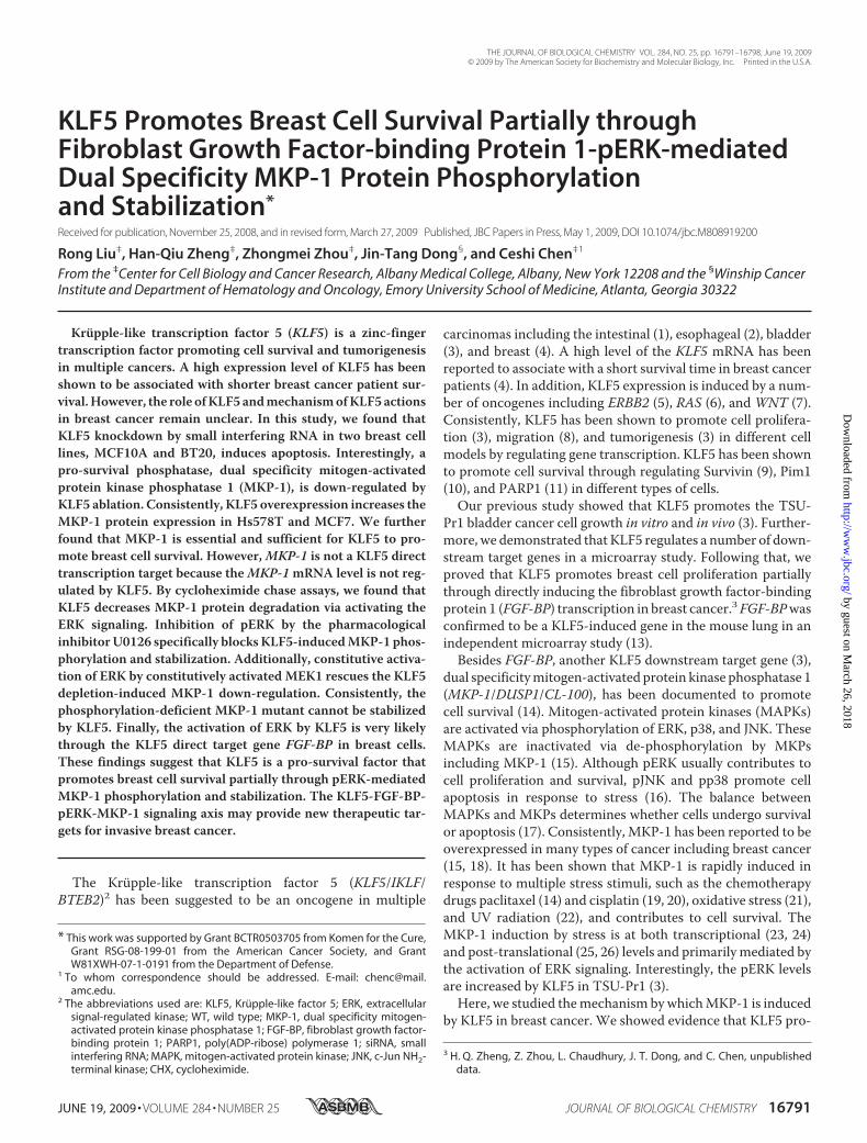
KLF5 Knockdown Induces Apoptosis and Decreases theMKP-1 Expression in Breast Cells—KLF5 has previously beenshown to express in estrogen receptor � negative basal-likebreast cells.3 To determine whether KLF5 promotes breast cellsurvival, we knocked down KLF5 in two KLF5 positive breastcell lines, MCF10A and BT20 (30). We examined the levels ofapoptosis markers, cleaved PARP, and caspase 3, in the controlluciferase siRNA (Lucsi) and well characterized KLF5 siRNA(KLF5si) (3, 31) transfected cells by immunoblotting.We foundthat KLF5si induces the cleavage of both PARP and caspase 3compared with Lucsi in MCF10A and BT20 (Fig. 1A). To fur-ther confirm that KLF5 knockdown decreases cell survivalthrough inducing apoptosis, we measured cell viability by the
KLF5 Promotes Breast Cell Survival through MKP-1
16792 JOURNAL OF BIOLOGICAL CHEMISTRY VOLUME 284 • NUMBER 25 • JUNE 19, 2009
by guest on March 26, 2018
http://ww
w.jbc.org/
Dow
nloaded from
SRB assay and Annexin V levels by flow cytometry. Consistentwith Western blot results, KLF5si significantly decreases cellviability (Fig. 1B) and increases Annexin V staining (data notshown) in both MCF10A and BT20. Interestingly, the proteinexpression levels of a potential KLF5 downstream gene, thepro-survival phosphatase MKP-1, are decreased by KLF5si inboth cell lines (Fig. 1A).To test if MKP-1 indeed promotes breast cell survival, we
knocked downMKP-1 by a pre-designed anti-MKP-1 siRNA in
bothMCF10A and BT20 and exam-ined apoptosis. As expected, knock-down of MKP-1 also induces thecleavage of both PARP and caspase 3and the decrease of cell viability likeknockdown of KLF5 in bothMCF10A and BT20 (Fig. 1,C andD).KLF5 Promotes Cell Survival Par-
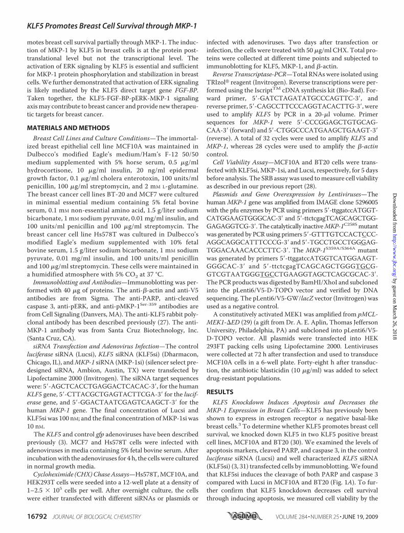
tially through MKP-1—Becausesilence of KLF5 induces apoptosisand down-regulates the expressionof the pro-survival MKP-1 proteinin breast cells, we wondered if KLF5functions partially through MKP-1.We performed a rescue experimentin MCF10A to determine whetherMKP-1 overexpression can blockthe KLF5si-induced apoptosis. Thewild-type (WT)MKP-1, the catalyt-ically inactive mutant MKP-1C258S(32), and the lacZ control geneswere forced overexpressed inMCF10A populations, respectively,by lentiviruses (Fig. 2A). In line withour previous observation, KLF5sidecreases the MKP-1 protein leveland induces apoptosis, indicated bycleavage of PARP and caspase 3 andloss of cell viability, in the controlLacZ overexpressing cells. Asexpected, forced overexpression ofWT MKP-1 clearly decreases thepERK levels and KLF5si-inducedapoptosis (Fig. 2). Similar resultswere obtained from two stableMKP-1 overexpressing MCF10Aclones (data not shown). Unexpect-edly, overexpression of the catalyti-cally inactive mutant MKP-1C258Salso blocks theKLF5si-induced apo-ptosis as efficiently as WT MKP-1.As a dominant negative MKP-1mutant, MKP-1C258S increases thepERK levels (Fig. 2A). Consistently,the expression level of MKP-1C258Sis higher than that of WT MKP-1presumably because a high level ofpERK stabilizes the MKP-1 protein(see below in detail). These findings
suggest that overexpression of MKP-1 can partially rescue theKLF5si-induced apoptosis in MCF10A.MKP-1 Expression Is Positively Regulated by KLF5 at the Pro-
tein Level but Not at the mRNA Level in Breast Cells—KLF5 is awell established transcriptional factor regulating transcriptionof a number of genes. To test whetherMKP-1 is a KLF5 directtranscriptional target, we examined MKP-1 expression at theprotein level by Western blot and the mRNA level by semi-quantitative reverse transcriptase-PCR after knocking down
FIGURE 1. Knockdown of KLF5 induces apoptosis and down-regulates the MKP-1 protein levels in breastcells. A, knockdown of KLF5 induces the PARP and caspase 3 cleavage and down-regulates the MKP-1 proteinlevels in MCF10A and BT20. A well characterized KLF5 siRNA was used to knockdown the KLF5 expression inMCF10A and BT20 cells. Luciferase siRNA (Lucsi) was used as the negative control. Protein levels were detectedby immunoblotting. B, KLF5 siRNA significantly reduces cell viability in MCF10A and BT20 as determined by theSRB assay. **, p � 0.001 (t test). C, knockdown of MKP-1 induces the PARP and caspase 3 cleavage in both MCF10Aand BT20 cell lines compared with the Lucsi negative control and the KLF5si positive control. D, the MKP-1 siRNAsignificantly reduces cell viability in MCF10A and BT20 compared with the Lucsi negative control and the KLF5sipositive control. Data are presented as the mean � S.D. (error bars) from three independent experiments.
FIGURE 2. MKP-1 partially rescues the KLF5 knockdown-induced apoptosis in MCF10A. A, overexpressionof either WT MKP-1 or the catalytically inactive MKP-1C258S mutant decreases the KLF5 siRNA-induced PARP andcaspase 3 cleavage. MCF10A cell populations stably expressing LacZ, MKP-1, or MKP-1C258S were transfectedwith Lucsi or KLF5si for 4 days. The apoptosis markers including cleaved PARP and caspase 3 were measured byimmunoblotting. B, overexpression of MKP-1 or MKP-1C258S significantly decreases the KLF5 knockdowninduced loss of cell viability as shown by the SRB assay. *, p � 0.05 (t test). Data are presented as the mean � S.D.(error bars) from three independent experiments.
KLF5 Promotes Breast Cell Survival through MKP-1
JUNE 19, 2009 • VOLUME 284 • NUMBER 25 JOURNAL OF BIOLOGICAL CHEMISTRY 16793
by guest on March 26, 2018
http://ww
w.jbc.org/
Dow
nloaded from
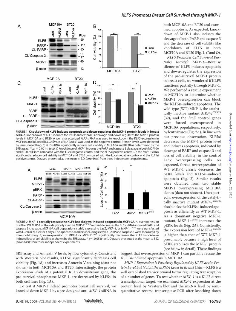
and overexpressing KLF5. To our surprise, KLF5si decreasesMKP-1 expression at the protein level but not at the mRNAlevel in both MCF10A and BT20 cell lines (Fig. 3, A and B).Additionally, KLF5 overexpression increases the expression ofMKP-1 at the protein level but not at the mRNA level in bothHs578T and MCF7 (Fig. 3, C and D). Finally, we found thatKLF5 cannot activate the MKP-1 promoter in MCF7 by dualluciferase reporter assays (data not shown). These results sug-gest thatMKP-1 is not a KLF5 direct transcription target genein breast cells.MKP-1 is a short-lived protein (the half-life is about 45 min
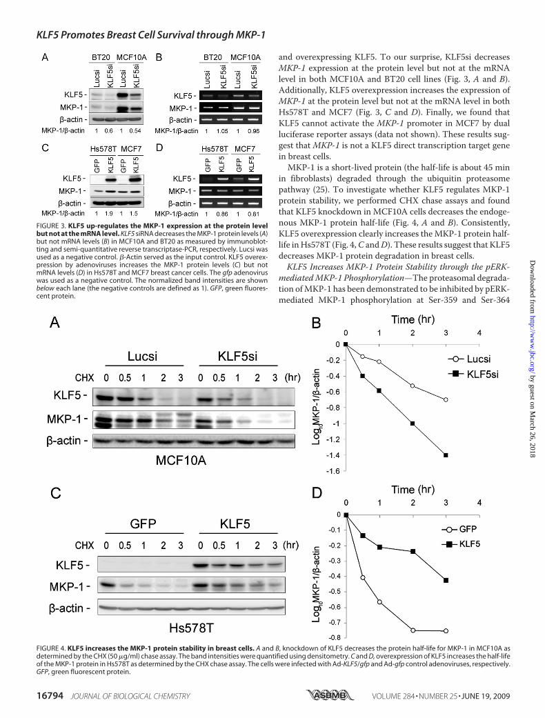
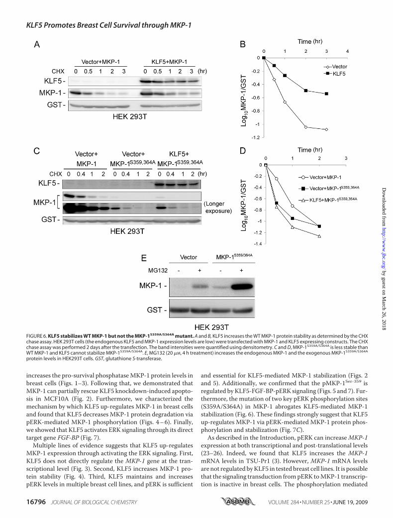
in fibroblasts) degraded through the ubiquitin proteasomepathway (25). To investigate whether KLF5 regulates MKP-1protein stability, we performed CHX chase assays and foundthat KLF5 knockdown in MCF10A cells decreases the endoge-nous MKP-1 protein half-life (Fig. 4, A and B). Consistently,KLF5 overexpression clearly increases theMKP-1 protein half-life inHs578T (Fig. 4,C andD). These results suggest that KLF5decreases MKP-1 protein degradation in breast cells.KLF5 Increases MKP-1 Protein Stability through the pERK-
mediatedMKP-1 Phosphorylation—The proteasomal degrada-tion ofMKP-1 has been demonstrated to be inhibited by pERK-mediated MKP-1 phosphorylation at Ser-359 and Ser-364
FIGURE 3. KLF5 up-regulates the MKP-1 expression at the protein levelbut not at the mRNA level. KLF5 siRNA decreases the MKP-1 protein levels (A)but not mRNA levels (B) in MCF10A and BT20 as measured by immunoblot-ting and semi-quantitative reverse transcriptase-PCR, respectively. Lucsi wasused as a negative control. �-Actin served as the input control. KLF5 overex-pression by adenoviruses increases the MKP-1 protein levels (C) but notmRNA levels (D) in Hs578T and MCF7 breast cancer cells. The gfp adenoviruswas used as a negative control. The normalized band intensities are shownbelow each lane (the negative controls are defined as 1). GFP, green fluores-cent protein.
FIGURE 4. KLF5 increases the MKP-1 protein stability in breast cells. A and B, knockdown of KLF5 decreases the protein half-life for MKP-1 in MCF10A asdetermined by the CHX (50 �g/ml) chase assay. The band intensities were quantified using densitometry. C and D, overexpression of KLF5 increases the half-lifeof the MKP-1 protein in Hs578T as determined by the CHX chase assay. The cells were infected with Ad-KLF5/gfp and Ad-gfp control adenoviruses, respectively.GFP, green fluorescent protein.
KLF5 Promotes Breast Cell Survival through MKP-1
16794 JOURNAL OF BIOLOGICAL CHEMISTRY VOLUME 284 • NUMBER 25 • JUNE 19, 2009
by guest on March 26, 2018
http://ww
w.jbc.org/
Dow
nloaded from
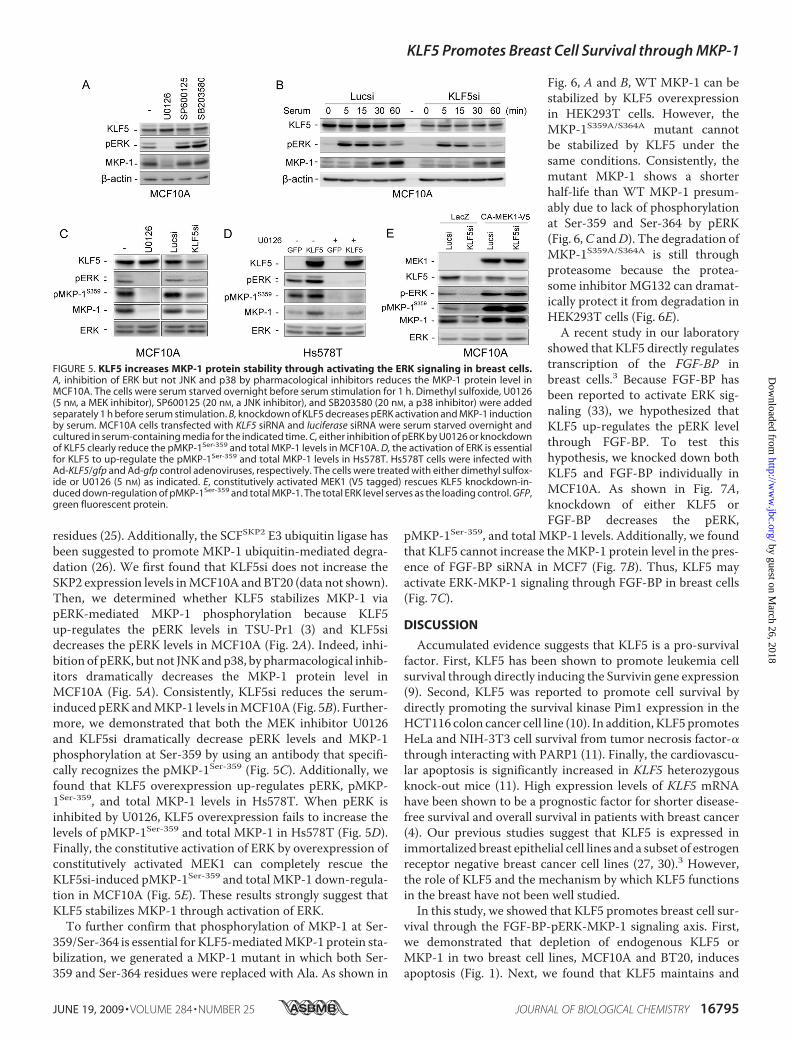
residues (25). Additionally, the SCFSKP2 E3 ubiquitin ligase hasbeen suggested to promote MKP-1 ubiquitin-mediated degra-dation (26). We first found that KLF5si does not increase theSKP2 expression levels inMCF10A andBT20 (data not shown).Then, we determined whether KLF5 stabilizes MKP-1 viapERK-mediated MKP-1 phosphorylation because KLF5up-regulates the pERK levels in TSU-Pr1 (3) and KLF5sidecreases the pERK levels in MCF10A (Fig. 2A). Indeed, inhi-bition of pERK, but not JNKandp38, by pharmacological inhib-itors dramatically decreases the MKP-1 protein level inMCF10A (Fig. 5A). Consistently, KLF5si reduces the serum-induced pERK andMKP-1 levels inMCF10A (Fig. 5B). Further-more, we demonstrated that both the MEK inhibitor U0126and KLF5si dramatically decrease pERK levels and MKP-1phosphorylation at Ser-359 by using an antibody that specifi-cally recognizes the pMKP-1Ser-359 (Fig. 5C). Additionally, wefound that KLF5 overexpression up-regulates pERK, pMKP-1Ser-359, and total MKP-1 levels in Hs578T. When pERK isinhibited by U0126, KLF5 overexpression fails to increase thelevels of pMKP-1Ser-359 and total MKP-1 in Hs578T (Fig. 5D).Finally, the constitutive activation of ERK by overexpression ofconstitutively activated MEK1 can completely rescue theKLF5si-induced pMKP-1Ser-359 and total MKP-1 down-regula-tion in MCF10A (Fig. 5E). These results strongly suggest thatKLF5 stabilizes MKP-1 through activation of ERK.To further confirm that phosphorylation of MKP-1 at Ser-
359/Ser-364 is essential for KLF5-mediatedMKP-1 protein sta-bilization, we generated a MKP-1 mutant in which both Ser-359 and Ser-364 residues were replaced with Ala. As shown in
Fig. 6, A and B, WT MKP-1 can bestabilized by KLF5 overexpressionin HEK293T cells. However, theMKP-1S359A/S364A mutant cannotbe stabilized by KLF5 under thesame conditions. Consistently, themutant MKP-1 shows a shorterhalf-life than WT MKP-1 presum-ably due to lack of phosphorylationat Ser-359 and Ser-364 by pERK(Fig. 6,C andD). The degradation ofMKP-1S359A/S364A is still throughproteasome because the protea-some inhibitor MG132 can dramat-ically protect it from degradation inHEK293T cells (Fig. 6E).A recent study in our laboratory
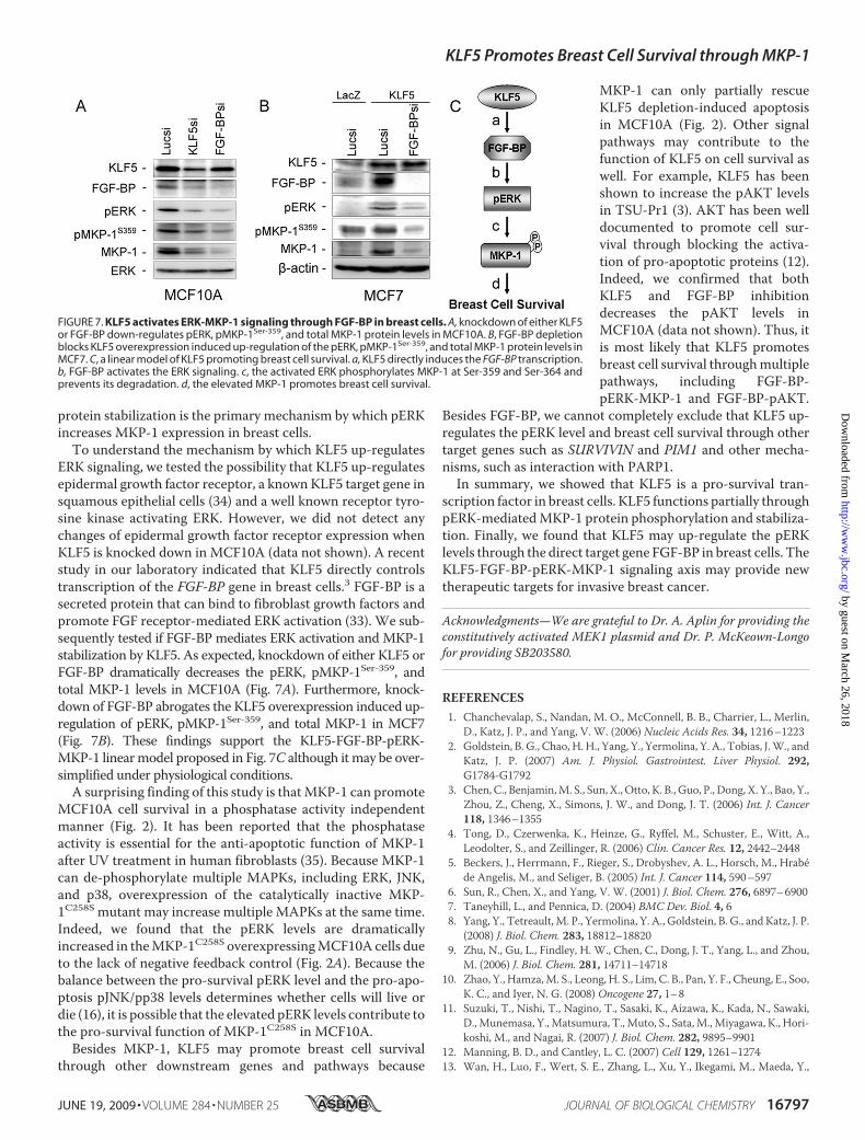
showed that KLF5 directly regulatestranscription of the FGF-BP inbreast cells.3 Because FGF-BP hasbeen reported to activate ERK sig-naling (33), we hypothesized thatKLF5 up-regulates the pERK levelthrough FGF-BP. To test thishypothesis, we knocked down bothKLF5 and FGF-BP individually inMCF10A. As shown in Fig. 7A,knockdown of either KLF5 orFGF-BP decreases the pERK,
pMKP-1Ser-359, and total MKP-1 levels. Additionally, we foundthat KLF5 cannot increase theMKP-1 protein level in the pres-ence of FGF-BP siRNA in MCF7 (Fig. 7B). Thus, KLF5 mayactivate ERK-MKP-1 signaling through FGF-BP in breast cells(Fig. 7C).
DISCUSSION
Accumulated evidence suggests that KLF5 is a pro-survivalfactor. First, KLF5 has been shown to promote leukemia cellsurvival through directly inducing the Survivin gene expression(9). Second, KLF5 was reported to promote cell survival bydirectly promoting the survival kinase Pim1 expression in theHCT116 colon cancer cell line (10). In addition, KLF5 promotesHeLa and NIH-3T3 cell survival from tumor necrosis factor-�through interacting with PARP1 (11). Finally, the cardiovascu-lar apoptosis is significantly increased in KLF5 heterozygousknock-out mice (11). High expression levels of KLF5 mRNAhave been shown to be a prognostic factor for shorter disease-free survival and overall survival in patients with breast cancer(4). Our previous studies suggest that KLF5 is expressed inimmortalized breast epithelial cell lines and a subset of estrogenreceptor negative breast cancer cell lines (27, 30).3 However,the role of KLF5 and the mechanism by which KLF5 functionsin the breast have not been well studied.In this study, we showed that KLF5 promotes breast cell sur-
vival through the FGF-BP-pERK-MKP-1 signaling axis. First,we demonstrated that depletion of endogenous KLF5 orMKP-1 in two breast cell lines, MCF10A and BT20, inducesapoptosis (Fig. 1). Next, we found that KLF5 maintains and
FIGURE 5. KLF5 increases MKP-1 protein stability through activating the ERK signaling in breast cells.A, inhibition of ERK but not JNK and p38 by pharmacological inhibitors reduces the MKP-1 protein level inMCF10A. The cells were serum starved overnight before serum stimulation for 1 h. Dimethyl sulfoxide, U0126(5 nM, a MEK inhibitor), SP600125 (20 nM, a JNK inhibitor), and SB203580 (20 nM, a p38 inhibitor) were addedseparately 1 h before serum stimulation. B, knockdown of KLF5 decreases pERK activation and MKP-1 inductionby serum. MCF10A cells transfected with KLF5 siRNA and luciferase siRNA were serum starved overnight andcultured in serum-containing media for the indicated time. C, either inhibition of pERK by U0126 or knockdownof KLF5 clearly reduce the pMKP-1Ser-359 and total MKP-1 levels in MCF10A. D, the activation of ERK is essentialfor KLF5 to up-regulate the pMKP-1Ser-359 and total MKP-1 levels in Hs578T. Hs578T cells were infected withAd-KLF5/gfp and Ad-gfp control adenoviruses, respectively. The cells were treated with either dimethyl sulfox-ide or U0126 (5 nM) as indicated. E, constitutively activated MEK1 (V5 tagged) rescues KLF5 knockdown-in-duced down-regulation of pMKP-1Ser-359 and total MKP-1. The total ERK level serves as the loading control. GFP,green fluorescent protein.
KLF5 Promotes Breast Cell Survival through MKP-1
JUNE 19, 2009 • VOLUME 284 • NUMBER 25 JOURNAL OF BIOLOGICAL CHEMISTRY 16795
by guest on March 26, 2018
http://ww
w.jbc.org/
Dow
nloaded from
increases the pro-survival phosphataseMKP-1 protein levels inbreast cells (Figs. 1–3). Following that, we demonstrated thatMKP-1 can partially rescue KLF5 knockdown-induced apopto-sis in MCF10A (Fig. 2). Furthermore, we characterized themechanism by which KLF5 up-regulates MKP-1 in breast cellsand found that KLF5 decreases MKP-1 protein degradation viapERK-mediated MKP-1 phosphorylation (Figs. 4–6). Finally,we showed that KLF5 activates ERK signaling through its directtarget gene FGF-BP (Fig. 7).
Multiple lines of evidence suggests that KLF5 up-regulatesMKP-1 expression through activating the ERK signaling. First,KLF5 does not directly regulate the MKP-1 gene at the tran-scriptional level (Fig. 3). Second, KLF5 increases MKP-1 pro-tein stability (Fig. 4). Third, KLF5 maintains and increasespERK levels in multiple breast cell lines, and pERK is sufficient
and essential for KLF5-mediated MKP-1 stabilization (Figs. 2and 5). Additionally, we confirmed that the pMKP-1Ser-359 isregulated byKLF5-FGF-BP-pERK signaling (Figs. 5 and 7). Fur-thermore, the mutation of two key pERK phosphorylation sites(S359A/S364A) in MKP-1 abrogates KLF5-mediated MKP-1stabilization (Fig. 6). These findings strongly suggest that KLF5up-regulates MKP-1 via pERK-mediated MKP-1 protein phos-phorylation and stabilization (Fig. 7C).As described in the Introduction, pERK can increaseMKP-1
expression at both transcriptional and post-translational levels(23–26). Indeed, we found that KLF5 increases the MKP-1mRNA levels in TSU-Pr1 (3). However, MKP-1 mRNA levelsare not regulated byKLF5 in tested breast cell lines. It is possiblethat the signaling transduction frompERK toMKP-1 transcrip-tion is inactive in breast cells. The phosphorylation mediated
FIGURE 6. KLF5 stabilizes WT MKP-1 but not the MKP-1S359A/S364A mutant. A and B, KLF5 increases the WT MKP-1 protein stability as determined by the CHXchase assay. HEK 293T cells (the endogenous KLF5 and MKP-1 expression levels are low) were transfected with MKP-1 and KLF5 expressing constructs. The CHXchase assay was performed 2 days after the transfection. The band intensities were quantified using densitometry. C and D, MKP-1S359A/S364A is less stable thanWT MKP-1 and KLF5 cannot stabilize MKP-1S359A/S364A. E, MG132 (20 �M, 4 h treatment) increases the endogenous MKP-1 and the exogenous MKP-1S359A/S364A
protein levels in HEK293T cells. GST, glutathione S-transferase.
KLF5 Promotes Breast Cell Survival through MKP-1
16796 JOURNAL OF BIOLOGICAL CHEMISTRY VOLUME 284 • NUMBER 25 • JUNE 19, 2009
by guest on March 26, 2018
http://ww
w.jbc.org/
Dow
nloaded from
protein stabilization is the primary mechanism by which pERKincreases MKP-1 expression in breast cells.To understand the mechanism by which KLF5 up-regulates
ERK signaling, we tested the possibility that KLF5 up-regulatesepidermal growth factor receptor, a known KLF5 target gene insquamous epithelial cells (34) and a well known receptor tyro-sine kinase activating ERK. However, we did not detect anychanges of epidermal growth factor receptor expression whenKLF5 is knocked down in MCF10A (data not shown). A recentstudy in our laboratory indicated that KLF5 directly controlstranscription of the FGF-BP gene in breast cells.3 FGF-BP is asecreted protein that can bind to fibroblast growth factors andpromote FGF receptor-mediated ERK activation (33). We sub-sequently tested if FGF-BP mediates ERK activation and MKP-1stabilization by KLF5. As expected, knockdown of either KLF5 orFGF-BP dramatically decreases the pERK, pMKP-1Ser-359, andtotal MKP-1 levels in MCF10A (Fig. 7A). Furthermore, knock-down of FGF-BP abrogates the KLF5 overexpression induced up-regulation of pERK, pMKP-1Ser-359, and total MKP-1 in MCF7(Fig. 7B). These findings support the KLF5-FGF-BP-pERK-MKP-1 linearmodel proposed in Fig. 7C although itmay be over-simplified under physiological conditions.A surprising finding of this study is thatMKP-1 can promote
MCF10A cell survival in a phosphatase activity independentmanner (Fig. 2). It has been reported that the phosphataseactivity is essential for the anti-apoptotic function of MKP-1after UV treatment in human fibroblasts (35). Because MKP-1can de-phosphorylate multiple MAPKs, including ERK, JNK,and p38, overexpression of the catalytically inactive MKP-1C258S mutant may increase multiple MAPKs at the same time.Indeed, we found that the pERK levels are dramaticallyincreased in theMKP-1C258S overexpressingMCF10A cells dueto the lack of negative feedback control (Fig. 2A). Because thebalance between the pro-survival pERK level and the pro-apo-ptosis pJNK/pp38 levels determines whether cells will live ordie (16), it is possible that the elevated pERK levels contribute tothe pro-survival function of MKP-1C258S in MCF10A.
Besides MKP-1, KLF5 may promote breast cell survivalthrough other downstream genes and pathways because
MKP-1 can only partially rescueKLF5 depletion-induced apoptosisin MCF10A (Fig. 2). Other signalpathways may contribute to thefunction of KLF5 on cell survival aswell. For example, KLF5 has beenshown to increase the pAKT levelsin TSU-Pr1 (3). AKT has been welldocumented to promote cell sur-vival through blocking the activa-tion of pro-apoptotic proteins (12).Indeed, we confirmed that bothKLF5 and FGF-BP inhibitiondecreases the pAKT levels inMCF10A (data not shown). Thus, itis most likely that KLF5 promotesbreast cell survival throughmultiplepathways, including FGF-BP-pERK-MKP-1 and FGF-BP-pAKT.
Besides FGF-BP, we cannot completely exclude that KLF5 up-regulates the pERK level and breast cell survival through othertarget genes such as SURVIVIN and PIM1 and other mecha-nisms, such as interaction with PARP1.In summary, we showed that KLF5 is a pro-survival tran-
scription factor in breast cells. KLF5 functions partially throughpERK-mediatedMKP-1 protein phosphorylation and stabiliza-tion. Finally, we found that KLF5 may up-regulate the pERKlevels through the direct target gene FGF-BP in breast cells. TheKLF5-FGF-BP-pERK-MKP-1 signaling axis may provide newtherapeutic targets for invasive breast cancer.
Acknowledgments—We are grateful to Dr. A. Aplin for providing theconstitutively activated MEK1 plasmid and Dr. P. McKeown-Longofor providing SB203580.
REFERENCES1. Chanchevalap, S., Nandan, M. O., McConnell, B. B., Charrier, L., Merlin,
D., Katz, J. P., and Yang, V. W. (2006) Nucleic Acids Res. 34, 1216–12232. Goldstein, B. G., Chao, H. H., Yang, Y., Yermolina, Y. A., Tobias, J.W., and
Katz, J. P. (2007) Am. J. Physiol. Gastrointest. Liver Physiol. 292,G1784-G1792
3. Chen, C., Benjamin,M. S., Sun, X.,Otto, K. B., Guo, P., Dong, X. Y., Bao, Y.,Zhou, Z., Cheng, X., Simons, J. W., and Dong, J. T. (2006) Int. J. Cancer118, 1346–1355
4. Tong, D., Czerwenka, K., Heinze, G., Ryffel, M., Schuster, E., Witt, A.,Leodolter, S., and Zeillinger, R. (2006) Clin. Cancer Res. 12, 2442–2448
5. Beckers, J., Herrmann, F., Rieger, S., Drobyshev, A. L., Horsch, M., Hrabede Angelis, M., and Seliger, B. (2005) Int. J. Cancer 114, 590–597
6. Sun, R., Chen, X., and Yang, V. W. (2001) J. Biol. Chem. 276, 6897–69007. Taneyhill, L., and Pennica, D. (2004) BMC Dev. Biol. 4, 68. Yang, Y., Tetreault,M. P., Yermolina, Y. A., Goldstein, B. G., and Katz, J. P.
(2008) J. Biol. Chem. 283, 18812–188209. Zhu, N., Gu, L., Findley, H. W., Chen, C., Dong, J. T., Yang, L., and Zhou,
M. (2006) J. Biol. Chem. 281, 14711–1471810. Zhao, Y., Hamza,M. S., Leong, H. S., Lim, C. B., Pan, Y. F., Cheung, E., Soo,
K. C., and Iyer, N. G. (2008) Oncogene 27, 1–811. Suzuki, T., Nishi, T., Nagino, T., Sasaki, K., Aizawa, K., Kada, N., Sawaki,
D.,Munemasa, Y.,Matsumura, T.,Muto, S., Sata,M.,Miyagawa, K., Hori-koshi, M., and Nagai, R. (2007) J. Biol. Chem. 282, 9895–9901
12. Manning, B. D., and Cantley, L. C. (2007) Cell 129, 1261–127413. Wan, H., Luo, F., Wert, S. E., Zhang, L., Xu, Y., Ikegami, M., Maeda, Y.,
FIGURE 7. KLF5 activates ERK-MKP-1 signaling through FGF-BP in breast cells. A, knockdown of either KLF5or FGF-BP down-regulates pERK, pMKP-1Ser-359, and total MKP-1 protein levels in MCF10A. B, FGF-BP depletionblocks KLF5 overexpression induced up-regulation of the pERK, pMKP-1Ser-359, and total MKP-1 protein levels inMCF7. C, a linear model of KLF5 promoting breast cell survival. a, KLF5 directly induces the FGF-BP transcription.b, FGF-BP activates the ERK signaling. c, the activated ERK phosphorylates MKP-1 at Ser-359 and Ser-364 andprevents its degradation. d, the elevated MKP-1 promotes breast cell survival.
KLF5 Promotes Breast Cell Survival through MKP-1
JUNE 19, 2009 • VOLUME 284 • NUMBER 25 JOURNAL OF BIOLOGICAL CHEMISTRY 16797
by guest on March 26, 2018
http://ww
w.jbc.org/
Dow
nloaded from
Bell, S. M., and Whitsett, J. A. (2008) Development 135, 2563–257214. Wu,W., Pew, T., Zou,M., Pang, D., andConzen, S. D. (2005) J. Biol. Chem.
280, 4117–412415. Keyse, S. M. (2008) Cancer Metastasis Rev. 27, 253–26116. Junttila, M. R., Li, S. P., andWestermarck, J. (2008) FASEB J. 22, 954–96517. Wada, T., and Penninger, J. M. (2004) Oncogene 23, 2838–284918. Loda, M., Capodieci, P., Mishra, R., Yao, H., Corless, C., Grigioni, W.,
Wang, Y., Magi-Galluzzi, C., and Stork, P. J. (1996) Am. J. Pathol. 149,1553–1564
19. Sanchez-Perez, I., Martínez-Gomariz, M., Williams, D., Keyse, S. M., andPerona, R. (2000) Oncogene 19, 5142–5152
20. Wang, J., Zhou, J. Y., and Wu, G. S. (2007) Cancer Res. 67, 11933–1194121. Zhou, J. Y., Liu, Y., and Wu, G. S. (2006) Cancer Res. 66, 4888–489422. Franklin, C. C., Srikanth, S., and Kraft, A. S. (1998) Proc. Natl. Acad. Sci.
U.S.A. 95, 3014–301923. Li, J., Gorospe, M., Hutter, D., Barnes, J., Keyse, S. M., and Liu, Y. (2001)
Mol. Cell. Biol. 21, 8213–822424. Wang, Z., Xu, J., Zhou, J. Y., Liu, Y., and Wu, G. S. (2006) Cancer Res. 66,
8870–887725. Brondello, J. M., Pouyssegur, J., and McKenzie, F. R. (1999) Science 286,
2514–2517
26. Lin, Y. W., and Yang, J. L. (2006) J. Biol. Chem. 281, 915–92627. Chen, C., Sun, X., Ran, Q., Wilkinson, K. D., Murphy, T. J., Simons, J. W.,
and Dong, J. T. (2005) Oncogene 24, 3319–332728. Chen, C., Zhou, Z., Ross, J. S., Zhou, W., and Dong, J. T. (2007) Int. J.
Cancer 121, 80–8729. Aplin, A. E., Stewart, S. A., Assoian, R. K., and Juliano, R. L. (2001) J. Cell
Biol. 153, 273–28230. Chen, C., Bhalala, H. V., Qiao, H., and Dong, J. T. (2002) Oncogene 21,
6567–657231. Aizawa, K., Suzuki, T., Kada, N., Ishihara, A., Kawai-Kowase, K., Mat-
sumura, T., Sasaki, K., Munemasa, Y., Manabe, I., Kurabayashi, M., Col-lins, T., and Nagai, R. (2004) J. Biol. Chem. 279, 70–76
32. Slack, D. N., Seternes, O. M., Gabrielsen, M., and Keyse, S. M. (2001)J. Biol. Chem. 276, 16491–16500
33. Tassi, E., Al-Attar, A., Aigner, A., Swift, M. R., McDonnell, K., Karavanov,A., and Wellstein, A. (2001) J. Biol. Chem. 276, 40247–40253
34. Yang, Y., Goldstein, B. G., Nakagawa, H., and Katz, J. P. (2007) FASEB J. 21,543–550
35. Hamdi, M., Kool, J., Cornelissen-Steijger, P., Carlotti, F., Popeijus, H. E.,van der Burgt, C., Janssen, J. M., Yasui, A., Hoeben, R. C., Terleth, C.,Mullenders, L. H., and van Dam, H. (2005) Oncogene 24, 7135–7144
KLF5 Promotes Breast Cell Survival through MKP-1
16798 JOURNAL OF BIOLOGICAL CHEMISTRY VOLUME 284 • NUMBER 25 • JUNE 19, 2009
by guest on March 26, 2018
http://ww
w.jbc.org/
Dow
nloaded from
Rong Liu, Han-Qiu Zheng, Zhongmei Zhou, Jin-Tang Dong and Ceshi ChenPhosphorylation and Stabilization
Factor-binding Protein 1-pERK-mediated Dual Specificity MKP-1 Protein KLF5 Promotes Breast Cell Survival Partially through Fibroblast Growth
doi: 10.1074/jbc.M808919200 originally published online May 1, 20092009, 284:16791-16798.J. Biol. Chem.
10.1074/jbc.M808919200Access the most updated version of this article at doi:
Alerts:
When a correction for this article is posted•
When this article is cited•
to choose from all of JBC's e-mail alertsClick here
http://www.jbc.org/content/284/25/16791.full.html#ref-list-1
This article cites 35 references, 20 of which can be accessed free at
by guest on March 26, 2018
http://ww
w.jbc.org/
Dow
nloaded from