Embed Size (px)
Citation preview
TitleKLINEFELTER'S SYNDROME ASSOCIATED WITHURETERAL POLYPS, RENAL PELVIC STONE ANDPRIMARY HYPERPARATHYROIDISM
Author(s) Baba, Shiro; Hayakawa, Masamichi; Matsushita, Kazuo;Nakamura, Hiroshi; Yoshimatsu, Hiroshi
Citation 泌尿器科紀要 (1982), 28(4): 445-451
Issue Date 1982-04
URL http://hdl.handle.net/2433/123065
Right
Type Departmental Bulletin Paper
Textversion publisher
Kyoto University
445
[Acta Ural. Jap. Vol. 28J
No.4, April 1982
KLINEFELTER'S SYNDROME ASSOCIATED WITH URETERAL POLYPS, RENAL
PELVIC STONE AND PRIMARY HYPERPARA THYROIDISM
Shiro BABA*, Masamichi HAYAKAwA*, Kazuo MATSUSHITA**,
Hiroshi NAKAMuRA* and Hiroshi YOSHIMATSU***
From the Department of Urology, National Defense Medical College, Saitama, Japan* and
the Department of Thoracic and Cardiovascular Surgery, School of Medicine, University of Occupational and Environmental Health, Japan, Fukuoka, Japan***
Herein is reported a case of 47 XXV-Klinefelter's syndrome associated with ureteral polyps,
renal pelvic stone and primary hyperparathyroidism. To the best of our knowledge, this is the second
patient reported to have Klinefelter's syndrome coexisting with primary hyperparathyroidism. Fre
quent endocrinological disorders in the patients with Klinefelter's syndrome and diagnostic problems
for hyperparathyroidism and ureteral polyps are discussed.
Key words: Klinefelter's syndrome, Primary hyperparathyroidism, Ureteral polyp.
According to the classical description, Klinefelter's syndrome is distinguished by small testes, gynecomastia and increased urinary gonadotropin activityll. Concepts of this syndrome have been modified and expanded to include a variety of additional physical characteristics, such as sparse body hair, eunuchoidal body habitus and feminine distribution of adipose tissue, which reflect the lack of androgen effects. Various other endocrinological disorders are reported to occur among the patients having this syndrome. These include diabetes mellitus2), low radioactive iodine uptake by thyroid gland3l, low response of the gland to thyroid-stimulating hormone (TSH)3,4), and abnormal pituitary response to thyrotropin releasing hormone (TRH) 5) •
Herein we report on a patient with Klinefelter's syndrome, who had right ureteral polyps, a stone in the ipsilateral renal pelvis and primary hyperparathyroidism. Review of the literature revealed only one similar case of primary hyperparathy-
** Present Adress: Department of Urology, School of Medicine, Tokai University.
roidism coexisting with Klinefelter's syndrome6).
CASE REPORT
A 55-year-old Japanese male was hospitalized, because of right flank pain and gross hematuria. The past medical record disclosed several episodes of stone passage mainly from the right kidney and bilateral mastectomy that had been done for gynecomastia 16 years previously. The patient experienced no penile erection nor ejaculation. Physical examination revealed eunuchoidal body habitus with a height of 171 cm and an arm span of 177.8 cm. The patient weighed 71 kg. The beard, pubic hair and axillary hair were all scanty. Nasal cavities were free of polyps. Vague pain was noted on the right costovertebral angle, radiating to the right lower abdomen. The penis was small, measuring 5 em in length and the bilateral testes were palpated to be small and firm, measuring about 1.5 X 2 X 2 cm. The prostate was normal in size and consistency. There was no evidence of hypospadias. The patient was normotensive and, psychologically, shy and
446 Acta Urol. J ap. Vol. 28 o. 4, 1982
(CCJ fJ .......... r ···················· 2 .............. .3 ......... .
4 5
........... I~ . ..... -.-....... I~ ..... )1 . ........... I{ II . ............ II ............. 1.1 ... 6 7 8 9 10 11 12
........... al ...... •• .......... .t II " II 13 14 15 16 17 18
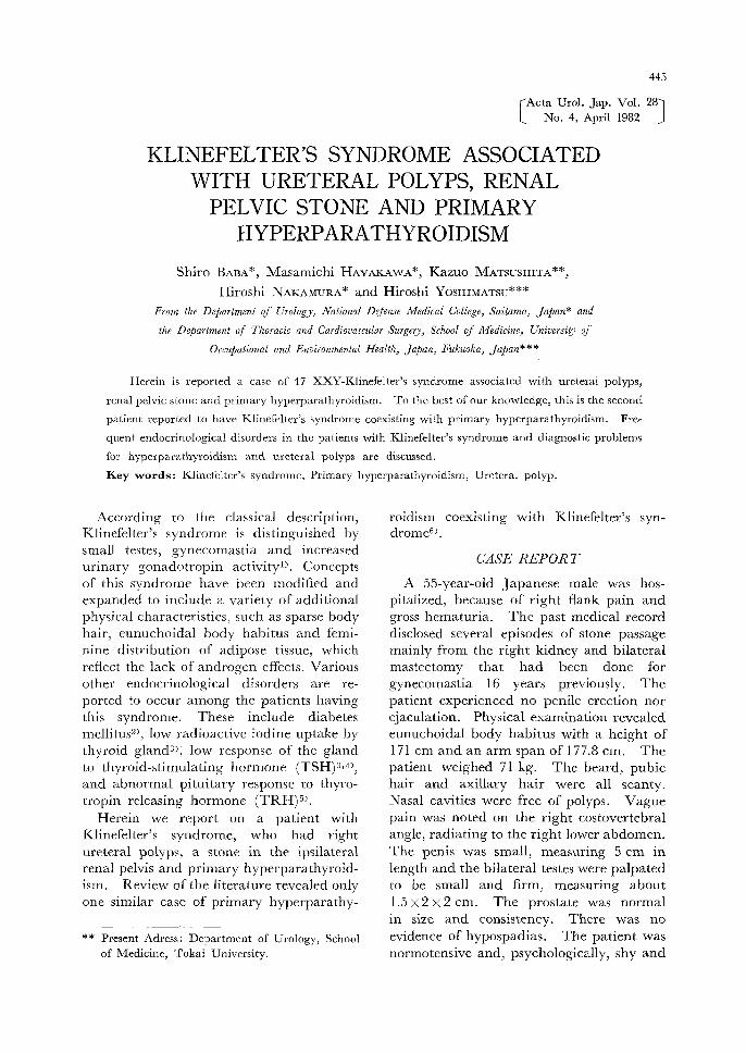
Fig. I. Chromosomal a na lys is showing 47 XXV pat tern (C iemsa sta in).
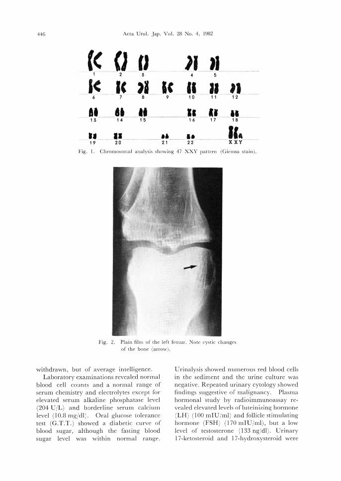
Fig. 2. Pla in fi lm of the left fem ur. Note cyst ic changes of the bone (arrow).
withdrawn , but of average intelligence. Labora tory examina tions revealed normal
blood cell counts a nd a norma l range of serum chemistry and electrolytes except for elevated serum alka line phospha tase level (204 U JL ) and borderline seru m calci urn level (10.8 mgJdl) . Ora l glucose tolerance tes t (G .T .T. ) showed a d iabetic curve of b lood sugar, although the fasting blood sugar level was within normal ra nge.
U rina lys is showed numerous red blood cells in the sediment a nd the uri ne culture was negative. R epeated urina ry cytology showed find ings suggestive of malignancy . Plasma hormonal study by radioimmunoassay revealed eleva ted levels ofl u teinizing hormone (LH) (100 mIU Jml) and foll icle st imu lating hormone (FSH) (170 mIUJml), but a low level of tes tosterone (133 ngJdl) . U r inary 17-ketosteroid and 17-hydroxysteroid were
Baba et a l. Kline felter 's syndrome with primary hyperparathyroidism 447
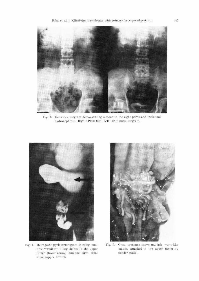
Fig . 3. Excretory urogram demonstrating a stone in the right pelvis and ipsilatera l hydronephros is. Right: Pl a in fi lm. Left: 10 minutes urogram .
Fig. 4. R etrograde pyeloureterogram showing multiple vermiform fi lling defects in the upper ureter (lower arrow) a nd th e right rena l
stone (upper arrow) .
Fig. 5. Gross specimen shows multiple worm-like masses, attached to the upper ureter by slender stalks.
448 Acta U ro1. .lap. Vol. 28 No.4, 1982
, .l:)
• . . , .. t' • ~, ., ... .. ..
.- .' "
~
" ...
" . - :
.. ',-
I • • -.
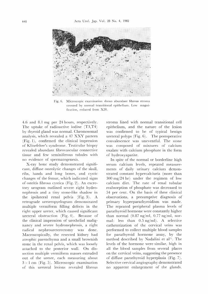
Fig. 6. Microscopic examination shows abunda nt fibrous stroma covered by normal trans itiona l epithelium. Low magnificatio n, reduced from X 20.
4.6 and 8. 1 mg per 24 hours , respectively. The uptake of radioactive iodine (T 3,T4) by thyroid gland was normal. Chromosomal analysis, which revealed a 47 XXY pattern (Fig. I), confirmed the clinical impression of Klinefd ter 's syndrome _ T esti cul ar biopsy revealed abundant fibrovascu lar connective tissue and few seminiferous tubules with no evidence of spermatogenesis.
X-ray bone study demonstrated significant, diffuse osteolytic cha nges of the skull , ribs, hands and long bones, and cystic cha nges of the femur, which indicated signs of osteitis fibrosa cystica (Fig . 2) . An excretory urogram outlined severe right hydronephros is and a tiny stone-like shadow in the ipsilateral renal pelvis (Fig. 3). A retrograde ureteropyelogram demonstrated multiple vermiform filling defects in the right upper ureter, which caused significant ureteral obstruction (Fig. 4) . Because of the clinical impression of urothelia l malignancy and severe hydronephrosis, a right radical nephroureterectomy was done. Macroscopically, the resected kidney had atrophic parenchyma and a small brownish stone in the rena l pelvis, which was loosely a ttached to the pos terior wall . On dissection multiple verm iform masses extruded out of the ureter, each measuring about 3 X I cm (Fig. 5) . Microscopic examination of this uretera l les ions revealed fibrous
stroma lined with normal transitional cell epithelium, and the nature of the lesion was confirmed to be of typical benign ureteral polyps (Fig. 6) . The postoperative convalescence was uneventful. The stone was composed of mixtures of calcium oxalate with calcium phosphate in the form of hydroxyapatite.
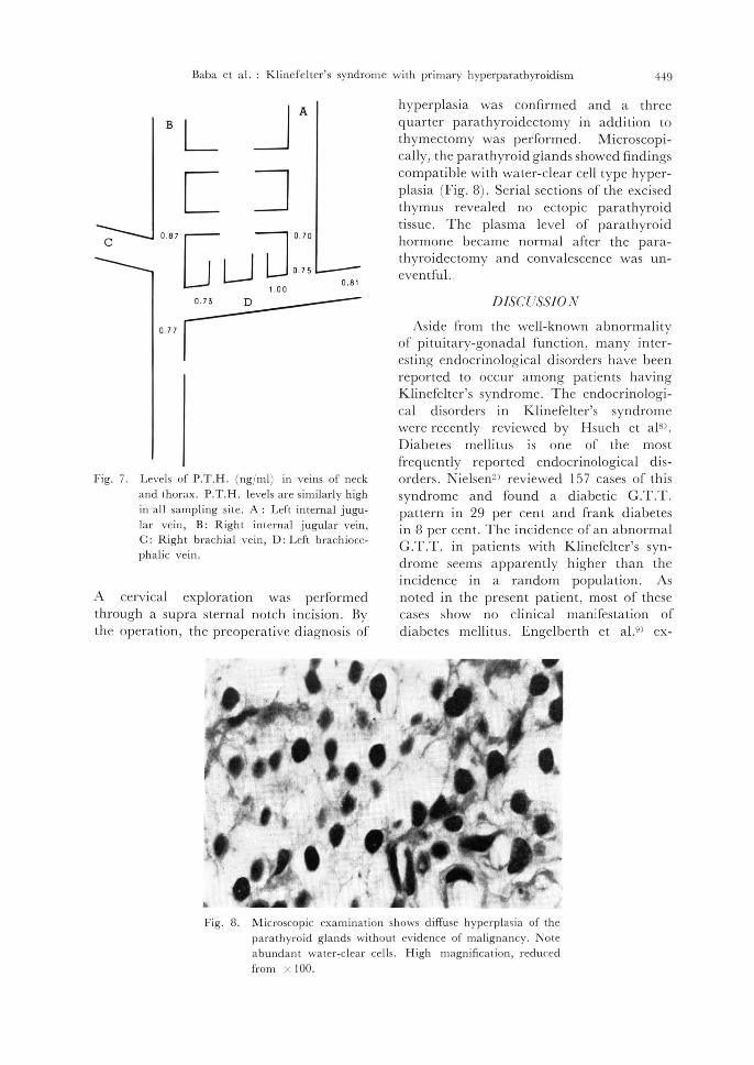
In spite of the normal or borderline high serum calcium levels, repeated measurements of daily urinary calcium demonstrated constant hypercalciuria (more than 300 mg/24 hr) under the regimen of low calcium diet. The rate of renal tubular reabsorption of phosphate was decreased to 54 per cent. On the basis of these clinical observations, a presumptive diagnosis of primary hyperparathyroidism was mad e. The repea ted peripheral plasma levels of parathyroid hormone were constantly higher than normal (0.87 ng/ml, 0.77 ng/ml , normal: less than 0.5 ng/ml). A selective catheterization of the cervical veins was performed to collect multiple blood samples for parathyro id hormone assay, by the method descri bed by N adalini et aj7 ' . The levels of the hormone were similar, high in a ll the blood samples from several places on the cervical veins, suggesting the presence of diffuse parathyroid hyperplasia (Fig. 7) . Selective cervical angiography demonstrated no apparent enlargement of the glands.
Baba et al. : Klinefelter's syndrome with primary hyperparathyroidism 449
J A B L L ~
c ." [J LJ LI:: 0.81
1.00
0.7~ D
0. 77 r Fig. 7. Levels of P.T.H . (ng/ml) in veins of neck
and thorax. P .T. H . levels are similarly high in a ll sampling site. A : Left in terna l jugular ve in, B: Right internal jugular vein, C: Righ t brachia l ve in, D: Left brach iocephalic vein.
A cervical exploration was performed through a supra sternal notch incision. By the operation , the preoperative diagnosis of
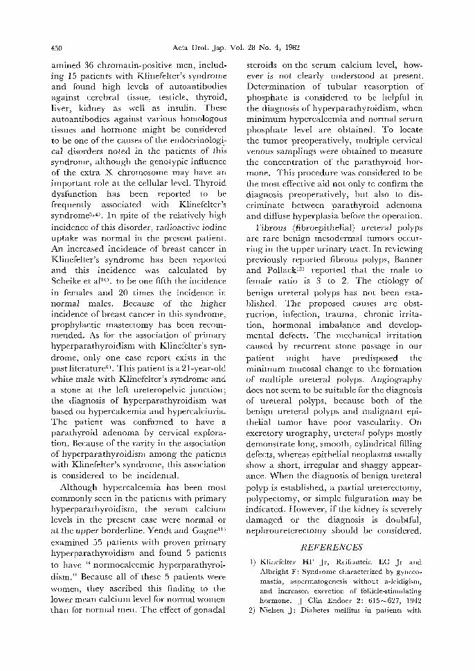
hyperplasia was confirmed and a three quarter parathyroidectomy in addition to thymectomy was performed. Microscopically, the parathyroid gla nds showed findings compatible with water-clear cell type hyperplasia (Fig. 8) . Serial sections of the excised thymus revealed no ectopic parathyroid tissue. The plasma level of pa rathyroid hormone became normal a fter the parathyroidectomy and convalescence was uneventful.
DiSCUSSiO N
Aside from the well-known abnormality of pituitary-gonadal function, many interesting endocrinological disorders have been reported to occur among patients having Klinefelter's syndrome. The endocrinological disorders in Klinefelter's syndrome were recently reviewed by Hsueh et aI8) .
Diabetes mellitus is one of the most frequently reported endocrinological disorders. Nielsen2) reviewed 157 cases of this syndrome and found a diabetic G.T.T. pattern in 29 per cent and frank diabetes in 8 per cent . The incidence of an abnormal G.T.T. in patients with Klinefelter's syndrome seems appa rently higher than the incidence in a random population. As noted in the present patient, most of these cases show no clinical manifestation of diabetes mellitus. Engelberth et aJ.9) ex-
Fig. 8. Microscopic examination shows diffuse hyperplasia of the parathyroid glands without evidence of malignancy. Note abundant water-clear cells. H igh magnifical·ion, reduced
frol11 X 100.
450 Acta Urol. Jap. Vol. 28 No.4, 1982
amined 36 chromatin-positive men, including 15 patients with Klinefelter's syndrome and found high levels of autoantibodies against cerebral tissue, testicle, thyroid, liver, kidney as well as insulin. These autoantibodies against various homologous tissues and hormone might be considered to be one of the causes of the endocrinological disorders noted in the patients of this syndrome, although the genotypic influence of the extra X chromosome may have an important role at the cellular level. Thyroid dysfunction has been reported to be frequently associated with Klinefelter's syndrome3 ,4). In spite of the relatively high
incidence of this disorder, radioactive iodine uptake was normal in the present patient. An increased incidence of breast cancer in Klinefelter's syndrome has been reported and this incidence was calculated by Scheike et aFO). to be one fifth the incidence
in females and 20 times the incidence in normal males. Because of the higher incidence of breast cancer in this syndrome, prophylactic mastectomy has been recomrnended. As for the associaLion of primary hyperparathyroidism with Klinefelter's syndrome, only one case report exists in the past literature6). This patient is a 21-year-old white male with Klinefelter's syndrome and a stone at the left ureteropelvic junction; the diagnosis of hyperparathyroidism was based on hypercalcemia and hypercalciuria. The patient was confirmed to have a parathyroid adenoma by cervical exploration. Because of the rarity in the association of hyperparathyroidism among the patients with Klinefelter's syndrome, this association is considered to be incidental.
Although hypercalcemia has been most commonly seen in the patients with primary hyperparathyroidism, the serum calcium levels in the present case were normal or at the upper borderline. Yendt and Gagnell)
examined 55 patients with proven primary hyperparathyroidism and found 5 patients
to have" normocalcemic hyperparathyroi
dism." Because all of these 5 patients were women, they ascribed this finding to the lower mean calcium level for normal women than for normal men. The effect of gonadal
steroids on the serum calcium level, however is not clearly understood at present. Determination of tubular reasorption of phosphate is considered to be helpful in the diagnosis of hyperparathyroidism, when minimum hypercalcemia and normal serum phosphate level are obtained. To locate the tumor preoperatively, multiple cervical venous samplings were obtained to measure the concentration of the parathyroid hormone. This procedure was considered to be the most effective aid not only to confirm the diagnosis preoperatively, but also to discriminate between parathyroid adenoma and diffuse hyperplasia before the operation.
Fibrous (fibroepithe1ial) ureteral polyps are rare benign mesodermal tumors occurring in the upper urinary tract. In reviewing previously reported fibrous polyps, Banner and Pollack12) reported that the male to female ratio is 3 to 2. The etiology of
benign ureteral polyps has not been established. The proposed causes are obstruction, infection, trauma, chronic irritation, hormonal imbalance and developmental defects. The mechanical irritation caused by recurrent stone passage in our
patient might have predisposed the minimum mucosal change to the formation of multiple ureteral polyps. Angiography does not seem to be suitable for the diagnosis of ureteral polyps, because both of the benign ureteral polyps and malignant epithelial tumor have poor vascularity. On excretory urography, ureteral polyps mostly demonstrate long, smooth, cylindrical filling defects, whereas epithelial neoplasms usually show a short, irregular and shaggy appearance. When the diagnosis of benign ureteral
polyp is established, a partial ureterectomy, polypectomy, or simple fulguration may be indicated. However, if the kidney is severely damaged or the diagnosis is doubtful, nephroureterectomy should be considered.
REFERENCES
I) Klinefelter HF Jr, Reifenstein EC Jr and Albright F: Syndrome characterized by gynecomastia, aspermatogenesis without a-Ieidigism, and increased excretion of follicle-stimulating hormone. J Clin Endocr 2: 615-627, 1942
2) Nielsen J: Diabetes mellitus in patients with
Baba et al. : Klinefelter's syndrome
aneuploid chromosome aberrations and in their
parents. Humangenetik 16: 165—.170, 19723) Davis TE, Canfield CJ and Herman RH, Goler
D: Thyroid function in patients with aspermio-
genesis and testicular tubular sclerosis. New Engl J Med 268: 178-182, 1963
4) Plunkett ER, Rangecroft G and Heagry FC: Thyroid function in patients with sex chromo-
somal anomalies. J Ment Defic Res 7: 25-34,
1964
5) Ozawa Y and Shishiba Y: Lack of TRH-induced TSH secretion in a patient with Klinefelter's
syndrome: a case report. Endocr Jap 22: 269-273, 1975
6) Spalding JA, Morrow GW Jr and Scholz DA: Coexisting Klinefelter's syndrome and primary
hyperparathyroidism: report of case. Metab 11: 732 —734, 1962
7) Nadalini VF, Positano N and Bruttini GP: Multiple sampling for parathyroid hormone by
with primary hyperparathyroidism451
subclavian approach. Urol 8: 163 165, 1979
8) Hsueh NA, Hsu TH and Federman DD: Endocrine features of Klinefelter's syndrome.
Medicine 57: 447--461, 19789) Engelberth 0, Charvat J, Jezkova Z and Raboch
J: Autoantibodies in chromatin-positive men. Lancet 2: 1194, 1966
10) Scheike 0, Visfeldt J, Petersen B: Male breast cancer 3. Breast carcinoma in association with
the Klinefelter's syndrome. Acta Pathol Micro- biol Scand (A) 81: 352-358, 1973
11) Yendt ER and Gagne RJ: Detection of primary hyperparathyroidism, with special reference to
its occurrence in hypercaiciuric females with "normal" or borderline serum calcium . Can Med Ass J 98: 331-..336, 1968
12) Banner MP and Pollack HM: Fibrous ureteral
polyps. Radiology 130: 73,--76, 1979 (Accepted for publication, August 14, 1981)
和文抄録
原 発 性 副 甲状 腺 機 能 充 進 症,尿 管 ポ リー プ お よび
腎 孟 結石 を 伴 ったKlinefelter症 候 群 の1例
馬
松
防衛医科大学校泌尿器科学教室
場 志 郎,早 川 正
下 一 男,中 村
道
宏
産業医科大学第2外 科学教室
吉 松 博
副 甲状腺様 能充進 症,右 尿管 ポ リープ,お よび右 腎
孟結石 を合併 したKlinefelter症 候群 の1例 を 経 験
し,そ の臨床経過 を報告す るとと もに文献的考察を加
えた.
〔症例〕 55歳 男性で右 側腹 部痛を主 訴 として来 院
した.理 学 的所見で外陰部 は男性型で あるが発育不良
で両 側睾丸 は小 さくeunuchoidal habitusを 呈 した.
既往歴に右 尿管結 石で5回 自然排石が認 め られたほか
に,16年 前gynec・mastiaの ためmastectomyを 受 け
た事が ある.身 長171 cm, arm span 177.8 cmで 体
毛は少 な く染色体検 査は47XXYでKlinefelter症
候群 と診 断した.血 清LH, FSHは 高値で テス トス テ
ロン値 は 低 値 を 示 した.排 泄性腎孟造影で右 水腎症
と右腎孟結石が認 め られ,逆 行性 腎孟 造影によ り右尿
管上部に水腎症の原 因 となる腫瘍 による多発性陰影欠
損がみ られ,尿 細胞診 も悪性腫瘍 を疑 わせたため,右
腎尿管全摘術 を施行 した.尿 管腫瘍 は組織学 的には多
発性の尿管 ポ リープで あった.血 清Ca, Pは 正常範
囲内であ るがhypercalciuriaが み られ,%TRPも54
%と 低値で副 甲状腺 ホルモ ン値 も高 いため,原 発性副
甲状腺機 能充進症 を疑いcervical explOrationを 施行
した.副 甲状 腺は,difltise hyperplasiaを 呈 し3/4副
甲状腺亜 全摘術 と胸腺摘 出術 を行 つた.摘 出 した胸腺
内には異所性副 甲状腺 組織は認め られ なか った.
文 献的に本症候群 には糖 尿病,甲 状腺機能不全,下 垂
体牲腺機 能不全 などのい くつ かの内分泌機能 障害が報
告 されてい る.し か し本症候 群に副 甲状腺機能充進 を
合併 した報告は過去 に1例 をみ るにす ぎない.副 甲状
腺機 能充進症の診 断上,%TRPと 静脈 カテーテル
による頸部 静脈血のmultiple samplingに よる副 甲状
腺 ホルモ ン測定 の意義 について言及 した.