Embed Size (px)
DESCRIPTION
Citation preview
BIOMECHANICS OF KNEEBIOMECHANICS OF KNEE
presented bypresented by
DR KRISHNA NAIK ADR KRISHNA NAIK A
PG IN MS (ORTHO) PG IN MS (ORTHO)
OGH HYD OGH HYD
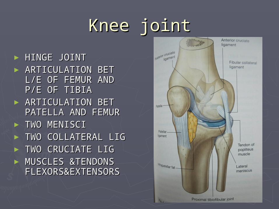
Knee jointKnee joint
► HINGE JOINTHINGE JOINT► ARTICULATION BET L/E ARTICULATION BET L/E
OF FEMUR AND P/E OF OF FEMUR AND P/E OF TIBIATIBIA
► ARTICULATION BET ARTICULATION BET PATELLA AND FEMURPATELLA AND FEMUR
► TWO MENISCITWO MENISCI► TWO COLLATERAL LIGTWO COLLATERAL LIG► TWO CRUCIATE LIGTWO CRUCIATE LIG► MUSCLES &TENDONS MUSCLES &TENDONS
FLEXORS&EXTENSORSFLEXORS&EXTENSORS
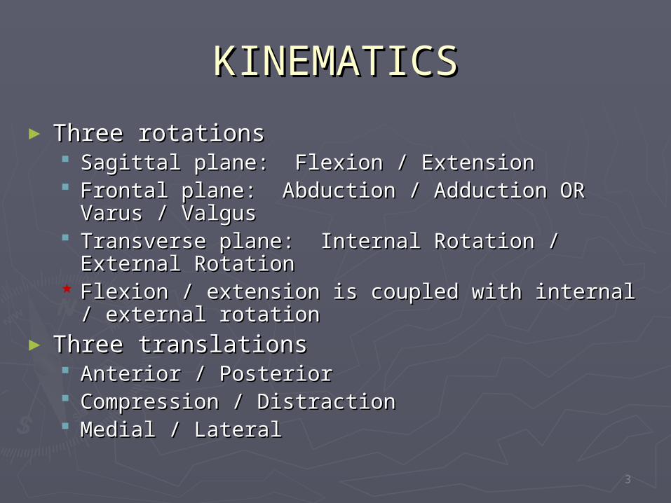
KINEMATICSKINEMATICS
► Three rotationsThree rotations Sagittal plane: Flexion / ExtensionSagittal plane: Flexion / Extension Frontal plane: Abduction / Adduction OR Varus / Frontal plane: Abduction / Adduction OR Varus /
ValgusValgus Transverse plane: Internal Rotation / External Transverse plane: Internal Rotation / External
RotationRotation Flexion / extension is coupled with internal / Flexion / extension is coupled with internal /
external rotationexternal rotation► Three translationsThree translations
Anterior / PosteriorAnterior / Posterior Compression / DistractionCompression / Distraction Medial / LateralMedial / Lateral
3
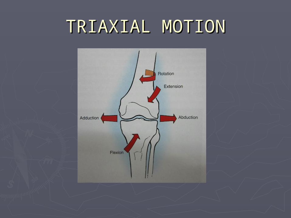
TRIAXIAL MOTIONTRIAXIAL MOTION
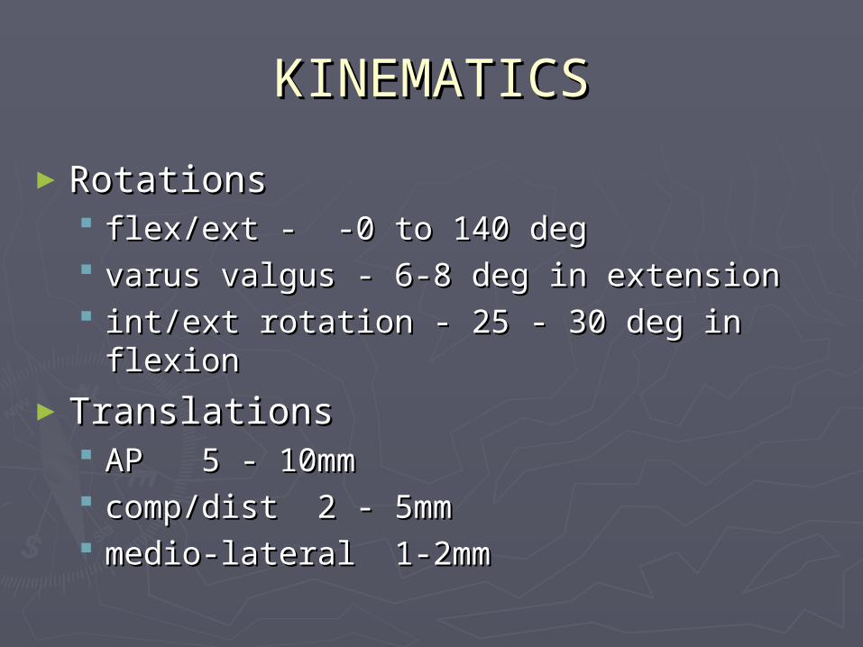
KINEMATICSKINEMATICS
►RotationsRotations flex/ext - -0 to 140 degflex/ext - -0 to 140 deg varus valgus - 6-8 deg in extensionvarus valgus - 6-8 deg in extension int/ext rotation - 25 - 30 deg in flexionint/ext rotation - 25 - 30 deg in flexion
►TranslationsTranslations AP 5 - 10mmAP 5 - 10mm comp/dist 2 - 5mmcomp/dist 2 - 5mm medio-lateral 1-2mm medio-lateral 1-2mm
FLEXION FLEXION
►During flexion of knee an average of During flexion of knee an average of 2mm and 21mm of posterior 2mm and 21mm of posterior translation of medial and laterel fem translation of medial and laterel fem condyles occurs respectively.condyles occurs respectively.
►This medially based pivoting of the This medially based pivoting of the knee with ext rot of tibia on femur knee with ext rot of tibia on femur during extension is called SCREW during extension is called SCREW HOME MECHANISM.HOME MECHANISM.
FLEXION OF KNEE FLEXION OF KNEE
►Swing phase -67 dSwing phase -67 d►Ascending stairs -83 dAscending stairs -83 d►Descending stairs -90 dDescending stairs -90 d►Arising from chair -93 dArising from chair -93 d
GAIT CYCLEGAIT CYCLE
► Just prior to heel strike - max Just prior to heel strike - max extension & max external rotationextension & max external rotation
►heel strike - max valgusheel strike - max valgus►Foot flat - flexion & internal rotation Foot flat - flexion & internal rotation
progressprogress►swing phase - internal rotation swing phase - internal rotation
continues, max flexion, max anterior continues, max flexion, max anterior translation.translation.
AXIS OF LOWER LIMBAXIS OF LOWER LIMB
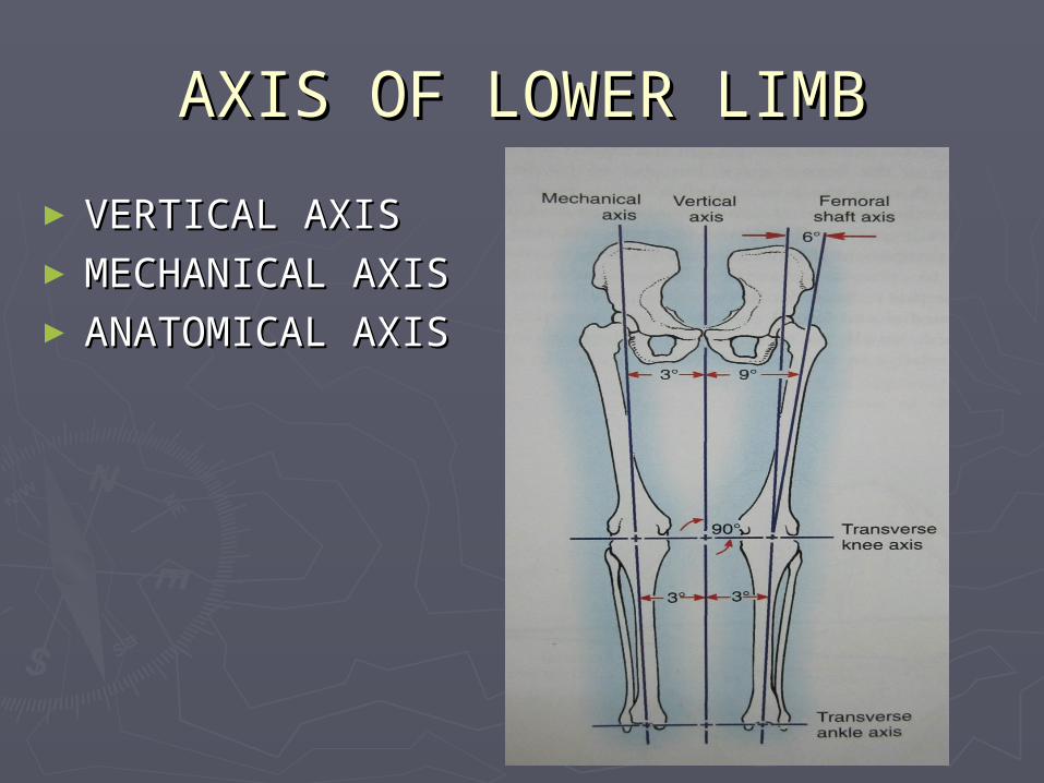
► VERTICAL AXISVERTICAL AXIS►MECHANICAL AXISMECHANICAL AXIS► ANATOMICAL AXISANATOMICAL AXIS
MECHANICAL AXISMECHANICAL AXIS►Line drawn on a standing long leg AP Line drawn on a standing long leg AP
radiograph from the centre of radiograph from the centre of femoral head to centre of talar dome.femoral head to centre of talar dome.
►This projects through the centre of This projects through the centre of the knee joint described as NEUTRAL the knee joint described as NEUTRAL mech axismech axis
►Mech axis lateral to knee centre-Mech axis lateral to knee centre-valgus deformity.valgus deformity.
►Mech axis medial to knee centre-Mech axis medial to knee centre-varus defvarus def
►Mech axis of LL is in 3 deg of valgus from Mech axis of LL is in 3 deg of valgus from vertical axis of body.vertical axis of body.
► Anat axis of femur is in 6 deg of valgus Anat axis of femur is in 6 deg of valgus from mech axis of LL and 9 deg of valgus from mech axis of LL and 9 deg of valgus from true vertical axis of body.from true vertical axis of body.
► Anat axis of tibia lies in 3 deg of varus Anat axis of tibia lies in 3 deg of varus from vertical axis of body.from vertical axis of body.
► By determining the tibial mechanical axis By determining the tibial mechanical axis using the center of the tibial plateau and using the center of the tibial plateau and the femoral mechanical axis using the the femoral mechanical axis using the center of the intercondylar notch, any center of the intercondylar notch, any medial or lateral subluxation through the medial or lateral subluxation through the knee joint is disregarded. knee joint is disregarded.
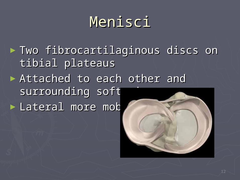
MenisciMenisci
►Two fibrocartilaginous discs on tibial Two fibrocartilaginous discs on tibial plateausplateaus
►Attached to each other and Attached to each other and surrounding soft tissuesurrounding soft tissue
►Lateral more mobileLateral more mobile
12
Function of menisciFunction of menisci
► StabilityStability Increases congruency by deepening the Increases congruency by deepening the
tibial plateautibial plateau Distribute loading and act as “shock Distribute loading and act as “shock
absorbers”absorbers”► MobilityMobility
Decreases frictionDecreases friction Must be mobile to avoid injuryMust be mobile to avoid injury
13
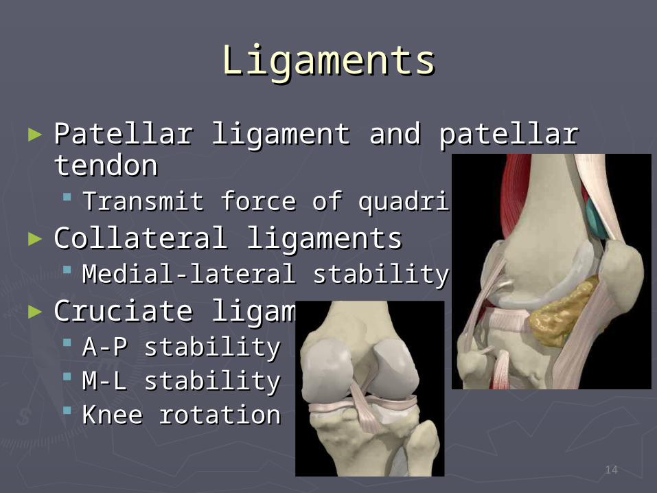
LigamentsLigaments
►Patellar ligament and patellar tendonPatellar ligament and patellar tendon Transmit force of quadricepsTransmit force of quadriceps
►Collateral ligamentsCollateral ligaments Medial-lateral stabilityMedial-lateral stability
►Cruciate ligamentsCruciate ligaments A-P stabilityA-P stability M-L stabilityM-L stability Knee rotationKnee rotation 1
14
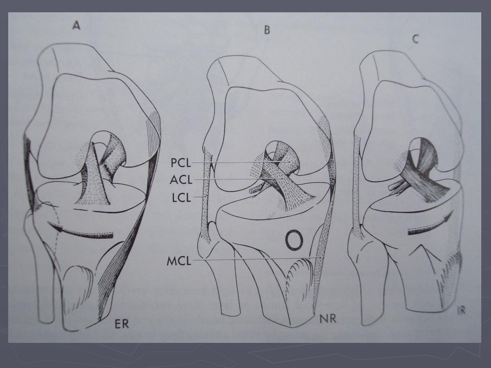
► MCLMCL Primary valgus restraint -57-78% Primary valgus restraint -57-78%
restraining moment of kneerestraining moment of knee Attached from med epicondyle to Attached from med epicondyle to
anterolat surface of tibia above&behind anterolat surface of tibia above&behind pes anserinuspes anserinus
Lax in flexionLax in flexion
►LCL LCL Primary varus restraintPrimary varus restraint Attached from lateral epicondyle to lat Attached from lateral epicondyle to lat
surface on head of fibula surface on head of fibula lax in flexionlax in flexion
CruciatesCruciates
ACLACL Primary static restraint to anterior Primary static restraint to anterior
displacement of tibia relative to femurdisplacement of tibia relative to femur secondary restraint on tibial rotation and secondary restraint on tibial rotation and
varus-valgus angulation at full extension. varus-valgus angulation at full extension. anteromedial band is tight in flexion, anteromedial band is tight in flexion,
providing the primary restraint, whereas providing the primary restraint, whereas the posterolateral bulky portion of this the posterolateral bulky portion of this ligament is tight in extension. ligament is tight in extension.
PCLPCL
Primary restraint to posterior displacement Primary restraint to posterior displacement it acts as a check of hyperextension only after it acts as a check of hyperextension only after
the anterior cruciate ligament has been ruptured. the anterior cruciate ligament has been ruptured. relaxed in extension, tense in flexionrelaxed in extension, tense in flexion It appears to guide the “screw-home” It appears to guide the “screw-home”
mechanism on internal rotation of the femur mechanism on internal rotation of the femur during terminal extension of the knee during terminal extension of the knee
Rotational stability is altered in flexion after the Rotational stability is altered in flexion after the posterior cruciate is cut. posterior cruciate is cut.
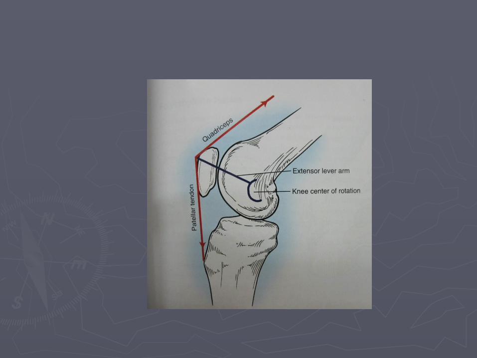
PATELLOFEMORAL JOINTPATELLOFEMORAL JOINT► Primary function of the patella is to Primary function of the patella is to
increase the lever arm of the extensor increase the lever arm of the extensor mechanism around the knee, improving the mechanism around the knee, improving the efficiency of quadriceps contraction. efficiency of quadriceps contraction.
► Displacement or lengthening of the Displacement or lengthening of the extensor lever arm changes throughout the extensor lever arm changes throughout the arc of knee motion. arc of knee motion.
► Length of the lever arm depends on Length of the lever arm depends on trochlea geometry, patellofemoral contact trochlea geometry, patellofemoral contact area and the varying center of rotation of area and the varying center of rotation of the knee. the knee.
► the extensor lever arm is greatest at 20 the extensor lever arm is greatest at 20 degrees of flexion, and the quadriceps degrees of flexion, and the quadriceps force required for knee extension increases force required for knee extension increases significantly in the last 20 degrees of significantly in the last 20 degrees of extension extension
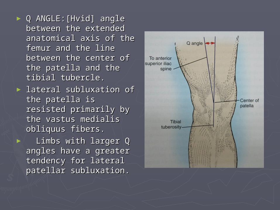
► Q ANGLE:[Hvid] angle Q ANGLE:[Hvid] angle between the extended between the extended anatomical axis of the anatomical axis of the femur and the line femur and the line between the center of between the center of the patella and the the patella and the tibial tubercle.tibial tubercle.
► lateral subluxation of lateral subluxation of the patella is resisted the patella is resisted primarily by the vastus primarily by the vastus medialis obliquus medialis obliquus fibers.fibers.
► Limbs with larger Q Limbs with larger Q angles have a greater angles have a greater tendency for lateral tendency for lateral patellar subluxation. patellar subluxation.
► As patella transmits the force of contraction As patella transmits the force of contraction of the quadriceps muscle to the patellar of the quadriceps muscle to the patellar tendon around a variably flexed knee,it tendon around a variably flexed knee,it experiences a joint reaction force[JRF] as the experiences a joint reaction force[JRF] as the trochlea opposes its posterior displacement. trochlea opposes its posterior displacement.
► JRF depends on the angle of knee flexion and JRF depends on the angle of knee flexion and the magnitude of the forces transmitted to the magnitude of the forces transmitted to the patella from the quadriceps and patellar the patella from the quadriceps and patellar tendons tendons
►During standing, the JRF increases with During standing, the JRF increases with increasing knee flexion as the force vectors increasing knee flexion as the force vectors of the quadriceps and patellar tendons of the quadriceps and patellar tendons become more parallel to the joint reaction become more parallel to the joint reaction force.force.
►During squatting to 120 degrees of knee During squatting to 120 degrees of knee flexion, the joint reaction force may be seven flexion, the joint reaction force may be seven to eight times body weight. to eight times body weight.
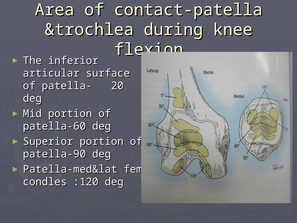
Area of contact-patella Area of contact-patella &trochlea during knee flexion&trochlea during knee flexion
► The inferior articular The inferior articular surface of patella- surface of patella- 20 deg20 deg
►Mid portion of Mid portion of patella-60 deg patella-60 deg
► Superior portion of Superior portion of patella-90 degpatella-90 deg
► Patella-med&lat fem Patella-med&lat fem condles :120 degcondles :120 deg

THANK YOUTHANK YOU