Embed Size (px)
Citation preview
T.R.N.C
NEAR EAST UNIVERSITY
INSTITUTE OF HEALTH SCIENCES
Knowledge and Practices of Electrocardiogram
Interpretation of Nurses
Obaydah Yaser Hamed Tahboub
In Partial Fulfilment of the Requirements for the
Degree of
Master of Nursing (Adult Acute Care)
NICOSIA 2018
T.R.N.C
NEAR EAST UNIVERSITY
INSTITUTE OF HEALTH SCIENCES
Knowledge and Practices of Electrocardiogram
Interpretation of Nurses
Obaydah Yaser Hamed Tahboub
In Partial Fulfilment of the Requirements for the
Degree of
Master of Nursing (Adult Acute Care)
Advisor: Assoc. Prof. Ümran Dal Yılmaz
NICOSIA 2018
I
APPROVAL
The Directorate of Graduate School of Health Sciences, this study has been accepted by the
thesis committee in nursing program as a master of Adult Acute Care nursing thesis.
Thesis Defence Date: 2018-08-08
Jury Members Signature
Supervisor: Assoc. Prof. Ümran Dal YILMAZ (Near East University) ............................
Chair: Assoc. Prof. Hatice Bebiş (Near East University) ..................................................
Member: Assist. Prof. Gülten Sucu Dağ (Eastern Mediterranean University) ..................
Approval:
According to the relevant article of the Near East University Postgraduate Study-Education
and Examination Regulation, this thesis has been approved by the above-Mentioned members
of the thesis committee and the decision of the board of Directors of the Institute.
Prof. Dr. Hüsnü Can Başer
Director of Graduate Institute of Health Sciences
II
DECLARATION
I hereby declare that the work in this thesis entitled “Knowledge and Practices of
Electrocardiogram Interpretation of Nurses” is the study of my own research efforts
undertaken under the supervision of Assoc. Prof. Ümran Dal Yılmaz.
My deepest thanks to Assoc. Prof. Ümran Dal Yılmaz, my supervisor, for her expertise, on-
going support and mentorship during my research.
I express my profound gratitude to my parents, brothers and my sister for their support,
constant encouragement through all my years of study and my life.
A special thankful to my friend Barış Yalçın my home mate, work mate and life mate here in
North Cyprus.
Also a special thankful to cardiology department and everyone working in Dr. Suat Günsel
Girne University Hospital for their supporting to me from first day working in hospital.
III
Knowledge and Practices of Electrocardiogram Interpretation of Nurses
ABSTRACT
Introduction: Electrocardiogram (ECG) interpretation for nurses is an important of the initial
evaluation for patients presenting with cardiac problems which maybe cause to potentially life
threatening complications. Nurse usually the first one who faces ECG interpretation which
made him be more creative to take appropriate dissection to save a life specially in critical area
in hospital. Which there is a need to increase knowledge and practice of nurses on ECG
interpretation to provide right intervention and avoid complications.
Objectives: The aim of the study is to determine the knowledge and practice of
electrocardiogram among nurses.
Methods: This descriptive study was conducted on the registered nurses who work in both
Near East university hospital and Dr. Suat Günsel Girne university hospital. Total 65 voluntary
nurses were composed the sample of the study. A questionnaire that was developed by the
researchers on the basis of the literature was used as data collection tool in this study. Data
were collected using a questionnaire in May 2018, after the ethical approval. Descriptive
statistics and Pearson Chi-Square tests were used in analysis of the data.
Results: Results of the present study showed high level of knowledge and practice of
electrocardiogram among nurses. Were statistically significant differences in terms of working
unit in hospital and previous ECG training courses affected and play important role in defining
the proffesinality of nurses to had experience in ECG interpretation.
Conclusions: Working unit in hospital and previous ECG training courses play important role
in defining the professionalism of nurses to had experience in ECG interpretation. Training
courses for nurses under the supervision of qualified well trained staff especially for nurses
who work in critical area in hospital with continuing self learning and staying up to date to any
changing and development of new protocols or technology to increase patient outcomes.
Keywords: Electrocardiogram, electrocardiogram interpretation, nursing, knowledge and
practices.

IV
List of Content
APPROVAL ............................................................................................................................ I
DECLARATION ................................................................................................................... II
ABSTRACT .......................................................................................................................... III
1. INTRODUCTION ............................................................................................................. 1
1.1. Problem Definition ........................................................................................................... 1
1.2. Aim of the Study .............................................................................................................. 4
2. BACKGROUND OF THE STUDY ................................................................................. 5
2.1. Definition of Electrocardiogram (ECG)............................................................................ 5
2.2. Coronary Artery Disease (CAD) ...................................................................................... 6
2.2.1. Stable angina .................................................................................................................. 7
2.2.2. Unstable angina .............................................................................................................. 7
2.2.3. Non-ST segment elevation myocardial infarction or heart attack (NSTEMI) ............... 8
2.2.4. ST segment elevation myocardial infarction or heart attack (STEMI) ….......…........... 8
2.3. Heart arrhythmias .............................................................................................................. 9
2.3.1. Atrial arrhythmias .......................................................................................................... 9
2.3.2. Ventricular arrhythmias ............................................................................................... 11
2.4. Importance of first interpretation of ECG ....................................................................... 12
2.5. Nursing considerations and responsibility about ECG ................................................... 12
2.6. Why study is important ................................................................................................... 13
3. METHODOLOGY ........................................................................................................... 14
3.1. Study Design ................................................................................................................... 14
3.2. Study Setting ................................................................................................................... 14
3.3. Sample Selection ..............................................................................................................15
3.4. Study Tools ..................................................................................................................... 15
3.5. Pilot Study ....................................................................................................................... 15
3.6. Data Collection ................................................................................................................ 16
3.7. Ethical Aspect ................................................................................................................. 16
3.8. Data Analysis ................................................................................................................... 16

V
4. RESULTS .......................................................................................................................... 17
5. DISCUSSION ................................................................................................................... 27
6. CONCLUSION ................................................................................................................. 30
7. FINDINGS AND RECOMMENDATIONS ................................................................... 30
7.1. Findings ........................................................................................................................... 30
7.2. Recommendations ........................................................................................................... 31
8. REFERENCES ................................................................................................................. 32
VI
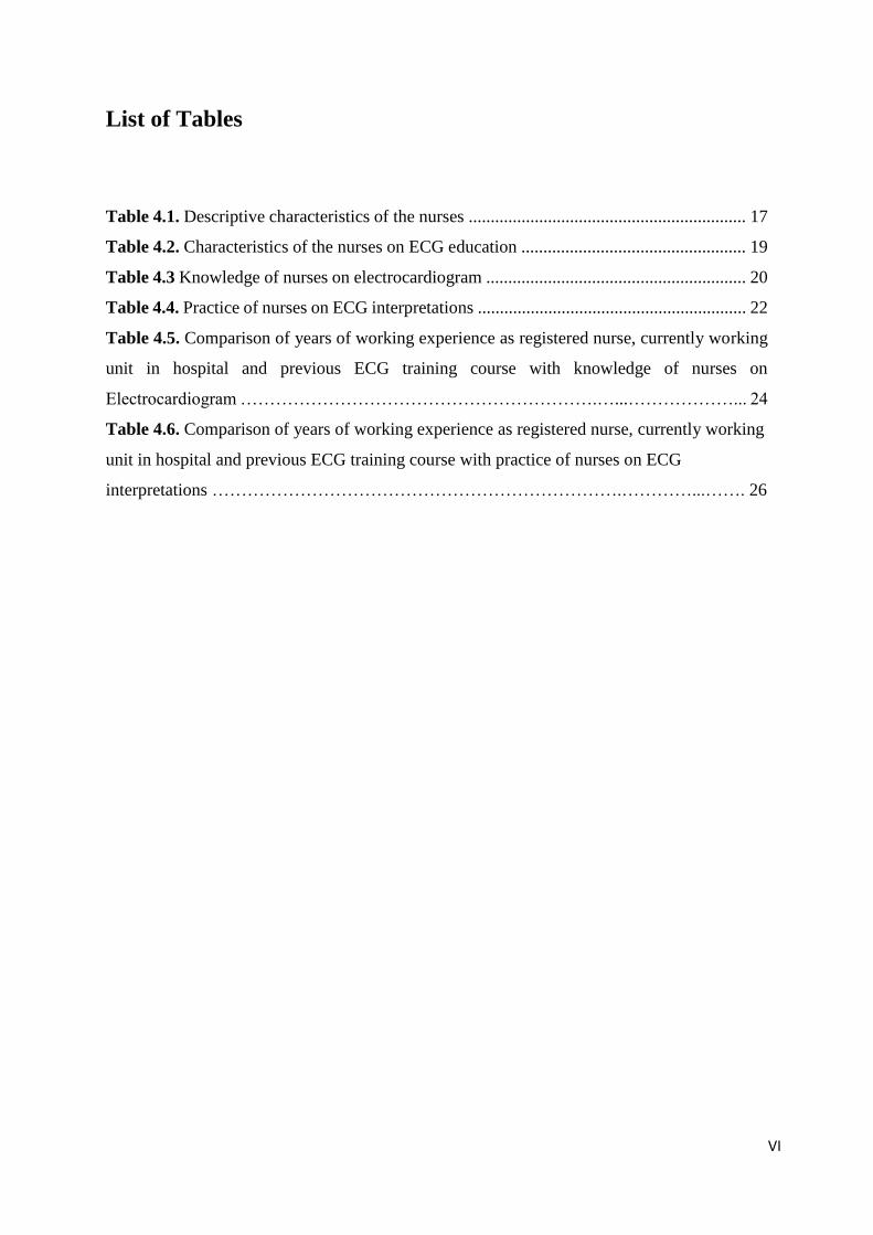
List of Tables
Table 4.1. Descriptive characteristics of the nurses ............................................................... 17
Table 4.2. Characteristics of the nurses on ECG education ................................................... 19
Table 4.3 Knowledge of nurses on electrocardiogram ........................................................... 20
Table 4.4. Practice of nurses on ECG interpretations ............................................................. 22
Table 4.5. Comparison of years of working experience as registered nurse, currently working
unit in hospital and previous ECG training course with knowledge of nurses on
Electrocardiogram …………………………………………………….…...………………... 24
Table 4.6. Comparison of years of working experience as registered nurse, currently working
unit in hospital and previous ECG training course with practice of nurses on ECG
interpretations …………………………………………………………….…………...……. 26

VII
List of Figure
Figure 4.1 Knowledge of nurses on Electrocardiogram ……………….…………………. 21
Figure 4.2 Practice of nurses on ECG interpretations ……………………………………. 23
VIII
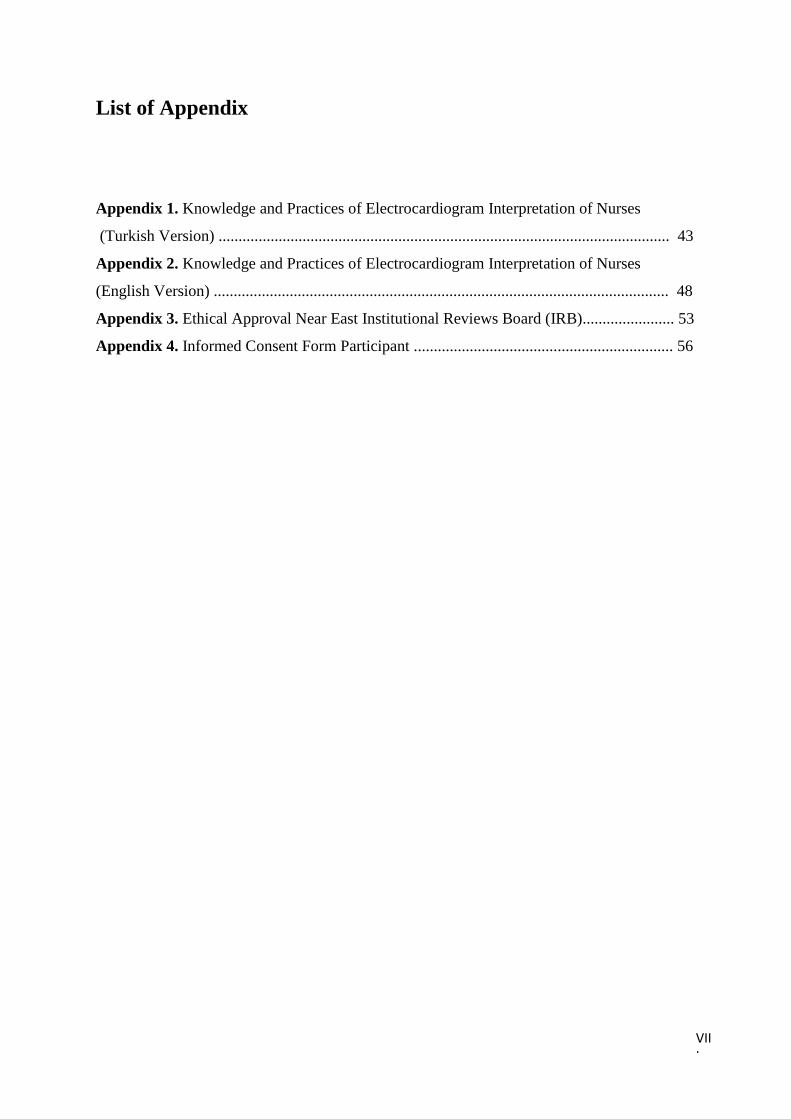
List of Appendix
Appendix 1. Knowledge and Practices of Electrocardiogram Interpretation of Nurses
(Turkish Version) ................................................................................................................. 43
Appendix 2. Knowledge and Practices of Electrocardiogram Interpretation of Nurses
(English Version) .................................................................................................................. 48
Appendix 3. Ethical Approval Near East Institutional Reviews Board (IRB)....................... 53
Appendix 4. Informed Consent Form Participant ................................................................. 56
IX
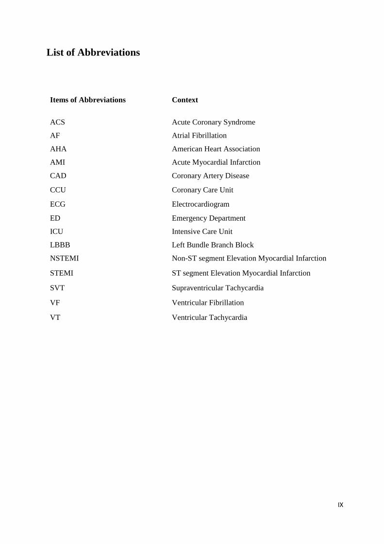
List of Abbreviations
Items of Abbreviations
Context
ACS Acute Coronary Syndrome
AF Atrial Fibrillation
AHA American Heart Association
AMI Acute Myocardial Infarction
CAD Coronary Artery Disease
CCU Coronary Care Unit
ECG Electrocardiogram
ED Emergency Department
ICU Intensive Care Unit
LBBB Left Bundle Branch Block
NSTEMI Non-ST segment Elevation Myocardial Infarction
STEMI ST segment Elevation Myocardial Infarction
SVT Supraventricular Tachycardia
VF Ventricular Fibrillation
VT Ventricular Tachycardia

1
1. INTRODUCTION
1.1 Problem Definition
Electrocardiography is a commonly used, non-invasive procedure for recording
electrical changes in the heart. The record,which is called an electrocardiogram (ECG),
shows the series of waves that relate to the electrical impulses which occur during each beat
of the heart. Nowadays ECG is an important of the initial evaluation for patients presenting
with cardiac problems, and its considered as the first diagnostic tool in chest pain and it
provide objective information’s about the structure and function of the heart (AlGhatrif
et al 2012). Also ECG make a focal point of modern medicine because it gives hole
background about diagnosing acute coronary syndromes and cardiac arrhythmias (George et al
2010).
Globally ischaemic heart disease and stroke takes the first place in the top 10 causes of
death worldwide which it accounting for a combined 15 million deaths in 2015 (WHO, January
2017). A previous studies show that most of patients coming to emergency department
suffering from symptoms of chest pain, one of these studies show that ECG done to 88.4% of
patient who comes to emergency and discussed that 59% of patients the most frequent
diagnoses for them were atypical chest pain (Martínez-Sellés, et al 2008).
In health care centres nurses are usually the first responders to an in-hospital cardiac arrest
and they must be master in basic resuscitation skills (Hernández-Padilla et al 2016). Every
nurse should be able to recognize basic ECG rhythms, such as normal sinus rhythm, sinus
tachycardia, sinus bradycardia, atrial fibrillation, atrial flutter, heart blocks, ventricular
fibrillation and asystole (Atwood et al 2015). This is required that the nurse must be responsible
for monitoring and clinical decision-making based on information obtained from the monitor
(Funk et al 2017).

2
Doğan and Dilek (2012) in their study stated that only 38.1% of the nurses were found to
be able to identify the ventricular fibrillation, 54,3% myocardial infarction, 33,3% third degree
atrioventricular block, 40,5% ventricular tachycardia. However, 20.5% of the nurses stated that
they could carry out defibrillation. 60,5% of the nurses expressed that they did not know the
right electrocardiography monitoring and thus could not recognize the type of the arrhythmia.
In Iraq study that was carried out to identify nurses’ knowledge concerning early
intervention for patients with ventricular tachycardia at Baghdad teaching hospitals which
show result of overall assessment of the studied sample’s knowledge was low (Mousa et al
2016). That led to a lot of responsibility on nurses to be qualified in continuous monitoring
especially in critical care units, to ensure ongoing safe and effective ECG monitoring and to
know the determine what courses needed and quality improvement program should be initiated
(Drew et al 2006). Also result of inappropriate interpretation use increases the cost of
healthcare and can delay the admission process an unpleasant burden which the hospital and
its patients must bear (Larson et al 2008).
In England it was established what called a Rapid Access Chest Pain Clinic (RACPC) that
made nurses to assess the patient and to form a care plan immediately without waiting for a
physician, and also when a patient with chest pain arrives in the clinic, an ECG recorded and
the nurse consultant then examines the patient and decides if further investigation is required
(Pottle, 2005).
Many methods had made and designed algorithms like CRISP (Cardiac Rhythm
Identification for Simple People) method to help nurses rapidly interpret ECGs that nurses
often confusing with identifying ECG rhythms (Atwood et al 2015). Also American Heart
Association designed what’s called PULSE trial (Practical Use of the Latest Standards for
Electrocardiography trial) which is online ECG monitoring education program and strategies
to implement and sustain change in practice, led by nurse champions on each unit and this trial
increase nurses’ knowledge of ECG monitoring, quality of care related to ECG monitoring and
patient outcomes (Funk et al 2010).
3
Facility work area affected the knowledge and practice toward ECG, one study done to
measured the effectiveness of an education program on nurses’ knowledge of ECG
interpretation which explain that pre test result of education show test scores of nurses in the
cardiology department were higher than those in the emergency department (ED) and intensive
care unit (ICU) (Zhang et al 2013). In other hand, study in Iraq show that most nurses worked
in coronary care unit (CCU), ICU and ED pass the questions regarding knowledge except
question concerning ECG changes regarding to new and old myocardial infarction which mean
that there is weakness in practical section which need intensive training courses (A. AL-
Husaunawy 2015).
The educated and qualified nurse play important role in preventing complication that
maybe occurred in patients who have arrhythmias, difficulties in interpreting the ECG patients
with Acute Myocardial Infarction (AMI) who present with Left Bundle Branch Block (LBBB)
may delay treatment and affects their prognosis (Spiers 2007). Previous studies (Fuenzalida et
al 2015) show that an educational intervention by nurses at discharge from the ED decreased
atrial fibrillation (AF) related complications at 3-month follow-up, one of these studies done
in tertiary hospital in Barcelona which also emphasise this result but for long time up to one-
year of follow-up (Fuenzalida et al 2017).
Also as known training and education play important role in being qualified in
interpretation ECG, one study held in CCU and ICU of Benha University hospital in Egypt that
show in it result improvement of nurse’s performance about recording a 12-lead ECG and
dysrhythmia interpretation after program implementation and the nurse's performance scores
were satisfactory (Refaey, 2012). In additional another study conducted in the southeast of
Spain which evaluated emergency nurse competence in ECG interpretation this study shows
that training within the previous 5 years have high score and level of knowledge was not
influenced by experience or hospital (Coll-Badell et al 2017).
4
The nurse working in ICU and ED of their patients’ critical conditions, are highly
responsible of determination and application of attempts as well as early detection of
diagnosing the signs and symptoms of their disease.
There is need to increase knowledge and practice of nurses on electrocardiogram to
provide health care outcomes for the patient and avoid any mistake in ECG interpretation and
interventions. Determination of knowledge and practice of nurses on electrocardiogram maybe
useful in improvement their level and enhance their initiation to become more professional in
interpretation ECG. However, no previous studies found about this subject in Turkish Republic
of North Cyprus.
1.2 Aim of the Study
The aim of the study is to determine the knowledge and practice of electrocardiogram
among nurses in University Hospital in North Cyprus. Questionnaire includes the following
questions:
1. What are knowledge of nurses on Electrocardiogram?
2. What are practices of nurses on ECG interpretations?
3. Are there any differences between descriptive characteristics and knowledge and the practice
of nursing on ECG interpretation?
5
2. BACKGROUND OF THE STUDY
2.1. Definition of Electrocardiogram
Electrocardiogram (ECG) is a procedure of recording the electrical activity of
the heart within a period of time using electrodes placed over chest. As known that heart has
four chambers two atria and two ventricles, the electrical discharge generated from sinoatrial
node (SA node) and move through atrial muscle fibres delay while the depolarization to
atrioventricular node (AV node) then it gone through out bundle of his to the right and left
bundle branch among purkinje fibres, The contraction of atria associated with the ECG wave
called P wave then when ventricular are depolarization it causes QRS complex finally when
ventricular repolarization T wave occurs (Hampton, 2008).
The standard 12-lead, ten electrodes are placed on the patient's limbs and on the surface of
the chest which records the electrical activity of the heart from 12 different viewpoints or leads
by attaching cables to the patient’s limbs and chest so the overall magnitude and direction of
the heart's electrical depolarization is captured at each moment throughout the cardiac cycle
(Jevon, 2009). It is important that a 12-lead ECG is recorded accurately because poor technique
can lead to misinterpretation of the results, mistaken diagnosis, mismanagement of the patient
and inappropriate transfer to hospital (Jevon, 2010).
ECG plays an important role in diagnosing of patients and providing whole information in
many clinical scenarios for examples; Arrhythmias, Coronary artery disease (CAD),
Electrolytes abnormalities, inherited cardiomyopathies and drug induced abnormalities
(Huitema et al 2014).
6
2.2 Coronary Artery Disease (CAD)
Coronary Artery Disease (CAD) is a blockage or narrowing of the coronary arteries
usually caused by atherosclerosis (Libby et al 2005) which may restrict flow of the blood to
the heart muscle by physically blocking the artery or by causing abnormal artery function and
tone (Cleveland clinic, June 2017). CAD is the most common cardiovascular disease
(Montalescot et al 2014). Which was in 2013 the most common cause of death globally
resulting in 8.14 million deaths (16.8%) up from 5.74 million deaths (12%) in 1990 (GBD
2015).
American heart association (AHA) statistical update show that the prevalence total of
11.5% of American adults (27.6 million) have been diagnosed with heart disease and mortality
in every year since 1919, CAD accounted for more deaths than any other major cause of death
in the United States (Benjamin et al 2017). Among Indian women with CAD continues to be a
major public health problem that represents a leading cause of death and disability (Pathak et
al 2017). Also the presence of diabetes, hypertension, low levels of high-density lipoprotein,
high levels of total cholesterol, low-density lipoprotein and triglycerides all are correlated with
CAD (Gupta et al 1999).
Almost two-thirds of heart failure cases are attributed to underlying CAD (Gheorghiade
et al 2006). Also CAD was a factor for heart failure in more than 50% of incident cases in
North America and Europe; 30% to 40% in Asia, Latin America, and the Caribbean; and less
than 10% in sub-Saharan Africa (Khatibzadeh et al 2012).

7
2.2.1 Stable angina
Stable angina result in chest pain during exertional activity while heart muscle doesn't get
as much blood as it needs which resolves with rest or sublingual administration of nitro-
glycerine (Tobin et al 2010). Angina reflects transient regional myocardial ischaemia caused
by inadequate coronary perfusion which The most common cause is atherosclerotic (Abrams J
2005).
Angina which increases prevalence with age for men 4-7% of who aged 45-64 to 12-14%
aged 65-84 and women 5-7% of who aged 45-64 to 10-12% aged 65-84 (Montalescot et al
2013). While there is is estimated that approximately 9 million patients in the USA suffer from
angina (Mensah et al 2007).
Angina clinical symptoms characterized by heaviness, tightness, pressure or burning across
the chest and may radiate to the jaw, arms or back which duration is brief and the sensation
generally resolves with rest or sublingual nitrates (Whittaker et al 2014). ECG maybe have
change if it done during an attack likely to show ST segment depression portends a poorer
prognosis than T-wave inversion alone or no ECG changes (Bhatheja et al 2007).
2.2.2 Unstable angina
Unstable angina is characterized by the clinical presentation of angina with or without
ischemic ECG changes (Bhatheja et al 2007). In unstable angina the chest pain usually is more
severe and longer lasting may occur at rest or at a lower level of physical exertion (Braunwald
et al 2002). The diagnosis of unstable angina still a clinical challenge and reflects one end of
the chain of Acute coronary syndrome (ACS) (Manning et al 2007)
ECG findings during an episode of unstable angina include ST depression, T wave
inversions or transient ST segment elevations (Fanaroff et al 2015). Medical management of
unstable angina includes therapies aimed at relief pain, inhibition of platelet aggregation and
thrombosis also consideration of revascularization of the stenosis vessels (Amsterdam et al
2014).
8
2.2.3 Non-ST segment elevation myocardial infarction or heart attack (NSTEMI)
NSTEMI is similar to unstable angina but it’s associated with positive biomarkers like
troponin or creatine kinase in the setting of angina (Bhatheja et al 2007). Also ECG changes
complemented by echocardiography, coronary angiography and other imaging modalities
(Basra et al 2016).
NSTEMI typically results from more distally located coronary thrombosis and coronary
arteries blockage often is only partial (Bassand et al 2011). Atherosclerotic changes in the
vessel wall or extent of calcification and subsequent intracoronary thrombus formation may
vary extremely in NSTEMI patients (de Winter et al 2012). European Society of Cardiology
(ESC) guidelines recommend urgent or immediate transfer to the catheterization laboratory for
patients with ongoing signs and symptoms of ischemia and for patients with hemodynamic or
electric instability (Hamm CW et al 2011).
2.2.4 ST segment elevation myocardial infarction or heart attack (STEMI)
STEMI continues to be one of the most dangerous acute complications of coronary artery
disease (Timmis et al 2017). Which need medical emergency and secondary prevention
promptly in order to not only improve the survival but also long-term prognosis of the patient
(Sachdewani et al 2018).
The most common cause is coronary artery narrowing from a disrupted atherosclerotic
plaque with overlay acute thrombosis that suddenly and significantly compromises coronary
blood flow but is usually not 100% occlusive (Anderson et al 2012). Therapeutic measures
focus on reperfusion of the occluded artery by pharmaceutical or by percutaneous coronary
intervention as early as possible (Deckers et al 2013).
9
2.3 Heart arrhythmias
Heart arrhythmias are abnormality or disturbance in the normal activation sequence of the
myocardium and can be classified by rate, mechanism, duration or site of origin (Bhaumik et
al 2016). Also arrhythmias could be indicative of structural heart disease which may lead to
complications like stroke or result in deterioration of hemodynamic and cardiac arrest
(Papadopoulos et al 2017).
ECG has been considered an important diagnostic and prognostic tool for the management
of patients with arrhythmias (Delgado et al 2016). Also evaluation of the ECG during
tachycardia can give us significant evidences regarding the arrhythmia mechanism and even
the arrhythmia site of origin in some particular cases (Cano et al 2017). Now most challenge
problem faced by today’s ECG examination is the huge difference in the morphologies of ECG
signs (Li X et al 2009). As arrhythmias classify by site of origin it might be Atrial arrhythmias,
Junctional arrhythmias, Ventricular arrhythmias and Heart blocks.
2.3.1 Atrial arrhythmias
Atrial arrhythmias are the most frequent rhythm disorder in humans and often lead to
severe complications such as heart failure and stroke (Virag et al 2002). Which consider those
arising from the atria like: sinus tachycardia and bradycardia, atrial fibrillation, atrial flutter,
and atrial tachycardia.
Atrial fibrillation (AF) is the most common cardiac arrhythmia (Kirchhof et al 2016). It is
estimated that approximately 7 million of patients in United States and European Union suffer
from AF (Fuster et al 2011). Up to 15% of all strokes in the United States can be attributable
this disorder (Rockson et al 2004). Unluckily these numbers maybe multiply by at least 2.5-
fold by 2050 (Alan S. Go et al 2001).

10
Patients with AF presented a lot of common symptoms include chest pain, shortness of
breath, tachycardia, palpitations, fatigue, anxiety, decreased activity tolerance heart failure,
light headedness, hypotension, stroke, and arterial embolization (Dewar et al 2007). Also
patients with AF the overall incidence of coronary artery disease is relatively high
(Michniewicz et al 2018)
The risk of stroke is significant in the patient with AF (Fuster et al 2011). So initial
assessment must be focused on determination of stability and prompt intervention (Cottrell et
al 2008). Medical treatment advanced Before cardioversion which can be used administration
of drugs such as amiodarone, ibutilide, propafenone flecainide or sotalol as there is evidence
of enhancement of the success of cardioversion and decreased recurrence of AF (Fuster et al
2011).
Medical treatment is often focused on control of heart rate and anticoagulation unless the
patient has significant symptoms. Controversy has existed for some time about rate control
versus rhythm control as the primary approach to treatment (Nattel et al 2006). Also conversion
of AF to normal sinus rhythm has been shown to decrease symptoms (January CT et al 2014).
Supraventricular tachycardia (SVT) is a clinical syndrome characterized by a rapid
tachycardia with an abrupt onset and termination (Badhwar N 2010). Presentation of SVT on
ECG is as a narrow-QRS-complex tachycardia which QRS interval of less than 120
milliseconds (Delacrétaz E 2006). SVT originate from supraventricular tissue or require it to
be a part of the re-entrant circuit which usually regular with heart rate of 160 to 200 beats per
minute (Lee et al 2008).
SVT account for approximately 50 000 patients visits ED each year (Huang et al 2013).
Patients with SVT present symptoms like palpitations, chest pain, Shortness of breath, syncope
and sudden cardiac death (Cheng et al 2000). Vagal maneuvers and adenosine are
recommended for treatment patients with regular SVT also synchronized cardioversion is
recommended for acute treatment in patients with hemodynamically stable SVT when
pharmacological therapy is ineffective or contraindicated (Page et al 2016).
11
2.3.2 Ventricular Arrhythmias
Ventricular arrhythmias cover a large spectrum of ventricular myocardium rhythm
disturbances, extending from premature ventricular complexes (PVCs) to ventricular
tachycardia (VT) and ventricular fibrillation (VF) (Papadopoulos et al 2017). Sustained
ventricular arrhythmias are an important cause of morbidity and the most common cause of
sudden cardiac death which accounting for 75–80% of cases (Zipes et al 2006). Risk increase
for sudden cardiac death or ventricular arrhythmias after AMI has been associated with scar
burden (Bello et al 2005).
Ventricular tachycardia is a heart rate more than 100 beats per min with three or more
consecutive beats originating from the ventricles which independent of atrial or atrioventricular
(AV) nodal conduction QRS more than 120 ms on ECG (Harris et al 2015). VT can be
classified based on its morphology which monomorphic VT has a single QRS morphology
suggesting a stable structural focus while polymorphic VT presents with continuously
changing QRS configuration from a more global activation sequence (Hunter et al 2008). Most
patients with VT receive an implantable cardioverter defibrillator (ICD) for the prevention of
sudden cardiac death and antiarrhythmic therapy which can prevent VT recurrence but does
not reduce mortality also catheter ablation is useful in preventing VT recurrence (Roberts et al
2011).
Ventricular fibrillation is a life-threatening with marked variability in cycle length and
morphology up to 300 beats per min with loss of cardiac output (Harris et al 2015). VF annual
incidence of 12.1 per 100,000 people which remains the leading cause of sudden cardiac death
and out-of-hospital cardiac arrest (Benjamin et al 2017). Current management guidelines for
VF emphasize the importance of early high-quality cardiopulmonary resuscitation (CPR) and
defibrillation and antiarrhythmic medications (Link et al 2015).
12
2.4 Importance of first interpretation of ECG
ECG interpretation is an important clinical skill as it allows rapid diagnosis of potentially
life-threatening arrhythmias and diseases (Salerno et al 2003). Which this interpretation shows
some abnormalities and first indication of ischemia, metabolic disturbance training and an
attention to detail to distinguish physiological ECG findings from abnormal ECG findings that
might indicate the presence of cardiac pathology (Sharma et al 2017).
The American College of Cardiology/American Heart Association (ACC/AHA) guidelines
specify that an ECG should be obtained and interpreted within 10 minutes of arrival to the ED
in patients with symptoms suspicious of ACS (Amsterdam et al 2014). Delay in acquiring a
diagnostic ECG to patients with suspected ACS could delay treatment and potentially lead to
increased tissue damage of the heart muscle (NHLBI, January 2015).
2.5 Nursing considerations and responsibility about ECG
Nurses play important role in obtaining, interpreting and communicating ECG findings
which knowledge and skills in detecting any arrhythmia are key factors in determining the
quality of nursing care (Sheiline, M 2008). Nurses must have sufficient knowledge to perform
these responsibilities in ways that increase care and patient outcomes (Pettersen et al 2014).
When applying continuous monitoring ECG to detect deterioration produces a situation
that may in fact subvert the nurse’s ability to provide proper clinical surveillance of patients
conditions (Larson et al 2008). So the nurse’s knowledge and skills in detecting any arrhythmia
on the ECG monitors are important key factors in determining the quality of nursing care
(Shieline 2008). Nurses in acute clinical areas are able to record and interpret 12-lead
electrocardiograms so the treatment can be initiated as soon as possible which leading to better
clinical outcomes for this patient group (Docherty 2003).
13
Also arrhythmias present a unique challenge to the ED nurse as knowledge of the
emergency procedures such as cardioversion and essential medications like patients with atrial
fibrillation is often complex and present with comorbid conditions leading to a myriad of
choices in medical treatment (Cottrell et al 2008).
2.6 Why study is important
ECG interpretation is an important clinical skill as it allows rapid diagnosis of potentially
life-threatening diseases (Salerno et al 2003). Because accuracy of the diagnosis derived from
an ECG tracing is key to patient outcome (Morrison et al 2006). Many hospitalized patients
exhibit signs of clinical deterioration, such as changes in vital signs prior to experiencing
critical events (Gazarian et al., 2010).
As known ECG is not the only diagnostic tool in the diagnosis of related diseases but it’s
benchmark door to balloon time of 90 min is highly dependent on quick and accurate ECG
Interpretation in chest pain cases (Calder, 2008). The national service framework for coronary
heart disease puts guidance on significant aspects of therapy that may make a substantial
difference to patient care which identify and fast-track patients with an ACS so that
thrombolysis or interventional therapy can be applied as soon as possible to decrease
myocardial damage and reduce door-to-needle time (Docherty 2003).
The ECG was and remains the most widely used initial screening test for evaluating
patients with chest pain (Lancia et al 2008). So there is evidence that continuous ECG
monitoring is of use in specific patient populations such as those with ACS and that
sophisticated monitoring parameters such as ST segment monitoring are useful in detecting
cardiac ischemia in those at risk (Drew et al 2004).
So that study so important to increase level of care outcome because most investigators
have observed that the level of knowledge of nursing professionals is lower than desired
(Stephens et al 2007). Also accuracy of the diagnosis derived from an ECG tracing is key to
patient outcome (Morrison et al 2006).

14
3. METHODOLOGY
3.1 Study Design
The study was planned as descriptive design.
3.2 Study Setting:
The study was conducted at the Near East University hospital in Lefkoşa city and at Dr. Suat
Günsel Girne University hospital in Girne city in North Cyprus. The Near East University
hospital the largest and leading university of Cyprus which is located in northern part of
Nicosia, the capital of North Cyprus. The services of Hospital of Near East University 209
private, single patient rooms, 8 operating theatres, 30-bed Intensive Care Unit, 17-bed Neonatal
Intensive Care Unit, an advanced laboratory where a wide array of medical and experimental
tests can be carried out, 22 other labs specializing on certain medical tests.
Dr. Suat Günsel Girne University Hospital, with its high technological equipment have about
150 bed capacity, there are a total of 15 beds in intensive care unit with 2 isolated rooms. These
intensive care units consist of 4 parts: child, cardiovascular surgeon, cardiology and general
intensive care unit. The hospital comprises 3 fully equipped operation theatres of which was
designed especially to carry out cardiac surgeries. There is one delivery unit, a blood bank,
sterilization and dialysis units, an emergency service; biochemistry, microbiology and
pathology labs; radiology, physiotherapy and rehabilitation clinics, cardiac centre, 20
policlinics, nutrition and dietetic and check-up centers, 4 sound-insulated classrooms with 120
people capacity, a restaurant with a capacity to serve up to 200 people, clinic pharmacy, normal
and cold air stores and a call center.
In both hospitals the nurses doing ECG to patients after taking orders from doctors in all
departments and following doctors’ orders in arrhythmias cases, also in both of hospital if any
emergency case happened the senior nurse or supervisor has responsibility to give order to do
ECG and dealing with any arrhythmias if there are no doctors.

15
3.3 Sample Selection:
The study was performed on the register nurses who work in critical care units like: intensive
care unit, coronary care unit, emergency department, recovery department and cardiology
department in both of the Near East University hospital and Dr. Suat Günsel Girne University
hospital. A total of 72 register nurses work in both hospitals. Total 65 voluntary nurses were
composed the sample of the study with 95% access rate.
3.4 Study Tools:
A questionnaire was developed by the researcher on the basis of the literatures were used
as data collection tool in this studies (Ahmed A. AL-Husaunawy 2015; Coll-Badell et al 2017;
Zhang et al 2013).
The questionnaire contains 3 sections:
- The first section regarding for demographics characteristics of nurses which consisted 11
questions.
-The second section consisted 15 questions with 2 choices (True and False) regarding
knowledge of nurses on electrocardiogram.
-Third section regarding practice of nurses on ECG interpretations which consisted 8 questions
with 4 choices (just one of choices is true and others choices are false).
Since all of nurses in both hospitals speaks Turkish; So the questionnaire prepared in Turkish
language and credence after reviewed by 3 specialist nurses’ and one cardiologist.
3.5 Pilot study
A pilot study was performed on ten nurses after approval from the Near East Institutional
Reviews Board (IRB) of Near East University hospital and Dr. Suat Günsel Girne University
hospital. After the pilot study, revision was not necessary and the nurses who included in pilot
study were added to main sample.
16
3.6 Data collection
Data were collected by researcher using questionnaire in May 2018. While the nurses on
their duty questionnaire was given to nurses with self completion method then collected from
them. Completion the questionnaire was taken about 15 minutes.
3.7 Ethical aspect
Ethical approval was obtained from the Near East Institutional Reviews Board (IRB)
(Appendix 3). Informed consent from the nurses and organizational permission were also
obtained (Appendix 4).
3.8. Data analysis
Statistical Package for the Social Sciences (SPSS) software version 22 MAC OS was used
to analyze the data. The analyzing of descriptive statistic variables like percentages and
frequency for categorical variables “True” and “false” statements were used in evaluation of
knowledge questions. Also comparisons were made between years of working experience as
registered nurse, currently working unit in hospital and previous ECG training course with only
correct answers of questions. The Pearson Chi-Square test was used in determined the
differences. Also the measurement of significance results are p < 0.05.

17
4. RESULTS
In this chapter, results of the study conducted to determine the knowledge and practice of
electrocardiogram among nurses.
Table 4.1 descriptive characteristics of the nurses (N=65)
Descriptive characteristics n %
Age (Mean: 26.94)
< = 25 29 44.6
26 – 30 28 43.1
> =31 8 12.3
Gender
Male 22 33.8
Female 43 66.2
Years of working experience as registered nurse
< 1 year 11 16.9
1-5 years 35 53.8
>= 6 years 19 29.2
Currently working unit in hospital
Emergency department 15 23.1
Intensive care unit 18 27.7
Coronary care unit 16 24.6
Cardiology department 7 10.8
Recovery unit 9 13.8
Education level
Bachelor’s 61 93.8
Master 4 6.2
Do you taking ECG for patients
Yes 58 89.2
No 7 10.8
18
Descriptive characteristics of the nurses are shown in table 4.1. A total of 65 questionnaires
were administered for this survey and the mean ages of the participants were 26.94 years.
Majority of the participants were female (66.2%), Most of the nurses had experience less than
five years as registered nurses (53.8%), The questionnaires were administered for this survey
in goal to target nurses which work in critical department: Emergency department (23.1%),
Intensive care unit (27.7%), Coronary care unit (24.6%), Cardiology department (10.8%) and
recovery unit (13.8%). Majority and most frequent of the nurses had bachelor degree (93.8%).
And most of participant nurses taking ECG for patients (89.2%). (Table 4.1).
19
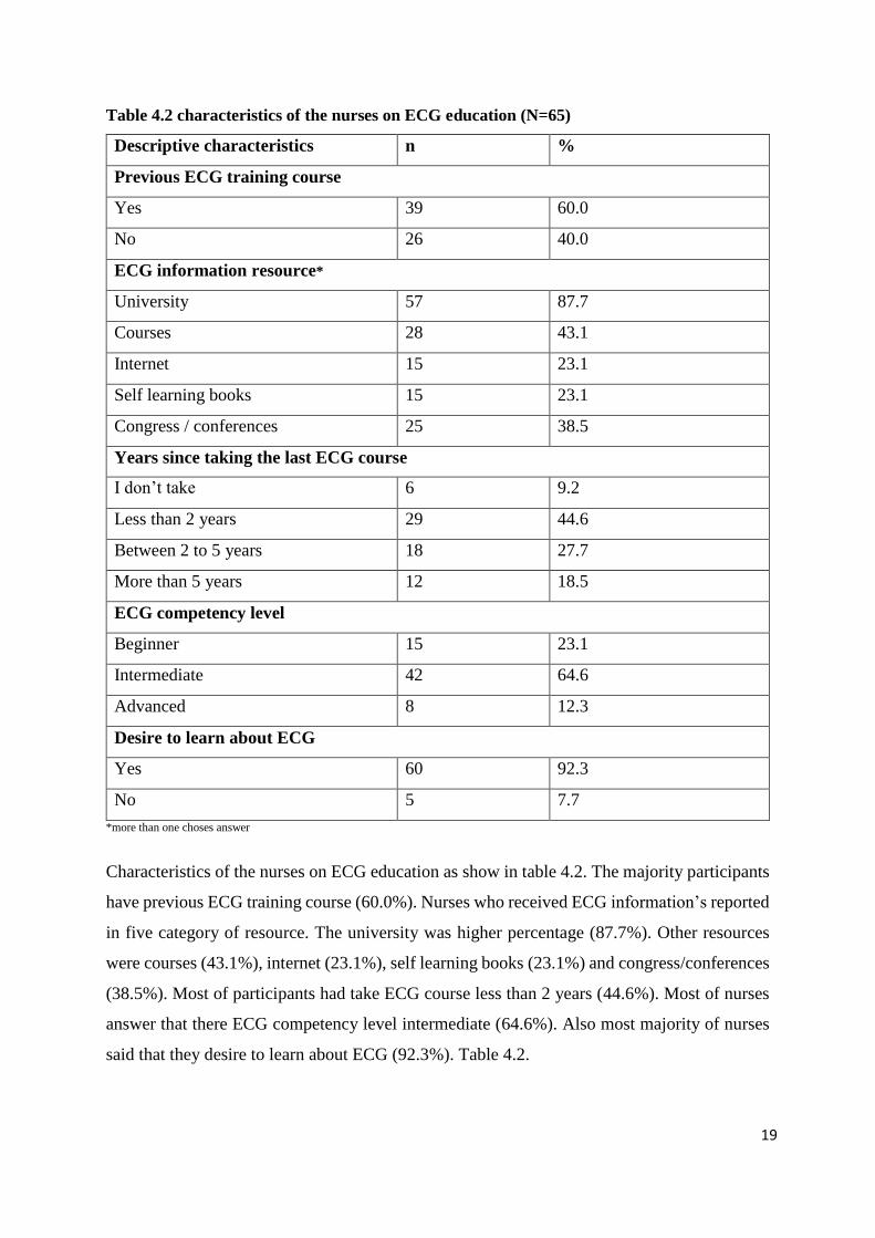
Table 4.2 characteristics of the nurses on ECG education (N=65)
Descriptive characteristics n %
Previous ECG training course
Yes 39 60.0
No 26 40.0
ECG information resource*
University 57 87.7
Courses 28 43.1
Internet 15 23.1
Self learning books 15 23.1
Congress / conferences 25 38.5
Years since taking the last ECG course
I don’t take 6 9.2
Less than 2 years 29 44.6
Between 2 to 5 years 18 27.7
More than 5 years 12 18.5
ECG competency level
Beginner 15 23.1
Intermediate 42 64.6
Advanced 8 12.3
Desire to learn about ECG
Yes 60 92.3
No 5 7.7
*more than one choses answer
Characteristics of the nurses on ECG education as show in table 4.2. The majority participants
have previous ECG training course (60.0%). Nurses who received ECG information’s reported
in five category of resource. The university was higher percentage (87.7%). Other resources
were courses (43.1%), internet (23.1%), self learning books (23.1%) and congress/conferences
(38.5%). Most of participants had take ECG course less than 2 years (44.6%). Most of nurses
answer that there ECG competency level intermediate (64.6%). Also most majority of nurses
said that they desire to learn about ECG (92.3%). Table 4.2.
20
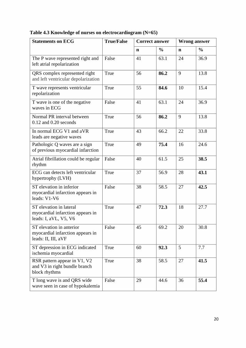
Table 4.3 Knowledge of nurses on electrocardiogram (N=65)
Statements on ECG True/False Correct answer Wrong answer
n % n %
The P wave represented right and
left atrial repolarization
False 41 63.1 24 36.9
QRS complex represented right
and left ventricular depolarization
True 56 86.2 9 13.8
T wave represents ventricular
repolarization
True 55 84.6 10 15.4
T wave is one of the negative
waves in ECG
False 41 63.1 24 36.9
Normal PR interval between
0.12 and 0.20 seconds
True 56 86.2 9 13.8
In normal ECG V1 and aVR
leads are negative waves
True 43 66.2 22 33.8
Pathologic Q waves are a sign
of previous myocardial infarction
True 49 75.4 16 24.6
Atrial fibrillation could be regular
rhythm
False 40 61.5 25 38.5
ECG can detects left ventricular
hypertrophy (LVH)
True 37 56.9 28 43.1
ST elevation in inferior
myocardial infarction appears in
leads: V1-V6
False 38 58.5 27 42.5
ST elevation in lateral
myocardial infarction appears in
leads: I, aVL, V5, V6
True 47 72.3 18 27.7
ST elevation in anterior
myocardial infarction appears in
leads: II, III, aVF
False 45 69.2 20 30.8
ST depression in ECG indicated
ischemia myocardial
True 60 92.3 5 7.7
RSR pattern appear in V1, V2
and V3 in right bundle branch
block rhythms
True 38 58.5 27 41.5
T long wave is and QRS wide
wave seen in case of hypokalemia
False 29 44.6 36 55.4
21
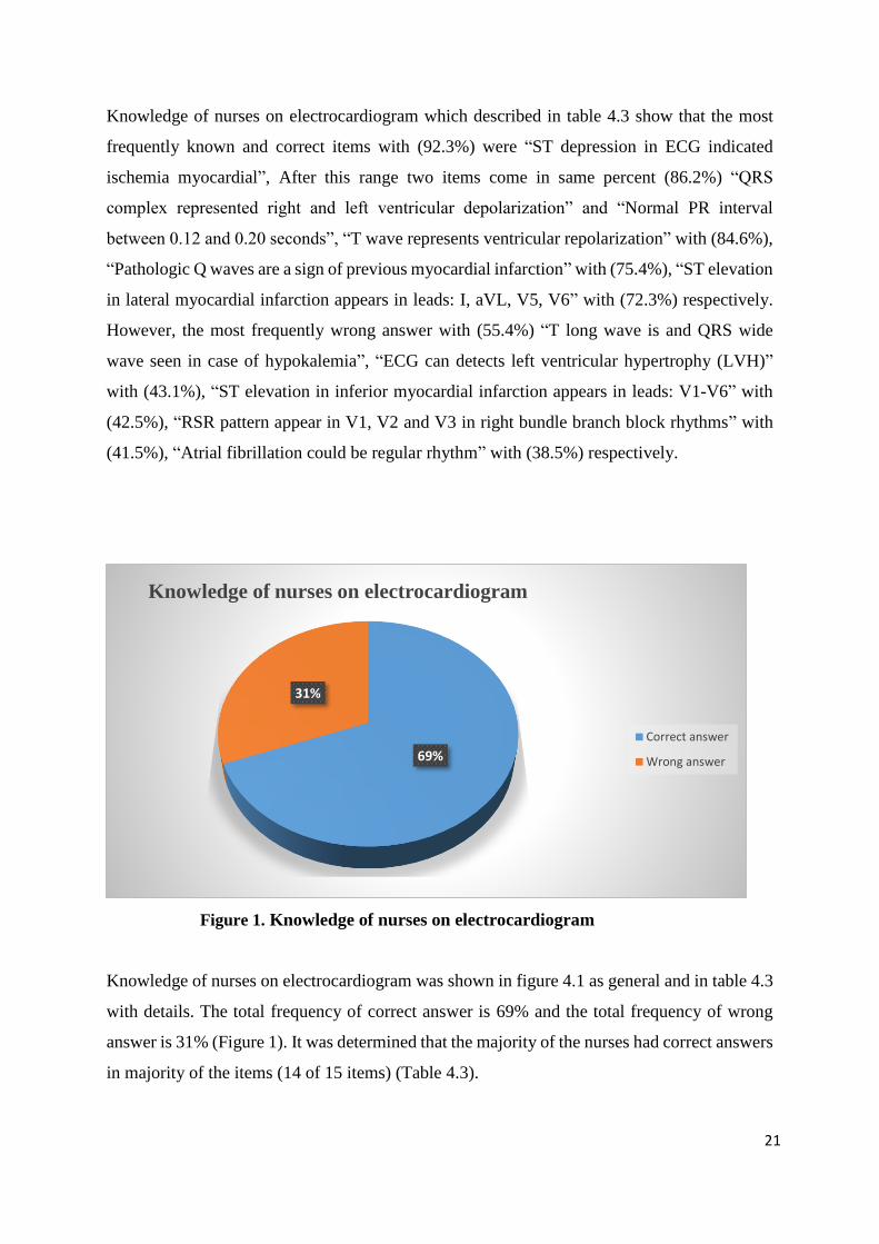
Knowledge of nurses on electrocardiogram which described in table 4.3 show that the most
frequently known and correct items with (92.3%) were “ST depression in ECG indicated
ischemia myocardial”, After this range two items come in same percent (86.2%) “QRS
complex represented right and left ventricular depolarization” and “Normal PR interval
between 0.12 and 0.20 seconds”, “T wave represents ventricular repolarization” with (84.6%),
“Pathologic Q waves are a sign of previous myocardial infarction” with (75.4%), “ST elevation
in lateral myocardial infarction appears in leads: I, aVL, V5, V6” with (72.3%) respectively.
However, the most frequently wrong answer with (55.4%) “T long wave is and QRS wide
wave seen in case of hypokalemia”, “ECG can detects left ventricular hypertrophy (LVH)”
with (43.1%), “ST elevation in inferior myocardial infarction appears in leads: V1-V6” with
(42.5%), “RSR pattern appear in V1, V2 and V3 in right bundle branch block rhythms” with
(41.5%), “Atrial fibrillation could be regular rhythm” with (38.5%) respectively.
Figure 1. Knowledge of nurses on electrocardiogram
Knowledge of nurses on electrocardiogram was shown in figure 4.1 as general and in table 4.3
with details. The total frequency of correct answer is 69% and the total frequency of wrong
answer is 31% (Figure 1). It was determined that the majority of the nurses had correct answers
in majority of the items (14 of 15 items) (Table 4.3).
69%
31%
Knowledge of nurses on electrocardiogram
Correct answer
Wrong answer
22
Table 4.4 Practice of nurses on ECG interpretations
Scenario of ECG
interpretations
True interpretations Correct answer
n %
You perform an ECG and
observe this rhythm. What do
you think it might be ?
An atrial flutter 55 84.6
You perform an ECG and
observe this rhythm. How would
you act ?
Ask for help without
leaving the patient alone
because it is a ventricular
fibrillation
44 67.7
A patient comes to the
emergency department due to a
respiratory distress. He has 140
beats per minute. You perform an
ECG and observe the following
It is an atrial fibrillation 41 63.1
A hospitalized patient who had
surgery due to an Acute
myocardial infarction, his vital
signs are unstable. You perform
an ECG and observe the
following
The patient presents a
Ventricular tachycardia
57 87.7
You performed ECG to patient
who have chest pain appeared
after leaving an important
meeting two hours ago. He is 52
years of age and hypertensive
and a few months ago he was
diagnosed with Diabetes Mellitus
II. The ECG as the following
It is an acute myocardial
infarction
47 72.3
A 24-year-old male comes to the
emergency department He is
athletic and slim. He reports
feeling a pricking sensation in
the left area of his chest since he
finished doing exercise (3 hours
earlier). You perform an ECG
and observe the following
It is a normal ECG 35 53.8
A 30-year-old woman comes to
the emergency department
reporting palpitations, chest
tightness and dyspnea. You
perform an ECG and observe the
following
It is an atrial tachycardia 33 50.8
What pathology you think the
patient with this ECG has?
A third-degree heart
block
39 60.0
23
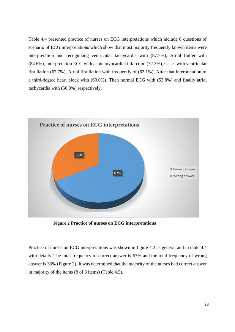
Table 4.4 presented practice of nurses on ECG interpretations which include 8 questions of
scenario of ECG interpretations which show that most majority frequently known items were
interpretation and recognizing ventricular tachycardia with (87.7%), Atrial flutter with
(84.6%), Interpretation ECG with acute myocardial infarction (72.3%), Cases with ventricular
fibrillation (67.7%), Atrial fibrillation with frequently of (63.1%), After that interpretation of
a third-degree heart block with (60.0%), Then normal ECG with (53.8%) and finally atrial
tachycardia with (50.8%) respectively.
Figure 2 Practice of nurses on ECG interpretations
Practice of nurses on ECG interpretations was shown in figure 4.2 as general and in table 4.4
with details. The total frequency of correct answer is 67% and the total frequency of wrong
answer is 33% (Figure 2). It was determined that the majority of the nurses had correct answer
in majority of the items (8 of 8 items) (Table 4.5).
67%
33%
Practice of nurses on ECG interpretations
Correct answer
Wrong answer
Table 4.5 comparison of years of working experience as registered nurse, currently working unit in hospital and previous ECG training course
with knowledge of nurses on electrocardiogram
Knowledge of
nurses on
Electrocardiogram
Years of working experience as
registered nurse
P
value
Likelihood
Ratio
Currently working unit in hospital
P
value
Likelihood
Ratio
Previous ECG training
course
P
value
Likelihood
Ratio < 1 1-5 >=6 ED ICU CCU CD RU YES No
Correct answer Correct answer Correct answer
N % N % N % N % N % N % N % N % N % N %
The p wave
represented right and
left atrial
repolarization
5 45.5 21 60.0 15 78.9 .160 .151 12 80.0 9 50.0 11 68.8 7 100.0 2 22.2 .008 .003 30 76.9 11 42.3 .005 .005
QRS complex
represented right and
left ventricular
depolarization
9 81.8 28 80.0 19 100.0 .114 .033 14 93.3 15 83.3 15 93.8 6 85.7 6 66.7 .354 .402 37 94.9 19 73.1 .013 .013
T wave represents
ventricular
repolarization
9 81.8 31 88.6 15 78.9 .620 .623 14 93.3 17 94.4 13 81.3 7 100.0 4 44.4 .005 .012 37 94.9 18 69.2 .005 .005
T wave is one of the
negative waves in
ECG
7 63.6 17 48.6 17 89.5 .012 .007 10 66.7 12 66.7 10 62.5 7 100.0 2 22.2 .030 .012 29 74.4 12 46.2 .021 .021
Normal PR interval
between 0.12 and 0.20
seconds
10 90.9 28 80.0 18 94.7 .228 .257 13 86.7 12 66.7 15 93.8 7 100.0 9 100 .059 .039 36 92.3 20 76.9 .079 .081
In normal ECG V1
and aVR leads are
negative waves
7 63.6 20 57.1 16 84.3 .131 .111 11 73.3 9 50 11 68.8 7 100.0 5 55.6 .163 .072 29 74.4 14 53.8 .087 .088
Pathologic Q waves
are a sign of previous
myocardial infarction
8 72.7 26 74.3 15 78.9 .907 .906 10 66.7 16 88.9 12 75 7 100.0 4 44.4 .054 .032 31 79.5 18 69.2 .347 .350
Atrial fibrillation
could be regular
rhythm
3 27.3 21 60.0 16 84.2 .008 .007 8 53.3 12 66.7 11 68.8 7 100.0 2 22.2 .024 .009 27 69.2 13 50.0 .118 .119
ECG can detects left
ventricular
hypertrophy (LVH) 3 27.3 20 57.1 14 73.7 .047 .044 6 40 11 61.1 9 56.3 7 100.0 4 44.4 .101 .035 24 61.5 13 50.0 .357 .358
ST elevation in
inferior myocardial
infarction appears in
leads: V1-V6
5 45.5 20 57.1 13 68.4 .457 .457 14 93.3 9 50 8 50 7 100.0 0 0.0 .001 .001 28 71.8 10 38.5 .008 .007
ST elevation in
lateral myocardial
infarction appears in
leads: I, aVL, V5,V6
7 63.6 25 71.4 15 78.9 .655 .655 13 86.7 13 72.3 10 62.5 7 100.0 4 44.4 .075 .038 35 89.7 12 46.2 .001 .001
24
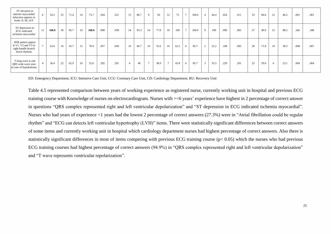
ED: Emergency Department, ICU: Intensive Care Unit, CCU: Coronary Care Unit, CD: Cardiology Department, RU: Recovery Unit
Table 4.5 represented comparison between years of working experience as registered nurse, currently working unit in hospital and previous ECG
training course with Knowledge of nurses on electrocardiogram. Nurses with >=6 years’ experience have highest in 2 percentage of correct answer
in questions “QRS complex represented right and left ventricular depolarization” and “ST depression in ECG indicated ischemia myocardial”.
Nurses who had years of experience <1 years had the lowest 2 percentage of correct answers (27.3%) were in “Atrial fibrillation could be regular
rhythm” and “ECG can detects left ventricular hypertrophy (LVH)” items. There were statistically significant differences between correct answers
of some items and currently working unit in hospital which cardiology department nurses had highest percentage of correct answers. Also there is
statistically significant differences in most of items compering with previous ECG training course (p< 0.05) which the nurses who had previous
ECG training courses had highest percentage of correct answers (94.9%) in “QRS complex represented right and left ventricular depolarization”
and “T wave represents ventricular repolarization”.
ST elevation in
anterior myocardial
infarction appears in
leads: II, III, aVF
6 54.5 25 71.4 14 73.7 .504 .521 13 86.7 9 50 12 75 7 100.0 4 44.4 .024 .011 33 84.6 12 46.2 .001 .001
ST depression in
ECG indicated
ischemia myocardial
11 100.0 30 85.7 19 100.0 .098 .038 14 93.3 14 77.8 16 100 7 100.0 9 100 .090 .065 37 49.9 23 88.5 .342 .348
RSR pattern appear
in V1, V2 and V3 in
right bundle branch
block rhythms
7 63.6 16 45.7 15 78.9 .057 .050 10 66.7 10 55.6 10 62.5 6 85.7 2 22.2 .108 .093 28 71.8 10 38.5 .008 .007
T long wave is and
QRS wide wave seen
in case of hypokalemia
4 36.4 22 62.9 10 52.6 .292 .291 6 40 7 38.9 7 43.8 6 85.7 3 33.3 .229 .201 23 59.0 6 23.1 .004 .004
25

26
Table 4.6 Comparison of years of working experience as registered nurse, currently working unit in hospital and previous ECG training
course with practice of nurses on ECG interpretations
Table 4.6 represented comparison between years of working experience as registered nurse, currently working unit in hospital and previous ECG
training course with practice of nurses on ECG interpretations. Study represented statistically significant differences in years of working experience
as registered nurse in scenario number 7 and 8 of ECG interpretations” (p< 0.05), Also currently working unit in hospital and previous ECG
training course represented the comparison with practice of nurses on ECG interpretations represented statistically significant differences in most
of items (p< 0.05).
Practice of
nurses on
ECG
interpretatio
n
Years of working experience as registered
nurse
P
value Likelihood
Ratio
Currently working unit in hospital
P
value Likelihood
Ratio
Previous ECG training
course
P
value Likelihood
Ratio
< 1 1-5 >=6 ED ICU CCU CD RU YES No
Correct answer Correct answer Correct answer
N % N % N % N % N % N % N % N % N % N %
Scenario
number 1 of
ECG
interpretation
8 72.7 29 82.9 18 94.7 .319 .285 14 93.3 14 77.8 15 93.8 7 100.0 5 55.6 .075 .081 38 97.4 17 65.4 .002 .001
Scenario
number 2 of
ECG
interpretation
6 54.5 22 62.9 16 84.2 .144 .147 11 73.3 12 66.7 12 75.0 7 100.0 2 22.2 .017 .012 29 74.4 15 57.7 .072 .044
Scenario
number 3 of
ECG
interpretation
6 54.5 20 57.1 15 78.9 .408 .247 13 86.7 10 55.6 9 56.3 7 100.0 2 22.2 .002 .001 32 82.1 9 34.6 .001 .001
Scenario
number 4 of
ECG
interpretation
8 72.7 32 91.4 17 89.5 .129 .160 14 93.3 12 66.7 16 100.0 7 100.0 8 88.9 .221 .145 37 94.9 20 76.9 .109 .071
Scenario
number 5 of
ECG
interpretation
6 54.5 25 71.4 16 84.2 .157 .133 15 100.0 13 72.2 12 75.0 7 100.0 0 0.00 .001 .001 35 89.7 12 46.2 .001 .001
Scenario
number 6 of
ECG
interpretation
4 36.4 16 45.7 15 78.9 .010 .017 9 60.0 7 38.9 8 50.0 7 100.0 4 44.4 .187 .099 25 64.1 10 38.8 .097 .071
Scenario
number 7 of
ECG
interpretation
1 9.1 19 54.3 13 68.4 .001 .001 10 66.7 7 38.9 9 56.3 7 100.0 0 0.00 .002 .001 23 59.0 10 38.5 .016 .006
Scenario
number 8 of
ECG
interpretation
4 36.4 21 60.0 14 73.7 .162 .063 10 66.7 12 66.7 10 62.5 7 100.0 0 0.00 .003 .001 31 79.5 8 30.8 .001 .001
26
27
5. DISCUSSION
The finding from this study was determined the knowledge and practice of
electrocardiogram among nurses. The study was conducted on 65 nurses with a different age,
experience and level of education. The study contact with critical care units like: Intensive care
unit, Coronary care unit, Emergency department, Recovery department and Cardiology
department which made knowledge and practice of ECG interpretation very important. In the
current study, no specific courses, educational programs or associations stated by participants
that they take it to improve their knowledge and practice of ECG.
Regarding to knowledge of nurses on electrocardiogram were shown in Table 4.3 it was
found that; majority of the nurses had correct answers about 69% in most of the items (14 of
15 items) and this is a satisfying result. In our study the lowest percent of correct answer was
“T long wave is and QRS wide wave seen in case of hypokalemia” about 55.4% of nurses’
wrong answer so nurses should attend to educational course to prepare them and endorse with
patients. In other hand the study had similar in results with AL-Husaunawy 2015 study that
indicate most nurses pass the questions regarding knowledge except question concerning ECG
changes regarding new and old myocardial infarction, comparing to current study with
question of “Pathologic Q waves are a sign of previous myocardial infarction” the percentage
of correct answer was 75.4% which indicate high knowledge. As important of educated nurses
they must had a critical thinking and knowledge in detection of myocardial infarction that
Stanfield L. 2018 explained in her study that the first-nurse educational intervention to include
other high-risk populations that need rapid intervention upon arrival at the emergency
department which in current study participant nurses had pass all questions related to
myocardial infarction that “ST elevation in lateral myocardial infarction appears in leads: I,
aVL, V5, V6” with 72.3% (True), “ST elevation in anterior myocardial infarction appears in
leads: II, III, aVF” with 69.2% (false) and “ST elevation in inferior myocardial infarction
appears in leads: V1-V6” with 58.5% (false) respectively. Also the the most frequently known
and correct item in this study with 92.3% were “ST depression in ECG indicated ischemia
myocardial”.
28
This study is one of studies which evaluate nurses’ competence those working in critical
area of there practice on ECG interpretation. In our study, the current practice of nurses on
ECG interpretation results are satisfying with 67% of nurses passing the questionnaire which
this score is higher than those of other studies like Werner et al 2016 which had 54% correct
answers on the test from participate nurses. In current study the lowest percent of correct
answer was identifying the case scenario of atrial tachycardia question which maybe confusing
the participate nurses because 32.3% of answers was “It is an atrial extra-systole”. Focusing
on literature regarding to practice of nurses on ECG interpretations the most majority
recognizing arrhythmias of different cases were ventricular tachycardia and atrial flutter which
this reflex finding similar with study conducted by Coll-Badell et al 2017. Also in our study
defining ECG that included acute myocardial infarction were 72.3% of answers cases which
also similar to Coll-Badell et al 2017 result which was of 71.9% of participates and compering
to Doğan et al 2012 study result was 54.3% of participates identify myocardial infarction which
mean that our study participates had high knowledge and practice to recognize this type of
critical case that may cause life threatening.
In comparison of years of working experience as registered nurse, currently working unit
in hospital and previous ECG training course with knowledge of nurses on electrocardiogram
(Table 4.5). In currently study it was showed that nurses who less than one year of experience
had lowest average of correct answers and nurses who experience >=6 years had highest
average of correct answers. As shown up the comparison of years of working experience with
knowledge of nurses on electrocardiogram wasn’t statistically significant differences in terms
of majority of the items (p>0.05). Results showed statistically significant differences in 3 items
of “T wave is one of the negative waves in ECG”, “Atrial fibrillation could be regular rhythm”
and “ECG can detects left ventricular hypertrophy (LVH)” were p< 0.05. Back to current result
in comparison of currently working unit in hospital with knowledge of nurses on
electrocardiogram which the cardiology department had the highest percent of correct answers
between nurses who work in hospital this result is similar to result of Zhang et al 2013 study
that show the test scores of nurses in the cardiology department were higher than those in ED
and ICU. There were statistically significant differences between correct answers of some
items on knowledge of nurses on electrocardiogram and currently working unit in hospital
which recovery unit nurses had the lowest percent of correct answers between participate
nurses which item that no one of these nurses answer correct “ST elevation in inferior
myocardial infarction appears in leads leads: V1-V6” (p< 0.05).

29
Also “The p wave represented right and left atrial repolarization”, “T wave represents
ventricular repolarization”, “T wave is one of the negative waves in ECG”, “Atrial fibrillation
could be regular Rhythm”. “ECG can detects left ventricular hypertrophy (LVH)” and “ST
elevation in anterior myocardial infarction appear in leads: II, III, aVF” which these differences
were found significant statistically (p<0.05). Also there were significant differences between
knowledge of nurses on electrocardiogram and previous ECG training course. Which the
percentage of nurses who had previous ECG training course in this study were 60% of total
participate, in addition the current study presented that the nurses who had previous ECG
training course score more percentage correct answers than who didn’t had which also the
comparison for most of items had differences were found significant statistically (p< 0.05) this
results are similar to Coll-Badell et al 2017 that shown nurses who had received training within
the previous five years scored significantly higher than those who had not.
In comparison years of working experience as registered nurse, currently working unit in
hospital and previous ECG training course with practice of nurses on ECG interpretations
(Table 4.6). As current study results the comparison of years of working experience with
practice of nurses on ECG interpretations had same result as knowledge comparison part that
nurses who less than one year of experience had lowest average of correct answers. Results
showed statistically significant differences only in 2 scenarios 6 and 7 that’s contact with
recognizing of case of normal ECG and atrial tachycardia that’s the participated nurses had
confusing to analyzed (p<0.05). And more of scenario items were highest average of correct
answers but no statistically significant differences in terms of majority of the items (p>0.05)
which similar to Lak et al 2013 study results. The current study represented that currently
working unit in hospital was significant statistically affected on practice of nurses on ECG
interpretations the nurses who working in cardiology department had correct answers which
also similar to Zhang et al 2013 study. Also CCU staff nurses had higher percent of correct
answers which similar to Lak et al 2013 study results that CCU working experience was
associated with better results on the ECG test. The nurses who work in recovery unit had lowest
average of correct answers were statistically significant differences in most of items (p< 0.05).
Referred to comparison between previous ECG training course with practice of nurses on ECG
interpretations the percentage of nurses who had previous ECG training course score more
correct answers than who didn’t had which also statistically significant differences in most of
items (p< 0.05).
30
6. CONCLUSION
Results of the present study showed high level of knowledge and practice of
electrocardiogram among nurses. Also working unit in hospital and previous ECG training
courses play important role in defining the professionalism of nurses to had experience in ECG
interpretation. Training courses for nurses abroad for knowledge and practice with more
intensive detailed lectures about ECG in universities. Also regular courses under the
supervision of qualified well trained staff especially for nurses who work in critical area in
hospital. Nurses must be continuing self learning and staying update to any changing and
development of new protocols or technology in the world.
7. FINDINGS AND RECOMMENDATIONS
7.1. Findings
Main findings of the study that was performed with the aim of determination of the
knowledge and practices of electrocardiogram interpretation of nurses were listed as
followings:
- The mean ages of the participants were (26.94) years. Majority of the participants were
female (66.2%), while (33.8%) of them were male. Majority of the nurses had bachelor
degree (93.8%). Most of the nurses had experience less than five years as registered
nurses (53.8%) (Table 4.1).
- The majority of (60.0%) of the participants had received previous ECG courses. The
higher percentage ECG education resource was university (87.7%) among the
resources (Table 4.2).
31
- Regarding to Knowledge of nurses on Electrocardiogram, it was found that; majority
of the nurses had correct answers (69%) (Figure 1). In most of the items (14 of 15
items) (Table 4.3).
- Regarding to practice of nurses on ECG interpretations, it was found that; majority of
the nurses had correct answers (67%) (Figure 2). In most of the items (8 of 8 items)
(Table 4.4).
- There were statistically significant differences in terms of currently working unit in
hospital and previous ECG training course with different items at Knowledge and
practice of nurses on ECG interpretations (Table 4.5, Table 4.6).
7.2. Recommendations
Based on the results of the study following recommendations were made:
- Refresher intensive training courses under the supervision of qualified well trained staff
in electrocardiography should be run at least every 2 years.
- Training courses for nurses abroad for knowledge and practice exchange more detailed
lectures about ECG in universities.
- Initiation of education program and self-learning handbook material were effective in
improving the nurses’ ECG knowledge.
32
8. REFERENCES
AlGhatrif, M., & Lindsay, J. (2012). A brief review: history to understand fundamentals of
electrocardiography. Journal of community hospital internal medicine perspectives, 2(1),
14383.
Atwood, D., & Wadlund, D. L. (2015). ECG interpretation using the CRISP method: A guide
for nurses. AORN journal, 102(4), 396-408.
Amsterdam, E. A., Wenger, N. K., Brindis, R. G., Casey, D. E., Ganiats, T. G., Holmes, D. R.,
... & Levine, G. N. (2014). 2014 AHA/ACC guideline for the management of patients with
non–ST-elevation acute coronary syndromes: a report of the American College of
Cardiology/American Heart Association Task Force on Practice Guidelines. Journal of the
American College of Cardiology, 64(24), e139-e228.
Authors/Task Force Members, Hamm, C. W., Bassand, J. P., Agewall, S., Bax, J., Boersma,
E., ... & Huber, K. (2011). ESC Guidelines for the management of acute coronary syndromes
in patients presenting without persistent ST-segment elevation: The Task Force for the
management of acute coronary syndromes (ACS) in patients presenting without persistent ST-
segment elevation of the European Society of Cardiology (ESC). European heart
journal, 32(23), 2999-3054.
Anderson JL, Adams CD, Antman EM, et al. (2012). ACCF/AHA focused update incorporated
into the ACCF/AHA 2007 guidelines for the management of patients with unstable
angina/non-ST-elevation myocardial infarction: a report of the American College of
Cardiology Foundation/American Heart Association Task Force on Practice
Guidelines;127(23): e663–828.
Atlas Writing Group, Timmis, A., Townsend, N., Gale, C., Grobbee, R., Maniadakis, N., ... &
Bax, J. (2017). European Society of Cardiology: cardiovascular disease statistics
2017. European heart journal, 39(7), 508-579.
33
Authors/Task Force Members, Hamm, C. W., Bassand, J. P., Agewall, S., Bax, J., Boersma,
E., ... & Huber, K. (2011). ESC Guidelines for the management of acute coronary syndromes
in patients presenting without persistent ST-segment elevation: The Task Force for the
management of acute coronary syndromes (ACS) in patients presenting without persistent ST-
segment elevation of the European Society of Cardiology (ESC). European heart
journal, 32(23), 2999-3054.
Abrams J.(2005) Clinical practice. Chronic stable angina. N Engl J Med; 352: 2524e33.
Benjamin, E. J., Blaha, M. J., Chiuve, S. E., Cushman, M., Das, S. R., Deo, R., ... & Jiménez,
M. C. (2017). Heart disease and stroke statistics-2017 update: a report from the American
Heart Association. Circulation, 135(10), e146-e603.
Bhaumik, A., Kumar, P. S., Saha, S., & Aishwarya, M. N. L. (2016). A review on cardiac
arrhythmia and cardiac ablation: Invasive techniques and future perspective. World journal of
pharmaceutical and medical research, 2, 105-111.
Badhwar, N. (2010). Introduction to Supraventricular Tachycardia. Cardiac Electrophysiology
Clinics 2, 179–181.
Bhatheja, R., & Mukherjee, D. (2007). Acute coronary syndromes: unstable angina/non-ST
elevation myocardial infarction. Critical care clinics, 23(4), 709-735.
Braunwald, E., Antman, E. M., Beasley, J. W., Califf, R. M., Cheitlin, M. D., Hochman, J. S.,
... & Pepine, C. J. (2002). ACC/AHA 2002 guideline update for the management of patients
with unstable angina and non–ST-segment elevation myocardial infarction—summary article:
a report of the American College of Cardiology/American Heart Association task force on
practice guidelines (Committee on the Management of Patients With Unstable
Angina). Journal of the American College of Cardiology, 40(7), 1366-1374.
Bello, D., Fieno, D. S., Kim, R. J., Pereles, F. S., Passman, R., Song, G., ... & Goldberger, J.
J. (2005). Infarct morphology identifies patients with substrate for sustained ventricular
tachycardia. Journal of the American College of Cardiology, 45(7), 1104-1108.
34
Basra, S. S., Virani, S. S., Paniagua, D., Kar, B., & Jneid, H. (2016). Acute Coronary
Syndromes: Unstable Angina and Non–ST Elevation Myocardial Infarction. Heart failure
clinics, 12(1), 31-48.
Cleveland clinic (June 22, 2017). Coronary Artery Disease. Retrieved from:
https://my.clevelandclinic.org/health/diseases/16898-coronary-artery-disease
Cheng, C. H., Sanders, G. D., Hlatky, M. A., Heidenreich, P., McDonald, K. M., Lee, B. K.,
... & Owens, D. K. (2000). Cost-effectiveness of radiofrequency ablation for supraventricular
tachycardia. Annals of internal medicine, 133(11), 864-876.
Coll-Badell, M., Jiménez-Herrera, M. F., & Llaurado-Serra, M. (2017). emergency nurse
competence in electrocardiographic interpretation in Spain: A cross-sectional study. Journal of
emergency nursing, 43(6), 560-570.
Calder, S., 2008. Clinical pearls and pitfalls of electrocardiogram interpretation in acute
myocardial infarction. Journal of Emergency Nursing 34, 324–329. ACP/ACC/AHA Task
Force, 1995. Clinical competence in electrocardiography. Journal of the American College of
Cardiology 25, 1465–1469.
Cottrell, D. B., & Mack, K. (2008). Atrial fibrillation: an emergency nurse’s rapid
response. Journal of emergency nursing, 34(3), 207-210.
Cano, Ó., Andrés, A., Alonso, P., Osca, J., Sancho-Tello, M. J., Rueda, J., ... & Martínez-Dolz,
L. (2017). Essential ECG clues in patients with congenital heart disease and
arrhythmias. Journal of electrocardiology, 50(2), 243-250.
Doğan, H. D., & Melek, M. (2012). Hemşirelerin acil kalp hastalıklarında g.rülen, EKG
bulgularını tanıyabilme ve uygun tedavi yaklaşımlarını değerlendirebilme düzeylerinin tespiti,
60-69.
Drew, B. J., & Funk, M. (2006). Practice standards for ECG monitoring in hospital settings:
executive summary and guide for implementation, 157-168.
35
Delacrétaz, E. (2006). Supraventricular tachycardia. New England Journal of
Medicine, 354(10), 1039-1051.
Deckers, J. W. (2013). Classification of myocardial infarction and unstable angina: a re-
assessment. International journal of cardiology, 167(6), 2387-2390.
Delgado, V., Bucciarelli-Ducci, C., & Bax, J. J. (2016). Diagnostic and prognostic roles of
echocardiography and cardiac magnetic resonance. Journal of Nuclear Cardiology, 23(6),
1399-1410.
Dewar, R. I., & Lip, G. Y. (2007). Guidelines Development Group for the NICE Clinica l
Guideline for the management of atrial fibrillation. Heart, 93.
Docherty, B., 2003. 12-Lead ECG interpretation and chest pain management. British Journal
of Nursing 12, 1248–1255.
de Winter, R. J., & Tijssen, J. G. (2012). Non–ST-Segment Elevation Myocardial Infarction:
Revascularization for Everyone? 903-905.
Drew, B.J.C., Califf, R.M., Funk, M., Kaufman, E.S., Krucoff, M.W., Laks, M.M., Macfarlane,
P.W., Sommargren, C., Swiryn, S., Van Hare, G.F., 2004. Practice standards for
electrocardiographic monitoring in hospital settings: an American Heart Association scientific
statement from the Councils on Cardiovascular Nursing, Clinical Cardiology, and
Cardiovascular Disease in the Young. Circulation 110 (17) 2721–2746.
Fuenzalida, C., Ferro, I., Ambrós, A., Siches, C., Sánchez, M., & Cabrera, J. (2015). An
educational intervention nursing at discharge from the emergency department reduces
complications and admissions in the short term in patients with atrial fibrillation. Emergencias,
27, 75-81.8
Fuenzalida, C., Hernández, G., Ferro, I., Siches, C., Ambrós, À., & Coll- Vinent, B. (2017).
Long-term benefits of education by emergency care nurses at discharge of patients with atrial
fibrillation. International emergency nursing, 35,7-12.
36
Funk, M., Fennie, K. P., Stephens, K. E., May, J. L., Winkler, C. G., Drew, B. J., & PULSE
site investigators. (2017). Association of implementation of practice standards for
electrocardiographic monitoring with nurses’ knowledge, quality of care, and patient
outcomes. circulation: cardiovascular quality and outcomes, 10(2), e003132.
Funk, M., Winkler, C. G., May, J. L., Stephens, K., Fennie, K. P., Rose, L. L., ... & Drew, B.
J. (2010). Unnecessary arrhythmia monitoring and underutilization of ischemia and QT
interval monitoring in current clinical practice: baseline results of the practical use of the latest
standards for electrocardiography trial. journal of electrocardiology, 43(6), 542-547.
Fuster, V., Rydén, L. E., Cannom, D. S., Crijns, H. J., Curtis, A. B., Ellenbogen, K. A., ... &
Olsson, S. B. (2011). 2011 ACCF/AHA/HRS focused updates incorporated into the
ACC/AHA/ESC 2006 Guidelines for the management of patients with atrial fibrillation: a
report of the American College of Cardiology Foundation/American Heart Association Task
Force on Practice Guidelines developed in partnership with the European Society of
Cardiology and in collaboration with the European Heart Rhythm Association and the Heart
Rhythm Society. Journal of the American College of Cardiology, 57(11), e101-e198.
Fanaroff AC, et al. (2015) Does this patient with chest pain have acute coronary syndrome?
The rational clinical examination systematic review. JAMA 314(18): 1955–1965.
GBD 2013 Mortality and Causes of Death Collaborators. (2015). Global, regional, and national
age-sex specific all-cause and cause-specific mortality for 240 causes of death, 1990-2013: a
systematic analysis for the Global Burden of Disease Study 2013. Lancet, 385(9963), 117–
171.
Gupta R, Puri V, Narain VV(1999). Cardiovascular risk profile in Indian women(Abstr).
Indian Heart J; 51:679.
Go, A. S., Hylek, E. M., Phillips, K. A., Chang, Y., Henault, L. E., Selby, J. V., & Singer, D.
E. (2001). Prevalence of diagnosed atrial fibrillation in adults: national implications for rhythm
management and stroke prevention: The Anticoagulation and Risk Factors in Atrial Fibrillation
(ATRIA) Study. Jama, 285(18), 2370-2375.

37
Gheorghiade, M., Sopko, G., De Luca, L., Velazquez, E. J., Parker, J. D., Binkley, P. F., ... &
Bonow, R. O. (2006). Navigating the crossroads of coronary artery disease and heart
failure. Circulation, 114(11), 1202-1213.
Gazarian, P.K., Henneman, E.A., Chandler, G.E., 2010. Nurse decision making in the prearrest
period. Clinical Nursing Research 19 (1) 21–37.
George, A., Arumugham, P. S., & Figueredo, V. M. (2010). aVR–the forgotten lead.
experimental & clinical cardiology, 15(2), e36.
Hernández-Padilla, J., Suthers, F., Fernández-Sola, C., & Granero-Molina, J. (2016).
Development and psychometric assessment of the basic resuscitation skills self-efficacy scale.
European journal of cardiovascular nursing, 15(3), e10-e18.
Hampton, J. R. (2008). The ECG Made Easy. Elsevier Health Sciences, (8), 4-5.
Huitema, A. A., Zhu, T., Alemayehu, M., & Lavi, S. (2014). Diagnostic accuracy of ST-
segment elevation myocardial infarction by various healthcare providers. International journal
of cardiology, 177(3), 825-829.
Huang HD, Tamarisa R, Mathur N, et al. (2013). Stellate ganglion block: a therapeutic
alternative for patients with medically refractory inappropriate sinus tachycardia? J
Electrocardiol.46:693-6.
Harris, P., & Lysitsas, D. (2015). Ventricular arrhythmias and sudden cardiac death. BJA
Education, 16(7), 221-229.
Hunter, J. D., Sharma, P., & Rathi, S. (2008). Long QT syndrome. Continuing Education in
Anaesthesia, Critical Care & Pain, 8(2), 67-70.
Iosr journals, & Ahmed A. AL-Husaunawy. (2015). Evaluation of nurses knowledge and
practical of electrocardiogram toward adolescent patient, 10-16.
38
Jevon, P. (2010). Procedure for recording a standard 12-lead electrocardiogram. British
Journal of Nursing, 19(10), 649-651.
Jevon, P. (2010). Recording an accurate ECG. Practice nursing, 21(8), 404-408.
January CT, et al. 2014 AHA/ACC/HRS guideline for the management of patients with atrial
fibrillation: a report of the American College of Cardiology/American Heart Association Task
Force on practice guidelines and the Heart Rhythm Society. Circulation 2014;130(23):e199-
e267.
Khatibzadeh, S., Farzadfar, F., Oliver, J., Ezzati, M., & Moran, A. (2013). Worldwide risk
factors for heart failure: a systematic review and pooled analysis. International journal of
cardiology, 168(2), 1186-1194.
Kirchhof, P., Benussi, S., Kotecha, D., Ahlsson, A., Atar, D., ... & Hendriks, J. (2016). 2016
ESC Guidelines for the management of atrial fibrillation developed in collaboration with
EACTS. EP Europace, 18(11), 1609-1678.
Lak, K., Zareie, F., Habibzadeh, H., Mohammadpour, Y., Rahnemoon, K., Zare, H., &
Zaviyeh, M. (2013). A survey on the effect of educational software method of arrhythmias
stimulator on the level of knowledge of electrocardiograms interpretation in nurses. Iran J Crit
Care Nurs, 6(3), 173-80.
Lancia, L., Pisegna Cerone, M., Vittorini, P., Romano, S., Penco, M., 2008. A comparison
between EASI system 12-lead ECGs and standard 12-lead ECGs for improved clinical nursing
practice. Journal of Clinical Nursing 17, 370– 377.
Libby, P., & Theroux, P. (2005). Pathophysiology of coronary artery
disease. Circulation, 111(25), 3481-3488.
Li X, Shu L, Hu H (2009) Kernel-based nonlinear dimensionality reduction for
electrocardiogram recognition. Neural Comput Appl 18(8):1013–1020.

39
Link, M. S., Berkow, L. C., Kudenchuk, P. J., Halperin, H. R., Hess, E. P., Moitra, V. K., ... &
White, R. D. (2015). Part 7: adult advanced cardiovascular life support: 2015 American Heart
Association guidelines update for cardiopulmonary resuscitation and emergency
cardiovascular care. Circulation, 132(18 suppl 2), S444-S464.
Larson, T. S., & Brady, W. J. (2008). Electrocardiographic monitoring in the hospitalized
patient: a diagnostic intervention of uncertain clinical impact. The american journal of
emergency medicine, 26(9), 1047-1055.
Lee, K. W., Badhwar, N., & Scheinman, M. M. (2008). Supraventricular tachycardia—part
I. Current problems in cardiology, 33(9), 467-546.
Montalescot, G., Sechtem, U., Achenbach, S., Andreotti, F., Arden, C., Budaj, A., ... &
Ferreira, J. R. (2014). 2013 ESC guidelines on the management of stable coronary artery
disease. The Task Force on the management of stable coronary artery disease of the European
Society of Cardiology. Revista Española de Cardiología, 67(02), 135-135.
Michniewicz, E., Mlodawska, E., Lopatowska, P., Tomaszuk-Kazberuk, A., & Malyszko, J.
(2018). Patients with atrial fibrillation and coronary artery disease–double trouble. Advances
in medical sciences, 63(1), 30-35.
Morrison, L. J., Brooks, S., Sawadsky, B., McDonald, A., & Verbeek, P. R. (2006). Prehospital
12‐ lead Electrocardiography Impact on Acute Myocardial Infarction Treatment Times and
Mortality: A Systematic Review. Academic emergency medicine, 13(1), 84-89.
Montalescot, G., Sechtem, U., Achenbach, S., Andreotti, F., Arden, C., ... & Di Mario, C.
(2013). 2013 ESC guidelines on the management of stable coronary artery disease: the Task
Force on the management of stable coronary artery disease of the European Society of
Cardiology. European heart journal, 34(38), 2949-3003.
Martínez-Sellés, M., Bueno, H., Sacristán, A., Estévez, Á., Ortiz, J., Gallegoa, L., &Fernández-
Avilés, F. (2008). Dolor torácico en urgencias: frecuencia, perfil clínico y estratificación de
riesgo. Revista española de cardiología, 61(9), 953-959.
40
Mousa, A. M., & Ahmed, H. A. O. R. S. (2016). Nurses' knowledge concerning early
interventions for patients with ventricular tachycardia at Baghdad teaching hospitals. Kufa
journal for nursing sciences 6(2).
Mensah, G. A., & Brown, D. W. (2007). An overview of cardiovascular disease burden in the
United States. Health affairs, 26(1), 38-48.
Manning, P., & Awtry, E. H. (2014). Unstable Angina: Presentation, Diagnosis, and
Management. Encyclopedia of Cardiovascular Research and Medicine, 606-615.
Nattel, S., & Opie, L. H. (2006). Controversies in atrial fibrillation. The Lancet, 367(9506),
262-272.
National Heart, Lung, and Blood Institute NHLBI (January 27, 2015). What is a heart attack?
Retrieved from: http://www.nhlbi.nih.gov/health/health-topics/topics/heart attack.
Pettersen, T. R., Fålun, N., & Norekvål, T. M. (2014). Improvement of in-hospital telemetry
monitoring in coronary care units: An intervention study for achieving optimal electrode
placement and attachment, hygiene and delivery of critical information to patients. European
Journal of Cardiovascular Nursing, 13(6), 515-523.
Pathak, L. A., Shirodkar, S., Ruparelia, R., & Rajebahadur, J. (2017). Coronary artery disease
in women. Indian Heart Journal, 69(4), 532-538.
Page, R. L., Joglar, J. A., Caldwell, M. A., Calkins, H., Conti, J. B., Deal, B. J., ... & Indik, J.
H. (2016). 2015 ACC/AHA/HRS guideline for the management of adult patients with
supraventricular tachycardia: a report of the American College of Cardiology/American Heart
Association Task Force on Clinical Practice Guidelines and the Heart Rhythm Society. Journal
of the American College of Cardiology, 67(13), e27-e115.
Papadopoulos, C. H., Oikonomidis, D., Lazaris, E., & Nihoyiannopoulos, P. (2017).
Echocardiography and Cardiac Arrhythmias. Hellenic Journal of Cardiology.

41
Pottle, A. (2005). A nurse-led rapid access chest pain clinic—experience from the first 3 years.
European journal of cardiovascular nursing, 4(3), 227-233.
Rockson, S. G., & Albers, G. W. (2004). Comparing the guidelines: anticoagulation therapy
to optimize stroke prevention in patients with atrial fibrillation. Journal of the American
College of Cardiology, 43(6), 929-935.
Refaey, A. S. T. (2012). Impact of designed teaching protocol about advanced cardiac life
support (ACLS) on critical care nurse’s knowledge and practices at Benha university hospital,
Cairo, Egypt. J Am Sci, 8(12), 838-50.
Roberts-Thomson, K. C., Lau, D. H., & Sanders, P. (2011). The diagnosis and management of
ventricular arrhythmias. Nature Reviews Cardiology, 8(6), 311.
Spiers, C. M. (2007). Using the 12-lead ECG to diagnose acute myocardial infarction in the
presence of left bundle branch block. Accident and emergency nursing, 15(1), 56-61.
Sharma, S., Drezner, J. A., Baggish, A., Papadakis, M., Wilson, M. G., Prutkin, J. M., ... &
Asif, I. M. (2017). International recommendations for electrocardiographic interpretation in
athletes. European heart journal, ehw631.
Stephens, K. E., Anderson, H., Carey, M. G., & Pelter, M. M. (2007). Interpreting 12-lead
electrocardiograms for acute ST-elevation myocardial infarction: what nurses know. Journal
of cardiovascular nursing, 22(3), 186-193.
Sheiline, M. (2008). Effectiveness of a competency program on ECG monitoring and
interpretation for staff nurses working in critical care areas and to determine the factors
influencing their competency. Asian Journal of Cardiovascular Nursing, 16(1), 1-7.
Salerno, S. M., Alguire, P. C., & Waxman, H. S. (2003). Training and competency evaluation
for interpretation of 12-lead electrocardiograms: recommendations from the American College
of Physicians. Annals of internal medicine, 138(9), 747-750.
42
Sachdewani, r. K., dingra, l. C., & memon, a. H. (2018). Acute stemi: prevalence of
hypertension and diabetes in patients of acute stemi admitted in ccu ghulam muhammad
teaching hospital, sukuur sindh pakistan. Professional medical journal, 25(5).
Tobin, K. J. (2010). Stable angina pectoris: what does the current clinical evidence tell
us? Journal of the American Osteopathic Association, 110(7), 364.
Virag, N., Jacquemet, V., Henriquez, C. S., Zozor, S., Blanc, O., Vesin, J. M., ... &
Kappenberger, L. (2002). Study of atrial arrhythmias in a computer model based on magnetic
resonance images of human atria. Chaos: An Interdisciplinary Journal of Nonlinear
Science, 12(3), 754-763.
WHO (2017, January). The top 10 causes of death. Retrieved from:
http://www.who.int/mediacentre/factsheets/fs310/en/
Whittaker, A., Jokhi, P., & Curzen, N. (2014). Ischaemic heart disease: stable
angina. Medicine, 42(9), 495-501.
Zipes, D. P. et al (2006). ACC/AHA/ESC 2006 Guidelines for Management of Patients with
Ventricular Arrhythmias and the Prevention of Sudden Cardiac Death: a report of the
American College of Cardiology/American Heart Association Task Force and the European
Society of Cardiology Committee for Practice Guidelines (writing committee to develop
Guidelines for Management of Patients with Ventricular Arrhythmias and the Prevention of
Sudden Cardiac Death): developed in collaboration with the European Heart Rhythm
Association and the Heart Rhythm Society. Circulation 114, e385–e484.
Zhang, H., & Hsu, L. L. (2013). The effectiveness of an education program on nurses’
knowledge of electrocardiogram interpretation. International emergency nursing, 21(4), 247-
251.
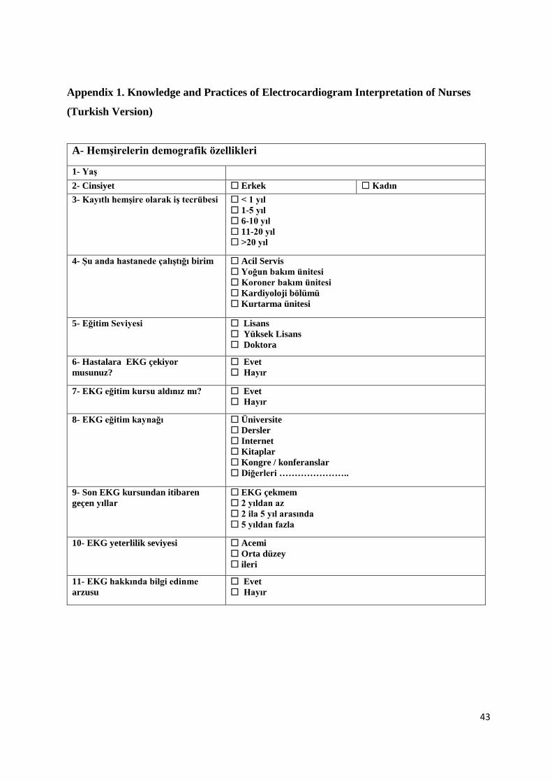
43
Appendix 1. Knowledge and Practices of Electrocardiogram Interpretation of Nurses
(Turkish Version)
A- Hemşirelerin demografik özellikleri
1- Yaş
2- Cinsiyet Erkek Kadın
3- Kayıtlı hemşire olarak iş tecrübesi < 1 yıl
1-5 yıl
6-10 yıl
11-20 yıl
>20 yıl
4- Şu anda hastanede çalıştığı birim Acil Servis
Yoğun bakım ünitesi
Koroner bakım ünitesi
Kardiyoloji bölümü
Kurtarma ünitesi
5- Eğitim Seviyesi
Lisans
Yüksek Lisans
Doktora
6- Hastalara EKG çekiyor
musunuz?
Evet
Hayır
7- EKG eğitim kursu aldınız mı? Evet
Hayır
8- EKG eğitim kaynağı Üniversite
Dersler
Internet
Kitaplar
Kongre / konferanslar
Diğerleri …………………..
9- Son EKG kursundan itibaren
geçen yıllar
EKG çekmem
2 yıldan az
2 ila 5 yıl arasında
5 yıldan fazla
10- EKG yeterlilik seviyesi Acemi
Orta düzey
ileri
11- EKG hakkında bilgi edinme
arzusu
Evet
Hayır
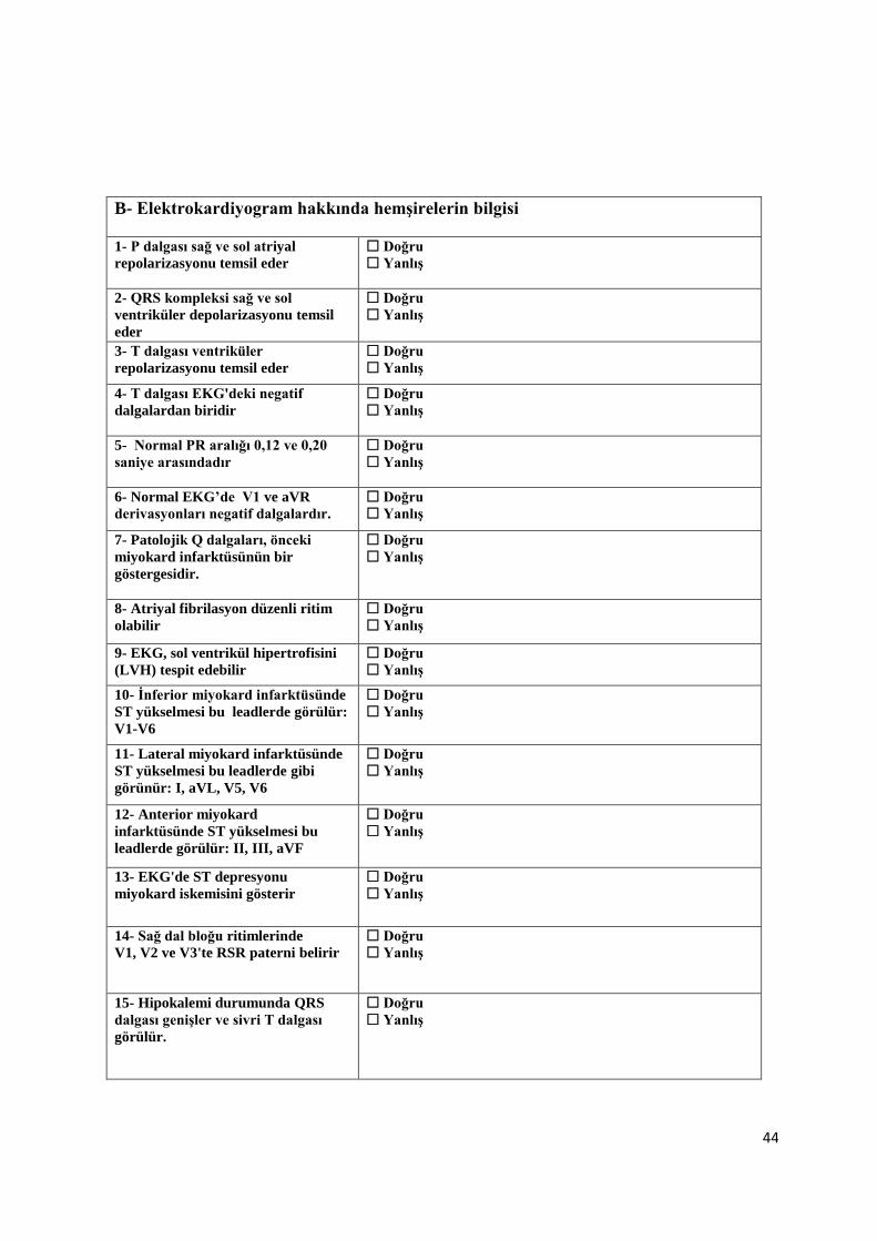
44
B- Elektrokardiyogram hakkında hemşirelerin bilgisi
1- P dalgası sağ ve sol atriyal
repolarizasyonu temsil eder
Doğru
Yanlış
2- QRS kompleksi sağ ve sol
ventriküler depolarizasyonu temsil
eder
Doğru
Yanlış
3- T dalgası ventriküler
repolarizasyonu temsil eder
Doğru
Yanlış
4- T dalgası EKG'deki negatif
dalgalardan biridir
Doğru
Yanlış
5- Normal PR aralığı 0,12 ve 0,20
saniye arasındadır
Doğru
Yanlış
6- Normal EKG’de V1 ve aVR
derivasyonları negatif dalgalardır.
Doğru
Yanlış
7- Patolojik Q dalgaları, önceki
miyokard infarktüsünün bir
göstergesidir.
Doğru
Yanlış
8- Atriyal fibrilasyon düzenli ritim
olabilir
Doğru
Yanlış
9- EKG, sol ventrikül hipertrofisini
(LVH) tespit edebilir
Doğru
Yanlış
10- İnferior miyokard infarktüsünde
ST yükselmesi bu leadlerde görülür:
V1-V6
Doğru
Yanlış
11- Lateral miyokard infarktüsünde
ST yükselmesi bu leadlerde gibi
görünür: I, aVL, V5, V6
Doğru
Yanlış
12- Anterior miyokard
infarktüsünde ST yükselmesi bu
leadlerde görülür: II, III, aVF
Doğru
Yanlış
13- EKG'de ST depresyonu
miyokard iskemisini gösterir
Doğru
Yanlış
14- Sağ dal bloğu ritimlerinde
V1, V2 ve V3'te RSR paterni belirir
Doğru
Yanlış
15- Hipokalemi durumunda QRS
dalgası genişler ve sivri T dalgası
görülür.
Doğru
Yanlış

45
C- Hemşirelerin EKG yorumları üzerine uygulaması
1- EKG çektiniz ve bu ritmi gözlemlediniz. Ne olabileceğini düşünürsünüz?
Üçüncü derece kalp bloğu
Atriyal flutter
Supraventriküler taşikardi
Bilmiyorum
2- EKG çektiniz ve bu ritmi gözlemlediniz. Nasıl davranırsınız?
Hastayı yalnız
bırakmadan yardım isterim
çünkü bir ventriküler
fibrilasyon
Hastayı yalnız
bırakmadan yardım isterim
çünkü bir atriyal fibrilasyon
Başka bir EKG
gerçekleştiririm çünkü
parazit olabilir
Nasıl davranacağını
bilmiyorsun ama bunun ciddi
bir sorun olduğunu
biliyorsun.
3- Bir hasta solunum sıkıntısı nedeniyle acil servise geldi. Dakikada 140 nabız
saptandı. EKG çektiniz ve aşağıdaki gibi saptadınız. Ne düşünürsünüz
Bu bir atriyal taşikardidir.
Bu bir Atriyal
fibrilasyondur
Bu bir atriyal ekstra
sistoldür
Bilmiyorum
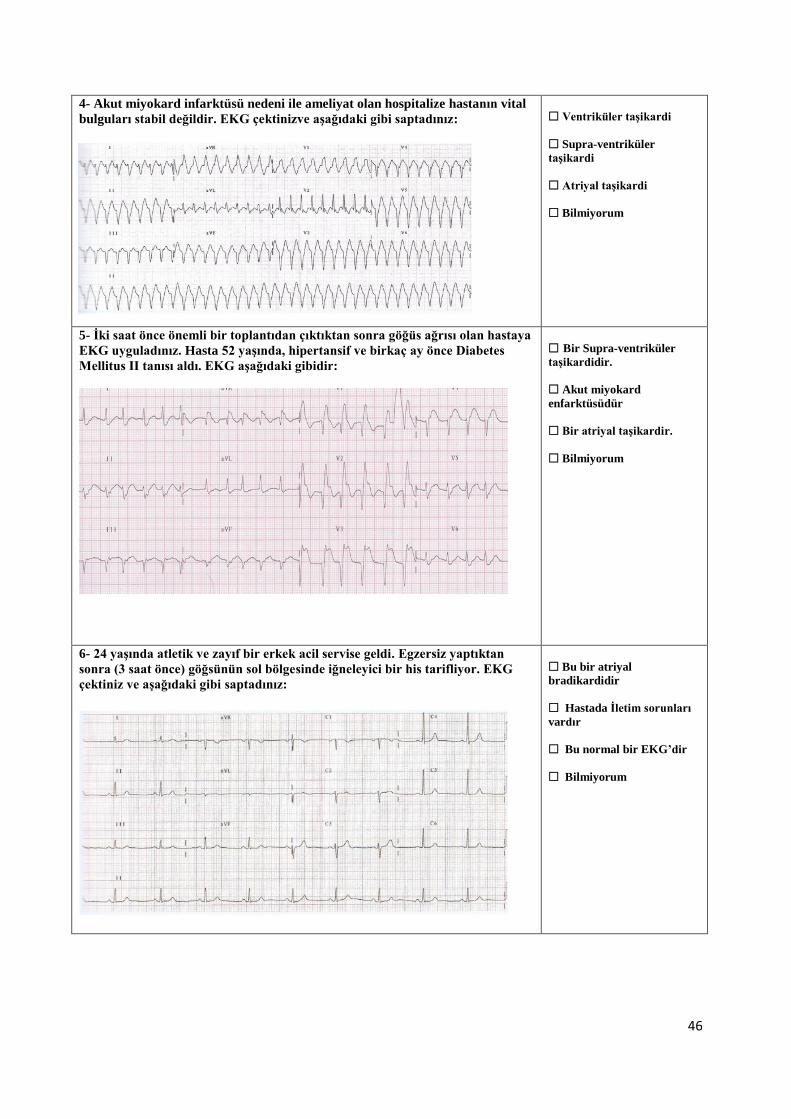
46
4- Akut miyokard infarktüsü nedeni ile ameliyat olan hospitalize hastanın vital
bulguları stabil değildir. EKG çektinizve aşağıdaki gibi saptadınız:
Ventriküler taşikardi
Supra-ventriküler
taşikardi
Atriyal taşikardi
Bilmiyorum
5- İki saat önce önemli bir toplantıdan çıktıktan sonra göğüs ağrısı olan hastaya
EKG uyguladınız. Hasta 52 yaşında, hipertansif ve birkaç ay önce Diabetes
Mellitus II tanısı aldı. EKG aşağıdaki gibidir:
Bir Supra-ventriküler
taşikardidir.
Akut miyokard
enfarktüsüdür
Bir atriyal taşikardir.
Bilmiyorum
6- 24 yaşında atletik ve zayıf bir erkek acil servise geldi. Egzersiz yaptıktan
sonra (3 saat önce) göğsünün sol bölgesinde iğneleyici bir his tarifliyor. EKG
çektiniz ve aşağıdaki gibi saptadınız:
Bu bir atriyal
bradikardidir
Hastada İletim sorunları
vardır
Bu normal bir EKG’dir
Bilmiyorum

47
7- Otuz yaşında bir kadın acil servise çarpıntı, göğüste sıkışma ve nefes darlığı
şikayeti ile başvurdu. EKG çektiniz ve aşağıdaki gibi saptadınız:
Bir ventriküler
taşikardidir
Bir atriyal ekstra
sistoldür
Bir atriyal taşikardidir
Bilmiyorum
8- Bu EKG'ye sahip hastada hangi patalojiyi düşünürsünüz?
Birinci derece kalp bloğu
Herhangi bir patolojisi
yok
Üçüncü derece kalp bloğu
Bilmiyorum
48
Appendix 2. Knowledge and Practices of Electrocardiogram Interpretation of Nurses
(English Version)
A- Demographics characteristics of nurses
1- Age
2- Gender Male Female
3- Years of working experience as
registered nurse
< 1 year
1-5 years
6-10 years
11-20 years
>20 years
4- Currently working unit in hospital Emergency department
Intensive care unit
Coronary care unit
Cardiology department
Recovery unit
5- Education level Bachelor’s
Master
PhD
6- Do you taking ECG for patients Yes
No
7- Previous ECG training course Yes
No
8- ECG education resource University
Courses
Internet
Self learning books
Congress / conferences
Others …………………..
9- Years since taking the last ECG
course
I don’t take
Less than 2 years
Between 2 to 5 years
More than 5 years
10- ECG competency level
Beginner
Intermediate
Advanced
11- Desire to learn about ECG Yes
No

49
B- Knowledge of nurses on Electrocardiogram
1- The p wave represented right and
left atrial repolarization
True
False
2- QRS complex represented right
and left ventricular depolarization
True
False
3- T wave represents ventricular
repolarization
True
False
4- T wave is one of the negative
waves in ECG
True
False
5- Normal PR interval between
0.12 and 0.20 seconds
True
False
6- In normal ECG V1 and aVR leads
are negative waves
True
False
7- Pathologic Q waves are a sign
of previous myocardial infarction
True
False
8- Atrial fibrillation could be regular
rhythm
True
False
9- ECG can detects left ventricular
hypertrophy (LVH)
True
False
10- ST elevation in inferior
myocardial infarction appear in
leads: V1-V6
True
False
11- ST elevation in lateral
myocardial infarction appear in
leads: I, aVL, V5, V6
True
False
12- ST elevation in anterior
myocardial infarction appear in
leads: II, III, aVF
True
False
13- ST depression in ECG indicated
ischemia myocardial
True
False
14- RSR pattern appear in V1, V2
and V3 in right bundle branch block
rhythms
True
False
15- T long wave is and QRS wide
wave seen in case of hypokalemia
True
False

50
C- practice of nurses on ECG interpretations
1- You perform an ECG and observe this rhythm. What do you think it might
be?
A third degree heart block
An atrial flutter
A supra-ventricular
tachycardia
I do not know
2-You perform an ECG and observe this rhythm. How would you act?
Ask for help without
leaving the patient alone
because it is a ventricular
fibrillation
Ask for help without
leaving the patient alone
because it is an atrial
fibrillation
Perform another ECG
because it looks like there
may be interference
You do not know how to
act but you know it must be a
serious problem
3- A patient comes to the emergency department due to a respiratory distress.
He has 140 beats per minute. You perform an ECG and observe the following:
It is an atrial tachycardia
It is an atrial fibrillation
It is an atrial extra-systole
I do not know

51
4- A hospitalized patient who had surgery due to an Acute myocardial
infarction, his vital signs are unstable. You perform an ECG and observe the
following:
The patient presents a
ventricular tachycardia
The patient presents a
supra-ventricular
tachycardia
The patient presents an
atrial tachycardia
I do not know
5- You performed ECG to patient who have chest pain appeared after leaving
an important meeting two hours ago. He is 52 years of age and hypertensive and
a few months ago he was diagnosed with Diabetes Mellitus II. The ECG as the
following:
It is a supra-ventricular
tachycardia
It is an acute myocardial
infarction
It is an atrial tachycardia
I do not know
6- A 24-year-old male comes to the emergency department He is athletic and
slim. He reports feeling a pricking sensation in the left area of his chest since he
finished doing exercise (3 hours earlier). You perform an ECG and observe the
following:
It is an atrial bradycardia
He has conduction
problems
It is a normal ECG
I do not know
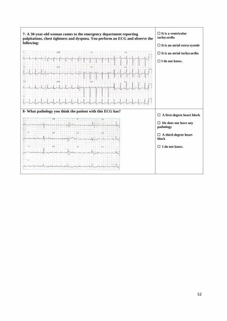
52
7- A 30-year-old woman comes to the emergency department reporting
palpitations, chest tightness and dyspnea. You perform an ECG and observe the
following:
It is a ventricular
tachycardia
It is an atrial extra-systole
It is an atrial tachycardia
I do not know.
8- What pathology you think the patient with this ECG has?
A first-degree heart block
He does not have any
pathology
A third-degree heart
block
I do not know.

53
Appendix 3: Ethical Approval Near East Institutional Reviews Board (IRB)
54

55

56
Appendix 4. Informed Consent Form Participant
INFORMED CONSENT FORM FOR ADULTS
(FOR THE PATIENTS / PARTICIPANTS)
You are invited to participate in a research study conducted by Obaydah Yaser Hamed
Tahboub Supervised by Assoc. Prof. Dr. Ümran Dal Yılmaz from the NEAR EAST
UNIVERSITY Faculty of Health Sciences, Nursing Department. I have learned that
Knowledge and Practices of Electrocardiogram Interpretation of Nurses. You were selected
as a possible participant in this study because findings of the study may be useful in
determine nurses’ Knowledge and Practices of Electrocardiogram Interpretation in health
care institutions. If you decide to participate, a questionnaire will be used as data collection
tool in this study. The questionnaire contains questions regarding for demographics,
knowledge and practices of nurses on ECG. However, I cannot guarantee that I personally
will receive any benefits from this research. Any information that is obtained in connection
with this study and that can be identified with you will remain confidential and will be
disclosed only with your permission or as required by law. Subject identities will be kept
confidential participation is voluntary. Your decision whether or not to participate will not
affect your relationship with Near East University hospital and Dr. Suat Günsel Girne
University hospital. If you decide to participate, you are free to withdraw your consent and
discontinue participation at any time without penalty. If you have any questions about the
study, please feel free to contact [+905428734098,
[email protected]]. [+905326603086, [email protected]]. If you have
questions regarding your rights as a research subject, please contact the NEAR EAST
INSTITUTIONAL REVIEW BOARD. You will be offered a copy of this form to keep.
Your signature indicates that you have read and understand the information provided above,
that you willingly agree to participate, that you may withdraw your consent at any time and
discontinue participation without penalty, that you will receive a copy of this form, and that
you are not waiving any legal claims.
Participant
Name, Surname:
Address:
Phone:
Signature:
Witness
Name, Surname:
Address:
Phone:
Signature:
Interviewer:
Name, Surname:
Address:
Phone:
Signature: