Embed Size (px)
Citation preview

17
Kudoa dianae sp. n. (Myxosporea: Multivalvulida), a new parasite ofbullseye puffer, Sphoeroides annulatus (Tetraodontiformes:Tetraodontidae)
Iva Dyková1, Emma Josefina Fajer Avila2 and Ivan Fiala3
1Institute of Parasitology, Academy of Sciences of the Czech Republic, Branišovská 31, 370 05 České Budějovice, CzechRepublic;
2Centro de Investigación en Alimentación y Desarollo, A.C. Unidad de Investigación en Acuicultura y Manejo Ambiental delCIAD, A.C. Av. Sábalo Cerritos s/a, Estero del Yugo, Mazatlán, Sinaloa, Mexico;
3Faculty of Biological Sciences, University of South Bohemia, Branišovská 31, 370 05 České Budějovice, Czech Republic
Key words: Myxosporea, Multivalvulida, taxonomy, SSU rDNA
Abstract. A new multivalvulid myxosporean species, Kudoa dianae sp. n., is described from bullseye puffer, Sphoeroidesannulatus (Jenyns) (Tetraodontiformes: Tetraodontidae). Plasmodia develop in extramuscular sites, in the wall of oesophagusand less frequently on mesenteries. Mature spores can reach lumen of the digestive tract directly by disruption of plasmodial wallor via macrophage transport to the oesophageal epithelium. New species is characterised by morphology of spores and by thecomplete sequence of SSU rRNA gene that differs from all hitherto known sequences of Kudoa species. Spore morphology(moderate-sized, simple non-ornate spores, quadrate in apical view) clusters with that of Kudoa scienae, K. cerebralis, K.chilkaensis, K. leiostomi, K. funduli, K. cascasia and K. ovivora. Analysis of phylogenetic relationships (using SSU rRNA genesequences) among five Kudoa species, the molecular data of which are available thus far, revealed that K. dianae is distinguish-able from these five species and that its closest relation is with K. miniauriculata.
The list of named species of the genus KudoaMeglitsch, 1947 (with 44 items in Moran et al. 1999 and45 in Swearer and Robertson 1999) expanded recentlyto 47, K. camarguensis and K. ramsayi being the lastnewly described species (Pampoulie et al. 1999, Kala-vati et al. 2000).
Regarding the type of tissue infected, muscle-infecting species (35) predominate. Other species havebeen found in extramuscular localisations, e.g., gills,brain, kidney, gallbladder, and ovaries (Swearer andRobertson 1999). Of the muscle-infecting species, theagents of post-mortem myoliquefaction (K. thyrsites, K.paniformis and K. miniauriculata), have been frequentlystudied and papers describing their biology, impact ofinfections on the aquaculture industry and commercialfisheries and diagnostic methods have been published(Egusa and Nakajima 1980, Kabata and Whitaker 1981,Patashnik et al. 1982, Langdon et al. 1992, Whitakerand Kent 1992, Moser and Kent 1994, Whitaker at al.1996, Moran et al. 1999). Species infecting smoothmuscles, e.g., K. ciliatae, K. intestinalis, K. sphyraeni,K. valamugili (Maeno et al. 1993, Dyková et al. 1994,Swearer and Robertson 1999) were found to developmostly in the wall of digestive tract. Multiple sites ofinfection in the same host specimen were recordedrather exceptionally, e.g., in Kudoa thyrsites infection inCoryphaena hippurus, Kudoa sp. infection in Morone
americana (Swearer and Robertson 1999) and Kudoasp. infection in cultured Sparus aurata (Paperna 1982).
Using cluster analysis of the dissimilarity coeffi-cients, Swearer and Robertson (1999) defined, among45 Kudoa species included in their study, 7 groupingswith unique combinations of taxonomic characters.Different patterns of similarity were discovered byHervio et al. (1997). They compared SSU rDNAsequences of four Kudoa species, analysed theirphylogenetic relationships and concluded, curiouslyenough, that Kudoa species cluster by geographiclocation rather than by morphology of spores. Thisstatement is a big challenge to use both morphologicaland molecular approach in identifying Kudoa species.As several papers (Paperna 1982, Langdon 1990, Lomet al. 1992, Maeno et al. 1993, Swearer and Robertson1999) evidenced, Kudoa spp. developing in uncommonsites or unusual types of tissues deserve attention evenwhen their impact on the host is not dramatic. Webelieve that the study of extramuscularly developingKudoa species can help to better understand the biologyof this group of myxosporeans. Below we describe anew species of the genus Kudoa Meglitsch, 1947 fromthe bullseye puffer, Sphoeroides annulatus (Jenyns)(Tetraodontidae), a fish species native to the coast ofPacific state of Sinaloa (Mexico). The cultivationpotential of this fish is currently being estimated(Duncan and Rodríguez 2001).
FOLIA PARASITOLOGICA 49: 17-23, 2002
Address for correspondence: I. Dyková, Institute of Parasitology ASCR, Branišovská 31, 370 05 České Budějovice, Czech Republic. Phone:++420 38 777 5423; Fax: ++420 38 5300 388; E-mail: [email protected]

18
MATERIALS AND METHODS
In total 150 bullseye puffers, Sphoeroides annulatus(Jenyns, 1842) were collected along the Pacific Coast, inBahía de La Paz, BCS, and off the coast of Mazatlán, Sinaloa,Mexico. Thirty-seven juvenile specimens were 17.2 (10.5-22.0) cm in length and their weight was 104.2 (40-204) g.Specimens (in total 113) of adult-age group were 28.3 (20.5-39.4) cm in length and their weight was 527.9 (250-1450) g.
The body cavity of fish was opened from the anal orifice tothe gills and buccal cavity and the internal organs wereexamined. A macroscopic inspection of organs was followedby examination of fresh mounts of tissue pieces about 3 mm indiameter, compressed between the slide and coverslip. Severalsamples from each organ and from different parts of bodymusculature were examined. Samples of all organs andsomatic muscles were routinely fixed with Davidson fixativeand processed for histology using Paraplast as the embeddingmedium and haematoxylin and eosin (H&E) and Giemsasolutions for staining.
The lesions that contained Kudoa spores were fixed alsofor transmission electron microscopy (in 3% glutaraldehydebuffered with sodium cacodylate), stored for 3 weeks inholding sodium cacodylate buffer and postfixed with 1%buffered osmium tetroxide. After dehydration in an acetonegradient series, the tissues were embedded in Spurr’s resin.The ultrathin sections were stained with uranyl acetate andlead citrate and observed in a JEOL JEM 1010 electronmicroscope.
Spores collected from fresh material were fixed in 80%ethanol (EM grade) and submitted to SSU rRNA genesequence analysis. Approximately 100 spores stored in ethanolwere centrifuged and washed two times in PBS. Three freeze-thaw cycles were performed in order to release the contents ofspores. DNA was extracted with DNeasy Tissue Kit (Qiagen)according to manufacturer’s protocol and resuspended in afinal step in 200 µl H2O. The almost entire SSU rRNA genewas amplified using forward primer 5’-GGTCATATGCTCGTCTCAAA-3’and reverse primer 5’-TACAAAGGGCAGAGAC-3’ designed out of alignment of four Kudoa speciesstudied by Hervio et al (1997): Kudoa paniformis(AF034640), K. miniauriculata (AF034639), K. amamiensis(AF034638) and K. thyrsites (AF031413). PCR reactions wereperformed in total volume of 25 µl containing PCR buffer(TaKaRa), 2 mM of MgCl2, 0.2 mM of dNTPs, 1 unit of TaqDNA polymerase (TaKaRa) and 25 pmol of each primer in aT3 Thermocycler (Biometra). Denaturation at 95°C for 3 minwas followed by 35 cycles consisting of 94°C for 1 min, 53°Cfor 1 min and 72°C for 2 min and ended by 10 min extensionat 72°C. PCR products were cloned into pCR® 2.1 TOPOCloning vector using the TOPO-TA Cloning Kit (Invitrogen)and sequenced on an automatic sequencer CEQTM 2000(Beckman Coulter) using CEQ DTCS Dye Kit (BeckmanCoulter) according to the manufacturer’s protocol. The almostentire SSU rRNA gene sequences were aligned usingMegAlign version 4.00 (DNASTAR package) with ClustalWalgorithm. Genetic distances were calculated using the Kimura2-parameter algorithm (Kimura 1980) out of alignment withexcluded gaps. Ambiguous regions, incomplete sequences,invariant sites and gaps were removed (1618 characters), and
remaining 580 variable sites were used for phylogeneticanalysis. Phylogenetic trees were constructed using maximumparsimony, neighbour-joining and maximum likelihoodmethods in the frame of PAUP program (Swofford 1998). Therobustness of the tree was tested by bootstrap analysis with1000 replicates.
RESULTS
Spores with all characteristics of the genus KudoaMeglitsch, 1947 were found in macroscopical lesionslocalised in two sites: in the wall of oesophagus, and onmesenteries. White cystic formations (plasmodialstages) and white or yellowish formations with lessdefined borders (masses of spores originated fromplasmodia) were easily detectable through the mucosaepithelium when oesophagus was cut open. Such lesionswere found in 7 (19%) out of 37 juvenile specimens ofSphoeroides annulatus and in 22 (20%) out of 113 adultspecimens. Less frequent were lesions localised onmesenteries (prevalence 5.4% in juveniles and 7.9% inadult specimens). Lesions localised in both sites werefound in 24% of juveniles and 34% of adult specimensexamined.Description
In apical view mature spores (Fig. 1) were quadratewith rounded edges. Four polar capsules were equal-sized, pyriform, 2.0 µm in length and 1.5 µm in width.The two turns of the polar filament were not clearlyseen in fresh spores, but were counted in electronmicrographs (Figs. 12, 13). The length of spores meas-ured in lateral view was 5 (4.5-5.5) µm, the width aswell as thickness was 6 (5.5-6.5) µm. The shape ofspores was very simple in both apical and lateral views,the posterior part of spores was rounded, suture lineswere only slightly indicated in fresh spores. Shell valveswere thickened in the apical, slightly protruding part ofspore (Figs. 1, 12).
Fig. 1. Spores of Kudoa dianae sp. n. in apical and lateralviews. Nomarski differential interference contrast.

Dyková et al.: Kudoa dianae sp. n.
19
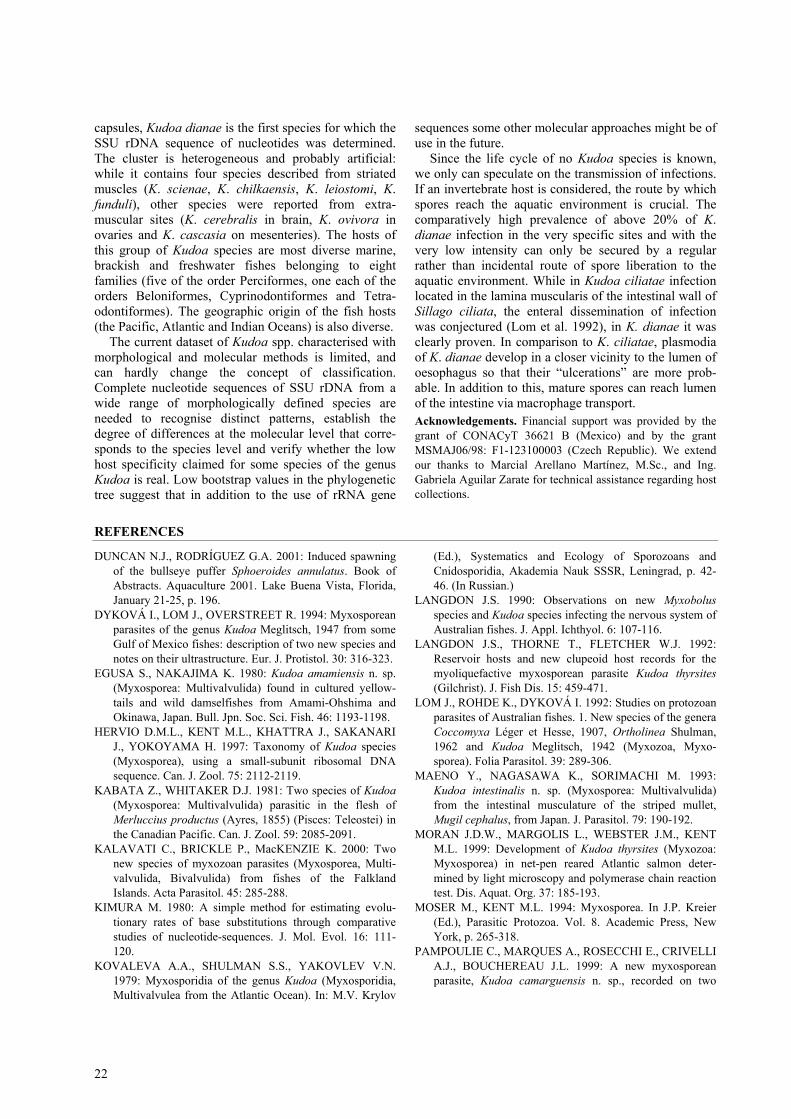
Figs. 2-6. Histology of Kudoa dianae infection in Sphoeroides annulatus. H&E. Fig. 2. Part of plasmodium localised under theepithelium of oesophagus, ×210. Fig. 3. Agglomeration of spores in the connective tissue of oesophageal wall. Note thedesquamation of mucosa epithelium (top), ×250. Fig. 4. Mature spores surrounding in huge amounts the blood vessel inoesophagus, ×860. Figs. 5, 6. Substitution of epithelial layer of oesophagus with mature spores transported via macrophages,×240 and ×840 respectively.

20
Figs. 7, 8. Kudoa dianae infection in Sphoeroides annulatus: host-parasite interface with numerous projections on the peripheryof plasmodial stage. Transmission electron micrographs. Figs. 9-13. Development of Kudoa dianae. Transmission electronmicrographs. Fig. 9. Sporoblast in the early phase of differentiation. Fig. 10. Capsulogenesis. Fig. 11. Capsulogenesis andsporogenesis. Figs. 12, 13. Mature spores.
In the process of sporogenesis (Figs. 9-11), thesporoplasm complex represented by one cell envelopingthe other (Fig. 10) and a lack of pansporoblast formationwere observed. Numerous, sometimes interconnectedprojections on the periphery of plasmodial stages (Figs.7, 8) characterised the host-parasite interface. Theyreminded to some extent of rich folding of plasma-lemma in Kudoa shkae (Dyková et al. 1994).
Polysporous plasmodia filled with mature sporeswere white, spherical or ovoid formations with amaximum diameter of 5.0 mm. Histological examina-
tion of oesophageal lesions revealed that plasmodiadeveloped in subepithelial connective tissue, laminamucosa and among muscle fibres of oesophageal wall.Molecular data
The amplified and sequenced SSU rRNA gene regionof this Kudoa species was 1568nt in length includingregions corresponding to forward (20nt) and reverse(16nt) primers. The G+C content of the sequenced genewas 46.17%. The sequence was deposited in theGenBank under accession number AF 414692. The SSUrRNA gene of this Kudoa species from S. annulatus
Dyková et al.: Kudoa dianae sp. n.
21
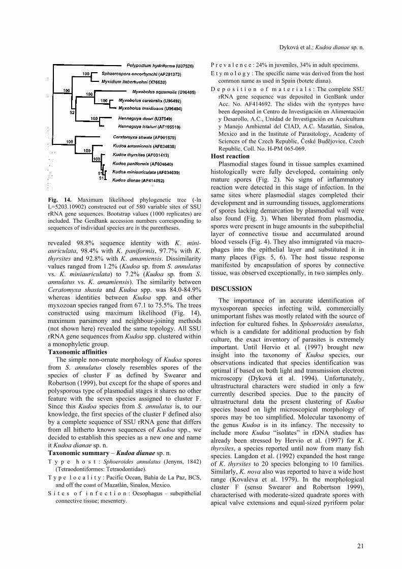
Fig. 14. Maximum likelihood phylogenetic tree (-lnL=5203.10902) constructed out of 580 variable sites of SSUrRNA gene sequences. Bootstrap values (1000 replicates) areincluded. The GenBank accession numbers corresponding tosequences of individual species are in the parentheses.
revealed 98.8% sequence identity with K. mini-auriculata, 98.4% with K. paniformis, 97.7% with K.thyrsites and 92.8% with K. amamiensis. Dissimilarityvalues ranged from 1.2% (Kudoa sp. from S. annulatusvs. K. miniauriculata) to 7.2% (Kudoa sp. from S.annulatus vs. K. amamiensis). The similarity betweenCeratomyxa shasta and Kudoa spp. was 84.0-84.9%whereas identities between Kudoa spp. and othermyxozoan species ranged from 67.1 to 75.5%. The treesconstructed using maximum likelihood (Fig. 14),maximum parsimony and neighbour-joining methods(not shown here) revealed the same topology. All SSUrRNA gene sequences from Kudoa spp. clustered withina monophyletic group.Taxonomic affinities
The simple non-ornate morphology of Kudoa sporesfrom S. annulatus closely resembles spores of thespecies of cluster F as defined by Swearer andRobertson (1999), but except for the shape of spores andpolysporous type of plasmodial stages it shares no otherfeature with the seven species assigned to cluster F.Since this Kudoa species from S. annulatus is, to ourknowledge, the first species of the cluster F defined alsoby a complete sequence of SSU rRNA gene that differsfrom all hitherto known sequences of Kudoa spp., wedecided to establish this species as a new one and nameit Kudoa dianae sp. n.Taxonomic summary – Kudoa dianae sp. n.T y p e h o s t : Sphoeroides annulatus (Jenyns, 1842)
(Tetraodontiformes: Tetraodontidae).T y p e l o c a l i t y : Pacific Ocean, Bahía de La Paz, BCS,
and off the coast of Mazatlán, Sinaloa, Mexico.S i t e s o f i n f e c t i o n : Oesophagus – subepithelial
connective tissue; mesentery.
P r e v a l e n c e : 24% in juveniles, 34% in adult specimens.E t y m o l o g y : The specific name was derived from the host
common name as used in Spain (botete diana).D e p o s i t i o n o f m a t e r i a l s : The complete SSU
rRNA gene sequence was deposited in GenBank underAcc. No. AF414692. The slides with the syntypes havebeen deposited in Centro de Investigación en Alimentacióny Desarollo, A.C., Unidad de Investigación en Acuiculturay Manejo Ambiental del CIAD, A.C. Mazatlán, Sinaloa,Mexico and in the Institute of Parasitology, Academy ofSciences of the Czech Republic, České Budějovice, CzechRepublic, Coll. No. H-PM 065-069.
Host reactionPlasmodial stages found in tissue samples examined
histologically were fully developed, containing onlymature spores (Fig. 2). No signs of inflammatoryreaction were detected in this stage of infection. In thesame sites where plasmodial stages completed theirdevelopment and in surrounding tissues, agglomerationsof spores lacking demarcation by plasmodial wall werealso found (Fig. 3). When liberated from plasmodia,spores were present in huge amounts in the subepitheliallayer of connective tissue and accumulated aroundblood vessels (Fig. 4). They also immigrated via macro-phages into the epithelial layer and substituted it inmany places (Figs. 5, 6). The host tissue responsemanifested by encapsulation of spores by connectivetissue, was observed exceptionally, in two samples only.
DISCUSSION
The importance of an accurate identification ofmyxosporean species infecting wild, commerciallyunimportant fishes was mostly related with the source ofinfection for cultured fishes. In Sphoeroides annulatus,which is a candidate for additional production by fishculture, the exact inventory of parasites is extremelyimportant. Until Hervio et al. (1997) brought newinsight into the taxonomy of Kudoa species, ourobservations indicated that species identification wasoptimal if based on both light and transmission electronmicroscopy (Dyková et al. 1994). Unfortunately,ultrastructural characters were studied in only a fewcurrently described species. Due to the paucity ofultrastructural data the present clustering of Kudoaspecies based on light microscopical morphology ofspores may be too simplified. Molecular taxonomy ofthe genus Kudoa is in its infancy. The necessity toinclude more Kudoa “isolates” in rDNA studies hasalready been stressed by Hervio et al. (1997) for K.thyrsites, a species reported until now from many fishspecies. Langdon et al. (1992) expanded the host rangeof K. thyrsites to 20 species belonging to 10 families.Similarly, K. nova also was reported to have a wide hostrange (Kovaleva et al. 1979). In the morphologicalcluster F (sensu Swearer and Robertson 1999),characterised with moderate-sized quadrate spores withapical valve extensions and equal-sized pyriform polar
22
capsules, Kudoa dianae is the first species for which theSSU rDNA sequence of nucleotides was determined.The cluster is heterogeneous and probably artificial:while it contains four species described from striatedmuscles (K. scienae, K. chilkaensis, K. leiostomi, K.funduli), other species were reported from extra-muscular sites (K. cerebralis in brain, K. ovivora inovaries and K. cascasia on mesenteries). The hosts ofthis group of Kudoa species are most diverse marine,brackish and freshwater fishes belonging to eightfamilies (five of the order Perciformes, one each of theorders Beloniformes, Cyprinodontiformes and Tetra-odontiformes). The geographic origin of the fish hosts(the Pacific, Atlantic and Indian Oceans) is also diverse.
The current dataset of Kudoa spp. characterised withmorphological and molecular methods is limited, andcan hardly change the concept of classification.Complete nucleotide sequences of SSU rDNA from awide range of morphologically defined species areneeded to recognise distinct patterns, establish thedegree of differences at the molecular level that corre-sponds to the species level and verify whether the lowhost specificity claimed for some species of the genusKudoa is real. Low bootstrap values in the phylogenetictree suggest that in addition to the use of rRNA gene
sequences some other molecular approaches might be ofuse in the future.
Since the life cycle of no Kudoa species is known,we only can speculate on the transmission of infections.If an invertebrate host is considered, the route by whichspores reach the aquatic environment is crucial. Thecomparatively high prevalence of above 20% of K.dianae infection in the very specific sites and with thevery low intensity can only be secured by a regularrather than incidental route of spore liberation to theaquatic environment. While in Kudoa ciliatae infectionlocated in the lamina muscularis of the intestinal wall ofSillago ciliata, the enteral dissemination of infectionwas conjectured (Lom et al. 1992), in K. dianae it wasclearly proven. In comparison to K. ciliatae, plasmodiaof K. dianae develop in a closer vicinity to the lumen ofoesophagus so that their “ulcerations” are more prob-able. In addition to this, mature spores can reach lumenof the intestine via macrophage transport.Acknowledgements. Financial support was provided by thegrant of CONACyT 36621 B (Mexico) and by the grantMSMAJ06/98: F1-123100003 (Czech Republic). We extendour thanks to Marcial Arellano Martínez, M.Sc., and Ing.Gabriela Aguilar Zarate for technical assistance regarding hostcollections.
REFERENCES
DUNCAN N.J., RODRÍGUEZ G.A. 2001: Induced spawningof the bullseye puffer Sphoeroides annulatus. Book ofAbstracts. Aquaculture 2001. Lake Buena Vista, Florida,January 21-25, p. 196.
DYKOVÁ I., LOM J., OVERSTREET R. 1994: Myxosporeanparasites of the genus Kudoa Meglitsch, 1947 from someGulf of Mexico fishes: description of two new species andnotes on their ultrastructure. Eur. J. Protistol. 30: 316-323.
EGUSA S., NAKAJIMA K. 1980: Kudoa amamiensis n. sp.(Myxosporea: Multivalvulida) found in cultured yellow-tails and wild damselfishes from Amami-Ohshima andOkinawa, Japan. Bull. Jpn. Soc. Sci. Fish. 46: 1193-1198.
HERVIO D.M.L., KENT M.L., KHATTRA J., SAKANARIJ., YOKOYAMA H. 1997: Taxonomy of Kudoa species(Myxosporea), using a small-subunit ribosomal DNAsequence. Can. J. Zool. 75: 2112-2119.
KABATA Z., WHITAKER D.J. 1981: Two species of Kudoa(Myxosporea: Multivalvulida) parasitic in the flesh ofMerluccius productus (Ayres, 1855) (Pisces: Teleostei) inthe Canadian Pacific. Can. J. Zool. 59: 2085-2091.
KALAVATI C., BRICKLE P., MacKENZIE K. 2000: Twonew species of myxozoan parasites (Myxosporea, Multi-valvulida, Bivalvulida) from fishes of the FalklandIslands. Acta Parasitol. 45: 285-288.
KIMURA M. 1980: A simple method for estimating evolu-tionary rates of base substitutions through comparativestudies of nucleotide-sequences. J. Mol. Evol. 16: 111-120.
KOVALEVA A.A., SHULMAN S.S., YAKOVLEV V.N.1979: Myxosporidia of the genus Kudoa (Myxosporidia,Multivalvulea from the Atlantic Ocean). In: M.V. Krylov
(Ed.), Systematics and Ecology of Sporozoans andCnidosporidia, Akademia Nauk SSSR, Leningrad, p. 42-46. (In Russian.)
LANGDON J.S. 1990: Observations on new Myxobolusspecies and Kudoa species infecting the nervous system ofAustralian fishes. J. Appl. Ichthyol. 6: 107-116.
LANGDON J.S., THORNE T., FLETCHER W.J. 1992:Reservoir hosts and new clupeoid host records for themyoliquefactive myxosporean parasite Kudoa thyrsites(Gilchrist). J. Fish Dis. 15: 459-471.
LOM J., ROHDE K., DYKOVÁ I. 1992: Studies on protozoanparasites of Australian fishes. 1. New species of the generaCoccomyxa Léger et Hesse, 1907, Ortholinea Shulman,1962 and Kudoa Meglitsch, 1942 (Myxozoa, Myxo-sporea). Folia Parasitol. 39: 289-306.
MAENO Y., NAGASAWA K., SORIMACHI M. 1993:Kudoa intestinalis n. sp. (Myxosporea: Multivalvulida)from the intestinal musculature of the striped mullet,Mugil cephalus, from Japan. J. Parasitol. 79: 190-192.
MORAN J.D.W., MARGOLIS L., WEBSTER J.M., KENTM.L. 1999: Development of Kudoa thyrsites (Myxozoa:Myxosporea) in net-pen reared Atlantic salmon deter-mined by light microscopy and polymerase chain reactiontest. Dis. Aquat. Org. 37: 185-193.
MOSER M., KENT M.L. 1994: Myxosporea. In J.P. Kreier(Ed.), Parasitic Protozoa. Vol. 8. Academic Press, NewYork, p. 265-318.
PAMPOULIE C., MARQUES A., ROSECCHI E., CRIVELLIA.J., BOUCHEREAU J.L. 1999: A new myxosporeanparasite, Kudoa camarguensis n. sp., recorded on two

Dyková et al.: Kudoa dianae sp. n.
23
goby species (Teleostei: Pisces) in the Rhône delta (Medi-terranean Sea, France). J. Euk. Microbiol. 46: 304-310.
PAPERNA I. 1982: Kudoa infection in the glomeruli,mesentery and peritoneum of cultured Sparus aurata L. J.Fish Dis. 5: 539-543.
PATASHNIK M., GRONINGER H.S., BARNETT H.,KUDO G., KOURY B. 1982: Pacific whiting, Merlucciusproductus: I. Abnormal muscle texture caused by myxo-sporidian-induced proteolysis. Mar. Fish. Rev. 44: 1-12.
SWEARER S.E., ROBERTSON D.R. 1999: Life history,pathology, and description of Kudoa ovivora n. sp.
(Myxozoa, Myxosporea): an ovarian parasite of Caribbeanlabroid fishes. J. Parasitol. 85: 337-353.
SWOFFORD D.U. 1998: PAUP*: Phylogenetic AnalysisUsing Parsimony (*and other methods). Version 4.0b4.Sinauer Associates, Sunderland, Massachusetts, USA.
WHITAKER D.J., KENT M.L. 1992: Kudoa thyrsites(Myxosporea) and soft flesh in pen-reared coho salmon.Newsl. Am. Fish. Soc. Fish Health Sect. 20: 4-5.
WHITAKER D.J., KENT M.L., SAKANARI J.A. 1996:Kudoa miniauriculata n. sp. (Myxozoa, Myxosporea)from the musculature of bocaccio (Sebastes paucispinus)from California. J. Parasitol. 82: 312-315.
Received 29 May 2001 Accepted 27 August 2001