Embed Size (px)
Citation preview

Cerebrovascular diseases
2nd most often reason of death and the most often reason of disability,
every year - 16.9 millions new strokes and a 5.9 millions of death
Disability-Adjusted Life Years and Death in relations to strokes
1990 vs 2013
From 3,54% to 4,62%
From 9,66% to 11,75%,
Feigin VL et al. Neuroepidemiology 2015;45:161–176; Feigin VL et al., Lancet Neurol 2009;8:355-369.

Epidemiology
Incidence – 125 – 446/100 000 inhabitants (Feigin V.L. et al.,
Lancet Neurol, 2009)
SLOVAKIA
Mortality: 100-200/100 000
Incidence: 224/100 000

Cerebrovascular diseases
Diseases with sudden onset, or rapid development, of focal cerebral dysfunction as the consequence of lesion of cerebral arteries. There are 2 types:
Brain ischemia (stroke) or
Brain haemorrhage

Stroke
Haemorrhage
•Intracerebral
•Subarachnoid
Ischemic
Ateroscle
rotic
Penetrating
Arteries
(lacunar)
Cardiogenic
embolic
•FP
•Valvular d.
•Atrial
trombus
•Others
Cryptogenic•Other rare
diseases
•Protrombotic
disease
•Dissection
•Arteritis
•Others
15%
85%
20% 20%25% 5%30%
Hypo
perfusion
AS
embolic

Cerebrovascular diseases

Cerebrovascular diseases

Cerebrovascular diseases
Subarachnoid
haemorhage
Brain haemorrhageBrain ischemia

Head injury, NO stroke
Subduralny hematoma Epidural hematoma

Anatomy of cerebral arteries


Vertebral arteries
Foramina transversaria

Regulation of cerebral circulation
Blod flow - 50 – 60 ml/100 g of brain tissue/min.
Blod flow below 20 ml/100 g/min. – functional changes of neurons – reversible dysfunction (few hours)
Blod flow below 12, or 10 ml/100 g/min structural changes irreversible changes – brain infarct,


Ford AL et al. Stroke. 2011 May ; 42(5): 1307–1313.doi:10.1161/STROKEAHA.110.600957.
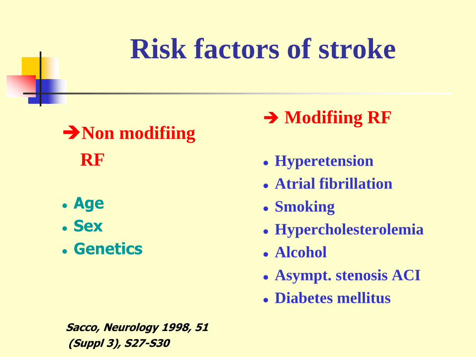
Risk factors of stroke
Non modifiing
RF
Age
Sex
Genetics
Sacco, Neurology 1998, 51
(Suppl 3), S27-S30
Modifiing RF
Hyperetension
Atrial fibrillation
Smoking
Hypercholesterolemia
Alcohol
Asympt. stenosis ACI
Diabetes mellitus

Nontreated arterial hypertension
A – at admission B – hours after admission

Arterial hypertension
The most severe risk factor for stroke
Recommendation of „International Society on Hypertension“ –targer values
- 135/85 Torr in patients without organs damage,
- 130/80 Torr in patients with organs damage, in correlation with AHA, ACC recommendations
According „National Heart, Lung, and Blood Institue-appointed“ panel for older people recommendation is:
Target value –
< 150/90 mm Hg for patients ˃ 60 years of age
< 140/90 mm Hg for younger patients
Meschia JF et al. Guidelines for the primary prevention of stroke. A statement for the healthcare professionals from the
American heart Association/American Stroke Association. Stroke 2014;45:3754-3832.
Feigin VL et al. Neuroepidemiology 2015;45:161–176; Feigin VL et al., Lancet Neurol 2009;8:355-369.

Arterial hypertension
According „National Heart, Lung, and Blood Institue-appointed“ panel for older people recomendation is:
Target value - < 150/90 mm Hg for patients ˃ 60 years of age
And < 140/90 mm Hg for younger patients pre mladších pacientov
Z každodennej klinickej praxe môžeme potvrdiť, že tieto hodnoty TK sú pacientmi lepšie tolerované
Nízke hodnoty TK sú v každodennej praxi často sprevádzane pocitmi závrativosti
V každodennej praxi - prudké znižovanie hodnôt tlaku, hlavne v akútnom štádiu iCMP – nežiadúce hypoperfúzia
mozgu.
Feigin VL et al. Neuroepidemiology 2015;45:161–176; Feigin VL et al., Lancet Neurol 2009;8:355-369.

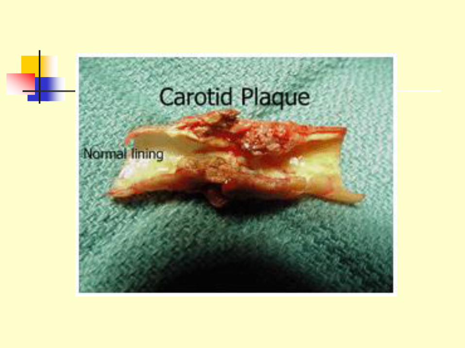
Atherosclerosis
Risk of embolism, hypoperfusion
Spagnoli L.G. et al.: J Nucl Med, 2007

Patients after stroke/TIA 2011 EFNS guidelines

AF 5-times increased risk of strokey
Marini C et al. Stroke 2005;36:1115–9
Patients with AF
Patients without AF
Recurrent stroke
Months after 1st stroke
Kum
ula
tívna p
ravdepodobnosť
rekure
nci
e (
%)
10
12
8
6
4
2
00 2 4 6 8 10
P=0,0398

AF depending on age
Feinberg W.M., Arch Intern Med 1995

Kardiálne ochorenia
Fibrilácia predsiení
Warfarin vs Aspirin
redukcia RR – 39% (ESC guidelines, Europace, 2010)
BAFTA – redukcia RR - 52% (INR: 2-3) (Mant J. et al.,
Lancet, 2007)
WASPO – nežiadúce účinky – 6% vs 33% (Hart R.G. et al.,
Ann Intern Med, 2007)
Riziko krvácania – INR: 3,5 – 4,0
HAS-BLED – skóre rizika krvácania 3 – vysoké riziko

Fatrial fibrillation
Risk of stroke
CHADS2 – (cardiac insufficiency, hypertension, age,
diabetes, stroke)
2 – high risk
NOACs - trombin inhibitor - Dabigatran, inhibitors
facator Xa – Rivaroxaban, Apixaban, Edoxaban
HAS-BLED – risk of bleeding 3 – high risk


71 years old man
Isch heart disease
AF
AH
TIA in history
Hypercholesterolemia
CHADS2 = 3
Treatment – ASA 100 mg, Corvitol, Dicardin, Rasilez
Archív Neurologickej kliniky UNLP a LF UPJŠ Košice

71 years old man
iv thrombolysis
Thrombectomy
mRS – 4 points
Archív Neurologickej kliniky UNLP a LF UPJŠ Košice

79 years old woman
Isch heart disease
AF
1 month after TEP
CHADS2 = 3
Treatment – LMWH from TEP, Isoptin, Digoxin, Furosemid, Cardilan, Zyllt

79 years old woman
Prefrontal syndrome, apathy, weakness of left lower extremity,
Out of time window – no IVT, no TE
mRS – 4 5 6 (after 2M)


Diabetes mellitus (DM)
Risk of atherotrombotic strokes, lacunes, dementia

Trombophilia
Z.K., female, 25 years
3 days after delivery
Posit. familial history
Deficit AT III
3a 3b 3c 4
Szilasiová J., .....Gdovinová Z.: Cerebrovasc Dis, 2007

Trombophilia
L.T., man, 55 years
Repeating strokes – leftside hemiparesis (2003, 2005), sekundary
epilepsy
Posit. familial history
FV Leiden, MTHFR homozygot
Szilasiová J., .....Gdovinová Z.: Cerebrovasc Dis, 2007

Cryptogenic stroke
Stroke with Unknown etiology
AF – 4,9-9,2%
Better detection – AF cca 25%
What is the best method – Holter 24 hours, 2 weeks, longer?
Duration of AF – 30 sek? 6 min?
Brachmann J. et al.: Circ Arrhytm Electrophysiol, 2016; 9:e003333. DOI:10.1161/CIRCEP.115.003333 ; Healey JS et al., NEJM 2012 ; 366:120-129.
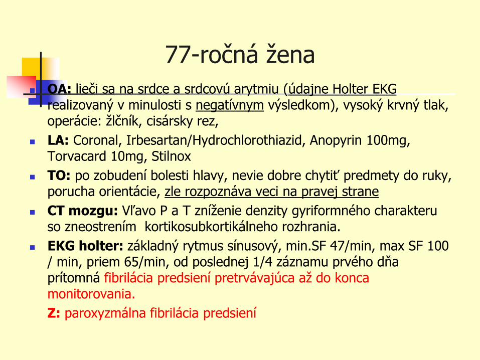
77-ročná žena
OA: lieči sa na srdce a srdcovú arytmiu (údajne Holter EKG realizovaný v minulosti s negatívnym výsledkom), vysoký krvný tlak, operácie: žlčník, cisársky rez,
LA: Coronal, Irbesartan/Hydrochlorothiazid, Anopyrin 100mg, Torvacard 10mg, Stilnox
TO: po zobudení bolesti hlavy, nevie dobre chytiť predmety do ruky, porucha orientácie, zle rozpoznáva veci na pravej strane
CT mozgu: Vľavo P a T zníženie denzity gyriformného charakteru so zneostrením kortikosubkortikálneho rozhrania.
EKG holter: základný rytmus sínusový, min.SF 47/min, max SF 100 / min, priem 65/min, od poslednej 1/4 záznamu prvého dňa prítomná fibrilácia predsiení pretrvávajúca až do konca monitorovania.
Z: paroxyzmálna fibrilácia predsiení

77-ročná žena

77-ročný muž Osobná anamnéza:
• st.p.DF Q IM, znížená EF 40-45%,
• art.hypertenzia III st.,
• incip. CHRI na podklade vaskulárnej nefrosklerózy a diabetickej nefropatie,
• Mi reg I.st s mierne dilatovaou ĽP,
• Trireg. I st. bez významnejšej PH,
• Dyslipidémia
• DM 2. typu na inzulíne s komplikáciami,
• Primárna hypotyreóza na podklade dif. lymfocyt. tyreoiditídy v substitučnej liečbe,
LA: metoprolol, Co-Valsacor, Trimetazidin SR 35mg, Anopyrin 100 mg, Ezetrol, Atoris 20 mg, Euthyrox, Inzulíny: Novorapid Penfil 3x5 jj s.c., inzulín Lantus 2 x 16 jj s.c.

77-ročný muž
TO: včera ráno, keď sa zobudil, všimol si, že nevidí predmety na pravej strane, spozoroval od včera bolesti hlavy, ktoré prakticky neustupujú, zle spal, potil sa, okrem toho má pocit, že nemá istý krok - pocit, akoby sa motal.
CT mozgu: Vľavo mediookcipitálne kortikosubkortikálne -ischemické zmeny v štádiu cytotoxického edému.
EKG Holter: striedanie fibrilácie predsiení so SR, opakované RR nad 2500 ms, max. RR > 4000ms, min. frekvencie pod 30/min. Komor. ektópia je ojedinelá. ST/T bez signif. dynamických zmien - sinonodálna dysfunkcia s indikáciou na implantáciu KS.
Odp. Implantácia TKS

Classification of stroke I.
Old defintion TIA – transitory ischemic atack - lasts 1 hour
Brain infarct – completed stroke
New „tissue-based“ definition of TIA
A brief episode of neurological dysfunction caused by
focal brain or retinal ischemia, with clinical symptoms
typically lasting less than one hour, and without evidence
of acute infarction
Ischemic stroke (brain infarct) is defined as an infarction of
central nervous system tissue.

Classification of stroke II.
Territory of a. cerebri media
Territory of a. cerebri anterior
Territory of a. cerebri posterior
Territory of a. bazilaris (vertebrobasilar)
Territory of a. carotis interna
Territory of a. carotis communis
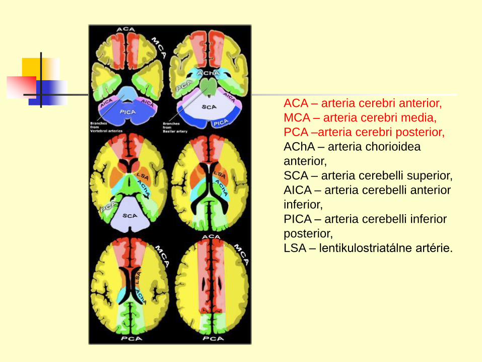
ACA – arteria cerebri anterior,
MCA – arteria cerebri media,
PCA –arteria cerebri posterior,
AChA – arteria chorioidea
anterior,
SCA – arteria cerebelli superior,
AICA – arteria cerebelli anterior
inferior,
PICA – arteria cerebelli inferior
posterior,
LSA – lentikulostriatálne artérie.

MCA territory
The most often embolic etiology – very sudden onset
Speech disorder, hemiparesis (dominantly on upper
extremity, central lesion of n. VII.
Wernicke – Mann position of the body

ACA territory
Central paresis of lower extremity
Disorders of behaviour – prefrontal sy

PCA territory
Visual field disorders – homonymous
hemianopsia

BA territory
Dizziness, diplopia, nystagmus,
hemiparesis or kvadruparesis,
hemiplegia alternans, cranial nerves
lesions, problems with deglutination and
speech

Classification of stroke III.
Brain infarct
Lacunar infarct – diameter less than 1,5
cm

Diagnostics of stroke
Clinical feature
Brain CT
Laboratory tests – RBC, SR, coagulation,
fibrinogen, Na, K, sugar, urea, kreatinin,
cholesterol, triglycerids, CRP, TPIT
Duplex US of carotid arteries
ECHO cardiography

Brain CT – early signs of ischemia
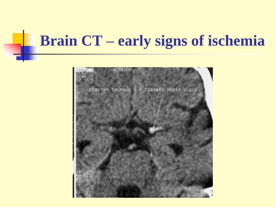
Brain CT – early signs of ischemia

Brain CT – ischemia

Brain CT – ischemia

Brain CT – ischemia

„Wake-up stroke“
Brain MR – FLAIR Brain MR – DWI

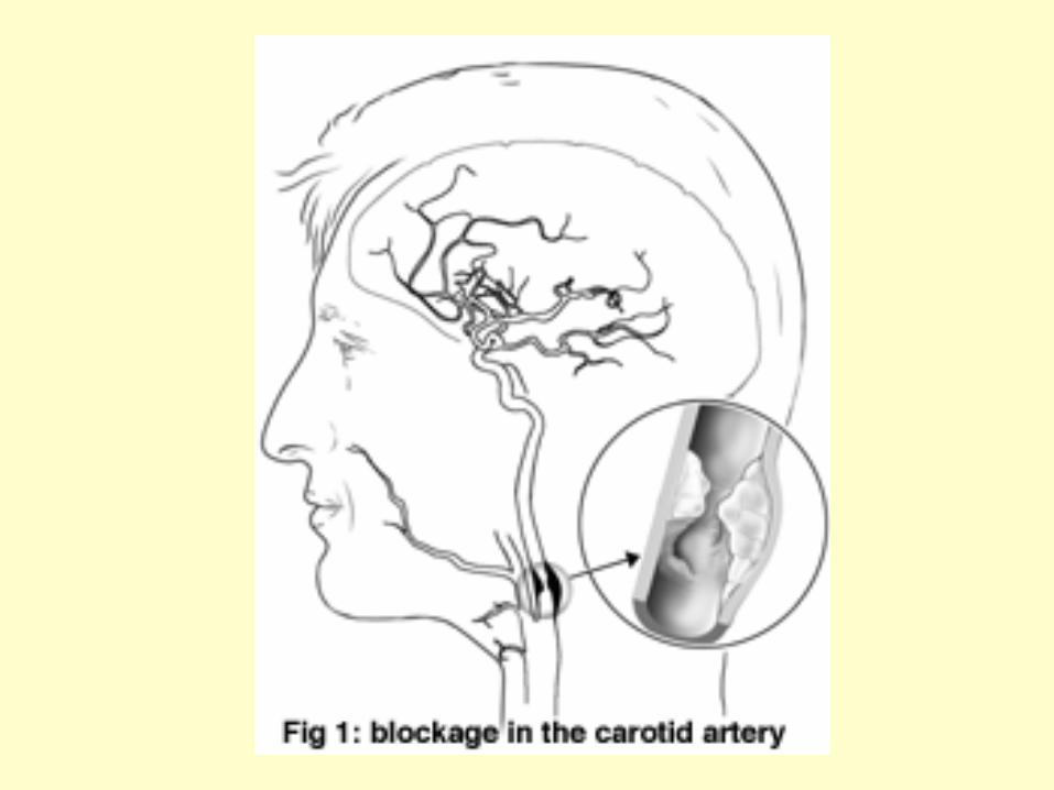
ICA stenosis

ICA stenosis

ICA stenosis

Duplex of carotid arteries
and AG

Stroke therapy
Acute
1/ Trombolysis - rt-PA (recombinant tissue plasminogen activator) -
≤ 4.5 hours after first symptoms!
2/ Thrombectomy
≤ 6 hours after first symptoms!
3/ ASA – 325 mg – later that 6 hours
1/ - i.v. rt-PA 0,9 mg/kg
Brain CT – negative, early signs ofischemia

Možnosť úpravy neurologického poškodenia trombolytickou reperfúziou
Saver. Stroke 2006;37:263-266.González. Am J Neuroradiol 2006;27:728-735.
Donnan. Lancet Neurol 2002;1:417-425.
Neliečný pacient stráca v
ischemickej oblasti
približne 1,9 milióna
neurónov každú minútu
Reperfúzia ponúka
možnosť redukcie rozsahu
ischemického poškodenia
Ischemické jadro
(mozgové tkanivo
smerujúce k nekróze)
Penumbra
(zachrániteľná časť
mozgu)

The goal of therapy

Development of ischemia

ECASS III – Clinical Rationale
ECASS
2
All Studies
< 3 hSITS-MOST
3-4.5 hECASS 3
> 4,5 tim
ECASS III
NNT – 2/90 min., 7/3h, 14/3-4.5h

Trombolysis
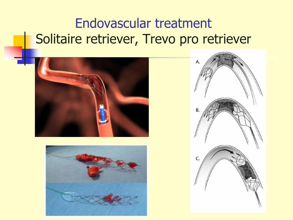
Endovascular treatment Solitaire retriever, Trevo pro retriever

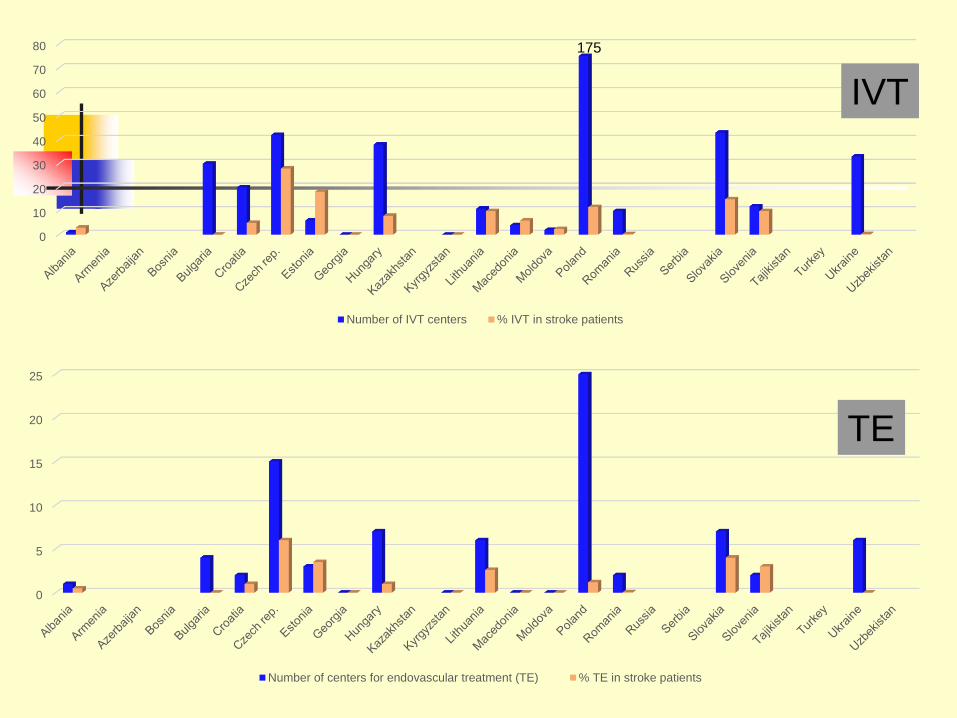
Intravenous thrombolysis/million inhabitans
deSousa DA, Fischer U et al., survey from 43 countries, zaslané do tlače

deSousa DA, Fischer U et al., survey from 43 countries, zaslané do tlače

Endovascular treatments/million inhabitans
deSousa DA, Fischer U et al., survey from 43 countries, zaslané do tlače

deSousa DA, Fischer U et al., survey from 43 countries, zaslané do tlače
0
10
20
30
40
50
60
70
80
Number of IVT centers % IVT in stroke patients
0
5
10
15
20
25
Number of centers for endovascular treatment (TE) % TE in stroke patients
IVT
TE
175

deSousa DA, Fischer U et al., survey from 43 countries, zaslané do tlače

New guidelines in Slovakia
Network of hospitals for IVT and ET – 24/7 hours
Recommendations for collaboration with emergency service
Výbor Cerebrovaskulárnej sekcie SNeS, 2017 - návrh

CTA/MRA
Brain CT and CT AG
Sugar
aPTT, PT (INR), - in patients with anticoagulant therapy
Mild neurological deficit, age ˃ 80 years, epileptic seizure
Mair G. et al, Stroke 2015, 46: 102 -107; Leary MC et al, Stroke 2003, 34:2636-2640; Goldmaker GV et al, Stroke 2009, 40: 134-139

Network of hospitals
Poskytovaná rekanalizačná liečba – trombolytická liečba (43)
Poskytovaná rekanalizačná liečba – trombolytická aj trombektomická liečba (6)
Poskytovaná rekanalizačná liečba – trombektomická liečba (2)

Počet pacientov potrebných liečiť - Numbers needed to treat (NNT) na
dosiahnutie modifikovaného Rankinovho skóre 0-1
NNT
14
NNT
4 - 5
NNT
9
5
4
3
2
0
Percento pravdepodobnosti odhadnuté modelom
95% konfidenčný interval pre
odhadované percento pravdepodobnosti
60 90 120 150 360330300270240210180
Čas od vzniku mozgovej príhody do začatia liečby (min)
Wahlgren et al. Lancet 2008;372:1303-1309.
Lees et al. Lancet 2010;375:1695-1703.
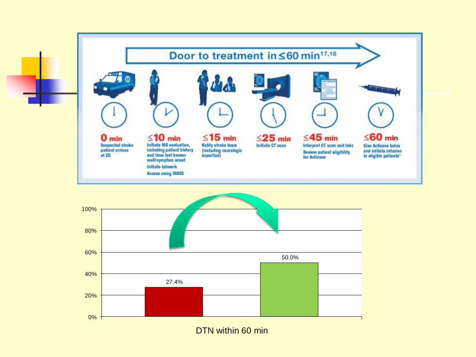
27.4%
50.0%
0%
20%
40%
60%
80%
100%
DTN within 60 min

Toni D., ESOC 2016, Kodaň.

Time delay (minutes) Slovensko
2014 2015 2016 2017
Onset to door time 80.0 82.0 88 85 - 89
Door to imaging 21.0 17.0 15 15 - 15
Door to treatment
(DNT)
60.0 45.0 45 40,5 - 35
Onset to
treatment/needle time
155.0 137.0 143 135 - 130
SITS, okt 2017

G-FAST
• G - Gaze
• F- Face
• A - Arm
• S – Speech
• T – Time

Výbor Cerebrovaskulárnej sekcie SNeS, 2017
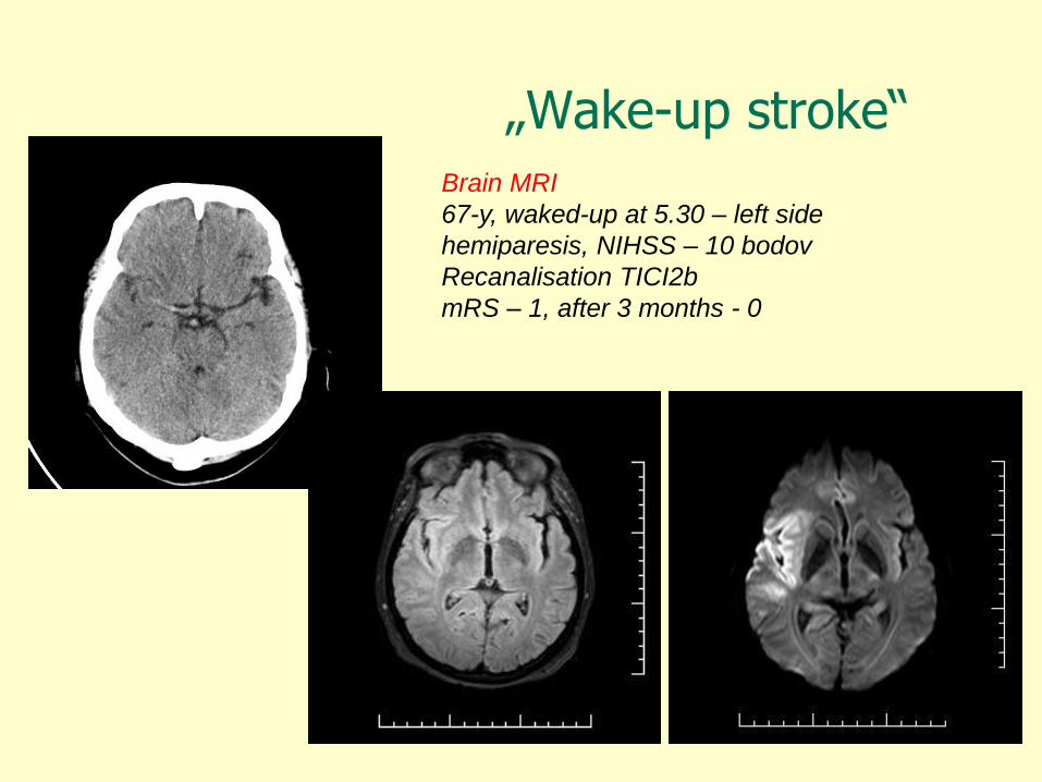
„Wake-up stroke“Brain MRI
67-y, waked-up at 5.30 – left side
hemiparesis, NIHSS – 10 bodov
Recanalisation TICI2b
mRS – 1, after 3 months - 0

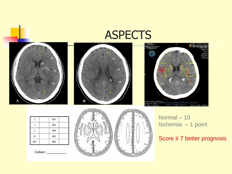
ASPECTS
Normal – 10
Ischemia – 1 point
Score ≥ 7 better prognosis

O.R. 52-years old woman
Wake up - 5.45, she felt down, aphasia, right side hemiparesis
Emergency
6.50 – hospital, NIHSS -11
7.05 – brain CT
7.45 – rTPA
9.05 – DSA, trombektomy

O.R. 52-years old woman
DSA before TE DSA after TE

O.R. 52-years old woman
Brain CT after 24 hours
mRS – 0 at time of discharge


T.T. 37-years old man
30.7. 2013 –orchiektomy and CHT
30.8.2013 at 16.00 weakness of left extremities
Admitted in hospital at 18.00, barin CT at 19.20
Admitted in our hospital at 21.15, 30.8.2013

Angiography Trombektomy 30.8.2013 at 22.45

CT after TE and decompressive craniotomy
After po 24 hours After 72 hours After 15 days

Eric Jordan sings Puccini


Therapy after acute stroke
Therapy of risk factors – prevention
Antithrombotics
Anticoagulants
Endarterectomy (CEA)
STENT
Rehabilitation

Guidelines for antiagregants
Antithrombotics
Non cardioembolic strokes
ASA, 50 – 325 mg 1xD
Combination ASA and dipyridamol 200 mg 2xD
Clopidogrel 75 mg 1xD
Albers GW a kol., Chest 2001;

Indications for anticoagulants
in patients with stroke and AF
Stroke, AF and
CHADS2 – (congestive heart failure,
hypertension, age 75, diabetes, stroke)
2 – high risk
Warfarin – INR 2,0-3,0
Direct oral anticoagulants
Direct inhibitor of thrombin – Dabigtran
Inhibitors of Xa – Apixaban, Rivaroxaban, Edoxaban

Charakteristika štúdiíRELY
dabigatran
ROCKET AF
rivaroxaban
ARISTOTLE
apixaban
Mechanizmus účinku IIa Xa Xa
Počet pacientov 18 113 14 264 18 201
Dávkovanie150 mg 2x/ day
110 mg 2x/ day
20mg 1x/ day
(15mg 1x/ day )
5mg 2x/ day
2,5 mg 2x/ day
Dizajn PROBE Double blind Double blind
Priemer CHADS2 2,1 3,5 2,1
Priemer TTR 64% 55% 62%
Medián TTR 67% 58% 66%
Prerušenie liečby
(Warfarín)21,2% (16,6) 23,9% (22,4) 25,3% (27,5)
RELY – ARISTOTLE – ROCKET AF
Connolly SJ et al. N Engl J Med 2009; 361:1139–1151.Connolly SJ et al. N Engl J Med 2010; 363:1875–1876 (letter to editor).
SPC Pradaxa tvrdé tobolky 110/150 mg, 8/2011;
Granger CB et al. NEJM 2011: 10..1056/NEJMoal 1107039. NEJM.org ; Patel MR et al. NEJM 2011;10.1056/NJMoal1009638.NEJM.org.
https://www.dcri.org/news-publications/slides-presentations/ROCKET-AF-LBCT FINAL.ppt/view?searchterm=rocket

Endarterectomy ICA
Indications
ACI stenosis > 70% (in ulcerating AS
plaques – risk of embolisation – > 60%)
Brain CT
After TIA – 2 days, small infarct within 2
weeks, others – 6 weeks after stroke

Endarterectomy

Endarterectomy

STENT

ICA High grade stenosis - Stent

ICAHigh grade stenosis - Stent

ICAHigh grade stenosis - Stent

Indications of STENT
Patients with
- operation risk
- older patiens
- risk of anaesthesia
- changes on the neck
Restenosis after CAE

Advantages of STENT
Less invasive method
Less patients with restenosis
Shorter hospitalization
Smaller risk of wound complications

Blood supply of spinal cord
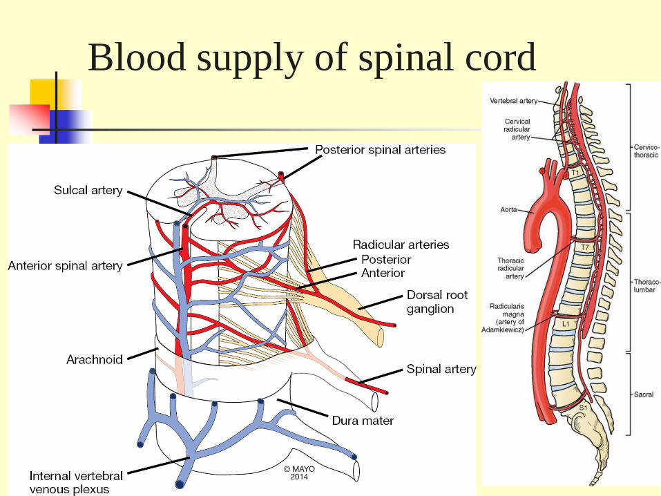
Blood supply of spinal cord

Acute spinal cord ischemia
Represents only 5-8% of acute myelopathies and <1% of
all strokes
The majority of patients developed symptoms quickly,
with maximal symptomatology reached within 12 hours
for >50% of patients and within 72 hours for the vast
majority of patients
Initial symptoms include severe back pain (60-70%), loss
of bladder control (60%) and bowel control (40%).

Clinical feature
anterior spinal artery syndrome
paralysis below affected level (initially flaccid; later spastic)
pain and temperature sensory loss
relative sparing of proprioception and vibration (dorsal columns)
posterior spinal artery syndrome
complete sensory loss at the level of injury
proprioception and vibration loss below level
minimal, typically transient, motor symptoms

MRI
T2-wieghted MRI

Owl's Eye in Spinal Magnetic Resonance Imaging

Haematomeylia
Bleeding to spinal cord
Etiology
AVM
Anti-coagulant therapy
Coagulopaties
Injury

Haematomeylia - etiology

Hematomyelia – clinical feature
Pain
Symptoms similar as in ischemia
Diagnosis – MRI
Treatment – conservative, surgery
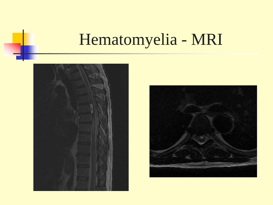
Hematomyelia - MRI

Cerebral venous thrombosis (CVT)
Rare type of stroke
Thrombosis occurs in the venous side of the brain circulation
Occlusion of one or more cerebral veins nad dural venous sinus.
Incidence – 1/100 000 inhabitans
The most frequent – children and young adults, more often in women

Cerebral venous thrombosis (CVT)
Etiology
Infections (in 70 % - Staphylococcus aureus, than Streptococcus pneumoniae, gramnegative bacterias, Aspergillus).
Focal infections on the head – sinusitis, meningitis, malignancy, otitis, tonsilitis, furunkul, penetrating head injury
Generalized infections – endokarditis, tuberkulosis, pneumonia, hepatitis, AIDS.
Lumbal puncture

Cerebral venous thrombosis (CVT)
Non infectious risk factors Oral contraceptives,
Drugs with protrombotic effect
Pregnancy, pueprerium
Thrombophilic disorders,
Antiphospholipid syndrome
Malignancies

Cerebral venous thrombosis Clinical feature
Subacute beginning
Different neurological symptoms
Later – hemorhagic transformation
Cefalea, nauzea, vomitus
Hemiparézis, paraparesis (sinus sagitalis superior),
Aphasia,
ataxia, chorea, hemianopsia,


Cerebral venous thrombosis Clinical feature
Epileptic seazure
Papil oedema
Cranial nerve lesions - (n. VI, n. VII, n. VIII).
syndrom foramen jugulare (n. IX – XII.)

Sinus cavernosus thrombosis (SC)

Sinus cavernosus thrombosis (SC)
very rare, life-threatening condition that can affect adults and children.
Symptoms
Severe headache
Swelling, redness, or irritation around one or both eyes
Drooping eyelids
Inability to move the eye
High fever
Pain or numbness around the face or eyes
Fatigue
Vision loss or double vision
Seizures

Sinus cavernosus thrombosis - MRI

Sinus transversus thrombosis
Sy intracranial hypertension
a temporal symptomatology

Sinus sagitalis superior thrombosis
Spastic monoparesis of lower extremity
Or spastic paraparesis of lower extremities
Or unilateral hemiparesis

Cortical vein htrombosis
Clinical feature
Focal deficit – aphasia, hemiparesis, hemianopsy, hemianopsy,
Diagnosis
Clinical feature – SIH
Diagnosis – MRI with contrast
CSF - proteino-cytologic association, in 10 % - CSF negative
Etiology

Treatment
Anticoagulants iv, or sc (heparin, alebo LMWH)
After stabilization – p.o. anticoagulants (Warfarin) INR 2,0 – 2,5 for 6 months, when thrombophilia is present –long lasting
When there is no effect of heparin – rTPA
Antibiotics (ceúhalosporins)
Symptomatic treatment (antiedematous treatment, antiepileptics)

![Schools & Universities Your Company. [Your Company] can help you… ˃ Alert and inform students and parents ˃ Keep everyone up-to-date ˃ Celebrate school](https://img.pdfslide.net/doc/110x75/56649ef45503460f94c072c6/schools-universities-your-company-your-company-can-help-you-alert.jpg)
![Restaurant Industry Your Company. [Your Company] can help you… ˃ Attract first time diners ˃ Bring in more repeat business ˃ Make all hours peak hours](https://img.pdfslide.net/doc/110x75/5518a6d1550346881f8b4ada/restaurant-industry-your-company-your-company-can-help-you-attract-first-time-diners-bring-in-more-repeat-business-make-all-hours-peak-hours.jpg)

![Medical Industry Your Company. [Your Company] can help you… [Your Company] ˃ Reduce missed appointments ˃ Keep patients informed ˃ Gain word-of-mouth](https://img.pdfslide.net/doc/110x75/56649de95503460f94ae4608/medical-industry-your-company-your-company-can-help-you-your-company.jpg)


![Retail Industry Your Company. [Your Company] can help you… ˃ Reel in more first-time shoppers ˃ Boost repeat customer visits ˃ Bring in shoppers during](https://img.pdfslide.net/doc/110x75/56649e4b5503460f94b4077e/retail-industry-your-company-your-company-can-help-you-reel-in-more.jpg)

![Religious Organizations Your Company. [Your Company] can help you… ˃ Welcome more members ˃ Increase attendance ˃ Get members involved ˃ Maintain lasting](https://img.pdfslide.net/doc/110x75/56649e0e5503460f94af82fd/religious-organizations-your-company-your-company-can-help-you-welcome.jpg)


![6 - NSPS OOOO and OOOOa (2018 0731 0952) · Perform reduced emissions c ompletions/green completions: ˃ ˃ ˃] ˃ ˃](https://img.pdfslide.net/doc/110x75/5ec14b553e700b7ae2081734/6-nsps-oooo-and-ooooa-2018-0731-0952-perform-reduced-emissions-c-ompletionsgreen.jpg)