Embed Size (px)
Citation preview
Kwaliteit van rectale kankerzorg
- Fase 1 - Een praktijkrichtlijn voor
rectale kanker
KCE reports 69A
Federaal Kenniscentrum voor de Gezondheidszorg Centre fédéral d’expertise des soins de santé
2007
Het Federaal Kenniscentrum voor de Gezondheidszorg
Voorstelling : Het Federaal Kenniscentrum voor de Gezondheidszorg is een parastatale, opgericht door de programma-wet van 24 december 2002 (artikelen 262 tot 266) die onder de bevoegdheid valt van de Minister van Volksgezondheid en Sociale Zaken. Het Centrum is belast met het realiseren van beleidsondersteunende studies binnen de sector van de gezondheidszorg en de ziekteverzekering.
Raad van Bestuur
Effectieve leden : Gillet Pierre (Voorzitter), Cuypers Dirk (Ondervoorzitter), Avontroodt Yolande, De Cock Jo (Ondervoorzitter), De Meyere Frank, De Ridder Henri, Gillet Jean-Bernard, Godin Jean-Noël, Goyens Floris, Kesteloot Katrien, Maes Jef, Mertens Pascal, Mertens Raf, Moens Marc, Perl François, Smiets Pierre, Van Massenhove Frank, Vandermeeren Philippe, Verertbruggen Patrick, Vermeyen Karel.
Plaatsvervangers : Annemans Lieven, Boonen Carine, Collin Benoît, Cuypers Rita, Dercq Jean-Paul, Désir Daniel, Lemye Roland, Palsterman Paul, Ponce Annick, Pirlot Viviane, Praet Jean-Claude, Remacle Anne, Schoonjans Chris, Schrooten Renaat, Vanderstappen Anne.
Regeringscommissaris : Roger Yves
Directie
Algemeen Directeur : Dirk Ramaekers
Adjunct-Algemeen Directeur : Jean-Pierre Closon
Contact
Federaal Kenniscentrum voor de Gezondheidszorg (KCE) Wetstraat 62 B-1040 Brussel Belgium
Tel: +32 [0]2 287 33 88 Fax: +32 [0]2 287 33 85
Email : [email protected] Web : http://www.kce.fgov.be
Kwaliteit van rectale kankerzorg
- Fase 1 -
Een praktijkrichtlijn voor rectale kanker
KCE reports 69A
F. PENNINCKX, S. ROELS, D. LEONARD, S. LAURENT, J. DECAESTECKER, C. DE VLEESCHOUWER, K. HAUSTERMANS, N. ECTORS,
M. PEETERS, E. VAN CUTSEM, E. DANSE, D. DE CONINCK, E. VAN EYCKEN, J. VLAYEN
Federaal Kenniscentrum voor de Gezondheidszorg Centre fédéral d’expertise des soins de santé
2007
KCE reports 69A
Titel : Kwaliteit van rectale kankerzorg, fase : Een praktijkrichtlijn voorrectale kanker
Auteurs : F. Penninckx (UZ Leuven), S. Roels (UZ Leuven), D. Leonard (UCL), S. Laurent (UGent), J. Decaestecker (UZ Leuven), C. De Vleeschouwer (UZ Leuven), K. Haustermans (UZ Leuven), N. Ectors (UZ Leuven), M. Peeters (UGent), E. Van Cutsem (UZ Leuven), E. Danse (UCL), D. De Coninck (AZ St.Lucas Brugge), E. Van Eycken (Stichting Kankerregister), J. Vlayen (KCE)
Externe experten : PROCARE Steering Group
Externe validatoren : Andrew Shorthouse (Department of Coloproctology, Northern General Hospital, and Faculty of Health and Wellbeing, Sheffield Hallam University, UK), Simon Van Belle (Department of Medical Oncology, University Hospital Ghent), Philippe Coucke (Department of Radiotherapy, CHU de Liège)
Conflict of interest : De meeste auteurs (behalve E. Van Eycken en J. Vlayen) en externe experts zijn werkzaam in een dienst waar patiënten met rectale kanker behandeld worden. F. Penninckx, K. Haustermans, M. Peeters en E. Van Cutsem ontvingen betalingen van diverse farmaceutische firma’s om te spreken, en onderzoeksfondsen (doch niet gerelateerd aan dit rapport).
Disclaimer : De externe experten hebben aan het wetenschappelijke rapport meegewerkt dat daarna aan de validatoren werd voorgelegd. De validatie van het rapport volgt uit een consensus of een meerderheidsstem tussen de validatoren. Alleen het KCE is verantwoordelijk voor de eventuele resterende vergissingen of onvolledigheden alsook voor de aanbevelingen aan de overheid..
Layout : Verhulst Ine
Brussel, 21 december 2007
Studie nr 2006-03-1 Domein : Good Clinical Practice (GCP)
MeSH : Rectal Neoplasms; Rectal Diseases; Practice Guidelines
NLM classification : WI 610
Taal : Nederlands, Engels Format : Adobe® PDF™ (A4)
Wettelijk depot : D/2006/10.273/54
Elke gedeeltelijke reproductie van dit document is toegestaan mits bronvermelding. Dit document is beschikbaar van op de website van het Federaal Kenniscentrum voor de gezondheidszorg.
Hoe refereren naar dit document? Penninckx F, Roels S, Leonard D, Laurent S, Decaestecker J, De Vleeschouwer C, et al. Kwaliteit van rectale kankerzorg, fase 1: Een praktijkrichtlijn voor rectale kanker. Good Clinical Practice (GCP). Brussel: Federaal Keniscentrum voor de Gezondheidszorg (KCE); 2007. KCE reports 69A (D/2007/10.273/54)
KCE Reports 69A Kwaliteit van rectale kankerzorg (I) i
Voorwoord
Beleidsmatig en in de media krijgen bepaalde kankers – terecht - veel aandacht. Denk maar aan borstkanker en recent nog de initiatieven om vroegtijdig colonkanker op te sporen. Over bepaalde kankers die nochtans ook relatief frequent voorkomen horen we veel minder. Eén daarvan is rectum- of endeldarmkanker.
Het lijkt er misschien op dat rectumkanker gemakkelijkheidshalve net zoals hoger gelegen colonkanker kan behandeld worden. Niets is minder waar. Er zijn natuurlijk vergelijkingspunten, maar qua aanpak – en dan op de eerste plaats chirurgisch – vergt deze kanker vaak een specifieke expertise. En dat inzicht groeide jaren geleden al bij een aantal vooraanstaande Belgische kankerspecialisten. Zij slaagden erin om over alle grenzen heen de grote groep van in rectumkanker betrokken specialistenverenigingen rond de tafel te brengen en gemeenschappelijk en boeiend kwaliteitsverbeterend project op de sporen te zetten. Het initiatief kreeg de afkorting PROCARE (PROject on CAncer of the REctum). Inhoudelijk wil men door middel van concrete klinische praktijkrichtlijnen en een educatief project aan de hand van wetenschappelijke onderbouwde kwaliteitsindicatoren de kwaliteit van de rectale kankerzorg verbeteren.
Er zijn in ons land – zoals trouwens in vele andere landen - inderdaad praktijkverschillen in de aanpak van rectumkanker tussen de verschillende ziekenhuizen. De vraag is dan steeds, wat doen we eraan, om vervolgens soms in simplistische beleidsvoorstellen te vervallen. Dit project is anders. Het is breed gedragen door de brede beroepsgroep. Hun meest vooraanstaande klinische experten werken – naast een veeleisende dagtaak – gedreven, bij wijlen gepassioneerd, mee aan PROCARE. Het Kenniscentrum bood dan ook maar al te graag de nodige ruggesteun aan een dergelijk initiatief.
Dit rapport, waarin naar analogie met de rapporten van het KCE voor andere kankertypes, evidence-based praktijkrichtlijnen werden ontwikkeld, is het eerste van twee deelrapporten. Dezelfde groep van klinische experten maakt momenteel de vertaalslag van de PROCARE aanbevelingen naar meetbare kwaliteitsindicatoren. Dit moet toelaten de kwaliteit van de rectale kankerzorg in de nabije toekomst op te volgen en een positief instrument te ontwikkelen voor kwaliteitsverbetering. De PROCARE oefening is uniek en de eerste in zijn aard in België. Het KCE is fier om samen met het Kankerregister en het RIZIV dit te mogen ondersteunen: uiteindelijk is het de patiënt wiens kansen erop vooruit zullen gaan. En dat is de essentie van goede kankerzorg.
Closon Jean-Pierre Ramaekers Dirk
Adjunct algemeen directeur Algemeen Directeur
ii Kwaliteit van rectale kankerzorg (I) KCE Reports 69A
Samenvatting
INLEIDING Vroeger onderzoek in België en in het buitenland toont een belangrijke variabiliteit tussen individuele ziekenhuizen in de soorten behandeling van rectumkanker en in de resultaten ervan. In meerdere Europese landen werd de afgelopen jaren een standaardisatie van de behandeling beoogd door het implementeren van diagnostische en therapeutische aanbevelingen. De kwaliteitscontrole vindt plaats aan de hand van gevalideerde indicatoren. De toepassing ervan leidde tot een significante verbetering van de uitkomst van rectumkanker in andere landen. De evaluatie van de kwaliteit van zorg aan de hand van kankerregistratiegegevens heeft in België een hele achterstand in te halen. In de internationale literatuur blijft België voorlopig een grijze vlek op de Europese kaart.
In 2004 startte in België het ‘PROject on CAncer of the Rectum’ project (PROCARE) met als doelstelling de kwaliteit van rectale kankerzorg in België te verbeteren door standaardisatie via de ontwikkeling en implementatie van specifieke aanbevelingen en via kwaliteitsbewaking door registratie en feedback. Alle medische specialismen betrokken in de behandeling van rectale kanker werden verenigd in een multidisciplinaire werkgroep met vertegenwoordigers van de respectievelijke wetenschappelijke verenigingen. Een eerste voorlopige versie van de PROCARE aanbevelingen werd geschreven in 2005 en werd gevolgd door workshops (chirurgie, pathologie, radiotherapie, chemotherapie en radiologie). Een prospectieve database van individuele patiëntengegevens werd ontwikkeld en vrijwillige registratie via de Stichting Kankerregister ging van start in 2006. Van de deelnemende centra worden alle relevante data (gaande van staging tot follow-up) van consecutieve patiënten met rectale kanker in deze prospectieve database ingebracht. Deze gegevens zullen de basis vormen voor nationale en internationale benchmarking.
In dit rapport wordt een geüpdate versie van de PROCARE aanbevelingen besproken. In een volgend rapport (2008) zal voor de eerste maal een set van kwaliteitsindicatoren getest worden op de prospectieve PROCARE gegevens en op gekoppelde gegevens van de Stichting Kankerregister, het Intermutualistisch Agentschap en de Federale Overheidsdienst Volksgezondheid, Veiligheid van de Voedselketen en Leefmilieu.
METHODOLOGIE Voor de ontwikkeling van deze richtlijn werd de ADAPTE methodologie gebruikt. In eerste instantie werden de belangrijkste klinische zoekvragen geformuleerd. Bestaande (inter)nationale richtlijnen werden gezocht in Medline, National Guideline Clearinghouse en websites van oncologische organisaties. De 33 gevonden richtlijnen werden door middel van het AGREE instrument beoordeeld op hun kwaliteit door vier onafhankelijke reviewers en al dan niet geselecteerd op basis van een algemeen kwaliteitsoordeel. Vervolgens werden de 17 geselecteerde richtlijnen geupdated voor elke klinische vraag, door bijkomende evidentie te zoeken in Medline en de Cochrane Database of Systematic Reviews. Een level of evidence werd toegekend aan elke originele aanbeveling en bijkomende studie door gebruik te maken van het GRADE systeem.
Gebaseerd op de gevonden evidence werden aanbevelingen geformuleerd door een multidisciplinaire richtlijnontwikkelingsgroep. Deze aanbevelingen werden op een formele manier beoordeeld door de PROCARE stuurgroep. Belangenconflicten werden genoteerd.
FINALE AANBEVELINGEN De details van de richtlijn kunnen teruggevonden worden in het scientific report onmiddellijk na deze samenvatting.
KCE Reports 69A Kwaliteit van rectale kankerzorg (I) iii
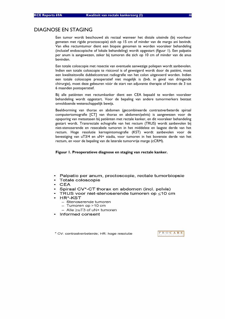
DIAGNOSE EN STAGING Een tumor wordt beschouwd als rectaal wanneer het distale uiteinde (bij voorkeur gemeten met rigide proctoscopie) zich op 15 cm of minder van de margo ani bevindt. Van elke rectumtumor dient een biopsie genomen te worden vooraleer behandeling (inclusief endoscopische of lokale behandeling) wordt opgestart (figuur 1). Een palpatio per anum is aangewezen, zeker bij tumoren die zich op 10 cm of minder van de anus bevinden.
Een totale coloscopie met resectie van eventuele aanwezige poliepen wordt aanbevolen. Indien een totale coloscopie te risicovol is of geweigerd wordt door de patiënt, moet een kwaliteitsvolle dubbelcontrast radiografie van het colon uitgevoerd worden. Indien een totale coloscopie preoperatief niet mogelijk is (bvb. in geval van dringende chirurgie), moet deze gebeuren vóór de start van adjuvante therapie of binnen de 3 tot 6 maanden postoperatief.
Bij alle patiënten met rectumkanker dient een CEA bepaald te worden vooraleer behandeling wordt opgestart. Voor de bepaling van andere tumormerkers bestaat onvoldoende wetenschappelijk bewijs.
Beeldvorming van thorax en abdomen (gecombineerde contrastverbeterde spiraal computertomografie [CT] van thorax en abdomen/pelvis) is aangewezen voor de opsporing van metastasen bij patiënten met rectale kanker, en dit vooraleer behandeling gestart wordt. Transrectale echografie van het rectum (TRUS) wordt aanbevolen bij niet-stenoserende en resecabele tumoren in het middelste en laagste derde van het rectum. Hoge resolutie kernspintomografie (KST) wordt aanbevolen voor de bevestiging van uT3/4 en uN+ stadia, voor tumoren in het bovenste derde van het rectum, en voor de bepaling van de laterale tumorvrije marge (cCRM).
Figuur 1. Preoperatieve diagnose en staging van rectale kanker.
iv Kwaliteit van rectale kankerzorg (I) KCE Reports 69A
BEHANDELING
Voorafgaande radio- en chemotherapie: neo-adjuvante behandeling
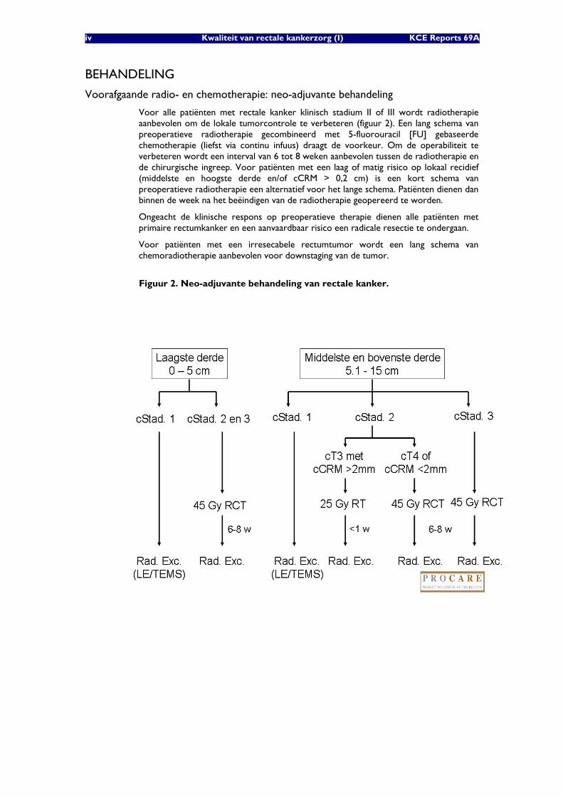
Voor alle patiënten met rectale kanker klinisch stadium II of III wordt radiotherapie aanbevolen om de lokale tumorcontrole te verbeteren (figuur 2). Een lang schema van preoperatieve radiotherapie gecombineerd met 5-fluorouracil [FU] gebaseerde chemotherapie (liefst via continu infuus) draagt de voorkeur. Om de operabiliteit te verbeteren wordt een interval van 6 tot 8 weken aanbevolen tussen de radiotherapie en de chirurgische ingreep. Voor patiënten met een laag of matig risico op lokaal recidief (middelste en hoogste derde en/of cCRM > 0,2 cm) is een kort schema van preoperatieve radiotherapie een alternatief voor het lange schema. Patiënten dienen dan binnen de week na het beëindigen van de radiotherapie geopereerd te worden.
Ongeacht de klinische respons op preoperatieve therapie dienen alle patiënten met primaire rectumkanker en een aanvaardbaar risico een radicale resectie te ondergaan.
Voor patiënten met een irresecabele rectumtumor wordt een lang schema van chemoradiotherapie aanbevolen voor downstaging van de tumor.
Figuur 2. Neo-adjuvante behandeling van rectale kanker.
KCE Reports 69A Kwaliteit van rectale kankerzorg (I) v
Chirurgie
Pre- en perioperatieve voorbereiding omvat de volgende zaken: darmvoorbereiding, tromboseprofylaxis (graduele compressiekousen en subcutane laag-moleculair-gewicht heparine), antibioticaprofylaxis (éénmalige preoperatieve dosis), voorbereiding van bloedtransfusie, bespreking van het risico op postoperatieve urogenitale dysfunctie (tumoren in het middelste en laagste derde), en preoperatieve stomavoorlichting indien een stoma voorzien wordt.
De anale sfincter dient waar mogelijk en wenselijk vrijwaard te worden. Een totale mesorectale excisie (TME) is aanbevolen voor tumoren in het middelste en laagste derde van het rectum, hetzij als onderdeel van een restoratieve proctectomie, een Hartmann procedure of een abdominoperineale resectie (APR). Voor tumoren in het bovenste derde wordt een partiële mesorectale excisie (PME) aanbevolen. Peroperatief (vooral tijdens APR) dient rectale perforatie of doorbreken van de tumor vermeden te worden.
Na een restoratieve proctectomie en TME dient een colon pouch, coloplastie of side-to-end colo-anale anastomose overwogen te worden om de functionele outcome en levenskwaliteit te verbeteren. Een tijdelijk stoma is te overwegen indien er een risico bestaat voor lekkage uit de anastomose (zeker bij infraperitoneale anastomose na TME).
Lokale excisie of transanale endoscopische microchirurgische resectie (TEMS) is geen standaardbehandeling voor vroegtijdige stadia van rectumkanker. Deze technieken kunnen aanbevolen worden voor kleine uT1 letsels (< 3 cm) met het uitzicht van een villeus adenoom en negatieve biopsies. Gezien het risico op kliermetastasen en een verminderde tumorcontrole dienen alle uT1 letsels een radicale TME resectie te ondergaan bij patiënten met een aanvaardbaar operatief risico.
Bij stenoserende tumoren kan een laparoscopische exploratie en het aanleggen van een derivatief stoma overwogen worden vóór de start van neo-adjuvante behandeling. Stenting in afwachting van curatieve chirurgie wordt niet aanbevolen.
Pathologie
Het resectiestuk dient ongeopend aan de patholoog geleverd te worden binnen de 2-3 uur na resectie. De exacte topografie van de tumor dient bepaald te worden. De kwaliteit (volledig, bijna volledig, onvolledig) van een totale mesorectale excisie dient beoordeeld te worden op het ongeopende staal. De volgende parameters dienen gemeten te worden na fixatie en sectie: diepste punt van tumorinvasie, afstand tot de dichtstbijzijnde circumferentiële oppervlakte. Een minimum van 12 lymfeklieren dient gevonden en geanalyseerd te worden in het resectiestuk.
Het pathologierapport dient gestandaardiseerd te zijn en alle belangrijke macroscopische en microscopische gegevens te bevatten. De resultaten dienen besproken te worden tijdens een multidisciplinair overleg met de patholoog, chirurg, radiotherapeut, oncoloog en gastro-enteroloog.
Aanvullende chemo- en radiotherapie: adjuvante behandeling
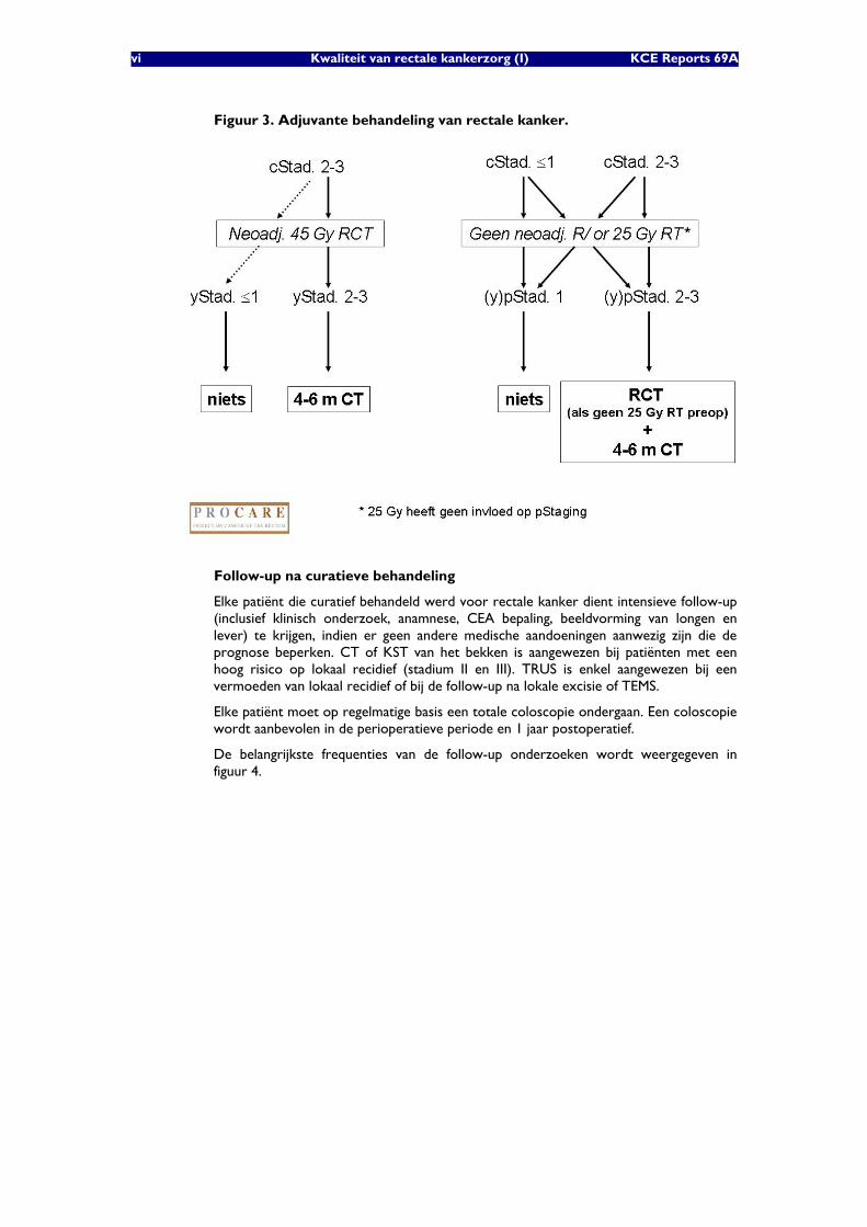
Bij alle patiënten met rectale kanker pathologisch stadium II of III die preoperatieve radiotherapie kregen zonder chemotherapie, dient adjuvante chemotherapie met 5FU overwogen te worden (figuur 3).
Bij patiënten met rectale kanker pathologisch stadium II of III die geen neo-adjuvante behandeling kregen, wordt de combinatie van adjuvante radiotherapie en chemotherapie aanbevolen. Dit is eveneens het geval voor patiënten die een R1 resectie ondergingen. Indien de chemotherapie 5FU bevat is een continu infuus effectiever dan een bolus infuus (figuur 3).
Adjuvante behandeling dient gestart te worden binnen de 3 maanden na chirurgie.
vi Kwaliteit van rectale kankerzorg (I) KCE Reports 69A
Figuur 3. Adjuvante behandeling van rectale kanker.
Follow-up na curatieve behandeling
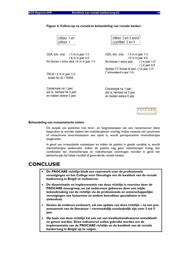
Elke patiënt die curatief behandeld werd voor rectale kanker dient intensieve follow-up (inclusief klinisch onderzoek, anamnese, CEA bepaling, beeldvorming van longen en lever) te krijgen, indien er geen andere medische aandoeningen aanwezig zijn die de prognose beperken. CT of KST van het bekken is aangewezen bij patiënten met een hoog risico op lokaal recidief (stadium II en III). TRUS is enkel aangewezen bij een vermoeden van lokaal recidief of bij de follow-up na lokale excisie of TEMS.
Elke patiënt moet op regelmatige basis een totale coloscopie ondergaan. Een coloscopie wordt aanbevolen in de perioperatieve periode en 1 jaar postoperatief.
De belangrijkste frequenties van de follow-up onderzoeken wordt weergegeven in figuur 4.
KCE Reports 69A Kwaliteit van rectale kankerzorg (I) vii
Figuur 4. Follow-up na curatieve behandeling van rectale kanker.
Behandeling van metastatische ziekte
De aanpak van patiënten met lever- en longmetastasen van een rectumtumor dient besproken te worden tijdens het multidisciplinair overleg. Indien resectie van synchrone of metachrone levermetastasen een optie is, wordt perioperatieve chemotherapie aangeraden.
In geval van irresecabele metastasen en indien de patiënt in goede conditie is, wordt chemotherapie aanbevolen. Indien de patiënt nog geen radiotherapie kreeg, kan combinatie van chemotherapie en radiotherapie overwogen worden in geval van pelvische pijn bij lokaal recidief of gevorderde rectale kanker.
CONCLUSIE • De PROCARE richtlijn biedt een raamwerk voor de professionele
verenigingen en het College voor Oncologie om de kwaliteit van de rectale kankerzorg in België te verbeteren.
• De disseminatie en implementatie van deze richtlijn is voorzien door de PROCARE stuurgroep, en zal ondermeer gebeuren door een wijde bekendmaking van de richtlijn via de professionele en wetenschappelijke verenigingen van huisartsen en andere betrokken specialisten in het ziekenhuis.
• Gezien de evidence evolueert, zal een update van deze richtlijn – na een pre-assessment van de literatuur – vermoedelijk noodzakelijk zijn over 3 tot 5 jaar.
• Op basis van deze richtlijn zal een set van kwaliteitsindicatoren ontwikkeld en getest worden. Deze indicatoren zullen gebruikt worden om de implementatie van de PROCARE richtlijn en de kwaliteit van de rectale kankerzorg in België op te volgen.

KCE reports 69 PROCARE 1
Scientific summary
Table of contents
1 GENERAL INTRODUCTION .......................................................................... 4 2 UPDATED PROCARE GUIDELINES FOR THE TREATMENT OF
RECTAL CANCER ............................................................................................ 6 2.1 INTRODUCTION ................................................................................................................................ 6 2.2 METHODOLOGY................................................................................................................................ 6
2.2.1 General approach ................................................................................................................ 6 2.2.2 Guideline development group composition.................................................................. 7 2.2.3 Clinical questions ................................................................................................................. 7 2.2.4 Search for evidence...........................................................................................................10 2.2.5 Quality appraisal.................................................................................................................11 2.2.6 Data extraction and summary ........................................................................................12 2.2.7 Formulation of recommendations .................................................................................12 2.2.8 External review ..................................................................................................................12
2.3 DEFINITIONS ....................................................................................................................................13 2.3.1 The rectum..........................................................................................................................13 2.3.2 Staging...................................................................................................................................13 2.3.3 Extent of resection (R) and radial margin....................................................................14 2.3.4 Other definitions related to surgery.............................................................................15 2.3.5 Definitions related to radiotherapy volume and International Commission of Radiation Units (ICRU) reference point ..................................................................................15
2.4 FINAL RECOMMENDATIONS ...........................................................................................................16 2.4.1 Access to treatment .........................................................................................................16 2.4.2 Diagnosis and staging ........................................................................................................16 2.4.3 Neoadjuvant treatment....................................................................................................21 2.4.4 Surgical treatment .............................................................................................................26 2.4.5 Pathology .............................................................................................................................31 2.4.6 Adjuvant therapy................................................................................................................37 2.4.7 Follow-up after curative treatment ...............................................................................42 2.4.8 Treatment of metastatic rectal cancer .........................................................................44
3 CONCLUSIONS.............................................................................................. 52 4 APPENDICES .................................................................................................. 53 5 REFERENCES ................................................................................................ 233
2 PROCARE KCE reports 69
ABBREVIATIONS
5-FU 5-fluorouracil
95% CI 95 percent confidence interval
AGREE Appraisal of Guidelines Research and Evaluation
AJCC American Joint Committee on Cancer
APR Abdomino-perineal resection of the rectum
ASA American Association of Anaesthetists score
ASCO American Society of Clinical Oncology
BED Biological effective doses
CBC Complete blood count
CBO Dutch Institute for Healthcare Improvement
CCO Cancer Care Ontario
CDSR Cochrane database of systematic reviews
CEA Carcinoembryonic antigen
CE-CT Contrast-enhanced computed tomography
CPG Clinical practice guideline
CRC Colorectal cancer
CRM Circumferential resection margin
CRT Chemoradiation therapy
CT Computed tomography
CTV Clinical target volume
DCBE Double contrast barium enema
DFS Disease-free survival
DVT Deep venous thrombosis
EBRT External beam radiotherapy
EGFR Epidermal growth factor receptor
EORTC European Organisation for Research and Treatment of Cancer
EUS Endoscopic ultrasonography
FAP Familial adenomatous polyposis
FBCR Foundation Belgian Cancer Registry
FNCLCC Fédération Nationale des Centres de Lutte Contre le Cancer
FUFA Fluorouracil/folinic acid
GRADE Grading of Recommendations Assessment, Development and Evaluation
GTV Gross tumour volume
Gy Gray
HCFU 1-hexylcarbamoyl-5-fluorouracil
HIPEC Hyperthermic Intraperitoneal Chemotherapy
HNPCC Hereditary nonpolyposis colorectal cancer
KCE reports 69 PROCARE 3
HR Hazard ratio
HR-MRI High-resolution magnetic resonance imaging
IBD Inflammatory bowel disease
ICD International classification of diseases
ICRU International Commission of Radiation Units
IMA Intermutualistisch Agentschap
IMRT Intensity-modulated radiotherapy
IOM Institute of Medicine
LE Local excision
LRR Local recurrence rate
LV Leucovorin
LVI Lymphovascular invasion
MDT Multidisciplinary team
MeSH Medical Subject Headings
MKG/RCM Minimale klinische gegevens/Résumé clinique minimum
MRI Magnetic resonance imaging
NCCN National Comprehensive Cancer Network
NICE National Institute for Health and Clinical Excellence
NIH National Institutes of Health
NQF National Quality Forum
PET Positron-emission tomography
PME Partial mesorectal excision
PROCARE PROject on CAncer of the Rectum
PTV Planning target volumes
PVI Protracted venous infusion
RC Rectal cancer
RCRG Rectal cancer regression grade
RCT Randomised controlled trial
RR Risk ratio
RT Radiotherapy
SIGN Scottish Intercollegiate Guidelines Network
SR Systematic review
TEMS Transanal endoscopic microsurgical resection
TME Total mesorectal excision
TRUS Transrectal ultrasonography
UICC International Union Against Cancer
US Ultrasonography
4 PROCARE KCE reports 69
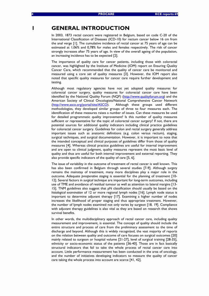
1 GENERAL INTRODUCTION In 2003, 1873 rectal cancers were registered in Belgium, based on code C-20 of the International Classification of Diseases (ICD-10) for rectum cancer below 16 cm from the anal verge [1]. The cumulative incidence of rectal cancer at 75 years of age can be estimated at 1,06% and 0,78% for males and females respectively. The risk of cancer strongly increases after 75 years of age. In view of the overall ageing of the population, an increasing incidence has to be expected [2].
The importance of quality care for cancer patients, including those with colorectal cancer, was highlighted by the Institute of Medicine (IOM) report on Ensuring Quality Cancer Care, which recommended that the quality of cancer care be monitored and measured using a core set of quality measures [3]. However, the IOM report also noted that specific quality measures for cancer care require further development and testing.
Although most regulatory agencies have not yet adopted quality measures for colorectal cancer surgery, quality measures for colorectal cancer care have been identified by the National Quality Forum (NQF) (http://www.qualityforum.org) and the American Society of Clinical Oncologists/National Comprehensive Cancer Network (http://www.asco.org/portal/site/ASCO). Although these groups used different methodologies, they developed similar groups of three to four measures each. The identification of these measures raises a number of issues. Can these measures be used for detailed programmatic quality improvement? Is this number of quality measures sufficient or representative for the topic of colorectal cancer surgery? If not, there are potential sources for additional quality indicators including clinical practice guidelines for colorectal cancer surgery. Guidelines for colon and rectal surgery generally address important issues such as anatomic definitions (e.g. colon versus rectum), staging, surgical techniques, and surgical documentation. However, it is important to note that the intended conceptual and clinical purposes of guidelines differ from those of quality measures [4]. Whereas clinical practice guidelines are useful for internal improvement and are open to clinical judgment, quality measures represent the most basic level of quality and thus are useful for both internal improvement and external reporting. They also provide specific indicators of the quality of care [5, 6].
The issue of variability in the outcome of treatment of rectal cancer is well known. This has also been confirmed in Belgium through several studies [7-9]. Although surgery remains the mainstay of treatment, many more disciplines play a major role in the outcome. Adequate preoperative staging is essential for the planning of treatment [10-12]. Several factors in surgical technique are important for long-term outcomes, including use of TME and avoidance of residual tumour as well as attention to lateral margins [13-15]. TNM guidelines also suggest that pN classification should usually be based on the histological examination of 12 or more regional lymph nodes [16]. Lymph node status is important to determine adjuvant therapy [17]. Examining a higher number of nodes increases the likelihood of proper staging and thus appropriate treatment. However, the number of lymph nodes examined not only varies by surgeon [18, 19]. Compliance with adjuvant therapy guidelines is also vital as they are based on research that shows survival benefits.
In other words, the multidisciplinary approach of rectal cancer care, including quality measurement and improvement, is essential. The concept of quality should include the entire structure and process of care from the preliminary assessment to the time of discharge and beyond. Although this is widely recognized, the vast majority of reports on the relation between quality and outcome of care focuses on surgical outcomes [20] mainly related to surgeon or hospital volume [21-27], level of surgical training [28-35], ethnicity or socio-economic status of the patients [36-40]. Those are in fact basically structural indicators that fail to take the whole process of rectal cancer care into account. Little performance measurement has been conducted in the area of oncology, and the number of initiatives developing indicators to measure the quality of cancer care taking the whole process into account are scarce [41, 42].
KCE reports 69 PROCARE 5
In view of published therapeutic variability and the reported benefit of national projects and trials, all Belgian scientific societies involved in the treatment of patients with rectal cancer at any stage, decided in December 2004 to set up a nationwide and multidisciplinary project PROCARE (PROject on CAncer of the REctum). The project aims to improve outcomes in patients with rectal cancer based on standardization through guidelines, implementation of these guidelines and quality assurance through registration and feedback.
A preliminary version of a guideline (CPG) was drafted in 2005, followed by workshops (surgery, pathology, radiotherapy, chemotherapy, radiology). A set for data entry of individual patients was constructed and voluntary registration in the PROCARE database at the Foundation Belgian Cancer Registry (FBCR) was started in 2006. Of the participating centres, all consecutive patients with rectal cancer (at any stage) are prospectively entered in this database. The PROCARE registration form entails all data relevant for any discipline on the staging and treatment of rectal cancer. Through feedback all centres will be able to position themselves in comparison to national (and possibly international) indicators and comparators. Above this, the opportunity will be given to call upon the expertise of accredited peers to analyze the results and support them in taking corrective actions if deemed useful or necessary.
In the present report, an updated version of the PROCARE CPG is presented. In a subsequent report, scheduled for 2008, a set of quality indicators will be pilot tested using the prospective PROCARE database and coupled data of the FBCR, the Minimal Clinical Data (MKG/RCM) and the Common Sickness Funds Agency (Intermutualistisch Agentschap, IMA). Also, an overview will be provided of international experiences with the measurement of quality indicators for rectal cancer.

6 PROCARE KCE reports 69
2 UPDATED PROCARE GUIDELINES FOR THE TREATMENT OF RECTAL CANCER
2.1 INTRODUCTION
Although several CPGs related to rectal or colorectal cancer already exist, most deal with specific aspect(s) of the disease. In July 2006, the PROCARE steering group (see below) established a working group to update and improve the quality of its multidisciplinary guideline in collaboration with the KCE. The following aspects of the management of patients with rectal cancer are covered: diagnosis and pre-treatment staging, indications and type of neoadjuvant therapy, surgical aspects related to elective and emergency surgery as well as to radical and local excision, pathological examination of the resected specimen, indications and type of adjuvant therapy, follow-up after curative treatment, and therapeutic aspects of patients with metastatic rectal cancer. This CPG does not cover screening and prevention (including symptom criteria to guide referral to a specialist and surveillance of patient groups at high risk), anal cancer, rectal cancer in the context of hereditary syndromes, and genetic counselling.
This CPG is intended to be used by all professionals involved in the care of patients with rectal cancer. The recommendations are based on the best available evidence and are adopted by the multidisciplinary steering group of PROCARE. This CPG is endorsed by the Belgian Section for Colorectal Surgery (BSCRS), a section of the Royal Belgian Society for Surgery (RBSS) represented in the PROCARE steering group by Bertrand C, De Coninck D, Duinslaeger M, Kartheuser A, Penninckx F, Van de Stadt J and Vaneerdeweg W, the Belgian Society of Surgical Oncology (BSSO) represented by Claeys D, the Belgian Group for Endoscopic Surgery (BGES) represented by Burnon D, the Belgian Society of Pathology and Digestive Pathology Club represented by Ectors N, Jouret A and Sempoux C, the Belgian Society of Radiotherapy – Oncology (BSRO) represented by Haustermans K, Scalliet P and Spaas P, the Belgian Group Digestive Oncology (BGDO) represented by Laurent S, Polus M, Van Cutsem E and Van Laethem JL, the Belgian Society Medical Oncology (BSMO) represented by Bleiberg H, Humblet Y and Van Cutsem E , the Royal Belgian Society Radiology (RBSR) represented by Danse E, Op De Beeck B and Smeets P, the Vlaamse Vereniging Gastro-Enterologie (VVGE) represented by Cabooter M, Pattyn P and Peeters M, the Société Royale Belge Gastro-Entérologie (SRBGE) represented by Melange M, Rahier J and Van Laethem JL, the Belgian Society Endoscopy represented by Buset M, the Belgian Professional Surgical Association (BPSA) represented by Haeck L and Mansvelt B, and the FBCR represented by Van Eycken E. The CPG is also endorsed by the College of Oncology, represented by Scalliet P. Nationwide implementation of highly recommended CPGs is warranted in order to reduce diagnostic and therapeutic variability. However, the ultimate decision about the appropriateness of any specific procedure must be made by the physician in the context of an individual patient.
2.2 METHODOLOGY
2.2.1 General approach
The present CPG was developed by adapting (inter)national CPGs to the Belgian context [43]. This approach is currently being structured in a formal methodology by the ADAPTE group, an international group of guideline developers and researchers [43]. The ADAPTE methodology generally consists of three major phases:
Set-up Phase: Outlines the necessary tasks to be completed prior to beginning the adaptation process (e.g., identifying necessary skills and resources).
KCE reports 69 PROCARE 7
Adaptation Phase: Assists guideline developers in moving from selection of a topic to identification of specific clinical questions; searching for and retrieving guidelines; assessing the consistency of the evidence therein, their quality, currency, content and applicability; decision making around adaptation; and preparing the draft adapted guideline.
Finalization Phase: Guides guideline developers through getting feedback on the document from stakeholders who will be impacted by the guideline, consulting with the source developers of guidelines used in the adaptation process, establishing a process for review and updating of the adapted guideline and the process of creating a final document.
This stepwise approach is currently being validated in an evaluation study using the (qualitative and quantitative) information from multiple case studies.
2.2.2 Guideline development group composition
The working group delegated by PROCARE consisted of 1 radiologist (Etienne Danse), 2 radiation oncologists (Karin Haustermans, Sarah Roels), 3 surgeons (Daniël De Coninck, Daniël Leonard, Freddy Penninckx), 1 pathologist (Nadine Ectors), and 5 gastrointestinal oncologists (Jochen Decaestecker, Caroline De Vleeschouwer, Stéphanie Laurent, Marc Peeters, Eric Van Cutsem). Methodological and organizational support was provided by experts from the KCE (Gert Peeters, Joan Vlayen). All persons involved were editorially independent.
2.2.3 Clinical questions
Clinical search questions were formulated for all aspects of rectal cancer management based on the PICO principle (patient, intervention, comparison, outcome). The clinical practice guideline addresses the following clinical questions:
1. Diagnosis and staging:
a. What method should be used for the detection of synchronous colonic lesions (polyps, cancer) in patients with rectal cancer?
b. Are tumour markers useful staging tools in patients with rectal cancer?
c. What imaging technique(s) can be recommended for the detection of metastatic disease in patients with rectal cancer?
d. What imaging technique(s) can be recommended for the locoregional cTN staging of patients with rectal cancer?
1. Can transrectal ultrasonography (TRUS) distinguish between a pT1 and a pT0 in patients with a benign looking, biopsy negative villous adenoma of the rectum?
2. What imaging technique should be used to identify transmural invasion in a patient with rectal cancer?
3. What imaging technique should be used to identify nodal involvement in patients with rectal cancer?
4. When there is no agreement between the results of different staging tools, what result is to be considered in the decision for neoadjuvant treatment in patients with resectable rectal cancer?
5. What imaging technique should be used to evaluate the cCRM (lateral margin) in patients with rectal cancer?
2. Neoadjuvant treatment:
a. Can preoperative radiotherapy improve the outcome in patients with resectable rectal cancer compared to surgery alone?
8 PROCARE KCE reports 69
b. Is preoperative chemoradiotherapy better than preoperative radiotherapy alone in the outcome of patients with resectable rectal cancer?
c. Is preoperative (chemo)radiotherapy better than postoperative chemoradiotherapy in the outcome of patients with resectable rectal cancer?
d. Is 5-FU continuous infusion superior to bolus 5-FU in combination with preoperative radiotherapy in the outcome of patients with resectable rectal cancer?
e. Is intravenous 5-FU better than oral 5-FU in the outcome of patients with resectable rectal cancer?
f. Is a long course of preoperative (chemo)radiation better than a short course of preoperative radiation in the outcome of patients with resectable rectal cancer?
g. Is a long treatment interval between preoperative (chemo)radiation and surgery better than a short interval in the outcome of patients with resectable rectal cancer?
h. Is there any benefit from alternative regimens of preoperative (chemo)radiotherapy compared to the standard regimen of (chemo)radiotherapy (short course or long course) in the outcome of patients with resectable rectal cancer? What is the role of brachytherapy/contact X-ray therapy in the preoperative treatment of resectable rectal cancer?
i. Is restaging after preoperative treatment useful in patients with resectable rectal cancer?
j. What is the role of (chemo)radiotherapy in patients with unresectable rectal cancer?
3. Surgery:
a. Can urinary or sexual dysfunction be avoided by good quality total mesorectal excision (TME) sphincter saving or abdominoperineal resection in rectal cancer patients for whom curative surgery is scheduled?
b. Can postoperative morbidity be reduced by preoperative bowel preparation in rectal cancer patients for whom curative surgery is scheduled?
c. Can postoperative deep venous thrombosis (DVT) be reduced by perioperative thromboprophylaxis in rectal cancer patients for whom curative surgery is scheduled?
d. Can postoperative septic complications be reduced by antibiotic prophylaxis in rectal cancer patients for whom curative surgery is scheduled?
e. Can preoperative stoma counselling, including stoma sitting, improve postoperative quality of life in rectal cancer patients for whom curative surgery is scheduled?
f. What is the impact of high versus low ligation of the inferior mesenteric artery on outcome in rectal cancer patients for whom curative surgery is scheduled?
g. What is the impact of lateral lymphatic dissection (iliac nodes) on outcome in rectal cancer patients for whom curative surgery is scheduled?

KCE reports 69 PROCARE 9
h. Can sphincter saving operation be performed for rectal cancer of the lower third of the rectum without compromising the (oncological and functional) outcome in patients for whom curative surgery is scheduled?
i. Can laparoscopic resection be performed without compromising the outcome in rectal cancer patients for whom curative surgery is scheduled?
j. Does inadvertent perforation of the rectum during surgery influence oncological outcome in rectal cancer patients for whom curative surgery is scheduled?
k. Does rectal stump wash-out prior to anastomosis decrease local recurrence in rectal cancer patients for whom curative surgery is scheduled?
l. Should a colonic pouch, a coloplasty or a straight coloanal anastomosis be performed for optimal functional outcome in rectal cancer patients for whom curative surgery is scheduled?
m. Should a temporary defunctioning stoma routinely or selectively be constructed at restorative proctectomy in order to reduce clinical leak rate in rectal cancer patients for whom curative surgery is scheduled?
n. Can a local resection or transanal endoscopic microsurgical resection be performed instead of a radical resection without compromising the outcome in rectal cancer patients for whom curative surgery is scheduled?
o. Is stenting an appropriate alternative for stoma construction as a bridge to radical surgery in case of stenosing rectal cancer?
p. Is stenting a valid alternative for stoma construction in a palliative setting?
4. Pathology
a. How should a rectal cancer resection specimen be assessed macroscopically (with specific criteria for the evaluation of TME quality)?
b. How should a rectal cancer resection specimen be assessed microscopically?
c. What are the data to be reported by the pathologist?
5. Adjuvant treatment
a. In patients who received neoadjuvant radio(chemo)therapy, when should adjuvant chemotherapy be considered?
b. In patients who received neoadjuvant radio(chemo)therapy, what chemotherapy is to be recommended?
c. In patients who did not receive neoadjuvant radio(chemo)therapy, when should adjuvant treatment be considered?
d. In patients who did not receive neoadjuvant radio(chemo)therapy, what type of adjuvant treatment and regimen is to be recommended: radiotherapy, chemotherapy or combined radiochemotherapy?
6. Follow-up:
a. Has follow-up an impact on survival and quality of life in patients curatively treated for rectal cancer?
10 PROCARE KCE reports 69
b. What clinical, biochemical or technical investigations have to be done in terms of local recurrence, distant recurrence and resectability of recurrence in patients curatively treated for rectal cancer?
c. How frequently and for how long clinical, biochemical or technical investigations have to be done in terms of local recurrence, distant recurrence in patients curatively treated for rectal cancer?
7. Metastatic disease:
a. What diagnostic tools can be used to determine the resectability of a metastatic disease? What are the resectability criteria?
b. What is the best management in patients with resectable primary tumour and resectable metastases?
1. Should induction treatment be applied in resectable metastatic rectal cancer?
2. What course of radiotherapy should be considered (long versus short)?
3. What is the best management in patients with resectable primary tumour and resectable metastases: sequential or synchronous surgery?
4. What is the best management in patients with metachronous resectable metastases, neoadjuvant or adjuvant chemotherapy?
c. Is radical treatment of a resectable primary tumour useful in patients with non resectable metastases?
d. Does first-line chemotherapy alone as compared to observation have an impact on prognosis in patients with synchronous or metachronous non resectable metastases?
e. Does second-line chemotherapy alone as compared to observation have an impact on prognosis in patients with synchronous or metachronous non resectable metastases?
f. What combination(s) should be considered for first- and second line chemotherapy?
g. How to manage non-resectable metastatic rectal cancer?
h. What is the management of isolated peritoneal carcinomatosis?
2.2.4 Search for evidence
2.2.4.1 Clinical practice guidelines
The search for guidelines on all or any aspect of the management of rectal cancer was performed in August 2006 by 2 members of the PROCARE panel (Daniel Leonard, Freddy Penninckx).
The following sources were consulted:
• National Guideline Clearinghouse: www.guideline.gov (search terms “rectal neoplasms”, “rectal cancer”);
• Medline (via PubMed; free text words “rectal neoplasms”, “rectal cancer”, “colorectal neoplasms”, “colorectal cancer” and “guideline”; MeSH-terms "Rectal Neoplasms" and "Practice Guideline");
• Sites of specific oncology organisations:
KCE reports 69 PROCARE 11
o ASCO: http://www.asco.org/portal/site/ASCO/menuitem.56bbfed7341ace64e7cba5b4320041a0/?vgnextoid=1c09201eb61a7010VgnVCM100000ed730ad1RCRD;
o NCCN: http://www.nccn.org/;
o FNCLCC: http://www.fnclcc.fr/sor/structure/index-sorspecialistes.html;
o Cancer Care Ontario: http://www.cancercare.on.ca/;
All retrieved hits were screened by title and abstract (and full-text if required), taking into account the following inclusion and exclusion criteria:
• Inclusion criteria:
o CPGs related to rectal or colorectal cancer;
o Publication and/or update in 2001 or thereafter;
o Publication in English, German, French or Dutch.
• Exclusion criteria: patient versions of CPGs on (colo)rectal cancer care; CPGs exclusively addressing population screening, primary prevention (including surveillance in patient groups at high risk), and/or genetic counselling; CPGs relating to anal cancer, familial adenomatous polyposis, hereditary non-polyposis colon cancer, and the Peutz-Jeghers syndrome.
2.2.4.2 Additional evidence
For each clinical question, the evidence – identified through the included CPGs – was updated by searching Medline (MeSH-term ‘Rectal Neoplasms’, not exploded; in combination with domain-specific MeSH-terms) and the Cochrane Database of Systematic Reviews (domain-specific free text words) from the search date of the CPG on.
The following inclusion criteria were applied:
• Design: systematic reviews, meta-analyses, randomized controlled trials (in the absence of these designs, also non-randomized controlled trials, cohort studies and/or case-control studies were included);
• Date of publication: 2001 – search date (August 2006)
• Language: English, French, German, Dutch.
Searches related to metastatic rectal cancer and palliative treatment were limited to meta-analyses and randomized controlled trails published in the last three years (11/2003 – 11/2006) in order to represent as much as possible the actual state of the art in this fastly evolving domain.
2.2.5 Quality appraisal
2.2.5.1 Clinical practice guidelines
The English version of the AGREE instrument (www.agreecollaboration.org) was used for the critical appraisal of the identified CPGs. All thirty-three guidelines were scored by 4 independent experts (see appendix for the scores per guideline). The score of the domain methodology was used as an important criterion in the final selection of guidelines.
At the end, 17 guidelines were included (see appendix).
2.2.5.2 Additional evidence
The quality of the retrieved systematic reviews and primary studies was assessed using the checklists of the Dutch Cochrane Centre (www.cochrane.nl).
12 PROCARE KCE reports 69
2.2.6 Data extraction and summary
For each included CPG the following data were extracted: organisation, scope, search date, publication year, relevant recommendations with supporting evidence.
For each systematic review, the search date, publication year, included studies and main results were extracted. For RCTs and observational studies, the following data were extracted: publication year, study population, study intervention, and outcomes.
For each clinical question, the recommendations from the identified CPGs and the additional evidence were summarized in evidence tables. A level of evidence was assigned to each recommendation and additional study using the GRADE system (see appendix) [44].
2.2.7 Formulation of recommendations
Based on the retrieved evidence, a first draft of recommendations was prepared by each expert responsible for its subdiscipline. This first draft together with the evidence tables was circulated to the guideline development group, and discussed during several face-to-face meetings and by email. Based on these discussion meetings a second draft of recommendations was prepared. A grade of recommendation was assigned to each recommendation using the GRADE system (see appendix), including ‘expert opinion’ where applicable. The second draft was once more circulated to the guideline development group for final approval.
2.2.8 External review
On February 9th 2007, the second draft of recommendations was circulated by e-mail to the PROCARE steering group: Bertrand C, Burnon D, Claeys D, De Coninck D, Duinslaeger M, Kartheuser A, Pattyn P, Penninckx F, Van de Stadt J, Vaneerdeweg W (surgeons), Ectors N, Jouret An , Rahier J, Sempoux C (pathologists), Danse E, Op De Beeck B, Smeets P (radiologists); Haustermans K, Scalliet P, Spaas P (radiation oncologists); Haeck L, Mansvelt B(surgeons representing the Belgian Professional Association), Bleiberg H, Humblet Y, Laurent S, Peeters M, Polus M, Van Cutsem E, (oncologists), Buset M, Cabooter M, Melange M, Van Laethem JL (gastroenterologists); Van Eycken E (Foundation Belgian Cancer Registry) . All steering group members were invited to discuss these recommendations and their grades (including expert opinion) during a consensus meeting on February 22nd 2007. As a preparation of the meeting, all steering group members were asked to score each recommendation on a 5-point Likert-scale to indicate their agreement with the recommendation, with a score ‘1’ indicating ‘completely disagree’, ‘2’ indicating ‘somewhat disagree’, ‘3’ indicating ‘unsure’, ‘4’ indicating ‘somewhat agree’, and ‘5’ indicating ‘completely agree’. The scorers were also able to answer ‘not applicable’ in case they were not familiar with the underlying evidence. In case of disagreement with the recommendation (scores ‘1’ or ‘2’), scientific evidence for the disagreement had to be provided. All received scores were anonymized and summarized into a mean score, standard deviation and % of ‘agree’-scores (score ‘4’ and ‘5’). Consensus agreement was defined as 60% ‘agree’-scores.
Fifteen individual colleagues returned there scores, as well as one group of 4 specialists working at the same institution (considered as one score). The latter score was reported but not used to calculate the global score (see appendix). All disciplines were represented. A copy of the individual and global scores per recommendation as well as the comments was provided at the face-to-face meeting.
All recommendations reached >60% agreement. However, items that were commented and/or items that had one or more individual scores of ‘1’ or ‘2’ were discussed. Items with scores ‘4’ and ‘5’ were not discussed. During the meeting a consensus was reached on all recommendations. The summary of the discussion and the final version of the recommendations were attached to the minutes of the meeting, and sent to all members of the PROCARE steering committee. No requests for further adaptation(s) were made.
KCE reports 69 PROCARE 13
2.3 DEFINITIONS
2.3.1 The rectum
Tumours with their distal edge at 15 cm or less from the anal verge, as measured with a rigid rectosigmoidoscope, are classified as rectal. Distances from the anal verge measured with a flexible sigmoido- or colonoscopy are not always reliable.
The anal verge should be the usual landmark. Nonetheless, the distance between the lower edge of the tumour and the upper limit of the anal canal can be useful. The distance between the lower edge of the tumour and the anal verge is very important for stratification and because it influences the type of neoadjuvant treatment, the type of surgery and outcome.
For international benchmarking, rectal tumours can be categorized according to their distal edge as “low” (up to 5.0 cm above the anal verge), “mid” (from 5.1 till 10.0 cm above the anal verge) and “high” (from 10.1 – 15.0 cm above the anal verge) [45-47].
2.3.2 Staging
The TNM classification of tumours described by the International Union Against Cancer (UICC) and the American Joint Committee on Cancer (AJCC) is used for tumour staging [16, 48]:
• cTNM: pre-treatment clinical classification, based on clinical examination, imaging, endoscopy, biopsy, surgical exploration or other;
• pTNM: post-surgical histopathological classification;
• ypTNM: post-surgical histopathological classification following preoperative therapy (radio- and/or chemotherapy).
2.3.2.1 Classification adapted from UICC and AJCC [16, 48]
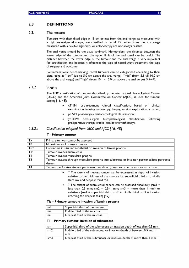
T - Primary tumour
Tx Primary tumour cannot be assessed T0 No evidence of primary tumour Tis* Carcinoma in situ: intraepithelial or invasion of lamina propria T1° Tumour invades submucosa T2 Tumour invades muscularis propria T3 Tumour invades through muscularis propria into subserosa or into non-peritonealized perirectal
tissues T4 Tumour perforates visceral peritoneum or directly invades other organs or structures
• * The extent of mucosal cancer can be expressed in depth of invasion relative to the thickness of the mucosa: i.e. superficial third m1, middle third m2 and deepest third m3.
• ° The extent of submucosal cancer can be assessed absolutely (sm1 = less than 0.5 mm; sm2 = 0.5–1 mm; sm3 = more than 1 mm) or relatively (sm1 = superficial third; sm2 = middle third; sm3 = invasion reaching the deepest third) [49].
Tis – Primary tumour: invasion of lamina propria
m1 Superficial third of the mucosa m2 Middle third of the mucosa m3 Deepest third of the mucosa
T1 – Primary tumour: invasion of submucosa
sm1 Superficial third of the submucosa or invasion depth of less than 0.5 mm sm2 Middle third of the submucosa or invasion depth of between 0.5 and 1
mm sm3 Deepest third of the submucosa or invasion depth of more than 1 mm
14 PROCARE KCE reports 69
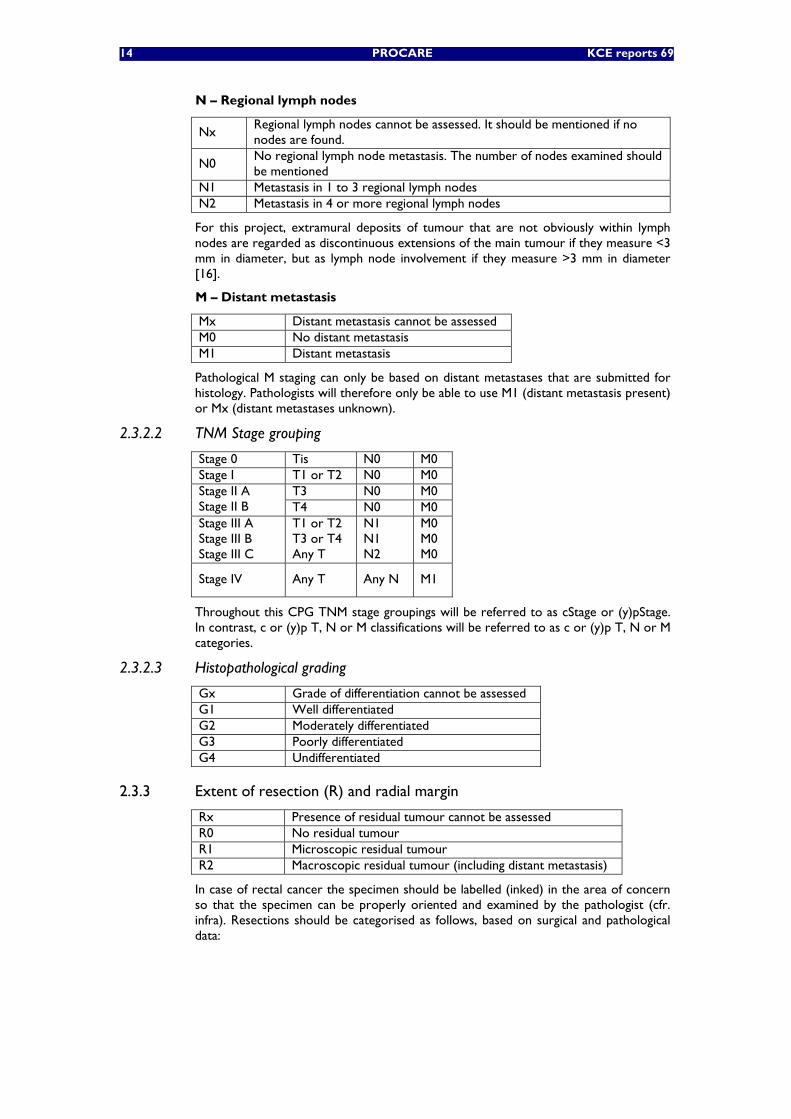
N – Regional lymph nodes
Nx Regional lymph nodes cannot be assessed. It should be mentioned if no nodes are found.
N0 No regional lymph node metastasis. The number of nodes examined should be mentioned
N1 Metastasis in 1 to 3 regional lymph nodes N2 Metastasis in 4 or more regional lymph nodes
For this project, extramural deposits of tumour that are not obviously within lymph nodes are regarded as discontinuous extensions of the main tumour if they measure <3 mm in diameter, but as lymph node involvement if they measure >3 mm in diameter [16].
M – Distant metastasis
Mx Distant metastasis cannot be assessed M0 No distant metastasis M1 Distant metastasis
Pathological M staging can only be based on distant metastases that are submitted for histology. Pathologists will therefore only be able to use M1 (distant metastasis present) or Mx (distant metastases unknown).
2.3.2.2 TNM Stage grouping
Stage 0 Tis N0 M0 Stage I T1 or T2 N0 M0
T3 N0 M0 Stage II A Stage II B T4 N0 M0 Stage III A Stage III B Stage III C
T1 or T2 T3 or T4 Any T
N1 N1 N2
M0 M0 M0
Stage IV Any T Any N M1
Throughout this CPG TNM stage groupings will be referred to as cStage or (y)pStage. In contrast, c or (y)p T, N or M classifications will be referred to as c or (y)p T, N or M categories.
2.3.2.3 Histopathological grading
Gx Grade of differentiation cannot be assessed G1 Well differentiated G2 Moderately differentiated G3 Poorly differentiated G4 Undifferentiated
2.3.3 Extent of resection (R) and radial margin
Rx Presence of residual tumour cannot be assessed R0 No residual tumour R1 Microscopic residual tumour R2 Macroscopic residual tumour (including distant metastasis)
In case of rectal cancer the specimen should be labelled (inked) in the area of concern so that the specimen can be properly oriented and examined by the pathologist (cfr. infra). Resections should be categorised as follows, based on surgical and pathological data:
KCE reports 69 PROCARE 15
• R0: all gross disease is resected by en bloc resection with margins histologically free of disease. Non-en-bloc resection, positive radial margin i.e. <1 mm, positive proximal or distal bowel margins, residual lymph node disease, Nx, or even intraoperative inadvertent perforation of the tumour bearing bowel segment should not be considered R0. These patients are candidates for adjuvant radiochemotherapy or adjuvant chemotherapy in case preoperative radiotherapy has been given in order to reduce recurrence rates. Non-en-bloc resection and inadvertent perforation of the tumour-bearing segment during dissection must be documented in the surgical report.
• R1: all gross disease is resected by en bloc resection with margins histologically positive for disease or with cancer at less than 1 mm from a margin (or intraoperative perforation, cf. supra).
• R2: residual macroscopic disease, either locoregional or distant, remains unresected (thus including distant disease).
2.3.4 Other definitions related to surgery
• Emergency: immediate operation within 2 hours of admission or in conjunction with resuscitation
• Urgent: operation carried out within 24-hrs of admission.
• Scheduled: an early operation, but not immediately life-saving.
• Elective: operation at the time to suit both patient and surgeon.
• Hartmann’s procedure: anterior resection of the rectum with closure of the distal resection margin and end colostomy.
• Partial mesorectal excision (PME): anterior resection with excision of part of the rectum and colorectal anastomosis. It is indicated for cancer of the rectosigmoid junction or the upper rectal third of the rectum. A partial mesorectal excision should be performed down to 5 cm below the lower edge of the tumour.
• Total mesorectal excision (TME): resection of the entire mesorectal fat, down to the levator plane, with respect of the circumferential mesorectal integrity (as proven by pathology) and preservation of the nerve plexuses and nerves surrounding the mesorectum. A TME is indicated for cancer in the mid and lower third of the rectum.
• Restorative proctectomy: sphincter-saving complete resection of the rectum with total mesorectal excision and colo-anal anastomosis (with or without pouch or coloplasty). It is indicated for tumours of the middle and lower third of the rectum.
• Abdomino-perineal excision of rectum (APR): excision of the whole rectum and anus with total mesorectal excision and terminal colostomy.
2.3.5 Definitions related to radiotherapy volume and International Commission of Radiation Units (ICRU) reference point
2.3.5.1 Clinical target volume (CTV)
The CTV is defined as the gross tumour volume (GTV) plus the areas at risk for microscopic tumour extension. The locoregional lymph nodes at risk for subclinical disease include the internal iliac lymph nodes, the presacral nodes and the mesorectal nodes for all patients. According to the level of the primary tumour and the involvement of other organs, additional lymph node regions become at risk. If there is involvement of adjacent organs or structures, nodal drainage can arise via the lymphatics of the involved organ. This involves the external iliac nodes when there is tumour extension to anterior organs (bladder/prostate/seminal vesicles/uterus) and the inguinal nodes if the anal canal and/or lower third of the vagina are involved.
16 PROCARE KCE reports 69
If the patient is planned to undergo an abdominoperineal resection or the lesion is within 6 cm from the anal margin and the surgeon aims at a sphincter saving procedure, the perineal region, defined as the anal sphincter complex and the surrounding ischiorectal fossa, should be included in the CTV. Further information and the rationale behind these delineation guidelines have been published [50].
The CTV will be delineated using a CT scan in the treatment position.
2.3.5.2 Planning target volumes (PTV)
The PTV includes the CTV plus a margin for set-up error and/or patient/organ motion. Additional margins may be required based upon clinical judgment.
Radiation beams are designed to adequately cover the PTV. This applies for the conventional treatment technique as well as for the 3D conformal treatment technique or intensity modulated radiation. With the latter technique, planning CT can help to adjust the field borders to ensure adequate coverage of the PTV.
2.3.5.3 International Committee on Radiation Units (ICRU) reference point
The ICRU reference point is to be located in the central part of PTV (ICRU 50.62). The specification of the target dose is in terms of a dose to a point at or near the centre of target volume:
• For arrangement of two or more intersecting beams: at the intersection of the central ray of the beams.
• Other or complex treatment arrangements: at the centre of the target area(s).
2.4 FINAL RECOMMENDATIONS
2.4.1 Access to treatment
No formal search was performed on this topic, but the following statements, derived from other guidelines seem to be appropriate and are to be recommended:
1. The interval between making a diagnosis of cancer and the start of treatment should be less than 4 weeks [51].
2. All patients should have the benefit of objective information [51].
3. The patient should be informed that rectal cancer treatment deserves a multidisciplinary approach. Rectal cancer should be treated by specialists (gastroenterologists, radiologists, surgeons, pathologists, radiation oncologists, oncologists) with appropriate training and experience [51]. The use of a single multidisciplinary document for informed consent is recommended when available.
4. The patient who develops colorectal cancer before the age of 45 years or who belongs to a family in which colorectal or associated cancers (endometrium,…) have occurred, must be informed about the risk for his/her relatives to develop the disease. The physician or specialist will insist on appropriate investigations and surveillance in the patient’s family members [51, 52].
2.4.2 Diagnosis and staging
2.4.2.1 Diagnosis of rectal cancer
Digital rectal examination should be carried out in all patients. Since the treatment of rectal cancer is invasive, the diagnosis should be based on the results of pathologic examination of biopsies, which should be obtained from all rectal tumours before the start of any type of treatment, including endoscopic or local excision. Pre-treatment staging is important for prognosis and for decision-making on the type of neoadjuvant
KCE reports 69 PROCARE 17
treatment and surgical resection/reconstruction. Also, it provides accurate case-mix data for stratification. Therefore, it should be of the best possible accuracy.
The distance between the lower edge of the tumour and the anal verge is very important, since it co-determines the indication for neoadjuvant treatment, the type of surgery and outcome. It is recommended to determine this distance at rigid proctoscopy (rectoscopy). Colonoscopy (at withdrawal) could be an alternative, but cannot be recommended because it is not always reliable [52]. A tumour with its distal edge at 15 cm or less from the anal verge is classified as rectal (cfr. definitions). Although the anal verge should be the usual landmark, the distance between the lower edge of the tumour and the upper limit of the anal canal (anal sphincters) can also be useful. However, for international benchmarking the tumour location as referred to the anal verge is used.
For tumours within 10cm of the anal verge, the operating surgeon should record fixation, location of the tumour in relation to the anal sphincters and quadrant(s) occupied by the tumour[52].
1 A tumour with its distal edge at 15 cm or less from the anal verge should be classified as rectal. A biopsy should be obtained from all rectal tumours before the start of any type of treatment (including endoscopic or local excision) (1C recommendation).
2 It is recommended that the distance from the lower edge of the tumour to the anal verge should always be determined by rigid proctoscopy (rectoscopy) before the start of neoadjuvant treatment. Colonoscopy (at withdrawal) is not always reliable for measurement of this distance (1C recommendation) [52].
3 A digital rectal examination should be performed in all patients with rectal cancer. The operating surgeon should record information on the fixity, location (longitudinal and circumferential) and proximity to the sphincters in patients with low or mid rectal tumours (1C recommendation) [52].
2.4.2.2 Detection of simultaneous colonic lesions in patients with rectal cancer
Extensive use of preoperative colonoscopy is recommended in the evaluation of colorectal cancer, in order to promote detection of synchronous tumours, reduce the incidence of 'early metachronous' cancer and avoid malignant degeneration of adenomatous polyp. The incidence of a synchronous polyp has been reported to be 14 % and the incidence of a synchronous carcinoma 4 % [53]. The highest incidence is to be expected in patients with a genetic predisposition (e.g. FAP, HNPCC, …) or in patients at increased risk for colorectal cancer (e.g. IBD).
Studies on the detection of simultaneous colonic lesions in patients with rectal cancer are limited and most have been poorly reported [52, 54, 55]. There are no good data directly comparing the performance of double contrast barium enema (DCBE) with colonoscopy for the detection of synchronous colon polyps or cancer in patients with rectal cancer. Thus, the accuracy of both examination in the screening and evaluation of patients presenting with symptoms suggestive of colorectal cancer were used (extrapolation). It has to be taken into account that these data are mainly based on studies that used a different referral pattern for the two techniques. Moreover, no good reference standard is available to verify the results of the techniques. Indeed, it is important to realize that colonoscopy is not a perfect (‘gold’) reference test. Nevertheless, it is recommended that patients with rectal cancer should have a total colonoscopy with resection of concomitant polyps if possible [52, 54, 55]. However, if total colonoscopy is judged to be too risky or if colonoscopy is refused after informed consent, a high quality DCBE should be performed [52, 54, 55]. In emergency circumstances, when a total colonoscopy is not possible preoperatively, it should be performed before the start of adjuvant therapy or at least within 3-6 months postoperatively [52, 55]. According to the NICE guideline, the quality of colonoscopy should be recorded with the aim to achieve a high total colonoscopy rate with a low perforation risk [54].
18 PROCARE KCE reports 69
Virtual CT or MRI based colonography are sensitive methods for the detection of colorectal cancer and/or large polyps, but not for polyps less than 10 mm in diameter [55]. Systematic reviews indicate that studies are poorly reported and that heterogeneity of sensitivity must raise concerns about consistency of performance and about technical variability [56, 57]. These issues must be resolved before virtual colonography can be advocated for routine use in the screening for synchronous colon cancer.
4 Patients with rectal cancer should have a total colonoscopy with resection of concomitant polyps if possible. If total colonoscopy is judged to be too risky or if colonoscopy is refused after informed consent, a high quality double contrast barium enema should be performed (1C recommendation) [52, 54, 55].
5 CT-colonography cannot (yet) be recommended for routine use. However, it may be useful in case of stenosing rectal cancer if the radiological equipment and expertise with audit is available (1C recommendation) [55-57].
6 In emergency circumstances, when a total colonoscopy is not possible preoperatively, it should be performed before the start of adjuvant therapy or at least within 3-6 months after surgery (1C recommendation) [52, 55].
7 The quality of colonoscopy should be recorded with the aim to achieve a high total colonoscopy rate with a low perforation risk (2C recommendation) [54].
2.4.2.3 Tumour markers in patients with rectal cancer
Lack of sensitivity and specificity preclude the use of any available serum marker for the early detection of colorectal cancer [52, 58]. However, pre-treatment carcinoembryonic antigen (CEA) levels have been related to cancer stage and survival (independent of pTN stage in nonmetastatic colorectal cancer). Significantly increased CEA levels may indicate the presence of metastatic disease, warranting further pre-treatment evaluation (e.g. using FDG PET or PET/CT scan).
8 The serum carcinoembryonic antigen (CEA) level should be determined in all patients before the start of any treatment (1B recommendation) [52, 58].
9 There is not enough evidence to recommend the routine use of other tumour markers (1B recommendation) [58].
2.4.2.4 Staging of rectal cancer
The TNM stage of a (colo)rectal cancer is a very important predictor of prognosis.
The aim of imaging techniques such as CT, MRI and PET is to detect hepatic and extra-hepatic metastatic disease. The recommendations presented below are mainly based on the French guidelines [59]. A recent meta-analysis of Bipat et al. included studies on CT, MRI and PET [60]. Per patient, PET was found to be the best technique. Per lesion MRI with intravenous injection of gadolinium had the best sensitivity. Nonetheless, spiral CE-CT (MSCT) is recommended for routine use. When contrast-CT can not be performed, MRI can be considered as a valid, even more accurate alternative. CT is to be combined with FDG-PET for the better staging of patients with potentially resectable metastatic disease.
All guidelines agree that patients with rectal cancer should have locoregional cTN staging [52, 54, 55]. Investigation with TRUS and MRI is recommended by most guidelines.
Polyps with/without dysplasia (T0 or Tis) do not infiltrate the submucosa, have virtually no risk of lymph node metastasis, and do not require full thickness excision of the rectal wall. However, the deep resection margin should be evaluable at pathology (one specimen) and microscopically negative (i.e. more than 1 mm margin). These lesions are usually small, although (very) large adenomas with focal invasion do occur.
KCE reports 69 PROCARE 19
Some endoscopic features (umbilication, non-elevation of the lesion after submucosal lifting) may be indicative of invasion. This setting requires accurate pre-treatment distinction between T1 versus T0 lesions. Until recently, a small cT1 rectal cancer with prognostically good pathological characteristics (‘low risk’) was generally considered an appropriate indication for full thickness local excision (LE, TEMS) (cfr. chapter 2.4.4 on surgery). Thus, pT0-T1 tumours have frequently been reported together in the past.
The aim was to distinguish T0-1 and T2 or more lesions. However, LE for pT1 has become controversial in view of significantly decreased local disease control after LE as compared with radical excision, except maybe for pT1sm1 [61-64]. TRUS is the best method to visualize the different layers of the rectal wall. In contrast with previous reports and opinions, T0 can be identified by TRUS with a high accuracy, but, higher frequency, higher resolution probes have to be used [65] Not only high-quality equipment but also highly-experienced examiners are essential in order to obtain valid US data. TRUS should preferentially be performed before or together with biopsy(ies) in order to avoid secondary effects potentially distording TRUS findings and interpretation.
Accurate identification of cStage II tumours (i.e. cT3-4N0M0), and cStage III tumours (i.e. cTanyN+M0) is relevant for the decision about neo-adjuvant radio(chemo)therapy (cfr. chapter 2.4.3 on neoadjuvant treatment). Overstaging of T2 lesions can occur because of peritumoral inflammatory reaction. Thus, it may be indicated to confirm the diagnosis of a cT3 lesion by a second morphologically oriented imaging modality. However, the relevance of differentiating a small T3 from a full T2 may be limited, as both are well away from the resection margin, except in the lower rectum. In the latter case, both tumour types receive neoadjuvant (chemo)radiation in most centres (cfr. infra).
Existing guidelines are mainly based on a systematic review performed by Kwok [66]. It was concluded that the performance of all imaging modalities (TRUS, CT and MRI) to distinguish between T3-4 and T1-2 are comparable. This was confirmed in the meta-analysis of Bipat [60]. However, TRUS is operator dependent, more difficult to perform for high rectal tumours and impossible in stenosing cancer. Also, it can not provide information on the depth of perirectal fat invasion and on the lateral tumour-free margin (cCRM). Therefore, MRI can be advocated as the single diagnostic tool able to provide these clinically important data in one session. CT induced much more understaging (hence potential undertreatment) than MRI. However, contrast enhanced multislice CT may be(come) a valid alternative. Also, UPSIO-MRI is still under investigation [67].
For clinical decision making, particularly related to neoadjuvant treatment, it is recommended to take into account the highest tumour and/or nodal category found by means of any imaging modality. However, no existing recommendations were found in guidelines, nor data in the recent literature. This recommendation has therefore to be regarded as expert opinion. In clinical practice, the ‘fail safe’ principle is usually applied. However, over-staging may result in over-treatment with its inherent complications. Thus, imaging should be of high quality. In order to avoid the harm of neoadjuvant treatment in small pT3 lesions with good CRM (i.e. located > 6 cm above the anal verge) it seems appropriate for decision making to take into account the result of the imaging modality with the lower T category for RC in the mid and upper third of the rectum if cN = 0 and cCRM not threatened. However, preoperative chemoradiation with an interval of 6-8 weeks to surgery results in about 20% of complete response (no viable tumour found in the resection specimen); this type of response as well as major tumour regression is reported to be related to improved outcome, including disease-free survival (DFS). These observations indicate that neoadjuvant treatment with the aim to downsize the tumour could be applied (at this time) in all except Stage I rectal cancer.
Transmural invasion (T3 or Stage II) and N+ (Stage III) are both related to increased local recurrence rate (LRR). N+ was found to have the most important effect on LRR despite TME surgery of good quality [45].
20 PROCARE KCE reports 69
Therefore, it is appropriate to take into account the result of the imaging modality with the highest N category, although it must be admitted that cN-staging is less accurate than cT staging.
There is no evidence to support the routine performance of preoperative re-staging. However, in some selected patients, it may be considered (cfr. chapter 2.4.3 on neoadjuvant treatment).
10 All patients with rectal cancer should have imaging of abdomen and chest for the detection of metastatic disease before elective treatment (1B recommendation) [52, 55, 59, 68].
11 A combined thorax and abdomen/pelvis spiral contrast-enhanced CT is recommended for the detection of metastatic disease. If a contrast-enhanced CT is contra-indicated, a thorax spiral CT without contrast and a contrast-enhanced magnetic resonance imaging (MRI) of the liver can be performed (1B recommendation) [59, 60, 68].
12 FDG-PET/CT can be recommended as an additional investigation, especially for the further staging of patients with apparently resectable metastasis, because of its high overall accuracy (1B recommendation) [54, 59, 60].
13 In case of emergency surgery, staging for metastatic disease should be performed intra-operatively and postoperatively, if not done pre-operatively (1C recommendation) [54, 55].
14 If cTN staging will drive therapeutic decisions, transrectal ultrasonography, if performed by an experienced examiner, is recommended for all non-stenosing, resectable tumours in the middle and lower third of the rectum (1B recommendation) [52, 54, 55, 60, 68, 69].
15 If cTN staging will drive therapeutic decisions, any uT3/4 and any uN+ category should be confirmed by phased array high resolution magnetic resonance imaging (HR-MRI). The clinical circumferential resection margin should also be determined by HR-MRI (1B recommendation) [52, 54, 55, 67, 69, 70].
16 If cTN staging will drive therapeutic decisions, a phased array high resolution magnetic resonance imaging (HR-MRI) is recommended for all tumours in the upper third of the rectum (1C recommendation) [52, 54, 68, 71].
17 Diagnostic imaging and its accuracy should be discussed and audited by all (colo)rectal cancer multidisciplinary teams (1C recommendation) [68, 70, 72].
18 Early rectal cancer as well as benign looking, biopsy negative villous adenomata of the rectum should be assessed with transrectal ultrasonography by an experienced examiner before any type of treatment (including excisional biopsy). Audits of diagnostic performance should be performed (1C recommendation) [54, 65, 73].
19 For identification of transmural penetration (T3 or more) and node positivity it could be recommended to use 2 staging modalities (transrectal ultrasonography [TRUS] and high resolution magnetic resonance imaging [HR-MRI], or TRUS and multislice CT are recommended) (expert opinion).
20 For clinical decision making, particularly related to neoadjuvant treatment, it is recommended to take into account the highest tumour and/or nodal category found by means of any imaging modality (expert opinion).
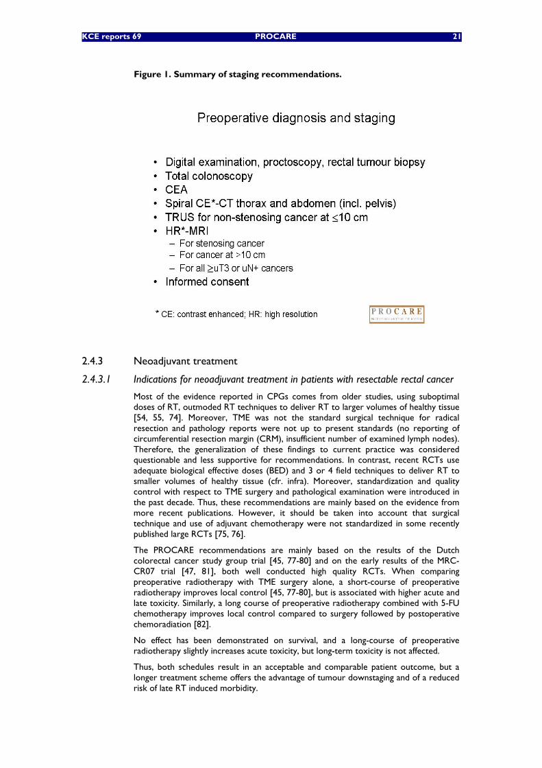
KCE reports 69 PROCARE 21
Figure 1. Summary of staging recommendations.
2.4.3 Neoadjuvant treatment
2.4.3.1 Indications for neoadjuvant treatment in patients with resectable rectal cancer
Most of the evidence reported in CPGs comes from older studies, using suboptimal doses of RT, outmoded RT techniques to deliver RT to larger volumes of healthy tissue [54, 55, 74]. Moreover, TME was not the standard surgical technique for radical resection and pathology reports were not up to present standards (no reporting of circumferential resection margin (CRM), insufficient number of examined lymph nodes). Therefore, the generalization of these findings to current practice was considered questionable and less supportive for recommendations. In contrast, recent RCTs use adequate biological effective doses (BED) and 3 or 4 field techniques to deliver RT to smaller volumes of healthy tissue (cfr. infra). Moreover, standardization and quality control with respect to TME surgery and pathological examination were introduced in the past decade. Thus, these recommendations are mainly based on the evidence from more recent publications. However, it should be taken into account that surgical technique and use of adjuvant chemotherapy were not standardized in some recently published large RCTs [75, 76].
The PROCARE recommendations are mainly based on the results of the Dutch colorectal cancer study group trial [45, 77-80] and on the early results of the MRC-CR07 trial [47, 81], both well conducted high quality RCTs. When comparing preoperative radiotherapy with TME surgery alone, a short-course of preoperative radiotherapy improves local control [45, 77-80], but is associated with higher acute and late toxicity. Similarly, a long course of preoperative radiotherapy combined with 5-FU chemotherapy improves local control compared to surgery followed by postoperative chemoradiation [82].
No effect has been demonstrated on survival, and a long-course of preoperative radiotherapy slightly increases acute toxicity, but long-term toxicity is not affected.
Thus, both schedules result in an acceptable and comparable patient outcome, but a longer treatment scheme offers the advantage of tumour downstaging and of a reduced risk of late RT induced morbidity.
22 PROCARE KCE reports 69
Data from univariate subgroup analyses in the Dutch Colorectal Cancer Group trial suggest an improved local control for middle and low seated tumours and for stage II and III RC, but not for high seated tumours and stage I and IV rectal cancer. However, a multivariate test for interaction between tumour stage and treatment group and between tumour level and treatment group was not significant, indicating that the local effect of preoperative RT is similar for all TNM stages and tumour levels [45, 77-80]. Results from the MRC CR07 trial confirm a benefit of short course RT on local control for all tumour levels and stages; results of local tumour control according to tumour level after a long course of chemoradiation followed by TME surgery have not been found [47, 81]. Although there is no strong evidence that patients with clinical stage I rectal cancer and patients with high seated (>10 cm) rectal cancer would not benefit from RT or chemoradiation before TME surgery, the absolute benefit in these cases is obviously more limited than in more advanced rectal cancer stages. If RT or chemoradiation is applied, it should be considered to outweigh (late) toxicity. In view of the absence of mesorectal fat in front of the distal third of the rectum, an exception has been made for full cT2 cancer in this location.
Acute or chronic toxicity may be associated with radio(chemo)therapy, such as enteritis, diarrhoea, bowel obstruction/stricture or perforation and fibrosis within the pelvis. Haematological and non-haematological adverse effects may occur when radiotherapy is combined with chemotherapy. Thus, patients to whom neoadjuvant radiotherapy or radiochemotherapy is proposed, should be informed of the potential harmful effects [55].
21 In order to improve local control, preoperative radiotherapy should be considered for resectable rectal cancer. It is recommended for all cStage II and cStage III lesions at any level. Radiotherapy is not recommended for cStage I lesions. However, it should be discussed in the multidisciplinary team for full cT2 lesions located ventrally in the lower third of the rectum because of the eccentric location of the rectum in the mesorectal fat (1A recommendation) [45, 47, 77-81].
22 Patients to whom neoadjuvant radiotherapy or radiochemotherapy is proposed, should be informed of the potential harmful effects (expert opinion).
2.4.3.2 Type of neoadjuvant treatment in patients with resectable rectal cancer
Two recent RCTs specifically addressing the value of additional chemotherapy to preoperative RT were found [75, 76]. Although both studies can be classified as high quality and provide the best evidence available at this time, TME was not the standard surgical procedure and pathology reports were not up to present standards; moreover, compliance to postoperative chemotherapy was poor in the EORTC trial [75]. These limitations can be of great consequence to the measured outcomes and should be taken into account in the interpretation of the results. The addition of 5-FU chemotherapy to a long course of preoperative radiotherapy was found to improve local control and to increase downsizing and downstaging compared to a long course of radiotherapy alone, resulting in more pathological complete responses. However, the rate of sphincter saving procedures was not influenced by the addition of chemotherapy. Preoperative chemoradiation resulted in higher acute grade 3/4 toxicity compared to RT alone, but postoperative complications were not significantly different. The incidence of late complications in the EORTC trial was not different in the 4 arms [75].
Whether preoperative (chemo)radiotherapy is better than postoperative chemoradiotherapy in patients with resectable rectal cancer can be answered with the findings of a German trial [82] comparing preoperative long course chemoradiation with a similar regimen given postoperatively. TME was performed but pathology quality assurance was not implemented. Both treatment modalities resulted in a similar overall and disease-free survival rate, but preoperative chemoradiation was associated with significantly less local recurrences and toxicity compared to postoperative chemoradiation. Also, compliance with preoperative CRT was remarkably better than with postoperative treatment.
KCE reports 69 PROCARE 23
Overall, no difference in sphincter saving procedures were observed; however, more patients who were intended to undergo an APR, received a sphincter sparing procedure after preoperative CRT, indicating that preoperative CRT can induce tumour shrinkage resulting in more sphincter saving operations in low-lying tumours.
Recommendations in existing guidelines [55] on the choice between continuous or bolus 5-FU in combination with preoperative radiotherapy are based on prospective cohort studies that have proven the safety and feasibility of the 3 following regimens: intermittently infused FUFA [83], continuous FU [84], or bolus FUFA [83, 85]. Bosset et al. reported the findings of 3 phase II studies, using the same preoperative CRT schedule, but different 5-FU doses [83]. The overall response rate was 87% for local disease, with 14,6% complete remissions among 41 macroscopically completely resected tumours. 29,3% of these tumours were downstaged. The authors concluded that a dose of 350 mg/m²/day was associated with an optimal toxicity and compliance profile. Another CT schedule, consisting of infusional 5-FU (300mg/m²/day) concomitant with each fraction of RT, was proven to be effective in the preoperative setting of locally advanced rectal cancer [85]. Rich et al. obtained an excellent local control of 96%. No RCTs were found that focussed on this subject in the preoperative setting. There is evidence from combined CRT in the postoperative setting in patients with high risk RC [86]. The authors found an increased time to relapse and improved survival with FU given by protracted venous infusion (PVI). The overall local control was good and slightly better in the PVI arm. Since these results only relate to postoperative CRT, the evidence is of a low quality level to support the use of a PVI in the preoperative setting.
There is low quality evidence from two small RCTs that continuous oral 5-FU is equivalent to bolus intravenous 5-FU in patients with T3N1 rectal cancer treated with preoperative chemoradiation [87, 88]. Overall, oral doxifluridine-based CRT showed comparable tumour response rates, local recurrences and systemic disease compared to IV FU-based CRT. Toxicity was not significantly increased, but more patients in the oral arm had a grade 1 or 2 diarrhoea [88]. Capecitabine was better tolerated than bolus intravenous 5-FU and was more effective in the promotion of down-staging [87]. Whether oral 5-FU is equivalent to a protracted infusion of 5-FU combined with preoperative radiotherapy remains unanswered.
Only one RCT directly compared a short course of preoperative RT with a long course of CRT in patients with low T3-4 rectal cancer [89]. Its results should be interpreted with caution because of several weaknesses. Although both regimens demonstrated comparable results in terms of patient outcome, the advantage of tumour regression and downstaging after a longer RT schedule combined with CT was confirmed. Despite these pronounced tumour responses after CRT, no more sphincter sparing procedures were performed in comparison with short-course RT. Acute RT toxicity was higher after CRT, postoperative complications were slightly lower in this group and late toxicity rates were comparable.
Similar downsizing and downstaging effects after a long course of CRT have been observed in other RCTs [75, 76]. On the contrary, short courses of preoperative RT followed by immediate surgery have failed to demonstrate any downstaging effect [45, 47].
If one considers the reported number of positive CRM in all these trials, it can be concluded that long-course preoperative RT with or without chemotherapy results in a positive CRM in about 4% to 7% of the patients [76, 89], compared to 10% to 18% [47, 78, 89] after a short-course of RT and 11% to 20% without any preoperative treatment [47, 78]. These findings indicate that a long course of CRT may induce a reduction in CRM positivity, which is an important prognostic factor for local control. An important finding in the Dutch trial is that the benefit for preoperative RT in terms of local control was only significant for patients with a wide (CRM > 2mm) and narrow margin (CRM 1-2 mm), but not for patients with a positive CRM (CRM ≤ 1mm) [78]. Thus, short course RT followed by TME surgery within one week should be reserved for cases where the CRM is certainly not at risk.
24 PROCARE KCE reports 69
The results of trials comparing preoperative short course RT with CRT, both followed by surgery after an interval of 6-8 weeks, have to be awaited.
The role of a long interval (6 to 8 weeks) between preoperative RT and surgery versus a short interval (2 weeks) was investigated in patients with low lying T2-3 RC [90]. Waiting for 6 to 8 weeks after RT resulted in an increased tumour response rate and downstaging effect, with no detrimental effect on survival, local control, morbidity and functional outcome. The increased downstaging was associated with more complete pathologic responses and a higher rate of sphincter saving resections, but these differences were not significant. Similar downstaging effects were observed in other RCT after a long course of CRT followed by a 3-10 weeks interval [75, 76, 82, 89].
In all RCTs using a short course of RT (5x5Gy), patients are operated within a week after completion of RT [45, 47, 81]. Until now no evidence is available that a longer treatment interval is safe and effective after a short course of RT. Therefore, tumour resection within a week after short course RT is recommended.
The results of one RCT are in favour of the use of high-dose preoperative RT and delayed surgery to increase tumour response and sphincter preservation in patients with low rectal cancer [91]. Higher doses of preoperative RT, given through endo-cavitary contact X-ray as additional boost to external beam RT (EBRT), could safely be administered, without increasing acute side effects. However, this study included a small number of patients and some patients received additional brachytherapy. Other studies that have investigated the role of brachytherapy with higher doses of preoperative RT in RC patients have only been conducted in phase II setting [92]. Higher doses of preoperative EBRT did not yield similar results in several RCTs [93-98]. In the future, intensity-modulated RT (IMRT) and tomotherapy could be used for dose escalation without jeopardizing the surrounding normal tissues [99, 100].
No evidence was found in existing guidelines on the value of restaging after preoperative RT or CRT. Review of the recent literature indicates a poor agreement between locoregional clinical and pathologic staging after preoperative chemoradiation. The main problem is overstaging, but some patients, considered as complete responders on preoperative re-evaluation still harbour viable tumour cells in the resected specimen. However, the interval between completion of CRT and re-staging could have been too short to allow maximum tumour necrosis. Furthermore, the precise role of microscopic residual tumour cells after irradiation is not determined [101-103].
Evaluation of tumour response after neoadjuvant therapy could be useful to select patients for more limited surgical interventions, such as local excision or sphincter saving surgery in low lying RC. Patients with a complete clinical tumour response could even be selected for a policy of close observation, avoiding surgical morbidity and mortality. In the study by Habr-Gama et al. [104] overall and disease-free 5-year survival were comparable in patients with incomplete clinical response but ypT0 after preoperative CRT (5-FU, Leucovorin and 5040 cGy), treated with radical surgery, and in a highly selected group of patients with complete clinical response after neoadjuvant CRT followed by close observation and salvage surgery as indicated. At this time, evidence in favour of observation after complete clinical response to neoadjuvant chemoradiation is too weak and radical surgery remains the standard treatment for rectal cancer.
KCE reports 69 PROCARE 25
23 A long-course of preoperative radiotherapy combined with some form of 5-FU based chemotherapy (pre- or postoperative) is recommended (1A recommendation) [75, 76, 105, 106].
24 A long course of preoperative radiotherapy (RT) (25 times 1.8 Gy combined with 5-FU based chemotherapy at a dose of 225 mg/m²/d during the RT) is recommended for patients with resectable Stage II or III rectal cancer, because it offers the advantage of tumour downsizing and downstaging (1A recommendation) [75, 76, 82, 89, 105-109].
25 Based on evidence from combined chemoradiation in the postoperative setting in patients with high risk rectal cancer, the use of a continuous infusion of FU during preoperative pelvic radiation is recommended (1C recommendation) [86].
26 The use of a protracted infusion of 5-FU during preoperative pelvic radiation is recommended for patients with Stage II-III rectal cancer. Oral 5-FU is an acceptable alternative to intravenous 5-FU during preoperative pelvic radiation (1B recommendation) [88].
27 A short-course of preoperative radiotherapy (RT) can be an alternative for a long course RT regimen in patients with a moderate or low risk for local recurrence (middle and high seated rectal cancer and/or circumferential resection margin [CRM] > 0,2 cm) (2A recommendation) [45, 47, 77-81].
28 A long course of radiotherapy (RT) (minimum 25 x 1,8Gy) should be followed by a long interval (6 to 8 weeks) to improve tumour resectability as a result of tumour downstaging. If a short course of RT (5 x 5Gy) is used, patients should be operated within a week after the end of RT (1A recommendation) [45, 47, 75-81, 89, 90, 105-109].
29 Higher doses of radiotherapy (> 28 x 1,8Gy) can be used in order to increase tumour response and tumour resectability, provided it is associated with an acceptable toxicity rate (2B recommendation) [91].
30 Brachytherapy/contact X-ray therapy is not a standard approach in resectable rectal cancer and the use should be limited to clinical trials and specialized centres with experience in these techniques (2B recommendation) [91].
31 Actually, clinical and imaging diagnostic tools, incl. digital rectal examination, proctoscopy with biopsies, transrectal ultrasonography (TRUS), CT, pelvic magnetic resonance imaging (MRI) and FDG-PET scan, do not allow a confident prediction of a histological complete response after chemoradiation. All acceptable-risk patients with a diagnosis of primary rectal cancer should undergo radical resection, regardless of their clinical response to preoperative therapy (1C recommendation) [110-128].
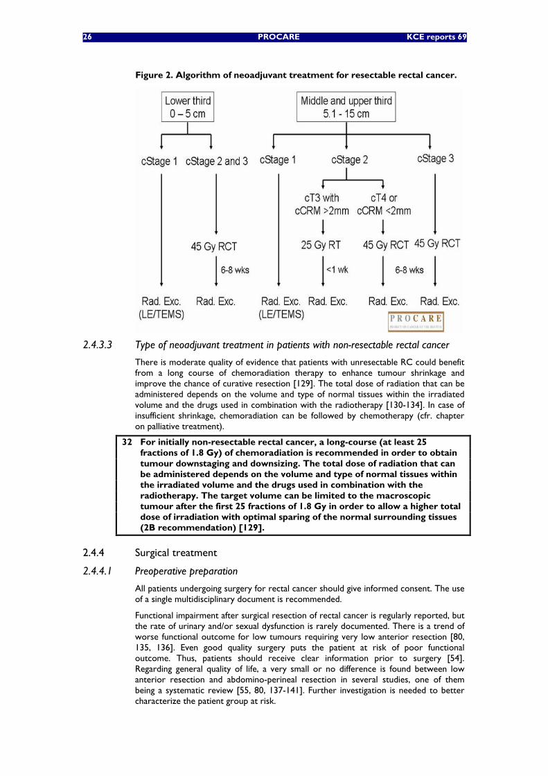
26 PROCARE KCE reports 69
Figure 2. Algorithm of neoadjuvant treatment for resectable rectal cancer.
2.4.3.3 Type of neoadjuvant treatment in patients with non-resectable rectal cancer
There is moderate quality of evidence that patients with unresectable RC could benefit from a long course of chemoradiation therapy to enhance tumour shrinkage and improve the chance of curative resection [129]. The total dose of radiation that can be administered depends on the volume and type of normal tissues within the irradiated volume and the drugs used in combination with the radiotherapy [130-134]. In case of insufficient shrinkage, chemoradiation can be followed by chemotherapy (cfr. chapter on palliative treatment).
32 For initially non-resectable rectal cancer, a long-course (at least 25 fractions of 1.8 Gy) of chemoradiation is recommended in order to obtain tumour downstaging and downsizing. The total dose of radiation that can be administered depends on the volume and type of normal tissues within the irradiated volume and the drugs used in combination with the radiotherapy. The target volume can be limited to the macroscopic tumour after the first 25 fractions of 1.8 Gy in order to allow a higher total dose of irradiation with optimal sparing of the normal surrounding tissues (2B recommendation) [129].
2.4.4 Surgical treatment
2.4.4.1 Preoperative preparation
All patients undergoing surgery for rectal cancer should give informed consent. The use of a single multidisciplinary document is recommended.
Functional impairment after surgical resection of rectal cancer is regularly reported, but the rate of urinary and/or sexual dysfunction is rarely documented. There is a trend of worse functional outcome for low tumours requiring very low anterior resection [80, 135, 136]. Even good quality surgery puts the patient at risk of poor functional outcome. Thus, patients should receive clear information prior to surgery [54]. Regarding general quality of life, a very small or no difference is found between low anterior resection and abdomino-perineal resection in several studies, one of them being a systematic review [55, 80, 137-141]. Further investigation is needed to better characterize the patient group at risk.
KCE reports 69 PROCARE 27
RCTs evaluating mechanical bowel preparation in elective colorectal surgery either show no benefit or a negative effect of mechanical bowel cleaning [54, 55, 142-145]. Studies have compared ethylene glycol mechanical bowel preparation with no preparation. Less is published about fleet enemas or low fibber diet prior to surgery. No definitive conclusions can be made for rectal cancer surgery, because patients with mid or low rectal cancer were either excluded or their number was very limited in all studies reported until now.
Patients undergoing oncological pelvic surgery are at risk for thromboembolic adverse events. Three Cochrane reviews suggest that the optimal prophylaxis in colorectal surgery is the combination of low-dose unfractionated heparin and compression stockings [54, 55, 146]. The unfractionated heparin can be replaced by low molecular weight heparin. These studies were not specifically related to rectal cancer patients.
Although the evidence is poor, preoperative stoma site marking and patient stoma education positively influence the outcome in terms of postoperative hospital stay, psychological adjustment [54, 55, 147]. They also reduce stoma related interventions.
Relevant blood loss during surgery, in particular cancer surgery, should be avoided as much as possible. Blood transfusion per se may not be a risk factor for poor prognosis after colorectal cancer surgery. However, the combination of perioperative blood transfusion and subsequent development of postoperative infectious complications may be associated with a poor prognosis [148]. Nonetheless, preparations for blood transfusion should be made in all patients undergoing surgery for rectal cancer, except when an individual patient refuses.
33 Before total mesorectal excision (TME) surgery, patients should be informed about the risk of urogenital dysfunction after resection for mid and low rectal cancer (1C recommendation) [54, 55, 80, 135-141].
34 In the absence of specific data, mechanical bowel preparation is recommended in the context of rectal cancer surgery, although no benefit was observed in the context of colon surgery (including anterior resection) (1C recommendation) [54, 55, 142-145].
35 Thromboembolism prophylaxis should be administered in the perioperative period of patients with rectal cancer using graduated compression stockings and appropriate doses of subcutaneous low molecular weight heparin, unless there is a specific contraindication (1B recommendation) [54, 55, 146].
36 All patients undergoing surgery for rectal cancer should have a single immediately preoperative dose of antibiotic prophylaxis. Several intravenous antibiotics appear to be effective, but only those covering aerobic and anaerobic germs should be used (1A recommendation) [54, 55].
37 Whenever (definitive or temporary) stoma construction is planned, preoperative counselling by a specialized nurse, and stoma site marking by the surgeon or by a specialized nurse under his/her supervision, are recommended (1B recommendation) [54, 55, 147].
38 Preparations for blood transfusion should be made in all patients undergoing surgery for rectal cancer except when an individual patient refuses (1C recommendation) [148].
2.4.4.2 Elective surgery for cure
Radical resection
The main emphasis of surgery is to obtain clear surgical margins yielding a curative R0 resection (no residual tumour). The term curative resection should be based on histological confirmation of complete excision of tumour with negative margins (proximal, distal and radial). The distal margin is the transsected full thickness edge and does not include the tissue donut from the endoluminal stapler if the tumour is at > 3 cm from the cut end of the main specimen.
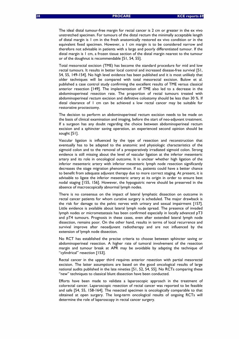
28 PROCARE KCE reports 69
The ideal distal tumour-free margin for rectal cancer is 2 cm or greater in the ex vivo unstretched specimen. For tumours of the distal rectum the minimally acceptable length of distal margin is 1 cm in the fresh anatomically restored ex vivo condition or in the equivalent fixed specimen. However, a 1 cm margin is to be considered narrow and therefore not advisable in patients with a large and poorly differentiated tumour. If the distal margin is 1 cm, a frozen tissue section of the distal margin nearest to the tumour or of the doughnut is recommendable [51, 54, 55].
Total mesorectal excision (TME) has become the standard procedure for mid and low rectal tumours. It results in better local control and increased disease-free survival [51, 54, 55, 149-154]. No high level evidence has been published and it is most unlikely that older techniques will be compared with total mesorectal excision. Bulow et al. published a case control study confirming the excellent results of TME versus classical anterior resection [149]. The implementation of TME also led to a decrease in the abdominoperineal resection rate. The proportion of rectal tumours treated with abdominoperineal rectum excision and definitive colostomy should be less than 30 %. If distal clearance of 1 cm can be achieved a low rectal cancer may be suitable for restorative proctectomy.
The decision to perform an abdominoperineal rectum excision needs to be made on the basis of clinical examination and imaging, before the start of neo-adjuvant treatment. If a surgeon has any doubt regarding the choice between abdominoperineal rectum excision and a sphincter saving operation, an experienced second opinion should be sought [51].
Vascular ligation is influenced by the type of resection and reconstruction that eventually has to be adapted to the anatomic and physiologic characteristics of the sigmoid colon and to the removal of a preoperatively irradiated sigmoid colon. Strong evidence is still missing about the level of vascular ligation at the inferior mesenteric artery and its role in oncological outcome. It is unclear whether high ligation of the inferior mesenteric artery with inferior mesenteric lymph node resection significantly decreases the stage migration phenomenon. If so, patients could have a better chance to benefit from adequate adjuvant therapy due to more correct staging. At present, it is advisable to ligate the inferior mesenteric artery at its origin in order to ensure best nodal staging [155, 156]. However, the hypogastric nerve should be preserved in the absence of macroscopically abnormal lymph nodes.
There is no consensus on the impact of lateral lymphatic dissection on outcome in rectal cancer patients for whom curative surgery is scheduled. The major drawback is the risk for damage to the pelvic nerves with urinary and sexual impairment [157]. Little evidence is available about lateral lymph node spread. The presence of invaded lymph nodes or micrometastasis has been confirmed especially in locally advanced pT3 and pT4 tumours. Prognosis in these cases, even after extended lateral lymph node dissection, remains poor. On the other hand, results in terms of local recurrence and survival improve after neoadjuvant radiotherapy and are not influenced by the extension of lymph node dissection.
No RCT has established the precise criteria to choose between sphincter saving or abdominoperineal resection. A higher rate of tumoral involvement of the resection margin and tumour break at APR may be avoidable by adapting the technique of “cylindrical” resection [153].
Rectal cancer in the upper third requires anterior resection with partial mesorectal excision. The latter assumptions are based on the good oncological results of large national audits published in the late nineties [51, 52, 54, 55]. No RCTs comparing these “new” techniques to classical blunt dissection have been conducted.
Efforts have been made to validate a laparoscopic approach in the treatment of colorectal cancer. Laparoscopic resection of rectal cancer was reported to be feasible and safe [54, 55, 158-164]. The resected specimen is oncologically comparable to that obtained at open surgery. The long-term oncological results of ongoing RCTs will determine the role of laparoscopy in rectal cancer surgery.
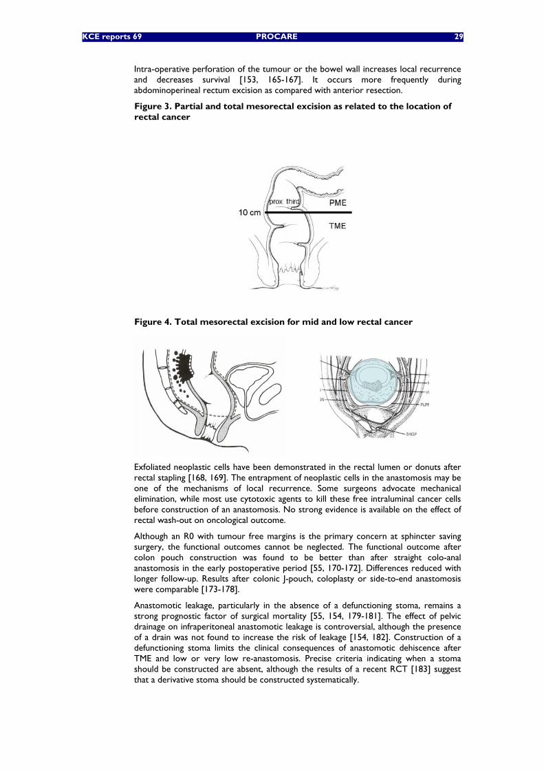
KCE reports 69 PROCARE 29
Intra-operative perforation of the tumour or the bowel wall increases local recurrence and decreases survival [153, 165-167]. It occurs more frequently during abdominoperineal rectum excision as compared with anterior resection.
Figure 3. Partial and total mesorectal excision as related to the location of rectal cancer
Figure 4. Total mesorectal excision for mid and low rectal cancer
Exfoliated neoplastic cells have been demonstrated in the rectal lumen or donuts after rectal stapling [168, 169]. The entrapment of neoplastic cells in the anastomosis may be one of the mechanisms of local recurrence. Some surgeons advocate mechanical elimination, while most use cytotoxic agents to kill these free intraluminal cancer cells before construction of an anastomosis. No strong evidence is available on the effect of rectal wash-out on oncological outcome.
Although an R0 with tumour free margins is the primary concern at sphincter saving surgery, the functional outcomes cannot be neglected. The functional outcome after colon pouch construction was found to be better than after straight colo-anal anastomosis in the early postoperative period [55, 170-172]. Differences reduced with longer follow-up. Results after colonic J-pouch, coloplasty or side-to-end anastomosis were comparable [173-178].
Anastomotic leakage, particularly in the absence of a defunctioning stoma, remains a strong prognostic factor of surgical mortality [55, 154, 179-181]. The effect of pelvic drainage on infraperitoneal anastomotic leakage is controversial, although the presence of a drain was not found to increase the risk of leakage [154, 182]. Construction of a defunctioning stoma limits the clinical consequences of anastomotic dehiscence after TME and low or very low re-anastomosis. Precise criteria indicating when a stoma should be constructed are absent, although the results of a recent RCT [183] suggest that a derivative stoma should be constructed systematically.
30 PROCARE KCE reports 69
Each team should audit its clinical leak rate and adapt clinical practice as required. Ileostomy or colostomy can be used equally, but there is a tendency to use more ileostomies.
39 It is advisable to ligate the inferior mesenteric artery at its origin in order to ensure best nodal staging. However, the hypogastric nerve should be preserved in the absence of macroscopically abnormal lymph nodes (2C recommendation) [155, 156].
40 During rectal surgery for cancer, lateral lymph node dissection (iliac nodes) is not recommended in the absence of macroscopic disease (2A recommendation) [157].
41 Surgeons should aim, wherever possible and desirable, to preserve the anal sphincter. A total mesorectal excision should be performed for tumours in the middle and lower third of the rectum either as part of a restorative proctectomy, a Hartmann's procedure or an abdominoperineal resection. If distal clearance of 1 cm can be achieved, a low rectal cancer may be suitable for restorative proctectomy. For tumours in the upper rectum, the mesorectum should be divided no less than 5 cm below the lower margin of the tumour (partial mesorectal excision). Care should be taken to preserve the pelvic autonomic nerves and plexuses whenever possible (1B recommendation) [54, 55, 149-154].
42 Laparoscopic or laparoscopy-assisted surgery for rectal cancer should only be performed by experienced laparoscopic surgeons who have been properly trained, who enter their patients in a trial or audit their results very carefully in a multidisciplinary context (1A recommendation) [54, 55, 158-164].
43 During surgery for rectal cancer, great care should be taken to avoid rectal perforation or tumoral break, especially during abdominoperineal resection. The occurrence of intra-operative perforation as well as its location in relation to the tumour site should be reported in the surgical note (1B recommendation) [153, 165-167].
44 A rectal wash-out before re-anastomosis may prevent tumour cell implantation and is recommended, although strong evidence is lacking (2C recommendation) [168, 169].
45 After restorative proctectomy and total mesorectal excision, the formation of a colonic pouch, coloplasty or side-to-end colo-anal anastomosis should be considered to improve functional outcome and quality of life (1A recommendation) [55, 170-178].
46 A temporary defunctioning stoma should be considered each time the anastomosis is at risk for leakage. This is particularly true for an infra-peritoneal anastomosis after total mesorectal excision (1A recommendation) [55, 154, 179-184].
Local excision and transanal endoscopic microsurgical resection
Local excision (LE) and transanal endoscopic microsurgical resection (TEMS) are attractive because of their low morbidity and functional sequellae as compared with radical resection. However, care should be taken not to forget the primary goal of surgery, namely to cure the patient.
Local full thickness disk excision for cure classically has been restricted to low risk pT1 rectal cancer that are technically suitable for a transanal approach: located in the lower third of the rectum (or up to about 7 cm), uT1N0, less than 3 cm diameter; postoperative pT1, G1 or G2, no lymphovascular invasion and tumour free resection margins [55]. In contrast with LE, TEMS allows transluminal excision of a (small) rectal tumour at any level, i.e. up to 15 cm [185]. In case of unfavourable pathology findings or positive margins, more radical surgery with restorative proctectomy or APR should follow immediately [55].
KCE reports 69 PROCARE 31
However, local full thickness excision (i.e. LE and, by analogy, TEMS) for pT1 has become controversial in view of significantly decreased local disease control after full thickness local excision as compared with radical excision, except maybe for pT1sm1 [61-64].
Promising results have been reported after neoadjuvant treatment for early rectal cancer (up to T2) followed by TEMS [186]. TEMS could also be applied for resection and pathological examination of remaining scar tissue after clinical complete response following chemoradiation. The results of ongoing trials have to be awaited before any general recommendation on this novel approach can be made.
47 Local excision (LE) or transanal endoscopic microsurgical resection (TEMS) should not be a standard curative approach for ‘early’ rectal cancer outside a clinical trial. However, patients not fit for radical resection or on a palliative course can benefit from these techniques (1B recommendation) [55, 185, 186].
48 The role of local excision (LE) for pT1 rectal cancer has become controversial. LE or transanal endoscopic microsurgical resection (TEMS) can be recommended for small (< 3 cm diameter) uT1 lesions with the appearance of a villous adenoma and with negative biopsies, located below the peritoneal reflection of Douglas (7-9 cm above the anal verge in men, 5-7.5 cm in women). For pT1 sm 2 and sm 3 lesions, radical resection or adjuvant treatment should follow LE or TEMS in patients fit for further therapy. However, for pT1sm1 lesions close observation is a valid alternative (1C recommendation) [55, 61-64, 187].
49 In view of the risk of nodal metastasis and decreased disease control, all uT1 lesions located above the peritoneal reflection of Douglas deserve radical total mesorectal excision (TME) (with low risk of urogenital dysfunction) if the patient is fit for surgery (1C recommendation) [61-64, 187].
2.4.4.3 Emergency surgery
The quality of care in emergency circumstances should be as high as possible. Therefore, emergency surgery should be carried out by or under supervision of an experienced surgeon and anaesthetist. Stoma formation should be carried out in the patient’s interests only. The overall mortality for emergency surgery should be less than 20%.
Intestinal obstruction in rectal cancer patients is rare. In first instance, a stoma should be constructed. Intraluminal stents have been proposed as an alternative. Originally, this endoscopic approach was developed for palliative settings (cfr. chapter 0 on palliative treatment). Many questions remain open on its use in a potentially curative setting. Although stenting as a bridge to curative surgery might be attractive, no recommendation can be made at this time.
50 In case of stenosing rectal cancer, a laparoscopic exploration and construction of a derivative stoma should be considered before starting neoadjuvant treatment. Stenting as a bridge to curative surgery can not yet be recommended. Stenting is a promising technique that should be considered for palliation in patients with extensive metastatic disease, who are not fit enough or who are unwilling to have a colostomy (2C recommendation) [54, 55].
2.4.5 Pathology Assessment of the completeness of tumour resection and of the pathological stage of rectal cancer is important for prognosis, choice of additional treatment, and control of the quality of the surgical resection. Standardisation of data, the application of well-defined criteria, and the acceptance of an identical and unique staging system allow integration and comparison of data.
32 PROCARE KCE reports 69
2.4.5.1 Macroscopic assessment
The mesorectal surface of a good resection specimen should be smooth without violation of the fat and with a good bulk to the mesorectum around the rectum. The distal margin should appear adequate without coning near the tumour. Defects should not be more than very superficial or 5 mm deep.
The quality of the mesorectum can be graded as complete, nearly complete, or incomplete [188]:
1. A complete mesorectum is an intact mesorectum with only minor irregularities on a smooth mesorectal surface. Defects are no deeper than 5 mm, and there is no coning toward the distal margin of the specimen. There is a smooth circumferential resection margin on slicing (cfr. infra).
2. A nearly complete mesorectum has a moderate bulk to the mesorectum, but irregularity of the mesorectal surface. Moderate coning of the specimen is allowed. At no site the muscularis propria is visible, with the exception of the insertion of the levator muscles.
3. An incomplete mesorectum has little bulk to the mesorectum with defects down onto the muscularis propria and/or very irregular circumferential resection margin on slicing (cfr. infra).
The distance between the deepest point of extension of the tumour and the surgical circumferential surface is defined as the circumferential margin, which needs to be assessed with great care. It can be measured by using a measurement device incorporated in the microscope itself (e.g. Vernier scale). Otherwise a sheet of graph paper that is photocopied onto a sheet of acetate and cut to size can be used.
51 The rectal cancer resection specimen should be delivered to the pathologist fresh (within 2 to 3 hours), unopened, and unpinned (except for local excision specimen; cf.). Administrative data, information on personal or family history, cTNM staging, the type of surgery performed, and preoperative treatment modalities should be provided by the surgeon (1C recommendation) [55, 188-193].
52 The resection specimen should be examined by the pathologist. It is mandatory to determine the exact topography of the tumour, also with reference to the serosal surface, i.e. above, at or below the peritoneal fold of Douglas. The quality of the mesorectal excision should be assessed on the unopened specimen and graded as complete, nearly complete or incomplete. Abdominoperineal rectal excision specimens require specific attention as the description of the quality of the total mesorectal excision is limited to the mesorectal surface; ideally, an abdomino-perineal resection specimen should have a monocylindrical shape. It is recommended to photograph the ventral and dorsal aspects of the specimen before inking or opening the specimen (1C recommendation) [55, 188-194].
53 After examination of the external surface, it should be inked before opening and fixating the specimen. After fixation, the specimen should be sectioned in parallel cuts of 3-4 mm perpendicular to the length of the bowel allowing to assess the deepest point of invasion and to measure the smallest distance between tumour extension and the nearest lateral surface. It is necessary to photograph the parallel cuts taken through the total mesorectal excision (TME) to document the quality of the surgical specimen and the extent of the disease and mandatory if large microscopic sections are not used. The deepest point of invasion should be sampled for microscopy, and the distance to the nearest circumferential surface should be measured and reported in mm. No distinction should be made between the various modes of involvement i.e. direct spread, involved lymph node, lymphatic or vascular spread (1C recommendation) [55, 188-195].
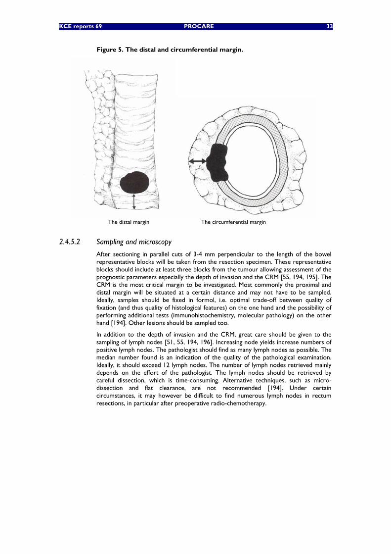
KCE reports 69 PROCARE 33
Figure 5. The distal and circumferential margin.
The distal margin The circumferential margin
2.4.5.2 Sampling and microscopy
After sectioning in parallel cuts of 3-4 mm perpendicular to the length of the bowel representative blocks will be taken from the resection specimen. These representative blocks should include at least three blocks from the tumour allowing assessment of the prognostic parameters especially the depth of invasion and the CRM [55, 194, 195]. The CRM is the most critical margin to be investigated. Most commonly the proximal and distal margin will be situated at a certain distance and may not have to be sampled. Ideally, samples should be fixed in formol, i.e. optimal trade-off between quality of fixation (and thus quality of histological features) on the one hand and the possibility of performing additional tests (immunohistochemistry, molecular pathology) on the other hand [194]. Other lesions should be sampled too.
In addition to the depth of invasion and the CRM, great care should be given to the sampling of lymph nodes [51, 55, 194, 196]. Increasing node yields increase numbers of positive lymph nodes. The pathologist should find as many lymph nodes as possible. The median number found is an indication of the quality of the pathological examination. Ideally, it should exceed 12 lymph nodes. The number of lymph nodes retrieved mainly depends on the effort of the pathologist. The lymph nodes should be retrieved by careful dissection, which is time-consuming. Alternative techniques, such as micro-dissection and flat clearance, are not recommended [194]. Under certain circumstances, it may however be difficult to find numerous lymph nodes in rectum resections, in particular after preoperative radio-chemotherapy.
34 PROCARE KCE reports 69
54
54.1 The number of blocks to be taken from the tumour is 3 at minimum (1C recommendation) [55, 194, 195].
54.2 One block at least should include the transition from the surrounding ‘normal’ mucosa to the tumour and at least one other should include the deepest point of invasion (1C recommendation) [55, 188-195].
54.3 Proximal and distal section margins do not have to be embedded if the tumour is situated at a distance of more than 3 cm from these margins. If the tumour is close to a margin, it is recommended to sample this margin and to demonstrate the relationship to the tumour by perpendicular sections. Biopsies have to be taken to assess the circumferential (radial, lateral) margin (1C recommendation) [16, 55, 188-193, 197].
55 Ideally, samples should be fixed in formol in order to allow additional molecular pathological examination. Frozen preserved biopsy samples may be important, especially if there are clinical arguments for hereditary nonpolyposis colorectal cancer (expert opinion) [194].
56 Associated lesions (polyps, inflammatory bowel disease [IBD], …) have to be sampled. In polyposis cases, a reasonable number of biopsies should be taken as well as the (proximal and distal) section margins. Proximal and distal section margins should also be embedded in IBD cases (expert opinion) [194].
57
57.1 All lymph nodes included in a resection specimen are considered to be regional. Distinction between paratumoral nodes and others i.e. local vs. regional lymph nodes is not requested. The number of lymph nodes analysed is important. At least 12 lymph nodes should be found and embedded. The numbers of lymph nodes retrieved depends mainly on the effort of the pathologist (1B recommendation) [55, 194, 196, 198, 199]. The number of positive lymph nodes relates to the number investigated. When less than 8 lymph nodes have been analysed, the proportion of cancers with lymph node involvement is underestimated (1C recommendation) [194, 196, 199]. However, it may be difficult to find numerous lymph nodes in rectum resections, in particular after preoperative radio-chemotherapy (1C recommendation) [194, 199] .
57.2 There is insufficient scientific evidence to recommend micro-dissection techniques or fat clearance (expert opinion) [194].
57.3 Extra-regional lymph nodes are classified as metastases and should be embedded and described separately (1C recommendation) [194].
KCE reports 69 PROCARE 35
2.4.5.3 The pathology report
Histologic type according to the WHO classification:
• Adenocarcinoma: the histological grade should be mentioned either in a four or three-tiers system as well (G1), moderately (G2), poorly differentiated (G3) and undifferentiated (G4), or in a two-tiers system as low (G1,G2) grade and high (G3, G4) grade. The high grade corresponds to less than 50% of glandular structures of the surface analysed.
• Mucinous carcinoma (colloid carcinoma): a tumour composed of at least 50% of this type of proliferation. It is considered as poorly differentiated adenocarcinoma.
• Signet ring cell carcinoma: a tumour composed of at least 50% of this type of proliferation. It is also considered as poorly differentiated adenocarcinoma.
• Adenosquamous or squamous carcinoma.
• Small cell carcinoma.
• Medullary carcinoma: is considered as undifferentiated carcinoma
• Undifferentiated carcinoma (G4): corresponds to less than 5% of glandular structures of the surface analysed.
The depth of invasion should be described in function of the anatomical structures i.e. mucosa, submucosa, muscularis propria, subserosa, serosa and translated into the new TNM classification.
• Tx and To: primary tumour cannot be assessed (Tx). No evidence of primary tumour (T0).
• Tis: carcinoma in situ includes cancer cells confined within the glandular basement membrane (intraepithelial) or lamina propria (intramucosal) with no extension through the muscularis mucosae into the submucosa. The term ‘high grade dysplasia’ and ‘severe dysplasia’ may be used as synonyms for intraepithelial (in situ) carcinoma.
• T1: tumour invades submucosa
• T2: tumour invades muscularis propria without breaching
• T3: tumour invades through the muscularis propria into the subserosa, or into the non-peritonealised pericolic and perirectal tissues. The subserosa corresponds to the adipous connective tissue situated in between the outer surface of the muscularis propria and the mesothelial lining.
• T4: tumour directly invades other organs or structures, and/or perforates the visceral peritoneum. “Direct invasion” in T4 includes invasion of other segments of the colorectum by way of the serosa. Tumour that is adherent to other organs or structures, macroscopically, is classified cT4. However if no tumour is present in the adhesion, microscopically, the classification should be pT3.
Note: The 3-mm rule was introduced in TNM5. This rule stated that any mesorectal tumour deposit 3 mm in size or greater should be thought of as an involved lymph node. Any deposit smaller than 3 mm in diameter should be included in the pT. In the current edition of the TNM staging system (TNM6), the 3-mm rule has been withdrawn and the definitions of lymph-node and venous invasion revised. TNM6 states that smooth metastatic nodules in the perirectal fat should be considered as lymph-node metastases and should, therefore, be staged in the N category. Although TNM5 contains the controversial 3-mm rule that seems to lack an evidence base, this rule does at least have the advantage of being quantitative and, therefore, reproducible. Thus, it has been advocated to stick to the 3-mm rule [198].
36 PROCARE KCE reports 69
Different systems have been developed and used to describe and to quantify regression of colorectal cancer after (chemo)radiation (ypTNM):
• the Rectal Cancer Regression Grade (RCRG) [200]. This system comprises three grades: RCRG 1 indicates “good” radioresponsiveness where the tumour is either sterilized or only microscopic foci of adenocarcinoma remain. RCRG 2 reflects marked fibrosis but with macroscopic tumour still present. RCRG 3 indicates a “poor” response with little or no fibrosis in the presence of abundant macroscopic tumour.
• the modified Mandard classification system which has been developed for oesophageal cancer initially [201]; this system uses 5 grades ranging from TRG1 (no tumour cells) to TRGR5 (no regression).
• the Dworak classification [202]; this system also uses 5 grades ranging from no evidence of any treatment effect to a complete response with no viable tumour identified. The following are characteristics of each grade:
o GR0 or no regression;
o GR1 or minor regression: dominant tumour mass with obvious fibrosis and/or vasculopathy (dominant tumour mass with obvious fibrosis in 25% or less of the tumour mass);
o GR2 or moderate regression: dominantly fibrotic changes with few tumour cells or groups easy to find (dominant tumour mass with obvious fibrosis in 26% to 50% of the tumour mass);
o GR3 or good regression: very few tumour cells (difficult to find microscopically) in fibrotic tissue with or without mucous substance (dominant fibrosis outgrowing the tumour mass; i.e., more than 50% tumour regression);
o GR4 or total regression: no tumour cells (no viable tumour cells, only fibrotic mass).
Problems relating to the finding of mucin pools with and especially without neoplastic epithelium are described. Tumour related mucin pools represent areas throughout the bowel wall that were previously occupied by tumour and could still be depending on sampling.
58 The pathology report should be standardised, providing all important macroscopic and microscopic data.
58.1 Mandatory macroscopic data are:
58.1.1 the measurements of the resection specimen, including those of adjacent structures and organs;
58.1.2 the localisation of the tumour in relationship to the peritoneal lining;
58.1.3 the proximal, distal and lateral (circumferential, radial) section margins; if the specimen can not be oriented, the section margins are described as the closest and most distant margin;
58.1.4 the maximal diameter of the tumour;
58.1.5 the macroscopic appearance of the lesion should be described as protruding/exophytic, ulcerating, infiltrating, flat;
58.1.6 the presence of perforation at the tumour site; the presence of peritoneal deposits.
58.1.7 the presence of associated lesions, e.g. synchronic cancers, polyps and chronic idiopathic inflammatory bowel disease;
(1C recommendation) [55, 194].
58.2 Mandatory microscopic data are:
58.2.1 the histological type;
KCE reports 69 PROCARE 37
58.2.2 the histologic grade of adenocarcinoma, using either a four or three-tiers system, i.e. well (G1), moderately(G2), poorly differentiated (G3) and undifferentiated (G4), or a two-tiers system, i.e. low (G1,G2) grade and high (G3, G4) grade; mucinous carcinoma (colloid carcinoma) and signet ring cell carcinoma are to be considered as poorly differentiated adenocarcinoma;
58.2.3 the depth of invasion should be described and translated into the new pTNM classification;
58.2.4 after irradiation (ypTNM), the grade of tumour regression should be described so that any of the existing grading systems can be applied; although scientific evidence is limited, the use of the 5-tierce Dworak classification system is recommended [200-202];
58.2.5 resection margins; a margin of <1 mm is considered positive;
58.2.6 the total number of examined and the number of involved regional lymph nodes; there is insufficient evidence to recommend semi-serial sectioning of lymph nodes or to perform immunohistochemical stains; it is recommended to report extramural tumour deposits as lymph node involvement if they measure >3 mm in diameter;
58.2.7 extramural deposits of tumour; defined as deposits that are not obviously within lymph nodes if they measure <3 mm in diameter;
58.2.8 the presence of vascular invasion into extramural veins;
58.2.9 the presence of perineural and/or lymphatic and/or intramural vascular invasion may be mentioned;
58.2.10 distant metastasis: the report should mention M1 if microscopic examination of a sample confirms the presence of a metastasis. This finding can relate to a liver biopsy or non-regional lymph nodes or peritoneal carcinomatous deposits;
58.2.11 cytological examination of peritoneal fluid revealing tumour cells equals M1. If the existence of distant metastasis can not be assessed, one should indicate pMx;
(1B recommendation) [16, 52, 54, 55, 194-196, 198, 203-205].
58.3 It is recommended to use a check-list (cfr. appendix) [54, 55, 194, 203].
59 The results of the pathology report should be discussed in a multidisciplinary meeting, involving the pathologist, surgeon, radiologist, radiotherapist, oncologist and gastroenterologist in order to determine further treatment (1C recommendation) [194].
2.4.6 Adjuvant therapy
2.4.6.1 Adjuvant chemotherapy in patients with clinical stage II-III rectal cancer who did receive neoadjuvant (chemo)radiotherapy
From former clinical practice guidelines [55] it is known that chemotherapy during six months is prolonging survival in stage III patients. Whether adjuvant chemotherapy is prolonging survival in stage II patients is not known. The recent EORTC 22921 trial showed no survival benefit of adding chemotherapy (a regimen that actually is no more optimal) to radiotherapy for stage II and III patients, but chemotherapy, regardless of whether administered before or after surgery had a significant benefit on local control [75]. In this trial postoperative chemotherapy was given during four months. Although this trial is a RCT, there were several shortcomings (TME was not the standard surgical procedure, pathology reports were not up to present standards, e.g. no CRM assessment, and there was a poor compliance of postoperative chemotherapy).
Two clinical practice guidelines concluded that FUFA given by IV injection for 5 days every 4 weeks for 6 cycles is the regimen for which the most evidence is available and that it is clearly effective in prolonging survival in patients with stage III [55, 206].
38 PROCARE KCE reports 69
The most recent guideline also concluded that infusional FUFA or capecitabine is more effective and less toxic, based on retrospective analysis and based on extrapolation of evidence from patients with advanced disease [55]. Two new European studies could not give new relevant information on this topic [207, 208]. There were also three new Japanese studies showing no benefit of adding one year oral 1-hexylcarbamoyl-5-fluorouracil (HCFU) to induction 5-FU [209], low dose (333 mg/m2 day 1-3 and day 6-8) versus high dose (1000 mg/m2 day 1-3 and day 6-8) induction therapy with 5-FU [210] or adding immunotherapy [211].
There is no direct evidence supporting the indication for adjuvant chemotherapy in Stage III rectal cancer patients treated with neoadjuvant chemoradiotherapy [55, 206]. However, the evidence from studies with adjuvant chemotherapy in patients with a resected stage III rectal cancer without neoadjuvant treatment suggests that further chemotherapy with 5-FU can be administered for at least 4 months (given that preoperatively already two months of chemotherapy was given). The new EORTC 22921 trial failed to show a benefit for postoperative chemotherapy if a patient already received neoadjuvant chemoradiotherapy [75], but again a regimen was used that actually is no more considered to be optimal. As there is no proven benefit of chemotherapy in patients with stage I or II disease, postoperative chemotherapy may not be indicated in case of a pathologic complete response.
Cohort studies and one published meta-analysis suggest a small but not significant survival benefit for portal vein infusional chemotherapy with 5-FU [55, 206]. The recent AXIS study could only demonstrate a benefit for curatively resected colon cancer patients (in subgroup analysis) and not for rectal cancer patients [212].
There is no direct evidence supporting the need to start adjuvant therapy within 3 months after surgery. This is a rather general recommendation based on expert opinion, in analogy with the treatment of other types of cancer and based on the oncologic rationale that adjuvant therapy is able to treat micrometastatic disease at a time when tumour burden is at a minimum.
60 Any patient with a pathological stage II or III after resection who received preoperative radiotherapy without chemotherapy, should be considered for adjuvant chemotherapy with 5-FU during four (Stage II) or six (Stage III) months (1A recommendation) [55, 75, 206].
61 Infusional FUFA or capecitabine are recommended because they are more effective and less toxic than bolus FUFA (1C evidence), which was shown to prolong survival in patients with pathological stage III disease (1A recommendation) [55, 206-211].
62 After neoadjuvant radiochemotherapy, the indication for adjuvant chemotherapy in Stage III rectal cancer can be based on the cStaging. However, the benefit of adjuvant chemotherapy (during 4 months) seems to be very limited and may not be indicated in case of a pathologic complete (or almost complete) response (2C recommendation) [55, 75, 206].
63 There is insufficient evidence to support the use of adjuvant treatment with portal vein infusion chemotherapy with 5-FU in patients with resected rectal cancer (1A recommendation) [55, 206, 212].
64 Adjuvant therapy should start within 3 months after surgery. It should not be started in the presence of pelvic septic complications (expert opinion).
2.4.6.2 Adjuvant therapy in patients who did not receive neoadjuvant therapy
In 2001, Cancer Care Ontario (CCO) performed a review of 25 randomized controlled trials, 4 meta-analyses, 2 evidence-based consensus statements on adjuvant radiotherapy and/or chemotherapy in stage II and III resected rectal cancer. [206]. Some multi-arm trials contributed to more than one of the following comparative analyses: radiotherapy versus observation, chemotherapy versus observation, chemotherapy versus radiotherapy, chemotherapy plus radiotherapy versus chemotherapy alone, and chemotherapy plus radiotherapy versus radiotherapy alone.
KCE reports 69 PROCARE 39
The resulting CCO guideline came to the same conclusion as the National Institutes of Health Consensus Development Conference 1990 [213], i.e. that patients with resected stage II or III rectal cancer should be offered adjuvant therapy with the combination of radiotherapy and chemotherapy. There was no evidence to support the use of radiotherapy alone, if the goal of adjuvant therapy is to improve survival. There was evidence that chemotherapy should include 5-fluorouracil (5-FU), but not semustine, and that intravenous infusion with 5-FU is more effective than bolus injection (6 RCTs).
Radiotherapy alone versus observation (8 RCTs) improved local control without a significant survival benefit [206]. The SIGN guideline summarized 27 RCTs and 2 meta-analyses on radiotherapy versus observation showing a 9% reduction in risk of loss of local control (Number Needed to Treat = 11) without benefit in overall survival (meta-analysis) and at the cost of a significantly worse bowel function with RT [55]. Also, the AXIS trial investigators did not observe a survival benefit in 761 patients randomized with respect to radiotherapy. Although not statistically significant, the impact on local recurrence rates in this trial was similar to that reported in the literature [212]. In an EORTC trial there was an increased toxicity without survival benefit of elective irradiation of para-aortic lymph nodes and liver in addition to postoperative pelvic radiotherapy [214].
Chemotherapy versus observation (6 RCTs and 2 meta-analyses) improved survival but not local control [206].
None of 3 RCTs comparing chemotherapy versus radiotherapy found a benefit for overall survival or disease-free survival [206].
However, this information has become less relevant since chemoradiation is to be preferred above observation (2 RCTs), radiotherapy alone (3 RCTs) or chemotherapy alone (3 RCTs) [206].
The pooled analysis of the 3 trials of chemotherapy plus radiotherapy versus radiotherapy revealed a benefit for chemotherapy plus radiotherapy for both survival (odds ratio, 0.58; 95% confidence interval, 0.37 to 0.92; p=0.019) and local control (odds ratio, 0.50; 95% confidence interval, 0.27 to 0.92; p=0.025) [206]. After this guideline was published, Cafiero et al. reported a trial in which postoperative radiotherapy was compared to radiotherapy and chemotherapy [215]. The group with combination therapy had a non-significantly increased relative risk of death, but there was an unbalance of stage II and stage III patient in the two groups, there was low adherence to chemotherapy, chemotherapy was with 5-FU and levamisole and chemotherapy and radiotherapy were not concurrent.
Pooled results from two trials showed no significant survival benefit for chemotherapy plus radiotherapy versus chemotherapy (odds ratio=0.80; 95% confidence interval, 0.48 to 1.32; p=0.37). Also, in a third trial, the addition of radiotherapy to chemotherapy did not significantly improve disease-free survival (hazard ratio, 0.99; 95% confidence interval, 0.80 to 1.22; p=0.90) or overall survival (hazard ratio, 0.98; 95% confidence interval, 0.78 to 1.24; p=0.89); however, a significant reduction in the cumulative incidence of locoregional recurrence was evident for patients randomized to combined CT+RT compared with chemotherapy alone (relative risk, 0.57; 95% CI, 0.36 to 0.92; 5% absolute decrease from 13% with CT alone to 8% with CT+RT at five years; p=0.02) [206]. Moreover, the level of evidence for adjuvant radiotherapy as monotherapy giving a better local control is high. On the other hand, it is known that adjuvant chemotherapy gives a better overall survival, most often without effect on local recurrence rate. Thus, postoperative adjuvant combined radiation and chemotherapy is to be recommended in patients who did not receive preoperative radiotherapy and who are at high risk of recurrence [55, 206].
From existing clinical practice guidelines [55] it is known that chemotherapy during six months is prolonging survival in stage III patients. Also a recent Japanese trial, the first with TME as standard surgery, showed a better relapse free survival (primary endpoint) and a better overall survival (secondary endpoint) with oral 5-FU based chemotherapy in stage III resected rectal cancer patients [216].
40 PROCARE KCE reports 69
As we recommend to use in these patients a long course of radiotherapy together with continuous 5-FU (which counts for two months), postoperative chemotherapy during an extra four months is warranted.
Existing clinical practice guidelines concluded that further adjuvant chemotherapy is not indicated in stage II patients [55]. Thereafter, data from four recent trials on adjuvant chemotherapy have been reported. The study of Taal et al. could not show a significant survival benefit for rectal cancer patients (in subgroup analysis) because there were too few rectal cancer patients included to draw conclusions [217]. There was a tendency for a better survival in these patients, more in stage III than in stage II patients. A systematic review by Glimelius et al. did not find a survival benefit for adjuvant chemotherapy in rectal cancer patients [218]. A Japanese study by Kato et al. on the other hand showed a clear improvement in disease free survival as well for colonic cancer patients as for rectal cancer patients, but again, there were too few rectal cancer patients to draw firm conclusions [219]. A Japanese meta-analysis of the effect of adjuvant chemotherapy with oral fluorinated pyrimidines in the rectal cancer group found an overall survival benefit, but with hazard ratio=0,92, CI 95% 0,79-1,07) [220].
Although there is no trial dealing with the specific setting of pStage II rectal cancer with “unfavourable prognostic features”, our recommendation is to apply the conclusions of the NIH consensus [213], i.e. to give postoperative chemoradiotherapy in stage II and III patients if they were not treated with neoadjuvant treatment.
Evidence from existing guidelines on the use of 5-FU given by a protracted venous infusion (PVI) during postoperative RT is mainly based on the results of chemoradiation in patients with high risk RC [55, 206]. There was an improved tumour response and distant control, suggesting an improved local and systemic effect with FU given by a PVI. This resulted in a benefit for overall survival in favour of PVI. The overall local control was good and slightly better in the PVI arm.
A recent American trial showed similar overall survival rates, disease free survival rates and locoregional failure rates between bolus 5-FU therapy and PVI therapy (with a non significant benefit for the PVI group) but with significantly less haematological toxicity in the PVI arm [221].
Enteritis, diarrhoea, bowel obstruction or perforation and fibrosis within the pelvis are associated with postoperative radiotherapy [206]. Delayed adverse effects from radiotherapy include radiation enteritis (4%), small-bowel obstruction (5%) and rectal stricture (5%). A greater number of haematological and non-haematological adverse effects were associated with chemotherapy plus radiotherapy than with chemotherapy, radiotherapy or observation. Postoperative chemotherapy plus radiotherapy was associated with acute gastrointestinal and rheumatologic adverse effects that may be severe or life-threatening.
A recent small study showed severe long-term anorectal dysfunction as result of a weakened, less sensitive anal sphincter and undistensible rectum with faecal incontinence in 60% vs. 8% of patients that received adjuvant radiotherapy or not [222]. Another small study demonstrated that the combination of postoperative radiotherapy with high-dose 5-FU was too toxic [223]. A detailed analysis of toxicity of a previously reported trial by the North Central Cancer Treatment Group showed that the rate of diarrhoea was significantly greater in the PVI group when compared to the bolus 5-FU group, and this effect was even more important in the group of patients that underwent an anterior resection [224].
There is no direct evidence supporting the need to start adjuvant therapy within 3 months after surgery. This is a rather general recommendation based on expert opinion, in analogy with the treatment of other types of cancer and based on the oncologic rationale that adjuvant therapy is able to treat micrometastatic disease at a time when tumour burden is at a minimum.
KCE reports 69 PROCARE 41
65 In patients with radically resected rectal cancer who did not receive neoadjuvant therapy, there is no superiority of adjuvant chemotherapy alone over adjuvant radiotherapy alone, or vice versa, with respect to overall or disease-free survival (1A recommendation) [206].
66 Although adjuvant radiotherapy alone decreases local recurrence rate and adjuvant chemotherapy alone improves survival, they are inferior to the combination of radiotherapy and chemotherapy in patients with radically resected pathological Stage II-III rectal cancer who did not receive neoadjuvant therapy (1A recommendation) [55, 206, 212, 214, 215].
67
67.1 Patients who did not receive neoadjuvant therapy and have a pathological stage III tumour of the rectum, or in whom an R1 resection (including a pCRM of <1 mm) was performed, should be considered for chemoradiotherapy, followed by 4 months of chemotherapy (1A recommendation) [55, 206, 212, 215, 216, 219].
67.2 Patients with a resected pathological stage II tumour with unfavourable prognostic features (inadequately sampled lymph nodes, perforation, T4 lesion, poorly differentiated histology), who did not receive neoadjuvant therapy, should also be considered for chemoradiotherapy (1B recommendation) [55, 206, 212, 215-220], followed by 4 months of chemotherapy (expert opinion).
67.3 Patients with a resected pathological stage II tumour without unfavourable prognostic features, who did not receive neoadjuvant therapy, should also be considered for chemoradiotherapy. However, the evidence supporting the use of 4 months extra adjuvant chemotherapy is weak [55, 206, 215-220].
68 When chemotherapy with 5-fluorouracil is given concurrently with postoperative radiotherapy, a continuous intravenous infusion is more effective than the drug administered by bolus infusion (1A recommendation) [206, 221].
69 Patients to whom adjuvant radiotherapy or radiochemotherapy is proposed, should be informed of the potential harmful effects, most often diarrhoea and faecal incontinence, following sphincter sparing surgery (expert opinion) [55, 206, 222-224].
70 Adjuvant therapy should start within 3 months after surgery. It should not be started in the presence of pelvic septic complications (expert opinion).
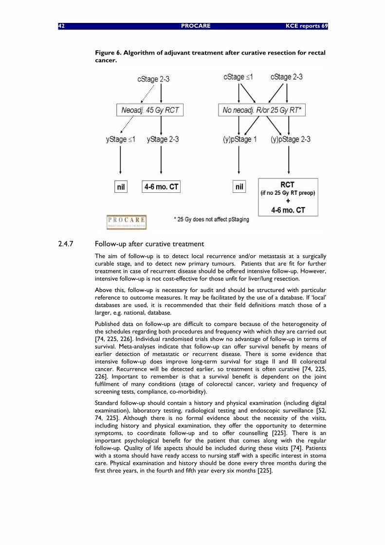
42 PROCARE KCE reports 69
Figure 6. Algorithm of adjuvant treatment after curative resection for rectal cancer.
2.4.7 Follow-up after curative treatment
The aim of follow-up is to detect local recurrence and/or metastasis at a surgically curable stage, and to detect new primary tumours. Patients that are fit for further treatment in case of recurrent disease should be offered intensive follow-up. However, intensive follow-up is not cost-effective for those unfit for liver/lung resection.
Above this, follow-up is necessary for audit and should be structured with particular reference to outcome measures. It may be facilitated by the use of a database. If ‘local’ databases are used, it is recommended that their field definitions match those of a larger, e.g. national, database.
Published data on follow-up are difficult to compare because of the heterogeneity of the schedules regarding both procedures and frequency with which they are carried out [74, 225, 226]. Individual randomised trials show no advantage of follow-up in terms of survival. Meta-analyses indicate that follow-up can offer survival benefit by means of earlier detection of metastatic or recurrent disease. There is some evidence that intensive follow-up does improve long-term survival for stage II and III colorectal cancer. Recurrence will be detected earlier, so treatment is often curative [74, 225, 226]. Important to remember is that a survival benefit is dependent on the joint fulfilment of many conditions (stage of colorectal cancer, variety and frequency of screening tests, compliance, co-morbidity).
Standard follow-up should contain a history and physical examination (including digital examination), laboratory testing, radiological testing and endoscopic surveillance [52, 74, 225]. Although there is no formal evidence about the necessity of the visits, including history and physical examination, they offer the opportunity to determine symptoms, to coordinate follow-up and to offer counselling [225]. There is an important psychological benefit for the patient that comes along with the regular follow-up. Quality of life aspects should be included during these visits [74]. Patients with a stoma should have ready access to nursing staff with a specific interest in stoma care. Physical examination and history should be done every three months during the first three years, in the fourth and fifth year every six months [225].
KCE reports 69 PROCARE 43
CEA is the only blood test that is supported by evidence regarding early diagnosis of recurrence. Routine blood tests (i.e. CBC, liver function tests), molecular markers or faecal occult blood testing have no prognostic or predictive value [52, 55, 225, 227]. CEA measurement should be done together with history and physical examination every three months during the first three years, in the fourth and fifth year every six months [52, 54, 55, 74, 225, 226].
Liver imaging is necessary since most metastases occur there. Ultrasound is a well-accepted imaging tool, but is less accurate than CT or MRI in diagnosing liver metastases at presentation (see chapter 2.4.2). This is likely also true for liver metastases that develop after curative surgery [68]. Above this, ultrasound is unable to assess for recurrent pelvic disease following rectal (or sigmoid) surgery. There is no obvious difference between CT and MRI for detecting recurrence, although MRI is more useful due to a higher theoretical ability to differentiate scar tissue from recurrence [68]. In patients with stage II and III rectal cancer, an abdominal/pelvic CT should be done annually during the first three years, while an ultrasound of the abdomen should be done in between the CT scans [52, 68, 225, 226].
Although there are insufficient data to recommend lung imaging, lung recurrences are as common as liver relapses in patients with rectal cancer, with the largest proportion of resectable recurrence found on thoracic CT [225]. Pulmonary recurrences are less associated with an elevated CEA [74, 225, 226]. An annual CT of the chest is therefore recommended during the first three years for patients with stage II and III rectal cancer. A chest X-ray should be done at six months after surgery and then annually for all patients. Thus, chest CT and X-ray will be done alternately at 6 months intervals during the first three years.
The endoscopic follow-up consists of a total colonoscopy in the peri-operative period and 1 year after the resection [52, 225, 227]. If this examination is normal, the next examination can be scheduled after 3 years. If this colonoscopy is normal, the interval until the next examination can be extended to 5 years. In patients with hereditary or familial predisposition, more intensive follow-up must be considered [52, 68, 74, 225, 227].
Chromo-endoscopy, magnification endoscopy and computed tomography colonography (virtual colonoscopy) are not established techniques for screening or surveillance [227]. There is also no place for EUS in routine follow-up, but EUS is a good tool for diagnosing local recurrence [52].
71 Every patient curatively treated for rectal cancer (all stages) should undergo intensive follow-up if there are no other medical conditions that limit the prognosis (1B recommendation) [74, 225, 226].
72 Every patient should undergo a physical examination and history, carcinoembryonic antigen (CEA) measurement, lung imaging (chest X-ray or CT-scan) and liver imaging (ultrasound or CT-scan). In patients at higher risk of local recurrent disease (i.e. stage II and III) a pelvic CT-scan or magnetic resonance imaging (MRI) is recommended (1B recommendation) [52, 54, 55, 68, 74, 225, 226].
73 Endoscopic ultrasound is only recommended when a local recurrence is suspected or in the follow-up after local excision/ transanal endoscopic microsurgical resection (TEMS) (1C recommendation) [52, 227].
74 A history, physical examination and carcinoembryonic antigen (CEA) testing should be done every three months for the first three years, during the fourth and fifth year every six months (1B recommendation) [52, 54, 55, 68, 74, 225, 226].
75 Patients at higher risk of recurrent disease (i.e. stage II and III) should undergo annually a CT-scan of the chest and abdomen/pelvis during the first three years (1B recommendation) [52, 68, 225, 226].
76 Liver ultrasound should be done every 6 months in the first three years (not when a CT-scan is done), annually in the fourth and fifth year (1B recommendation) [52, 68, 74, 226].
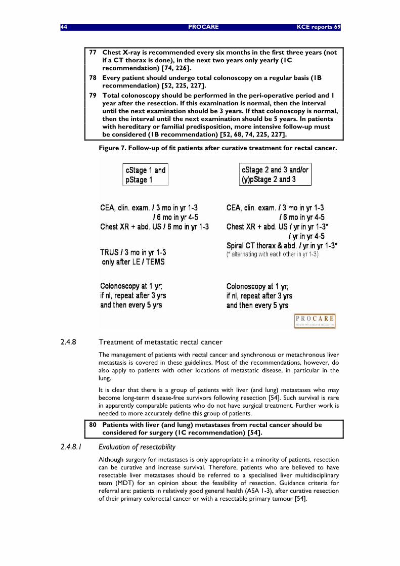
44 PROCARE KCE reports 69
77 Chest X-ray is recommended every six months in the first three years (not if a CT thorax is done), in the next two years only yearly (1C recommendation) [74, 226].
78 Every patient should undergo total colonoscopy on a regular basis (1B recommendation) [52, 225, 227].
79 Total colonoscopy should be performed in the peri-operative period and 1 year after the resection. If this examination is normal, then the interval until the next examination should be 3 years. If that colonoscopy is normal, then the interval until the next examination should be 5 years. In patients with hereditary or familial predisposition, more intensive follow-up must be considered (1B recommendation) [52, 68, 74, 225, 227].
Figure 7. Follow-up of fit patients after curative treatment for rectal cancer.
2.4.8 Treatment of metastatic rectal cancer
The management of patients with rectal cancer and synchronous or metachronous liver metastasis is covered in these guidelines. Most of the recommendations, however, do also apply to patients with other locations of metastatic disease, in particular in the lung.
It is clear that there is a group of patients with liver (and lung) metastases who may become long-term disease-free survivors following resection [54]. Such survival is rare in apparently comparable patients who do not have surgical treatment. Further work is needed to more accurately define this group of patients.
80 Patients with liver (and lung) metastases from rectal cancer should be considered for surgery (1C recommendation) [54].
2.4.8.1 Evaluation of resectability
Although surgery for metastases is only appropriate in a minority of patients, resection can be curative and increase survival. Therefore, patients who are believed to have resectable liver metastases should be referred to a specialised liver multidisciplinary team (MDT) for an opinion about the feasibility of resection. Guidance criteria for referral are: patients in relatively good general health (ASA 1-3), after curative resection of their primary colorectal cancer or with a resectable primary tumour [54].
KCE reports 69 PROCARE 45
Percutaneous biopsy of a liver tumour may be associated with extrahepatic cancer cell dissemination and results in a reduced long-term survival even when resection of hepatic metastases is undertaken [228]. Biopsy of hepatic lesions should therefore not be performed without discussion within the multidisciplinary team.
Positron emission tomography (PET) scanning is an emerging technology and its optimum role in relation to more established imaging methods is not yet defined [54, 59]. PET is capable of identifying local recurrence, liver and other distant metastases from colorectal origin. PET is certainly useful before resection of liver metastases to exclude extra-hepatic dissemination of the disease.
Metastatic liver lesions can be characterized with MRI. This also allows evaluation of the liver volume in case a large resection is considered [59].
81 Patients who are believed, on the basis of imaging, to have resectable liver metastases should be referred to a specialised liver multidisciplinary team, for an opinion about the feasibility of resection, if they are in relatively good general health (ASA 1-3), have undergone curative resection of their primary colorectal cancer or have a resectable primary tumour (1C recommendation) [54].
82 Biopsy of hepatic lesions should not be performed without discussion with the multidisciplinary team (1C recommendation) [228].
83 In conjunction with other imaging modalities, PET can be recommended in the further staging of the extent of metastatic disease, and influences decisions on patient management. PET is useful before resection of metastases to evaluate the extra-hepatic dissemination of the disease (2C recommendation) [54, 59].
84 Magnetic resonance imaging (MRI) can be useful to characterize metastatic liver lesions and to evaluate the volume of liver in case of large resection (1C recommendation) [59].
85 The morphology of the metastatic disease must be discussed in a multidisciplinary team (MDT) to identify non-resectability and to evaluate the possibility of reversibility. If necessary, a magnetic resonance imaging (MRI) liver and/or PET scan will be performed if they would influence the management of the disease (expert opinion).
2.4.8.2 Resectability criteria
Long-term survival can be achieved in patients with hepatic metastasis from colorectal origin after radical resection of the primary cancer and appropriate local treatment for hepatic metastases. The influence of the number or location of the metastases on survival after complete macroscopic resection is controversial [228]. Duration of survival is shortened by the presence of inadequate or involved resection margins [229]. A number of studies have supported the view that poorer overall and disease free survival are associated with resection margin less than 1 cm although others have produced evidence to suggest that a lesser margin may be acceptable as long as the tumour pseudocapsule is resected during dissection [228].
It has been increasingly evident that tumours which were previously thought to be unresectable can be treated by a combination of advanced techniques with a curative intent and long term survival [230-233]. Thus, it is suggested to subdivide patients according to their metastatic status in those with resectable metastases, potentially resectable metastases, and those with metastases unlikely to ever become resectable [234].
Resectability of liver tumours requires assessment by a radiologist in conjunction with a liver surgeon experienced in the management of colorectal metastases as there is also a need to define acceptable residual functioning volume in order to avoid postoperative liver failure. Concerns regarding compromised hepatic functional reserve following extended hepatic resection have led to consider preoperative portal vein embolisation in an attempt to increase the volume of the intended residual liver [231]. Others have suggested two-stage hepatic resection [230, 232].
46 PROCARE KCE reports 69
86
86.1 The ability to achieve clear margins (R0 resection) should be determined by a radiologist and a surgeon in the liver multidisciplinary team (MDT);
86.2 The acceptable residual functioning liver volume should be taken into account;
86.3 Resectability may be achieved by portal vein embolisation or two stage hepatectomy to increase hepatic functional reserve and also by the combination of surgery and ablation;
86.4 Patients with extrahepatic disease that should be considered for liver resection include resectable/ablatable pulmonary metastases, resectable/ablatable extrahepatic sites and local direct extension of liver metastases;
86.5 Those patients with tumours thought to be borderline for resection may have resectable or ablatable disease and should be referred for discussion with the specialized hepatobiliary unit before treatment
(1C recommendation) [59, 228, 235, 236].
2.4.8.3 Patients with resectable liver metastasis
Synchronous resectable liver metastasis
Long-term survival can be achieved in patients with hepatic metastasis from colorectal origin after radical resection of the primary cancer and curative local treatment for hepatic metastases. The influence of the number or location of the metastases on survival after complete macroscopic resection is controversial [228]. There is a consensus that the primary tumour should be operated, with or without neoadjuvant chemoradiation, if the tumour is symptomatic, irrespective of the resectability of the metastases. If the primary tumour is asymptomatic with resectable metastases, standard practice is to resect the primary tumour and the metastases, either at the same time or in a stepwise fashion, followed by chemotherapy [234]. The EORTC intergroup randomised phase III study 40983 evaluating the benefit of peri-operative Folfox4 chemotherapy in patients with potentially resectable colorectal cancer liver metastases, demonstrated improved progression-free survival over surgery alone in patients whose metachronous or synchronous metastases were actually resected [237]. Perioperative Folfox4-chemotherapy was proposed as the new standard of care. However, the results have not yet been published in full paper.
87 Although there is no evidence, optimal local control should be obtained in patients with a resectable primary rectal cancer and synchronous resectable metastases, including preoperative radiotherapy, radiochemotherapy, or chemotherapy. Multidisciplinary team (MDT) discussion is recommended for decision-making in this setting. A specialized liver and colorectal MDT should decide about the opportunity of synchronous resection of the primary rectal cancer and liver metastasis (1C recommendation) [228, 237].
88 Perioperative chemotherapy is recommended in patients with synchronous resectable liver metastases (1B recommendation) [237].
89 Usually, rectal cancer resection and liver resection has not been performed synchronously but management of accessible small metastases detected peri-operatively may be considered for combined resection. Simultaneous colon and liver resection has been shown to be safe and efficient when performed in high volume centres with appropriate experience in liver resectional surgery (2C recommendation) [52, 228].
90 It is also appropriate to provide recovery time after resection of the primary rectal cancer resection and to refer the patient to a specialist liver multidisciplinary team (MDT) for consideration of liver resection (1C recommendation) [228].
KCE reports 69 PROCARE 47
91 Patients with unfavourable primary pathology such as perforated primary tumour or extensive nodal involvement should be considered for chemotherapy prior to liver resection and be restaged after 3 months (1C recommendation) [228].
Metachronous resectable liver metastasis
There are only a few RCTs with low power addressing the treatment of patients with metachronous resectable metastases. More studies are needed to answer the question whether these patients should have pre- and/or postoperative chemotherapy. The EORTC intergroup randomised phase III study 40983 evaluating the benefit of peri-operative Folfox4 chemotherapy in patients with potentially resectable CRC liver metastases, demonstrated improved progression-free survival over surgery alone in patients whose metachronous or synchronous metastases were actually resected [237]. Perioperative Folfox4-chemotherapy was proposed as the new standard of care. However, the results have not yet been published in full paper.
Chemotherapy for non-resectable metastatic colorectal cancer improves survival and should be considered in all patients (cfr. infra). In some cases, initially non-resectable tumours should be considered for downsizing with chemotherapy.
In a large cohort study, combination chemotherapy with oxaliplatin, fluorouracil and folinic acid (Folfox) allowed resection in 13.5% of patients presenting initially non-resectable liver metastases; survival of these patients was similar to comparable series of operable patients treated by surgical resection [238].
Both Folfox and Folfiri therapy used and tested in phase III randomized trials provide a similar response rate, progression free survival and overall survival [239]. Both Folfox and Folfiri regimens can make unresectable patients resectable. There are arguments in favour of an oxaliplatin-based chemotherapy, which could increase the resection rate [240]. Folfox, Folfiri, Folfoxiri or the combination of 2 cytotoxics and a biological (cetuximab or bevacizumab) may lead to resection in +/- 20 % of the patients. Larger phase 3 studies report a lower resection rate. In general a correlation between the response rate and resection rate has been reported [241, 242].
92 Perioperative chemotherapy is recommended in patients with metachronous resectable liver metastases (1B recommendation) [237].
93 Patients with potentially resectable disease and who have undergone radical resection of the primary tumour should be considered for resection, with perioperative chemotherapy (1B recommendation) [59, 228, 237].
94 Neoadjuvant treatment is of interest to shrink liver metastases thought to be irresectable by a specialist liver multidisciplinary team (MDT) (1C recommendation) [52, 54, 59, 228, 240].
95 Several types of chemotherapy could be used to decrease liver metastases with the aim to increase the resection rate. The best regimen appropriate to reduce liver metastases in the hope of resection has not yet been established (expert opinion).
Adjuvant chemotherapy after metastasectomy
There are few RCTs with low power examining the use of adjuvant chemotherapy after metastasectomy. Further work is needed to determine whether the addition of adjuvant treatment results in improved survival. However, adjuvant intravenous systemic chemotherapy with 5-FU/LV significantly increases the disease-free survival in patients with completely resected liver metastases from colorectal cancer [243]. However the FOLFOX regimen is more active than the 5-FU/LV alone in patients with metastases and is considered as a better option.
Thus, perioperative Folfox4-chemotherapy, consisting of 6 cycles Folfox4 during 3 months preoperatively and the same regimen postoperatively, was proposed as the new standard of care in patients with respectable liver metastases from colorectal origin
48 PROCARE KCE reports 69
[237]. This regimen was found to be safe and to increase the progression free survival as compared with surgery alone. However, the results have not yet been published in full paper.
Few RCTs with limited power are available on the use of intra-arterial chemotherapy in combination with systemic chemotherapy. The interest of intra-arterial chemotherapy combined with systemic chemotherapy is limited because of the complexity of the technique, the costs, and the morbidity [59].
96 After R0 resection of colorectal metastases, chemotherapy using systemic 5-FU/folinic acid with or without irinotecan or oxaliplatin is recommended (1C recommendation) [237, 243]. However, the evidence suggests that perioperative chemotherapy with FOLFOX can be recommended (cfr. supra).
97 The benefit of intra-arterial chemotherapy in combination with systemic chemotherapy is limited and not applicable outside clinical trials. Therefore, routine adjuvant hepatic arterial infusion after curative resection for colorectal cancer of the liver cannot be recommended (2C recommendation) [59].
2.4.8.4 Patients with non-resectable liver metastasis
Primary treatment of patients with synchronous non-resectable liver metastasis
The prognosis of the patients with non-resectable metastases unlikely to ever become resectable is conditioned by the metastases and not by the primary tumour itself. If the primary tumour is not symptomatic, it is reasonable to start with chemotherapy without any treatment of the primary [234]. However, there is some discussion on the indication for resection of the primary or administration of radiochemotherapy in order to prevent local complications before initiating a systemic chemotherapy. In the literature, there are few reports looking on the feasibility of non surgical treatments for rectal tumour with synchronous non-resectable metastases [244]. There is no RCT to guide the therapeutic choices. The first aim of therapy is to maintain the quality of life and avoid invasive procedures.
98 In the presence of synchronous non-resectable metastases, and without any hope of future resection, and in the absence of signs of local complication, resection of the primary tumour is not recommended (1C recommendation) [59, 228, 237].
99 In the presence of non-resectable metastases, symptoms related to the primary rectal cancer should be palliated by local therapy, such as coagulation, radiotherapy, stenting (1C recommendation) [52, 54, 55].
Chemotherapy for non-resectable synchronous or metachronous (liver) metastasis
There is evidence from two systematic reviews that chemotherapy for metastatic colorectal cancer can improve survival and should be considered in all patients not suitable for surgery [245, 246]. In advanced disease, early chemotherapy can increase survival time, reduce symptoms and improve quality of life. Good condition is required to have the greatest benefit of systemic chemotherapy. Patients should be informed of the potential benefits and morbidity of treatment and should be fully involved in decision-making. First-line chemotherapy should also be proposed to elderly patients in good condition since the benefit on survival was the same as that observed in younger patients.
FIRST-LINE TREATMENT In first-line therapy, a combination of irinotecan with fluorouracil-leucovorin (bolus or continuous infusion) leads to significant increase in response rate, progression free survival, and overall survival compared with standard fluorouracil-leucovorin. Quality of life is comparable [247, 248]. 5-FU/folinic acid plus oxaliplatin compared with 5-
KCE reports 69 PROCARE 49
FU/folinic acid alone in first-line failed to show survival benefit, but there is improvement in response rate [249, 250].
In other words, there is good evidence to support initial combination chemotherapy for patients with metastatic CRC, but any benefit of the use of these regimens has to be set against increased toxicity compared with 5-FU/folinic acid alone.
Oral capecitabine as single agent yields higher response rates than 5-FU plus leucovorin [251, 252]. Similar median time to progression and median duration of survival were observed with capecitabine and 5-FU plus leucovorin. Therefore, oral fluoropyrimidines can be proposed as an alternative to intravenous 5-FU. Oral fluoropyrimidine in monotherapy can also be proposed as an alternative to the combination in case of contra-indication to IV therapy or increased risk of toxicity or for the patient’s convenience.
The addition of bevacizumab to Irinotecan/5-FU/Leucovorin in first-line treatment of metastatic CRC has been reported to improve overall survival, progression-free survival, objective response rate, and duration of response compared with Irinotecan/5-FU/Leucovorin alone [253]. For patients with advanced colorectal cancer receiving 5-FU-based chemotherapy as first-line therapy, the addition of bevacizumab is recommended to improve overall survival in patients with no contraindications to bevacizumab.
Ralitrexed is as effective as the Mayo FU/FA regimen, but evidence concerning its toxicity is conflicting [254]. Therefore, raltitrexed is not recommended as first-line therapy, but may be considered as an alternative for patients intolerant of 5-FU regimens or for patients in whom 5-FU is contraindicated due to cardiotoxicity in monotherapy or in combination with irinotecan or oxaliplatin.
Neuropathy, one of the most important side effects of oxaliplatin, can be irreversible and decreases the quality of life of the patients. The Optimox strategy (Folfox-7) can be considered as first-line to decrease the exposure to oxaliplatin with the consequence of decreasing its side-effects [255]. Comparable median progression-free survival and survival times were observed after Folfox4 and Folfox7 (lower dose versus higher dose of oxaliplatin).
SECOND-LINE TREATMENT
Patients who have failed to respond to, or who have progressed during treatment with 5-FU/folinic acid may respond to treatment with irinotecan [256, 257]. The responses in second-line irinotecan may translate into improved survival although the benefits are modest: an increase of 10 weeks in median survival but converging survival curves at 2 years. As for first line chemotherapy, second line chemotherapy must also be proposed to elderly patients since the benefit on survival is the same as that observed in younger patients.
The addition of bevacizumab to 5-FU/LV/oxaliplatin increases the activity of the FOLFOX regimen in patients with advanced colorectal cancer receiving second-line therapy if they did not receive bevacizumab as a part of their initial irinotecan-based therapy. However, the potential toxicity of bevacizumab must be evaluated in function of the patient’s condition and potential contra-indications [258]. Bevacizumab is not yet approved in this setting in Europe.
A randomised phase II study confirmed the activity of cetuximab in 329 patients with EGFR-positive, irinotecan-refractory metastatic CRC [259]. Response rate, median time to progression and overall survival were significantly better after retreatment with cetuximab and irinotecan than with cetuximab alone.
Second-line treatment with irinotecan, either alone or in combination with infusional 5-FU/LV, is supported after failure to 5-FU [256, 257].
The effectiveness of oxaliplatin as single agent in first- or second-line palliative therapy is limited [260]. Improved response rate, time to tumour progression and alleviation of tumour-related symptoms has been demonstrated with oxaliplatin in combination with fluorouracil-leucovorin in irinotecan failing patients [261].
50 PROCARE KCE reports 69
100 Chemotherapy must be proposed to patients with non-resectable metastases in good condition (1A recommendation) [54, 55, 59, 262, 263].
101 First-line chemotherapy should also be proposed to elderly patients in good condition since the benefit on survival was the same as that observed in younger patients (1A recommendation) [264, 265].
102 Folfox or Folfiri are recommended as first-line chemotherapy for non-resectable metastases from rectal cancer (1A recommendation) [52, 262, 266].
103 Oral fluoropyrimidines can be proposed as an alternative to intravenous 5-FU. Oral fluoropyrimidine in monotherapy can also be proposed as an alternative to the combination in case of contra-indication to IV therapy or increased risk of toxicity or for the patient’s convenience (1A recommendation) [55, 262, 267].
104 First-line bevacizumab in combination with a 5-FU based regimen is an option since bevacizumab increases survival in association with a 5-FU based regimen. However, the potential toxicity of bevacizumab must be evaluated in function of the patient’s condition and potential contra-indications (1A recommendation) [262, 263, 268, 269].
105 Raltitrexed is not recommended as first-line therapy but may be considered as an alternative in those patients intolerant of 5-FU regimens or in whom 5-FU is contraindicated due to cardiotoxicity in monotherapy or in combination with irinotecan or oxaliplatin (1C recommendation) [55, 270].
106 The sequential (optimox) strategy can be used safely to avoid the toxicity related to the administration of oxaliplatin (1A recommendation) [255].
107 After progression under first-line chemotherapy, taking into account the benefit in survival and quality of life, second-line chemotherapy should be proposed to informed patients in good condition (1A recommendation) [55, 59, 271].
108 Second line chemotherapy must also be proposed to elderly patients since the benefit on survival is the same as that observed in younger patients (1A recommendation) [59, 272].
109 In case of disease progression several options remain valuable:
109.1 Cetuximab is a good option in combination with irinotecan for chemotherapy-resistant patients (1B recommendation) [259, 273].
109.2 Shift to Folfiri for patients resistant to Folfox and vice versa, is another option (1B recommendation) [52, 262, 274].
2.4.8.5 Patients with non-resectable rectal cancer and metastases
The aim of palliative systemic therapy is to improve survival and quality of live in patients with metastatic colorectal cancer. Fluorouracil (5-FU) with LV modulation has a marginal but positive effect on survival in these patients [245, 246]. The incorporation of irinotecan (CPT-11) and oxaliplatin for the management of metastatic colorectal cancer has generated improvement in survival. The development of oral fluoropyrimidines, mimicking continuous infusion 5-FU, is convenient to use. An additional increase in the effectiveness of systemic therapies can be expected from new agents such as anti-angiogenesis drugs, tyrosine kinase inhibitors, and epidermal growth factor blockers.
Palliative chemotherapy should be available for every patient with metastatic colorectal cancer. Older patients without clinical contraindications benefit just like younger patients and should not be excluded from treatment [265]. Infusional 5-FU was shown to be more effective than bolus 5-FU in both age groups.
Four to seven percent of the patients with rectal cancer develop bone metastases. Palliative radiotherapy has been shown to be effective for pain relief in such patients.
KCE reports 69 PROCARE 51
Therefore, a short course of radiotherapy (one to five fractions) should be available without delay for patients with metastatic disease in bones [54].
Although there is no high-quality evidence, radiotherapy can provide valuable palliation in patients with non-resectable rectal cancer and pelvic pain. However, the choice of the regimen will depend upon a number of factors including the patient’s preference and general condition, and the severity of symptoms.
A systematic review on the efficacy and safety of stenting in colorectal obstruction identified 29 case series describing 598 attempted stent insertions [275]. Fifty-six percent of stent insertions were palliative. Use of a stent can avoid the need for a stoma. Expanding metal stents usually remain effective for more than a year, and in many cases provide palliation until death.
Patients who develop small or large bowel obstruction, in whom surgery is inappropriate, can be managed in most cases without intravenous fluids or a nasogastric tube. The symptoms can often be controlled for weeks using analgesic, antiemetic and antisecretory drugs parenterally [55]. Parenteral hydratation is sometimes indicated.
110 Chemotherapy must be proposed to patients with non-resectable primary and metastatic disease in good condition (1A recommendation) [54, 55, 59, 262, 263].
111 Elderly patients with non-resectable primary and metastatic colorectal cancer should also be considered for chemotherapy (1A recommendation) [264, 265].
112 Short course of radiotherapy (one to five fractions) should be available without delay for patients with metastatic disease in bones (1C recommendation) [54].
113 Radiotherapy in combination with chemotherapy should also be offered to those patients with locally recurrent or advanced rectal cancer and pelvic pain, who have not previously undergone radiotherapy (1C recommendation) [52, 54, 55].
114 Palliative surgery to relieve intestinal obstruction can have an important role in the management of patients with advanced colorectal cancer (1C recommendation) [54].
115 Stenting is a promising technique that should be offered to patients not fit enough or unwilling to undergo colostomy (2C recommendation) [52, 54, 55].
116 Medical measures such as analgesics, antiemetics and antisecretory drugs should be used alone or in combination to relieve the symptoms of bowel obstruction (1B recommendation) [55].
2.4.8.6 Patients with peritoneal carcinomatosis
The correct management of peritoneal carcinomatosis in CRC patients has to be further explored. Criteria of patient’s selection are not already determined. In a recent systematic review the level of evidence was low in 13 of 14 eligible studies [276]. A limited number of studies show that cytoreductive surgery associated with perioperative intraperitoneal chemotherapy improves overall survival when compared with systemic chemotherapy. However, several studies indicate that prognosis improves in patients receiving a complete cytoreduction, achieving a median survival of 28-60 months and a 5-year survival of 22-49%.
117 Cytoreductive surgery with hyperthermic intraperitoneal chemotherapy (HIPEC) can be considered in a selected subset of patients with peritoneal carcinomatosis from colorectal origin, in whom a complete resection can be obtained (1B recommendation) [262, 276-281].
118 In each patient with peritoneal carcinomatosis, the decision of cytoreductive surgery should be based on a multidisciplinary discussion (expert opinion).

52 PROCARE KCE reports 69
3 CONCLUSIONS • The presented PROCARE guideline offers a framework for the Professional
Societies and the College of Oncology to improve the quality of rectal cancer care in Belgium.
• The dissemination and implementation of this guideline will be prepared by the PROCARE Steering Group, and will be done by a broad distribution of the guideline through the professional and scientific associations of hospital specialists involved in the care of rectal cancer patients, and of general practitioners.
• In view of the evolving evidence, an update of the guideline will be necessary within 3 – 5 years after a pre-assessment of the literature.
• Next, based on this guideline a set of quality indicators will be developed and pilot tested. These indicators will be used to evaluate the implementation of the guideline and the quality of rectal cancer care in Belgium.
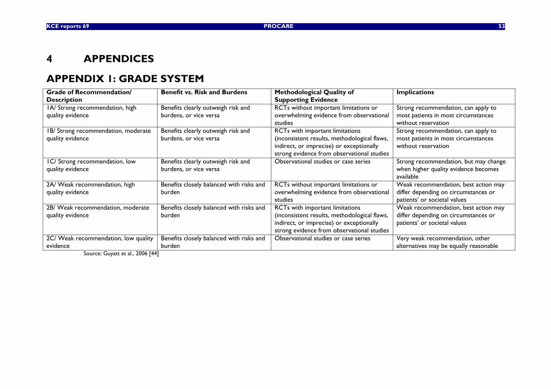
KCE reports 69 PROCARE 53
4 APPENDICES
APPENDIX 1: GRADE SYSTEM Grade of Recommendation/ Description
Benefit vs. Risk and Burdens Methodological Quality of Supporting Evidence
Implications
1A/ Strong recommendation, high quality evidence
Benefits clearly outweigh risk and burdens, or vice versa
RCTs without important limitations or overwhelming evidence from observational studies
Strong recommendation, can apply to most patients in most circumstances without reservation
1B/ Strong recommendation, moderate quality evidence
Benefits clearly outweigh risk and burdens, or vice versa
RCTs with important limitations (inconsistent results, methodological flaws, indirect, or imprecise) or exceptionally strong evidence from observational studies
Strong recommendation, can apply to most patients in most circumstances without reservation
1C/ Strong recommendation, low quality evidence
Benefits clearly outweigh risk and burdens, or vice versa
Observational studies or case series Strong recommendation, but may change when higher quality evidence becomes available
2A/ Weak recommendation, high quality evidence
Benefits closely balanced with risks and burden
RCTs without important limitations or overwhelming evidence from observational studies
Weak recommendation, best action may differ depending on circumstances or patients’ or societal values
2B/ Weak recommendation, moderate quality evidence
Benefits closely balanced with risks and burden
RCTs with important limitations (inconsistent results, methodological flaws, indirect, or imprecise) or exceptionally strong evidence from observational studies
Weak recommendation, best action may differ depending on circumstances or patients’ or societal values
2C/ Weak recommendation, low quality evidence
Benefits closely balanced with risks and burden
Observational studies or case series Very weak recommendation, other alternatives may be equally reasonable
Source: Guyatt et al., 2006 [44]
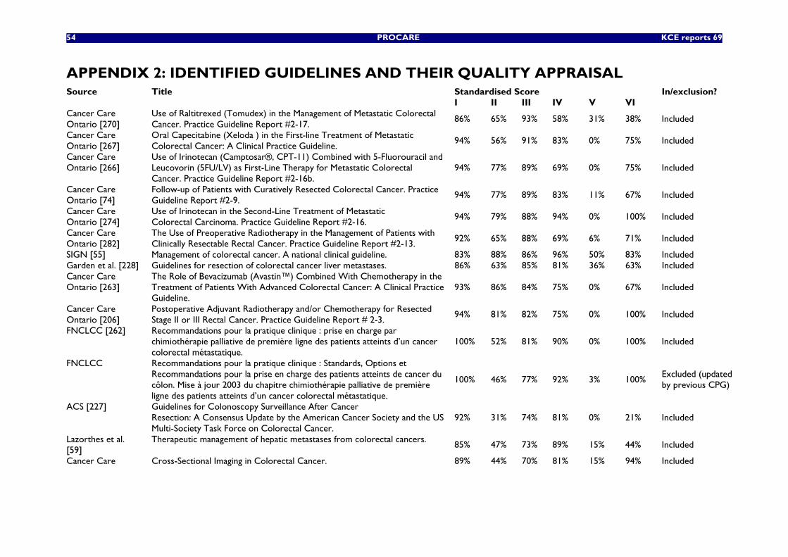
54 PROCARE KCE reports 69
APPENDIX 2: IDENTIFIED GUIDELINES AND THEIR QUALITY APPRAISAL Source Title Standardised Score In/exclusion? I II III IV V VI Cancer Care Ontario [270]
Use of Raltitrexed (Tomudex) in the Management of Metastatic Colorectal Cancer. Practice Guideline Report #2-17.
86% 65% 93% 58% 31% 38% Included
Cancer Care Ontario [267]
Oral Capecitabine (Xeloda ) in the First-line Treatment of Metastatic Colorectal Cancer: A Clinical Practice Guideline.
94% 56% 91% 83% 0% 75% Included
Cancer Care Ontario [266]
Use of Irinotecan (Camptosar®, CPT-11) Combined with 5-Fluorouracil and Leucovorin (5FU/LV) as First-Line Therapy for Metastatic Colorectal Cancer. Practice Guideline Report #2-16b.
94% 77% 89% 69% 0% 75% Included
Cancer Care Ontario [74]
Follow-up of Patients with Curatively Resected Colorectal Cancer. Practice Guideline Report #2-9.
94% 77% 89% 83% 11% 67% Included
Cancer Care Ontario [274]
Use of Irinotecan in the Second-Line Treatment of Metastatic Colorectal Carcinoma. Practice Guideline Report #2-16.
94% 79% 88% 94% 0% 100% Included
Cancer Care Ontario [282]
The Use of Preoperative Radiotherapy in the Management of Patients with Clinically Resectable Rectal Cancer. Practice Guideline Report #2-13.
92% 65% 88% 69% 6% 71% Included
SIGN [55] Management of colorectal cancer. A national clinical guideline. 83% 88% 86% 96% 50% 83% Included Garden et al. [228] Guidelines for resection of colorectal cancer liver metastases. 86% 63% 85% 81% 36% 63% Included Cancer Care Ontario [263]
The Role of Bevacizumab (Avastin™) Combined With Chemotherapy in the Treatment of Patients With Advanced Colorectal Cancer: A Clinical Practice Guideline.
93% 86% 84% 75% 0% 67% Included
Cancer Care Ontario [206]
Postoperative Adjuvant Radiotherapy and/or Chemotherapy for Resected Stage II or III Rectal Cancer. Practice Guideline Report # 2-3.
94% 81% 82% 75% 0% 100% Included
FNCLCC [262] Recommandations pour la pratique clinique : prise en charge par chimiothérapie palliative de première ligne des patients atteints d’un cancer colorectal métastatique.
100% 52% 81% 90% 0% 100% Included
FNCLCC Recommandations pour la pratique clinique : Standards, Options et Recommandations pour la prise en charge des patients atteints de cancer du côlon. Mise à jour 2003 du chapitre chimiothérapie palliative de première ligne des patients atteints d’un cancer colorectal métastatique.
100% 46% 77% 92% 3% 100% Excluded (updated by previous CPG)
ACS [227] Guidelines for Colonoscopy Surveillance After Cancer Resection: A Consensus Update by the American Cancer Society and the US Multi-Society Task Force on Colorectal Cancer.
92% 31% 74% 81% 0% 21% Included
Lazorthes et al. [59]
Therapeutic management of hepatic metastases from colorectal cancers. 85% 47% 73% 89% 15% 44% Included
Cancer Care Cross-Sectional Imaging in Colorectal Cancer. 89% 44% 70% 81% 15% 94% Included
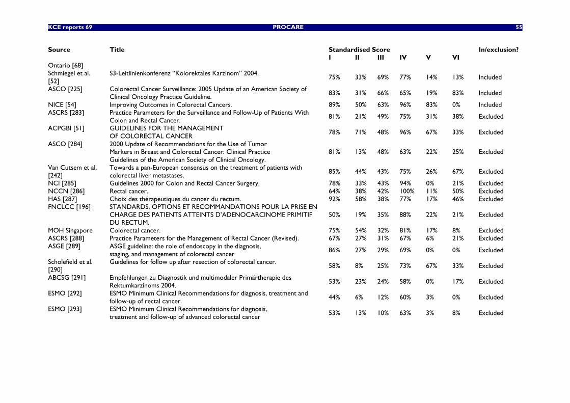
KCE reports 69 PROCARE 55
Source Title Standardised Score In/exclusion? I II III IV V VI Ontario [68] Schmiegel et al. [52]
S3-Leitlinienkonferenz “Kolorektales Karzinom” 2004. 75% 33% 69% 77% 14% 13% Included
ASCO [225] Colorectal Cancer Surveillance: 2005 Update of an American Society of Clinical Oncology Practice Guideline.
83% 31% 66% 65% 19% 83% Included
NICE [54] Improving Outcomes in Colorectal Cancers. 89% 50% 63% 96% 83% 0% Included ASCRS [283] Practice Parameters for the Surveillance and Follow-Up of Patients With
Colon and Rectal Cancer. 81% 21% 49% 75% 31% 38% Excluded
ACPGBI [51] GUIDELINES FOR THE MANAGEMENT OF COLORECTAL CANCER
78% 71% 48% 96% 67% 33% Excluded
ASCO [284] 2000 Update of Recommendations for the Use of Tumor Markers in Breast and Colorectal Cancer: Clinical Practice Guidelines of the American Society of Clinical Oncology.
81% 13% 48% 63% 22% 25% Excluded
Van Cutsem et al. [242]
Towards a pan-European consensus on the treatment of patients with colorectal liver metastases.
85% 44% 43% 75% 26% 67% Excluded
NCI [285] Guidelines 2000 for Colon and Rectal Cancer Surgery. 78% 33% 43% 94% 0% 21% Excluded NCCN [286] Rectal cancer. 64% 38% 42% 100% 11% 50% Excluded HAS [287] Choix des thérapeutiques du cancer du rectum. 92% 58% 38% 77% 17% 46% Excluded FNCLCC [196] STANDARDS, OPTIONS ET RECOMMANDATIONS POUR LA PRISE EN
CHARGE DES PATIENTS ATTEINTS D’ADENOCARCINOME PRIMITIF DU RECTUM.
50% 19% 35% 88% 22% 21% Excluded
MOH Singapore Colorectal cancer. 75% 54% 32% 81% 17% 8% Excluded ASCRS [288] Practice Parameters for the Management of Rectal Cancer (Revised). 67% 27% 31% 67% 6% 21% Excluded ASGE [289] ASGE guideline: the role of endoscopy in the diagnosis,
staging, and management of colorectal cancer 86% 27% 29% 69% 0% 0% Excluded
Scholefield et al. [290]
Guidelines for follow up after resection of colorectal cancer. 58% 8% 25% 73% 67% 33% Excluded
ABCSG [291] Empfehlungen zu Diagnostik und multimodaler Primärtherapie des Rektumkarzinoms 2004.
53% 23% 24% 58% 0% 17% Excluded
ESMO [292] ESMO Minimum Clinical Recommendations for diagnosis, treatment and follow-up of rectal cancer.
44% 6% 12% 60% 3% 0% Excluded
ESMO [293] ESMO Minimum Clinical Recommendations for diagnosis, treatment and follow-up of advanced colorectal cancer
53% 13% 10% 63% 3% 8% Excluded
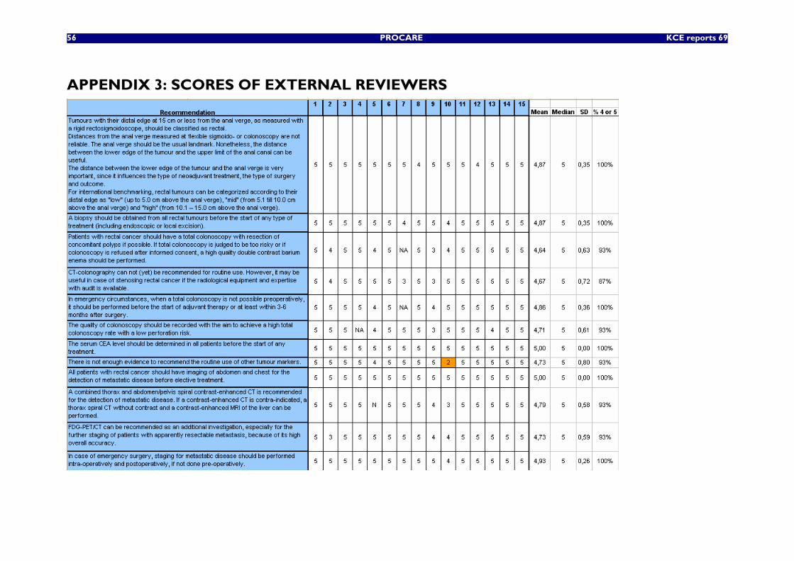
56 PROCARE KCE reports 69
APPENDIX 3: SCORES OF EXTERNAL REVIEWERS
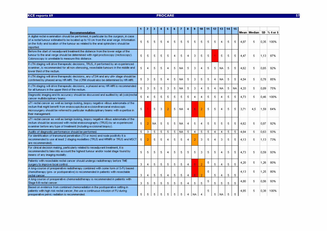
KCE reports 69 PROCARE 57
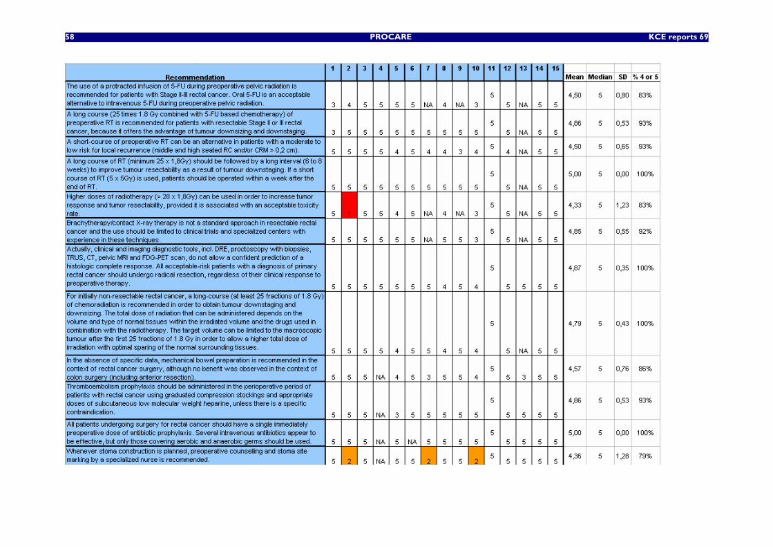
58 PROCARE KCE reports 69
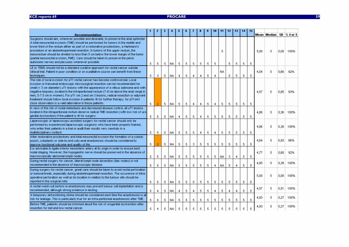
KCE reports 69 PROCARE 59
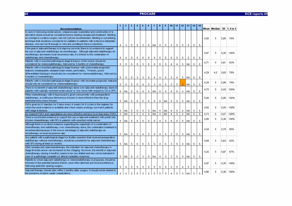
60 PROCARE KCE reports 69
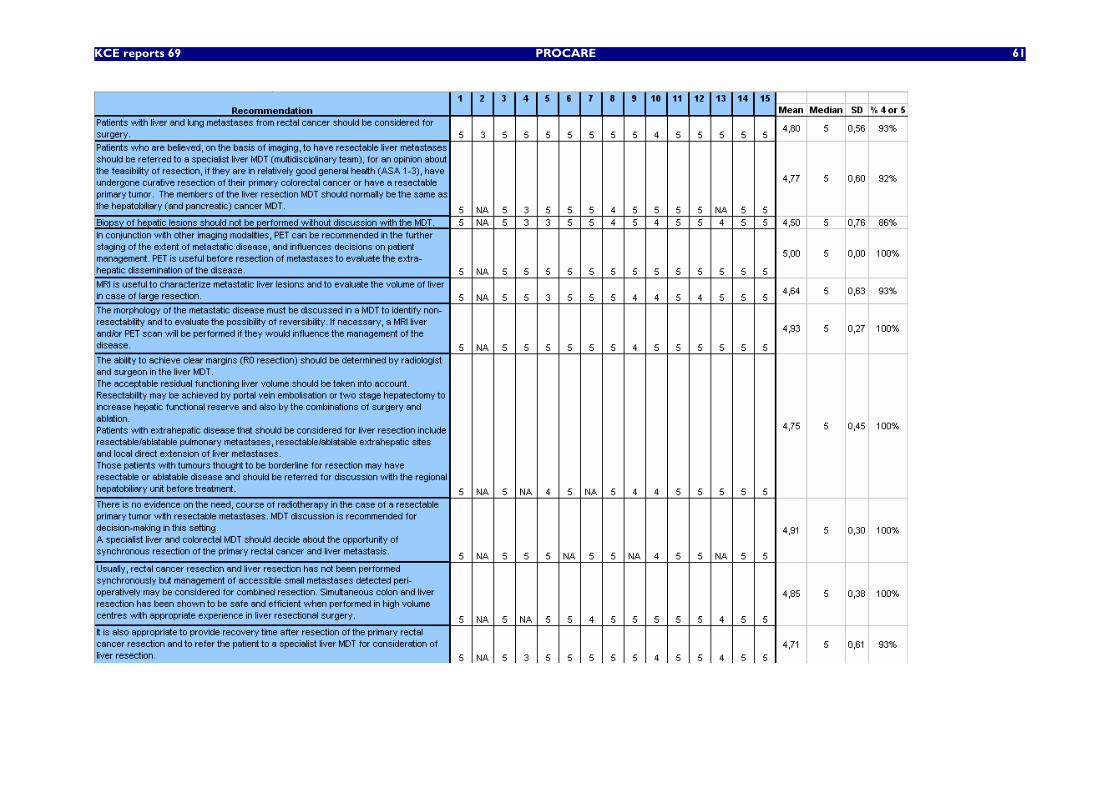
KCE reports 69 PROCARE 61
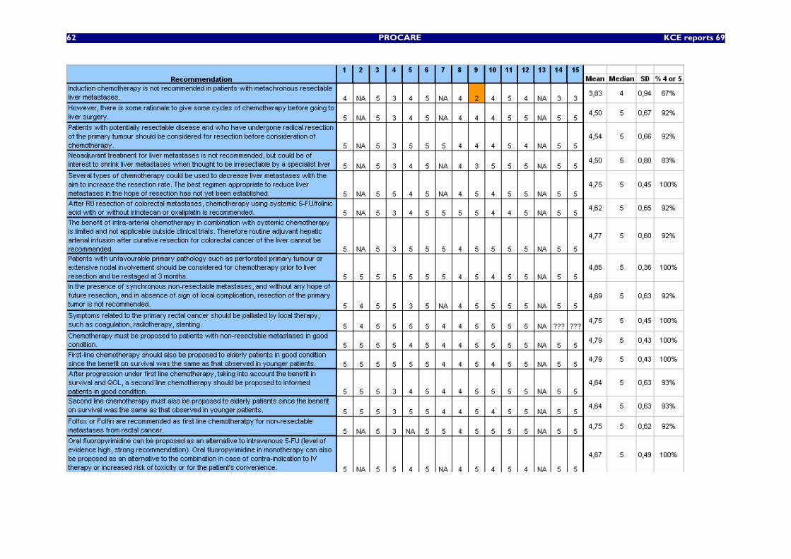
62 PROCARE KCE reports 69
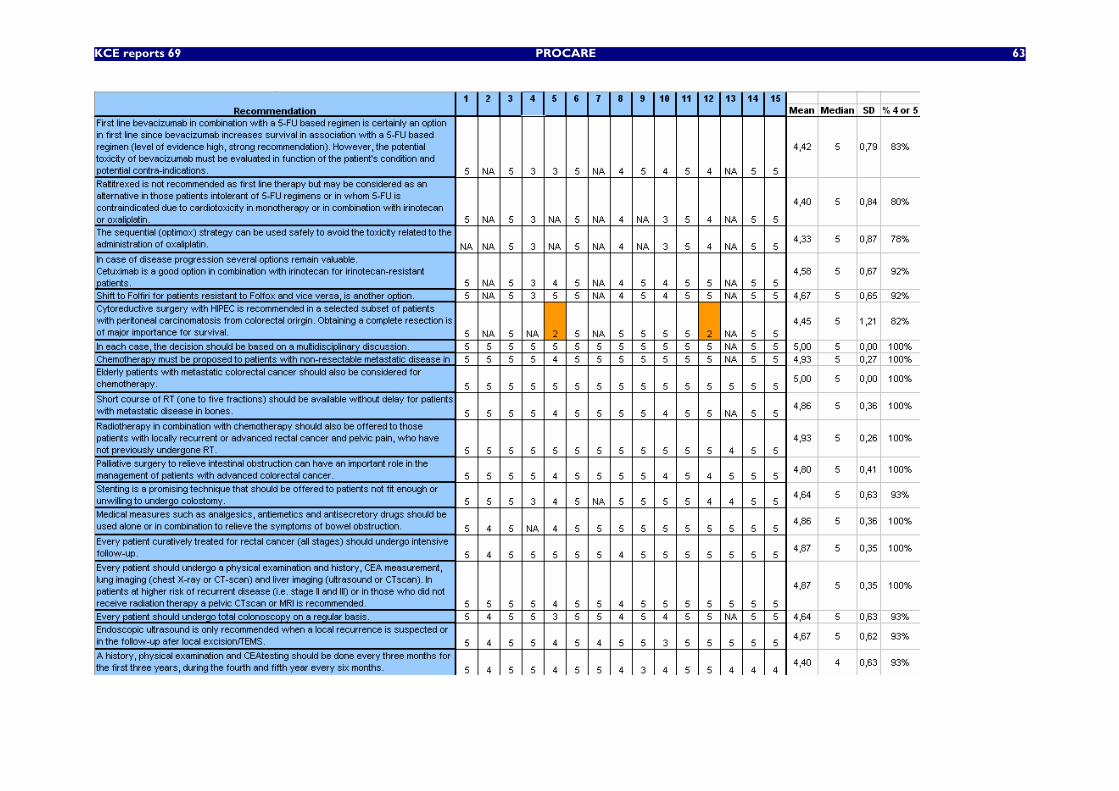
KCE reports 69 PROCARE 63
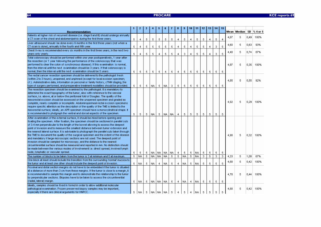
64 PROCARE KCE reports 69
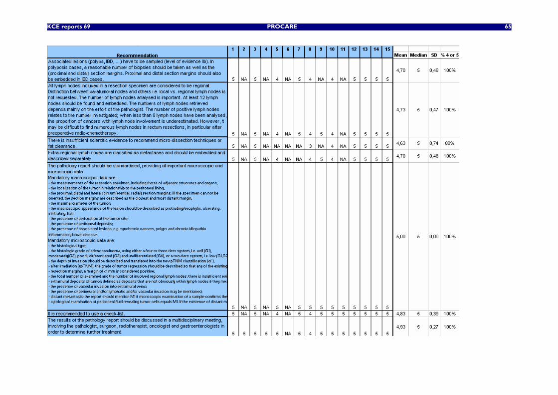
KCE reports 69 PROCARE 65
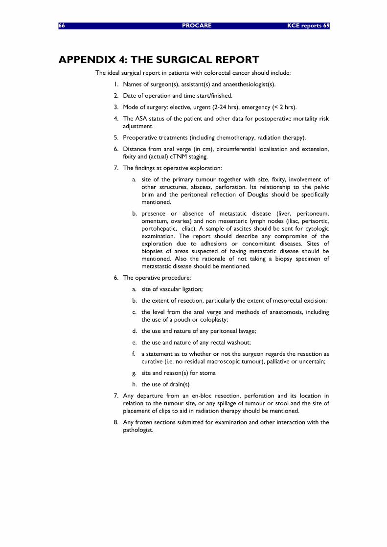
66 PROCARE KCE reports 69
APPENDIX 4: THE SURGICAL REPORT The ideal surgical report in patients with colorectal cancer should include:
1. Names of surgeon(s), assistant(s) and anaesthesiologist(s).
2. Date of operation and time start/finished.
3. Mode of surgery: elective, urgent (2-24 hrs), emergency (< 2 hrs).
4. The ASA status of the patient and other data for postoperative mortality risk adjustment.
5. Preoperative treatments (including chemotherapy, radiation therapy).
6. Distance from anal verge (in cm), circumferential localisation and extension, fixity and (actual) cTNM staging.
7. The findings at operative exploration:
a. site of the primary tumour together with size, fixity, involvement of other structures, abscess, perforation. Its relationship to the pelvic brim and the peritoneal reflection of Douglas should be specifically mentioned.
b. presence or absence of metastatic disease (liver, peritoneum, omentum, ovaries) and non mesenteric lymph nodes (iliac, periaortic, portohepatic, eliac). A sample of ascites should be sent for cytologic examination. The report should describe any compromise of the exploration due to adhesions or concomitant diseases. Sites of biopsies of areas suspected of having metastatic disease should be mentioned. Also the rationale of not taking a biopsy specimen of metastastic disease should be mentioned.
6. The operative procedure:
a. site of vascular ligation;
b. the extent of resection, particularly the extent of mesorectal excision;
c. the level from the anal verge and methods of anastomosis, including the use of a pouch or coloplasty;
d. the use and nature of any peritoneal lavage;
e. the use and nature of any rectal washout;
f. a statement as to whether or not the surgeon regards the resection as curative (i.e. no residual macroscopic tumour), palliative or uncertain;
g. site and reason(s) for stoma
h. the use of drain(s)
7. Any departure from an en-bloc resection, perforation and its location in relation to the tumour site, or any spillage of tumour or stool and the site of placement of clips to aid in radiation therapy should be mentioned.
8. Any frozen sections submitted for examination and other interaction with the pathologist.
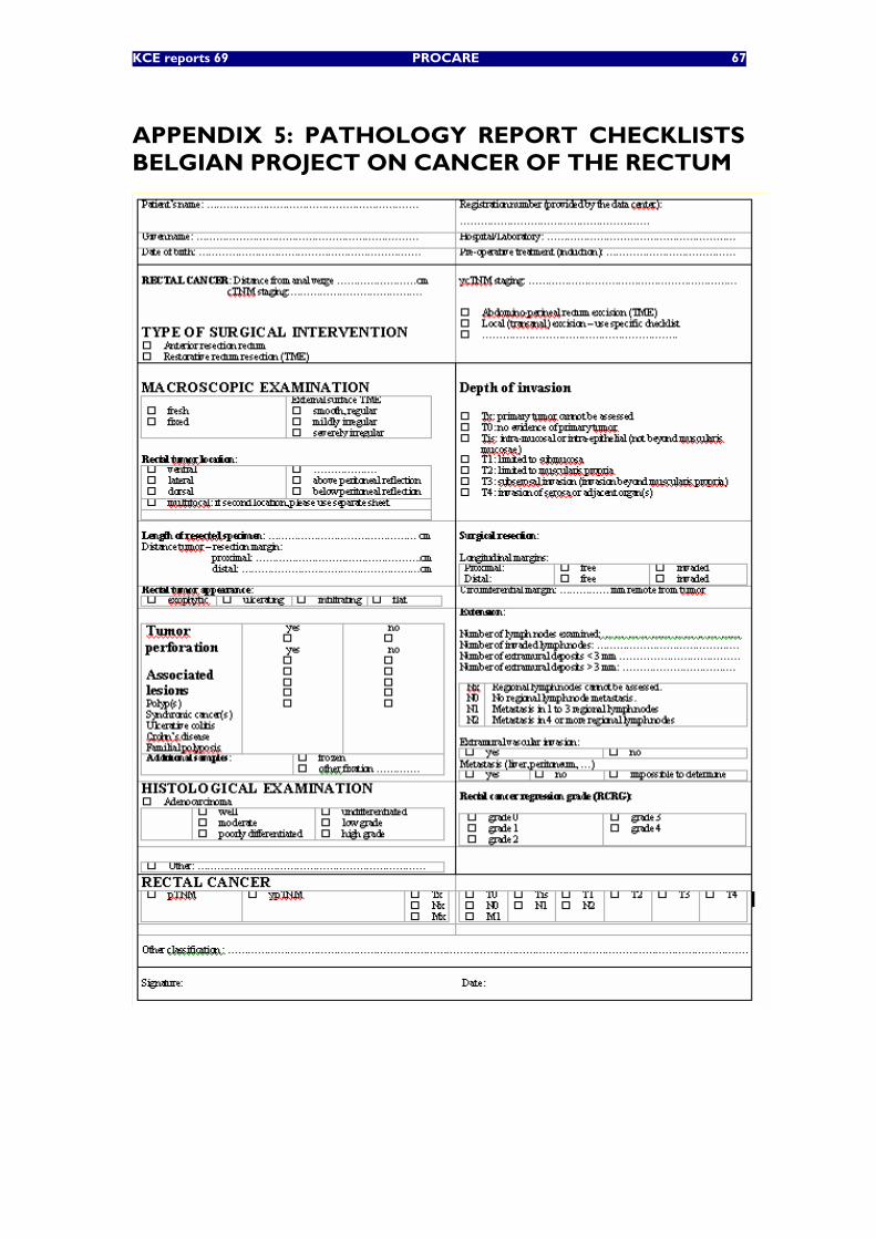
KCE reports 69 PROCARE 67
APPENDIX 5: PATHOLOGY REPORT CHECKLISTS BELGIAN PROJECT ON CANCER OF THE RECTUM
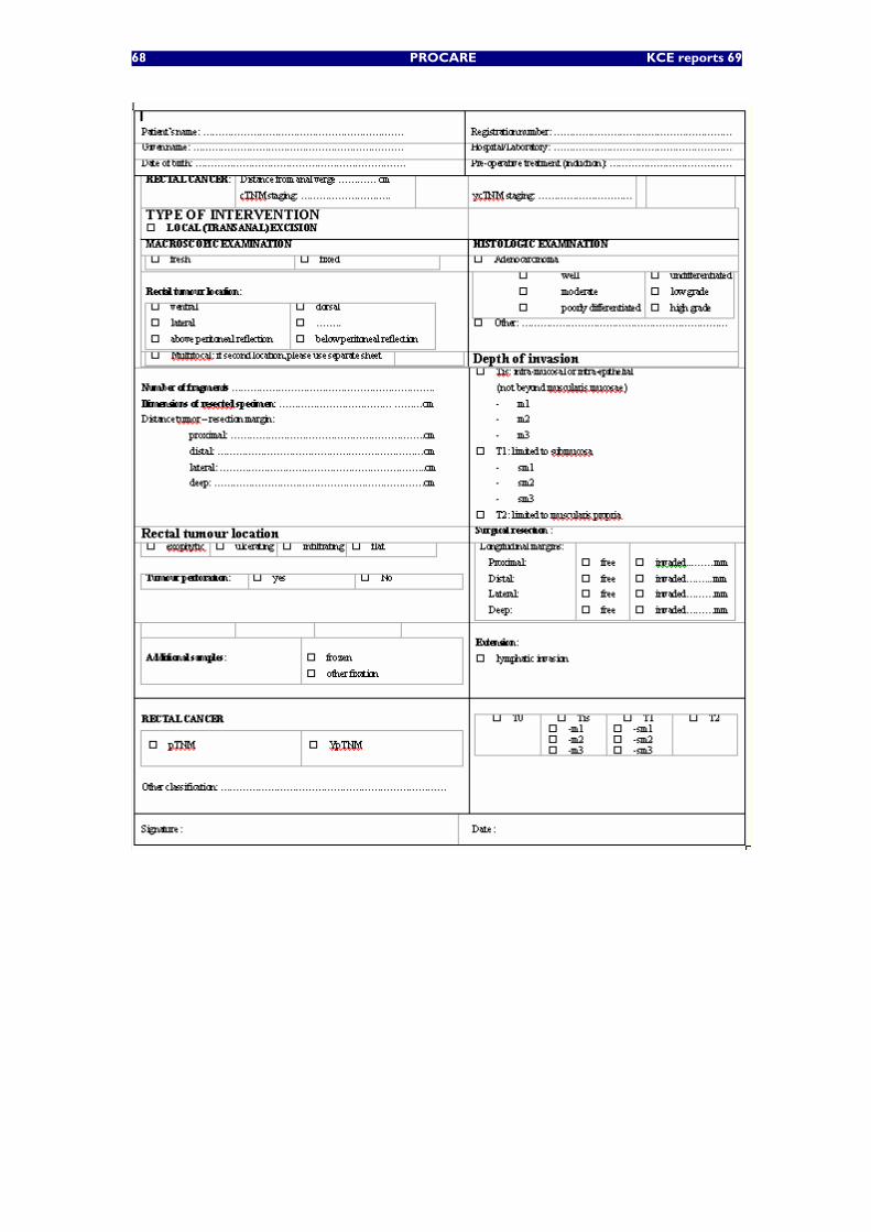
68 PROCARE KCE reports 69
KCE reports 69 PROCARE 69
APPENDIX 6: THE HANDLING OF THE SPECIMEN AFTER LOCAL EXCISION FOR (EARLY) RECTAL CANCER
"Early" rectal cancer can have different presentations varying from the so-called "malignant" polyp (invasive adenocarcinoma arisen in a pedunculated adenoma) up to the (de novo) infiltrating carcinoma limited to the superficial layers of the bowel wall. Compared to other segments of the gastro-intestinal tract, the colon is unique in that the colonic mucosa appears to contain very few lymphatic vessels, especially so under normal circumstances. This anatomical peculiarity underlies 1) the potential therapeutic value of local excisions and 2) the specific TNM classification : pTis = carcinoma in situ : intraepithelial (within basement membrane) i.e. non-invasive or invasion in the lamina propria (intramucosal) with no extension throughout the muscularis mucosae into the submucosa ; while pT1 equals tumour invading the submucosa. From the point of view of the pathologist the finding of an early rectal cancer can be fortuitous e.g. a malignant polyp or consist of a "first intention" therapeutic excision. There are no specific guidelines describing the handling of the latter local excision specimens, especially the local excisions of non-pedunculated lesions. The following description is mainly based on analogies with the handling of other specimens, a.o. EMR (endoscopic mucosal resection for e.g. Barrett lesions).
Assessment of the completeness of excision (R0) and of histologic features "negatively" influencing prognosis a.o. related to the risk of lymph node metastasis are important to predict prognosis and especially to assess the need for additional treatment i.e. more radical surgery. Standardisation of data, the application of well-defined criteria, and the acceptance of an identical and unique staging system allow integration and comparison of data.
Handling of the specimen
• The specimen should preferentially be received fresh, i.e. unfixed, and should be examined by a pathologist preferentially together with the clinician who performed the intervention (gastroenterologist or surgeon). The latter is mandatory if the specimen is received fragmented and (tentative) assessment of the surgical margins is aimed for.
• Administrative data, information on personal and family history, cTNM staging, type of intervention performed and if applicable preoperative treatment modalities should be provided. Additional information is needed if the specimen was received fragmented in order to aid orientation.
• It is important to inspect the specimen and to identify the lesion(s) and the margins most at risk of involvement as this will influence the orientation of the sectioning / embedding of the specimen. Usually, the lesion can be better visualized in the unfixed state.
• Macroscopic inspection of the specimen can be improved by the use of a dissection microscope.
• The margins of the specimen should be marked with ink. Different colours can be used although this is not mandatory.
• Before fixation the specimen should be pinned out – taken care not to over-stretch the specimen – on a cork or wax support. It is advisable to photograph the specimen to document the lesion.
• Ideally the specimen will be fixed in formol in order to allow molecular pathological examination. Taking frozen preserved biopsy samples may be important, especially if there are clinical arguments for HNPCC. Care should however be taken not to interfere with the assessment of completeness of excision.
70 PROCARE KCE reports 69
• Depending on the size of the specimen it may either be entirely sectioned in parallel cuts of 2-3 mm thickness perpendicular to the most critical section margin or it may be sectioned in parallel cuts of 2-3 mm thickness from one side to the other side and additional cuts may be taken perpendicularly to assess the remaining margins. If technically feasible the first option should be preferred.
• All the cuts should be embedded. The number of cuts in one cassette should be limited. If the lesion is polypoid / villous it is likely that small fragments will get detached from the primary lesion during handling. These fragments should be embedded separately.
Pathology report
The pathology report should be standardised, providing all important macroscopic and microscopic data. As already mentioned the need for additional treatment will be based on : the completeness of excision (R0), less than 1 mm is considered positive ; the depth of invasion and especially the risk of lymph node metastasis : i.e. the degree of differentiation (G3) and the presence of lymphatic invasion (L1). The presence of "budding" has been described as a risk factor for lymph node metastasis, especially by Japanese authors. Most often "budding" is described as 1 or a few (5) cells budding of the adjacent tumoral glands at the actively invading region and invading into the stromal component. The definition, but especially the grading of budding is ill-defined. The scientific evidence for a predictive value is therefore limited. The presence of vascular invasion is related to local recurrence and distant (haematogenous) metastasis. The value of perineural invasion, which is unlikely to be seen in local excisions, is not documented in this specific setting.
• Mandatory macroscopic data are :
o the measurement of the specimen
o the maximum diameter of the lesion
o localisation of the lesion in relationship to the margins
o the macroscopic appearance of the lesion should be described as protruding/exophytic, ulcerating, infiltrating, flat
o the presence of perforation at the site of the lesion
• Mandatory microscopic data are :
o the layers of the bowel wall included in the specimen : mucosa, submucosa, muscularis propria, …
o the presence of artefacts hampering interpretation (e.g. extreme coagulation artefacts)
o the histological type of the lesion (i.e. adenocarcinoma)
o the histologic grade of the adenocarcinoma, using a four-tiers system i.e. well (G1), moderately (G2), poorly (G3) or undifferentiated (G4) ; mucinous carcinoma (colloid) and signet ring cell carcinoma are to be considered as poorly differentiated carcinoma
o the depth of invasion should be described and translated into the appropriate classification ; the depth of invasion of the submucosa can be expressed in relative depth (1/3 of thickness ; Kudo e.a.) or in absolute depth of invasion (µm). If the submucosa is not entirely included in the specimen the latter classification system should be used. The Haggit RC e.a. (1985) classification of colorectal carcinomas arising in adenomas has never achieved much clinical impact and should be avoided.
o m1 : upper third of the mucosal thickness
o m2 : middle third of the mucosal thickness
o m3 : deepest / lower third of the mucosal thickness
KCE reports 69 PROCARE 71
o sm1 : upper third of the submucosal thickness alternatively less than 500 µm of depth of invasion into the submucosa
o sm2 : middle third of the submucosal thickness alternatively between 500 and 1000 µm of depth of invasion into the submucosa
o sm3 : deepest / lower third of the submucosal thickness alternatively more than 500 µm of depth of invasion into the submucosa
o resection margins : lateral and deep
o the presence of lymphatic invasion
o the presence of vascular invasion
o the presence of perineural invasion may be mentioned
o the presence of budding may be mentioned
• It is recommended to use a check-list.
The results of the pathology report should be discussed in a multidisciplinary meeting, involving the pathologist, surgeon, radiotherapist, oncologist and gastroenterologists in order to determine further treatment.
72 PROCARE KCE reports 69
APPENDIX 7: RADIOLOGY: TECHNICAL RECOMMENDATIONS
Staging of rectal cancer with multi-slice CT: suggested parameters
• spiral CT of the thorax and the abdomen during the same session
o at least four rows of detectors
o with intravenous contrast injection
dose and injection rate : 120 cc 2-3 cc/sec
o Chest CT:
Acquisition : 35 seconds after IV injection
Parameters: slice thickness 5 mm or lower
o Abdominal CT:
Acquisition: 65-70 seconds after IV injection
Parameters : slice thickness 5 mm or lower
Oral contrast: 750-1000 cc of 3% Hypaque or equivalent
Imaging of rectal cancer with MRI: suggested sequences
Lymph nodes: whole pelvis
• transverse T2 - weighted spin echo without fat suppression (<= 7 mm)
• transverse true FISP ( < = 7 mm)
Tumour:
• transverse T2 - weighted spin echo without fat suppression, 5 mm /0,5 mm
• transverse T2 fat suppression; 5 mm /0.5 mm
• other plan , without fat suppression
o coronal or sagittal depending on the location of the tumour
• transverse T1 fat suppression, before and with IV contrast
o 5 mm / 0,5 mm
Recommended MRI report:
The MRI report has to contain a T and N staging and the CRM (circumferential resection margin) estimated in mm:
• Estimation of the T category:
o T1: The tumour is located in the submucosa, appears with a lower signal intensity than the submucosa and does not extend into the circular muscle layer.
o T2: The tumour is located in the submucosa and in the muscular layer. There is a disappearance of the interface between the submucosa and the muscular propria. The lesion appears with an intermediate signal intensity (higher signal than muscle, lower signal than submucosa) within muscularis propria. The lesion does not extend into the perirectal fat (i.e a hypointense rim persists around the tumour).
o T3: The tumour invades the mesorectal fat with the loss of the interface between the muscular propria and the perirectal fat tissue. The tumour bulges or has nodular projections beyond the outer muscle layer. Spiculations are more indicative of fibrodesmoplatic reaction.

KCE reports 69 PROCARE 73
o T4: The tumour extends into adjacent organs (prostate, seminal vesicles,…) and /or perforates visceral peritoneum.
o The limitations of MRI for the distinction between T2 and T 3 categories are well known (overstaging).
• Estimation of the N category:
o Size of the lymph nodes : not relevant (threshold : 4 mm)
o The shape of the lymph node has to be considered:
Irregular aspect
Signal heterogeneity
These signs are indicative of tumour invasion
• Circumferential Resection Margin:
o 5 mm with MRI corresponds to 1 mm at surgery
74 PROCARE KCE reports 69
APPENDIX 8: RADIOTHERAPY: TECHNICAL CONSIDERATIONS
Radiation dose
The radiation dose will be specified at the ICRU-50 reference point, which is to be located in the central part of the clinical target volume (CTV). This reference point is further described above. The isodose curve representing 95% of the prescription dose must encompass the entire planning target volume (PTV) which is defined above. The standard deviation of the dose within the PTV should be as small as possible and not superior to 2% (≤ 2%) provided the Dmean and Dmedian are close to each other. Each field is to be treated every day. A volumetric treatment planning CT study is required to define the CTV and the PTV. Contiguous CT slices with 3-5 mm separation of the whole pelvis should be taken. The CTV will be outlined on all appropriate CT slices and displayed using beam’s eye views. The PTV is to be treated with any combination of coplanar or non-coplaner three-dimensional conformal fields shaped to deliver the specified dose while restricting the dose to the normal tissues. Field arrangements will be determined by 3D planning to produce the optimal conformal plan in accordance with volume definitions. A planned radiotherapy volume using at least 3 or 4 beams is recommended as this reduces morbidity and mortality.
Beam energy
Radiation therapy is delivered by photon radiation generated by a linear accelerator. Megavoltage equipment is required with effective photon energies ≥ 6MV. Mixed beams are allowed with higher energy for the lateral beams compared to the posterior beam. The use of 3D conformal radiotherapy capabilities is recommended.
Dose prescription
The dose will be prescribed at the center of the target area or at the intersection of central rays of the beam.
Patient treatment position
Patients must be reproducibly immobilized. Measures should be taken to reduce the volume of small bowel e.g. by using a belly board and/or treatment of the patient with a full bladder.
Shielding and verification
The radiation target volume will be defined by shaped ports with custom-made blocks or multileaf collimation. Portal verification shall be done for all treated fields. A maximum of 0.5 cm of deviation will be accepted.
Compensating filters or wedges
In the case of a large sloping contour, wedges or compensating filters should be used. If necessary, appropriate reduction in field size must be done to avoid excessive irradiation to critical structures.
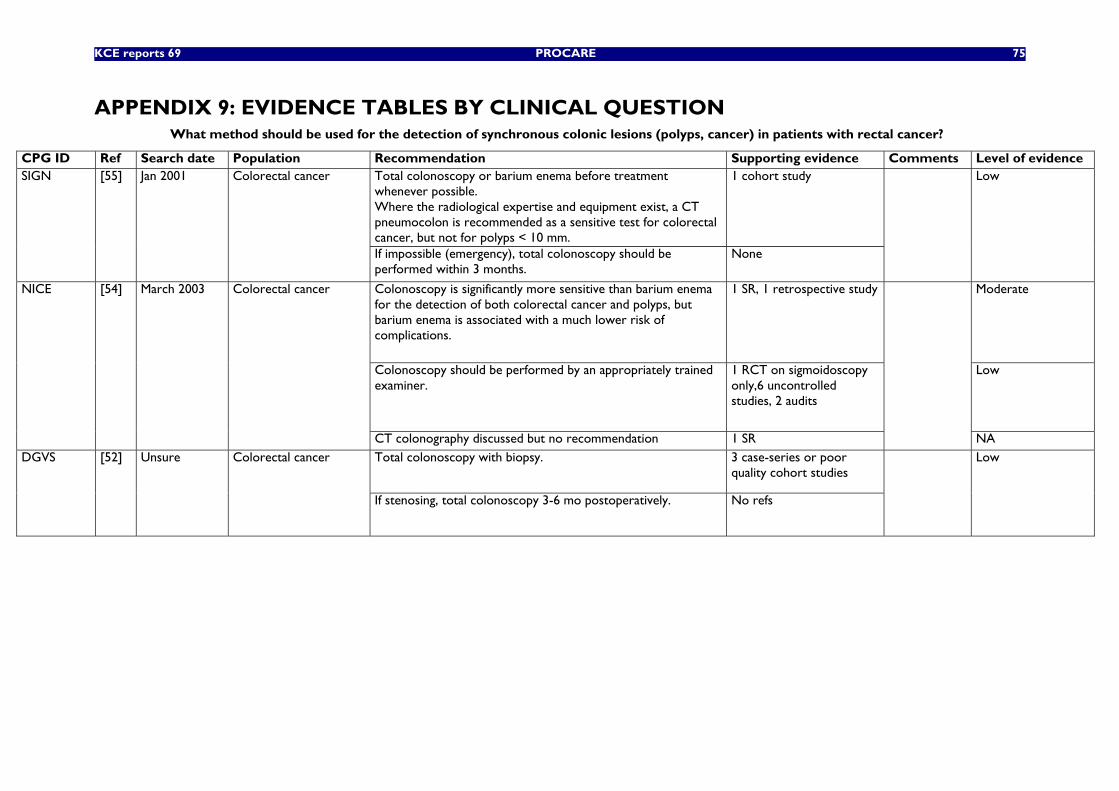
KCE reports 69 PROCARE 75
APPENDIX 9: EVIDENCE TABLES BY CLINICAL QUESTION What method should be used for the detection of synchronous colonic lesions (polyps, cancer) in patients with rectal cancer?
CPG ID Ref Search date Population Recommendation Supporting evidence Comments Level of evidence Total colonoscopy or barium enema before treatment whenever possible. Where the radiological expertise and equipment exist, a CT pneumocolon is recommended as a sensitive test for colorectal cancer, but not for polyps < 10 mm.
1 cohort study SIGN [55] Jan 2001 Colorectal cancer
If impossible (emergency), total colonoscopy should be performed within 3 months.
None
Low
Colonoscopy is significantly more sensitive than barium enema for the detection of both colorectal cancer and polyps, but barium enema is associated with a much lower risk of complications.
1 SR, 1 retrospective study Moderate
Colonoscopy should be performed by an appropriately trained examiner.
1 RCT on sigmoidoscopy only,6 uncontrolled studies, 2 audits
Low
NICE [54] March 2003 Colorectal cancer
CT colonography discussed but no recommendation 1 SR
NA Total colonoscopy with biopsy. 3 case-series or poor
quality cohort studies DGVS [52] Unsure Colorectal cancer
If stenosing, total colonoscopy 3-6 mo postoperatively. No refs
Low
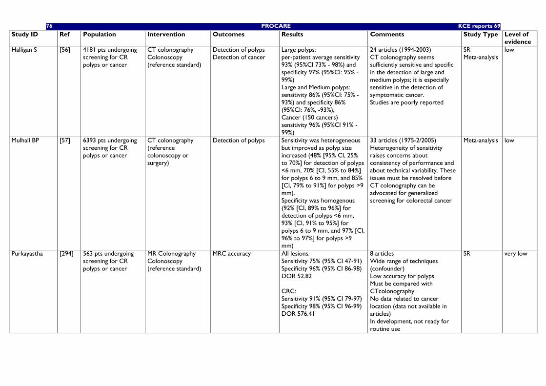
76 PROCARE KCE reports 69
Study ID Ref Population Intervention Outcomes Results Comments Study Type Level of evidence
Halligan S [56] 4181 pts undergoing screening for CR polyps or cancer
CT colonography Colonoscopy (reference standard)
Detection of polyps Detection of cancer
Large polyps: per-patient average sensitivity 93% (95%CI 73% - 98%) and specificity 97% (95%CI: 95% - 99%) Large and Medium polyps: sensitivity 86% (95%CI: 75% - 93%) and specificity 86% (95%CI: 76%, -93%), Cancer (150 cancers) sensitivity 96% (95%CI 91% - 99%)
24 articles (1994-2003) CT colonography seems sufficiently sensitive and specific in the detection of large and medium polyps; it is especially sensitive in the detection of symptomatic cancer. Studies are poorly reported
SR Meta-analysis
low
Mulhall BP [57] 6393 pts undergoing screening for CR polyps or cancer
CT colonography (reference colonoscopy or surgery)
Detection of polyps Sensitivity was heterogeneous but improved as polyp size increased (48% [95% CI, 25% to 70%] for detection of polyps <6 mm, 70% [CI, 55% to 84%] for polyps 6 to 9 mm, and 85% [CI, 79% to 91%] for polyps >9 mm). Specificity was homogenous (92% [CI, 89% to 96%] for detection of polyps <6 mm, 93% [CI, 91% to 95%] for polyps 6 to 9 mm, and 97% [CI, 96% to 97%] for polyps >9 mm)
33 articles (1975-2/2005) Heterogeneity of sensitivity raises concerns about consistency of performance and about technical variability. These issues must be resolved before CT colonography can be advocated for generalized screening for colorectal cancer
Meta-analysis low
Purkayastha [294] 563 pts undergoing screening for CR polyps or cancer
MR Colonography Colonoscopy (reference standard)
MRC accuracy All lesions: Sensitivity 75% (95% CI 47-91) Specificity 96% (95% CI 86-98) DOR 52.82 CRC: Sensitivity 91% (95% CI 79-97) Specificity 98% (95% CI 96-99) DOR 576.41
8 articles Wide range of techniques (confounder) Low accuracy for polyps Must be compared with CTcolonography No data related to cancer location (data not available in articles) In development, not ready for routine use
SR very low
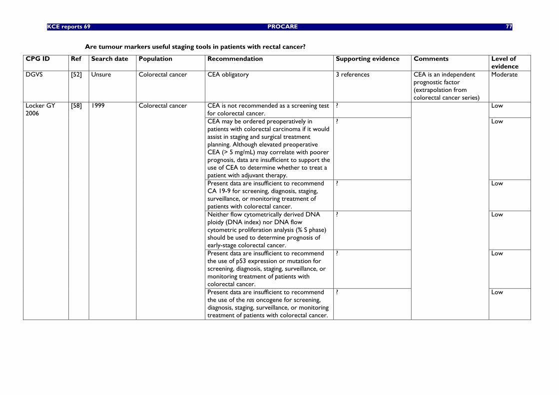
KCE reports 69 PROCARE 77
Are tumour markers useful staging tools in patients with rectal cancer?
CPG ID Ref Search date Population Recommendation Supporting evidence Comments Level of evidence
DGVS [52] Unsure Colorectal cancer CEA obligatory 3 references CEA is an independent prognostic factor (extrapolation from colorectal cancer series)
Moderate
CEA is not recommended as a screening test for colorectal cancer.
? Low
CEA may be ordered preoperatively in patients with colorectal carcinoma if it would assist in staging and surgical treatment planning. Although elevated preoperative CEA (> 5 mg/mL) may correlate with poorer prognosis, data are insufficient to support the use of CEA to determine whether to treat a patient with adjuvant therapy.
? Low
Present data are insufficient to recommend CA 19-9 for screening, diagnosis, staging, surveillance, or monitoring treatment of patients with colorectal cancer.
? Low
Neither flow cytometrically derived DNA ploidy (DNA index) nor DNA flow cytometric proliferation analysis (% S phase) should be used to determine prognosis of early-stage colorectal cancer.
? Low
Present data are insufficient to recommend the use of p53 expression or mutation for screening, diagnosis, staging, surveillance, or monitoring treatment of patients with colorectal cancer.
? Low
Locker GY 2006
[58] 1999 Colorectal cancer
Present data are insufficient to recommend the use of the ras oncogene for screening, diagnosis, staging, surveillance, or monitoring treatment of patients with colorectal cancer.
?
Low
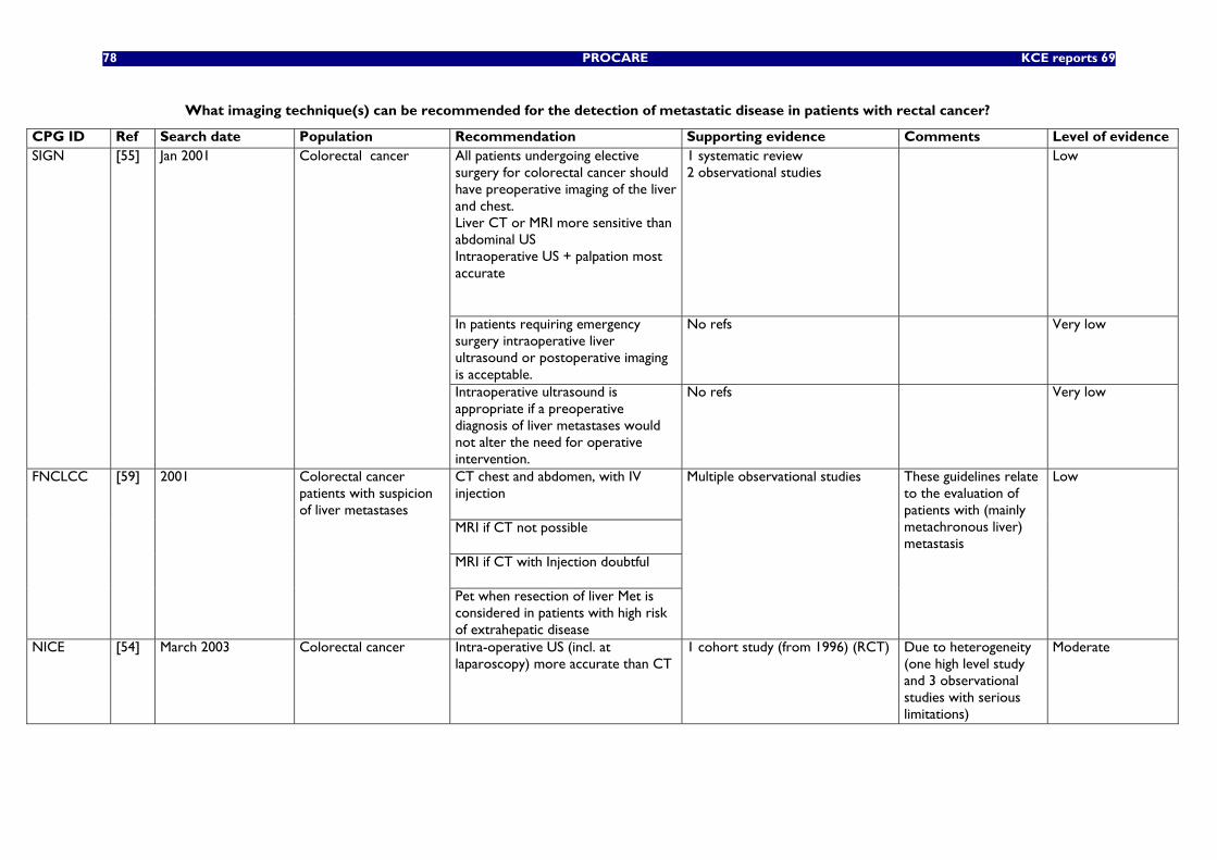
78 PROCARE KCE reports 69
What imaging technique(s) can be recommended for the detection of metastatic disease in patients with rectal cancer?
CPG ID Ref Search date Population Recommendation Supporting evidence Comments Level of evidence All patients undergoing elective surgery for colorectal cancer should have preoperative imaging of the liver and chest. Liver CT or MRI more sensitive than abdominal US Intraoperative US + palpation most accurate
1 systematic review 2 observational studies
Low
In patients requiring emergency surgery intraoperative liver ultrasound or postoperative imaging is acceptable.
No refs
Very low
SIGN [55] Jan 2001 Colorectal cancer
Intraoperative ultrasound is appropriate if a preoperative diagnosis of liver metastases would not alter the need for operative intervention.
No refs Very low
CT chest and abdomen, with IV injection MRI if CT not possible MRI if CT with Injection doubtful
FNCLCC [59] 2001 Colorectal cancer patients with suspicion of liver metastases
Pet when resection of liver Met is considered in patients with high risk of extrahepatic disease
Multiple observational studies
These guidelines relate to the evaluation of patients with (mainly metachronous liver) metastasis
Low
NICE [54] March 2003 Colorectal cancer Intra-operative US (incl. at laparoscopy) more accurate than CT
1 cohort study (from 1996) (RCT)
Due to heterogeneity (one high level study and 3 observational studies with serious limitations)
Moderate
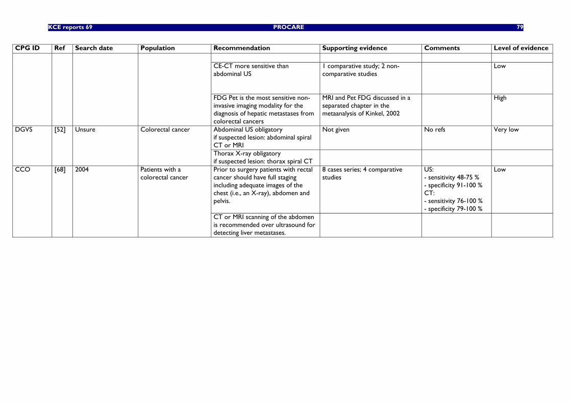
KCE reports 69 PROCARE 79
CPG ID Ref Search date Population Recommendation Supporting evidence Comments Level of evidence
CE-CT more sensitive than abdominal US
1 comparative study; 2 non-comparative studies
Low
FDG Pet is the most sensitive non-invasive imaging modality for the diagnosis of hepatic metastases from colorectal cancers
MRI and Pet FDG discussed in a separated chapter in the metaanalysis of Kinkel, 2002
High
Abdominal US obligatory if suspected lesion: abdominal spiral CT or MRI
Not given
No refs Very low DGVS [52] Unsure Colorectal cancer
Thorax X-ray obligatory if suspected lesion: thorax spiral CT
Prior to surgery patients with rectal cancer should have full staging including adequate images of the chest (i.e., an X-ray), abdomen and pelvis.
8 cases series; 4 comparative studies
US: - sensitivity 48-75 % - specificity 91-100 % CT: - sensitivity 76-100 % - specificity 79-100 %
Low CCO [68] 2004 Patients with a colorectal cancer
CT or MRI scanning of the abdomen is recommended over ultrasound for detecting liver metastases.
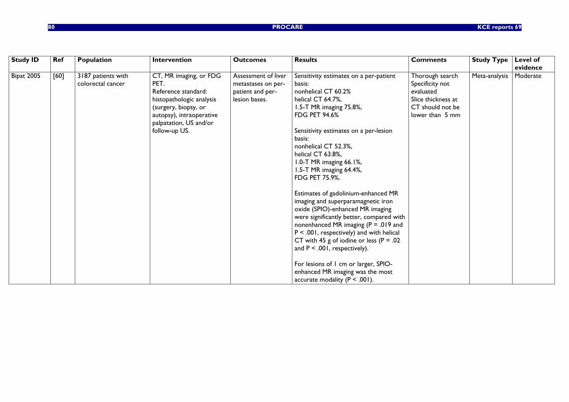
80 PROCARE KCE reports 69
Study ID Ref Population Intervention Outcomes Results Comments Study Type Level of
evidence Bipat 2005
[60] 3187 patients with colorectal cancer
CT, MR imaging, or FDG PET. Reference standard: histopathologic analysis (surgery, biopsy, or autopsy), intraoperative palpatation, US and/or follow-up US.
Assessment of liver metastases on per-patient and per-lesion bases.
Sensitivity estimates on a per-patient basis: nonhelical CT 60.2% helical CT 64.7%, 1.5-T MR imaging 75.8%, FDG PET 94.6% Sensitivity estimates on a per-lesion basis: nonhelical CT 52.3%, helical CT 63.8%, 1.0-T MR imaging 66.1%, 1.5-T MR imaging 64.4%, FDG PET 75.9%. Estimates of gadolinium-enhanced MR imaging and superparamagnetic iron oxide (SPIO)-enhanced MR imaging were significantly better, compared with nonenhanced MR imaging (P = .019 and P < .001, respectively) and with helical CT with 45 g of iodine or less (P = .02 and P < .001, respectively). For lesions of 1 cm or larger, SPIO-enhanced MR imaging was the most accurate modality (P < .001).
Thorough search Specificity not evaluated Slice thickness at CT should not be lower than 5 mm
Meta-analysis Moderate
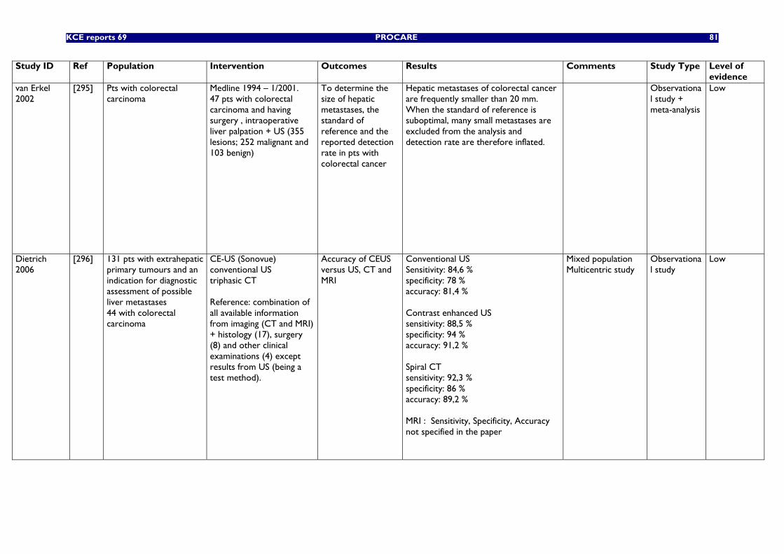
KCE reports 69 PROCARE 81
Study ID Ref Population Intervention Outcomes Results Comments Study Type Level of evidence
van Erkel 2002
[295] Pts with colorectal carcinoma
Medline 1994 – 1/2001. 47 pts with colorectal carcinoma and having surgery , intraoperative liver palpation + US (355 lesions; 252 malignant and 103 benign)
To determine the size of hepatic metastases, the standard of reference and the reported detection rate in pts with colorectal cancer
Hepatic metastases of colorectal cancer are frequently smaller than 20 mm. When the standard of reference is suboptimal, many small metastases are excluded from the analysis and detection rate are therefore inflated.
Observational study + meta-analysis
Low
Dietrich 2006
[296] 131 pts with extrahepatic primary tumours and an indication for diagnostic assessment of possible liver metastases 44 with colorectal carcinoma
CE-US (Sonovue) conventional US triphasic CT Reference: combination of all available information from imaging (CT and MRI) + histology (17), surgery (8) and other clinical examinations (4) except results from US (being a test method).
Accuracy of CEUS versus US, CT and MRI
Conventional US Sensitivity: 84,6 % specificity: 78 % accuracy: 81,4 % Contrast enhanced US sensitivity: 88,5 % specificity: 94 % accuracy: 91,2 % Spiral CT sensitivity: 92,3 % specificity: 86 % accuracy: 89,2 % MRI : Sensitivity, Specificity, Accuracy not specified in the paper
Mixed population Multicentric study
Observational study
Low
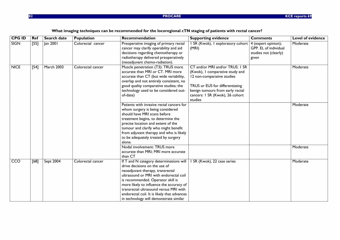
82 PROCARE KCE reports 69
What imaging techniques can be recommended for the locoregional cTN staging of patients with rectal cancer?
CPG ID Ref Search date Population Recommendation Supporting evidence Comments Level of evidence SIGN [55] Jan 2001 Colorectal cancer Preoperative imaging of primary rectal
cancer may clarify operability and aid decisions regarding chemotherapy or radiotherapy delivered preoperatively (neoadjuvant chemo-radiation).
1 SR (Kwok), 1 exploratory cohort (MRI)
4 (expert opinion); GPP. EL of individual studies not (clearly) given
Moderate
Muscle penetration (T3): TRUS more accurate than MRI or CT. MRI more accurate than CT (but wide variability, overlap and not entirely consistent, no good quality comparative studies; the technology used to be considered out-of-date)
CT and/or MRI and/or TRUS: 1 SR (Kwok), 1 comparative study and 12 non-comparative studies TRUS or EUS for differentiating benign tumours from early rectal cancers: 1 SR (Kwok), 26 cohort studies
Moderate
Patients with invasive rectal cancers for whom surgery is being considered should have MRI scans before treatment begins, to determine the precise location and extent of the tumour and clarify who might benefit from adjuvant therapy and who is likely to be adequately treated by surgery alone.
Moderate
NICE [54] March 2003 Colorectal cancer
Nodal involvement: TRUS more accurate than MRI; MRI more accurate than CT
Moderate
CCO [68] Sept 2004 Colorectal cancer If T and N category determinations will drive decisions on the use of neoadjuvant therapy, transrectal ultrasound or MRI with endorectal coil is recommended. Operator skill is more likely to influence the accuracy of transrectal ultrasound versus MRI with endorectal coil. It is likely that advances in technology will demonstrate similar
1 SR (Kwok), 22 case series Moderate
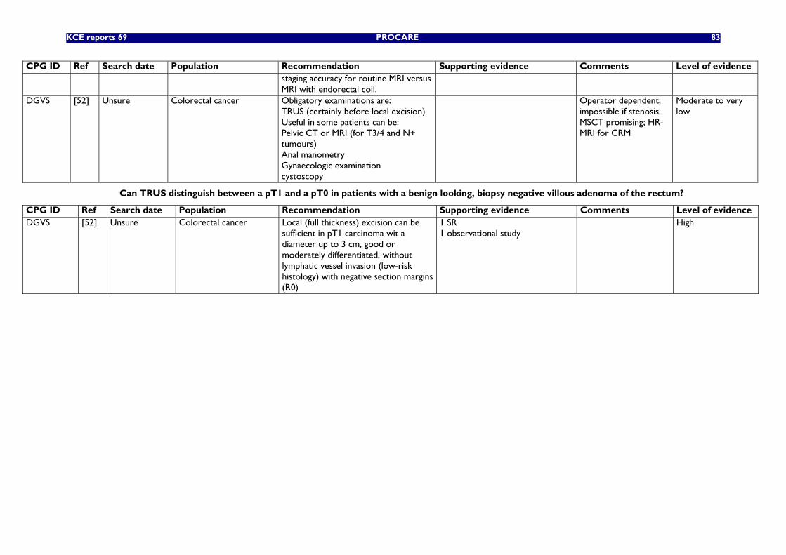
KCE reports 69 PROCARE 83
CPG ID Ref Search date Population Recommendation Supporting evidence Comments Level of evidence staging accuracy for routine MRI versus MRI with endorectal coil.
DGVS [52] Unsure Colorectal cancer Obligatory examinations are: TRUS (certainly before local excision) Useful in some patients can be: Pelvic CT or MRI (for T3/4 and N+ tumours) Anal manometry Gynaecologic examination cystoscopy
Operator dependent; impossible if stenosis MSCT promising; HR-MRI for CRM
Moderate to very low
Can TRUS distinguish between a pT1 and a pT0 in patients with a benign looking, biopsy negative villous adenoma of the rectum?
CPG ID Ref Search date Population Recommendation Supporting evidence Comments Level of evidence DGVS [52] Unsure Colorectal cancer Local (full thickness) excision can be
sufficient in pT1 carcinoma wit a diameter up to 3 cm, good or moderately differentiated, without lymphatic vessel invasion (low-risk histology) with negative section margins (R0)
1 SR 1 observational study
High

84 PROCARE KCE reports 69
Study ID
Ref Population Intervention Outcomes Results Comments Study Type Level of evidence
Worrell S 2004
[65] 258 pts biopsy negative for cancer rectal villous adenoma
Biopsy only Biopsy + TRUS Histology as the reference standard
Prediction pT1 vs pT0
Prevalence of pT1 = 24% (62/258) False negative biopsy in 24% TRUS sensitivity 81% CI 69-90 (50/62) TRUS specificity 88%CI 83-92 (172/196) TRUS accuracy 86% (222/258) TRUS PPV = 68% (50/74) TRUS NPV = 93% (172/184)
5 articles (1986-2003) TRUS false + results can be reduced by performing TRUS before snare excision, by using higher freq, higher resolution US probes Expertise is required!
SR Moderate
Kneist W 2004
286 pts with adenomas (175) or pT1-3 (111) up to 15 cm. Excised by TEMS or LE
DRE TRUS (1 examiner)
Prediction pT2-3 vs pT0-1
Prevalence pT2-3 = 15% (43/286) DRE sensitivity 78% DRE specificity 58% DRE PPV 85% DRE NPV 51% TRUS sensitivity 62% (25/43) TRUS specificity 93% (230/243) TRUS accuracy 89% (255/286) TRUS PPV 66% (25/38) TRUS NPV 93% (230/248)
TRUS is more performant than DRE and essential before TEMS or LE. The authors consider ‘low risk’ T1 as an appropriate indication for TEMS/LE Does not answer the question
Cohort study Very low
Kulig J 2006
[297] 29 patients with uT1
TRUS pT2 vs. T1 sensitivity 50% specificity 92.3% accuracy 89.2% PPV ? NPV ?
Retrospective Small series Single center N of pT0 patients = ? Does not answer the question
Cohort Very low
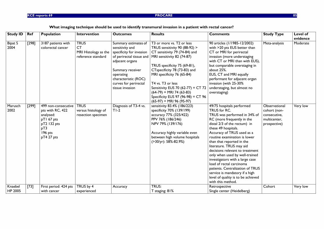
KCE reports 69 PROCARE 85
What imaging technique should be used to identify transmural invasion in a patient with rectal cancer?
Study ID Ref Population Intervention Outcomes Results Comments Study Type Level of evidence
Bipat S 2004
[298] 3187 patients with colorectal cancer
TRUS CT MRI Histology as the reference standard
Summary estimates of sensitivity and specificity for invasion of perirectal tissue and adjacent organs Summary receiver operating characteristic (ROC) curves for perirectal tissue invasion
T3 or more vs. T2 or less TRUS sensitivity 90 (88-92) > CT sensitivity 79 (74-84) and MRI sensitivity 82 (74-87) TRUS specificity 75 (69-81), CTspecificity 78 (73-83) and MRI specificity 76 (65-84) T4 vs. T3 or less Sensitivity EUS 70 (62-77) = CT 72 (64-79) = MRI 74 (63-83) Specificity EUS 97 (96-98) = CT 96 (65-97) = MRI 96 (95-97)
90 articles (1/1985-12/2002) with >20 pts EUS better than CT or MRI for perirectal invasion (more understaging with CT or MRI than with EUS), but comparable overstaging in about 25%. EUS, CT and MRI equally performant for adjacent organ invasion (with 25-30% understaging, but almost no overstaging)
Meta-analysis Moderate
Marusch 2002
[299] 499 non-consecutive pts with RC, 422 analysed: pT1 67 pts pT2 132 pts pT3 196 pts pT4 27 pts
TRUS versus histology of resection specimen
Diagnosis of T3-4 vs. T1-2
sensitivity 83.4% (186/223) specificity 70% (139/199) accuracy 77% (325/422) PPV 76% (186/246) NPV 79% (139/176) Accuracy highly variable even between high volume hospitals (>30/yr): 58%-82.9%)
49/75 hospitals performed TRUS for RC. TRUS was performed in 34% of RC (more frequently in the distal 2/3 of the rectum) in these 49 hospitals. Accuracy of TRUS used as a routine examination is lower than that reported in the literature. TRUS may aid decisions relevant to treatment only when used by well-trained investigators with a large case load of rectal carcinoma patients. Centralization of TRUS service is mandatory if a high level of quality is to be achieved with this method.
Observational cohort (non-consecutive, multicenter, prospective)
Very low
Knaebel HP 2005
[73] First period: 424 pts with cancer
TRUS by 4 experienced
Accuracy TRUS: T staging: 81%
Retrospective Single center (Heidelberg)
Cohort Very low
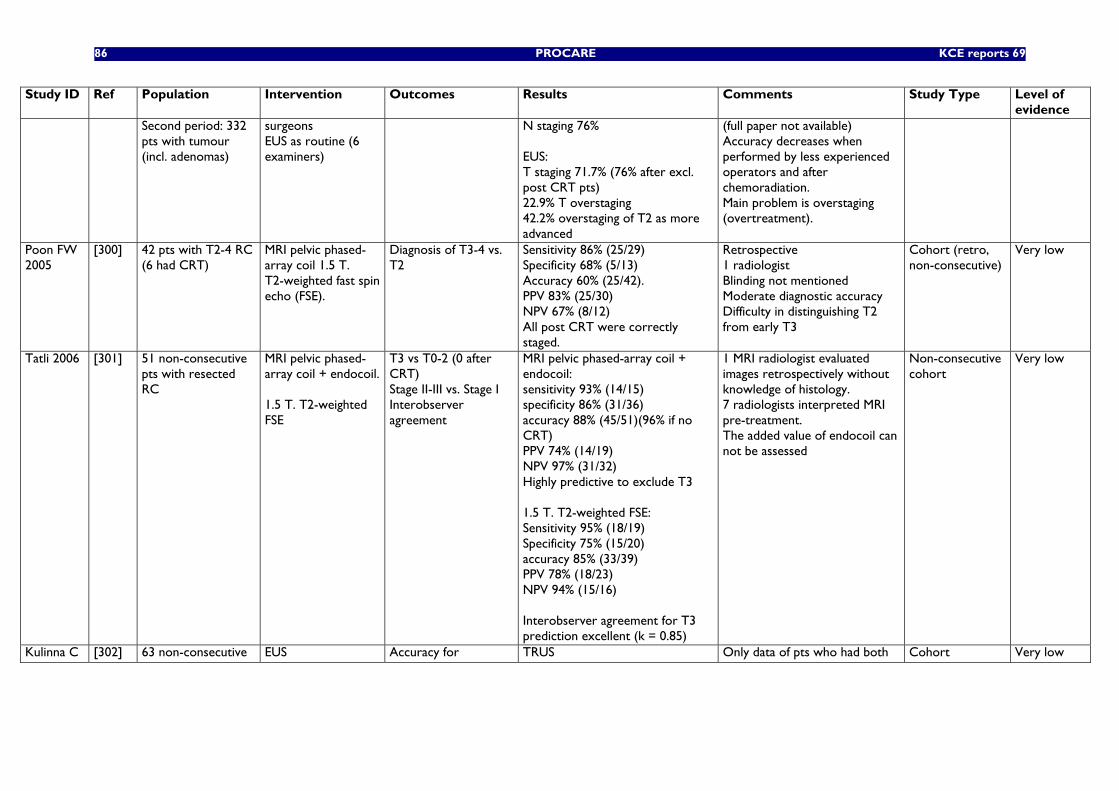
86 PROCARE KCE reports 69
Study ID Ref Population Intervention Outcomes Results Comments Study Type Level of evidence
Second period: 332 pts with tumour (incl. adenomas)
surgeons EUS as routine (6 examiners)
N staging 76% EUS: T staging 71.7% (76% after excl. post CRT pts) 22.9% T overstaging 42.2% overstaging of T2 as more advanced
(full paper not available) Accuracy decreases when performed by less experienced operators and after chemoradiation. Main problem is overstaging (overtreatment).
Poon FW 2005
[300] 42 pts with T2-4 RC (6 had CRT)
MRI pelvic phased-array coil 1.5 T. T2-weighted fast spin echo (FSE).
Diagnosis of T3-4 vs. T2
Sensitivity 86% (25/29) Specificity 68% (5/13) Accuracy 60% (25/42). PPV 83% (25/30) NPV 67% (8/12) All post CRT were correctly staged.
Retrospective 1 radiologist Blinding not mentioned Moderate diagnostic accuracy Difficulty in distinguishing T2 from early T3
Cohort (retro, non-consecutive)
Very low
Tatli 2006 [301] 51 non-consecutive pts with resected RC
MRI pelvic phased-array coil + endocoil. 1.5 T. T2-weighted FSE
T3 vs T0-2 (0 after CRT) Stage II-III vs. Stage I Interobserver agreement
MRI pelvic phased-array coil + endocoil: sensitivity 93% (14/15) specificity 86% (31/36) accuracy 88% (45/51)(96% if no CRT) PPV 74% (14/19) NPV 97% (31/32) Highly predictive to exclude T3 1.5 T. T2-weighted FSE: Sensitivity 95% (18/19) Specificity 75% (15/20) accuracy 85% (33/39) PPV 78% (18/23) NPV 94% (15/16) Interobserver agreement for T3 prediction excellent (k = 0.85)
1 MRI radiologist evaluated images retrospectively without knowledge of histology. 7 radiologists interpreted MRI pre-treatment. The added value of endocoil can not be assessed
Non-consecutive cohort
Very low
Kulinna C [302] 63 non-consecutive EUS Accuracy for TRUS Only data of pts who had both Cohort Very low
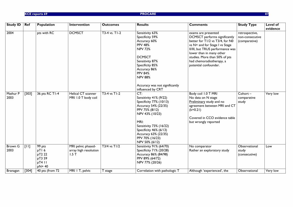
KCE reports 69 PROCARE 87
Study ID Ref Population Intervention Outcomes Results Comments Study Type Level of evidence
2004 pts with RC DCMSCT T3-4 vs. T1-2 Sensitivity 63% Specificity 59% Accuracy 60% PPV 48% NPV 72% DCMSCT Sensitivity 87% Specificity 85% Accuracy 86% PPV 84% NPV 88% Accuracy was not significantly influenced by CRT
exams are presented DCMSCT performs significantly better for T1/2 vs T3/4, for N0 vs N+ and for Stage I vs Stage II/III, but TRUS performance was lower than in many other studies. More than 50% of pts had chemoradiotherapy, a potential confounder.
retrospective, non-consecutive (comparative)
Mathur P 2003
[303] 36 pts RC T1-4 Helical CT scanner MRI 1.0 T body coil
T3-4 vs T1-2 CT: Sensitivity 41% (9/22) Specificity 77% (10/13) Accuracy 54% (22/35) PPV 75% (8/12) NPV 43% (10/23) MRI: Sensitivity 73% (16/22) Specificity 46% (6/13) Accuracy 63% (22/35) PPV 70% (16/23) NPV 50% (6/12)
Body coil 1.0 T MRI No data on N stage Preliminary study and no agreement between MRI and CT (k=0.21) Covered in CCO evidence table but wrongly reported
Cohort – comparative study
Very low
Brown G 2003
[11] 99 pts pT1 6 pT2 22 pT3 59 pT4 11 pN+ 40
MRI pelvic phased-array high resolution 1.5 T
T3/4 vs T1/2 Sensitivity 91% (64/70) Specificity 71% (20/28) Accuracy 86% (84/98) PPV 89% (64/72) NPV 77% (20/26)
No comparator Rather an exploratory study
Observational study (consecutive)
Low
Branagan [304] 40 pts (from 72 MRI 1 T, pelvic T stage Correlation with pathologic T Although ‘experienced’, the Observational Very low
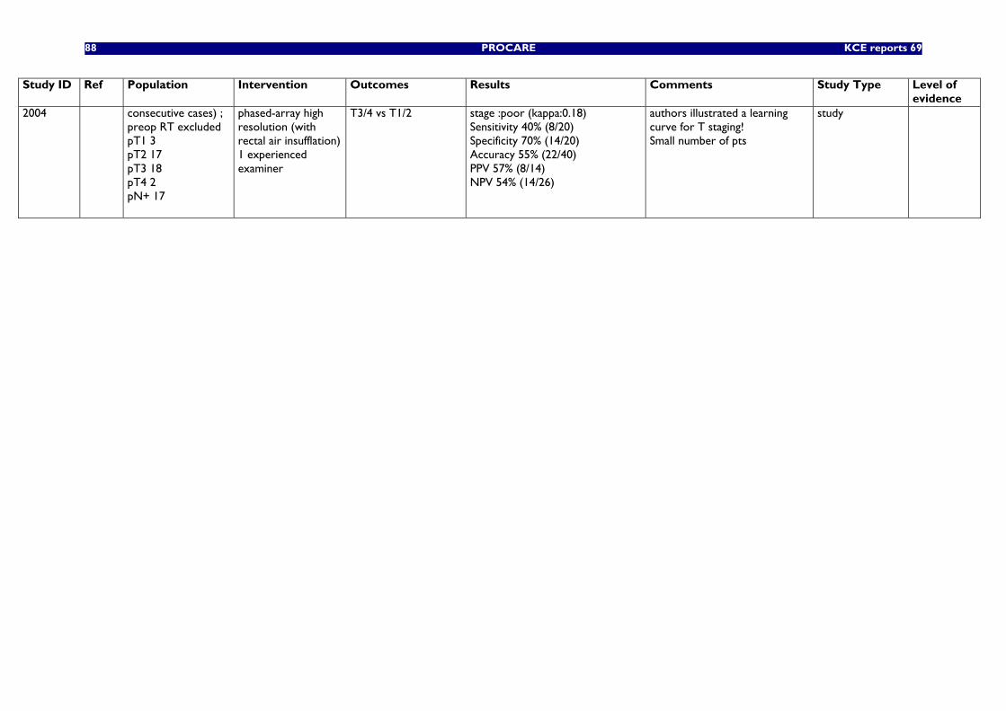
88 PROCARE KCE reports 69
Study ID Ref Population Intervention Outcomes Results Comments Study Type Level of evidence
2004 consecutive cases) ; preop RT excluded pT1 3 pT2 17 pT3 18 pT4 2 pN+ 17
phased-array high resolution (with rectal air insufflation) 1 experienced examiner
T3/4 vs T1/2
stage :poor (kappa:0.18) Sensitivity 40% (8/20) Specificity 70% (14/20) Accuracy 55% (22/40) PPV 57% (8/14) NPV 54% (14/26)
authors illustrated a learning curve for T staging! Small number of pts
study
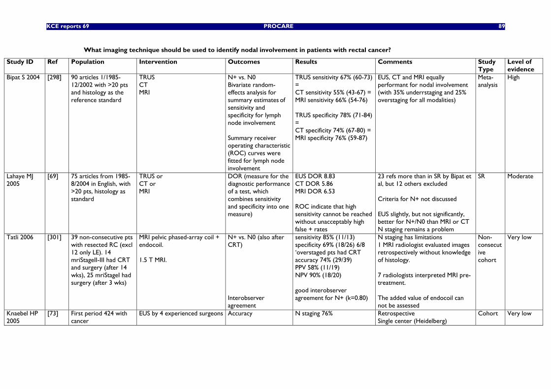
KCE reports 69 PROCARE 89
What imaging technique should be used to identify nodal involvement in patients with rectal cancer?
Study ID Ref Population Intervention Outcomes Results Comments Study Type
Level of evidence
Bipat S 2004 [298] 90 articles 1/1985-12/2002 with >20 pts and histology as the reference standard
TRUS CT MRI
N+ vs. N0 Bivariate random-effects analysis for summary estimates of sensitivity and specificity for lymph node involvement Summary receiver operating characteristic (ROC) curves were
fitted for lymph node involvement
TRUS sensitivity 67% (60-73) = CT sensitivity 55% (43-67) = MRI sensitivity 66% (54-76) TRUS specificity 78% (71-84) = CT specificity 74% (67-80) = MRI specificity 76% (59-87)
EUS, CT and MRI equally performant for nodal involvement (with 35% underrstaging and 25% overstaging for all modalities)
Meta-analysis
High
Lahaye MJ 2005
[69] 75 articles from 1985-8/2004 in English, with >20 pts, histology as standard
TRUS or CT or MRI
DOR (measure for the diagnostic performance of a test, which combines sensitivity and specificity into one measure)
EUS DOR 8.83 CT DOR 5.86 MRI DOR 6.53 ROC indicate that high sensitivity cannot be reached without unacceptably high false + rates
23 refs more than in SR by Bipat et al, but 12 others excluded Criteria for N+ not discussed EUS slightly, but not significantly, better for N+/N0 than MRI or CT N staging remains a problem
SR Moderate
Tatli 2006 [301] 39 non-consecutive pts with resected RC (excl 12 only LE). 14 mriStageII-III had CRT and surgery (after 14 wks), 25 mriStageI had surgery (after 3 wks)
MRI pelvic phased-array coil + endocoil. 1.5 T MRI.
N+ vs. N0 (also after CRT) Interobserver agreement
sensitivity 85% (11/13) specificity 69% (18/26) 6/8 ‘overstaged pts had CRT accuracy 74% (29/39) PPV 58% (11/19) NPV 90% (18/20) good interobserver agreement for N+ (k=0.80)
N staging has limitations 1 MRI radiologist evaluated images retrospectively without knowledge of histology. 7 radiologists interpreted MRI pre-treatment. The added value of endocoil can not be assessed
Non-consecutive cohort
Very low
Knaebel HP 2005
[73] First period 424 with cancer
EUS by 4 experienced surgeons
Accuracy N staging 76%
Retrospective Single center (Heidelberg)
Cohort Very low
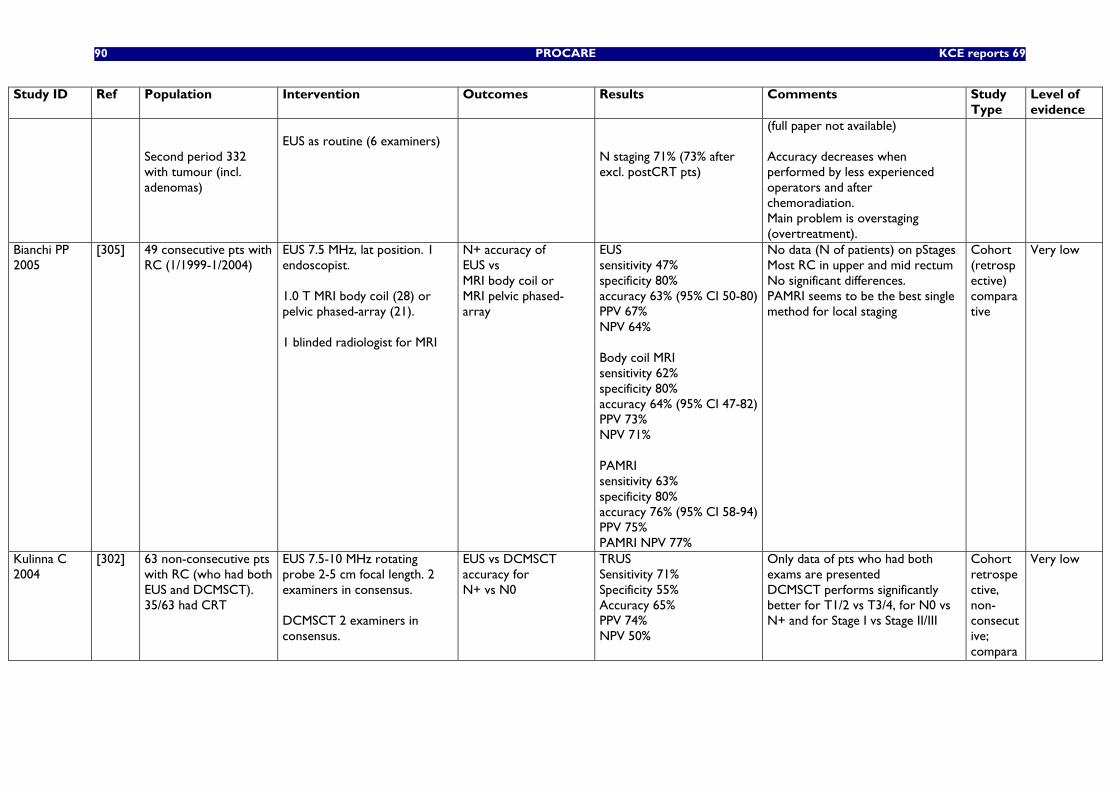
90 PROCARE KCE reports 69
Study ID Ref Population Intervention Outcomes Results Comments Study Type
Level of evidence
Second period 332 with tumour (incl. adenomas)
EUS as routine (6 examiners)
N staging 71% (73% after excl. postCRT pts)
(full paper not available) Accuracy decreases when performed by less experienced operators and after chemoradiation. Main problem is overstaging (overtreatment).
Bianchi PP 2005
[305] 49 consecutive pts with RC (1/1999-1/2004)
EUS 7.5 MHz, lat position. 1 endoscopist. 1.0 T MRI body coil (28) or pelvic phased-array (21). 1 blinded radiologist for MRI
N+ accuracy of EUS vs MRI body coil or MRI pelvic phased-array
EUS sensitivity 47% specificity 80% accuracy 63% (95% CI 50-80) PPV 67% NPV 64% Body coil MRI sensitivity 62% specificity 80% accuracy 64% (95% CI 47-82) PPV 73% NPV 71% PAMRI sensitivity 63% specificity 80% accuracy 76% (95% CI 58-94) PPV 75% PAMRI NPV 77%
No data (N of patients) on pStages Most RC in upper and mid rectum No significant differences. PAMRI seems to be the best single method for local staging
Cohort (retrospective) comparative
Very low
Kulinna C 2004
[302] 63 non-consecutive pts with RC (who had both EUS and DCMSCT). 35/63 had CRT
EUS 7.5-10 MHz rotating probe 2-5 cm focal length. 2 examiners in consensus. DCMSCT 2 examiners in consensus.
EUS vs DCMSCT accuracy for N+ vs N0
TRUS Sensitivity 71% Specificity 55% Accuracy 65% PPV 74% NPV 50%
Only data of pts who had both exams are presented DCMSCT performs significantly better for T1/2 vs T3/4, for N0 vs N+ and for Stage I vs Stage II/III
Cohort retrospective, non-consecutive; compara
Very low
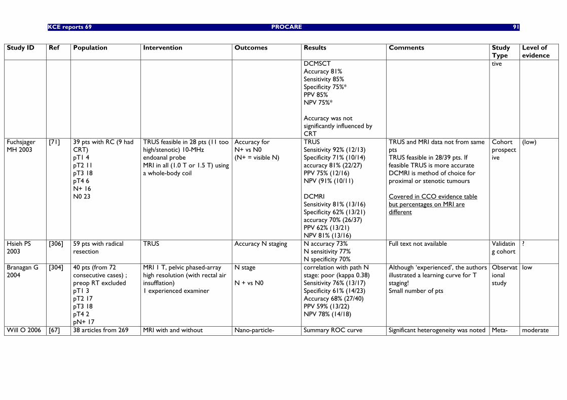
KCE reports 69 PROCARE 91
Study ID Ref Population Intervention Outcomes Results Comments Study Type
Level of evidence
DCMSCT Accuracy 81% Sensitivity 85% Specificity 75%* PPV 85% NPV 75%* Accuracy was not significantly influenced by CRT
tive
Fuchsjager MH 2003
[71] 39 pts with RC (9 had CRT) pT1 4 pT2 11 pT3 18 pT4 6 N+ 16 N0 23
TRUS feasible in 28 pts (11 too high/stenotic) 10-MHz endoanal probe MRI in all (1.0 T or 1.5 T) using a whole-body coil
Accuracy for N+ vs N0 (N+ = visible N)
TRUS Sensitivity 92% (12/13) Specificity 71% (10/14) accuracy 81% (22/27) PPV 75% (12/16) NPV (91% (10/11) DCMRI Sensitivity 81% (13/16) Specificity 62% (13/21) accuracy 70% (26/37) PPV 62% (13/21) NPV 81% (13/16)
TRUS and MRI data not from same pts TRUS feasible in 28/39 pts. If feasible TRUS is more accurate DCMRI is method of choice for proximal or stenotic tumours Covered in CCO evidence table but percentages on MRI are different
Cohort prospective
(low)
Hsieh PS 2003
[306] 59 pts with radical resection
TRUS Accuracy N staging N accuracy 73% N sensitivity 77% N specificity 70%
Full text not available
Validating cohort
?
Branagan G 2004
[304] 40 pts (from 72 consecutive cases) ; preop RT excluded pT1 3 pT2 17 pT3 18 pT4 2 pN+ 17
MRI 1 T, pelvic phased-array high resolution (with rectal air insufflation) 1 experienced examiner
N stage N + vs N0
correlation with path N stage: poor (kappa 0.38) Sensitivity 76% (13/17) Specificity 61% (14/23) Accuracy 68% (27/40) PPV 59% (13/22) NPV 78% (14/18)
Although ‘experienced’, the authors illustrated a learning curve for T staging! Small number of pts
Observational study
low
Will O 2006 [67] 38 articles from 269 MRI with and without Nano-particle- Summary ROC curve Significant heterogeneity was noted Meta- moderate
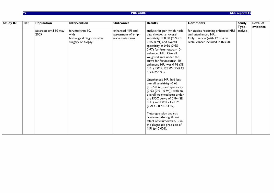
92 PROCARE KCE reports 69
Study ID Ref Population Intervention Outcomes Results Comments Study Type
Level of evidence
abstracts until 10 may 2005
ferumoxtran-10, with histological diagnosis after surgery or biopsy.
enhanced MRI and assessment of lymph node metastases
analysis for per-lymph-node data showed an overall sensitivity of 0·88 (95% CI 0·85–0·91) and overall specificity of 0·96 (0·95–0·97) for ferumoxtran-10-enhanced MRI. Overall weighted area under the curve for ferumoxtran-10-enhanced MRI was 0·96 (SE 0·01), DOR 123·05 (95% CI 5·93–256·93). Unenhanced MRI had less overall sensitivity (0·63 [0·57–0·69]) and specificity (0·93 [0·91–0·94]), with an overall weighted area under the ROC curve of 0·84 (SE 0·11) and DOR of 26·75 (95% CI 8·48–84·42). Metaregression analysis confirmed the significant effect of ferumoxtran-10 in the diagnostic precision of MRI (p=0·001).
for studies reporting enhanced MRI and unenhanced MRI. Only 1 article (with 12 pts) on rectal cancer included in this SR.
analysis
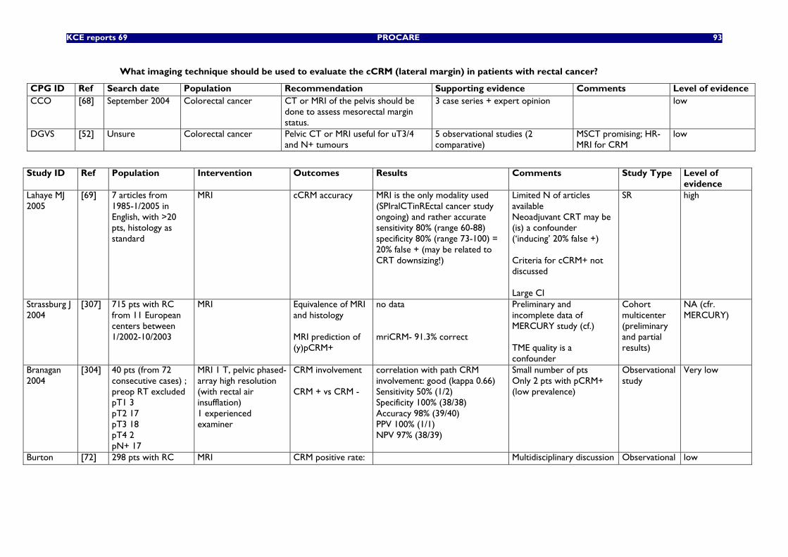
KCE reports 69 PROCARE 93
What imaging technique should be used to evaluate the cCRM (lateral margin) in patients with rectal cancer?
CPG ID Ref Search date Population Recommendation Supporting evidence Comments Level of evidence CCO [68] September 2004 Colorectal cancer CT or MRI of the pelvis should be
done to assess mesorectal margin status.
3 case series + expert opinion low
DGVS [52] Unsure Colorectal cancer Pelvic CT or MRI useful for uT3/4 and N+ tumours
5 observational studies (2 comparative)
MSCT promising; HR-MRI for CRM
low
Study ID Ref Population Intervention Outcomes Results Comments Study Type Level of
evidence Lahaye MJ 2005
[69] 7 articles from 1985-1/2005 in English, with >20 pts, histology as standard
MRI cCRM accuracy MRI is the only modality used (SPIralCTinREctal cancer study ongoing) and rather accurate sensitivity 80% (range 60-88) specificity 80% (range 73-100) = 20% false + (may be related to CRT downsizing!)
Limited N of articles available Neoadjuvant CRT may be (is) a confounder (‘inducing’ 20% false +) Criteria for cCRM+ not discussed Large CI
SR high
Strassburg J 2004
[307] 715 pts with RC from 11 European centers between 1/2002-10/2003
MRI Equivalence of MRI and histology MRI prediction of (y)pCRM+
no data mriCRM- 91.3% correct
Preliminary and incomplete data of MERCURY study (cf.) TME quality is a confounder
Cohort multicenter (preliminary and partial results)
NA (cfr. MERCURY)
Branagan 2004
[304] 40 pts (from 72 consecutive cases) ; preop RT excluded pT1 3 pT2 17 pT3 18 pT4 2 pN+ 17
MRI 1 T, pelvic phased-array high resolution (with rectal air insufflation) 1 experienced examiner
CRM involvement CRM + vs CRM -
correlation with path CRM involvement: good (kappa 0.66) Sensitivity 50% (1/2) Specificity 100% (38/38) Accuracy 98% (39/40) PPV 100% (1/1) NPV 97% (38/39)
Small number of pts Only 2 pts with pCRM+ (low prevalence)
Observational study
Very low
Burton [72] 298 pts with RC MRI CRM positive rate: Multidisciplinary discussion Observational low
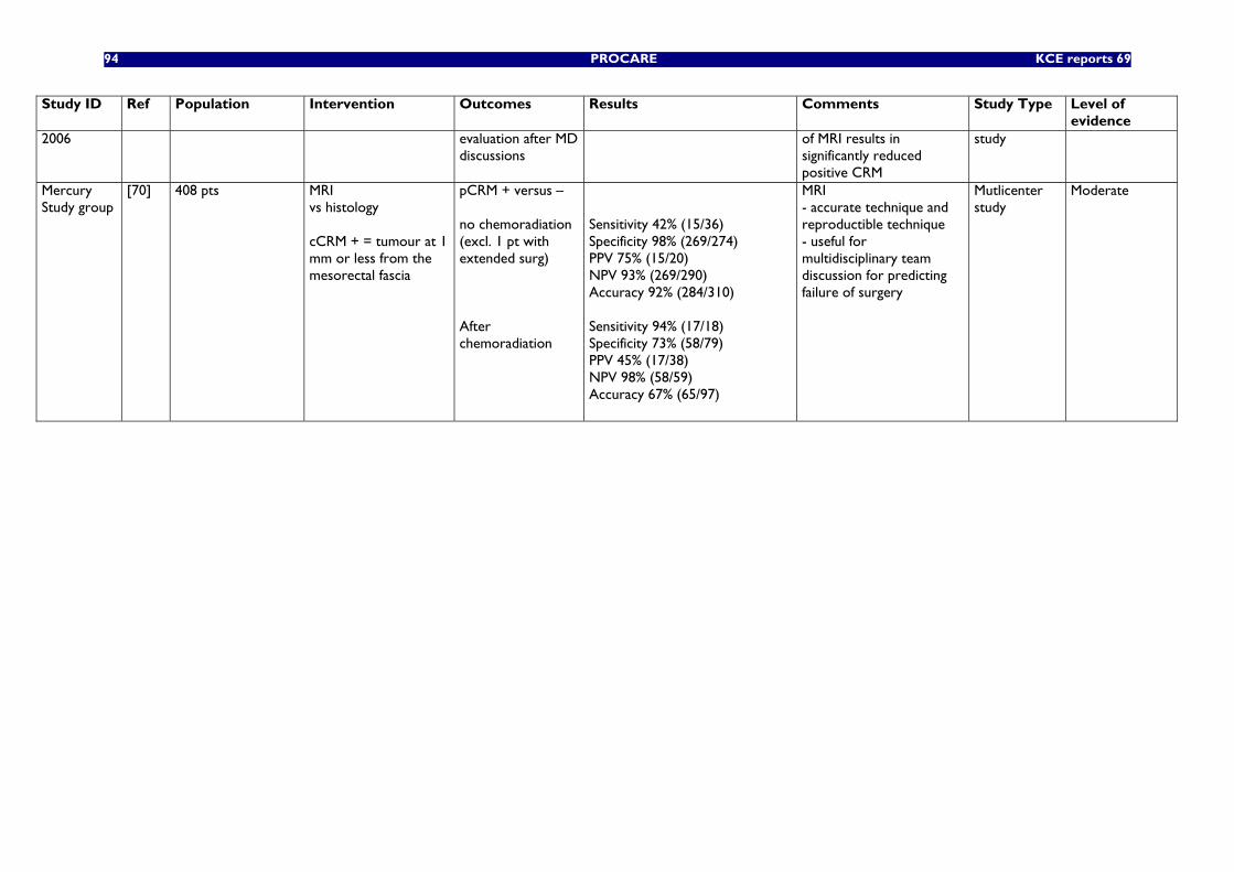
94 PROCARE KCE reports 69
Study ID Ref Population Intervention Outcomes Results Comments Study Type Level of evidence
2006 evaluation after MD discussions
of MRI results in significantly reduced positive CRM
study
Mercury Study group
[70] 408 pts MRI vs histology cCRM + = tumour at 1 mm or less from the mesorectal fascia
pCRM + versus – no chemoradiation (excl. 1 pt with extended surg) After chemoradiation
Sensitivity 42% (15/36) Specificity 98% (269/274) PPV 75% (15/20) NPV 93% (269/290) Accuracy 92% (284/310) Sensitivity 94% (17/18) Specificity 73% (58/79) PPV 45% (17/38) NPV 98% (58/59) Accuracy 67% (65/97)
MRI - accurate technique and reproductible technique - useful for multidisciplinary team discussion for predicting failure of surgery
Mutlicenter study
Moderate
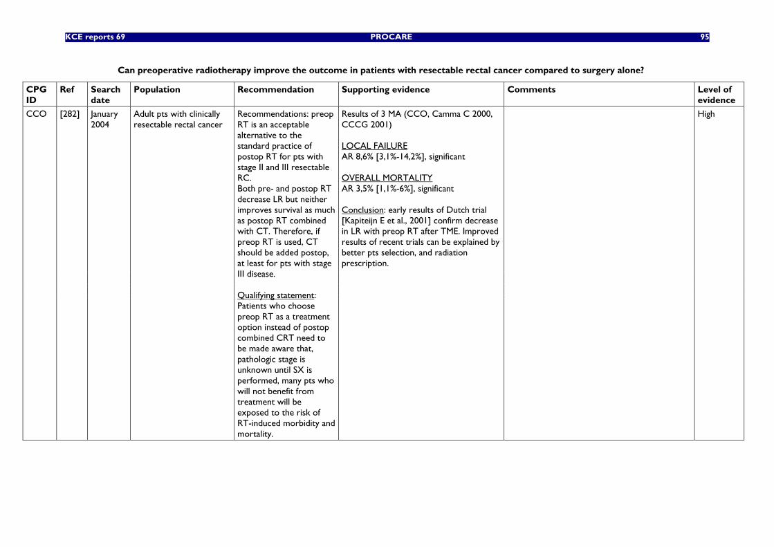
KCE reports 69 PROCARE 95
Can preoperative radiotherapy improve the outcome in patients with resectable rectal cancer compared to surgery alone?
CPG ID
Ref Search date
Population Recommendation Supporting evidence Comments Level of evidence
Results of 3 MA (CCO, Camma C 2000, CCCG 2001) LOCAL FAILURE AR 8,6% [3,1%-14,2%], significant OVERALL MORTALITY AR 3,5% [1,1%-6%], significant Conclusion: early results of Dutch trial [Kapiteijn E et al., 2001] confirm decrease in LR with preop RT after TME. Improved results of recent trials can be explained by better pts selection, and radiation prescription.
High
CCO [282] January 2004
Adult pts with clinically resectable rectal cancer
Recommendations: preop RT is an acceptable alternative to the standard practice of postop RT for pts with stage II and III resectable RC. Both pre- and postop RT decrease LR but neither improves survival as much as postop RT combined with CT. Therefore, if preop RT is used, CT should be added postop, at least for pts with stage III disease. Qualifying statement: Patients who choose preop RT as a treatment option instead of postop combined CRT need to be made aware that, pathologic stage is unknown until SX is performed, many pts who will not benefit from treatment will be exposed to the risk of RT-induced morbidity and mortality.
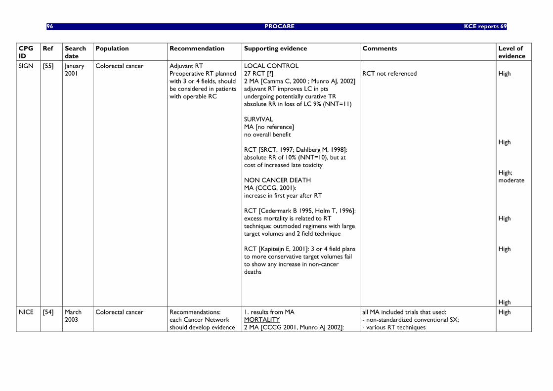
96 PROCARE KCE reports 69
CPG ID
Ref Search date
Population Recommendation Supporting evidence Comments Level of evidence
SIGN [55] January 2001
Colorectal cancer Adjuvant RT Preoperative RT planned with 3 or 4 fields, should be considered in patients with operable RC
LOCAL CONTROL 27 RCT [?] 2 MA [Camma C, 2000 ; Munro AJ, 2002] adjuvant RT improves LC in pts undergoing potentially curative TR absolute RR in loss of LC 9% (NNT=11) SURVIVAL MA [no reference] no overall benefit RCT [SRCT, 1997; Dahlberg M, 1998]: absolute RR of 10% (NNT=10), but at cost of increased late toxicity NON CANCER DEATH MA (CCCG, 2001): increase in first year after RT RCT [Cedermark B 1995, Holm T, 1996]: excess mortality is related to RT technique: outmoded regimens with large target volumes and 2 field technique RCT [Kapiteijn E, 2001]: 3 or 4 field plans to more conservative target volumes fail to show any increase in non-cancer deaths
RCT not referenced
High High High; moderate High High High
NICE [54] March 2003
Colorectal cancer Recommendations: each Cancer Network should develop evidence
1. results from MA MORTALITY 2 MA [CCCG 2001, Munro AJ 2002]:
all MA included trials that used: - non-standardized conventional SX; - various RT techniques
High
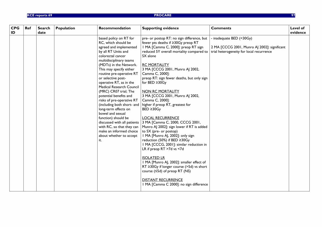
KCE reports 69 PROCARE 97
CPG ID
Ref Search date
Population Recommendation Supporting evidence Comments Level of evidence
based policy on RT for RC, which should be agreed and implemented by all RT Units and colorectal cancer multidisciplinary teams (MDTs) in the Network. This may specify either routine pre-operative RT or selective post-operative RT, as in the Medical Research Council (MRC) CR07 trial; The potential benefits and risks of pre-operative RT (including both short- and long-term effects on bowel and sexual function) should be discussed with all patients with RC, so that they can make an informed choice about whether to accept it.
pre- or postop RT: no sign difference, but fewer pts deaths if ≥30Gy preop RT 1 MA [Camma C, 2000]: preop RT sign reduced 5Y overall mortality compared to SX alone RC MORTALITY 3 MA [CCCG 2001, Munro AJ 2002, Camma C, 2000]: preop RT: sign fewer deaths, but only sign for BED ≥30Gy NON RC MORTALITY 3 MA [CCCG 2001, Munro AJ 2002, Camma C, 2000]: higher if preop RT, greatest for BED ≥30Gy LOCAL RECURRENCE 3 MA [Camma C, 2000, CCCG 2001, Munro AJ 2002]: sign lower if RT is added to SX (pre- or postop) 1 MA [Munro AJ, 2002]: only sign reduction (50%) if BED ≥30Gy 1 MA [CCCG, 2001]: similar reduction in LR if preop RT >7d vs <7d ISOLATED LR 1 MA [Munro AJ, 2002]: smaller effect of RT ≥30Gy if longer course (>5d) vs short course (≤5d) of preop RT (NS) DISTANT RECURRENCE 1 MA [Camma C 2000]: no sign difference
- inadequate BED (<30Gy) 2 MA [CCCG 2001, Munro AJ 2002]: significant trial heterogeneity for local recurrence
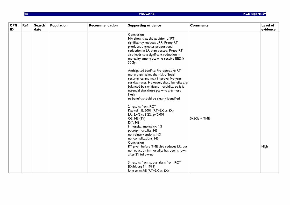
98 PROCARE KCE reports 69
CPG ID
Ref Search date
Population Recommendation Supporting evidence Comments Level of evidence
Conclusion: MA show that the addition of RT significantly reduces LRR. Preop RT produces a greater proportional reduction in LR than postop. Preop RT also leads to a significant reduction in mortality among pts who receive BED ≥ 30Gy Anticipated benifits: Pre-operative RT more than halves the risk of local recurrence and may improve five-year survival rates. However, these benefits are balanced by significant morbidity, so it is essential that those pts who are most likely to benefit should be clearly identified. 2. results from RCT Kapiteijn E, 2001 (RT+SX vs SX) LR: 2,4% vs 8,2%, p<0,001 OS: NS (2Y) DM: NS in hospital mortality: NS postop mortality: NS no. reinterventions: NS no. complications: NS Conclusion RT given before TME also reduces LR, but no reduction in mortality has been shown after 2Y follow-up 3. results from sub-analysis from RCT [Dahlberg M, 1998] long term AE (RT+SX vs SX)
5x5Gy + TME
High
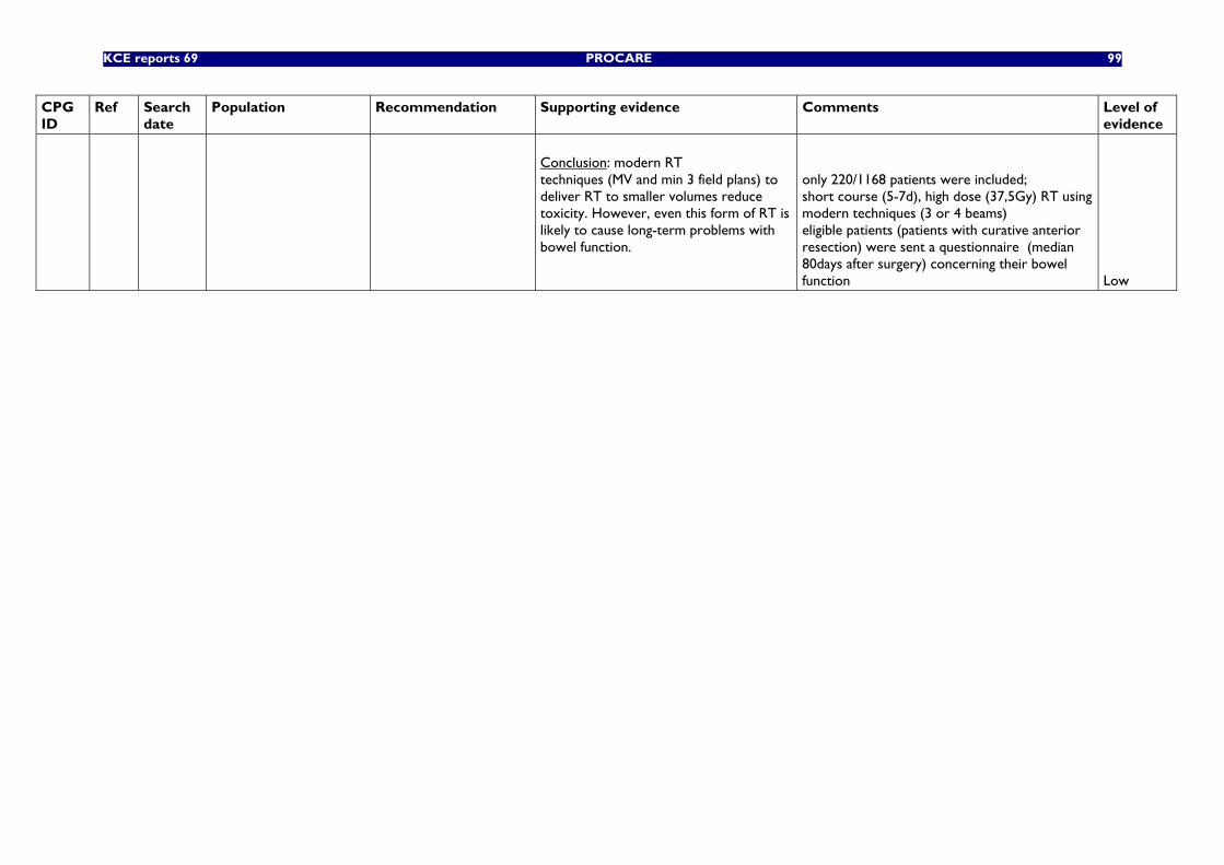
KCE reports 69 PROCARE 99
CPG ID
Ref Search date
Population Recommendation Supporting evidence Comments Level of evidence
Conclusion: modern RT techniques (MV and min 3 field plans) to deliver RT to smaller volumes reduce toxicity. However, even this form of RT is likely to cause long-term problems with bowel function.
only 220/1168 patients were included; short course (5-7d), high dose (37,5Gy) RT using modern techniques (3 or 4 beams) eligible patients (patients with curative anterior resection) were sent a questionnaire (median 80days after surgery) concerning their bowel function
Low
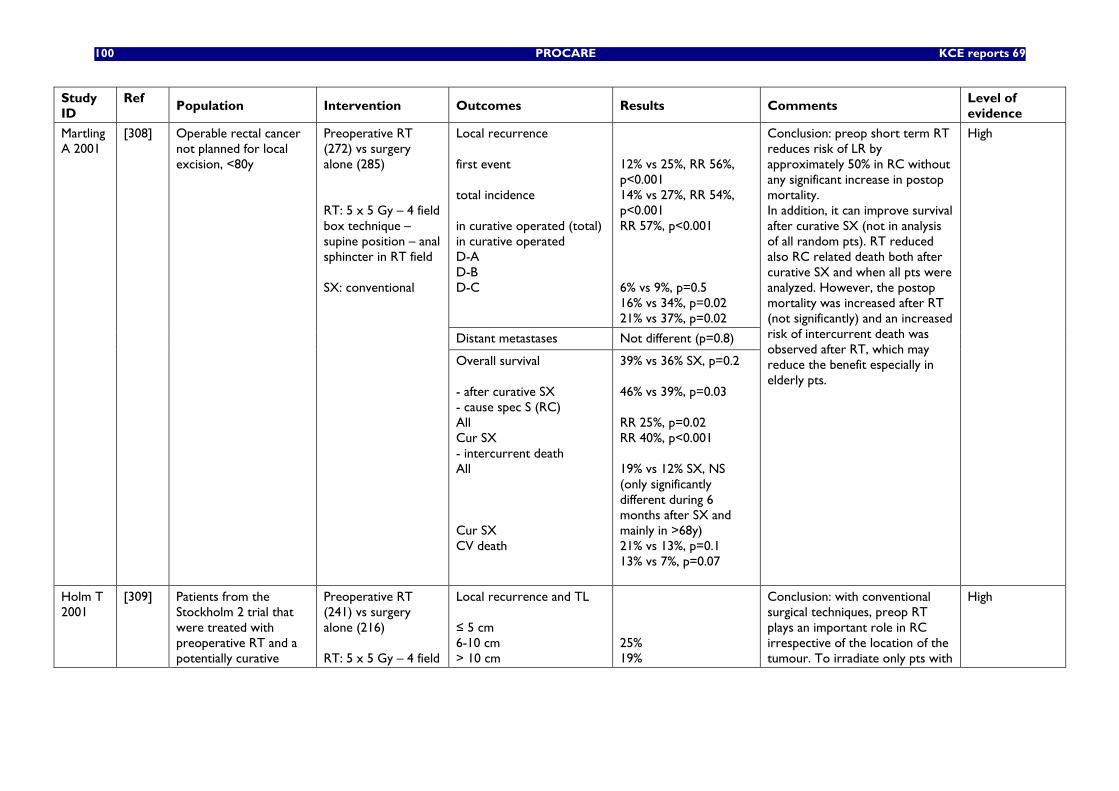
100 PROCARE KCE reports 69
Study ID
Ref Population Intervention Outcomes Results Comments
Level of evidence
Local recurrence first event total incidence in curative operated (total) in curative operated D-A D-B D-C
12% vs 25%, RR 56%, p<0.001 14% vs 27%, RR 54%, p<0.001 RR 57%, p<0.001 6% vs 9%, p=0.5 16% vs 34%, p=0.02 21% vs 37%, p=0.02
Distant metastases Not different (p=0.8)
Martling A 2001
[308] Operable rectal cancer not planned for local excision, <80y
Preoperative RT (272) vs surgery alone (285) RT: 5 x 5 Gy – 4 field box technique – supine position – anal sphincter in RT field SX: conventional
Overall survival - after curative SX - cause spec S (RC) All Cur SX - intercurrent death All Cur SX CV death
39% vs 36% SX, p=0.2 46% vs 39%, p=0.03 RR 25%, p=0.02 RR 40%, p<0.001 19% vs 12% SX, NS (only significantly different during 6 months after SX and mainly in >68y) 21% vs 13%, p=0.1 13% vs 7%, p=0.07
Conclusion: preop short term RT reduces risk of LR by approximately 50% in RC without any significant increase in postop mortality. In addition, it can improve survival after curative SX (not in analysis of all random pts). RT reduced also RC related death both after curative SX and when all pts were analyzed. However, the postop mortality was increased after RT (not significantly) and an increased risk of intercurrent death was observed after RT, which may reduce the benefit especially in elderly pts.
High
Holm T 2001
[309] Patients from the Stockholm 2 trial that were treated with preoperative RT and a potentially curative
Preoperative RT (241) vs surgery alone (216) RT: 5 x 5 Gy – 4 field
Local recurrence and TL ≤ 5 cm 6-10 cm > 10 cm
25% 19%
Conclusion: with conventional surgical techniques, preop RT plays an important role in RC irrespective of the location of the tumour. To irradiate only pts with
High
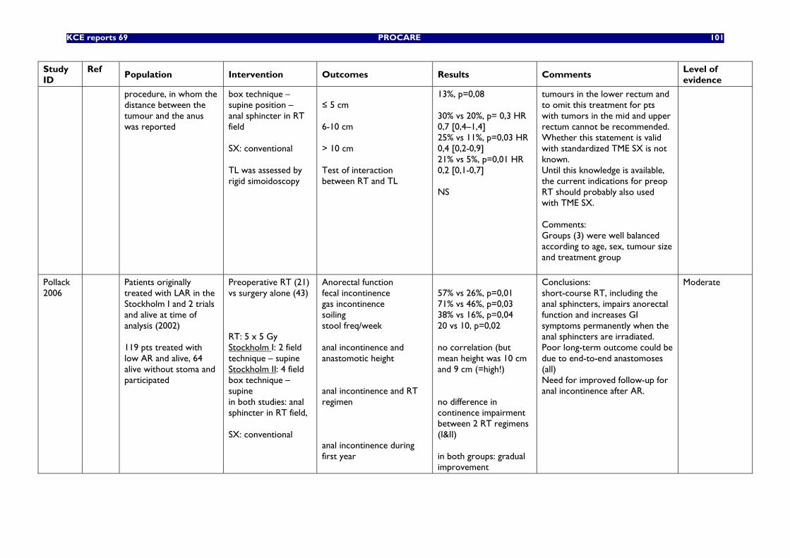
KCE reports 69 PROCARE 101
Study ID
Ref Population Intervention Outcomes Results Comments
Level of evidence
procedure, in whom the distance between the tumour and the anus was reported
box technique – supine position – anal sphincter in RT field SX: conventional TL was assessed by rigid simoidoscopy
≤ 5 cm 6-10 cm > 10 cm Test of interaction between RT and TL
13%, p=0,08 30% vs 20%, p= 0,3 HR 0,7 [0,4–1,4] 25% vs 11%, p=0,03 HR 0,4 [0,2-0,9] 21% vs 5%, p=0,01 HR 0,2 [0,1-0,7] NS
tumours in the lower rectum and to omit this treatment for pts with tumors in the mid and upper rectum cannot be recommended. Whether this statement is valid with standardized TME SX is not known. Until this knowledge is available, the current indications for preop RT should probably also used with TME SX. Comments: Groups (3) were well balanced according to age, sex, tumour size and treatment group
Pollack 2006
Patients originally treated with LAR in the Stockholm I and 2 trials and alive at time of analysis (2002) 119 pts treated with low AR and alive, 64 alive without stoma and participated
Preoperative RT (21) vs surgery alone (43) RT: 5 x 5 Gy Stockholm I: 2 field technique – supine Stockholm II: 4 field box technique – supine in both studies: anal sphincter in RT field, SX: conventional
Anorectal function fecal incontinence gas incontinence soiling stool freq/week anal incontinence and anastomotic height anal incontinence and RT regimen anal incontinence during first year
57% vs 26%, p=0,01 71% vs 46%, p=0,03 38% vs 16%, p=0,04 20 vs 10, p=0,02 no correlation (but mean height was 10 cm and 9 cm (=high!) no difference in continence impairment between 2 RT regimens (I&II) in both groups: gradual improvement
Conclusions: short-course RT, including the anal sphincters, impairs anorectal function and increases GI symptoms permanently when the anal sphincters are irradiated. Poor long-term outcome could be due to end-to-end anastomoses (all) Need for improved follow-up for anal incontinence after AR.
Moderate
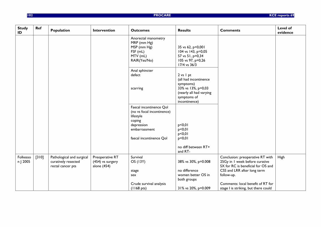
102 PROCARE KCE reports 69
Study ID
Ref Population Intervention Outcomes Results Comments
Level of evidence
Anorectal manometry MRP (mm Hg) MSP (mm Hg) FSF (mL) MTV (mL) RAIR(Yes/No)
35 vs 62, p<0,001 104 vs 143, p=0,05 57 vs 51, p=0,34 105 vs 97, p=0,26 17/4 vs 36/3
Anal sphincter defect scarring
2 vs 1 pt (all had incontinence symptoms) 33% vs 13%, p=0,03 (nearly all had varying symptoms of incontinence)
Faecal incontinence Qol (no vs focal incontinence) lifestyle coping depression embarrassment faecal incontinence Qol
p<0,01 p<0,01 p<0,01 p<0,01 no diff between RT+ and RT-
Folkesson J 2005
[310] Pathological and surgical curatively resected rectal cancer pts
Preoperative RT (454) vs surgery alone (454)
Survival OS (13Y) stage sex Crude survival analysis (1168 pts)
38% vs 30%, p=0.008 no difference women better OS in both groups 31% vs 20%, p=0.009
Conclusion: preoperative RT with 25Gy in 1 week before curative SX for RC is beneficial for OS and CSS and LRR after long term follow-up. Comments: local benefit of RT for stage I is striking, but there could
High
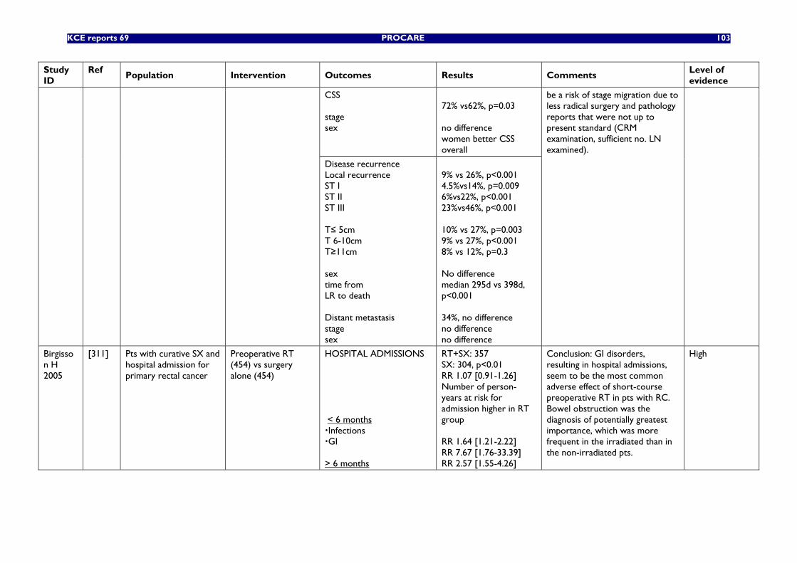
KCE reports 69 PROCARE 103
Study ID
Ref Population Intervention Outcomes Results Comments
Level of evidence
CSS stage sex
72% vs62%, p=0.03 no difference women better CSS overall
Disease recurrence Local recurrence ST I ST II ST III T≤ 5cm T 6-10cm T≥11cm sex time from LR to death Distant metastasis stage sex
9% vs 26%, p<0.001 4.5%vs14%, p=0.009 6%vs22%, p<0.001 23%vs46%, p<0.001 10% vs 27%, p=0.003 9% vs 27%, p<0.001 8% vs 12%, p=0.3 No difference median 295d vs 398d, p<0.001 34%, no difference no difference no difference
be a risk of stage migration due to less radical surgery and pathology reports that were not up to present standard (CRM examination, sufficient no. LN examined).
Birgisson H 2005
[311] Pts with curative SX and hospital admission for primary rectal cancer
Preoperative RT (454) vs surgery alone (454)
HOSPITAL ADMISSIONS < 6 months Infections GI
> 6 months
RT+SX: 357 SX: 304, p<0.01 RR 1.07 [0.91-1.26] Number of person-years at risk for admission higher in RT group RR 1.64 [1.21-2.22] RR 7.67 [1.76-33.39] RR 2.57 [1.55-4.26]
Conclusion: GI disorders, resulting in hospital admissions, seem to be the most common adverse effect of short-course preoperative RT in pts with RC. Bowel obstruction was the diagnosis of potentially greatest importance, which was more frequent in the irradiated than in the non-irradiated pts.
High
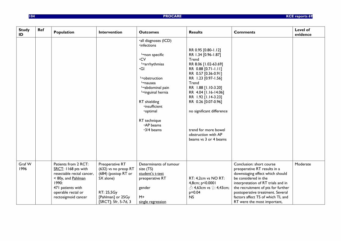
104 PROCARE KCE reports 69
Study ID
Ref Population Intervention Outcomes Results Comments
Level of evidence
all diagnoses (ICD) infections
non specific CV
arrhythmias GI
obstruction nausea abdominal pain inguinal hernia RT shielding insufficient optimal RT technique AP beams 3/4 beams
RR 0.95 [0.80-1.12] RR 1.34 [0.96-1.87] Trend RR 8.06 [1.02-63.69] RR 0.88 [0.71-1.11] RR 0.57 [0.36-0.91] RR 1.23 [0.97-1.56] Trend RR 1.88 [1.10-3.20] RR 4.04 [1.16-14.06] RR 1.92 [1.14-3.23] RR 0.26 [0.07-0.96] no significant difference trend for more bowel obstruction with AP beams vs 3 or 4 beams
Graf W 1996
Patients from 2 RCT: SRCT: 1168 pts with resectable rectal cancer, < 80y, and Pahlman 1990: 471 patients with operable rectal or rectosigmoid cancer
Preoperative RT (632) vs no preop RT (684) (postop RT or SX alone) RT: 25,5Gy [Pahlman] or 25Gy [SRCT]; 5fr, 5-7d, 3
Determinants of tumour size (TS) student’s t-test preoperative RT gender M+ single regression
RT: 4,2cm vs NO RT: 4,8cm; p<0.0001 ♂: 4,63cm vs ♀: 4,43cm; p=0.04 NS
Conclusion: short course preoperative RT results in a downstaging effect which should be considered in the interpretation of RT trials and in the recruitment of pts for further postoperative treatment. Several factors affect TS of which TL and RT were the most important,
Moderate
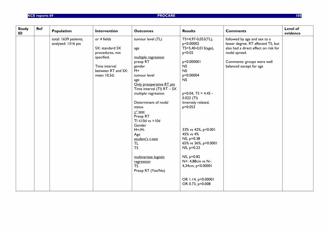
KCE reports 69 PROCARE 105
Study ID
Ref Population Intervention Outcomes Results Comments
Level of evidence
total: 1639 patients, analyzed: 1316 pts
or 4 fields SX: standard SX procedures, not specified, Time interval between RT and SX: mean 10,5d;
tumour level (TL) age multiple regression preop RT gender M+ tumour level age Only preoperative RT pts Time interval (TI) RT – SX multiple regression Determinant of nodal status χ² test Preop RT TI ≤10d vs >10d Gender M+/M- Age student’s t-test TL TS multivariate logistic regression TS Preop RT (Yes/No)
TS=4,97-0,053(TL), p<0.00002 TS=5,40-0,013(age), p<0.02 p<0.000001 NS NS p<0.00004 NS p=0.04, TS = 4.45 - 0.022 (TI) Inversely related, p=0.053 33% vs 42%, p<0.001 45% vs 4% NS, p=0.38 65% vs 36%, p<0.0001 NS, p=0.23 NS, p=0.82 N+: 4,88cm vs N-: 4,34cm, p<0.00001 OR 1.14, p<0.00001 OR 0.73, p=0.008
followed by age and sex to a lesser degree. RT affected TS, but also had a direct effect on risk for nodal spread. Comments: groups were well balanced except for age
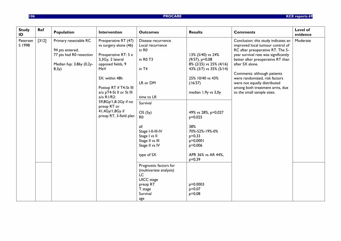
106 PROCARE KCE reports 69
Study ID
Ref Population Intervention Outcomes Results Comments
Level of evidence
Disease recurrence Local recurrence in R0 in R0 T3 in T4 LR or DM time to LR
13% (5/40) vs 24% (9/37), p=0,08 8% (2/25) vs 25% (4/16) 43% (3/7) vs 35% (5/14) 25% 10/40 vs 43% (16/37) median 1,9y vs 3,0y
[312]
Survival OS (5y) R0 all Stage I-II-III-IV Stage I vs II Stage II vs III Stage II vs IV type of SX
49% vs 28%, p=0,027 p=0,025 38% 70%-52%-19%-0% p=0,33 p=0,0001 p=0,006 APR 36% vs AR 44%, p=0,39
Moderate Petersen S 1998
Primary resectable RC 94 pts entered, 77 pts had R0 resection Median fup: 3,86y (0,2y-8,3y)
Preoperative RT (47) vs surgery alone (46) Preoperative RT: 5 x 3,3Gy, 2 lateral opposed fields, 9 MeV SX: within 48h Postop RT if T4-St III a/o pT4-St II or St III a/o R1/R2: 59,8Gy/1,8-2Gy if no preop RT or 41,4Gy/1,8Gy if preop RT, 3-field plan
Prognostic factors for (multivariate analysis) LC UICC stage preop RT T stage Survival age
p=0.0003 p=0.07 p=0,08
Conclusion: this study indicates an improved local tumour control of RC after preoperative RT. The 5-year survival rate was significantly better after preoperative RT than after SX alone. Comments: although patients were randomized, risk factors were not equally distributed among both treatment arms, due to the small sample sizes.
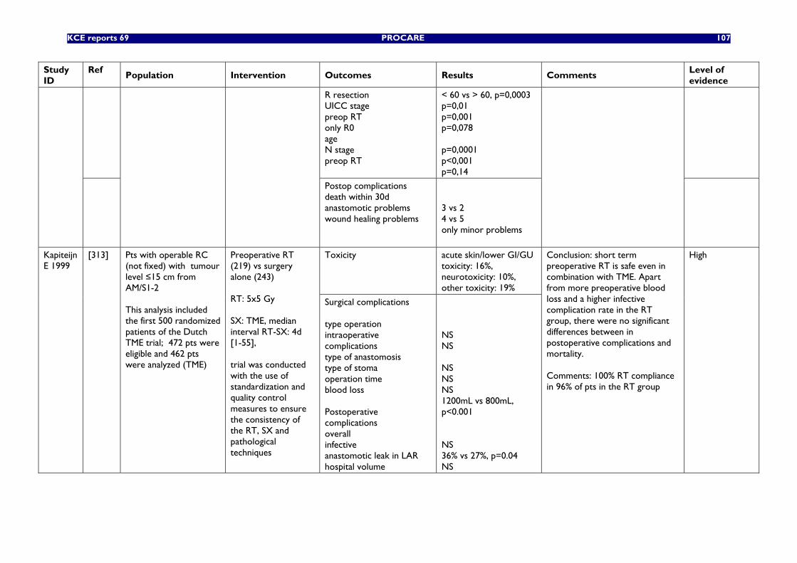
KCE reports 69 PROCARE 107
Study ID
Ref Population Intervention Outcomes Results Comments
Level of evidence
R resection UICC stage preop RT only R0 age N stage preop RT
< 60 vs > 60, p=0,0003 p=0,01 p=0,001 p=0,078 p=0,0001 p<0,001 p=0,14
Postop complications death within 30d anastomotic problems wound healing problems
3 vs 2 4 vs 5 only minor problems
Toxicity acute skin/lower GI/GU toxicity: 16%, neurotoxicity: 10%, other toxicity: 19%
Kapiteijn E 1999
[313] Pts with operable RC (not fixed) with tumour level ≤15 cm from AM/S1-2 This analysis included the first 500 randomized patients of the Dutch TME trial; 472 pts were eligible and 462 pts were analyzed (TME)
Preoperative RT (219) vs surgery alone (243) RT: 5x5 Gy SX: TME, median interval RT-SX: 4d [1-55], trial was conducted with the use of standardization and quality control measures to ensure the consistency of the RT, SX and pathological techniques
Surgical complications type operation intraoperative complications type of anastomosis type of stoma operation time blood loss Postoperative complications overall infective anastomotic leak in LAR hospital volume
NS NS NS NS NS 1200mL vs 800mL, p<0.001 NS 36% vs 27%, p=0.04 NS
Conclusion: short term preoperative RT is safe even in combination with TME. Apart from more preoperative blood loss and a higher infective complication rate in the RT group, there were no significant differences between in postoperative complications and mortality. Comments: 100% RT compliance in 96% of pts in the RT group
High
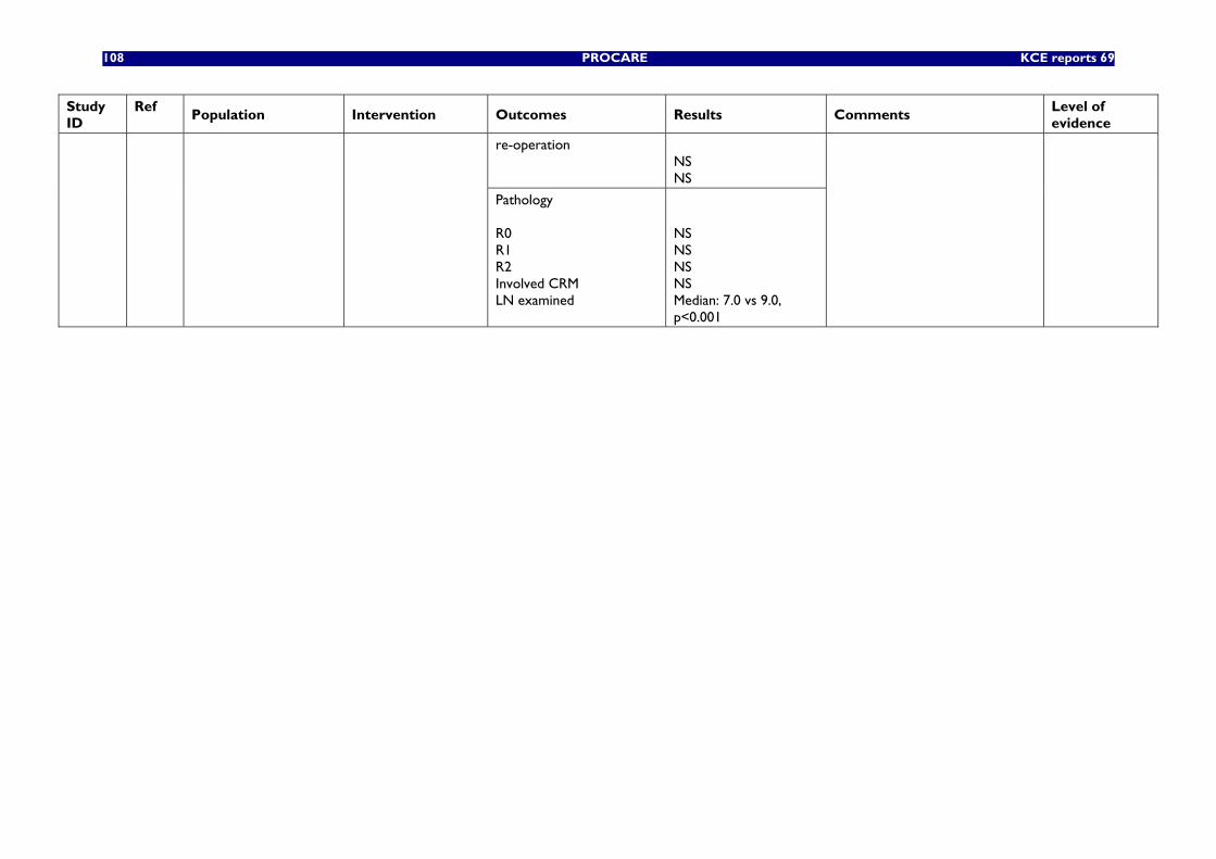
108 PROCARE KCE reports 69
Study ID
Ref Population Intervention Outcomes Results Comments
Level of evidence
re-operation NS NS
Pathology R0 R1 R2 Involved CRM LN examined
NS NS NS NS Median: 7.0 vs 9.0, p<0.001
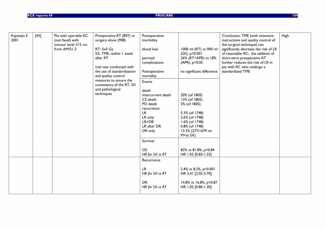
KCE reports 69 PROCARE 109
Postoperative morbidity blood loss perineal complications Postoperative mortality
1000 ml (RT) vs 900 ml (SX), p<0.001 26% (RT+APR) vs 18% (APR), p=0.05 no significant difference
Events death intercurrent death CS death PO death recurrence LR LR only LR+DR LR after DR DR only
20% (of 1805) 13% (of 1805) 3% (of 1805) 5.3% (of 1748) 2.6% (of 1748) 1.6% (of 1748) 0.8% (of 1748) 13.5% (277/1679 no M+at SX)
Kapiteijn E 2001
[45] Pts with operable RC (not fixed) with tumour level ≤15 cm from AM/S1-2
Preoperative RT (897) vs surgery alone (908) RT: 5x5 Gy SX: TME, within 1 week after RT trial was conducted with the use of standardization and quality control measures to ensure the consistency of the RT, SX and pathological techniques
Survival OS HR for SX vs RT
82% vs 81.8%, p=0.84 HR 1.02 [0.83-1.25]
Conclusion: TME (with extensive instructions and quality control of the surgical technique) can significantly decrease the risk of LR of resectable RC; the addition of short-term preoperative RT further reduces the risk of LR in pts with RC who undergo a standardized TME
High
Recurrence LR HR for SX vs RT DR HR for SX vs RT
2.4% vs 8.2%, p<0.001 HR 3.41 [2.05-5.70] 14.8% vs 16.8%, p=0.87 HR 1.02 [0.80-1.30]
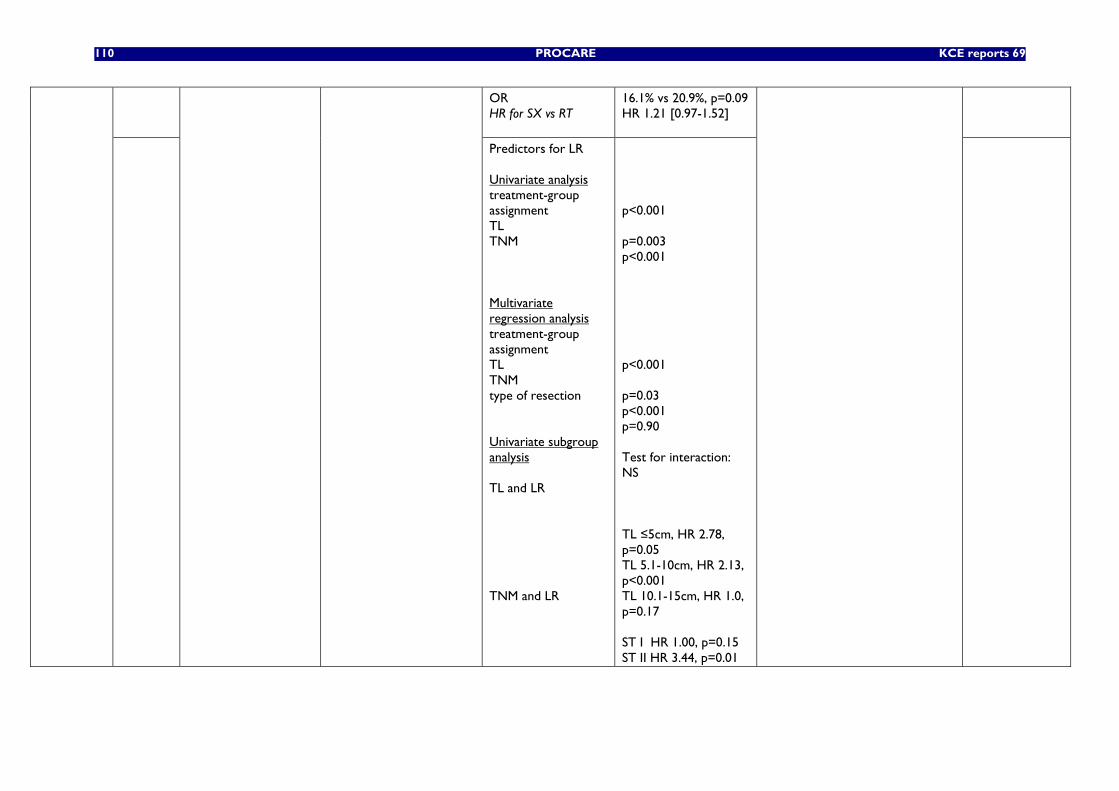
110 PROCARE KCE reports 69
OR HR for SX vs RT
16.1% vs 20.9%, p=0.09 HR 1.21 [0.97-1.52]
Predictors for LR Univariate analysis treatment-group assignment TL TNM Multivariate regression analysis treatment-group assignment TL TNM type of resection Univariate subgroup analysis TL and LR TNM and LR
p<0.001 p=0.003 p<0.001 p<0.001 p=0.03 p<0.001 p=0.90 Test for interaction: NS TL ≤5cm, HR 2.78, p=0.05 TL 5.1-10cm, HR 2.13, p<0.001 TL 10.1-15cm, HR 1.0, p=0.17 ST I HR 1.00, p=0.15 ST II HR 3.44, p=0.01
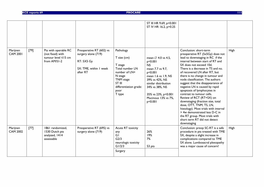
KCE reports 69 PROCARE 111
ST III HR 9.69, p<0.001 ST IV HR 16.2, p=0.25
Marijnen CAM 2001
[79] Pts with operable RC (not fixed) with tumour level ≤15 cm from AM/S1-2
Preoperative RT (602) vs surgery alone (719) RT: 5X5 Gy SX: TME, within 1 week after RT
Pathology T size (cm) T stage Total number LN number of LN+ N stage TNM stage ST III differentiation grade: poor T type
mean ∅ 4.0 vs 4.5, p<0.001 NS mean 7.7 vs 9.7, p<0.001 mean 1.6 vs 1.9, NS 39% vs 42%, NS similar distribution 34% vs 38%, NS 35% vs 23%, p<0.001 Mucinous 13% vs 7%, p<0.001
Conclusion: short-term preoperative RT (5x5Gy) does not lead to downstaging in RC if the interval between start of RT and SX does not exceed 10d. There is a decrease in TS and no. of recovered LN after RT, but there is no change in tumour and node classification. The authors suggest that the disappearance of negative LN is caused by rapid apoptosis of lymphocytes in contrast to tumour cells. Review of RCT (RT+SX) on downstaging (fraction size, total dose, OTT, TNM, TS, LN, histology). Most trials with interval > 4w demonstrated less D-C in the RT group. Most trials with short term RT did not detect downstaging
High
Acute RT toxicity any G1 G2/3 neurologic toxicity G1/2/3
26% 19% 7% 53 pts
Marijnen CAM 2002
[77] 1861 randomized, 1530 Dutch pts analyzed, 1414 assessable
Preoperative RT (695) vs surgery alone (719)
Surgery
Conclusion: preop SC-RT is a safe procedure in pts treated with TME SX, despite a slight increase in complications compared to TME SX alone. Lumbosacral plexopathy was a major cause of concern!
High
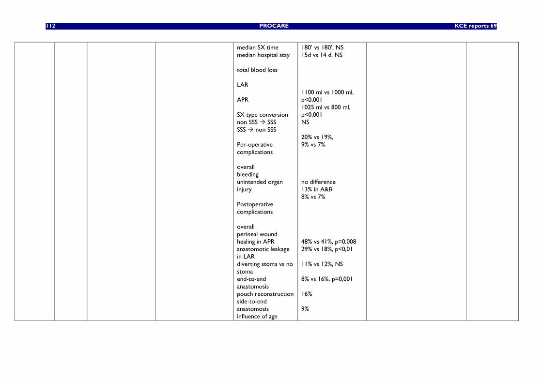
112 PROCARE KCE reports 69
median SX time median hospital stay total blood loss LAR APR SX type conversion non SSS SSS SSS non SSS Per-operative complications overall bleeding unintended organ injury Postoperative complications overall perineal wound healing in APR anastomotic leakage in LAR diverting stoma vs no stoma end-to-end anastomosis pouch reconstruction side-to-end anastomosis influence of age
180’ vs 180’, NS 15d vs 14 d, NS 1100 ml vs 1000 ml, p<0,001 1025 ml vs 800 ml, p<0,001 NS 20% vs 19%, 9% vs 7% no difference 13% in A&B 8% vs 7% 48% vs 41%, p=0,008 29% vs 18%, p<0,01 11% vs 12%, NS 8% vs 16%, p=0,001 16% 9%
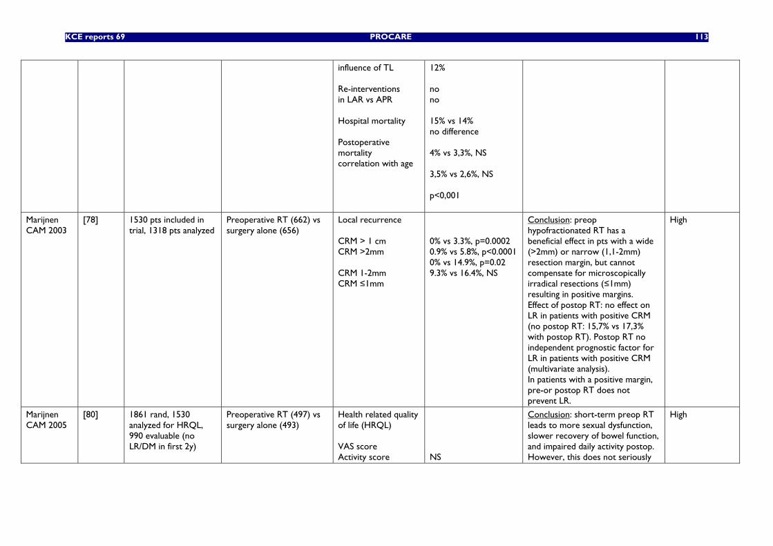
KCE reports 69 PROCARE 113
influence of TL Re-interventions in LAR vs APR Hospital mortality Postoperative mortality correlation with age
12% no no 15% vs 14% no difference 4% vs 3,3%, NS 3,5% vs 2,6%, NS p<0,001
Marijnen CAM 2003
[78] 1530 pts included in trial, 1318 pts analyzed
Preoperative RT (662) vs surgery alone (656)
Local recurrence CRM > 1 cm CRM >2mm CRM 1-2mm CRM ≤1mm
0% vs 3.3%, p=0.0002 0.9% vs 5.8%, p<0.0001 0% vs 14.9%, p=0.02 9.3% vs 16.4%, NS
Conclusion: preop hypofractionated RT has a beneficial effect in pts with a wide (>2mm) or narrow (1,1-2mm) resection margin, but cannot compensate for microscopically irradical resections (≤1mm) resulting in positive margins. Effect of postop RT: no effect on LR in patients with positive CRM (no postop RT: 15,7% vs 17,3% with postop RT). Postop RT no independent prognostic factor for LR in patients with positive CRM (multivariate analysis). In patients with a positive margin, pre-or postop RT does not prevent LR.
High
Marijnen CAM 2005
[80] 1861 rand, 1530 analyzed for HRQL, 990 evaluable (no LR/DM in first 2y)
Preoperative RT (497) vs surgery alone (493)
Health related quality of life (HRQL) VAS score Activity score
NS
Conclusion: short-term preop RT leads to more sexual dysfunction, slower recovery of bowel function, and impaired daily activity postop. However, this does not seriously
High
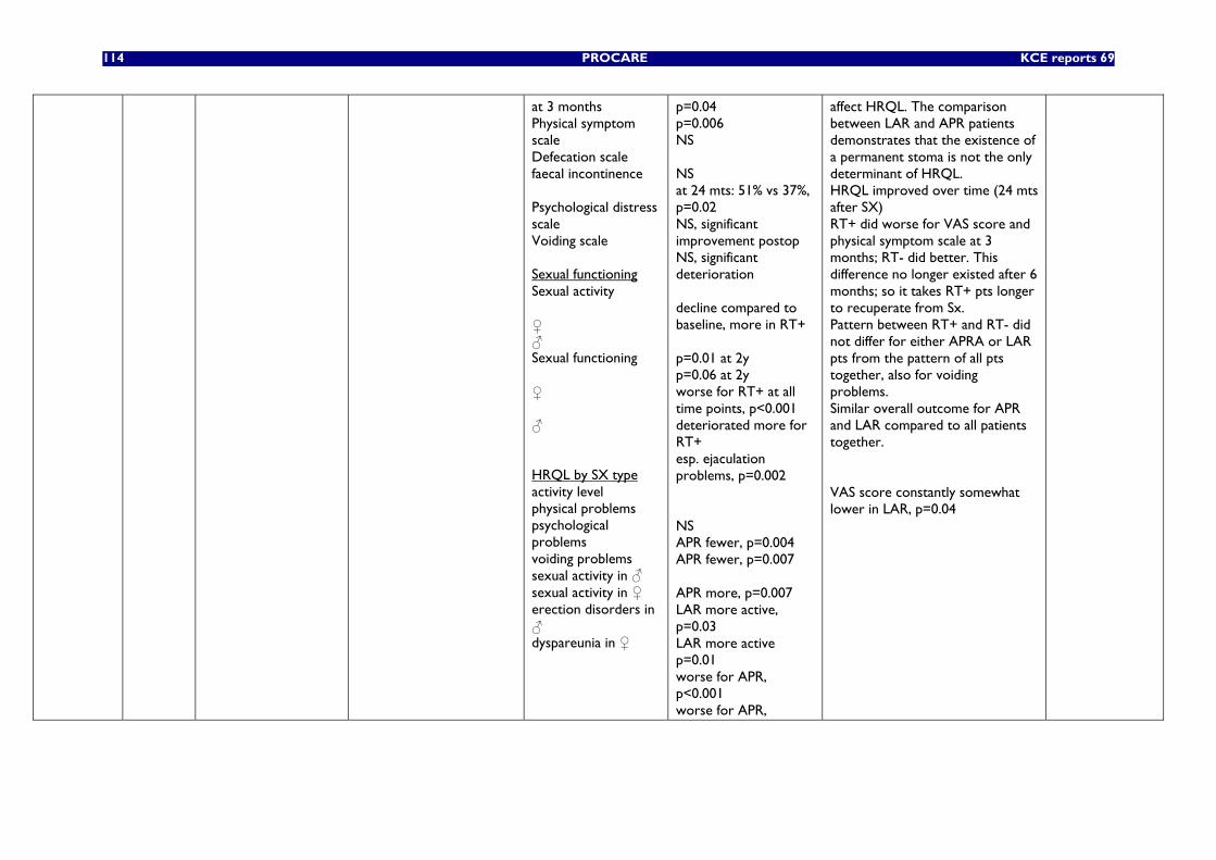
114 PROCARE KCE reports 69
at 3 months Physical symptom scale Defecation scale faecal incontinence Psychological distress scale Voiding scale Sexual functioning Sexual activity ♀ ♂ Sexual functioning ♀ ♂ HRQL by SX type activity level physical problems psychological problems voiding problems sexual activity in ♂ sexual activity in ♀ erection disorders in ♂ dyspareunia in ♀
p=0.04 p=0.006 NS NS at 24 mts: 51% vs 37%, p=0.02 NS, significant improvement postop NS, significant deterioration decline compared to baseline, more in RT+ p=0.01 at 2y p=0.06 at 2y worse for RT+ at all time points, p<0.001 deteriorated more for RT+ esp. ejaculation problems, p=0.002 NS APR fewer, p=0.004 APR fewer, p=0.007 APR more, p=0.007 LAR more active, p=0.03 LAR more active p=0.01 worse for APR, p<0.001 worse for APR,
affect HRQL. The comparison between LAR and APR patients demonstrates that the existence of a permanent stoma is not the only determinant of HRQL. HRQL improved over time (24 mts after SX) RT+ did worse for VAS score and physical symptom scale at 3 months; RT- did better. This difference no longer existed after 6 months; so it takes RT+ pts longer to recuperate from Sx. Pattern between RT+ and RT- did not differ for either APRA or LAR pts from the pattern of all pts together, also for voiding problems. Similar overall outcome for APR and LAR compared to all patients together. VAS score constantly somewhat lower in LAR, p=0.04
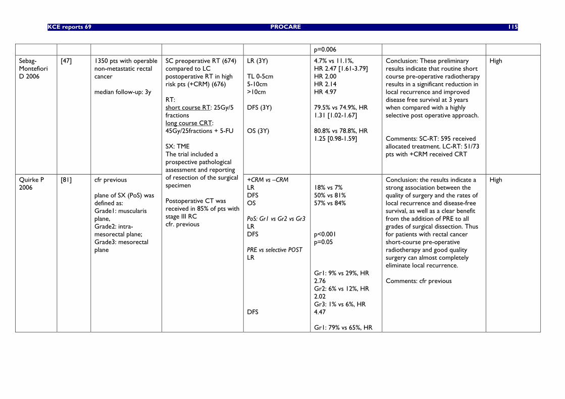
KCE reports 69 PROCARE 115
p=0.006
Sebag-Montefiori D 2006
[47] 1350 pts with operable non-metastatic rectal cancer median follow-up: 3y
LR (3Y) TL 0-5cm 5-10cm >10cm DFS (3Y) OS (3Y)
4.7% vs 11.1%, HR 2.47 [1.61-3.79] HR 2.00 HR 2.14 HR 4.97 79.5% vs 74.9%, HR 1.31 [1.02-1.67] 80.8% vs 78.8%, HR 1.25 [0.98-1.59]
Conclusion: These preliminary results indicate that routine short course pre-operative radiotherapy results in a significant reduction in local recurrence and improved disease free survival at 3 years when compared with a highly selective post operative approach. Comments: SC-RT: 595 received allocated treatment. LC-RT: 51/73 pts with +CRM received CRT
High
Quirke P 2006
[81] cfr previous plane of SX (PoS) was defined as: Grade1: muscularis plane, Grade2: intra-mesorectal plane; Grade3: mesorectal plane
SC preoperative RT (674) compared to LC postoperative RT in high risk pts (+CRM) (676) RT: short course RT: 25Gy/5 fractions long course CRT: 45Gy/25fractions + 5-FU SX: TME The trial included a prospective pathological assessment and reporting of resection of the surgical specimen Postoperative CT was received in 85% of pts with stage III RC cfr. previous
+CRM vs –CRM LR DFS OS PoS: Gr1 vs Gr2 vs Gr3 LR DFS PRE vs selective POST LR DFS
18% vs 7% 50% vs 81% 57% vs 84% p<0.001 p=0.05 Gr1: 9% vs 29%, HR 2.76 Gr2: 6% vs 12%, HR 2.02 Gr3: 1% vs 6%, HR 4.47 Gr1: 79% vs 65%, HR
Conclusion: the results indicate a strong association between the quality of surgery and the rates of local recurrence and disease-free survival, as well as a clear benefit from the addition of PRE to all grades of surgical dissection. Thus for patients with rectal cancer short-course pre-operative radiotherapy and good quality surgery can almost completely eliminate local recurrence. Comments: cfr previous
High
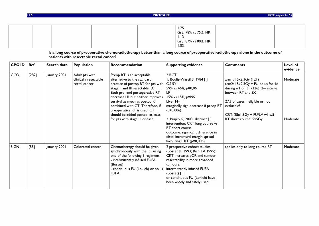
116 PROCARE KCE reports 69
1.75 Gr2: 78% vs 75%, HR 1.13 Gr3: 87% vs 80%, HR 1.53
Is a long course of preoperative chemoradiotherapy better than a long course of preoperative radiotherapy alone in the outcome of patients with resectable rectal cancer?
CPG ID Ref Search date Population Recommendation Supporting evidence
Comments Level of evidence
CCO [282] January 2004 Adult pts with clinically resectable rectal cancer
Preop RT is an acceptable alternative to the standard practice of postop RT for pts with stage II and III resectable RC. Both pre- and postoperative RT decrease LR but neither improves survival as much as postop RT combined with CT. Therefore, if preoperative RT is used, CT should be added postop, at least for pts with stage III disease
2 RCT 1. Boulis-Wassif S, 1984 [ ] OS 5Y 59% vs 46%, p=0,06 LF 15% vs 15%, p=NS Liver M+ marginally sign decrease if preop RT (p=0,006) 2. Buijko K, 2003, abstract [ ] intervention: CRT long course vs RT short course outcome: significant difference in distal intramural margin spread favouring CRT (p=0,006)
arm1: 15x2,3Gy (121) arm2: 15x2,3Gy + FU bolus for 4d during w1 of RT (126); 2w interval between RT and SX 27% of cases ineligible or not evaluable! CRT: 28x1,8Gy + FU/LV w1,w5 RT short course: 5x5Gy
Moderate Moderate
SIGN [55] January 2001 Colorectal cancer Chemotherapy should be given synchronously with the RT using one of the following 3 regimens: - intermittently infused FUFA (Bosset) - continuous FU (Lokich) or bolus FUFA
2 prospective cohort studies (Bosset JF, 1993; Rich TA 1995): CRT increases pCR and tumour resectability in more advanced tumours; intermittently infused FUFA (Bosset) [ ] or continuous FU (Lokich) have been widely and safely used
applies only to long course RT
Moderate
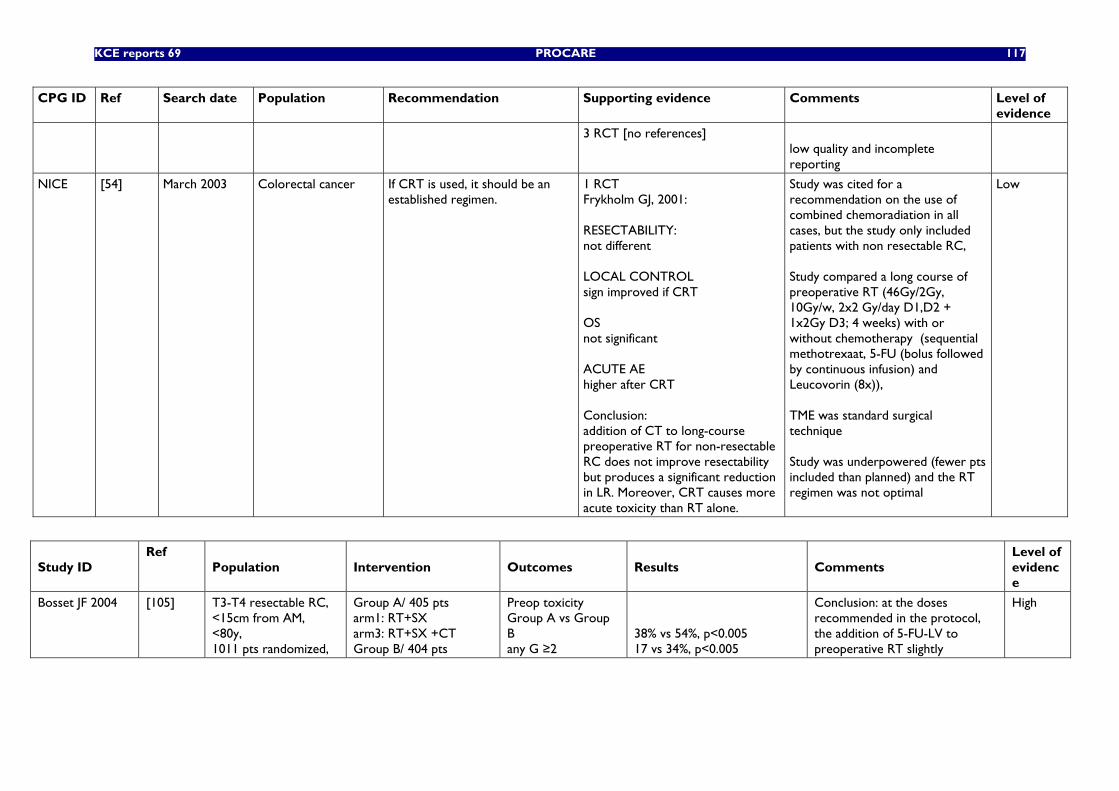
KCE reports 69 PROCARE 117
CPG ID Ref Search date Population Recommendation Supporting evidence
Comments Level of evidence
3 RCT [no references] low quality and incomplete reporting
NICE [54] March 2003 Colorectal cancer If CRT is used, it should be an established regimen.
1 RCT Frykholm GJ, 2001: RESECTABILITY: not different LOCAL CONTROL sign improved if CRT OS not significant ACUTE AE higher after CRT Conclusion: addition of CT to long-course preoperative RT for non-resectable RC does not improve resectability but produces a significant reduction in LR. Moreover, CRT causes more acute toxicity than RT alone.
Study was cited for a recommendation on the use of combined chemoradiation in all cases, but the study only included patients with non resectable RC, Study compared a long course of preoperative RT (46Gy/2Gy, 10Gy/w, 2x2 Gy/day D1,D2 + 1x2Gy D3; 4 weeks) with or without chemotherapy (sequential methotrexaat, 5-FU (bolus followed by continuous infusion) and Leucovorin (8x)), TME was standard surgical technique Study was underpowered (fewer pts included than planned) and the RT regimen was not optimal
Low
Study ID Ref
Population Intervention Outcomes Results Comments Level of evidence
Bosset JF 2004 [105] T3-T4 resectable RC, <15cm from AM, <80y, 1011 pts randomized,
Group A/ 405 pts arm1: RT+SX arm3: RT+SX +CT Group B/ 404 pts
Preop toxicity Group A vs Group B any G ≥2
38% vs 54%, p<0.005 17 vs 34%, p<0.005
Conclusion: at the doses recommended in the protocol, the addition of 5-FU-LV to preoperative RT slightly
High
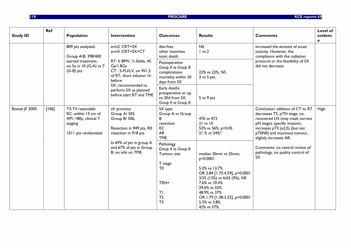
118 PROCARE KCE reports 69
Study ID Ref
Population Intervention Outcomes Results Comments Level of evidence
diarrhea other toxicities toxic death
NS 1 vs 2
Postoperative Group A vs Group B complications mortality within 30 days from SX
22% vs 23%, NS 3 vs 5 pts
809 pts analyzed, Group A/B: 398/400 started treatment, no Sx in 10 (G-A) vs 7 (G-B) pts
arm2: CRT+SX arm4 :CRT+SX+CT RT: ≥ 8MV, ¾ fields, 45 Gy/1.8Gy CT : 5-FU/LV, on W1,5 of RT, short infusion 1h before SX: recommended to perform SX as planned before start RT and TME
Early deaths preoperative or up to 30d from SX Group A vs Group B
5 vs 9 pts
increased the amount of acute toxicity. However, the compliance with the radiation protocol or the feasibility of SX did not decrease
SX type Group A vs Group B resection R2 AR TME
476 vs 473 21 vs 10 52% vs 56%, p=0.05 21 % vs 24%*
Bosset JF 2005 [106] T3-T4 resectable RC ,within 15 cm of AM, <80y, clinical T staging 1011 pts randomized
cfr previous Group A/ 505 Group B/ 506, Resection in 949 pts, R0 resection in 918 pts In 69% of pts in group A and 67% of pts in Group B: no info on TME
Pathology Group A vs Group B Tumour size T stage T0 T0N+ T1 T2 T3
median 30mm vs 25mm, p<0.0001 5.3% vs 13.7% OR 2.84 [1.75-4.59], p<0.0001 3/25 (12%) vs 6/65 (9%), NS 7.6% vs 10.4% 29.6% vs 33% 48.9% vs 37% OR 1.79 [1.38-2.32], p<0.0001 5.3% vs 3.8% 42% vs 57%,
Conclusion: addition of CT to RT decreases TS, pTN stage, no. recovered LN (may mask correct pN stage), specific invasion; increases pT0 (x2,5) (but not pT0N0) and mucinous tumour, slightly increases AR. Comments: no central review of pathology, no quality control of SX
High
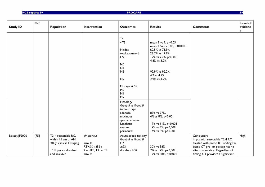
KCE reports 69 PROCARE 119
Study ID Ref
Population Intervention Outcomes Results Comments Level of evidence
T4 <T3 Nodes total examined LN+ N0 N1 N2 Nx M stage at SX M0 M1 Mx
mean 9 vs 7, p<0.05 mean 1.52 vs 0.86, p<0.0001 60.5% vs 71.9% 22.7% vs 17.8% 12% vs 7.2%, p<0.001 4.8% vs 3.2% 92.9% vs 92.2% 4.2 vs 4.7% 2.9% vs 3.2%
Histology Group A vs Group B tumour type adenoca mucinous specific invasion lymphatic venous perineural
87% vs 77%, 4% vs 8%, p<0,001 17% vs 11%, p=0,008 14% vs 9%, p=0,008 14% vs 8%, p=0,001
Bosset JF2006 [75] T3-4 resectable RC, within 15 cm of AM, <80y, clinical T staging 1011 pts randomized and analyzed
cfr previous arm 1: RT+SX : 252 : 2 no RT, 13 no TR arm 2:
Acute preop toxicity Group A vs Group B G2 ≥G3 diarrhea ≥G2
30% vs 38% 7% vs 14%, p<0,001 17% vs 38%, p<0,001
Conclusion: in pts with resectable T3/4 RC treated with preop RT, adding FU based CT pre- or postop has no effect on survival. Regardless of timing, CT provides a significant
High
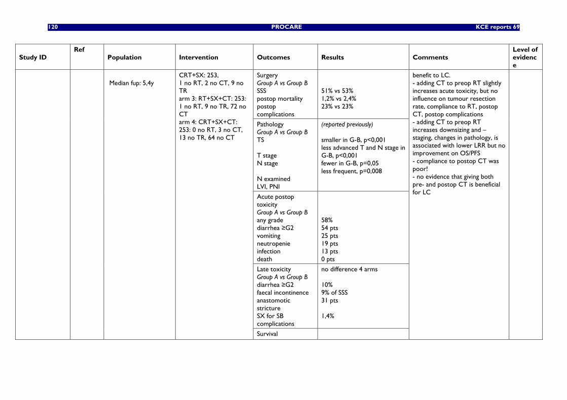
120 PROCARE KCE reports 69
Study ID Ref
Population Intervention Outcomes Results Comments Level of evidence
Surgery Group A vs Group B SSS postop mortality postop complications
51% vs 53% 1,2% vs 2,4% 23% vs 23%
Pathology Group A vs Group B TS T stage N stage N examined LVI, PNI
(reported previously) smaller in G-B, p<0,001 less advanced T and N stage in G-B, p<0,001 fewer in G-B, p=0,05 less frequent, p=0,008
Acute postop toxicity Group A vs Group B any grade diarrhea ≥G2 vomiting neutropenie infection death
58% 54 pts 25 pts 19 pts 13 pts 0 pts
Late toxicity Group A vs Group B diarrhea ≥G2 faecal incontinence anastomotic stricture SX for SB complications
no difference 4 arms 10% 9% of SSS 31 pts 1,4%
Median fup: 5,4y
CRT+SX: 253, 1 no RT, 2 no CT, 9 no TR arm 3: RT+SX+CT: 253: 1 no RT, 9 no TR, 72 no CT arm 4: CRT+SX+CT: 253: 0 no RT, 3 no CT, 13 no TR, 64 no CT
Survival
benefit to LC. - adding CT to preop RT slightly increases acute toxicity, but no influence on tumour resection rate, compliance to RT, postop CT, postop complications - adding CT to preop RT increases downsizing and –staging, changes in pathology, is associated with lower LRR but no improvement on OS/PFS - compliance to postop CT was poor! - no evidence that giving both pre- and postop CT is beneficial for LC
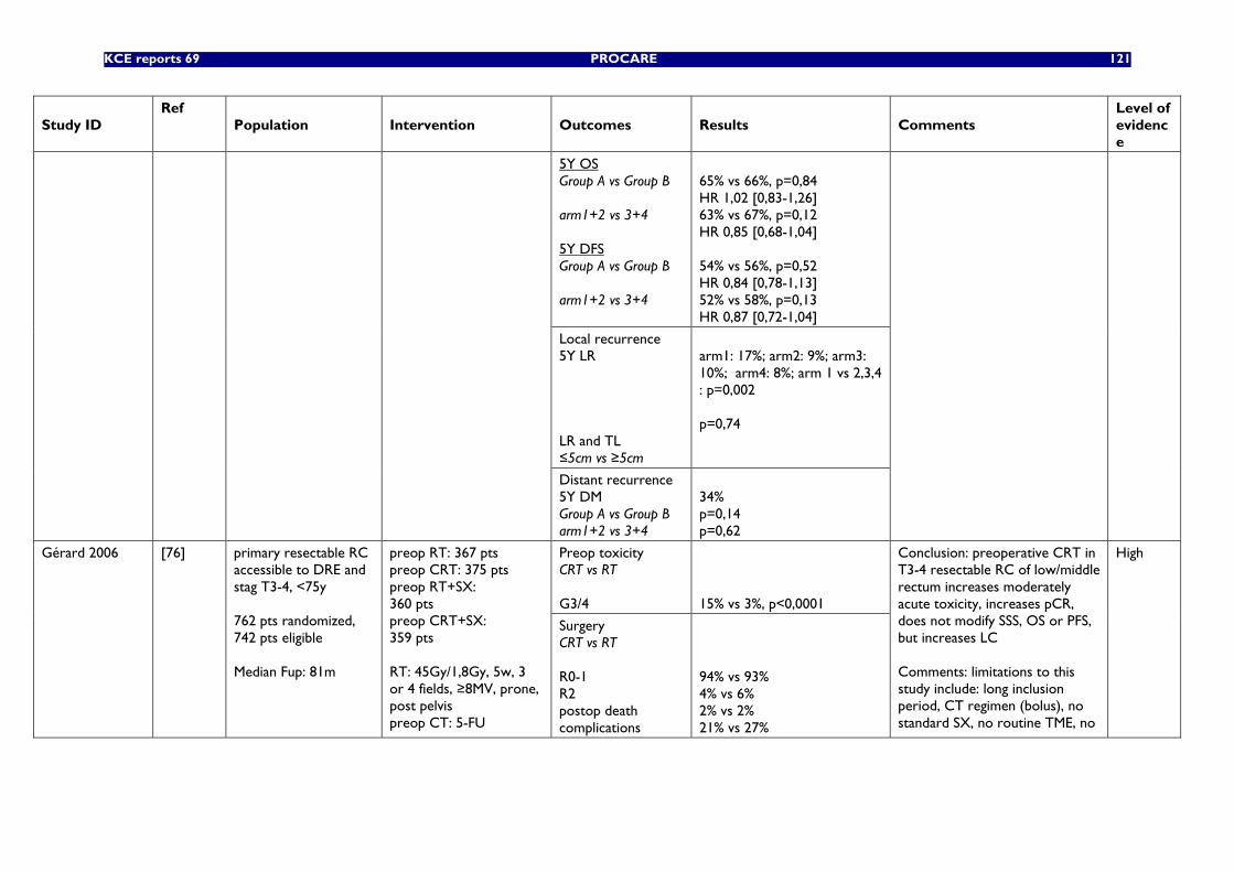
KCE reports 69 PROCARE 121
Study ID Ref
Population Intervention Outcomes Results Comments Level of evidence
5Y OS Group A vs Group B arm1+2 vs 3+4 5Y DFS Group A vs Group B arm1+2 vs 3+4
65% vs 66%, p=0,84 HR 1,02 [0,83-1,26] 63% vs 67%, p=0,12 HR 0,85 [0,68-1,04] 54% vs 56%, p=0,52 HR 0,84 [0,78-1,13] 52% vs 58%, p=0,13 HR 0,87 [0,72-1,04]
Local recurrence 5Y LR LR and TL ≤5cm vs ≥5cm
arm1: 17%; arm2: 9%; arm3: 10%; arm4: 8%; arm 1 vs 2,3,4 : p=0,002 p=0,74
Distant recurrence 5Y DM Group A vs Group B arm1+2 vs 3+4
34% p=0,14 p=0,62
Preop toxicity CRT vs RT G3/4
15% vs 3%, p<0,0001
Gérard 2006 [76] primary resectable RC accessible to DRE and stag T3-4, <75y 762 pts randomized, 742 pts eligible Median Fup: 81m
preop RT: 367 pts preop CRT: 375 pts preop RT+SX: 360 pts preop CRT+SX: 359 pts RT: 45Gy/1,8Gy, 5w, 3 or 4 fields, ≥8MV, prone, post pelvis preop CT: 5-FU
Surgery CRT vs RT R0-1 R2 postop death complications
94% vs 93% 4% vs 6% 2% vs 2% 21% vs 27%
Conclusion: preoperative CRT in T3-4 resectable RC of low/middle rectum increases moderately acute toxicity, increases pCR, does not modify SSS, OS or PFS, but increases LC Comments: limitations to this study include: long inclusion period, CT regimen (bolus), no standard SX, no routine TME, no
High
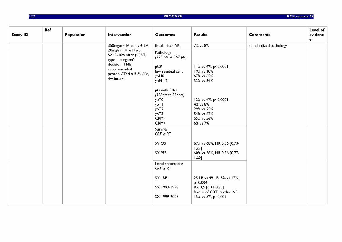
122 PROCARE KCE reports 69
Study ID Ref
Population Intervention Outcomes Results Comments Level of evidence
fistula after AR 7% vs 8%
Pathology (375 pts vs 367 pts) pCR few residual cells ypN0 ypN1-2 pts with R0-1 (338pts vs 336pts) ypT0 ypT1 ypT2 ypT3 CRM- CRM+
11% vs 4%, p<0,0001 19% vs 10% 67% vs 65% 33% vs 34% 12% vs 4%, p<0,0001 4% vs 8% 29% vs 25% 54% vs 62% 55% vs 56% 6% vs 7%
Survival CRT vs RT 5Y OS 5Y PFS
67% vs 68%, HR 0,96 [0,73-1,27] 60% vs 56%, HR 0,96 [0,77-1,20]
350mg/m² IV bolus + LV 20mg/m² IV w1+w5 SX: 3-10w after (C)RT, type = surgeon’s decision, TME recommended postop CT: 4 x 5-FU/LV, 4w interval
Local recurrence CRT vs RT 5Y LRR SX 1993-1998 SX 1999-2003
25 LR vs 49 LR, 8% vs 17%, p=0,004 RR 0,5 [0,31-0,80] favour of CRT, p value NR 15% vs 5%, p=0,007
standardized pathology
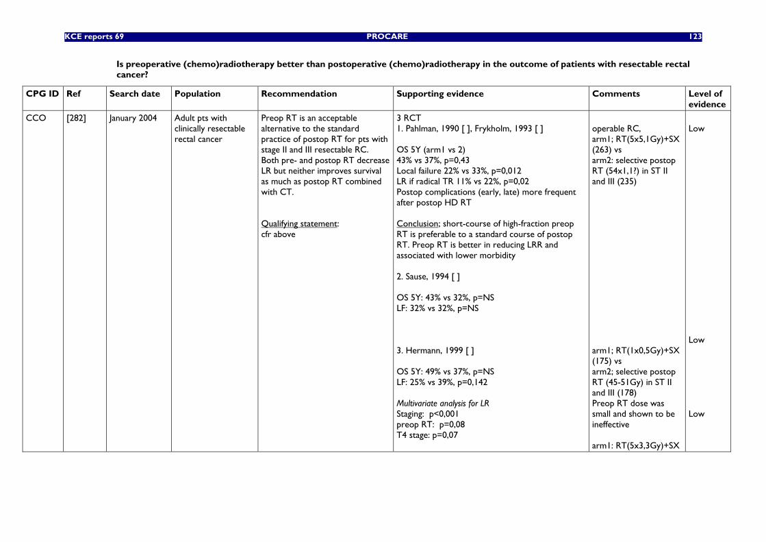
KCE reports 69 PROCARE 123
Is preoperative (chemo)radiotherapy better than postoperative (chemo)radiotherapy in the outcome of patients with resectable rectal cancer?
CPG ID Ref Search date Population Recommendation Supporting evidence
Comments Level of evidence
CCO [282] January 2004 Adult pts with clinically resectable rectal cancer
Preop RT is an acceptable alternative to the standard practice of postop RT for pts with stage II and III resectable RC. Both pre- and postop RT decrease LR but neither improves survival as much as postop RT combined with CT. Qualifying statement: cfr above
3 RCT 1. Pahlman, 1990 [ ], Frykholm, 1993 [ ] OS 5Y (arm1 vs 2) 43% vs 37%, p=0,43 Local failure 22% vs 33%, p=0,012 LR if radical TR 11% vs 22%, p=0,02 Postop complications (early, late) more frequent after postop HD RT Conclusion; short-course of high-fraction preop RT is preferable to a standard course of postop RT. Preop RT is better in reducing LRR and associated with lower morbidity 2. Sause, 1994 [ ] OS 5Y: 43% vs 32%, p=NS LF: 32% vs 32%, p=NS 3. Hermann, 1999 [ ] OS 5Y: 49% vs 37%, p=NS LF: 25% vs 39%, p=0,142 Multivariate analysis for LR Staging: p<0,001 preop RT: p=0,08 T4 stage: p=0,07
operable RC, arm1; RT(5x5,1Gy)+SX (263) vs arm2: selective postop RT (54x1,1?) in ST II and III (235) arm1; RT(1x0,5Gy)+SX (175) vs arm2; selective postop RT (45-51Gy) in ST II and III (178) Preop RT dose was small and shown to be ineffective arm1: RT(5x3,3Gy)+SX
Low Low Low
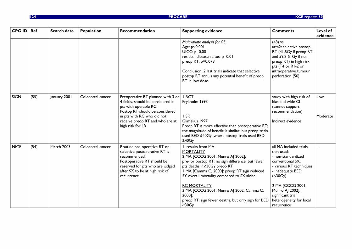
124 PROCARE KCE reports 69
CPG ID Ref Search date Population Recommendation Supporting evidence
Comments Level of evidence
Multivariate analysis for OS Age: p<0,001 UICC: p=0,001 residual disease status: p=0,01 preop RT: p=0,078 Conclusion: 2 last trials indicate that selective postop RT annuls any potential benefit of preop RT in low dose.
(48) vs arm2: selective postop RT (41,5Gy if preop RT and 59,8-51Gy if no preop RT) in high risk pts (T4 or R1-2 or intraoperative tumour perforation (56)
SIGN [55] January 2001 Colorectal cancer Preoperative RT planned with 3 or 4 fields, should be considered in pts with operable RC Postop RT should be considered in pts with RC who did not receive preop RT and who are at high risk for LR
1 RCT Frykholm 1993 1 SR Glimelius 1997 Preop RT is more effective than postoperative RT; the magnitude of benefit is similar, but preop trials used BED ≤40Gy, where postop trials used BED ≥40Gy
study with high risk of bias and wide CI (cannot support recommendation) Indirect evidence
Low Moderate
NICE
[54] March 2003 Colorectal cancer Routine pre-operative RT or selective postoperative RT is recommended. Postoperative RT should be reserved for pts who are judged after SX to be at high risk of recurrence
1. results from MA MORTALITY 2 MA [CCCG 2001, Munro AJ 2002]: pre- or postop RT: no sign difference, but fewer pts deaths if ≥30Gy preop RT 1 MA [Camma C, 2000]: preop RT sign reduced 5Y overall mortality compared to SX alone RC MORTALITY 3 MA [CCCG 2001, Munro AJ 2002, Camma C, 2000]: preop RT: sign fewer deaths, but only sign for BED ≥30Gy
all MA included trials that used: - non-standardized conventional SX; - various RT techniques - inadequate BED (<30Gy) 2 MA [CCCG 2001, Munro AJ 2002]: significant trial heterogeneity for local recurrence
-
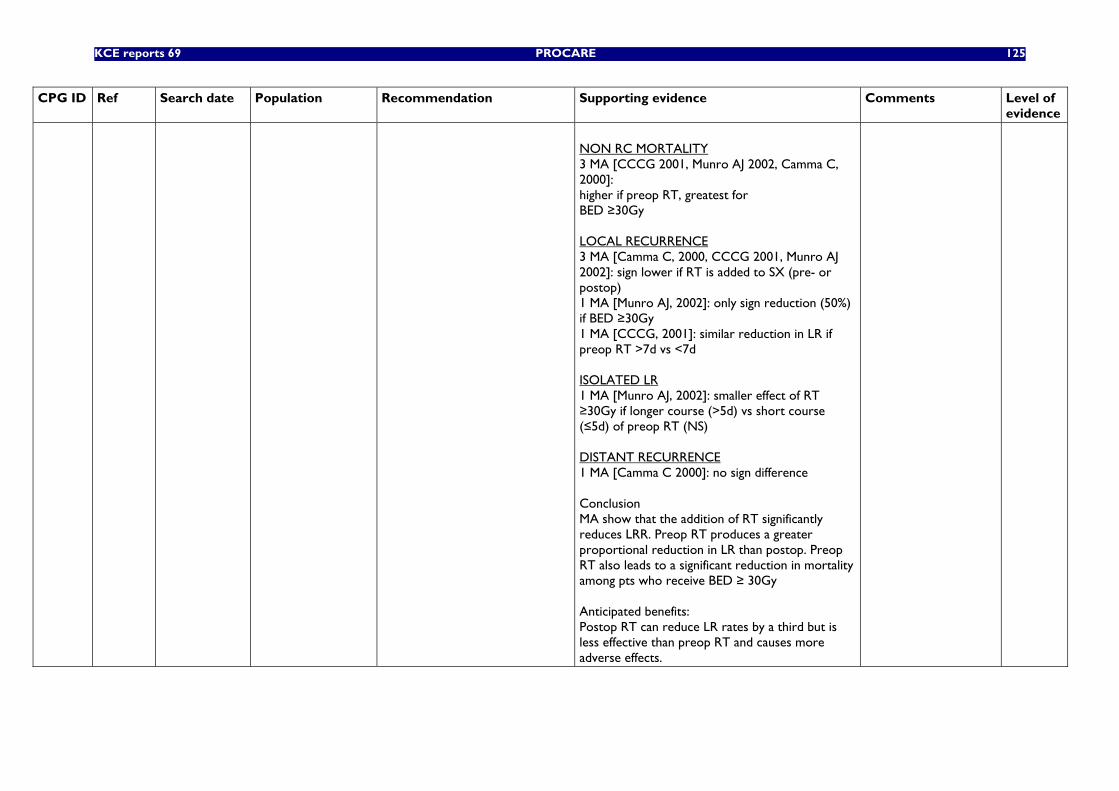
KCE reports 69 PROCARE 125
CPG ID Ref Search date Population Recommendation Supporting evidence
Comments Level of evidence
NON RC MORTALITY 3 MA [CCCG 2001, Munro AJ 2002, Camma C, 2000]: higher if preop RT, greatest for BED ≥30Gy LOCAL RECURRENCE 3 MA [Camma C, 2000, CCCG 2001, Munro AJ 2002]: sign lower if RT is added to SX (pre- or postop) 1 MA [Munro AJ, 2002]: only sign reduction (50%) if BED ≥30Gy 1 MA [CCCG, 2001]: similar reduction in LR if preop RT >7d vs <7d ISOLATED LR 1 MA [Munro AJ, 2002]: smaller effect of RT ≥30Gy if longer course (>5d) vs short course (≤5d) of preop RT (NS) DISTANT RECURRENCE 1 MA [Camma C 2000]: no sign difference Conclusion MA show that the addition of RT significantly reduces LRR. Preop RT produces a greater proportional reduction in LR than postop. Preop RT also leads to a significant reduction in mortality among pts who receive BED ≥ 30Gy Anticipated benefits: Postop RT can reduce LR rates by a third but is less effective than preop RT and causes more adverse effects.
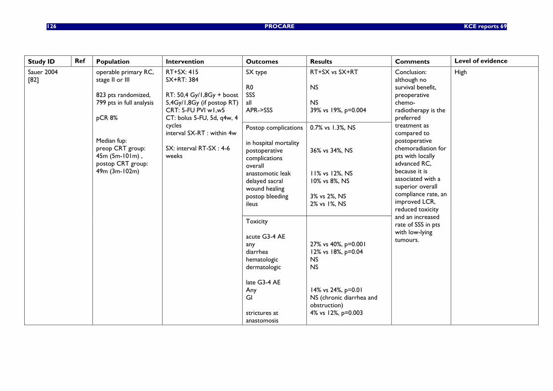
126 PROCARE KCE reports 69
Study ID Ref Population Intervention Outcomes Results Comments Level of evidence
SX type R0 SSS all APR->SSS
RT+SX vs SX+RT NS NS 39% vs 19%, p=0.004
Postop complications in hospital mortality postoperative complications overall anastomotic leak delayed sacral wound healing postop bleeding ileus
0.7% vs 1.3%, NS 36% vs 34%, NS 11% vs 12%, NS 10% vs 8%, NS 3% vs 2%, NS 2% vs 1%, NS
Sauer 2004 [82]
operable primary RC, stage II or III 823 pts randomized, 799 pts in full analysis pCR 8% Median fup: preop CRT group: 45m (5m-101m) , postop CRT group: 49m (3m-102m)
RT+SX: 415 SX+RT: 384 RT: 50,4 Gy/1,8Gy + boost 5,4Gy/1,8Gy (if postop RT) CRT: 5-FU PVI w1,w5 CT: bolus 5-FU, 5d, q4w, 4 cycles interval SX-RT : within 4w SX: interval RT-SX : 4-6 weeks
Toxicity acute G3-4 AE any diarrhea hematologic dermatologic late G3-4 AE Any GI strictures at anastomosis
27% vs 40%, p=0.001 12% vs 18%, p=0.04 NS NS 14% vs 24%, p=0.01 NS (chronic diarrhea and obstruction) 4% vs 12%, p=0.003
Conclusion: although no survival benefit, preoperative chemo-radiotherapy is the preferred treatment as compared to postoperative chemoradiation for pts with locally advanced RC, because it is associated with a superior overall compliance rate, an improved LCR, reduced toxicity and an increased rate of SSS in pts with low-lying tumours.
High
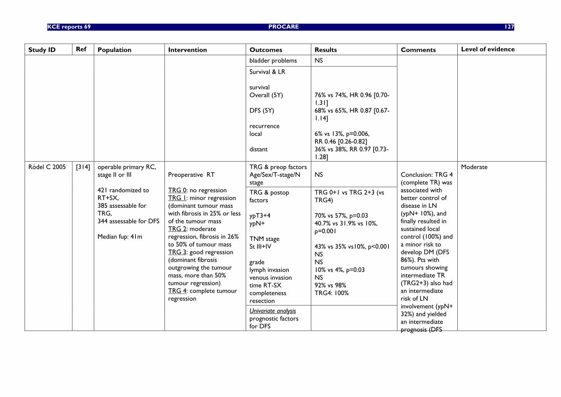
KCE reports 69 PROCARE 127
Study ID Ref Population Intervention Outcomes Results Comments Level of evidence
bladder problems NS
Survival & LR survival Overall (5Y) DFS (5Y) recurrence local distant
76% vs 74%, HR 0.96 [0.70-1.31] 68% vs 65%, HR 0.87 [0.67-1.14] 6% vs 13%, p=0.006, RR 0.46 [0.26-0.82] 36% vs 38%, RR 0.97 [0.73-1.28]
TRG & preop factors Age/Sex/T-stage/N stage
NS
TRG & postop factors ypT3+4 ypN+ TNM stage St III+IV grade lymph invasion venous invasion time RT-SX completeness resection
TRG 0+1 vs TRG 2+3 (vs TRG4) 70% vs 57%, p=0.03 40.7% vs 31.9% vs 10%, p=0.001 43% vs 35% vs10%, p<0.001 NS NS 10% vs 4%, p=0.03 NS 92% vs 98% TRG4: 100%
Rödel C 2005 [314] operable primary RC, stage II or III 421 randomized to RT+SX, 385 assessable for TRG, 344 assessable for DFS Median fup: 41m
Preoperative RT TRG 0: no regression TRG 1: minor regression (dominant tumour mass with fibrosis in 25% or less of the tumour mass TRG 2: moderate regression, fibrosis in 26% to 50% of tumour mass TRG 3: good regression (dominant fibrosis outgrowing the tumour mass, more than 50% tumour regression) TRG 4: complete tumour regression Univariate analysis
prognostic factors for DFS
Conclusion: TRG 4 (complete TR) was associated with better control of disease in LN (ypN+ 10%), and finally resulted in sustained local control (100%) and a minor risk to develop DM (DFS 86%). Pts with tumours showing intermediate TR (TRG2+3) also had an intermediate risk of LN involvement (ypN+ 32%) and yielded an intermediate prognosis (DFS
Moderate
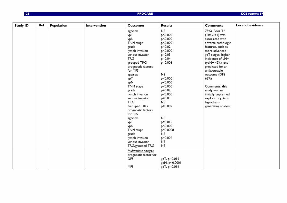
128 PROCARE KCE reports 69
Study ID Ref Population Intervention Outcomes Results Comments Level of evidence
age/sex ypT ypN TNM stage grade lymph invasion venous invasion TRG grouped TRG prognostic factors for MFS age/sex ypT ypN TNM stage grade lymph invasion venous invasion TRG Grouped TRG prognostic factors for RFS age/sex ypT ypN TNM stage grade lymph invasion venous invasion TRG/grouped TRG
NS p<0.0001 p<0.0001 p<0.0001 p=0.02 p<0.0001 p=0.03 p=0.04 p=0.006 NS p<0.0001 p<0.0001 p<0.0001 p=0.02 p<0.0001 p=0.03 NS p=0.009 NS p=0.015 p<0.0001 p=0.0008 NS p=0.002 NS NS
Multivariate analysis prognostic factor for DFS MFS
ypT, p=0.016 ypN, p<0.0001 ypT, p=0.014
75%). Poor TR (TRG0+1) was associated with adverse pathologic features, such as more advanced ypT stages, higher incidence of LN+ (ypN+ 42%), and predicted for an unfavourable outcome (DFS 63%) Comments: this study was an initially unplanned exploratory; ie. a hypothesis generating analysis
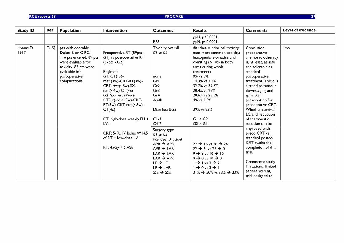
KCE reports 69 PROCARE 129
Study ID Ref Population Intervention Outcomes Results Comments Level of evidence
RFS
ypN, p<0.0001 ypN, p<0.0001
Toxicity overall G1 vs G2 none Gr1 Gr2 Gr3 Gr4 death Diarrhea ≥G3 C1-3 C4-7
diarrhea = principal toxicity; next most common toxicity: leucopenia, stomatitis and vomiting (< 10% in both arms during whole treatment) 0% vs 5% 14.3% vs 7.5% 32.7% vs 37.5% 20.4% vs 25% 28.6% vs 22.5% 4% vs 2.5% 39% vs 23% G1 > G2 G2 > G1
Hyams D 1997
[315] pts with operable Dukes B or C RC. 116 pts entered, 89 pts were evaluable for toxicity, 82 pts were evaluable for postoperative complications
Preoperative RT (59pts - G1) vs postoperative RT (57pts - G2) Regimen: G1: CT(1x)- rest (3w)-CRT-RT(3w)-CRT-rest(<8w)-SX-rest(<4w)-CT(4x) G2: SX-rest (<4w)-CT(1x)-rest (3w)-CRT-RT(3w)-CRT-rest(<8w)-CT(4x) CT: high-dose weekly FU + LV; CRT: 5-FU IV bolus W1&5 of RT + low-dose LV RT: 45Gy + 5.4Gy
Surgery type G1 vs G2 intended actual APR APR APR LAR LAR LAR LAR APR LE LE LE LAR SSS SSS
22 16 vs 26 26 22 6 vs 26 0 9 9 vs 10 10 9 0 vs 10 0 1 1 vs 3 2 1 0 vs 3 1 31% 50% vs 33% 33%
Conclusion: preoperative chemoradiotherapy is, at least, as safe and tolerable as standard postoperative treatment. There is a trend to tumour downstaging and sphincter preservation for preoperative CRT. Whether survival, LC and reduction of therapeutic sequelae can be improved with preop CRT vs standard postop CRT awaits the completion of this trial. Comments: study limitations: limited patient accrual, trial designed to
Low
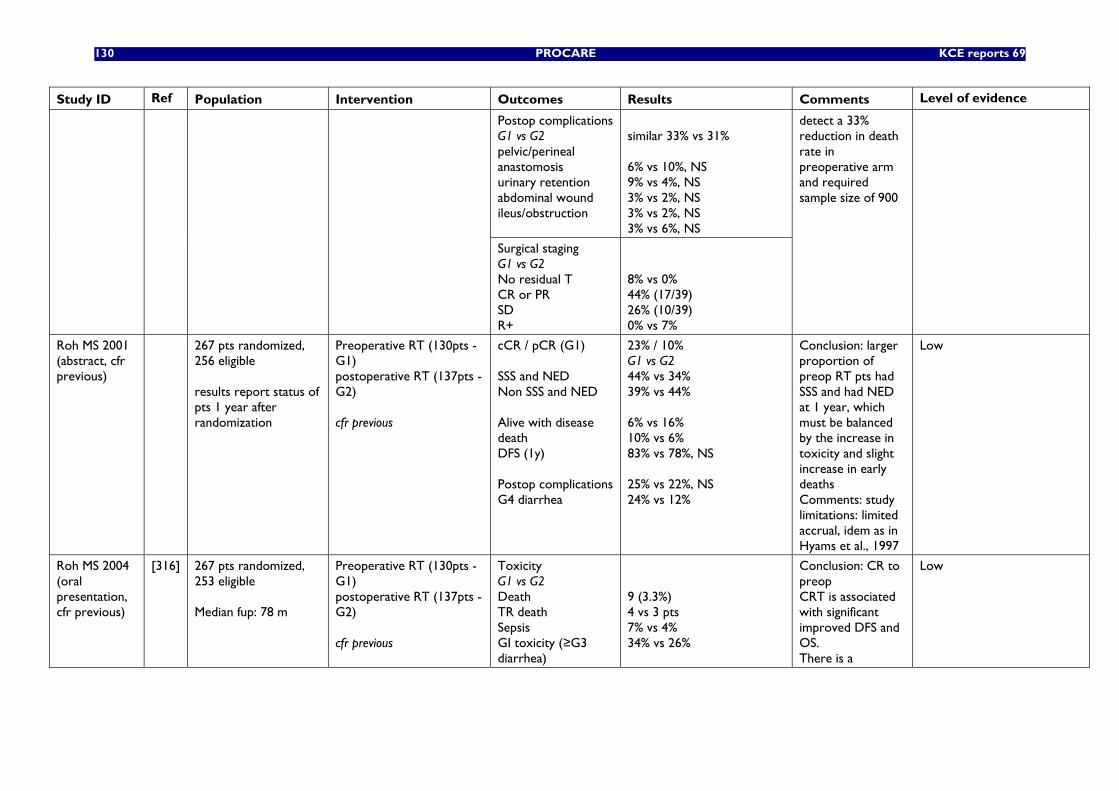
130 PROCARE KCE reports 69
Study ID Ref Population Intervention Outcomes Results Comments Level of evidence
Postop complications G1 vs G2 pelvic/perineal anastomosis urinary retention abdominal wound ileus/obstruction
similar 33% vs 31% 6% vs 10%, NS 9% vs 4%, NS 3% vs 2%, NS 3% vs 2%, NS 3% vs 6%, NS
Surgical staging G1 vs G2 No residual T CR or PR SD R+
8% vs 0% 44% (17/39) 26% (10/39) 0% vs 7%
detect a 33% reduction in death rate in preoperative arm and required sample size of 900
Roh MS 2001 (abstract, cfr previous)
267 pts randomized, 256 eligible results report status of pts 1 year after randomization
Preoperative RT (130pts - G1) postoperative RT (137pts - G2) cfr previous
cCR / pCR (G1) SSS and NED Non SSS and NED Alive with disease death DFS (1y) Postop complications G4 diarrhea
23% / 10% G1 vs G2 44% vs 34% 39% vs 44% 6% vs 16% 10% vs 6% 83% vs 78%, NS 25% vs 22%, NS 24% vs 12%
Conclusion: larger proportion of preop RT pts had SSS and had NED at 1 year, which must be balanced by the increase in toxicity and slight increase in early deaths Comments: study limitations: limited accrual, idem as in Hyams et al., 1997
Low
Roh MS 2004 (oral presentation, cfr previous)
[316] 267 pts randomized, 253 eligible Median fup: 78 m
Preoperative RT (130pts - G1) postoperative RT (137pts - G2) cfr previous
Toxicity G1 vs G2 Death TR death Sepsis GI toxicity (≥G3 diarrhea)
9 (3.3%) 4 vs 3 pts 7% vs 4% 34% vs 26%
Conclusion: CR to preop CRT is associated with significant improved DFS and OS. There is a
Low
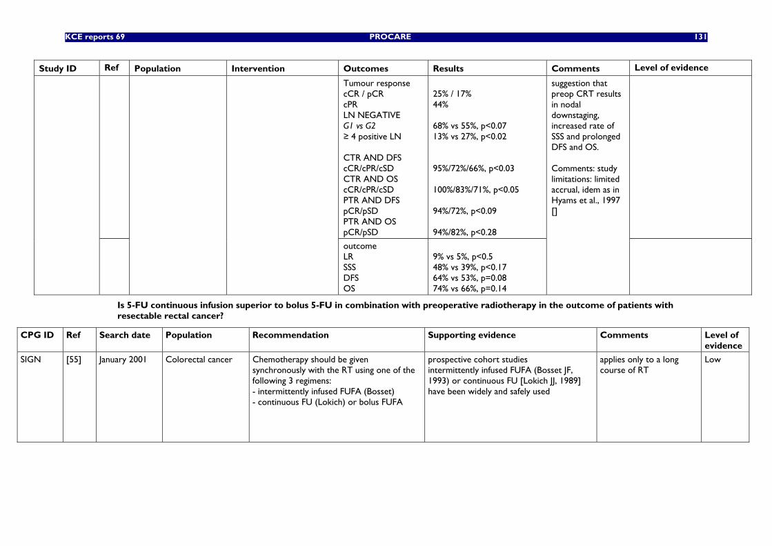
KCE reports 69 PROCARE 131
Study ID Ref Population Intervention Outcomes Results Comments Level of evidence
Tumour response cCR / pCR cPR LN NEGATIVE G1 vs G2 ≥ 4 positive LN CTR AND DFS cCR/cPR/cSD CTR AND OS cCR/cPR/cSD PTR AND DFS pCR/pSD PTR AND OS pCR/pSD
25% / 17% 44% 68% vs 55%, p<0.07 13% vs 27%, p<0.02 95%/72%/66%, p<0.03 100%/83%/71%, p<0.05 94%/72%, p<0.09 94%/82%, p<0.28
outcome LR SSS DFS OS
9% vs 5%, p<0.5 48% vs 39%, p<0.17 64% vs 53%, p=0.08 74% vs 66%, p=0.14
suggestion that preop CRT results in nodal downstaging, increased rate of SSS and prolonged DFS and OS. Comments: study limitations: limited accrual, idem as in Hyams et al., 1997 []
Is 5-FU continuous infusion superior to bolus 5-FU in combination with preoperative radiotherapy in the outcome of patients with resectable rectal cancer?
CPG ID Ref Search date Population Recommendation Supporting evidence
Comments Level of evidence
SIGN [55] January 2001 Colorectal cancer Chemotherapy should be given synchronously with the RT using one of the following 3 regimens: - intermittently infused FUFA (Bosset) - continuous FU (Lokich) or bolus FUFA
prospective cohort studies intermittently infused FUFA (Bosset JF, 1993) or continuous FU [Lokich JJ, 1989] have been widely and safely used
applies only to a long course of RT
Low
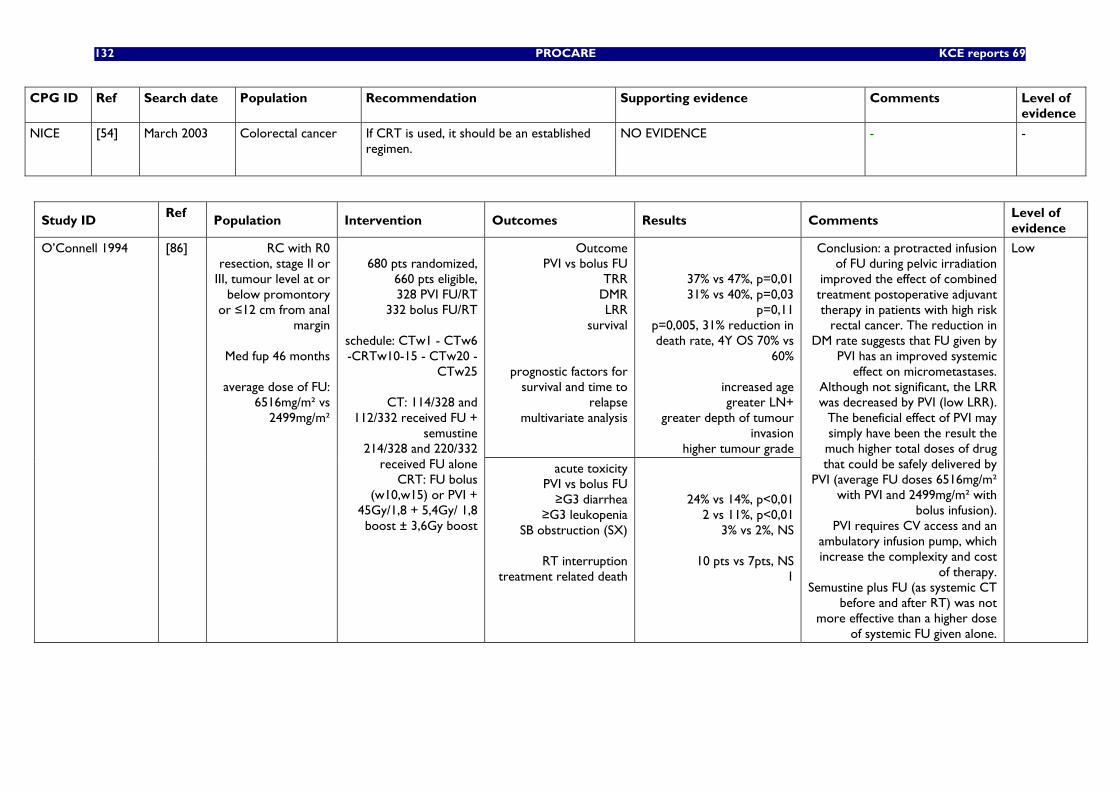
132 PROCARE KCE reports 69
CPG ID Ref Search date Population Recommendation Supporting evidence
Comments Level of evidence
NICE
[54] March 2003 Colorectal cancer If CRT is used, it should be an established regimen.
NO EVIDENCE - -
Study ID Ref
Population Intervention Outcomes Results Comments Level of evidence
Outcome PVI vs bolus FU
TRR DMR LRR
survival
prognostic factors for survival and time to
relapse multivariate analysis
37% vs 47%, p=0,01 31% vs 40%, p=0,03
p=0,11 p=0,005, 31% reduction in death rate, 4Y OS 70% vs
60%
increased age greater LN+
greater depth of tumour invasion
higher tumour grade
O’Connell 1994 [86] RC with R0 resection, stage II or
III, tumour level at or below promontory
or ≤12 cm from anal margin
Med fup 46 months
average dose of FU:
6516mg/m² vs 2499mg/m²
680 pts randomized,
660 pts eligible, 328 PVI FU/RT
332 bolus FU/RT
schedule: CTw1 - CTw6 -CRTw10-15 - CTw20 -
CTw25
CT: 114/328 and 112/332 received FU +
semustine 214/328 and 220/332
received FU alone CRT: FU bolus
(w10,w15) or PVI + 45Gy/1,8 + 5,4Gy/ 1,8 boost ± 3,6Gy boost
acute toxicity PVI vs bolus FU ≥G3 diarrhea
≥G3 leukopenia SB obstruction (SX)
RT interruption
treatment related death
24% vs 14%, p<0,01 2 vs 11%, p<0,01
3% vs 2%, NS
10 pts vs 7pts, NS 1
Conclusion: a protracted infusion of FU during pelvic irradiation
improved the effect of combined treatment postoperative adjuvant therapy in patients with high risk
rectal cancer. The reduction in DM rate suggests that FU given by
PVI has an improved systemic effect on micrometastases.
Although not significant, the LRR was decreased by PVI (low LRR).
The beneficial effect of PVI may simply have been the result the much higher total doses of drug that could be safely delivered by
PVI (average FU doses 6516mg/m² with PVI and 2499mg/m² with
bolus infusion). PVI requires CV access and an
ambulatory infusion pump, which increase the complexity and cost
of therapy. Semustine plus FU (as systemic CT
before and after RT) was not more effective than a higher dose
of systemic FU given alone.
Low
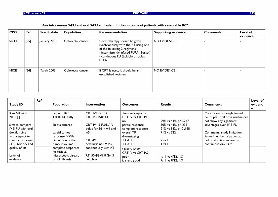
KCE reports 69 PROCARE 133
Are intravenous 5-FU and oral 5-FU equivalent in the outcome of patients with resectable RC?
CPG Ref Search date Population Recommendation Supporting evidence
Comments Level of evidence
SIGN [55] January 2001 Colorectal cancer Chemotherapy should be given synchronously with the RT using one of the following 3 regimens: - intermittently infused FUFA (Bosset) - continuous FU (Lokich) or bolus FUFA
NO EVIDENCE - -
NICE
[54] March 2003 Colorectal cancer If CRT is used, it should be an established regimen.
NO EVIDENCE - -
Study ID Ref
Population Intervention Outcomes Results Comments Level of evidence
Tumour response CRT IV vs CRT PO no partial response complete response overall TR downstaging T3 -> T0 T4 -> T0
29% vs 43%, p=0,247 50% vs 43%, p=,235 21% vs 14%, p=0 ,168 71% vs 52% 2 vs 1 1 vs 1
Kim NK et al., 2001 [ ] aim: to compare IV 5-FU with oral doxifluridine with respect to tumour response (TR), toxicity and quality of life. Level of evidence:
pts with RC, T3N1/T4, <70y 28 pts entered partial tumour response: >50% diminution of the tumour volume complete response: no residual microscopic disease or RT fibrosis
CRT IV+SX : 14 CRT PO+SX: 14 CRT-IV : 5-FU/LV IV bolus for 5d in w1 and w5, CRT-PO: doxifluridine/LV PO continuously with RT RT: 50,4Gy/1,8 Gy, 3 field box
Quality of life CRT IV vs CRT PO poor fair and good
4/11 vs 4/12, NS 7/11 vs 8/12, NS
Conclusion: although limited no. of pts., oral doxifluridine did not show any significant advantages over IV 5-FU Comments: study limitation: limited number of patients, bolus 5-FU is compared to continuous oral FU!!
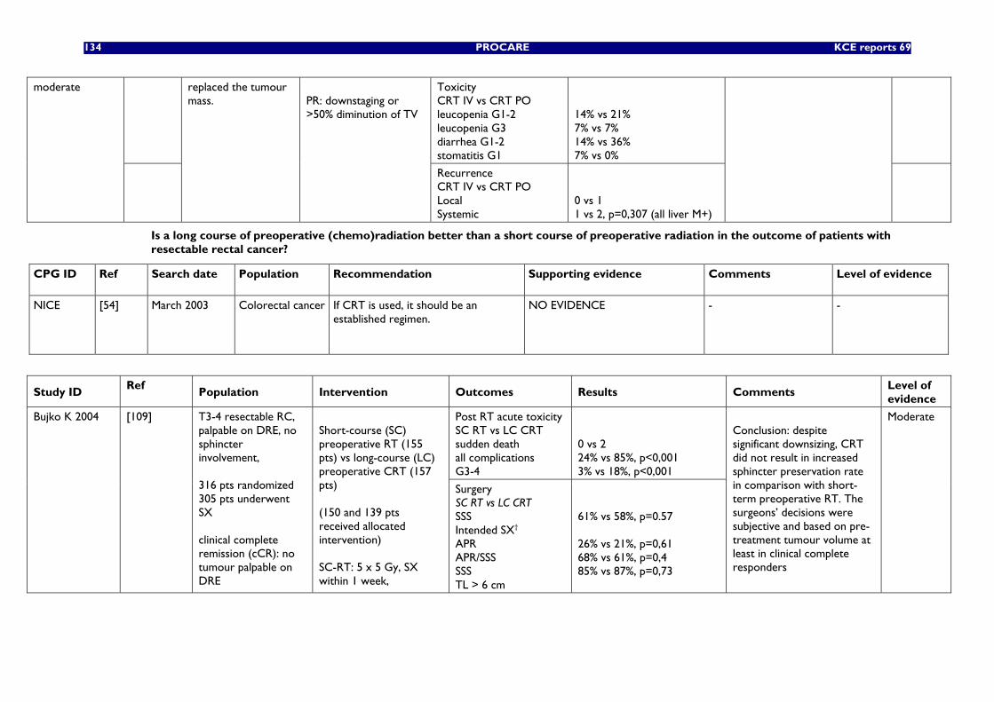
134 PROCARE KCE reports 69
Toxicity CRT IV vs CRT PO leucopenia G1-2 leucopenia G3 diarrhea G1-2 stomatitis G1
14% vs 21% 7% vs 7% 14% vs 36% 7% vs 0%
moderate
replaced the tumour mass.
PR: downstaging or >50% diminution of TV
Recurrence CRT IV vs CRT PO Local Systemic
0 vs 1 1 vs 2, p=0,307 (all liver M+)
Is a long course of preoperative (chemo)radiation better than a short course of preoperative radiation in the outcome of patients with resectable rectal cancer?
CPG ID Ref Search date Population Recommendation Supporting evidence
Comments Level of evidence
NICE
[54] March 2003 Colorectal cancer If CRT is used, it should be an established regimen.
NO EVIDENCE - -
Study ID Ref
Population Intervention Outcomes Results Comments Level of evidence
Post RT acute toxicity SC RT vs LC CRT sudden death all complications G3-4
0 vs 2 24% vs 85%, p<0,001 3% vs 18%, p<0,001
Bujko K 2004 [109] T3-4 resectable RC, palpable on DRE, no sphincter involvement, 316 pts randomized 305 pts underwent SX clinical complete remission (cCR): no tumour palpable on DRE
Short-course (SC) preoperative RT (155 pts) vs long-course (LC) preoperative CRT (157 pts) (150 and 139 pts received allocated intervention) SC-RT: 5 x 5 Gy, SX within 1 week,
Surgery SC RT vs LC CRT SSS Intended SX† APR APR/SSS SSS TL > 6 cm
61% vs 58%, p=0.57 26% vs 21%, p=0,61 68% vs 61%, p=0,4 85% vs 87%, p=0,73
Conclusion: despite significant downsizing, CRT did not result in increased sphincter preservation rate in comparison with short-term preoperative RT. The surgeons’ decisions were subjective and based on pre-treatment tumour volume at least in clinical complete responders
Moderate
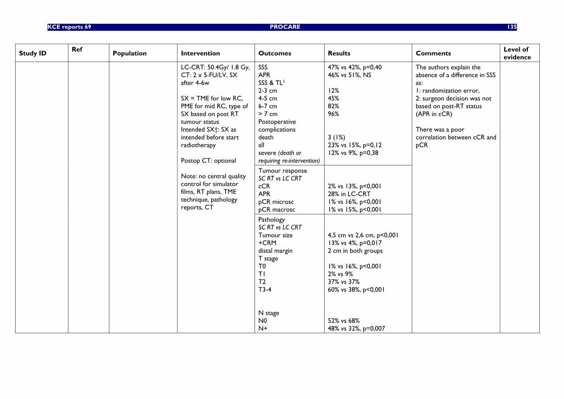
KCE reports 69 PROCARE 135
Study ID Ref
Population Intervention Outcomes Results Comments Level of evidence
SSS APR SSS & TL‡ 2-3 cm 4-5 cm 6-7 cm > 7 cm Postoperative complications death all severe (death or requiring re-intervention)
47% vs 42%, p=0,40 46% vs 51%, NS 12% 45% 82% 96% 3 (1%) 23% vs 15%, p=0,12 12% vs 9%, p=0,38
Tumour response SC RT vs LC CRT cCR APR pCR microsc pCR macrosc
2% vs 13%, p<0,001 28% in LC-CRT 1% vs 16%, p<0,001 1% vs 15%, p<0,001
LC-CRT: 50.4Gy/ 1.8 Gy, CT: 2 x 5-FU/LV, SX after 4-6w SX = TME for low RC, PME for mid RC, type of SX based on post RT tumour status Intended SX†: SX as intended before start radiotherapy Postop CT: optional Note: no central quality control for simulator films, RT plans, TME technique, pathology reports, CT
Pathology SC RT vs LC CRT Tumour size +CRM distal margin T stage T0 T1 T2 T3-4 N stage N0 N+
4,5 cm vs 2,6 cm, p<0,001 13% vs 4%, p=0,017 2 cm in both groups 1% vs 16%, p<0,001 2% vs 9% 37% vs 37% 60% vs 38%, p<0,001 52% vs 68% 48% vs 32%, p=0,007
The authors explain the absence of a difference in SSS as: 1: randomization error, 2: surgeon decision was not based on post-RT status (APR in cCR) There was a poor correlation between cCR and pCR
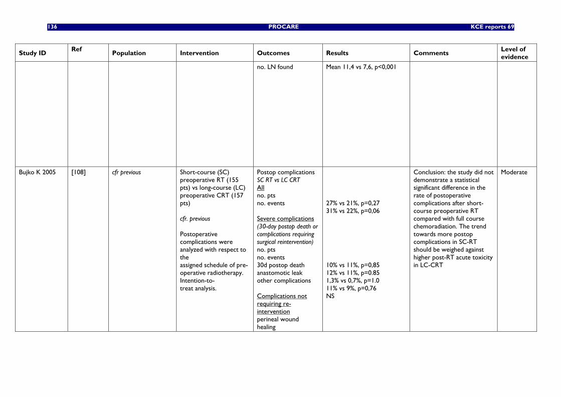
136 PROCARE KCE reports 69
Study ID Ref
Population Intervention Outcomes Results Comments Level of evidence
no. LN found Mean 11,4 vs 7,6, p<0,001
Bujko K 2005 [108] cfr previous
Short-course (SC) preoperative RT (155 pts) vs long-course (LC) preoperative CRT (157 pts) cfr. previous Postoperative complications were analyzed with respect to the assigned schedule of pre-operative radiotherapy. Intention-to- treat analysis.
Postop complications SC RT vs LC CRT All no. pts no. events Severe complications (30-day postop death or complications requiring surgical reintervention) no. pts no. events 30d postop death anastomotic leak other complications Complications not requiring re-intervention perineal wound healing
27% vs 21%, p=0,27 31% vs 22%, p=0,06 10% vs 11%, p=0,85 12% vs 11%, p=0.85 1,3% vs 0,7%, p=1.0 11% vs 9%, p=0,76 NS
Conclusion: the study did not demonstrate a statistical significant difference in the rate of postoperative complications after short-course preoperative RT compared with full course chemoradiation. The trend towards more postop complications in SC-RT should be weighed against higher post-RT acute toxicity in LC-CRT
Moderate
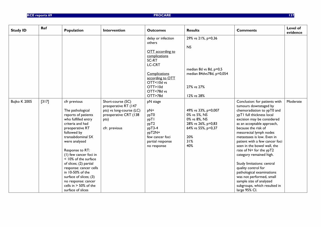
KCE reports 69 PROCARE 137
Study ID Ref
Population Intervention Outcomes Results Comments Level of evidence
delay or infection others OTT according to complications SC-RT LC-CRT Complications according to OTT OTT<10d vs OTT>10d OTT<78d vs OTT>78d
29% vs 21%, p=0,36 NS median 8d vs 8d, p=0,5 median 84dvs78d, p=0,054 27% vs 27% 12% vs 28%
Bujko K 2005 [317] cfr previous The pathological reports of patients who fulfilled entry criteria and had preoperative RT followed by transabdominal SX were analysed Response to RT: (1) few cancer foci in < 10% of the surface of slices; (2) partial response: cancer cells in 10-50% of the surface of slices; (3) no response: cancer cells in > 50% of the surface of slices
Short-course (SC) preoperative RT (147 pts) vs long-course (LC) preoperative CRT (138 pts) cfr. previous
pN stage pN+ ypT0 ypT1 ypT2 ypT3-4 ypT2N+ few cancer foci partial response no response
49% vs 33%, p=0,007 0% vs 5%, NS 0% vs 8%, NS 28% vs 26%, p=0,83 64% vs 55%, p=0,37 20% 31% 40%
Conclusion: for patients with tumours downstaged by chemoradiation to ypT0 and ypT1 full thickness local excision may be considered as an acceptable approach, because the risk of mesorectal lymph nodes metastases is low. Even in patient with a few cancer foci seen in the bowel wall, the rate of N+ for the ypT2 category remained high. Study limitations: central quality control for pathological examinations was not performed, small sample size of analyzed subgroups, which resulted in large 95% CI.
Moderate
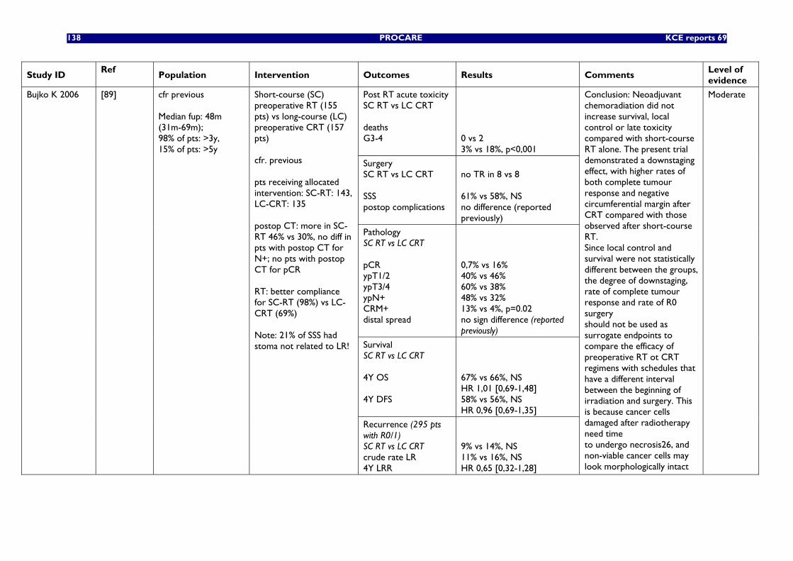
138 PROCARE KCE reports 69
Study ID Ref
Population Intervention Outcomes Results Comments Level of evidence
Post RT acute toxicity SC RT vs LC CRT deaths G3-4
0 vs 2 3% vs 18%, p<0,001
Surgery SC RT vs LC CRT SSS postop complications
no TR in 8 vs 8 61% vs 58%, NS no difference (reported previously)
Pathology SC RT vs LC CRT pCR ypT1/2 ypT3/4 ypN+ CRM+ distal spread
0,7% vs 16% 40% vs 46% 60% vs 38% 48% vs 32% 13% vs 4%, p=0.02 no sign difference (reported previously)
Survival SC RT vs LC CRT 4Y OS 4Y DFS
67% vs 66%, NS HR 1,01 [0,69-1,48] 58% vs 56%, NS HR 0,96 [0,69-1,35]
Bujko K 2006 [89] cfr previous Median fup: 48m (31m-69m); 98% of pts: >3y, 15% of pts: >5y
Short-course (SC) preoperative RT (155 pts) vs long-course (LC) preoperative CRT (157 pts) cfr. previous pts receiving allocated intervention: SC-RT: 143, LC-CRT: 135 postop CT: more in SC-RT 46% vs 30%, no diff in pts with postop CT for N+; no pts with postop CT for pCR RT: better compliance for SC-RT (98%) vs LC-CRT (69%) Note: 21% of SSS had stoma not related to LR!
Recurrence (295 pts with R0/1) SC RT vs LC CRT crude rate LR 4Y LRR
9% vs 14%, NS 11% vs 16%, NS HR 0,65 [0,32-1,28]
Conclusion: Neoadjuvant chemoradiation did not increase survival, local control or late toxicity compared with short-course RT alone. The present trial demonstrated a downstaging effect, with higher rates of both complete tumour response and negative circumferential margin after CRT compared with those observed after short-course RT. Since local control and survival were not statistically different between the groups, the degree of downstaging, rate of complete tumour response and rate of R0 surgery should not be used as surrogate endpoints to compare the efficacy of preoperative RT ot CRT regimens with schedules that have a different interval between the beginning of irradiation and surgery. This is because cancer cells damaged after radiotherapy need time to undergo necrosis26, and non-viable cancer cells may look morphologically intact
Moderate
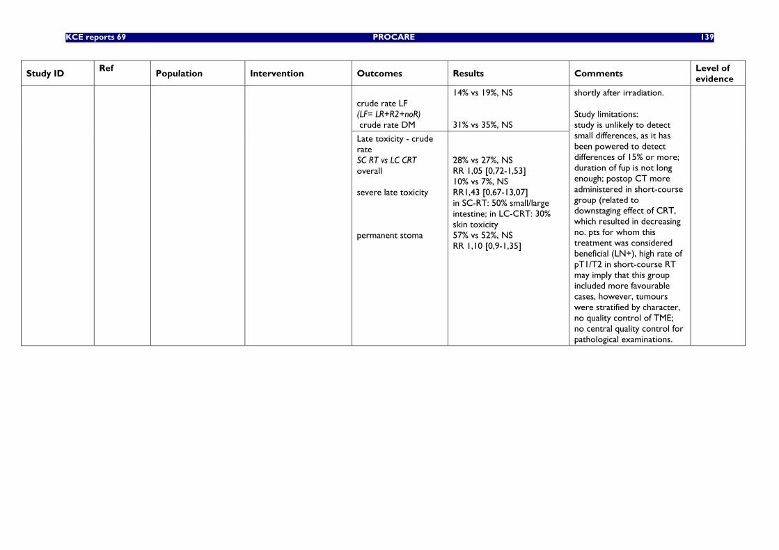
KCE reports 69 PROCARE 139
Study ID Ref
Population Intervention Outcomes Results Comments Level of evidence
crude rate LF (LF= LR+R2+noR) crude rate DM
14% vs 19%, NS 31% vs 35%, NS
Late toxicity - crude rate SC RT vs LC CRT overall severe late toxicity permanent stoma
28% vs 27%, NS RR 1,05 [0,72-1,53] 10% vs 7%, NS RR1,43 [0,67-13,07] in SC-RT: 50% small/large intestine; in LC-CRT: 30% skin toxicity 57% vs 52%, NS RR 1,10 [0,9-1,35]
shortly after irradiation. Study limitations: study is unlikely to detect small differences, as it has been powered to detect differences of 15% or more; duration of fup is not long enough; postop CT more administered in short-course group (related to downstaging effect of CRT, which resulted in decreasing no. pts for whom this treatment was considered beneficial (LN+), high rate of pT1/T2 in short-course RT may imply that this group included more favourable cases, however, tumours were stratified by character, no quality control of TME; no central quality control for pathological examinations.
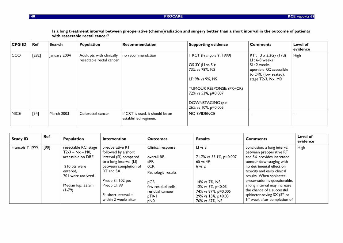
140 PROCARE KCE reports 69
Is a long treatment interval between preoperative (chemo)radiation and surgery better than a short interval in the outcome of patients with resectable rectal cancer?
CPG ID Ref Search Population Recommendation Supporting evidence
Comments Level of evidence
CCO [282] January 2004 Adult pts with clinically resectable rectal cancer
no recommendation 1 RCT (François Y, 1999) OS 3Y (LI vs SI): 73% vs 78%, NS LF: 9% vs 9%, NS TUMOUR RESPONSE: (PR+CR) 72% vs 53%, p=0,007 DOWNSTAGING (p): 26% vs 10%, p=0,005
RT : 13 x 3,3Gy (17d) LI : 6-8 weeks SI : 2 weeks operable RC accessible to DRE (low seated), stage T2-3, Nx, M0
High
NICE [54] March 2003 Colorectal cancer If CRT is used, it should be an established regimen.
NO EVIDENCE - -
Study ID Ref
Population Intervention Outcomes Results Comments Level of evidence
Clinical response overall RR cPR cCR
LI vs SI 71.7% vs 53.1%, p=0.007 65 vs 49 6 vs 2
François Y 1999 [90] resectable RC, stage T2-3 – Nx – M0, accessible on DRE 210 pts were entered, 201 were analyzed Median fup: 33,5m (1-79)
preoperative RT followed by a short interval (SI) compared to a long interval (LI) between completion of RT and SX. Preop SI: 102 pts Preop LI: 99 SI: short interval = within 2 weeks after
Pathologic results pCR few residual cells residual tumour pT0-1 pN0
14% vs 7%, NS 12% vs 3%, p<0.03 74% vs 87%, p=0.005 29% vs 15%, p<0.03 76% vs 67%, NS
conclusion: a long interval between preoperative RT and SX provides increased tumour downstaging with no detrimental effect on toxicity and early clinical results. When sphincter preservation is questionable, a long interval may increase the chance of a successful sphincter-saving SX (5th or 6th week after completion of
High
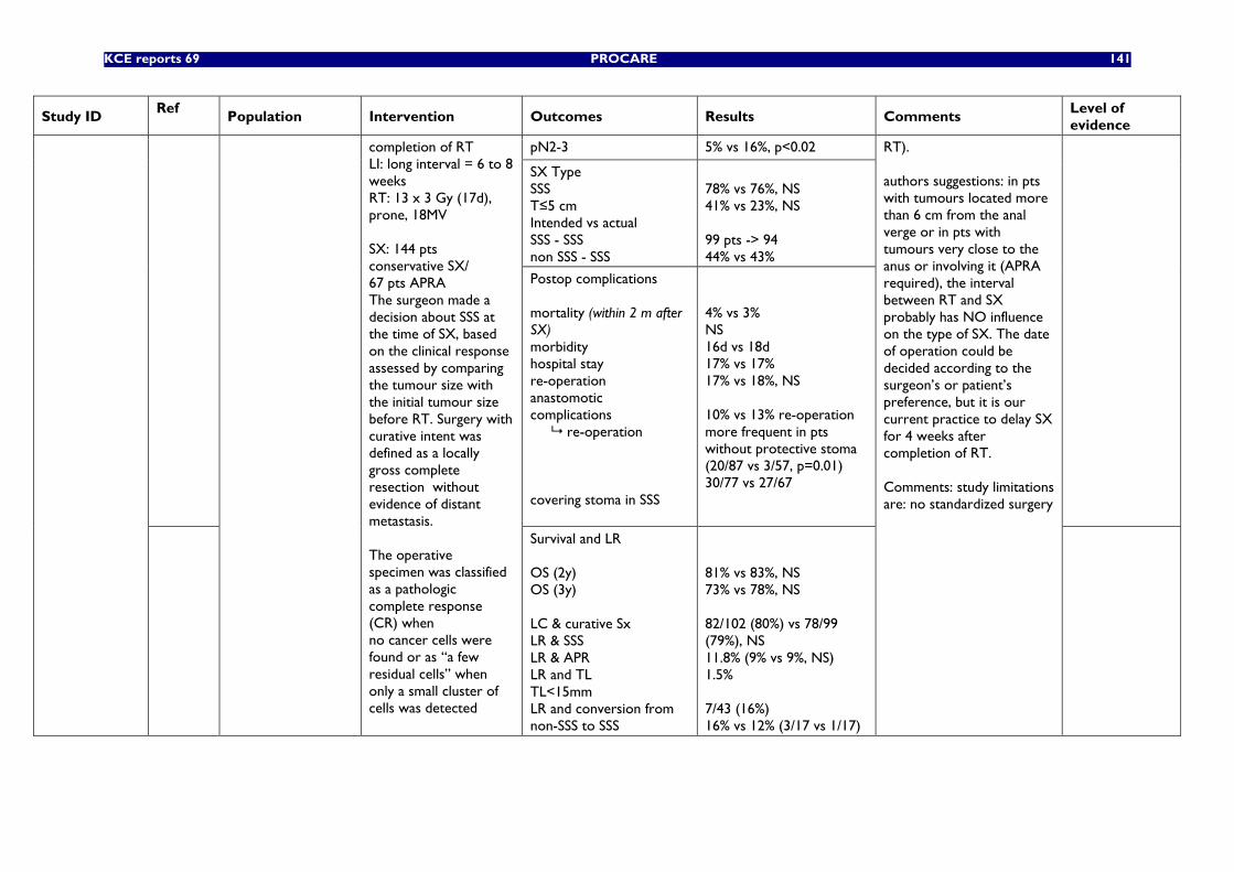
KCE reports 69 PROCARE 141
Study ID Ref
Population Intervention Outcomes Results Comments Level of evidence
pN2-3 5% vs 16%, p<0.02
SX Type SSS T≤5 cm Intended vs actual SSS - SSS non SSS - SSS
78% vs 76%, NS 41% vs 23%, NS 99 pts -> 94 44% vs 43%
Postop complications mortality (within 2 m after SX) morbidity hospital stay re-operation anastomotic complications re-operation covering stoma in SSS
4% vs 3% NS 16d vs 18d 17% vs 17% 17% vs 18%, NS 10% vs 13% re-operation more frequent in pts without protective stoma (20/87 vs 3/57, p=0.01) 30/77 vs 27/67
completion of RT LI: long interval = 6 to 8 weeks RT: 13 x 3 Gy (17d), prone, 18MV SX: 144 pts conservative SX/ 67 pts APRA The surgeon made a decision about SSS at the time of SX, based on the clinical response assessed by comparing the tumour size with the initial tumour size before RT. Surgery with curative intent was defined as a locally gross complete resection without evidence of distant metastasis. The operative specimen was classified as a pathologic complete response (CR) when no cancer cells were found or as ‘‘a few residual cells’’ when only a small cluster of cells was detected
Survival and LR OS (2y) OS (3y) LC & curative Sx LR & SSS LR & APR LR and TL TL<15mm LR and conversion from non-SSS to SSS
81% vs 83%, NS 73% vs 78%, NS 82/102 (80%) vs 78/99 (79%), NS 11.8% (9% vs 9%, NS) 1.5% 7/43 (16%) 16% vs 12% (3/17 vs 1/17)
RT). authors suggestions: in pts with tumours located more than 6 cm from the anal verge or in pts with tumours very close to the anus or involving it (APRA required), the interval between RT and SX probably has NO influence on the type of SX. The date of operation could be decided according to the surgeon’s or patient’s preference, but it is our current practice to delay SX for 4 weeks after completion of RT. Comments: study limitations are: no standardized surgery
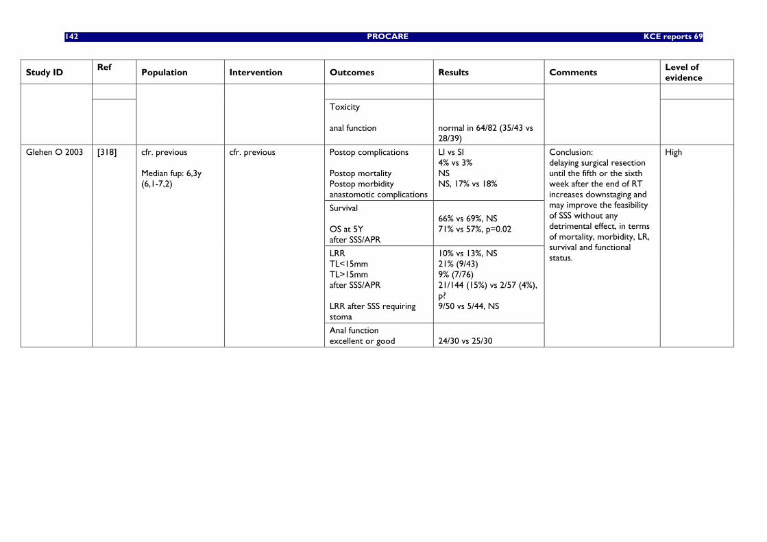
142 PROCARE KCE reports 69
Study ID Ref
Population Intervention Outcomes Results Comments Level of evidence
Toxicity anal function
normal in 64/82 (35/43 vs 28/39)
Postop complications Postop mortality Postop morbidity anastomotic complications
LI vs SI 4% vs 3% NS NS, 17% vs 18%
Survival OS at 5Y after SSS/APR
66% vs 69%, NS 71% vs 57%, p=0.02
LRR TL<15mm TL>15mm after SSS/APR LRR after SSS requiring stoma
10% vs 13%, NS 21% (9/43) 9% (7/76) 21/144 (15%) vs 2/57 (4%), p? 9/50 vs 5/44, NS
Glehen O 2003 [318] cfr. previous Median fup: 6,3y (6,1-7,2)
cfr. previous
Anal function excellent or good
24/30 vs 25/30
Conclusion: delaying surgical resection until the fifth or the sixth week after the end of RT increases downstaging and may improve the feasibility of SSS without any detrimental effect, in terms of mortality, morbidity, LR, survival and functional status.
High
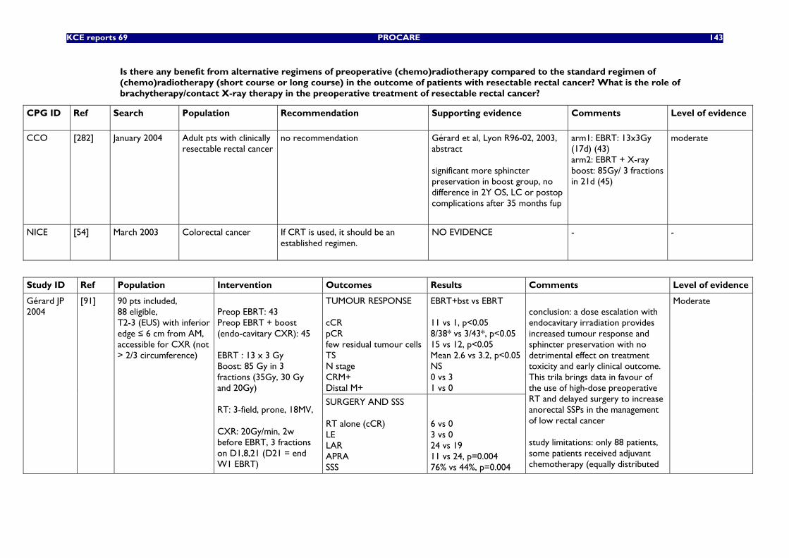
KCE reports 69 PROCARE 143
Is there any benefit from alternative regimens of preoperative (chemo)radiotherapy compared to the standard regimen of (chemo)radiotherapy (short course or long course) in the outcome of patients with resectable rectal cancer? What is the role of brachytherapy/contact X-ray therapy in the preoperative treatment of resectable rectal cancer?
CPG ID Ref Search Population Recommendation Supporting evidence
Comments Level of evidence
CCO [282] January 2004 Adult pts with clinically resectable rectal cancer
no recommendation Gérard et al, Lyon R96-02, 2003, abstract significant more sphincter preservation in boost group, no difference in 2Y OS, LC or postop complications after 35 months fup
arm1: EBRT: 13x3Gy (17d) (43) arm2: EBRT + X-ray boost: 85Gy/ 3 fractions in 21d (45)
moderate
NICE [54] March 2003 Colorectal cancer If CRT is used, it should be an established regimen.
NO EVIDENCE - -
Study ID Ref Population Intervention Outcomes Results Comments Level of evidence
TUMOUR RESPONSE cCR pCR few residual tumour cells TS N stage CRM+ Distal M+
EBRT+bst vs EBRT 11 vs 1, p<0.05 8/38* vs 3/43*, p<0.05 15 vs 12, p<0.05 Mean 2.6 vs 3.2, p<0.05 NS 0 vs 3 1 vs 0
Gérard JP 2004
[91] 90 pts included, 88 eligible, T2-3 (EUS) with inferior edge ≤ 6 cm from AM, accessible for CXR (not > 2/3 circumference)
Preop EBRT: 43 Preop EBRT + boost (endo-cavitary CXR): 45 EBRT : 13 x 3 Gy Boost: 85 Gy in 3 fractions (35Gy, 30 Gy and 20Gy) RT: 3-field, prone, 18MV, CXR: 20Gy/min, 2w before EBRT, 3 fractions on D1,8,21 (D21 = end W1 EBRT)
SURGERY AND SSS RT alone (cCR) LE LAR APRA SSS
6 vs 0 3 vs 0 24 vs 19 11 vs 24, p=0.004 76% vs 44%, p=0.004
conclusion: a dose escalation with endocavitary irradiation provides increased tumour response and sphincter preservation with no detrimental effect on treatment toxicity and early clinical outcome. This trila brings data in favour of the use of high-dose preoperative RT and delayed surgery to increase anorectal SSPs in the management of low rectal cancer study limitations: only 88 patients, some patients received adjuvant chemotherapy (equally distributed
Moderate
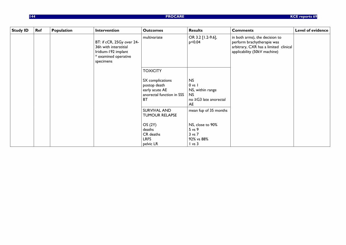
144 PROCARE KCE reports 69
Study ID Ref Population Intervention Outcomes Results Comments Level of evidence
multivariate OR 3.2 [1.2-9.6], p<0.04
TOXICITY SX complications postop death early acute AE anorectal function in SSS BT
NS 0 vs 1 NS, within range NS no ≥G3 late anorectal AE
BT: if cCR, 25Gy over 24-36h with interstitial Iridium-192 implant * examined operative specimens
SURVIVAL AND TUMOUR RELAPSE OS (2Y) deaths CR deaths LRFS pelvic LR
mean fup of 35 months NS, close to 90% 5 vs 9 3 vs 7 92% vs 88% 1 vs 3
in both arms), the decision to perform brachytherapie was arbitrary, CXR has a limited clinical applicability (50kV machine)
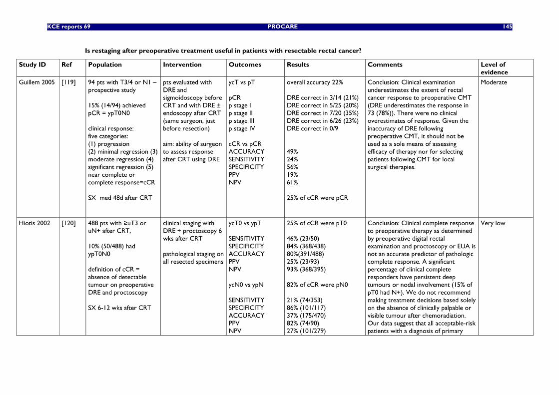
KCE reports 69 PROCARE 145
Is restaging after preoperative treatment useful in patients with resectable rectal cancer?
Study ID Ref Population Intervention Outcomes Results Comments Level of evidence
Guillem 2005 [119] 94 pts with T3/4 or N1 – prospective study 15% (14/94) achieved pCR = ypT0N0 clinical response: five categories: (1) progression (2) minimal regression (3) moderate regression (4) significant regression (5) near complete or complete response=cCR SX med 48d after CRT
pts evaluated with DRE and sigmoidoscopy before CRT and with DRE ± endoscopy after CRT (same surgeon, just before resection) aim: ability of surgeon to assess response after CRT using DRE
ycT vs pT pCR p stage I p stage II p stage III p stage IV cCR vs pCR ACCURACY SENSITIVITY SPECIFICITY PPV NPV
overall accuracy 22% DRE correct in 3/14 (21%) DRE correct in 5/25 (20%) DRE correct in 7/20 (35%) DRE correct in 6/26 (23%) DRE correct in 0/9 49% 24% 56% 19% 61% 25% of cCR were pCR
Conclusion: Clinical examination underestimates the extent of rectal cancer response to preoperative CMT (DRE underestimates the response in 73 (78%)). There were no clinical overestimates of response. Given the inaccuracy of DRE following preoperative CMT, it should not be used as a sole means of assessing efficacy of therapy nor for selecting patients following CMT for local surgical therapies.
Moderate
Hiotis 2002 [120] 488 pts with ≥uT3 or uN+ after CRT, 10% (50/488) had ypT0N0 definition of cCR = absence of detectable tumour on preoperative DRE and proctoscopy SX 6-12 wks after CRT
clinical staging with DRE + proctoscopy 6 wks after CRT pathological staging on all resected specimens
ycT0 vs ypT SENSITIVITY SPECIFICITY ACCURACY PPV NPV ycN0 vs ypN SENSITIVITY SPECIFICITY ACCURACY PPV NPV
25% of cCR were pT0 46% (23/50) 84% (368/438) 80%(391/488) 25% (23/93) 93% (368/395) 82% of cCR were pN0 21% (74/353) 86% (101/117) 37% (175/470) 82% (74/90) 27% (101/279)
Conclusion: Clinical complete response to preoperative therapy as determined by preoperative digital rectal examination and proctoscopy or EUA is not an accurate predictor of pathologic complete response. A significant percentage of clinical complete responders have persistent deep tumours or nodal involvement (15% of pT0 had N+). We do not recommend making treatment decisions based solely on the absence of clinically palpable or visible tumour after chemoradiation. Our data suggest that all acceptable-risk patients with a diagnosis of primary
Very low
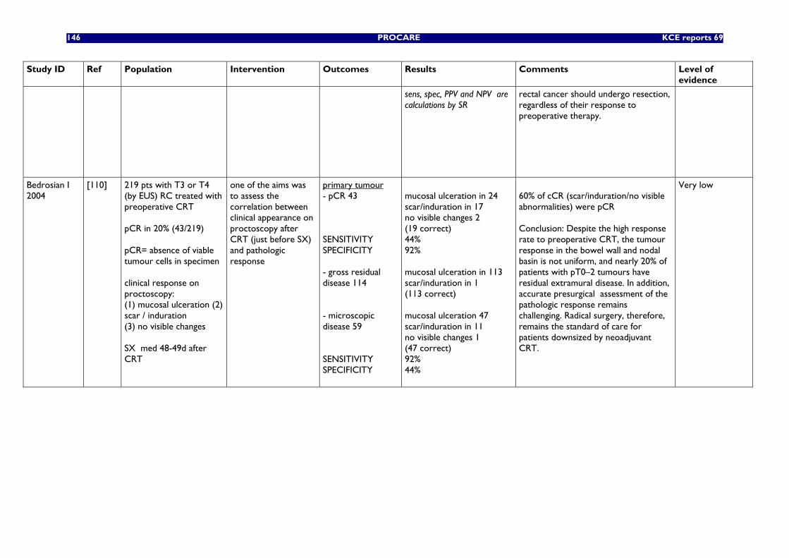
146 PROCARE KCE reports 69
Study ID Ref Population Intervention Outcomes Results Comments Level of evidence
sens, spec, PPV and NPV are calculations by SR
rectal cancer should undergo resection, regardless of their response to preoperative therapy.
Bedrosian I 2004
[110] 219 pts with T3 or T4 (by EUS) RC treated with preoperative CRT pCR in 20% (43/219) pCR= absence of viable tumour cells in specimen clinical response on proctoscopy: (1) mucosal ulceration (2) scar / induration (3) no visible changes SX med 48-49d after CRT
one of the aims was to assess the correlation between clinical appearance on proctoscopy after CRT (just before SX) and pathologic response
primary tumour - pCR 43 SENSITIVITY SPECIFICITY - gross residual disease 114 - microscopic disease 59 SENSITIVITY SPECIFICITY
mucosal ulceration in 24 scar/induration in 17 no visible changes 2 (19 correct) 44% 92% mucosal ulceration in 113 scar/induration in 1 (113 correct) mucosal ulceration 47 scar/induration in 11 no visible changes 1 (47 correct) 92% 44%
60% of cCR (scar/induration/no visible abnormalities) were pCR Conclusion: Despite the high response rate to preoperative CRT, the tumour response in the bowel wall and nodal basin is not uniform, and nearly 20% of patients with pT0–2 tumours have residual extramural disease. In addition, accurate presurgical assessment of the pathologic response remains challenging. Radical surgery, therefore, remains the standard of care for patients downsized by neoadjuvant CRT.
Very low
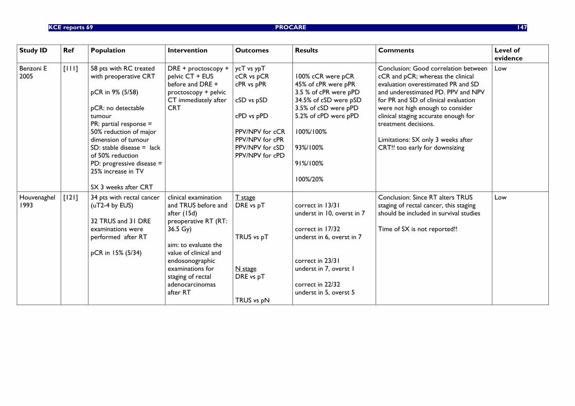
KCE reports 69 PROCARE 147
Study ID Ref Population Intervention Outcomes Results Comments Level of evidence
Benzoni E 2005
[111] 58 pts with RC treated with preoperative CRT pCR in 9% (5/58) pCR: no detectable tumour PR: partial response = 50% reduction of major dimension of tumour SD: stable disease = lack of 50% reduction PD: progressive disease = 25% increase in TV SX 3 weeks after CRT
DRE + proctoscopy + pelvic CT + EUS before and DRE + proctoscopy + pelvic CT immediately after CRT
ycT vs ypT cCR vs pCR cPR vs pPR cSD vs pSD cPD vs pPD PPV/NPV for cCR PPV/NPV for cPR PPV/NPV for cSD PPV/NPV for cPD
100% cCR were pCR 45% of cPR were pPR 3.5 % of cPR were pPD 34.5% of cSD were pSD 3.5% of cSD were pPD 5.2% of cPD were pPD 100%/100% 93%/100% 91%/100% 100%/20%
Conclusion: Good correlation between cCR and pCR; whereas the clinical evaluation overestimated PR and SD and underestimated PD. PPV and NPV for PR and SD of clinical evaluation were not high enough to consider clinical staging accurate enough for treatment decisions. Limitations: SX only 3 weeks after CRT!! too early for downsizing
Low
Houvenaghel 1993
[121] 34 pts with rectal cancer (uT2-4 by EUS) 32 TRUS and 31 DRE examinations were performed after RT pCR in 15% (5/34)
clinical examination and TRUS before and after (15d) preoperative RT (RT: 36.5 Gy) aim: to evaluate the value of clinical and endosonographic examinations for staging of rectal adenocarcinomas after RT
T stage DRE vs pT TRUS vs pT N stage DRE vs pT TRUS vs pN
correct in 13/31 underst in 10, overst in 7 correct in 17/32 underst in 6, overst in 7 correct in 23/31 underst in 7, overst 1 correct in 22/32 underst in 5, overst 5
Conclusion: Since RT alters TRUS staging of rectal cancer, this staging should be included in survival studies Time of SX is not reported!!
Low
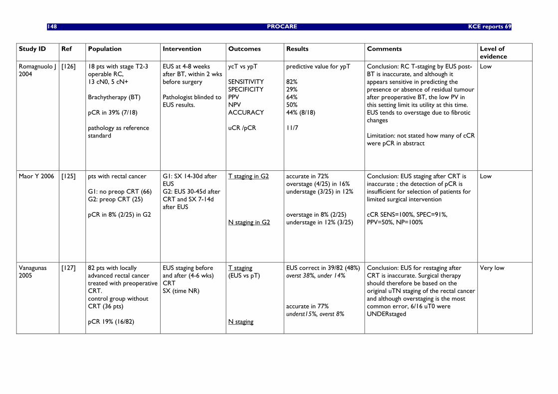
148 PROCARE KCE reports 69
Study ID Ref Population Intervention Outcomes Results Comments Level of evidence
Romagnuolo J 2004
[126] 18 pts with stage T2-3 operable RC, 13 cN0, 5 cN+ Brachytherapy (BT) pCR in 39% (7/18) pathology as reference standard
EUS at 4-8 weeks after BT, within 2 wks before surgery Pathologist blinded to EUS results.
ycT vs ypT SENSITIVITY SPECIFICITY PPV NPV ACCURACY uCR /pCR
predictive value for ypT 82% 29% 64% 50% 44% (8/18) 11/7
Conclusion: RC T-staging by EUS post-BT is inaccurate, and although it appears sensitive in predicting the presence or absence of residual tumour after preoperative BT, the low PV in this setting limit its utility at this time. EUS tends to overstage due to fibrotic changes Limitation: not stated how many of cCR were pCR in abstract
Low
Maor Y 2006 [125] pts with rectal cancer G1: no preop CRT (66) G2: preop CRT (25) pCR in 8% (2/25) in G2
G1: SX 14-30d after EUS G2: EUS 30-45d after CRT and SX 7-14d after EUS
T staging in G2 N staging in G2
accurate in 72% overstage (4/25) in 16% understage (3/25) in 12% overstage in 8% (2/25) understage in 12% (3/25)
Conclusion: EUS staging after CRT is inaccurate ; the detection of pCR is insufficient for selection of patients for limited surgical intervention cCR SENS=100%, SPEC=91%, PPV=50%, NP=100%
Low
Vanagunas 2005
[127] 82 pts with locally advanced rectal cancer treated with preoperative CRT. control group without CRT (36 pts) pCR 19% (16/82)
EUS staging before and after (4-6 wks) CRT SX (time NR)
T staging (EUS vs pT) N staging
EUS correct in 39/82 (48%) overst 38%, under 14% accurate in 77% underst15%, overst 8%
Conclusion: EUS for restaging after CRT is inaccurate. Surgical therapy should therefore be based on the original uTN staging of the rectal cancer and although overstaging is the most common error, 6/16 uT0 were UNDERstaged
Very low
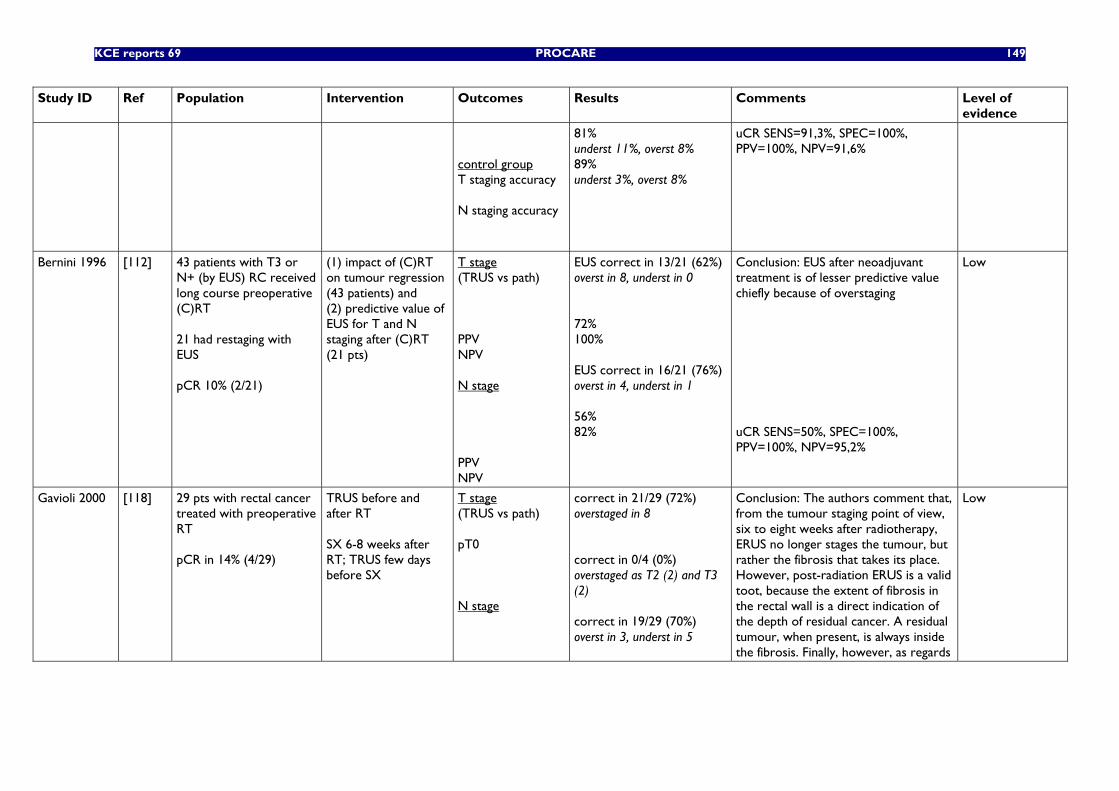
KCE reports 69 PROCARE 149
Study ID Ref Population Intervention Outcomes Results Comments Level of evidence
control group T staging accuracy N staging accuracy
81% underst 11%, overst 8% 89% underst 3%, overst 8%
uCR SENS=91,3%, SPEC=100%, PPV=100%, NPV=91,6%
Bernini 1996 [112] 43 patients with T3 or N+ (by EUS) RC received long course preoperative (C)RT 21 had restaging with EUS pCR 10% (2/21)
(1) impact of (C)RT on tumour regression (43 patients) and (2) predictive value of EUS for T and N staging after (C)RT (21 pts)
T stage (TRUS vs path) PPV NPV N stage PPV NPV
EUS correct in 13/21 (62%) overst in 8, underst in 0 72% 100% EUS correct in 16/21 (76%) overst in 4, underst in 1 56% 82%
Conclusion: EUS after neoadjuvant treatment is of lesser predictive value chiefly because of overstaging uCR SENS=50%, SPEC=100%, PPV=100%, NPV=95,2%
Low
Gavioli 2000 [118] 29 pts with rectal cancer treated with preoperative RT pCR in 14% (4/29)
TRUS before and after RT SX 6-8 weeks after RT; TRUS few days before SX
T stage (TRUS vs path) pT0 N stage
correct in 21/29 (72%) overstaged in 8 correct in 0/4 (0%) overstaged as T2 (2) and T3 (2) correct in 19/29 (70%) overst in 3, underst in 5
Conclusion: The authors comment that, from the tumour staging point of view, six to eight weeks after radiotherapy, ERUS no longer stages the tumour, but rather the fibrosis that takes its place. However, post-radiation ERUS is a valid toot, because the extent of fibrosis in the rectal wall is a direct indication of the depth of residual cancer. A residual tumour, when present, is always inside the fibrosis. Finally, however, as regards
Low
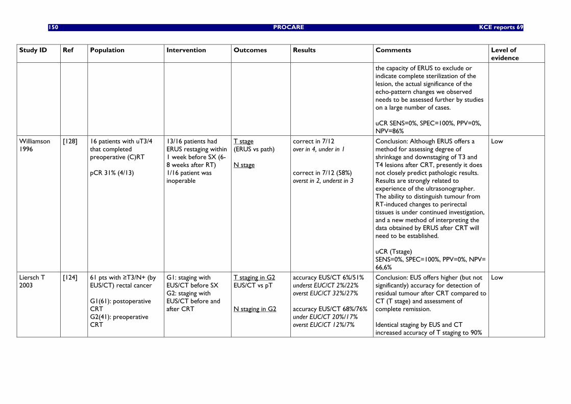
150 PROCARE KCE reports 69
Study ID Ref Population Intervention Outcomes Results Comments Level of evidence
the capacity of ERUS to exclude or indicate complete sterilization of the lesion, the actual significance of the echo-pattern changes we observed needs to be assessed further by studies on a large number of cases. uCR SENS=0%, SPEC=100%, PPV=0%, NPV=86%
Williamson 1996
[128] 16 patients with uT3/4 that completed preoperative (C)RT pCR 31% (4/13)
13/16 patients had ERUS restaging within 1 week before SX (6-8 weeks after RT) 1/16 patient was inoperable
T stage (ERUS vs path) N stage
correct in 7/12 over in 4, under in 1 correct in 7/12 (58%) overst in 2, underst in 3
Conclusion: Although ERUS offers a method for assessing degree of shrinkage and downstaging of T3 and T4 lesions after CRT, presently it does not closely predict pathologic results. Results are strongly related to experience of the ultrasonographer. The ability to distinguish tumour from RT-induced changes to perirectal tissues is under continued investigation, and a new method of interpreting the data obtained by ERUS after CRT will need to be established. uCR (Tstage) SENS=0%, SPEC=100%, PPV=0%, NPV= 66,6%
Low
Liersch T 2003
[124] 61 pts with ≥T3/N+ (by EUS/CT) rectal cancer G1(61): postoperative CRT G2(41): preoperative CRT
G1: staging with EUS/CT before SX G2: staging with EUS/CT before and after CRT
T staging in G2 EUS/CT vs pT N staging in G2
accuracy EUS/CT 6%/51% underst EUC/CT 2%/22% overst EUC/CT 32%/27% accuracy EUS/CT 68%/76% under EUC/CT 20%/17% overst EUC/CT 12%/7%
Conclusion: EUS offers higher (but not significantly) accuracy for detection of residual tumour after CRT compared to CT (T stage) and assessment of complete remission. Identical staging by EUS and CT increased accuracy of T staging to 90%
Low
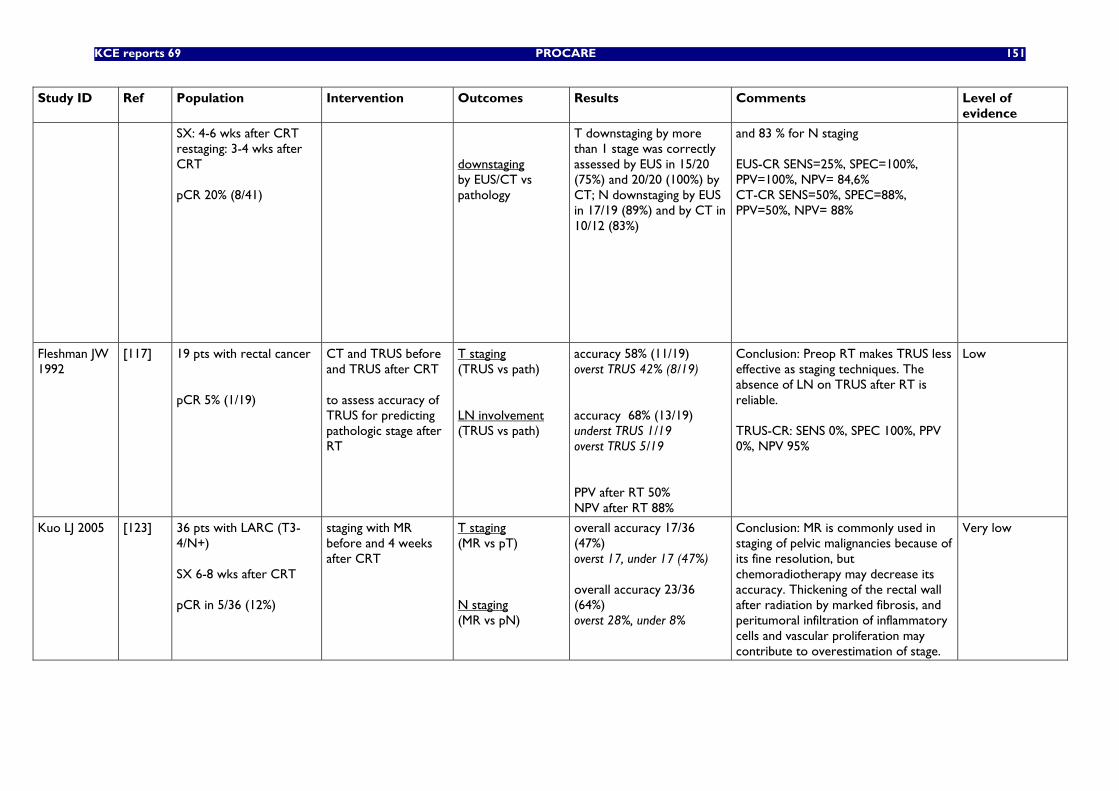
KCE reports 69 PROCARE 151
Study ID Ref Population Intervention Outcomes Results Comments Level of evidence
SX: 4-6 wks after CRT restaging: 3-4 wks after CRT pCR 20% (8/41)
downstaging by EUS/CT vs pathology
T downstaging by more than 1 stage was correctly assessed by EUS in 15/20 (75%) and 20/20 (100%) by CT; N downstaging by EUS in 17/19 (89%) and by CT in 10/12 (83%)
and 83 % for N staging EUS-CR SENS=25%, SPEC=100%, PPV=100%, NPV= 84,6% CT-CR SENS=50%, SPEC=88%, PPV=50%, NPV= 88%
Fleshman JW 1992
[117] 19 pts with rectal cancer pCR 5% (1/19)
CT and TRUS before and TRUS after CRT to assess accuracy of TRUS for predicting pathologic stage after RT
T staging (TRUS vs path) LN involvement (TRUS vs path)
accuracy 58% (11/19) overst TRUS 42% (8/19) accuracy 68% (13/19) underst TRUS 1/19 overst TRUS 5/19 PPV after RT 50% NPV after RT 88%
Conclusion: Preop RT makes TRUS less effective as staging techniques. The absence of LN on TRUS after RT is reliable. TRUS-CR: SENS 0%, SPEC 100%, PPV 0%, NPV 95%
Low
Kuo LJ 2005 [123] 36 pts with LARC (T3-4/N+) SX 6-8 wks after CRT pCR in 5/36 (12%)
staging with MR before and 4 weeks after CRT
T staging (MR vs pT) N staging (MR vs pN)
overall accuracy 17/36 (47%) overst 17, under 17 (47%) overall accuracy 23/36 (64%) overst 28%, under 8%
Conclusion: MR is commonly used in staging of pelvic malignancies because of its fine resolution, but chemoradiotherapy may decrease its accuracy. Thickening of the rectal wall after radiation by marked fibrosis, and peritumoral infiltration of inflammatory cells and vascular proliferation may contribute to overestimation of stage.
Very low
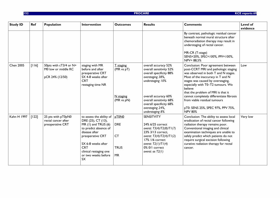
152 PROCARE KCE reports 69
Study ID Ref Population Intervention Outcomes Results Comments Level of evidence
By contrast, pathologic residual cancer beneath normal mural structure after chemoradiation therapy may result in understaging of rectal cancer. MR-CR (T-stage) SENS=20%, SPEC=100%, PPV=100%, NPV= 88,5%
Chen 2005 [116] 50pts with cT3/4 or N+ M0 low or middle RC pCR 24% (12/50)
staging with MR before and after preoperative CRT SX 4-8 weeks after CRT restaging time NR
T staging (MR vs pT) N staging (MR vs pN)
overall accuracy 52% overall sensitivity 52% overall specificity 88% overstaging 38%, understaging 10% overall accuracy 60% overall sensitivity 68% overall specificity 68% overstaging 24%, understaging 8%
Conclusion: Poor agreement between post-CCRT MRI and pathologic staging was observed in both T and N stages. Most of the inaccuracy in T and N stages was caused by overstaging, especially with T0–T2 tumours. We believe that the problem of MRI is that it cannot completely differentiate fibrosis from viable residual tumours pT0: SENS 25%, SPEC 97%, PPV 75%, NPV 80%
Low
Kahn H 1997 [122] 25 pts with pT0pN0 rectal cancer after preoperative CRT
to assess the ability of DRE (25), CT (13), MR (1) and TRUS (6) to predict absence of disease after preoperative CRT SX 6-8 weeks after CRT clinical restaging one or two weeks before SX
pT0N0 DRE CT TRUS MR
SENSITVITY 24% 6/25 correct overst: T3(4)/T2(8)/T1(7) 23% 3/13 correct, overst: T3(4)/T2(4)/T1(2) 17% 1/6 correct overst: T2(1)/T1(4) 0% 0/1 correct overst: as T2(1)
Conclusion: The ability to assess local eradication of rectal cancer following radiation therapy remains poor. Conventional imaging and clinical examination techniques are unable to safely predict which patients do not require surgical excision following curative radiation therapy for rectal cancer.
Very low
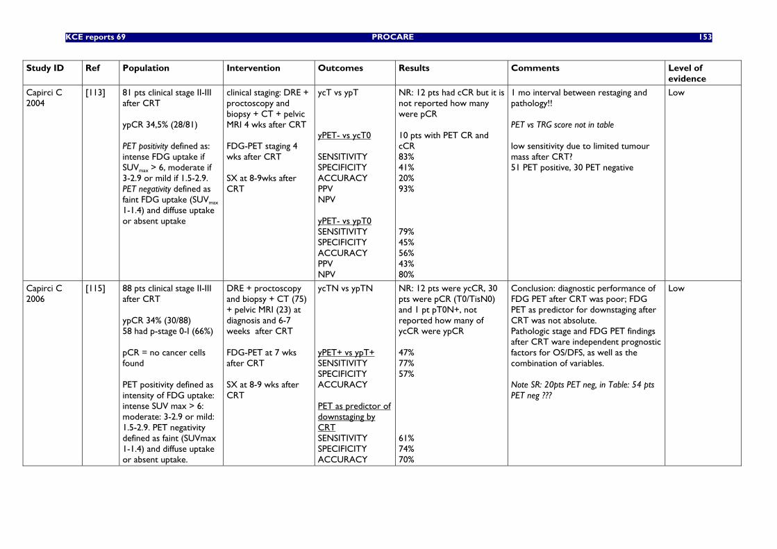
KCE reports 69 PROCARE 153
Study ID Ref Population Intervention Outcomes Results Comments Level of evidence
Capirci C 2004
[113] 81 pts clinical stage II-III after CRT ypCR 34,5% (28/81) PET positivity defined as: intense FDG uptake if SUVmax > 6, moderate if 3-2.9 or mild if 1.5-2.9. PET negativity defined as faint FDG uptake (SUVmax 1-1.4) and diffuse uptake or absent uptake
clinical staging: DRE + proctoscopy and biopsy + CT + pelvic MRI 4 wks after CRT FDG-PET staging 4 wks after CRT SX at 8-9wks after CRT
ycT vs ypT yPET- vs ycT0 SENSITIVITY SPECIFICITY ACCURACY PPV NPV yPET- vs ypT0 SENSITIVITY SPECIFICITY ACCURACY PPV NPV
NR: 12 pts had cCR but it is not reported how many were pCR 10 pts with PET CR and cCR 83% 41% 20% 93% 79% 45% 56% 43% 80%
1 mo interval between restaging and pathology!! PET vs TRG score not in table low sensitivity due to limited tumour mass after CRT? 51 PET positive, 30 PET negative
Low
Capirci C 2006
[115] 88 pts clinical stage II-III after CRT ypCR 34% (30/88) 58 had p-stage 0-I (66%) pCR = no cancer cells found PET positivity defined as intensity of FDG uptake: intense SUV max > 6: moderate: 3-2.9 or mild: 1.5-2.9. PET negativity defined as faint (SUVmax 1-1.4) and diffuse uptake or absent uptake.
DRE + proctoscopy and biopsy + CT (75) + pelvic MRI (23) at diagnosis and 6-7 weeks after CRT FDG-PET at 7 wks after CRT SX at 8-9 wks after CRT
ycTN vs ypTN yPET+ vs ypT+ SENSITIVITY SPECIFICITY ACCURACY PET as predictor of downstaging by CRT SENSITIVITY SPECIFICITY ACCURACY
NR: 12 pts were ycCR, 30 pts were pCR (T0/TisN0) and 1 pt pT0N+, not reported how many of ycCR were ypCR 47% 77% 57% 61% 74% 70%
Conclusion: diagnostic performance of FDG PET after CRT was poor; FDG PET as predictor for downstaging after CRT was not absolute. Pathologic stage and FDG PET findings after CRT ware independent prognostic factors for OS/DFS, as well as the combination of variables. Note SR: 20pts PET neg, in Table: 54 pts PET neg ???
Low
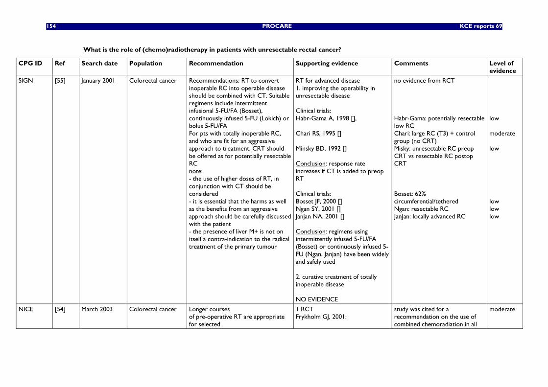
154 PROCARE KCE reports 69
What is the role of (chemo)radiotherapy in patients with unresectable rectal cancer?
CPG ID Ref Search date Population Recommendation Supporting evidence
Comments Level of evidence
SIGN [55] January 2001 Colorectal cancer Recommendations: RT to convert inoperable RC into operable disease should be combined with CT. Suitable regimens include intermittent infusional 5-FU/FA (Bosset), continuously infused 5-FU (Lokich) or bolus 5-FU/FA For pts with totally inoperable RC, and who are fit for an aggressive approach to treatment, CRT should be offered as for potentially resectable RC note: - the use of higher doses of RT, in conjunction with CT should be considered - it is essential that the harms as well as the benefits from an aggressive approach should be carefully discussed with the patient - the presence of liver M+ is not on itself a contra-indication to the radical treatment of the primary tumour
RT for advanced disease 1. improving the operability in unresectable disease Clinical trials: Habr-Gama A, 1998 [], Chari RS, 1995 [] Minsky BD, 1992 [] Conclusion: response rate increases if CT is added to preop RT Clinical trials: Bosset JF, 2000 [] Ngan SY, 2001 [] Janjan NA, 2001 [] Conclusion: regimens using intermittently infused 5-FU/FA (Bosset) or continuously infused 5-FU (Ngan, Janjan) have been widely and safely used 2. curative treatment of totally inoperable disease NO EVIDENCE
no evidence from RCT Habr-Gama: potentially resectable low RC Chari: large RC (T3) + control group (no CRT) Misky: unresectable RC preop CRT vs resectable RC postop CRT Bosset: 62% circumferential/tethered Ngan: resectable RC JanJan: locally advanced RC
low moderate low low low low
NICE [54] March 2003 Colorectal cancer Longer courses of pre-operative RT are appropriate for selected
1 RCT Frykholm GJ, 2001:
study was cited for a recommendation on the use of combined chemoradiation in all
moderate
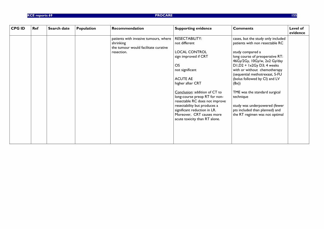
KCE reports 69 PROCARE 155
CPG ID Ref Search date Population Recommendation Supporting evidence
Comments Level of evidence
patients with invasive tumours, where shrinking the tumour would facilitate curative resection.
RESECTABILITY: not different LOCAL CONTROL sign improved if CRT OS not significant ACUTE AE higher after CRT Conclusion: addition of CT to long-course preop RT for non-resectable RC does not improve resectability but produces a significant reduction in LR. Moreover, CRT causes more acute toxicity than RT alone.
cases, but the study only included patients with non resectable RC study compared a long course of preoperative RT: 46Gy/2Gy, 10Gy/w, 2x2 Gy/day D1,D2 + 1x2Gy D3; 4 weeks with or without chemotherapy (sequential methotrexaat, 5-FU (bolus followed by CI) and LV (8x)) TME was the standard surgical technique study was underpowered (fewer pts included than planned) and the RT regimen was not optimal
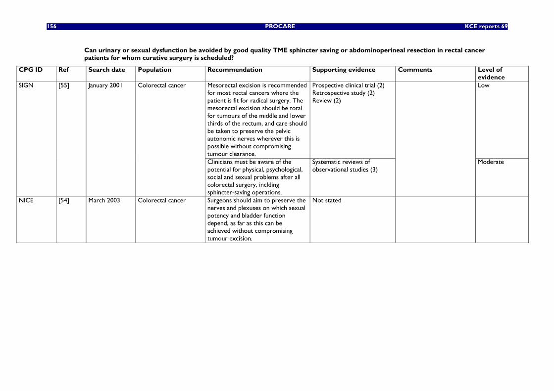
156 PROCARE KCE reports 69
Can urinary or sexual dysfunction be avoided by good quality TME sphincter saving or abdominoperineal resection in rectal cancer patients for whom curative surgery is scheduled?
CPG ID Ref Search date Population Recommendation Supporting evidence Comments Level of evidence
Mesorectal excision is recommended for most rectal cancers where the patient is fit for radical surgery. The mesorectal excision should be total for tumours of the middle and lower thirds of the rectum, and care should be taken to preserve the pelvic autonomic nerves wherever this is possible without compromising tumour clearance.
Prospective clinical trial (2) Retrospective study (2) Review (2)
Low SIGN [55] January 2001 Colorectal cancer
Clinicians must be aware of the potential for physical, psychological, social and sexual problems after all colorectal surgery, inclding sphincter-saving operations.
Systematic reviews of observational studies (3)
Moderate
NICE [54] March 2003 Colorectal cancer Surgeons should aim to preserve the nerves and plexuses on which sexual potency and bladder function depend, as far as this can be achieved without compromising tumour excision.
Not stated
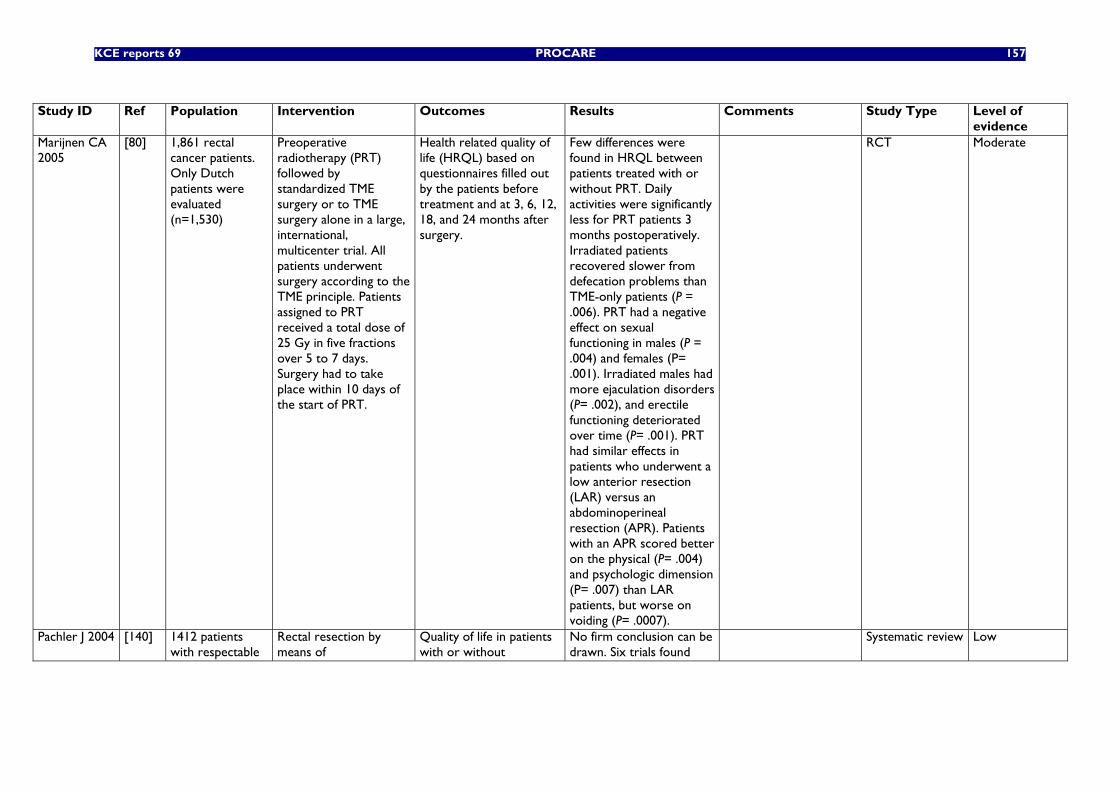
KCE reports 69 PROCARE 157
Study ID Ref Population Intervention Outcomes Results Comments Study Type Level of
evidence Marijnen CA 2005
[80] 1,861 rectal cancer patients. Only Dutch patients were evaluated (n=1,530)
Preoperative radiotherapy (PRT) followed by standardized TME surgery or to TME surgery alone in a large, international, multicenter trial. All patients underwent surgery according to the TME principle. Patients assigned to PRT received a total dose of 25 Gy in five fractions over 5 to 7 days. Surgery had to take place within 10 days of the start of PRT.
Health related quality of life (HRQL) based on questionnaires filled out by the patients before treatment and at 3, 6, 12, 18, and 24 months after surgery.
Few differences were found in HRQL between patients treated with or without PRT. Daily activities were significantly less for PRT patients 3 months postoperatively. Irradiated patients recovered slower from defecation problems than TME-only patients (P = .006). PRT had a negative effect on sexual functioning in males (P = .004) and females (P= .001). Irradiated males had more ejaculation disorders (P= .002), and erectile functioning deteriorated over time (P= .001). PRT had similar effects in patients who underwent a low anterior resection (LAR) versus an abdominoperineal resection (APR). Patients with an APR scored better on the physical (P= .004) and psychologic dimension (P= .007) than LAR patients, but worse on voiding (P= .0007).
RCT Moderate
Pachler J 2004 [140] 1412 patients with respectable
Rectal resection by means of
Quality of life in patients with or without
No firm conclusion can be drawn. Six trials found
Systematic review Low
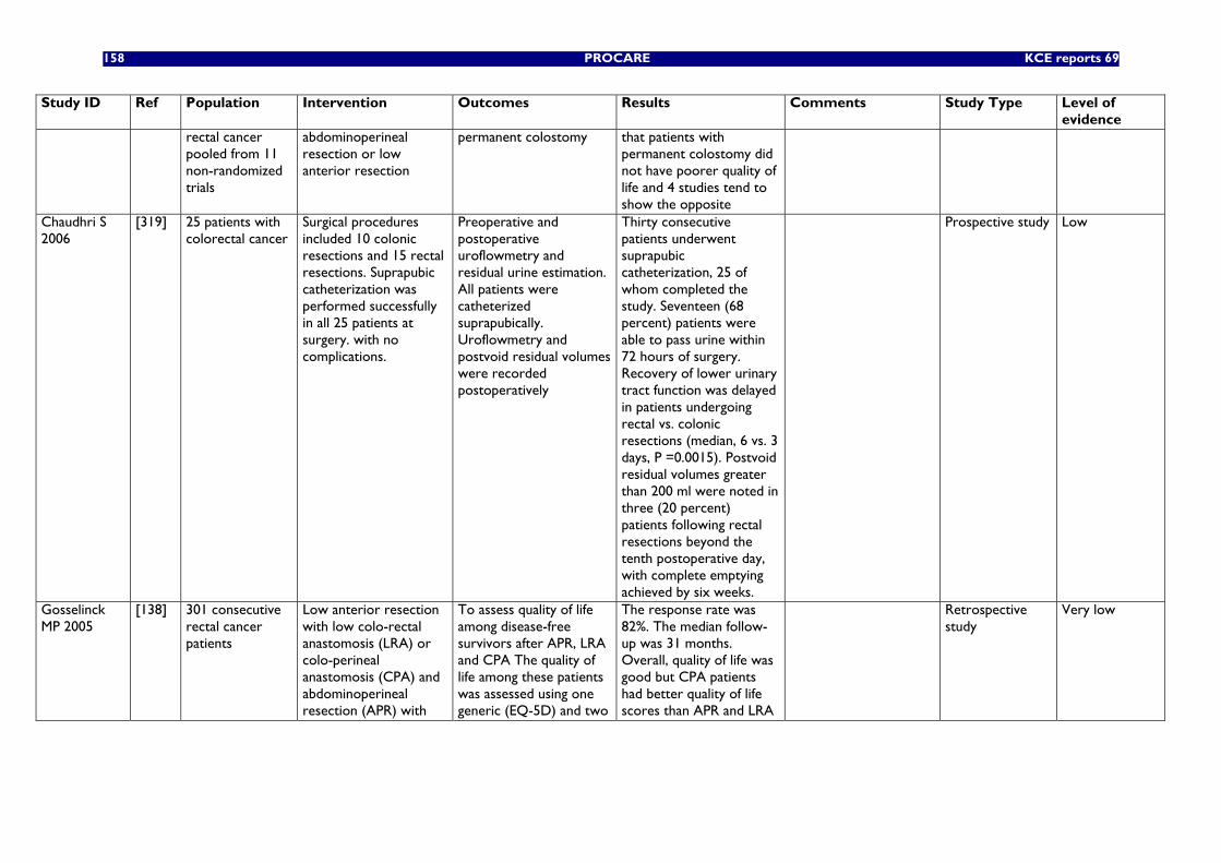
158 PROCARE KCE reports 69
Study ID Ref Population Intervention Outcomes Results Comments Study Type Level of evidence
rectal cancer pooled from 11 non-randomized trials
abdominoperineal resection or low anterior resection
permanent colostomy that patients with permanent colostomy did not have poorer quality of life and 4 studies tend to show the opposite
Chaudhri S 2006
[319] 25 patients with colorectal cancer
Surgical procedures included 10 colonic resections and 15 rectal resections. Suprapubic catheterization was performed successfully in all 25 patients at surgery. with no complications.
Preoperative and postoperative uroflowmetry and residual urine estimation. All patients were catheterized suprapubically. Uroflowmetry and postvoid residual volumes were recorded postoperatively
Thirty consecutive patients underwent suprapubic catheterization, 25 of whom completed the study. Seventeen (68 percent) patients were able to pass urine within 72 hours of surgery. Recovery of lower urinary tract function was delayed in patients undergoing rectal vs. colonic resections (median, 6 vs. 3 days, P =0.0015). Postvoid residual volumes greater than 200 ml were noted in three (20 percent) patients following rectal resections beyond the tenth postoperative day, with complete emptying achieved by six weeks.
Prospective study Low
Gosselinck MP 2005
[138] 301 consecutive rectal cancer patients
Low anterior resection with low colo-rectal anastomosis (LRA) or colo-perineal anastomosis (CPA) and abdominoperineal resection (APR) with
To assess quality of life among disease-free survivors after APR, LRA and CPA The quality of life among these patients was assessed using one generic (EQ-5D) and two
The response rate was 82%. The median follow-up was 31 months. Overall, quality of life was good but CPA patients had better quality of life scores than APR and LRA
Retrospective study
Very low
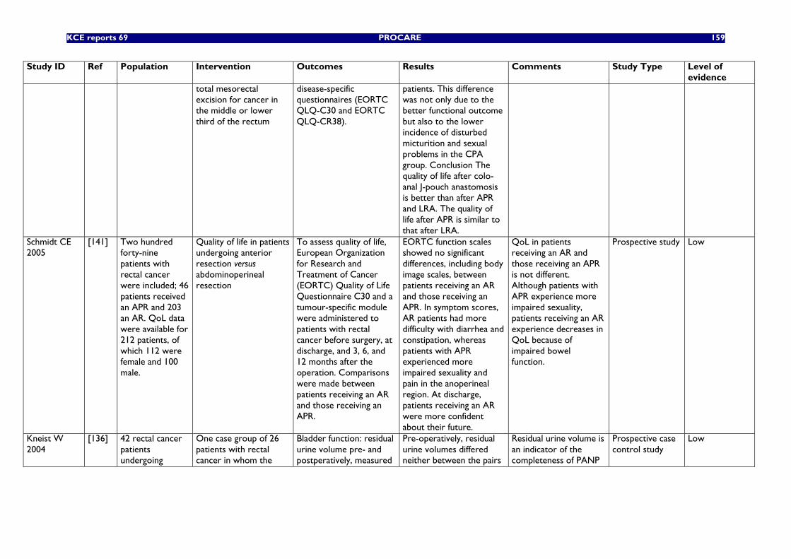
KCE reports 69 PROCARE 159
Study ID Ref Population Intervention Outcomes Results Comments Study Type Level of evidence
total mesorectal excision for cancer in the middle or lower third of the rectum
disease-specific questionnaires (EORTC QLQ-C30 and EORTC QLQ-CR38).
patients. This difference was not only due to the better functional outcome but also to the lower incidence of disturbed micturition and sexual problems in the CPA group. Conclusion The quality of life after colo-anal J-pouch anastomosis is better than after APR and LRA. The quality of life after APR is similar to that after LRA.
Schmidt CE 2005
[141] Two hundred forty-nine patients with rectal cancer were included; 46 patients received an APR and 203 an AR. QoL data were available for 212 patients, of which 112 were female and 100 male.
Quality of life in patients undergoing anterior resection versus abdominoperineal resection
To assess quality of life, European Organization for Research and Treatment of Cancer (EORTC) Quality of Life Questionnaire C30 and a tumour-specific module were administered to patients with rectal cancer before surgery, at discharge, and 3, 6, and 12 months after the operation. Comparisons were made between patients receiving an AR and those receiving an APR.
EORTC function scales showed no significant differences, including body image scales, between patients receiving an AR and those receiving an APR. In symptom scores, AR patients had more difficulty with diarrhea and constipation, whereas patients with APR experienced more impaired sexuality and pain in the anoperineal region. At discharge, patients receiving an AR were more confident about their future.
QoL in patients receiving an AR and those receiving an APR is not different. Although patients with APR experience more impaired sexuality, patients receiving an AR experience decreases in QoL because of impaired bowel function.
Prospective study Low
Kneist W 2004
[136] 42 rectal cancer patients undergoing
One case group of 26 patients with rectal cancer in whom the
Bladder function: residual urine volume pre- and postperatively, measured
Pre-operatively, residual urine volumes differed neither between the pairs
Residual urine volume is an indicator of the completeness of PANP
Prospective case control study
Low
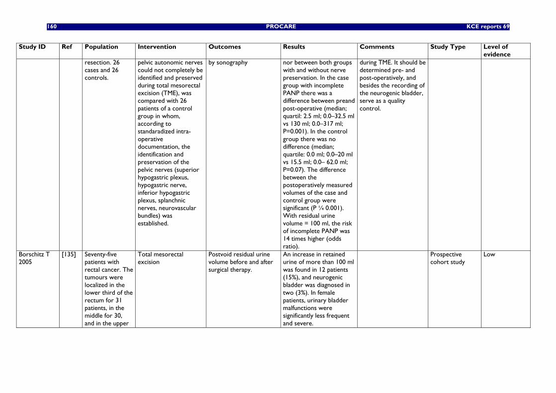
160 PROCARE KCE reports 69
Study ID Ref Population Intervention Outcomes Results Comments Study Type Level of evidence
resection. 26 cases and 26 controls.
pelvic autonomic nerves could not completely be identified and preserved during total mesorectal excision (TME), was compared with 26 patients of a control group in whom, according to standaradized intra-operative documentation, the identification and preservation of the pelvic nerves (superior hypogastric plexus, hypogastric nerve, inferior hypogastric plexus, splanchnic nerves, neurovascular bundles) was established.
by sonography nor between both groups with and without nerve preservation. In the case group with incomplete PANP there was a difference between preand post-operative (median; quartil: 2.5 ml; 0.0–32.5 ml vs 130 ml; 0.0–317 ml; P=0.001). In the control group there was no difference (median; quartile: 0.0 ml; 0.0–20 ml vs 15.5 ml; 0.0– 62.0 ml; P=0.07). The difference between the postoperatively measured volumes of the case and control group were significant (P ¼ 0.001). With residual urine volume = 100 ml, the risk of incomplete PANP was 14 times higher (odds ratio).
during TME. It should be determined pre- and post-operatively, and besides the recording of the neurogenic bladder, serve as a quality control.
Borschitz T 2005
[135] Seventy-five patients with rectal cancer. The tumours were localized in the lower third of the rectum for 31 patients, in the middle for 30, and in the upper
Total mesorectal excision
Postvoid residual urine volume before and after surgical therapy.
An increase in retained urine of more than 100 ml was found in 12 patients (15%), and neurogenic bladder was diagnosed in two (3%). In female patients, urinary bladder malfunctions were significantly less frequent and severe.
Prospective cohort study
Low
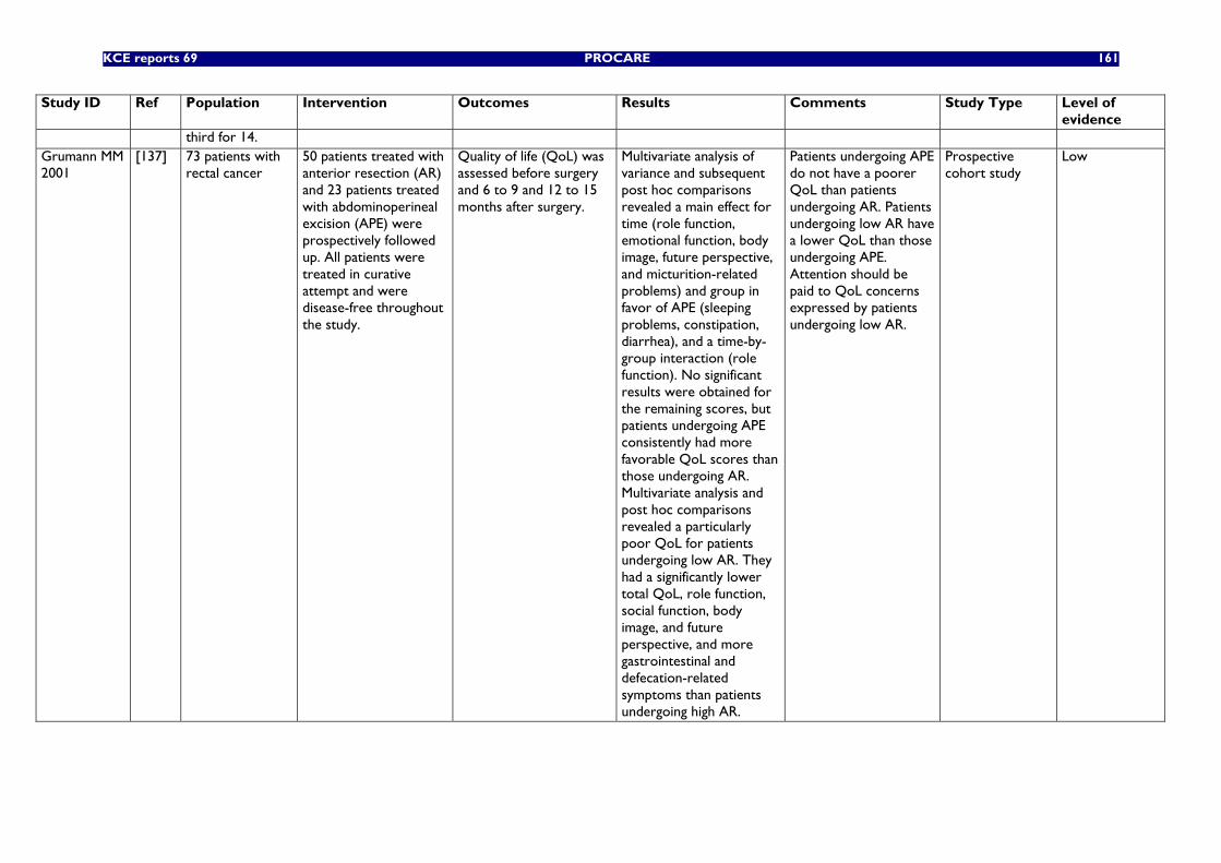
KCE reports 69 PROCARE 161
Study ID Ref Population Intervention Outcomes Results Comments Study Type Level of evidence
third for 14. Grumann MM 2001
[137] 73 patients with rectal cancer
50 patients treated with anterior resection (AR) and 23 patients treated with abdominoperineal excision (APE) were prospectively followed up. All patients were treated in curative attempt and were disease-free throughout the study.
Quality of life (QoL) was assessed before surgery and 6 to 9 and 12 to 15 months after surgery.
Multivariate analysis of variance and subsequent post hoc comparisons revealed a main effect for time (role function, emotional function, body image, future perspective, and micturition-related problems) and group in favor of APE (sleeping problems, constipation, diarrhea), and a time-by-group interaction (role function). No significant results were obtained for the remaining scores, but patients undergoing APE consistently had more favorable QoL scores than those undergoing AR. Multivariate analysis and post hoc comparisons revealed a particularly poor QoL for patients undergoing low AR. They had a significantly lower total QoL, role function, social function, body image, and future perspective, and more gastrointestinal and defecation-related symptoms than patients undergoing high AR.
Patients undergoing APE do not have a poorer QoL than patients undergoing AR. Patients undergoing low AR have a lower QoL than those undergoing APE. Attention should be paid to QoL concerns expressed by patients undergoing low AR.
Prospective cohort study
Low
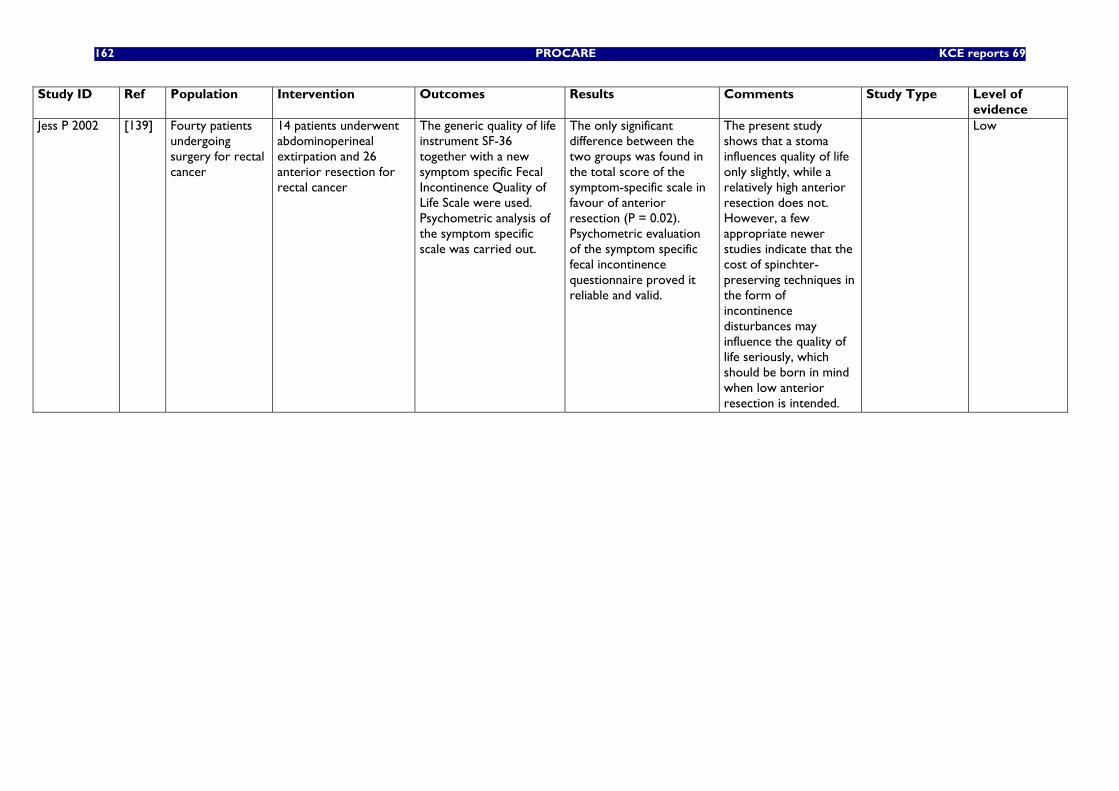
162 PROCARE KCE reports 69
Study ID Ref Population Intervention Outcomes Results Comments Study Type Level of evidence
Jess P 2002 [139] Fourty patients undergoing surgery for rectal cancer
14 patients underwent abdominoperineal extirpation and 26 anterior resection for rectal cancer
The generic quality of life instrument SF-36 together with a new symptom specific Fecal Incontinence Quality of Life Scale were used. Psychometric analysis of the symptom specific scale was carried out.
The only significant difference between the two groups was found in the total score of the symptom-specific scale in favour of anterior resection (P = 0.02). Psychometric evaluation of the symptom specific fecal incontinence questionnaire proved it reliable and valid.
The present study shows that a stoma influences quality of life only slightly, while a relatively high anterior resection does not. However, a few appropriate newer studies indicate that the cost of spinchter-preserving techniques in the form of incontinence disturbances may influence the quality of life seriously, which should be born in mind when low anterior resection is intended.
Low
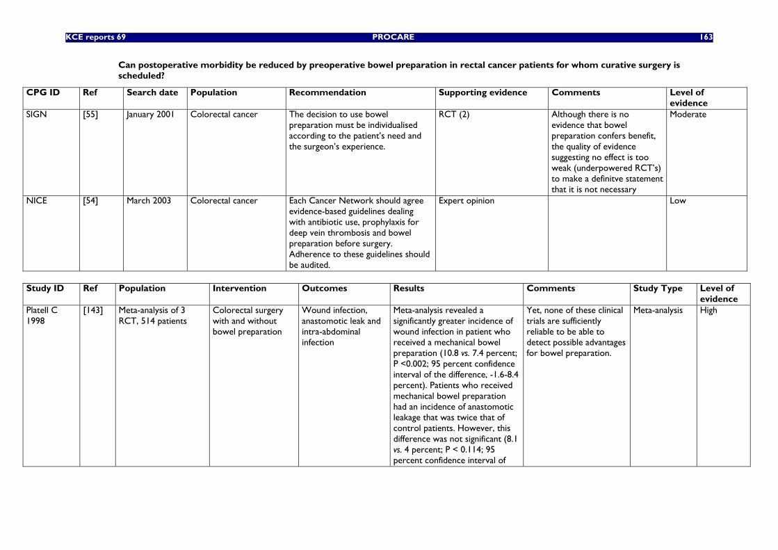
KCE reports 69 PROCARE 163
Can postoperative morbidity be reduced by preoperative bowel preparation in rectal cancer patients for whom curative surgery is scheduled?
CPG ID Ref Search date Population Recommendation Supporting evidence Comments Level of evidence
SIGN [55] January 2001 Colorectal cancer The decision to use bowel preparation must be individualised according to the patient’s need and the surgeon’s experience.
RCT (2) Although there is no evidence that bowel preparation confers benefit, the quality of evidence suggesting no effect is too weak (underpowered RCT’s) to make a definitve statement that it is not necessary
Moderate
NICE [54] March 2003 Colorectal cancer Each Cancer Network should agree evidence-based guidelines dealing with antibiotic use, prophylaxis for deep vein thrombosis and bowel preparation before surgery. Adherence to these guidelines should be audited.
Expert opinion Low
Study ID Ref Population Intervention Outcomes Results Comments Study Type Level of
evidence Platell C 1998
[143] Meta-analysis of 3 RCT, 514 patients
Colorectal surgery with and without bowel preparation
Wound infection, anastomotic leak and intra-abdominal infection
Meta-analysis revealed a significantly greater incidence of wound infection in patient who received a mechanical bowel preparation (10.8 vs. 7.4 percent; P <0.002; 95 percent confidence interval of the difference, -1.6-8.4 percent). Patients who received mechanical bowel preparation had an incidence of anastomotic leakage that was twice that of control patients. However, this difference was not significant (8.1 vs. 4 percent; P < 0.114; 95 percent confidence interval of
Yet, none of these clinical trials are sufficiently reliable to be able to detect possible advantages for bowel preparation.
Meta-analysis High
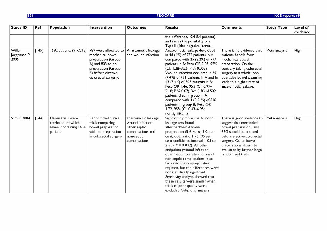
164 PROCARE KCE reports 69
Study ID Ref Population Intervention Outcomes Results Comments Study Type Level of evidence
the difference, -0.4-8.4 percent) and raises the possibility of a Type II (false-negative) error.
Wille-Jorgensen P 2005
[145] 1592 patients (9 RCTs) 789 were allocated to mechanical bowel preparation (Group A) and 803 to no preparation (Group B) before elective colorectal surgery.
Anastomotic leakage and wound infection
Anastomotic leakage developed in 48 (6%) of 772 patients in A compared with 25 (3.2%) of 777 patients in B; Peto OR 2.03, 95% (CI: 1.28–3.26; P ¼ 0.003). Wound infection occurred in 59 (7.4%) of 791 patients in A and in 43 (5.4%) of 803 patients in B; Peto OR 1.46, 95% (CI: 0.97–2.18; P ¼ 0.07);Five (1%) of 509 patients died in group in A compared with 3 (0.61%) of 516 patients in group B; Peto OR 1.72, 95% (CI: 0.43–6.95; nonsignificant)
There is no evidence that patients benefit from mechanical bowel preparation. On the contrary taking colorectal surgery as a whole, pre-operative bowel cleansing leads to a higher rate of anastomotic leakage.
Meta-analysis High
Slim K 2004 [144] Eleven trials were retrieved, of which seven, containing 1454 patients
Randomized clinical trials comparing bowel preparation with no preparation in colorectal surgery
anastomotic leakage, wound infection, other septic complications and non-septic complications
Significantly more anastomotic leakage was found aftermechanical bowel preparation (5·6 versus 3·2 per cent; odds ratio 1·75 (95 per cent confidence interval 1·05 to 2·90); P = 0·032). All other endpoints (wound infection, other septic complications and non-septic complications) also favoured the no-preparation regimen, but the differences were not statistically significant. Sensitivity analysis showed that these results were similar when trials of poor quality were excluded. Subgroup analysis
There is good evidence to suggest that mechanical bowel preparation using PEG should be omitted before elective colorectal surgery. Other bowel preparations should be evaluated by further large randomized trials.
Meta-analysis High
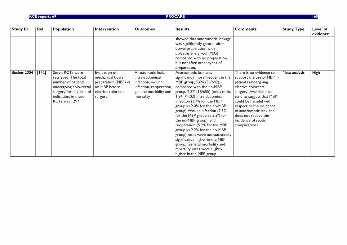
KCE reports 69 PROCARE 165
Study ID Ref Population Intervention Outcomes Results Comments Study Type Level of evidence
showed that anastomotic leakage was significantly greater after bowel preparation with polyethylene glycol (PEG) compared with no preparation, but not after other types of preparation.
Bucher 2004 [142] Seven RCTs were retrieved. The total number of patients undergoing colo-rectal surgery for any kind of indication, in these RCTs was 1297
Evaluation of mechanical bowel preparation (MBP) vs no MBP before elective colorectal surgery
Anastomotic leak, intra-abdominal infection, wound infection, reoperation, general morbidity and mortality
Anastomotic leak was significantly more frequent in the MBP group, 5.6% (36/642), compared with the no-MBP group, 2.8% (18/655) (odds ratio, 1.84; P=.03) Intra-abdominal infection (3.7% for the MBP group vs 2.0% for the no-MBP group) Wound infection (7.5% for the MBP group vs 5.5% for the no-MBP group), and reoperation (5.2% for the MBP group vs 2.2% for the no-MBP group) rates were nonstatistically significantly higher in the MBP group. General morbidity and mortality rates were slightly higher in the MBP group
There is no evidence to support the use of MBP in patients undergoing elective colorectal surgery. Available data tend to suggest that MBP could be harmful with respect to the incidence of anastomotic leak and does not reduce the incidence of septic complications.
Meta-analysis High
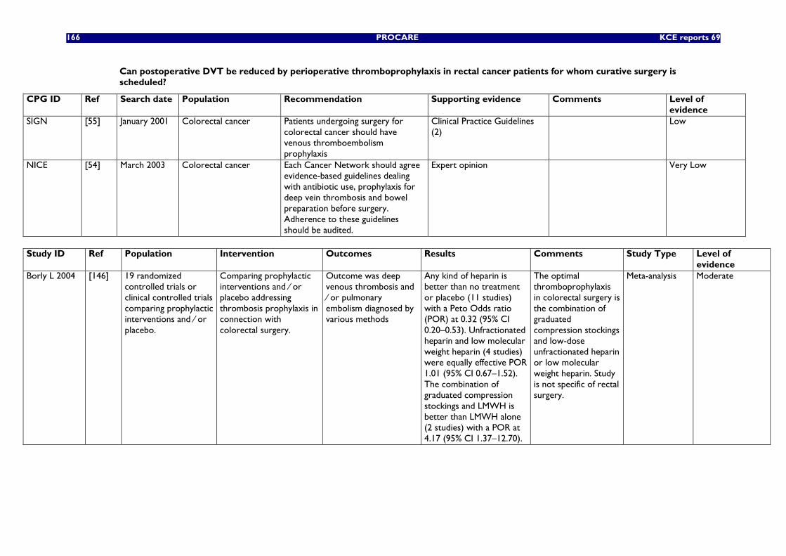
166 PROCARE KCE reports 69
Can postoperative DVT be reduced by perioperative thromboprophylaxis in rectal cancer patients for whom curative surgery is scheduled?
CPG ID Ref Search date Population Recommendation Supporting evidence Comments Level of evidence
SIGN [55] January 2001 Colorectal cancer Patients undergoing surgery for colorectal cancer should have venous thromboembolism prophylaxis
Clinical Practice Guidelines (2)
Low
NICE [54] March 2003 Colorectal cancer Each Cancer Network should agree evidence-based guidelines dealing with antibiotic use, prophylaxis for deep vein thrombosis and bowel preparation before surgery. Adherence to these guidelines should be audited.
Expert opinion Very Low
Study ID Ref Population Intervention Outcomes Results Comments Study Type Level of
evidence Borly L 2004 [146] 19 randomized
controlled trials or clinical controlled trials comparing prophylactic interventions and ⁄ or placebo.
Comparing prophylactic interventions and ⁄ or placebo addressing thrombosis prophylaxis in connection with colorectal surgery.
Outcome was deep venous thrombosis and ⁄ or pulmonary embolism diagnosed by various methods
Any kind of heparin is better than no treatment or placebo (11 studies) with a Peto Odds ratio (POR) at 0.32 (95% CI 0.20–0.53). Unfractionated heparin and low molecular weight heparin (4 studies) were equally effective POR 1.01 (95% CI 0.67–1.52). The combination of graduated compression stockings and LMWH is better than LMWH alone (2 studies) with a POR at 4.17 (95% CI 1.37–12.70).
The optimal thromboprophylaxis in colorectal surgery is the combination of graduated compression stockings and low-dose unfractionated heparin or low molecular weight heparin. Study is not specific of rectal surgery.
Meta-analysis Moderate
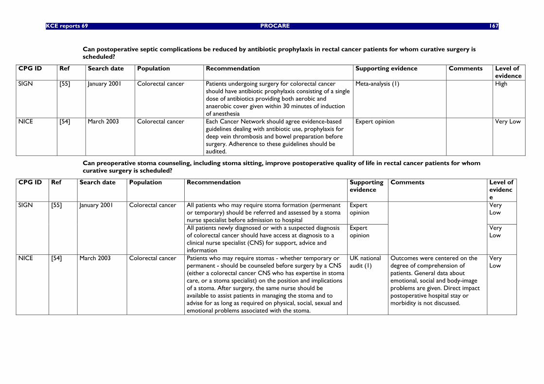
KCE reports 69 PROCARE 167
Can postoperative septic complications be reduced by antibiotic prophylaxis in rectal cancer patients for whom curative surgery is scheduled?
CPG ID Ref Search date Population Recommendation Supporting evidence Comments Level of evidence
SIGN [55] January 2001 Colorectal cancer Patients undergoing surgery for colorectal cancer should have antibiotic prophylaxis consisting of a single dose of antibiotics providing both aerobic and anaerobic cover given within 30 minutes of induction of anesthesia
Meta-analysis (1) High
NICE [54] March 2003 Colorectal cancer Each Cancer Network should agree evidence-based guidelines dealing with antibiotic use, prophylaxis for deep vein thrombosis and bowel preparation before surgery. Adherence to these guidelines should be audited.
Expert opinion Very Low
Can preoperative stoma counseling, including stoma sitting, improve postoperative quality of life in rectal cancer patients for whom curative surgery is scheduled?
CPG ID Ref Search date Population Recommendation Supporting evidence
Comments Level of evidence
All patients who may require stoma formation (permenant or temporary) should be referred and assessed by a stoma nurse specialist before admission to hospital
Expert opinion
Very Low
SIGN [55] January 2001 Colorectal cancer
All patients newly diagnosed or with a suspected diagnosis of colorectal cancer should have access at diagnosis to a clinical nurse specialist (CNS) for support, advice and information
Expert opinion
Very Low
NICE [54] March 2003 Colorectal cancer Patients who may require stomas - whether temporary or permanent - should be counseled before surgery by a CNS (either a colorectal cancer CNS who has expertise in stoma care, or a stoma specialist) on the position and implications of a stoma. After surgery, the same nurse should be available to assist patients in managing the stoma and to advise for as long as required on physical, social, sexual and emotional problems associated with the stoma.
UK national audit (1)
Outcomes were centered on the degree of comprehension of patients. General data about emotional, social and body-image problems are given. Direct impact postoperative hospital stay or morbidity is not discussed.
Very Low
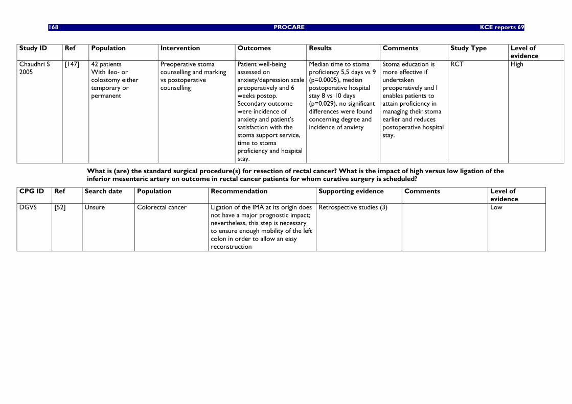
168 PROCARE KCE reports 69
Study ID Ref Population Intervention Outcomes Results Comments Study Type Level of evidence
Chaudhri S 2005
[147] 42 patients With ileo- or colostomy either temporary or permanent
Preoperative stoma counselling and marking vs postoperative counselling
Patient well-being assessed on anxiety/depression scale preoperatively and 6 weeks postop. Secondary outcome were incidence of anxiety and patient’s satisfaction with the stoma support service, time to stoma proficiency and hospital stay.
Median time to stoma proficiency 5,5 days vs 9 (p=0.0005), median postoperative hospital stay 8 vs 10 days (p=0,029), no significant differences were found concerning degree and incidence of anxiety
Stoma education is more effective if undertaken preoperatively and I enables patients to attain proficiency in managing their stoma earlier and reduces postoperative hospital stay.
RCT High
What is (are) the standard surgical procedure(s) for resection of rectal cancer? What is the impact of high versus low ligation of the inferior mesenteric artery on outcome in rectal cancer patients for whom curative surgery is scheduled?
CPG ID Ref Search date Population Recommendation Supporting evidence Comments Level of evidence
DGVS [52] Unsure Colorectal cancer Ligation of the IMA at its origin does not have a major prognostic impact; nevertheless, this step is necessary to ensure enough mobility of the left colon in order to allow an easy reconstruction
Retrospective studies (3) Low
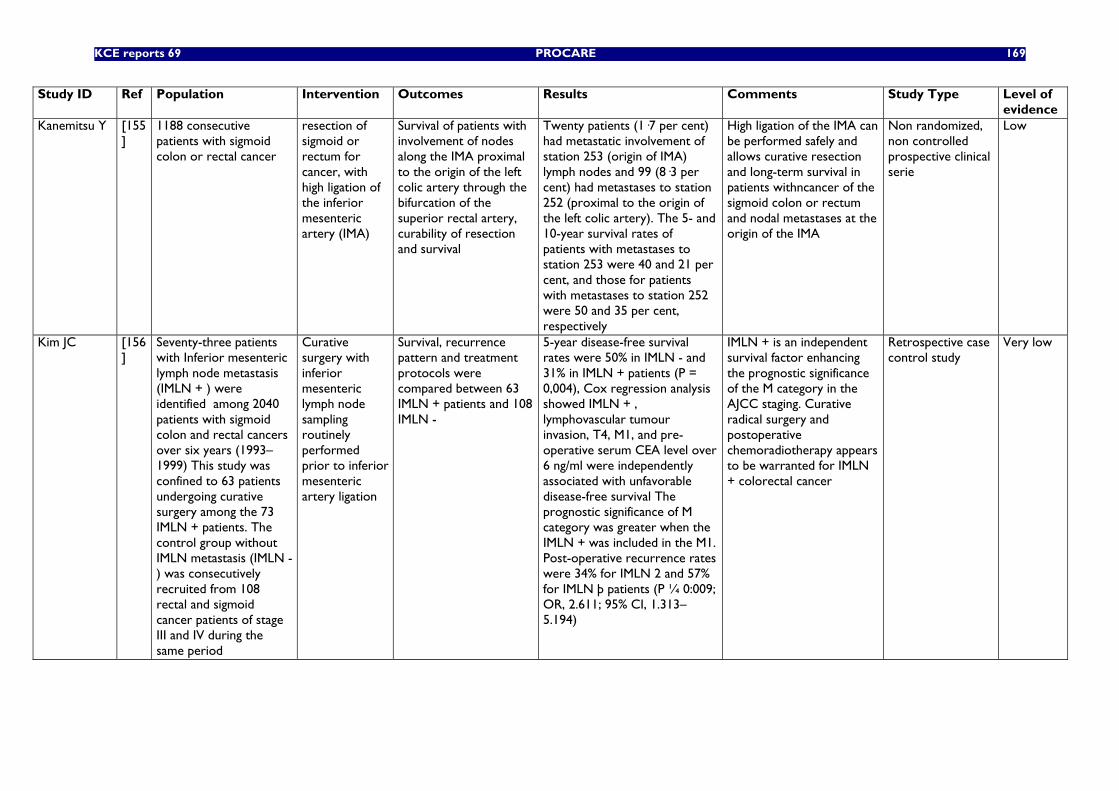
KCE reports 69 PROCARE 169
Study ID Ref Population Intervention Outcomes Results Comments Study Type Level of evidence
Kanemitsu Y [155]
1188 consecutive patients with sigmoid colon or rectal cancer
resection of sigmoid or rectum for cancer, with high ligation of the inferior mesenteric artery (IMA)
Survival of patients with involvement of nodes along the IMA proximal to the origin of the left colic artery through the bifurcation of the superior rectal artery, curability of resection and survival
Twenty patients (1·7 per cent) had metastatic involvement of station 253 (origin of IMA) lymph nodes and 99 (8·3 per cent) had metastases to station 252 (proximal to the origin of the left colic artery). The 5- and 10-year survival rates of patients with metastases to station 253 were 40 and 21 per cent, and those for patients with metastases to station 252 were 50 and 35 per cent, respectively
High ligation of the IMA can be performed safely and allows curative resection and long-term survival in patients withncancer of the sigmoid colon or rectum and nodal metastases at the origin of the IMA
Non randomized, non controlled prospective clinical serie
Low
Kim JC [156]
Seventy-three patients with Inferior mesenteric lymph node metastasis (IMLN + ) were identified among 2040 patients with sigmoid colon and rectal cancers over six years (1993–1999) This study was confined to 63 patients undergoing curative surgery among the 73 IMLN + patients. The control group without IMLN metastasis (IMLN -) was consecutively recruited from 108 rectal and sigmoid cancer patients of stage III and IV during the same period
Curative surgery with inferior mesenteric lymph node sampling routinely performed prior to inferior mesenteric artery ligation
Survival, recurrence pattern and treatment protocols were compared between 63 IMLN + patients and 108 IMLN -
5-year disease-free survival rates were 50% in IMLN - and 31% in IMLN + patients (P = 0,004), Cox regression analysis showed IMLN + , lymphovascular tumour invasion, T4, M1, and pre-operative serum CEA level over 6 ng/ml were independently associated with unfavorable disease-free survival The prognostic significance of M category was greater when the IMLN + was included in the M1. Post-operative recurrence rates were 34% for IMLN 2 and 57% for IMLN þ patients (P ¼ 0:009; OR, 2.611; 95% CI, 1.313–5.194)
IMLN + is an independent survival factor enhancing the prognostic significance of the M category in the AJCC staging. Curative radical surgery and postoperative chemoradiotherapy appears to be warranted for IMLN + colorectal cancer
Retrospective case control study
Very low

170 PROCARE KCE reports 69
What is the impact of lateral lymphatic dissection (iliac nodes) on outcome in rectal cancer patients for whom curative surgery is scheduled?
Study ID Ref Population Intervention Outcomes Results Comments Study Type Level of evidence
Nagawa H 2001
[157]
51 patients with respectable lower rectum cancer
Randomly allocated to complete autonomic nerve-preserving surgery without lateral node dissection (D1), or surgery with dissection of the lateral lymph nodes including autonomic nerves (D2) after preoperative radiation therapy
Function of pelvic organs, local recurrences
No difference was observed in either survival, disease-free survival or recurrence rate between D1 and D2 groups. Sexual and urinary functions were significantly worse in the D2 group one year after surgery.
This study suggests that lateral node dissection is not necessary in terms of curability for patients with advanced carcinoma of the lower rectum who undergo preoperative radiotherapy.
RCT High
Can sphincter saving operation be performed for rectal cancer of the lower third of the rectum without compromising the (oncological and functional) outcome in patients for whom curative surgery is scheduled?
CPG ID Ref Search date
Population Recommendation Supporting evidence Comments Level of evidence
Mesorectal excision is recommended for most rectal cancers where the patient is fit for radical surgery. The mesorectal excision should be total for tumours of the middle and lower thirds of the rectum, and care should be taken to preserve the pelvic autonomic nerves wherever this is possible without compromising tumour clearance.
Prospective clinical trial (2) Retrospective study (2) Review (2)
Low SIGN [55] January 2001
Colorectal cancer
Surgery for colorectal cancer should only be carried out by appropriately trained surgeons whose work is audited. Low rectal cancer should only be performed by those trained to carry out TME.
Systematic review (2) Retrospective study (1)
Moderate
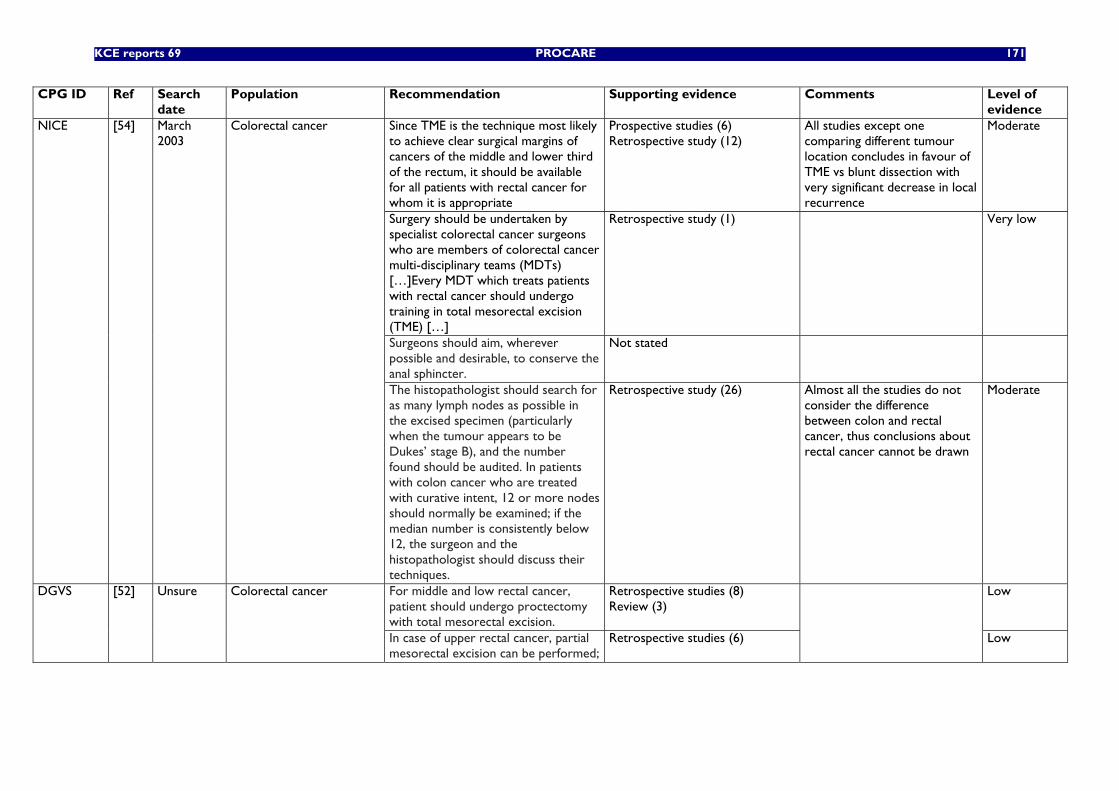
KCE reports 69 PROCARE 171
CPG ID Ref Search date
Population Recommendation Supporting evidence Comments Level of evidence
Since TME is the technique most likely to achieve clear surgical margins of cancers of the middle and lower third of the rectum, it should be available for all patients with rectal cancer for whom it is appropriate
Prospective studies (6) Retrospective study (12)
All studies except one comparing different tumour location concludes in favour of TME vs blunt dissection with very significant decrease in local recurrence
Moderate
Surgery should be undertaken by specialist colorectal cancer surgeons who are members of colorectal cancer multi-disciplinary teams (MDTs) […]Every MDT which treats patients with rectal cancer should undergo training in total mesorectal excision (TME) […]
Retrospective study (1) Very low
Surgeons should aim, wherever possible and desirable, to conserve the anal sphincter.
Not stated
NICE [54] March 2003
Colorectal cancer
The histopathologist should search for as many lymph nodes as possible in the excised specimen (particularly when the tumour appears to be Dukes’ stage B), and the number found should be audited. In patients with colon cancer who are treated with curative intent, 12 or more nodes should normally be examined; if the median number is consistently below 12, the surgeon and the histopathologist should discuss their techniques.
Retrospective study (26) Almost all the studies do not consider the difference between colon and rectal cancer, thus conclusions about rectal cancer cannot be drawn
Moderate
For middle and low rectal cancer, patient should undergo proctectomy with total mesorectal excision.
Retrospective studies (8) Review (3)
Low DGVS [52] Unsure Colorectal cancer
In case of upper rectal cancer, partial mesorectal excision can be performed;
Retrospective studies (6)
Low
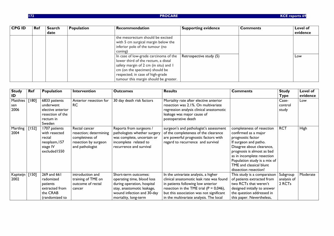
172 PROCARE KCE reports 69
CPG ID Ref Search date
Population Recommendation Supporting evidence Comments Level of evidence
the mesorectum should be excised with 5 cm surgical margin below the inferior pole of the tumour (no coning)
In case of low-grade carcinoma of the lower third of the rectum, a distal safety margin of 2 cm (in situ) and 1 cm (on the specimen) should be respected; in case of high-grade tumour this margin should be greater.
Retrospective study (5)
Low
Study ID
Ref Population Intervention Outcomes Results Comments Study Type
Level of evidence
Matthiessen 2006
[180] 6833 patients underwent elective anterior resection of the rectum in Sweden
Anterior resection for RC
30 day death risk factors Mortality rate after elective anterior resection was 2.1%. On multivariate regression analysis clinical anastomotic leakage was major cause of postoperative death
Case-control study
Low
Martling 2004
[152] 1707 patients with resected rectal neoplasm,157 stage IV excluded1550
Rectal cancer resection; determining completness of resection by surgeon and pathologist
Reports from surgeons / pathologists whether surgery was complete, uncertain or incomplete related to recurrence and survival
surgeon’s and pathologist’s assessment of the completeness of the clearance are powerful prognostic factors with regard to recurrence and survival
completeness of resection confirmed as a major prognostic factor If surgeon and patho. Disagree about clearance, prognosis is almost as bad as in incomplete resection Population study is a mix of TME and classical blunt dissection resection!
RCT High
Kapiteijn 2002
[150] 269 and 661 radomized patients extracted from the CRAB (randomized to
introduction and training of TME on outcome of rectal cancer
Short-term outcomes: operating time, blood loss during operation, hospital stay, anastomotic leakage, wound infection and 30-day mortality, long-term
In the univariate analysis, a higher clinical anastomotic leak rate was found in patients following low anterior resection in the TME trial (P = 0,046), but this association was not significant in the multivariate analysis. The local
This study is a comparaison of patients extracted from two RCTs that weren’t designed initially to answer the question addressed in this paper. Nevertheless,
Subgroup analysis of 2 RCTs
Moderate
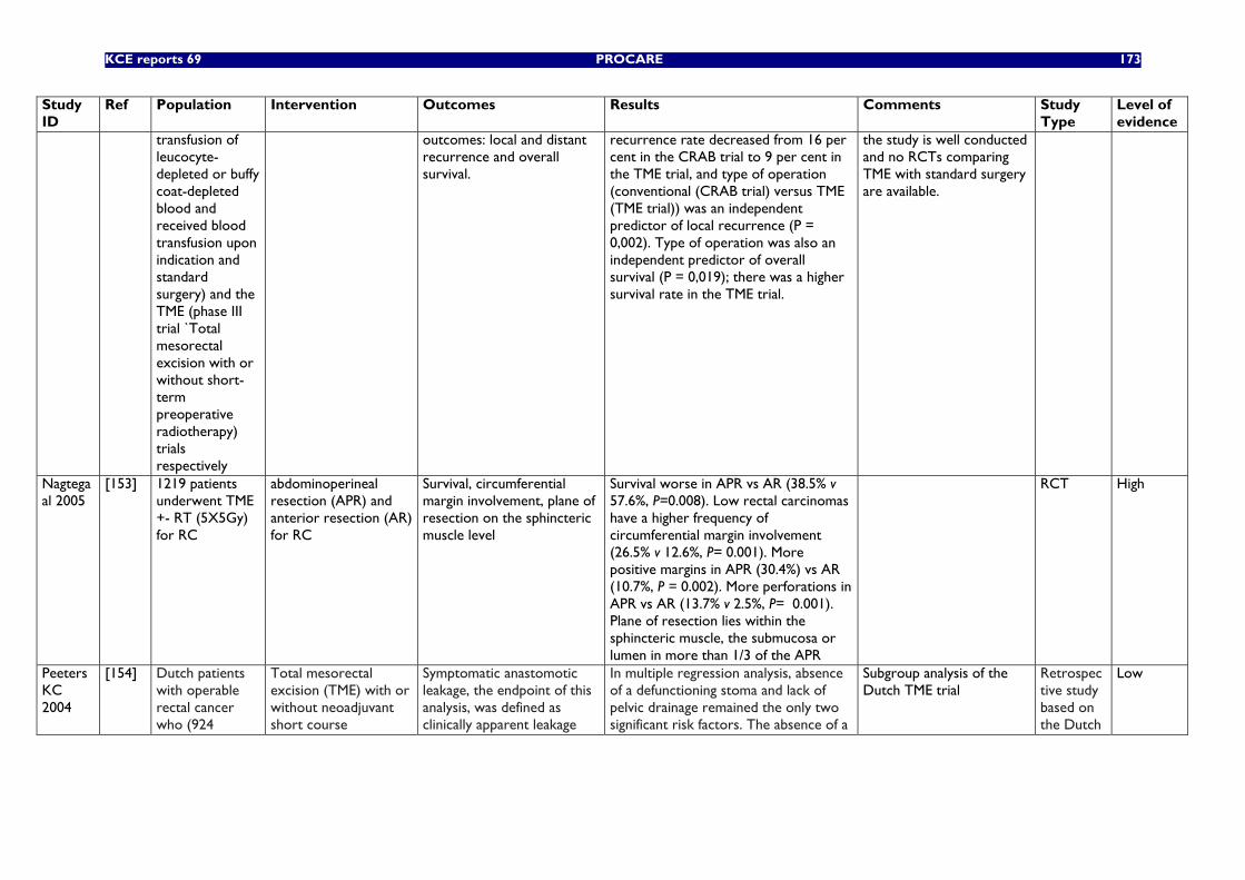
KCE reports 69 PROCARE 173
Study ID
Ref Population Intervention Outcomes Results Comments Study Type
Level of evidence
transfusion of leucocyte-depleted or buffy coat-depleted blood and received blood transfusion upon indication and standard surgery) and the TME (phase III trial `Total mesorectal excision with or without short-term preoperative radiotherapy) trials respectively
outcomes: local and distant recurrence and overall survival.
recurrence rate decreased from 16 per cent in the CRAB trial to 9 per cent in the TME trial, and type of operation (conventional (CRAB trial) versus TME (TME trial)) was an independent predictor of local recurrence (P = 0,002). Type of operation was also an independent predictor of overall survival (P = 0,019); there was a higher survival rate in the TME trial.
the study is well conducted and no RCTs comparing TME with standard surgery are available.
Nagtegaal 2005
[153] 1219 patients underwent TME +- RT (5X5Gy) for RC
abdominoperineal resection (APR) and anterior resection (AR) for RC
Survival, circumferential margin involvement, plane of resection on the sphincteric muscle level
Survival worse in APR vs AR (38.5% v 57.6%, P=0.008). Low rectal carcinomas have a higher frequency of circumferential margin involvement (26.5% v 12.6%, P= 0.001). More positive margins in APR (30.4%) vs AR (10.7%, P = 0.002). More perforations in APR vs AR (13.7% v 2.5%, P= 0.001). Plane of resection lies within the sphincteric muscle, the submucosa or lumen in more than 1/3 of the APR
RCT High
Peeters KC 2004
[154] Dutch patients with operable rectal cancer who (924
Total mesorectal excision (TME) with or without neoadjuvant short course
Symptomatic anastomotic leakage, the endpoint of this analysis, was defined as clinically apparent leakage
In multiple regression analysis, absence of a defunctioning stoma and lack of pelvic drainage remained the only two significant risk factors. The absence of a
Subgroup analysis of the Dutch TME trial
Retrospective study based on the Dutch
Low
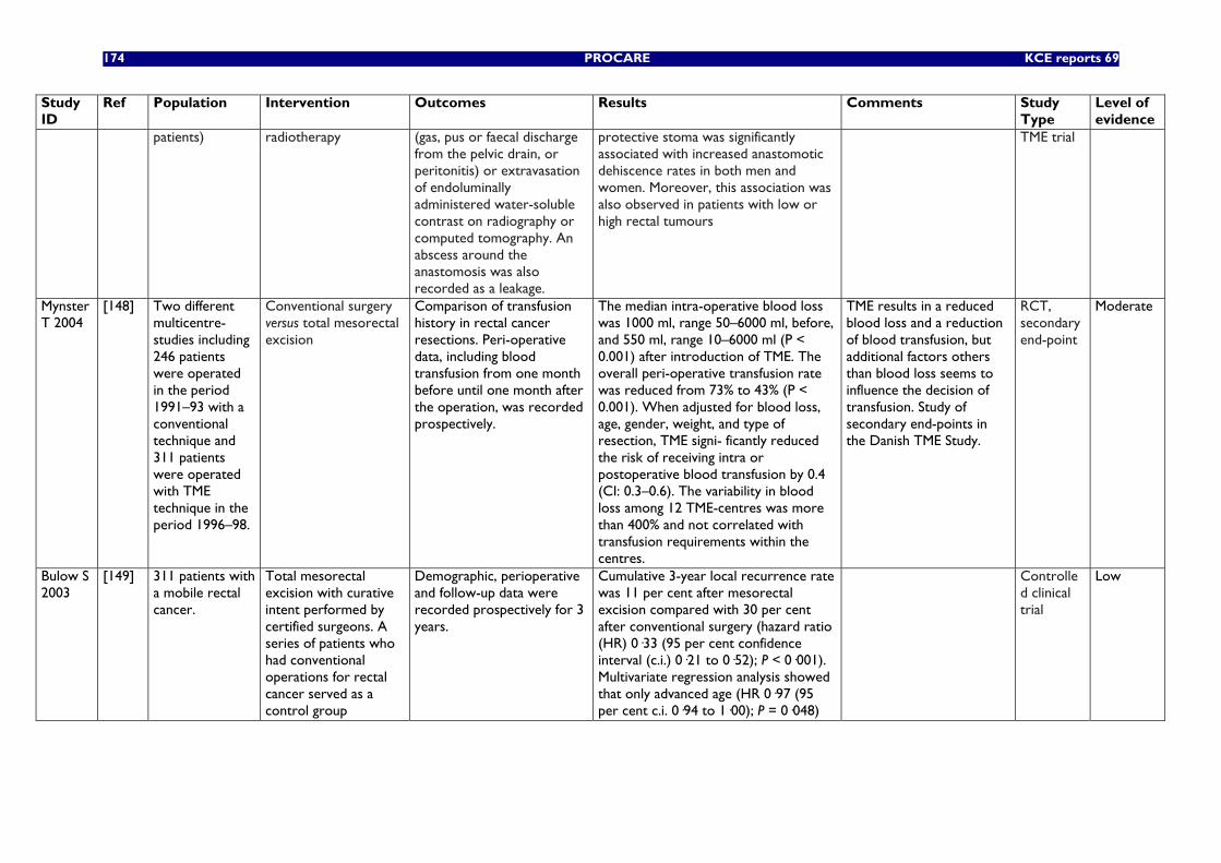
174 PROCARE KCE reports 69
Study ID
Ref Population Intervention Outcomes Results Comments Study Type
Level of evidence
patients) radiotherapy (gas, pus or faecal discharge from the pelvic drain, or peritonitis) or extravasation of endoluminally administered water-soluble contrast on radiography or computed tomography. An abscess around the anastomosis was also recorded as a leakage.
protective stoma was significantly associated with increased anastomotic dehiscence rates in both men and women. Moreover, this association was also observed in patients with low or high rectal tumours
TME trial
Mynster T 2004
[148] Two different multicentre- studies including 246 patients were operated in the period 1991–93 with a conventional technique and 311 patients were operated with TME technique in the period 1996–98.
Conventional surgery versus total mesorectal excision
Comparison of transfusion history in rectal cancer resections. Peri-operative data, including blood transfusion from one month before until one month after the operation, was recorded prospectively.
The median intra-operative blood loss was 1000 ml, range 50–6000 ml, before, and 550 ml, range 10–6000 ml (P < 0.001) after introduction of TME. The overall peri-operative transfusion rate was reduced from 73% to 43% (P < 0.001). When adjusted for blood loss, age, gender, weight, and type of resection, TME signi- ficantly reduced the risk of receiving intra or postoperative blood transfusion by 0.4 (CI: 0.3–0.6). The variability in blood loss among 12 TME-centres was more than 400% and not correlated with transfusion requirements within the centres.
TME results in a reduced blood loss and a reduction of blood transfusion, but additional factors others than blood loss seems to influence the decision of transfusion. Study of secondary end-points in the Danish TME Study.
RCT, secondary end-point
Moderate
Bulow S 2003
[149] 311 patients with a mobile rectal cancer.
Total mesorectal excision with curative intent performed by certified surgeons. A series of patients who had conventional operations for rectal cancer served as a control group
Demographic, perioperative and follow-up data were recorded prospectively for 3 years.
Cumulative 3-year local recurrence rate was 11 per cent after mesorectal excision compared with 30 per cent after conventional surgery (hazard ratio (HR) 0·33 (95 per cent confidence interval (c.i.) 0·21 to 0·52); P < 0·001). Multivariate regression analysis showed that only advanced age (HR 0·97 (95 per cent c.i. 0·94 to 1·00); P = 0·048)
Controlled clinical trial
Low
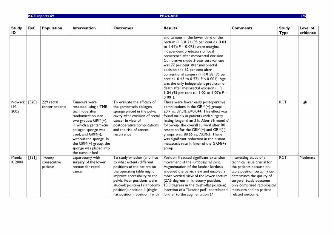
KCE reports 69 PROCARE 175
Study ID
Ref Population Intervention Outcomes Results Comments Study Type
Level of evidence
and tumour in the lower third of the rectum (HR 0·21 (95 per cent c.i. 0·04 to 1·97); P = 0·075) were marginal independent predictors of local recurrence after mesorectal excision. Cumulative crude 3-year survival rate was 77 per cent after mesorectal excision and 62 per cent after conventional surgery (HR 0·58 (95 per cent c.i. 0·43 to 0·77); P < 0·001). Age was the only independent predictor of death after mesorectal excision (HR 1·04 (95 per cent c.i. 1·02 to 1·07); P = 0·001).
Nowacki M 2005
[320] 229 rectal cancer patients
Tumours were resected using a TME technique after randomization into two groups: GRM(+), in which a gentamycin collagen sponge was used, and GRM(-), without the sponge. In the GRM(+) group, the sponge was placed into the tumour bed
To evaluate the efficacy of the gentamycin collagen sponge placed in the pelvic cavity after excision of rectal cancer in view of postoperative complications and the risk of cancer recurrence
There were fewer early postoperative complications in the GRM(+) group: 20.7 vs. 37.5%; p=0.044. This effect was found mainly in patients with surgery lasting longer than 3 h. After 36 months’ follow-up, the overall survival after R0 resection for the GRM(+) and GRM(-) groups was: 88.66 vs. 73.96%. There was significant reduction in the distant metastasis rate in favor of the GRM(+) group
RCT High
Maeda K 2004
[151] Twenty consecutive patients
Laparotomy with surgery of the lower rectum for rectal cancer
To study whether (and if so to what extent) different positions of the patient on the operating table might improve accessibility to the pelvis. Four positions were studied: position I (lithotomy position), position II (thighs-flat position), position I with
Position II caused significant extension movement of the lumbosacral joint. Augmentation of the lumbar lordosis widened the pelvic view and enabled a more vertical view of the lower rectum (27.5 degrees in lithotomy position, 13.0 degrees in the thighs-flat position). Insertion of a “lumbar pad“ contributed further to the augmentation (7
Interesting study of a technical issue crucial for the patients because on-table position certainly co-determines the quality of surgery. Study outcome only comprised radiological measures and no patient related outcome.
RCT Moderate
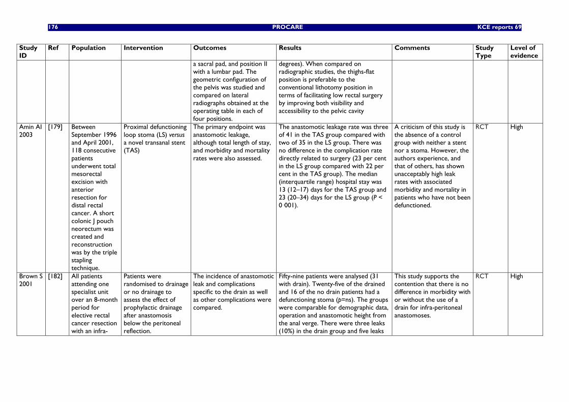
176 PROCARE KCE reports 69
Study ID
Ref Population Intervention Outcomes Results Comments Study Type
Level of evidence
a sacral pad, and position II with a lumbar pad. The geometric configuration of the pelvis was studied and compared on lateral radiographs obtained at the operating table in each of four positions.
degrees). When compared on radiographic studies, the thighs-flat position is preferable to the conventional lithotomy position in terms of facilitating low rectal surgery by improving both visibility and accessibility to the pelvic cavity
Amin AI 2003
[179] Between September 1996 and April 2001, 118 consecutive patients underwent total mesorectal excision with anterior resection for distal rectal cancer. A short colonic J pouch neorectum was created and reconstruction was by the triple stapling technique.
Proximal defunctioning loop stoma (LS) versus a novel transanal stent (TAS)
The primary endpoint was anastomotic leakage, although total length of stay, and morbidity and mortality rates were also assessed.
The anastomotic leakage rate was three of 41 in the TAS group compared with two of 35 in the LS group. There was no difference in the complication rate directly related to surgery (23 per cent in the LS group compared with 22 per cent in the TAS group). The median (interquartile range) hospital stay was 13 (12–17) days for the TAS group and 23 (20–34) days for the LS group (P < 0·001).
A criticism of this study is the absence of a control group with neither a stent nor a stoma. However, the authors experience, and that of others, has shown unacceptably high leak rates with associated morbidity and mortality in patients who have not been defunctioned.
RCT High
Brown S 2001
[182] All patients attending one specialist unit over an 8-month period for elective rectal cancer resection with an infra-
Patients were randomised to drainage or no drainage to assess the effect of prophylactic drainage after anastomosis below the peritoneal reflection.
The incidence of anastomotic leak and complications specific to the drain as well as other complications were compared.
Fifty-nine patients were analysed (31 with drain). Twenty-five of the drained and 16 of the no drain patients had a defunctioning stoma (p=ns). The groups were comparable for demographic data, operation and anastomotic height from the anal verge. There were three leaks (10%) in the drain group and five leaks
This study supports the contention that there is no difference in morbidity with or without the use of a drain for infra-peritoneal anastomoses.
RCT High
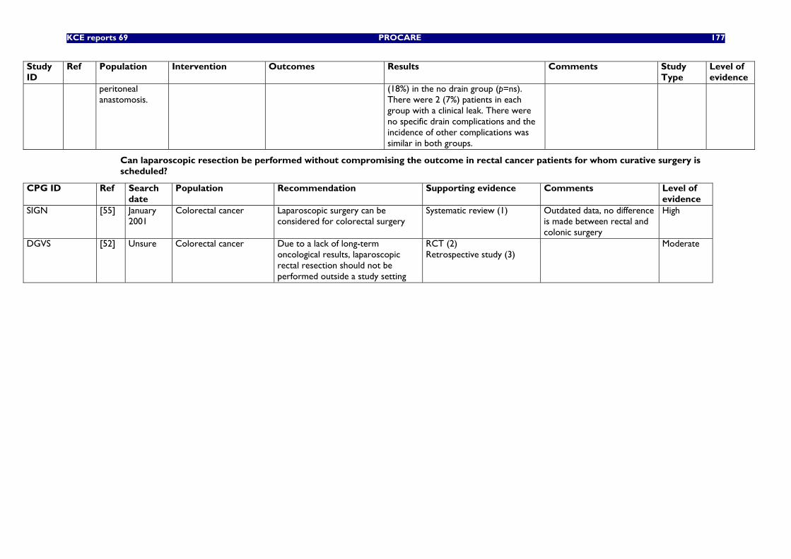
KCE reports 69 PROCARE 177
Study ID
Ref Population Intervention Outcomes Results Comments Study Type
Level of evidence
peritoneal anastomosis.
(18%) in the no drain group (p=ns). There were 2 (7%) patients in each group with a clinical leak. There were no specific drain complications and the incidence of other complications was similar in both groups.
Can laparoscopic resection be performed without compromising the outcome in rectal cancer patients for whom curative surgery is scheduled?
CPG ID Ref Search date
Population Recommendation Supporting evidence Comments Level of evidence
SIGN [55] January 2001
Colorectal cancer Laparoscopic surgery can be considered for colorectal surgery
Systematic review (1) Outdated data, no difference is made between rectal and colonic surgery
High
DGVS [52] Unsure Colorectal cancer Due to a lack of long-term oncological results, laparoscopic rectal resection should not be performed outside a study setting
RCT (2) Retrospective study (3)
Moderate
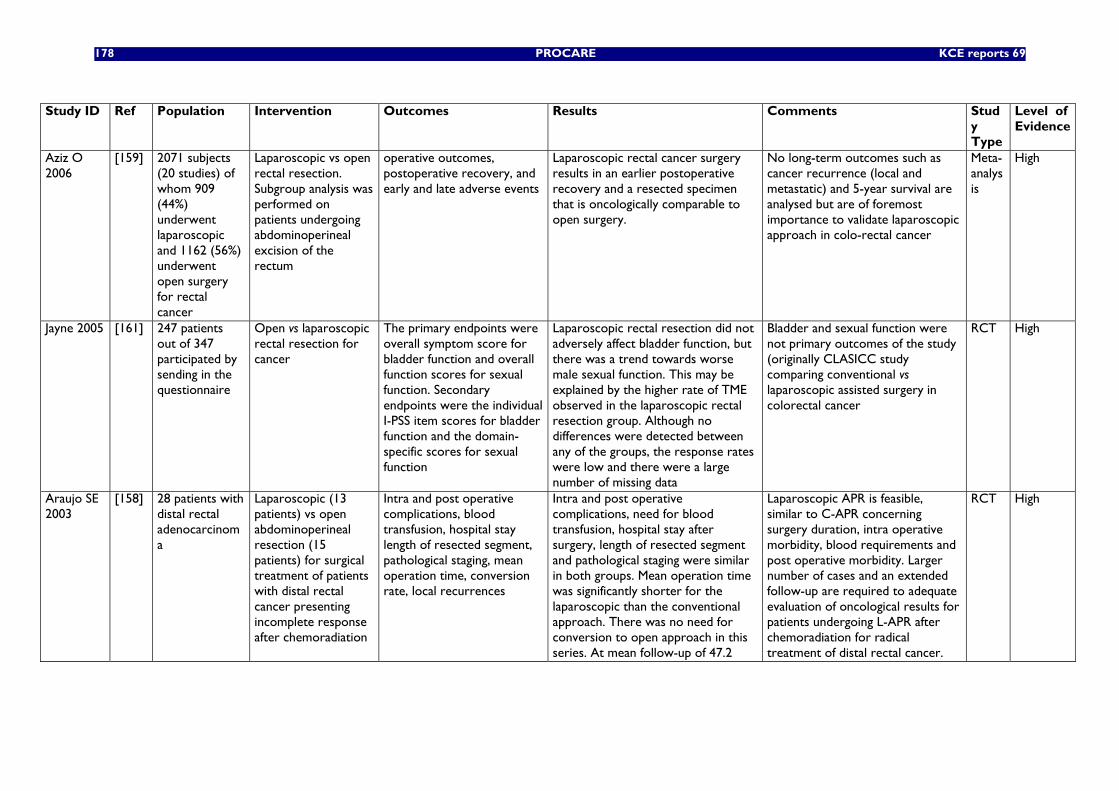
178 PROCARE KCE reports 69
Study ID Ref Population Intervention Outcomes Results Comments Stud
y Type
Level of Evidence
Aziz O 2006
[159] 2071 subjects (20 studies) of whom 909 (44%) underwent laparoscopic and 1162 (56%) underwent open surgery for rectal cancer
Laparoscopic vs open rectal resection. Subgroup analysis was performed on patients undergoing abdominoperineal excision of the rectum
operative outcomes, postoperative recovery, and early and late adverse events
Laparoscopic rectal cancer surgery results in an earlier postoperative recovery and a resected specimen that is oncologically comparable to open surgery.
No long-term outcomes such as cancer recurrence (local and metastatic) and 5-year survival are analysed but are of foremost importance to validate laparoscopic approach in colo-rectal cancer
Meta-analysis
High
Jayne 2005 [161] 247 patients out of 347 participated by sending in the questionnaire
Open vs laparoscopic rectal resection for cancer
The primary endpoints were overall symptom score for bladder function and overall function scores for sexual function. Secondary endpoints were the individual I-PSS item scores for bladder function and the domain-specific scores for sexual function
Laparoscopic rectal resection did not adversely affect bladder function, but there was a trend towards worse male sexual function. This may be explained by the higher rate of TME observed in the laparoscopic rectal resection group. Although no differences were detected between any of the groups, the response rates were low and there were a large number of missing data
Bladder and sexual function were not primary outcomes of the study (originally CLASICC study comparing conventional vs laparoscopic assisted surgery in colorectal cancer
RCT High
Araujo SE 2003
[158] 28 patients with distal rectal adenocarcinoma
Laparoscopic (13 patients) vs open abdominoperineal resection (15 patients) for surgical treatment of patients with distal rectal cancer presenting incomplete response after chemoradiation
Intra and post operative complications, blood transfusion, hospital stay length of resected segment, pathological staging, mean operation time, conversion rate, local recurrences
Intra and post operative complications, need for blood transfusion, hospital stay after surgery, length of resected segment and pathological staging were similar in both groups. Mean operation time was significantly shorter for the laparoscopic than the conventional approach. There was no need for conversion to open approach in this series. At mean follow-up of 47.2
Laparoscopic APR is feasible, similar to C-APR concerning surgery duration, intra operative morbidity, blood requirements and post operative morbidity. Larger number of cases and an extended follow-up are required to adequate evaluation of oncological results for patients undergoing L-APR after chemoradiation for radical treatment of distal rectal cancer.
RCT High
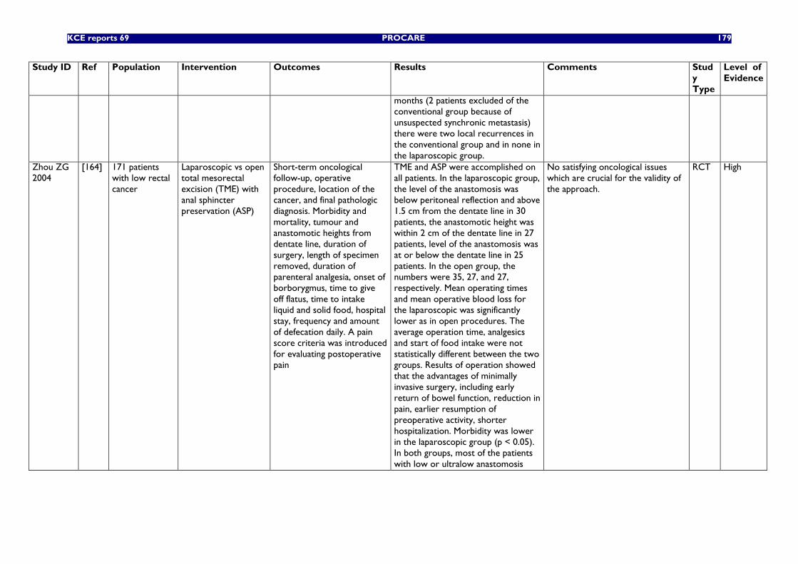
KCE reports 69 PROCARE 179
Study ID Ref Population Intervention Outcomes Results Comments Study Type
Level of Evidence
months (2 patients excluded of the conventional group because of unsuspected synchronic metastasis) there were two local recurrences in the conventional group and in none in the laparoscopic group.
Zhou ZG 2004
[164] 171 patients with low rectal cancer
Laparoscopic vs open total mesorectal excision (TME) with anal sphincter preservation (ASP)
Short-term oncological follow-up, operative procedure, location of the cancer, and final pathologic diagnosis. Morbidity and mortality, tumour and anastomotic heights from dentate line, duration of surgery, length of specimen removed, duration of parenteral analgesia, onset of borborygmus, time to give off flatus, time to intake liquid and solid food, hospital stay, frequency and amount of defecation daily. A pain score criteria was introduced for evaluating postoperative pain
TME and ASP were accomplished on all patients. In the laparoscopic group, the level of the anastomosis was below peritoneal reflection and above 1.5 cm from the dentate line in 30 patients, the anastomotic height was within 2 cm of the dentate line in 27 patients, level of the anastomosis was at or below the dentate line in 25 patients. In the open group, the numbers were 35, 27, and 27, respectively. Mean operating times and mean operative blood loss for the laparoscopic was significantly lower as in open procedures. The average operation time, analgesics and start of food intake were not statistically different between the two groups. Results of operation showed that the advantages of minimally invasive surgery, including early return of bowel function, reduction in pain, earlier resumption of preoperative activity, shorter hospitalization. Morbidity was lower in the laparoscopic group (p < 0.05). In both groups, most of the patients with low or ultralow anastomosis
No satisfying oncological issues which are crucial for the validity of the approach.
RCT High
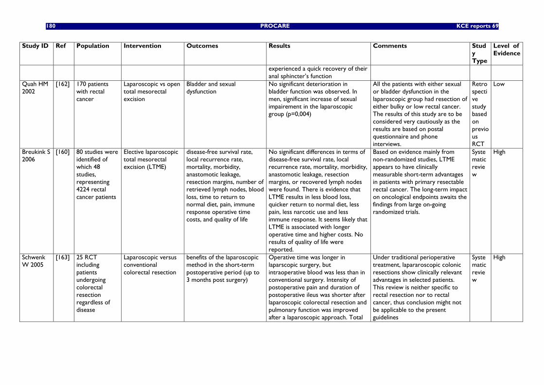
180 PROCARE KCE reports 69
Study ID Ref Population Intervention Outcomes Results Comments Study Type
Level of Evidence
experienced a quick recovery of their anal sphincter’s function
Quah HM 2002
[162] 170 patients with rectal cancer
Laparoscopic vs open total mesorectal excision
Bladder and sexual dysfunction
No significant deterioration in bladder function was observed. In men, significant increase of sexual impairement in the laparoscopic group (p=0,004)
All the patients with either sexual or bladder dysfunction in the laparoscopic group had resection of either bulky or low rectal cancer. The results of this study are to be considered very cautiously as the results are based on postal questionnaire and phone interviews.
Retrospective study based on previous RCT
Low
Breukink S 2006
[160] 80 studies were identified of which 48 studies, representing 4224 rectal cancer patients
Elective laparoscopic total mesorectal excision (LTME)
disease-free survival rate, local recurrence rate, mortality, morbidity, anastomotic leakage, resection margins, number of retrieved lymph nodes, blood loss, time to return to normal diet, pain, immune response operative time costs, and quality of life
No significant differences in terms of disease-free survival rate, local recurrence rate, mortality, morbidity, anastomotic leakage, resection margins, or recovered lymph nodes were found. There is evidence that LTME results in less blood loss, quicker return to normal diet, less pain, less narcotic use and less immune response. It seems likely that LTME is associated with longer operative time and higher costs. No results of quality of life were reported.
Based on evidence mainly from non-randomized studies, LTME appears to have clinically measurable short-term advantages in patients with primary resectable rectal cancer. The long-term impact on oncological endpoints awaits the findings from large on-going randomized trials.
Systematic review
High
Schwenk W 2005
[163] 25 RCT including patients undergoing colorectal resection regardless of disease
Laparoscopic versus conventional colorectal resection
benefits of the laparoscopic method in the short-term postoperative period (up to 3 months post surgery)
Operative time was longer in laparscopic surgery, but intraoperative blood was less than in conventional surgery. Intensity of postoperative pain and duration of postoperative ileus was shorter after laparoscopic colorectal resection and pulmonary function was improved after a laparoscopic approach. Total
Under traditional perioperative treatment, lapararoscopic colonic resections show clinically relevant advantages in selected patients. This review is neither specific to rectal resection nor to rectal cancer, thus conclusion might not be applicable to the present guidelines
Systematic review
High
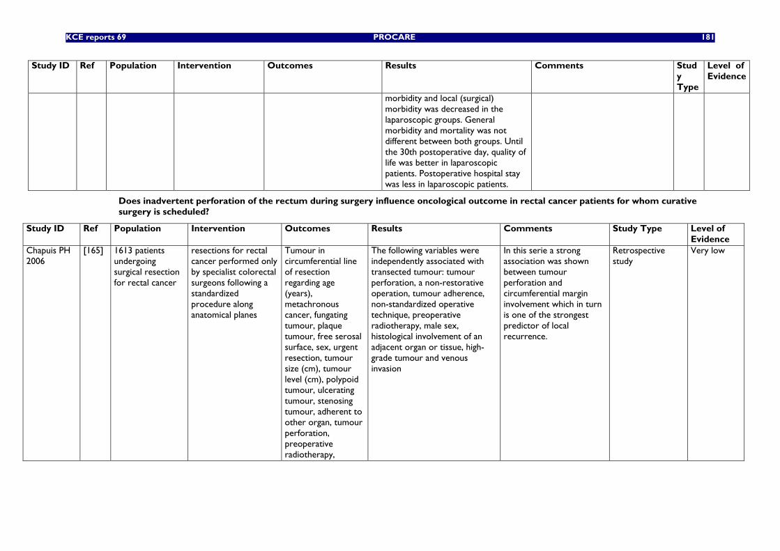
KCE reports 69 PROCARE 181
Study ID Ref Population Intervention Outcomes Results Comments Study Type
Level of Evidence
morbidity and local (surgical) morbidity was decreased in the laparoscopic groups. General morbidity and mortality was not different between both groups. Until the 30th postoperative day, quality of life was better in laparoscopic patients. Postoperative hospital stay was less in laparoscopic patients.
Does inadvertent perforation of the rectum during surgery influence oncological outcome in rectal cancer patients for whom curative surgery is scheduled?
Study ID Ref Population Intervention Outcomes Results Comments Study Type Level of Evidence
Chapuis PH 2006
[165] 1613 patients undergoing surgical resection for rectal cancer
resections for rectal cancer performed only by specialist colorectal surgeons following a standardized procedure along anatomical planes
Tumour in circumferential line of resection regarding age (years), metachronous cancer, fungating tumour, plaque tumour, free serosal surface, sex, urgent resection, tumour size (cm), tumour level (cm), polypoid tumour, ulcerating tumour, stenosing tumour, adherent to other organ, tumour perforation, preoperative radiotherapy,
The following variables were independently associated with transected tumour: tumour perforation, a non-restorative operation, tumour adherence, non-standardized operative technique, preoperative radiotherapy, male sex, histological involvement of an adjacent organ or tissue, high-grade tumour and venous invasion
In this serie a strong association was shown between tumour perforation and circumferential margin involvement which in turn is one of the strongest predictor of local recurrence.
Retrospective study
Very low
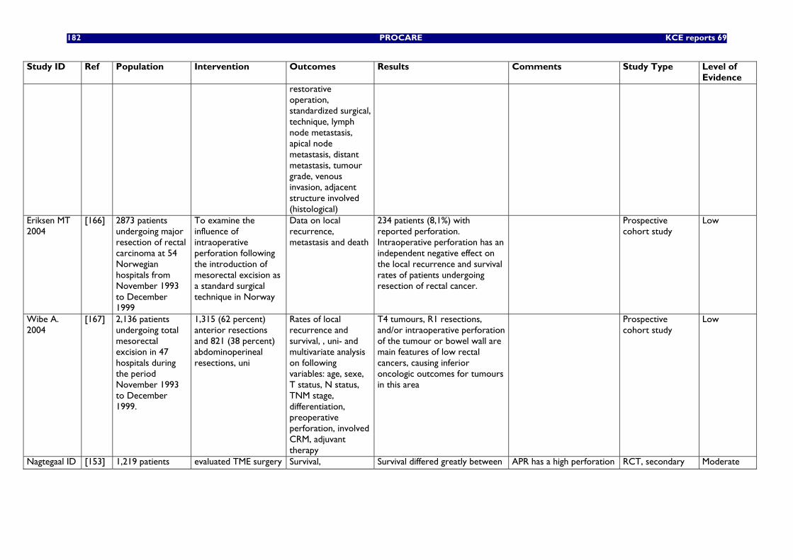
182 PROCARE KCE reports 69
Study ID Ref Population Intervention Outcomes Results Comments Study Type Level of Evidence
restorative operation, standardized surgical, technique, lymph node metastasis, apical node metastasis, distant metastasis, tumour grade, venous invasion, adjacent structure involved (histological)
Eriksen MT 2004
[166] 2873 patients undergoing major resection of rectal carcinoma at 54 Norwegian hospitals from November 1993 to December 1999
To examine the influence of intraoperative perforation following the introduction of mesorectal excision as a standard surgical technique in Norway
Data on local recurrence, metastasis and death
234 patients (8,1%) with reported perforation. Intraoperative perforation has an independent negative effect on the local recurrence and survival rates of patients undergoing resection of rectal cancer.
Prospective cohort study
Low
Wibe A. 2004
[167] 2,136 patients undergoing total mesorectal excision in 47 hospitals during the period November 1993 to December 1999.
1,315 (62 percent) anterior resections and 821 (38 percent) abdominoperineal resections, uni
Rates of local recurrence and survival, , uni- and multivariate analysis on following variables: age, sexe, T status, N status, TNM stage, differentiation, preoperative perforation, involved CRM, adjuvant therapy
T4 tumours, R1 resections, and/or intraoperative perforation of the tumour or bowel wall are main features of low rectal cancers, causing inferior oncologic outcomes for tumours in this area
Prospective cohort study
Low
Nagtegaal ID [153] 1,219 patients evaluated TME surgery Survival, Survival differed greatly between APR has a high perforation RCT, secondary Moderate
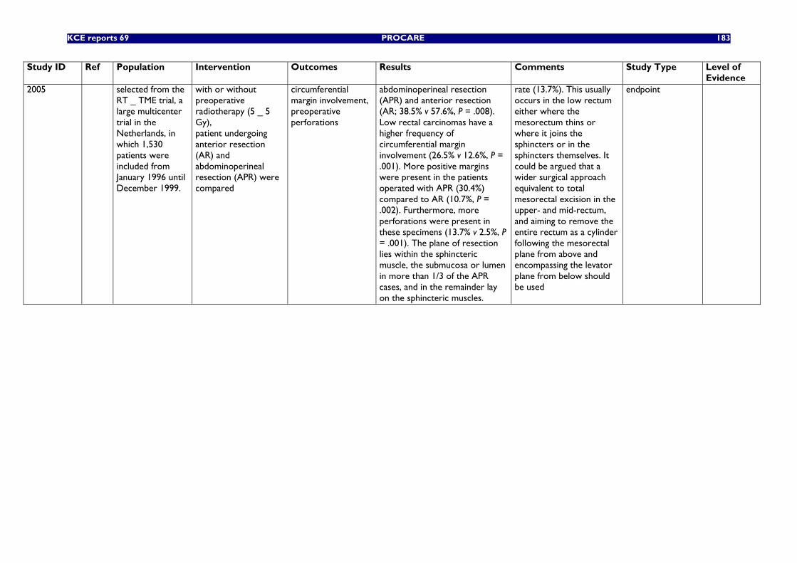
KCE reports 69 PROCARE 183
Study ID Ref Population Intervention Outcomes Results Comments Study Type Level of Evidence
2005 selected from the RT _ TME trial, a large multicenter trial in the Netherlands, in which 1,530 patients were included from January 1996 until December 1999.
with or without preoperative radiotherapy (5 _ 5 Gy), patient undergoing anterior resection (AR) and abdominoperineal resection (APR) were compared
circumferential margin involvement, preoperative perforations
abdominoperineal resection (APR) and anterior resection (AR; 38.5% v 57.6%, P = .008). Low rectal carcinomas have a higher frequency of circumferential margin involvement (26.5% v 12.6%, P = .001). More positive margins were present in the patients operated with APR (30.4%) compared to AR (10.7%, P = .002). Furthermore, more perforations were present in these specimens (13.7% v 2.5%, P = .001). The plane of resection lies within the sphincteric muscle, the submucosa or lumen in more than 1/3 of the APR cases, and in the remainder lay on the sphincteric muscles.
rate (13.7%). This usually occurs in the low rectum either where the mesorectum thins or where it joins the sphincters or in the sphincters themselves. It could be argued that a wider surgical approach equivalent to total mesorectal excision in the upper- and mid-rectum, and aiming to remove the entire rectum as a cylinder following the mesorectal plane from above and encompassing the levator plane from below should be used
endpoint
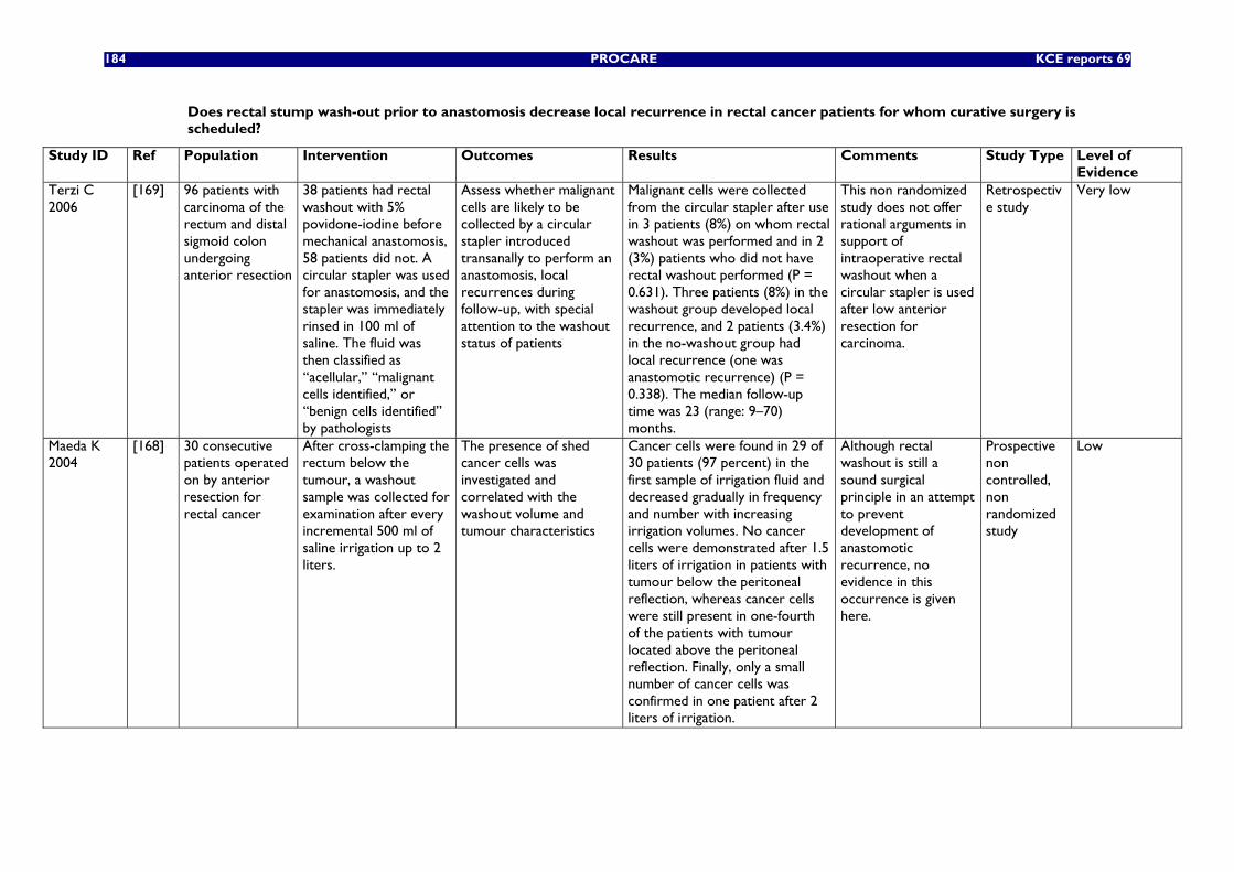
184 PROCARE KCE reports 69
Does rectal stump wash-out prior to anastomosis decrease local recurrence in rectal cancer patients for whom curative surgery is scheduled?
Study ID Ref Population Intervention Outcomes Results Comments Study Type Level of Evidence
Terzi C 2006
[169] 96 patients with carcinoma of the rectum and distal sigmoid colon undergoing anterior resection
38 patients had rectal washout with 5% povidone-iodine before mechanical anastomosis, 58 patients did not. A circular stapler was used for anastomosis, and the stapler was immediately rinsed in 100 ml of saline. The fluid was then classified as ‘‘acellular,’’ ‘‘malignant cells identified,’’ or ‘‘benign cells identified’’ by pathologists
Assess whether malignant cells are likely to be collected by a circular stapler introduced transanally to perform an anastomosis, local recurrences during follow-up, with special attention to the washout status of patients
Malignant cells were collected from the circular stapler after use in 3 patients (8%) on whom rectal washout was performed and in 2 (3%) patients who did not have rectal washout performed (P = 0.631). Three patients (8%) in the washout group developed local recurrence, and 2 patients (3.4%) in the no-washout group had local recurrence (one was anastomotic recurrence) (P = 0.338). The median follow-up time was 23 (range: 9–70) months.
This non randomized study does not offer rational arguments in support of intraoperative rectal washout when a circular stapler is used after low anterior resection for carcinoma.
Retrospective study
Very low
Maeda K 2004
[168] 30 consecutive patients operated on by anterior resection for rectal cancer
After cross-clamping the rectum below the tumour, a washout sample was collected for examination after every incremental 500 ml of saline irrigation up to 2 liters.
The presence of shed cancer cells was investigated and correlated with the washout volume and tumour characteristics
Cancer cells were found in 29 of 30 patients (97 percent) in the first sample of irrigation fluid and decreased gradually in frequency and number with increasing irrigation volumes. No cancer cells were demonstrated after 1.5 liters of irrigation in patients with tumour below the peritoneal reflection, whereas cancer cells were still present in one-fourth of the patients with tumour located above the peritoneal reflection. Finally, only a small number of cancer cells was confirmed in one patient after 2 liters of irrigation.
Although rectal washout is still a sound surgical principle in an attempt to prevent development of anastomotic recurrence, no evidence in this occurrence is given here.
Prospective non controlled, non randomized study
Low
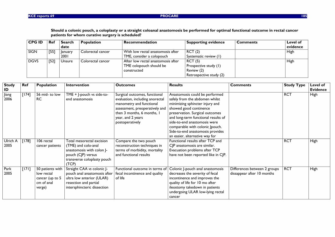
KCE reports 69 PROCARE 185
Should a colonic pouch, a coloplasty or a straight coloanal anastomosis be performed for optimal functional outcome in rectal cancer patients for whom curative surgery is scheduled?
CPG ID Ref Search date
Population Recommendation Supporting evidence Comments Level of evidence
SIGN [55] January 2001
Colorectal cancer With low rectal anastomosis after TME, consider a colopouch
RCT (2) Systematic review (1)
High
DGVS [52] Unsure Colorectal cancer After low rectal anastomosis after TME colopouch should be constructed
RCT (5) Prospective study (1) Review (2) Retrospective study (2)
High
Study ID
Ref Population Intervention Outcomes Results Comments Study Type Level of Evidence
Jiang 2006
[174] 56 mid- to low RC
TME + J-pouch vs side-to-end anastomosis
Surgical outcomes, functional evaluation, including anorectal manometry and functional assessment, preoperatively and then 3 months, 6 months, 1 year, and 2 years postoperatively
Anastomosis could be performed safely from the abdomen whilst minimizing sphincter injury and showed good continence preservation. Surgical outcomes and long-term functional results of side-to-end anastomosis were comparable with colonic Jpouch. Side-to-end anastomosis provides an easier, alternative way for
RCT High
Ulrich A 2005
[178] 106 rectal cancer patients
Total mesorectal excision (TME) and colo-anal anastomosis with colon J-pouch (CJP) versus transverse coloplasty pouch (TCP)
Compare the two pouch reconstruction techniques in terms of morbidity, mortality and functional results
Functional results after TCP and CJP anastomosis are similar. Evacuation problems after TCP have not been reported like in CJP.
RCT High
Park 2005
[171] 50 patients with low rectal cancer (up to 5 cm of anal verge)
Straight CAA vs colonic J-pouch anal anastomosis after ultra low anterior (ULAR) resection and partial intersphincteric dissection
Functional outcome in terms of fecal incontinence and quality of life
Colonic J-pouch anal anastomosis decreases the severity of fecal incontinence and improves the quality of life for 10 mo after ileostomy takedown in patients undergoing ULAR low-lying rectal cancer
Differences between 2 groups dissappear after 10 months
RCT High
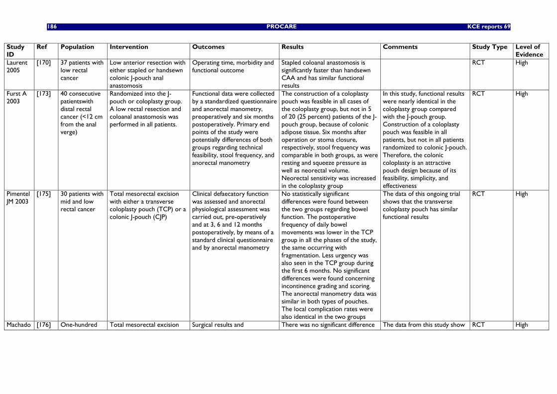
186 PROCARE KCE reports 69
Study ID
Ref Population Intervention Outcomes Results Comments Study Type Level of Evidence
Laurent 2005
[170] 37 patients with low rectal cancer
Low anterior resection with either stapled or handsewn colonic J-pouch anal anastomosis
Operating time, morbidity and functional outcome
Stapled coloanal anastomosis is significantly faster than handsewn CAA and has similar functional results
RCT High
Furst A 2003
[173] 40 consecutive patientswith distal rectal cancer (<12 cm from the anal verge)
Randomized into the J-pouch or coloplasty group. A low rectal resection and coloanal anastomosis was performed in all patients.
Functional data were collected by a standardized questionnaire and anorectal manometry, preoperatively and six months postoperatively. Primary end points of the study were potentially differences of both groups regarding technical feasibility, stool frequency, and anorectal manometry
The construction of a coloplasty pouch was feasible in all cases of the coloplasty group, but not in 5 of 20 (25 percent) patients of the J-pouch group, because of colonic adipose tissue. Six months after operation or stoma closure, respectively, stool frequency was comparable in both groups, as were resting and squeeze pressure as well as neorectal volume. Neorectal sensitivity was increased in the coloplasty group
In this study, functional results were nearly identical in the coloplasty group compared with the J-pouch group. Construction of a coloplasty pouch was feasible in all patients, but not in all patients randomized to colonic J-pouch. Therefore, the colonic coloplasty is an attractive pouch design because of its feasibility, simplicity, and effectiveness
RCT High
Pimentel JM 2003
[175] 30 patients with mid and low rectal cancer
Total mesorectal excision with either a transverse coloplasty pouch (TCP) or a colonic J-pouch (CJP)
Clinical defaecatory function was assessed and anorectal physiological assessment was carried out, pre-operatively and at 3, 6 and 12 months postoperatively, by means of a standard clinical questionnaire and by anorectal manometry
No statistically significant differences were found between the two groups regarding bowel function. The postoperative frequency of daily bowel movements was lower in the TCP group in all the phases of the study, the same occurring with fragmentation. Less urgency was also seen in the TCP group during the first 6 months. No significant differences were found concerning incontinence grading and scoring. The anorectal manometry data was similar in both types of pouches. The local complication rates were also identical in the two groups
The data of this ongoing trial shows that the transverse coloplasty pouch has similar functional results
RCT High
Machado [176] One-hundred Total mesorectal excision Surgical results and There was no significant difference The data from this study show RCT High
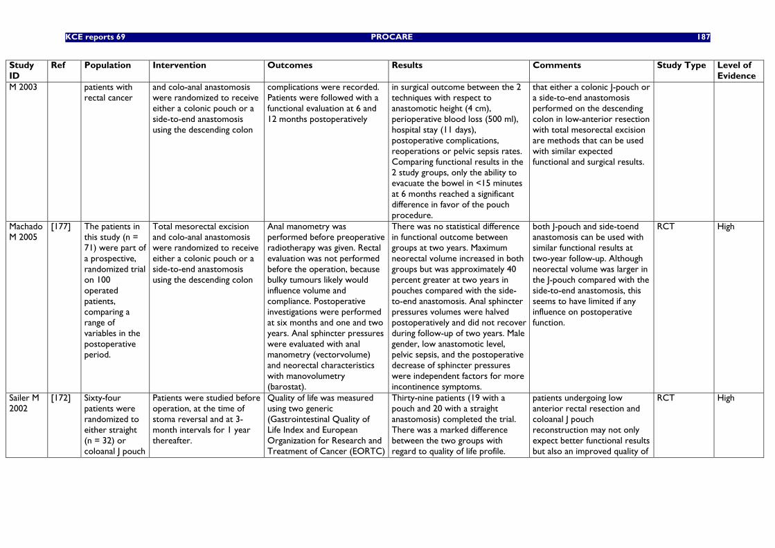
KCE reports 69 PROCARE 187
Study ID
Ref Population Intervention Outcomes Results Comments Study Type Level of Evidence
M 2003 patients with rectal cancer
and colo-anal anastomosis were randomized to receive either a colonic pouch or a side-to-end anastomosis using the descending colon
complications were recorded. Patients were followed with a functional evaluation at 6 and 12 months postoperatively
in surgical outcome between the 2 techniques with respect to anastomotic height (4 cm), perioperative blood loss (500 ml), hospital stay (11 days), postoperative complications, reoperations or pelvic sepsis rates. Comparing functional results in the 2 study groups, only the ability to evacuate the bowel in <15 minutes at 6 months reached a significant difference in favor of the pouch procedure.
that either a colonic J-pouch or a side-to-end anastomosis performed on the descending colon in low-anterior resection with total mesorectal excision are methods that can be used with similar expected functional and surgical results.
Machado M 2005
[177] The patients in this study (n = 71) were part of a prospective, randomized trial on 100 operated patients, comparing a range of variables in the postoperative period.
Total mesorectal excision and colo-anal anastomosis were randomized to receive either a colonic pouch or a side-to-end anastomosis using the descending colon
Anal manometry was performed before preoperative radiotherapy was given. Rectal evaluation was not performed before the operation, because bulky tumours likely would influence volume and compliance. Postoperative investigations were performed at six months and one and two years. Anal sphincter pressures were evaluated with anal manometry (vectorvolume) and neorectal characteristics with manovolumetry (barostat).
There was no statistical difference in functional outcome between groups at two years. Maximum neorectal volume increased in both groups but was approximately 40 percent greater at two years in pouches compared with the side-to-end anastomosis. Anal sphincter pressures volumes were halved postoperatively and did not recover during follow-up of two years. Male gender, low anastomotic level, pelvic sepsis, and the postoperative decrease of sphincter pressures were independent factors for more incontinence symptoms.
both J-pouch and side-toend anastomosis can be used with similar functional results at two-year follow-up. Although neorectal volume was larger in the J-pouch compared with the side-to-end anastomosis, this seems to have limited if any influence on postoperative function.
RCT High
Sailer M 2002
[172] Sixty-four patients were randomized to either straight (n = 32) or coloanal J pouch
Patients were studied before operation, at the time of stoma reversal and at 3-month intervals for 1 year thereafter.
Quality of life was measured using two generic (Gastrointestinal Quality of Life Index and European Organization for Research and Treatment of Cancer (EORTC)
Thirty-nine patients (19 with a pouch and 20 with a straight anastomosis) completed the trial. There was a marked difference between the two groups with regard to quality of life profile.
patients undergoing low anterior rectal resection and coloanal J pouch reconstruction may not only expect better functional results but also an improved quality of
RCT High
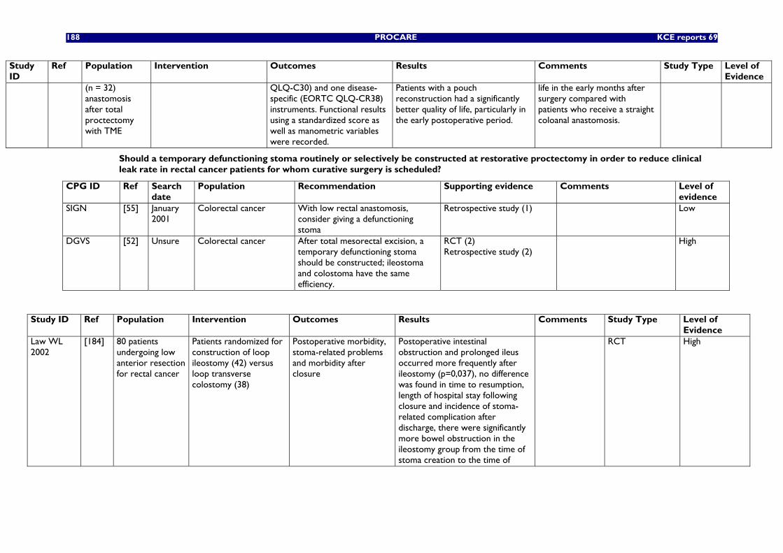
188 PROCARE KCE reports 69
Study ID
Ref Population Intervention Outcomes Results Comments Study Type Level of Evidence
(n = 32) anastomosis after total proctectomy with TME
QLQ-C30) and one disease-specific (EORTC QLQ-CR38) instruments. Functional results using a standardized score as well as manometric variables were recorded.
Patients with a pouch reconstruction had a significantly better quality of life, particularly in the early postoperative period.
life in the early months after surgery compared with patients who receive a straight coloanal anastomosis.
Should a temporary defunctioning stoma routinely or selectively be constructed at restorative proctectomy in order to reduce clinical leak rate in rectal cancer patients for whom curative surgery is scheduled?
CPG ID Ref Search date
Population Recommendation Supporting evidence Comments Level of evidence
SIGN [55] January 2001
Colorectal cancer With low rectal anastomosis, consider giving a defunctioning stoma
Retrospective study (1) Low
DGVS [52] Unsure Colorectal cancer After total mesorectal excision, a temporary defunctioning stoma should be constructed; ileostoma and colostoma have the same efficiency.
RCT (2) Retrospective study (2)
High
Study ID Ref Population Intervention Outcomes Results Comments Study Type Level of Evidence
Law WL 2002
[184] 80 patients undergoing low anterior resection for rectal cancer
Patients randomized for construction of loop ileostomy (42) versus loop transverse colostomy (38)
Postoperative morbidity, stoma-related problems and morbidity after closure
Postoperative intestinal obstruction and prolonged ileus occurred more frequently after ileostomy (p=0,037), no difference was found in time to resumption, length of hospital stay following closure and incidence of stoma-related complication after discharge, there were significantly more bowel obstruction in the ileostomy group from the time of stoma creation to the time of
RCT High
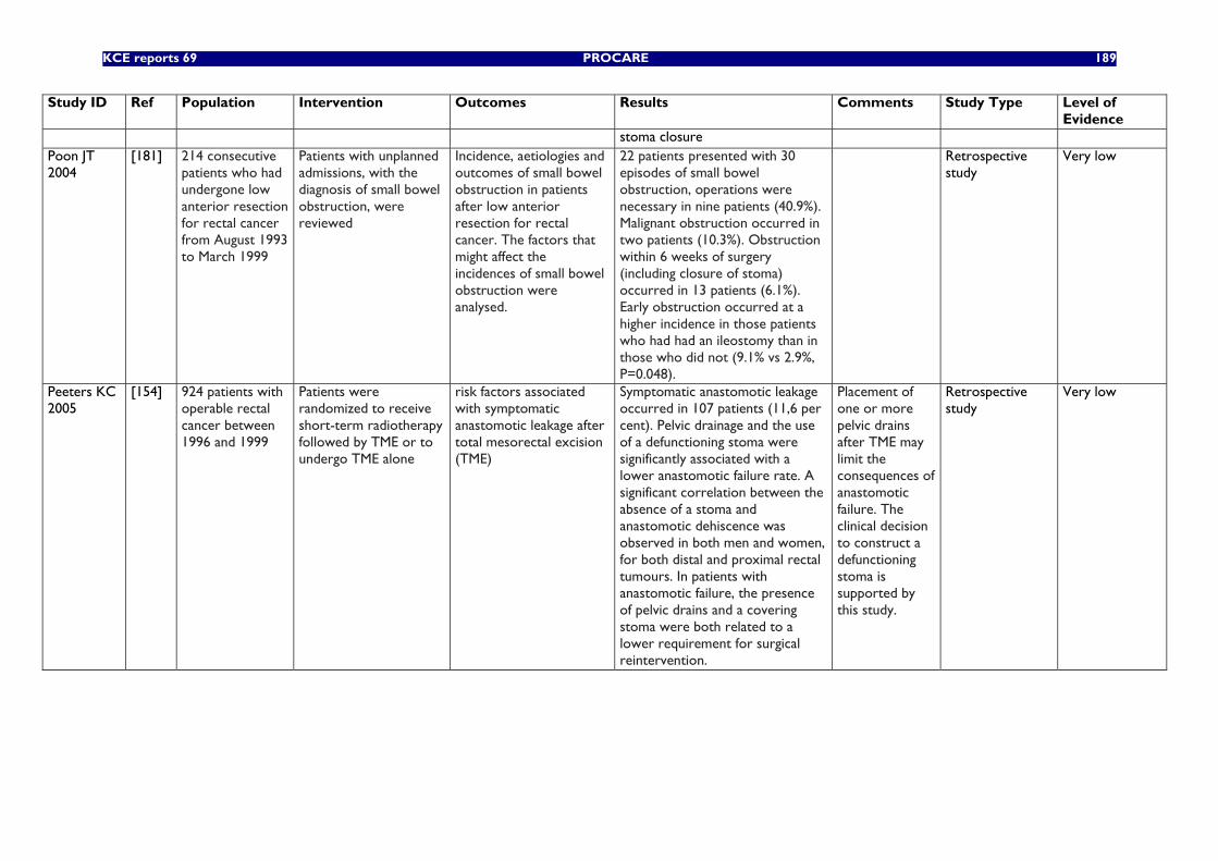
KCE reports 69 PROCARE 189
Study ID Ref Population Intervention Outcomes Results Comments Study Type Level of Evidence
stoma closure Poon JT 2004
[181] 214 consecutive patients who had undergone low anterior resection for rectal cancer from August 1993 to March 1999
Patients with unplanned admissions, with the diagnosis of small bowel obstruction, were reviewed
Incidence, aetiologies and outcomes of small bowel obstruction in patients after low anterior resection for rectal cancer. The factors that might affect the incidences of small bowel obstruction were analysed.
22 patients presented with 30 episodes of small bowel obstruction, operations were necessary in nine patients (40.9%). Malignant obstruction occurred in two patients (10.3%). Obstruction within 6 weeks of surgery (including closure of stoma) occurred in 13 patients (6.1%). Early obstruction occurred at a higher incidence in those patients who had had an ileostomy than in those who did not (9.1% vs 2.9%, P=0.048).
Retrospective study
Very low
Peeters KC 2005
[154] 924 patients with operable rectal cancer between 1996 and 1999
Patients were randomized to receive short-term radiotherapy followed by TME or to undergo TME alone
risk factors associated with symptomatic anastomotic leakage after total mesorectal excision (TME)
Symptomatic anastomotic leakage occurred in 107 patients (11,6 per cent). Pelvic drainage and the use of a defunctioning stoma were significantly associated with a lower anastomotic failure rate. A significant correlation between the absence of a stoma and anastomotic dehiscence was observed in both men and women, for both distal and proximal rectal tumours. In patients with anastomotic failure, the presence of pelvic drains and a covering stoma were both related to a lower requirement for surgical reintervention.
Placement of one or more pelvic drains after TME may limit the consequences of anastomotic failure. The clinical decision to construct a defunctioning stoma is supported by this study.
Retrospective study
Very low
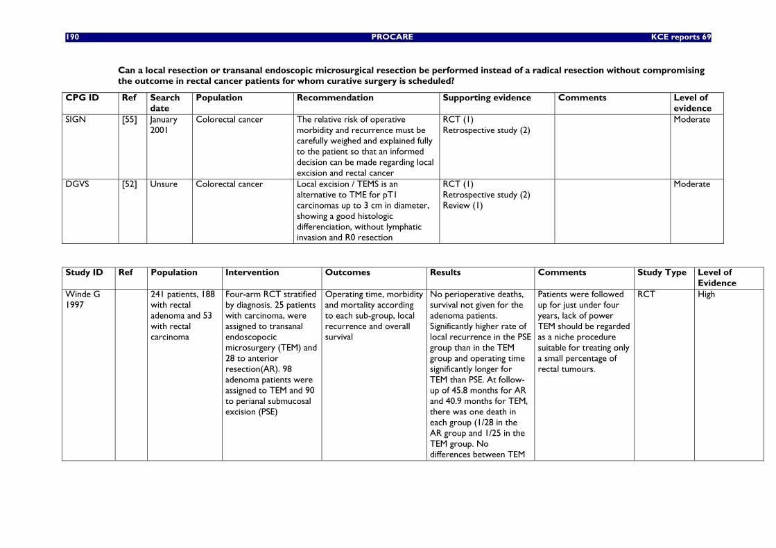
190 PROCARE KCE reports 69
Can a local resection or transanal endoscopic microsurgical resection be performed instead of a radical resection without compromising the outcome in rectal cancer patients for whom curative surgery is scheduled?
CPG ID Ref Search date
Population Recommendation Supporting evidence Comments Level of evidence
SIGN [55] January 2001
Colorectal cancer The relative risk of operative morbidity and recurrence must be carefully weighed and explained fully to the patient so that an informed decision can be made regarding local excision and rectal cancer
RCT (1) Retrospective study (2)
Moderate
DGVS [52] Unsure Colorectal cancer Local excision / TEMS is an alternative to TME for pT1 carcinomas up to 3 cm in diameter, showing a good histologic differenciation, without lymphatic invasion and R0 resection
RCT (1) Retrospective study (2) Review (1)
Moderate
Study ID Ref Population Intervention Outcomes Results Comments Study Type Level of
Evidence Winde G 1997
241 patients, 188 with rectal adenoma and 53 with rectal carcinoma
Four-arm RCT stratified by diagnosis. 25 patients with carcinoma, were assigned to transanal endoscopocic microsurgery (TEM) and 28 to anterior resection(AR). 98 adenoma patients were assigned to TEM and 90 to perianal submucosal excision (PSE)
Operating time, morbidity and mortality according to each sub-group, local recurrence and overall survival
No perioperative deaths, survival not given for the adenoma patients. Significantly higher rate of local recurrence in the PSE group than in the TEM group and operating time significantly longer for TEM than PSE. At follow-up of 45.8 months for AR and 40.9 months for TEM, there was one death in each group (1/28 in the AR group and 1/25 in the TEM group. No differences between TEM
Patients were followed up for just under four years, lack of power TEM should be regarded as a niche procedure suitable for treating only a small percentage of rectal tumours.
RCT High
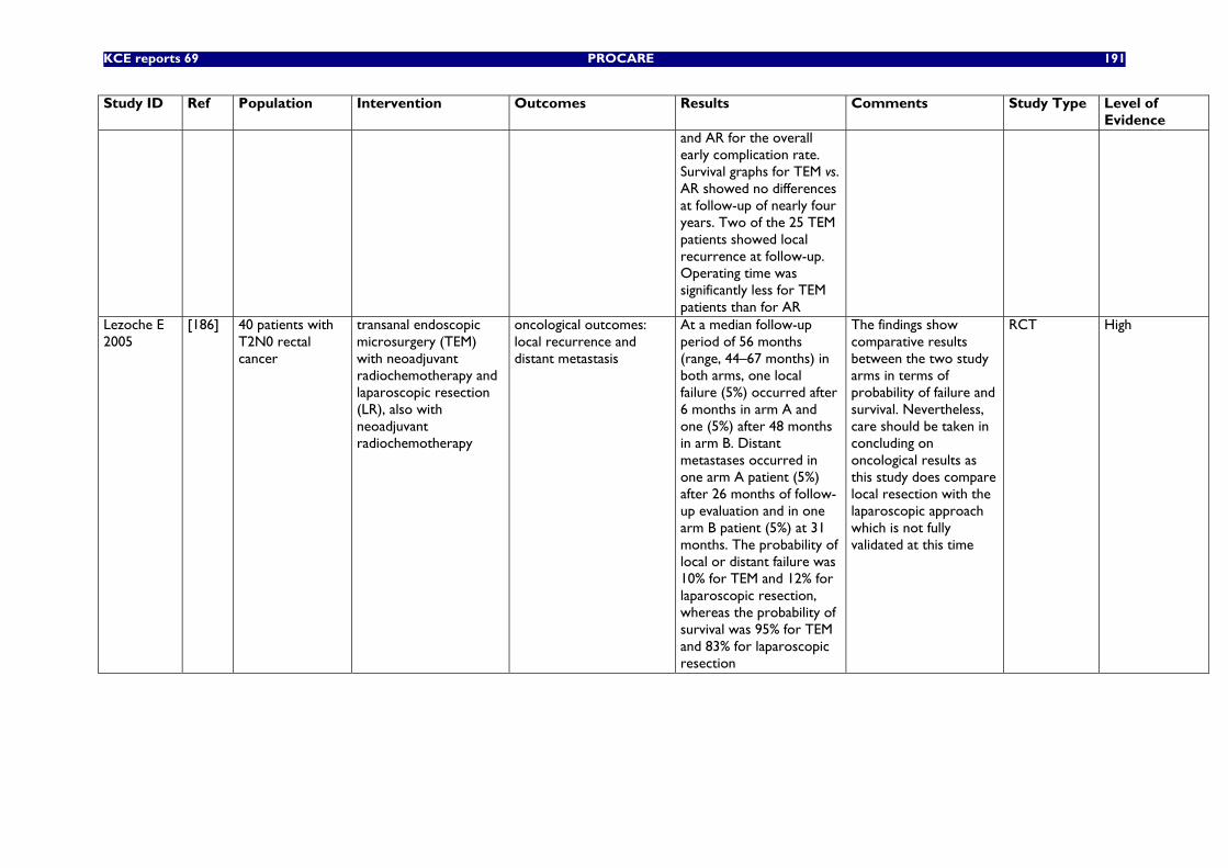
KCE reports 69 PROCARE 191
Study ID Ref Population Intervention Outcomes Results Comments Study Type Level of Evidence
and AR for the overall early complication rate. Survival graphs for TEM vs. AR showed no differences at follow-up of nearly four years. Two of the 25 TEM patients showed local recurrence at follow-up. Operating time was significantly less for TEM patients than for AR
Lezoche E 2005
[186] 40 patients with T2N0 rectal cancer
transanal endoscopic microsurgery (TEM) with neoadjuvant radiochemotherapy and laparoscopic resection (LR), also with neoadjuvant radiochemotherapy
oncological outcomes: local recurrence and distant metastasis
At a median follow-up period of 56 months (range, 44–67 months) in both arms, one local failure (5%) occurred after 6 months in arm A and one (5%) after 48 months in arm B. Distant metastases occurred in one arm A patient (5%) after 26 months of follow-up evaluation and in one arm B patient (5%) at 31 months. The probability of local or distant failure was 10% for TEM and 12% for laparoscopic resection, whereas the probability of survival was 95% for TEM and 83% for laparoscopic resection
The findings show comparative results between the two study arms in terms of probability of failure and survival. Nevertheless, care should be taken in concluding on oncological results as this study does compare local resection with the laparoscopic approach which is not fully validated at this time
RCT High
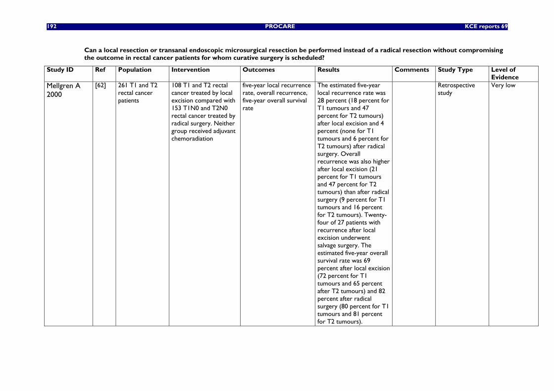
192 PROCARE KCE reports 69
Can a local resection or transanal endoscopic microsurgical resection be performed instead of a radical resection without compromising the outcome in rectal cancer patients for whom curative surgery is scheduled?
Study ID Ref Population Intervention Outcomes Results Comments Study Type Level of Evidence
Mellgren A 2000
[62] 261 T1 and T2 rectal cancer patients
108 T1 and T2 rectal cancer treated by local excision compared with 153 T1N0 and T2N0 rectal cancer treated by radical surgery. Neither group received adjuvant chemoradiation
five-year local recurrence rate, overall recurrence, five-year overall survival rate
The estimated five-year local recurrence rate was 28 percent (18 percent for T1 tumours and 47 percent for T2 tumours) after local excision and 4 percent (none for T1 tumours and 6 percent for T2 tumours) after radical surgery. Overall recurrence was also higher after local excision (21 percent for T1 tumours and 47 percent for T2 tumours) than after radical surgery (9 percent for T1 tumours and 16 percent for T2 tumours). Twenty-four of 27 patients with recurrence after local excision underwent salvage surgery. The estimated five-year overall survival rate was 69 percent after local excision (72 percent for T1 tumours and 65 percent after T2 tumours) and 82 percent after radical surgery (80 percent for T1 tumours and 81 percent for T2 tumours).
Retrospective study
Very low
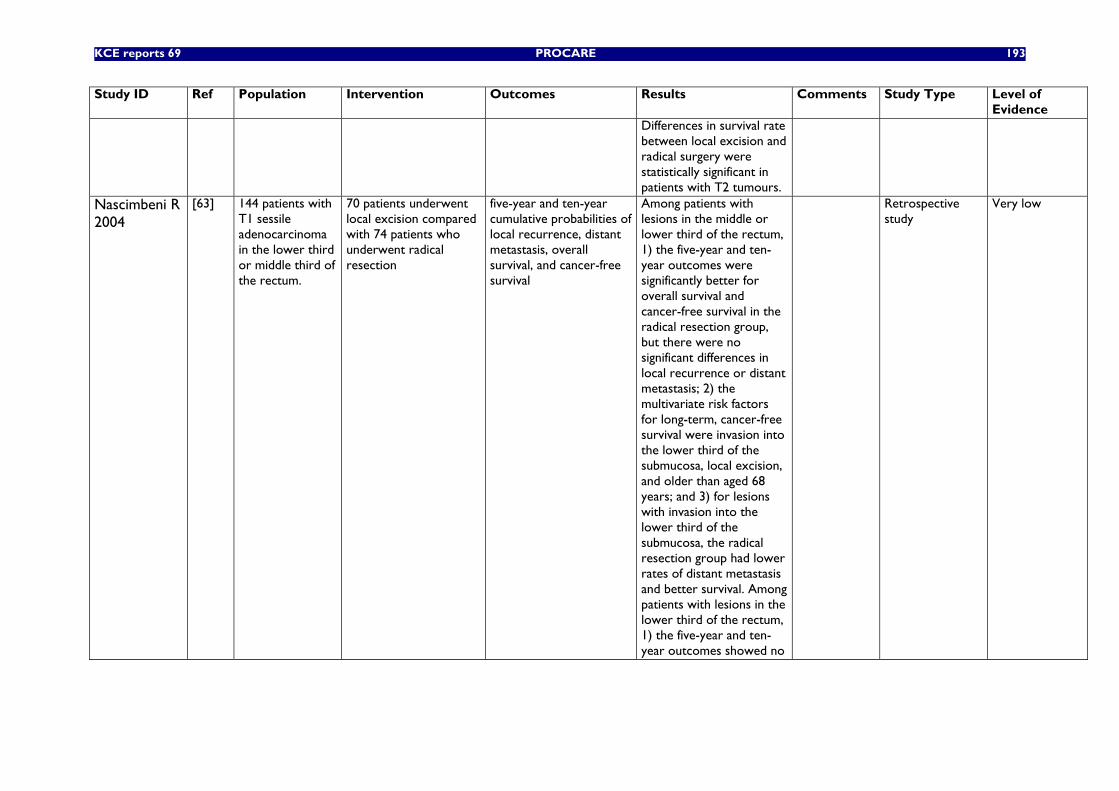
KCE reports 69 PROCARE 193
Study ID Ref Population Intervention Outcomes Results Comments Study Type Level of Evidence
Differences in survival rate between local excision and radical surgery were statistically significant in patients with T2 tumours.
Nascimbeni R 2004
[63] 144 patients with T1 sessile adenocarcinoma in the lower third or middle third of the rectum.
70 patients underwent local excision compared with 74 patients who underwent radical resection
five-year and ten-year cumulative probabilities of local recurrence, distant metastasis, overall survival, and cancer-free survival
Among patients with lesions in the middle or lower third of the rectum, 1) the five-year and ten-year outcomes were significantly better for overall survival and cancer-free survival in the radical resection group, but there were no significant differences in local recurrence or distant metastasis; 2) the multivariate risk factors for long-term, cancer-free survival were invasion into the lower third of the submucosa, local excision, and older than aged 68 years; and 3) for lesions with invasion into the lower third of the submucosa, the radical resection group had lower rates of distant metastasis and better survival. Among patients with lesions in the lower third of the rectum, 1) the five-year and ten-year outcomes showed no
Retrospective study
Very low
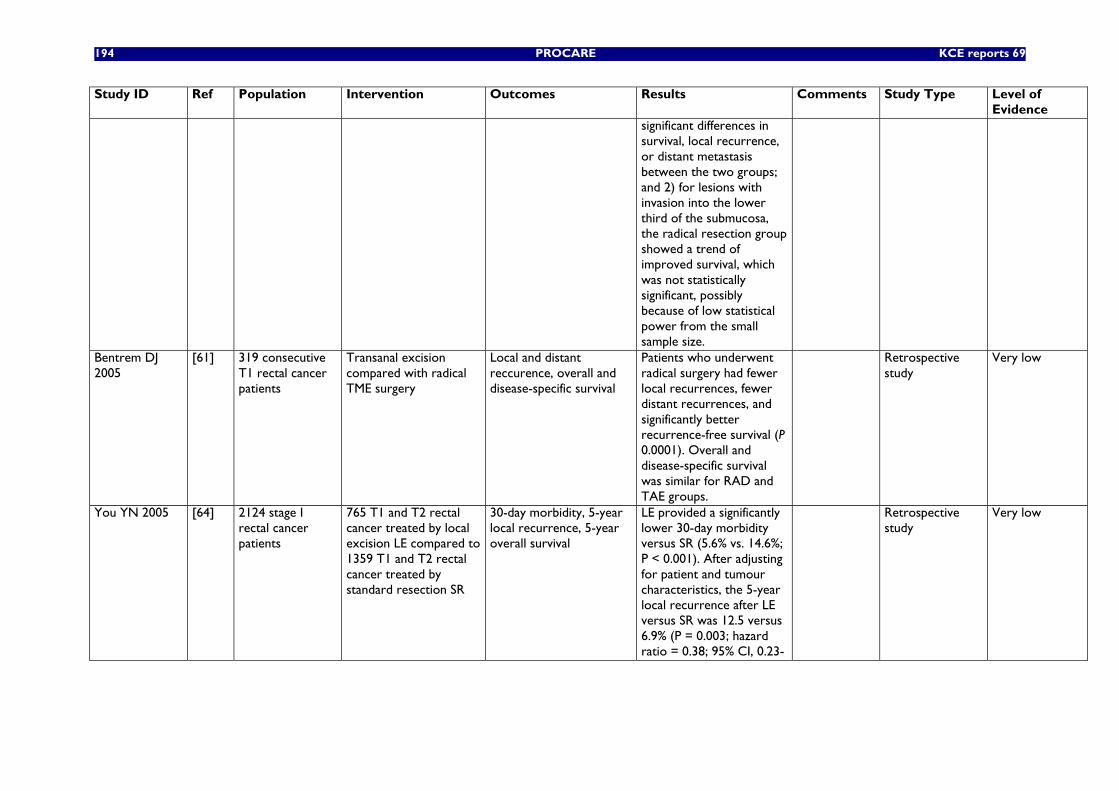
194 PROCARE KCE reports 69
Study ID Ref Population Intervention Outcomes Results Comments Study Type Level of Evidence
significant differences in survival, local recurrence, or distant metastasis between the two groups; and 2) for lesions with invasion into the lower third of the submucosa, the radical resection group showed a trend of improved survival, which was not statistically significant, possibly because of low statistical power from the small sample size.
Bentrem DJ 2005
[61] 319 consecutive T1 rectal cancer patients
Transanal excision compared with radical TME surgery
Local and distant reccurence, overall and disease-specific survival
Patients who underwent radical surgery had fewer local recurrences, fewer distant recurrences, and significantly better recurrence-free survival (P 0.0001). Overall and disease-specific survival was similar for RAD and TAE groups.
Retrospective study
Very low
You YN 2005 [64] 2124 stage I rectal cancer patients
765 T1 and T2 rectal cancer treated by local excision LE compared to 1359 T1 and T2 rectal cancer treated by standard resection SR
30-day morbidity, 5-year local recurrence, 5-year overall survival
LE provided a significantly lower 30-day morbidity versus SR (5.6% vs. 14.6%; P < 0.001). After adjusting for patient and tumour characteristics, the 5-year local recurrence after LE versus SR was 12.5 versus 6.9% (P = 0.003; hazard ratio = 0.38; 95% CI, 0.23-
Retrospective study
Very low
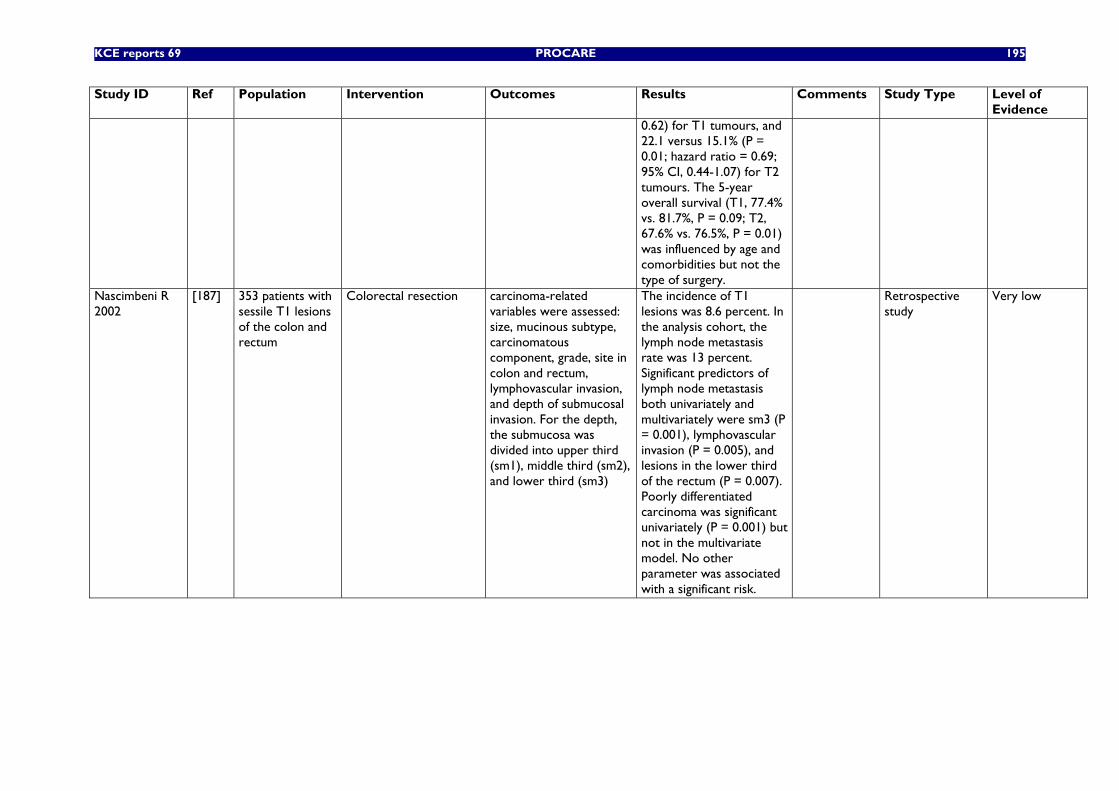
KCE reports 69 PROCARE 195
Study ID Ref Population Intervention Outcomes Results Comments Study Type Level of Evidence
0.62) for T1 tumours, and 22.1 versus 15.1% (P = 0.01; hazard ratio = 0.69; 95% CI, 0.44-1.07) for T2 tumours. The 5-year overall survival (T1, 77.4% vs. 81.7%, P = 0.09; T2, 67.6% vs. 76.5%, P = 0.01) was influenced by age and comorbidities but not the type of surgery.
Nascimbeni R 2002
[187] 353 patients with sessile T1 lesions of the colon and rectum
Colorectal resection carcinoma-related variables were assessed: size, mucinous subtype, carcinomatous component, grade, site in colon and rectum, lymphovascular invasion, and depth of submucosal invasion. For the depth, the submucosa was divided into upper third (sm1), middle third (sm2), and lower third (sm3)
The incidence of T1 lesions was 8.6 percent. In the analysis cohort, the lymph node metastasis rate was 13 percent. Significant predictors of lymph node metastasis both univariately and multivariately were sm3 (P = 0.001), lymphovascular invasion (P = 0.005), and lesions in the lower third of the rectum (P = 0.007). Poorly differentiated carcinoma was significant univariately (P = 0.001) but not in the multivariate model. No other parameter was associated with a significant risk.
Retrospective study
Very low
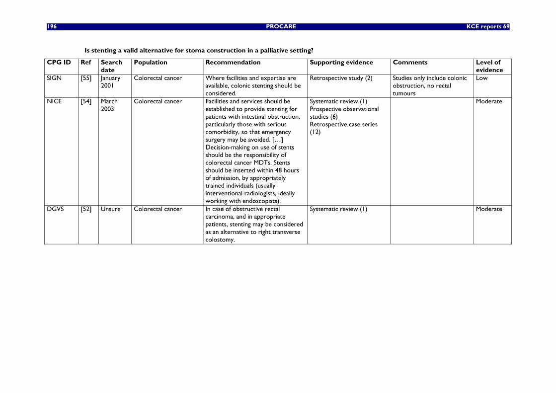
196 PROCARE KCE reports 69
Is stenting a valid alternative for stoma construction in a palliative setting?
CPG ID Ref Search date
Population Recommendation Supporting evidence Comments Level of evidence
SIGN [55] January 2001
Colorectal cancer Where facilities and expertise are available, colonic stenting should be considered.
Retrospective study (2) Studies only include colonic obstruction, no rectal tumours
Low
NICE [54] March 2003
Colorectal cancer Facilities and services should be established to provide stenting for patients with intestinal obstruction, particularly those with serious comorbidity, so that emergency surgery may be avoided. […] Decision-making on use of stents should be the responsibility of colorectal cancer MDTs. Stents should be inserted within 48 hours of admission, by appropriately trained individuals (usually interventional radiologists, ideally working with endoscopists).
Systematic review (1) Prospective observational studies (6) Retrospective case series (12)
Moderate
DGVS [52] Unsure Colorectal cancer In case of obstructive rectal carcinoma, and in appropriate patients, stenting may be considered as an alternative to right transverse colostomy.
Systematic review (1) Moderate
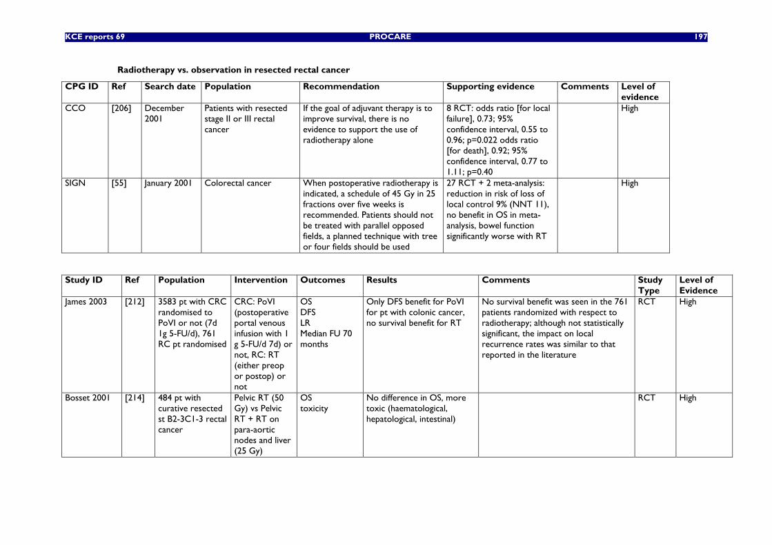
KCE reports 69 PROCARE 197
Radiotherapy vs. observation in resected rectal cancer
CPG ID Ref Search date Population Recommendation Supporting evidence Comments Level of evidence
CCO [206] December 2001
Patients with resected stage II or III rectal cancer
If the goal of adjuvant therapy is to improve survival, there is no evidence to support the use of radiotherapy alone
8 RCT: odds ratio [for local failure], 0.73; 95% confidence interval, 0.55 to 0.96; p=0.022 odds ratio [for death], 0.92; 95% confidence interval, 0.77 to 1.11; p=0.40
High
SIGN [55] January 2001 Colorectal cancer When postoperative radiotherapy is indicated, a schedule of 45 Gy in 25 fractions over five weeks is recommended. Patients should not be treated with parallel opposed fields, a planned technique with tree or four fields should be used
27 RCT + 2 meta-analysis: reduction in risk of loss of local control 9% (NNT 11), no benefit in OS in meta-analysis, bowel function significantly worse with RT
High
Study ID Ref Population Intervention Outcomes Results Comments Study Type
Level of Evidence
James 2003 [212] 3583 pt with CRC randomised to PoVI or not (7d 1g 5-FU/d), 761 RC pt randomised
CRC: PoVI (postoperative portal venous infusion with 1 g 5-FU/d 7d) or not, RC: RT (either preop or postop) or not
OS DFS LR Median FU 70 months
Only DFS benefit for PoVI for pt with colonic cancer, no survival benefit for RT
No survival benefit was seen in the 761 patients randomized with respect to radiotherapy; although not statistically significant, the impact on local recurrence rates was similar to that reported in the literature
RCT High
Bosset 2001 [214] 484 pt with curative resected st B2-3C1-3 rectal cancer
Pelvic RT (50 Gy) vs Pelvic RT + RT on para-aortic nodes and liver (25 Gy)
OS toxicity
No difference in OS, more toxic (haematological, hepatological, intestinal)
RCT High
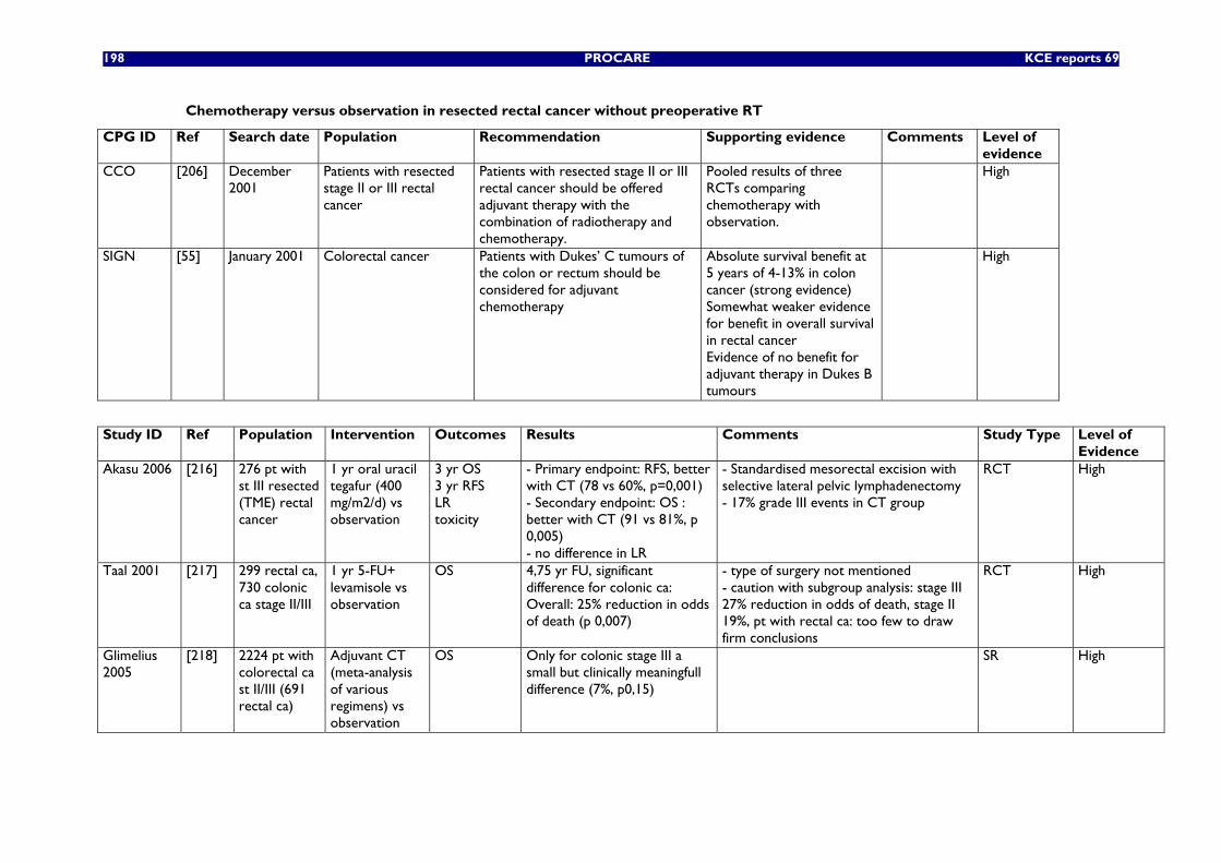
198 PROCARE KCE reports 69
Chemotherapy versus observation in resected rectal cancer without preoperative RT
CPG ID Ref Search date Population Recommendation Supporting evidence Comments Level of evidence
CCO [206] December 2001
Patients with resected stage II or III rectal cancer
Patients with resected stage II or III rectal cancer should be offered adjuvant therapy with the combination of radiotherapy and chemotherapy.
Pooled results of three RCTs comparing chemotherapy with observation.
High
SIGN [55] January 2001 Colorectal cancer Patients with Dukes’ C tumours of the colon or rectum should be considered for adjuvant chemotherapy
Absolute survival benefit at 5 years of 4-13% in colon cancer (strong evidence) Somewhat weaker evidence for benefit in overall survival in rectal cancer Evidence of no benefit for adjuvant therapy in Dukes B tumours
High
Study ID Ref Population Intervention Outcomes Results Comments Study Type Level of
Evidence Akasu 2006 [216] 276 pt with
st III resected (TME) rectal cancer
1 yr oral uracil tegafur (400 mg/m2/d) vs observation
3 yr OS 3 yr RFS LR toxicity
- Primary endpoint: RFS, better with CT (78 vs 60%, p=0,001) - Secondary endpoint: OS : better with CT (91 vs 81%, p 0,005) - no difference in LR
- Standardised mesorectal excision with selective lateral pelvic lymphadenectomy - 17% grade III events in CT group
RCT High
Taal 2001 [217] 299 rectal ca, 730 colonic ca stage II/III
1 yr 5-FU+ levamisole vs observation
OS 4,75 yr FU, significant difference for colonic ca: Overall: 25% reduction in odds of death (p 0,007)
- type of surgery not mentioned - caution with subgroup analysis: stage III 27% reduction in odds of death, stage II 19%, pt with rectal ca: too few to draw firm conclusions
RCT High
Glimelius 2005
[218] 2224 pt with colorectal ca st II/III (691 rectal ca)
Adjuvant CT (meta-analysis of various regimens) vs observation
OS Only for colonic stage III a small but clinically meaningfull difference (7%, p0,15)
SR High
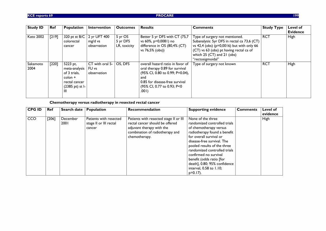
KCE reports 69 PROCARE 199
Study ID Ref Population Intervention Outcomes Results Comments Study Type Level of Evidence
Kato 2002 [219] 320 pt st B/C colorectal cancer
2 yr UFT 400 mg/d vs observation
5 yr OS 5 yr DFS LR, toxicity
Better 5 yr DFS with CT (75,7 vs 60%, p=0,0081) no difference in OS (80,4% (CT) vs 76,5% (obs))
Type of surgery not mentioned. Subanalysis: 5yr DFS in rectal ca 73,6 (CT) vs 42,4 (obs) (p=0,0016) but with only 66 (CT) vs 63 (obs) pt having rectal ca of which 25 (CT) and 21 (obs) “rectosigmoidal”
RCT High
Sakamoto 2004
[220] 5223 pt, meta-analysis of 3 trials, colon + rectal cancer (2385 pt) st I-III
CT with oral 5-FU vs observation
OS, DFS overall hazard ratio in favor of oral therapy 0.89 for survival (95% CI, 0.80 to 0.99; P=0.04), and 0.85 for disease-free survival (95% CI, 0.77 to 0.93; P<0 .001)
Type of surgery not known RCT High
Chemotherapy versus radiotherapy in resected rectal cancer
CPG ID Ref Search date Population Recommendation Supporting evidence Comments Level of evidence
CCO [206] December 2001
Patients with resected stage II or III rectal cancer
Patients with resected stage II or III rectal cancer should be offered adjuvant therapy with the combination of radiotherapy and chemotherapy.
None of the three randomized controlled trials of chemotherapy versus radiotherapy found a benefit for overall survival or disease-free survival. The pooled results of the three randomized controlled trials confirmed no survival benefit (odds ratio [for death], 0.80; 95% confidence interval, 0.58 to 1.10; p=0.17).
High
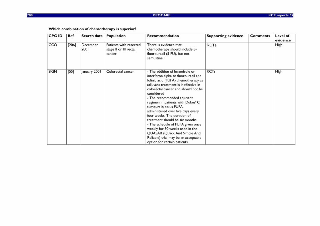
200 PROCARE KCE reports 69
Which combination of chemotherapy is superior?
CPG ID Ref Search date Population Recommendation Supporting evidence Comments Level of evidence
CCO [206] December 2001
Patients with resected stage II or III rectal cancer
There is evidence that chemotherapy should include 5-fluorouracil (5-FU), but not semustine.
RCTs High
SIGN [55] January 2001 Colorectal cancer - The addition of levamisole or interferon alpha to fluorouracil and folinic acid (FUFA) chemotherapy as adjuvant treatment is ineffective in colorectal cancer and should not be considered - The recommended adjuvant regimen in patients with Dukes' C tumours is bolus FUFA, administered over five days every four weeks. The duration of treatment should be six months - The schedule of FUFA given once weekly for 30 weeks used in the QUASAR (QUick And Simple And Reliable) trial may be an acceptable option for certain patients.
RCTs High
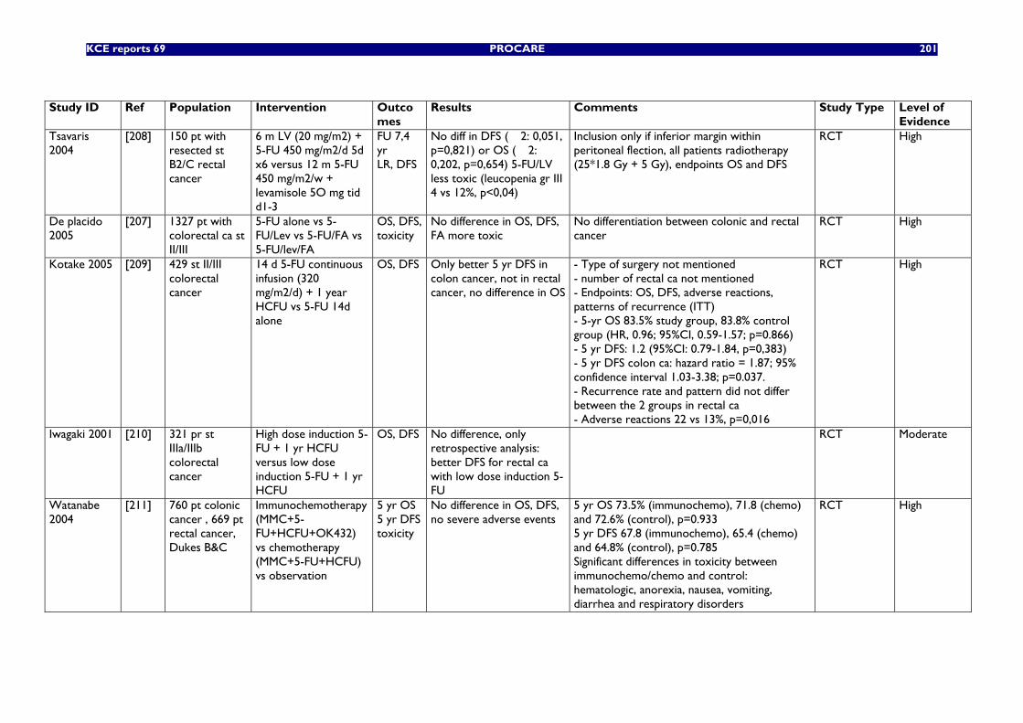
KCE reports 69 PROCARE 201
Study ID Ref Population Intervention Outco
mes Results Comments Study Type Level of
Evidence Tsavaris 2004
[208] 150 pt with resected st B2/C rectal cancer
6 m LV (20 mg/m2) + 5-FU 450 mg/m2/d 5d x6 versus 12 m 5-FU 450 mg/m2/w + levamisole 5O mg tid d1-3
FU 7,4 yr LR, DFS
No diff in DFS (�2: 0,051, p=0,821) or OS (�2: 0,202, p=0,654) 5-FU/LV less toxic (leucopenia gr III 4 vs 12%, p<0,04)
Inclusion only if inferior margin within peritoneal flection, all patients radiotherapy (25*1.8 Gy + 5 Gy), endpoints OS and DFS
RCT High
De placido 2005
[207] 1327 pt with colorectal ca st II/III
5-FU alone vs 5-FU/Lev vs 5-FU/FA vs 5-FU/lev/FA
OS, DFS, toxicity
No difference in OS, DFS, FA more toxic
No differentiation between colonic and rectal cancer
RCT High
Kotake 2005 [209] 429 st II/III colorectal cancer
14 d 5-FU continuous infusion (320 mg/m2/d) + 1 year HCFU vs 5-FU 14d alone
OS, DFS Only better 5 yr DFS in colon cancer, not in rectal cancer, no difference in OS
- Type of surgery not mentioned - number of rectal ca not mentioned - Endpoints: OS, DFS, adverse reactions, patterns of recurrence (ITT) - 5-yr OS 83.5% study group, 83.8% control group (HR, 0.96; 95%CI, 0.59-1.57; p=0.866) - 5 yr DFS: 1.2 (95%CI: 0.79-1.84, p=0,383) - 5 yr DFS colon ca: hazard ratio = 1.87; 95% confidence interval 1.03-3.38; p=0.037. - Recurrence rate and pattern did not differ between the 2 groups in rectal ca - Adverse reactions 22 vs 13%, p=0,016
RCT High
Iwagaki 2001 [210] 321 pr st IIIa/IIIb colorectal cancer
High dose induction 5-FU + 1 yr HCFU versus low dose induction 5-FU + 1 yr HCFU
OS, DFS No difference, only retrospective analysis: better DFS for rectal ca with low dose induction 5-FU
RCT Moderate
Watanabe 2004
[211] 760 pt colonic cancer , 669 pt rectal cancer, Dukes B&C
Immunochemotherapy (MMC+5-FU+HCFU+OK432) vs chemotherapy (MMC+5-FU+HCFU) vs observation
5 yr OS 5 yr DFS toxicity
No difference in OS, DFS, no severe adverse events
5 yr OS 73.5% (immunochemo), 71.8 (chemo) and 72.6% (control), p=0.933 5 yr DFS 67.8 (immunochemo), 65.4 (chemo) and 64.8% (control), p=0.785 Significant differences in toxicity between immunochemo/chemo and control: hematologic, anorexia, nausea, vomiting, diarrhea and respiratory disorders
RCT High
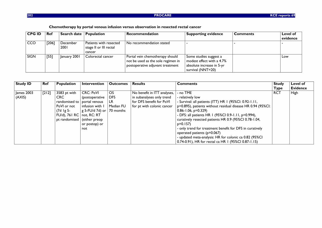
202 PROCARE KCE reports 69
Chemotherapy by portal venous infusion versus observation in resected rectal cancer
CPG ID Ref Search date Population Recommendation Supporting evidence Comments Level of evidence
CCO [206] December 2001
Patients with resected stage II or III rectal cancer
No recommendation stated - - -
SIGN [55] January 2001 Colorectal cancer Portal vein chemotherapy should not be used as the sole regimen in postoperative adjuvant treatment
Some studies suggest a modest effect with a 4.7% absolute increase in 5-yr survival (NNT=20)
Low
Study ID Ref Population Intervention Outcomes Results Comments Study Type
Level of Evidence
James 2003 (AXIS)
[212] 3583 pt with CRC randomised to PoVI or not (7d 1g 5-FU/d), 761 RC pt randomised
CRC: PoVI (postoperative portal venous infusion with 1 g 5-FU/d 7d) or not, RC: RT (either preop or postop) or not
OS DFS LR Median FU 70 months
No benefit in ITT analyses, in subanalyses only trend for DFS benefit for PoVI for pt with colonic cancer
- no TME - relatively low - Survival: all patients (ITT) HR 1 (95%CI: 0.92-1.11, p=0.895), patients without residual disease HR 0.94 (95%CI: 0.86-1.06, p=0.329) - DFS: all patients HR 1 (95%CI 0.9-1.11, p=0.994), curatively resected patients HR 0.9 (95%CI 0.78-1.04, p=0.157) - only trend for treatment benefit for DFS in curatively operated patients (p=0.067) - updated meta-analysis: HR for colonic ca 0.82 (95%CI 0.74-0.91), HR for rectal ca HR 1 (95%CI 0.87-1.15)
RCT High
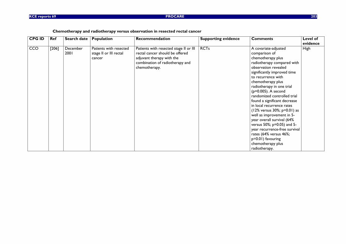
KCE reports 69 PROCARE 203
Chemotherapy and radiotherapy versus observation in resected rectal cancer
CPG ID Ref Search date Population Recommendation Supporting evidence Comments Level of evidence
CCO [206] December 2001
Patients with resected stage II or III rectal cancer
Patients with resected stage II or III rectal cancer should be offered adjuvant therapy with the combination of radiotherapy and chemotherapy.
RCTs A covariate-adjusted comparison of chemotherapy plus radiotherapy compared with observation revealed significantly improved time to recurrence with chemotherapy plus radiotherapy in one trial (p=0.005). A second randomized controlled trial found a significant decrease in local recurrence rates (12% versus 30%; p=0.01) as well as improvement in 5-year overall survival (64% versus 50%; p=0.05) and 5-year recurrence-free survival rates (64% versus 46%; p=0.01) favouring chemotherapy plus radiotherapy.
High
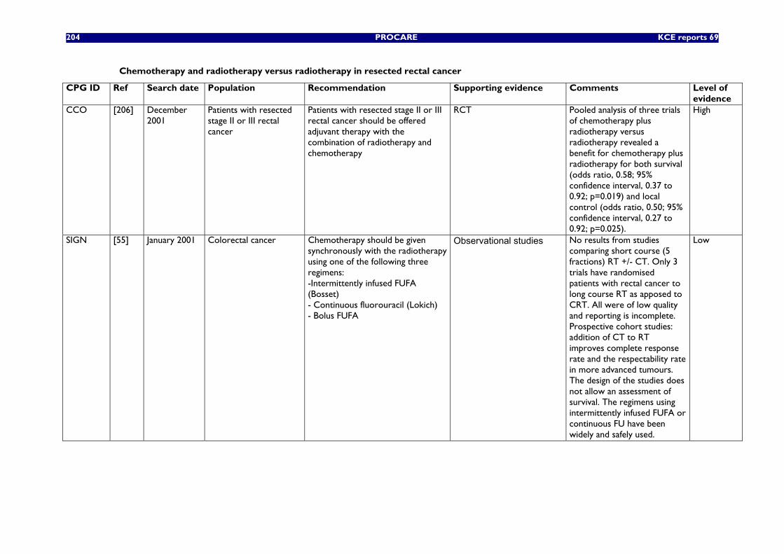
204 PROCARE KCE reports 69
Chemotherapy and radiotherapy versus radiotherapy in resected rectal cancer
CPG ID Ref Search date Population Recommendation Supporting evidence Comments Level of evidence
CCO [206] December 2001
Patients with resected stage II or III rectal cancer
Patients with resected stage II or III rectal cancer should be offered adjuvant therapy with the combination of radiotherapy and chemotherapy
RCT Pooled analysis of three trials of chemotherapy plus radiotherapy versus radiotherapy revealed a benefit for chemotherapy plus radiotherapy for both survival (odds ratio, 0.58; 95% confidence interval, 0.37 to 0.92; p=0.019) and local control (odds ratio, 0.50; 95% confidence interval, 0.27 to 0.92; p=0.025).
High
SIGN [55] January 2001 Colorectal cancer Chemotherapy should be given synchronously with the radiotherapy using one of the following three regimens: -Intermittently infused FUFA (Bosset) - Continuous fluorouracil (Lokich) - Bolus FUFA
Observational studies No results from studies comparing short course (5 fractions) RT +/- CT. Only 3 trials have randomised patients with rectal cancer to long course RT as apposed to CRT. All were of low quality and reporting is incomplete. Prospective cohort studies: addition of CT to RT improves complete response rate and the respectability rate in more advanced tumours. The design of the studies does not allow an assessment of survival. The regimens using intermittently infused FUFA or continuous FU have been widely and safely used.
Low
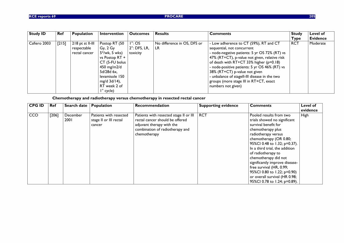
KCE reports 69 PROCARE 205
Study ID Ref Population Intervention Outcomes Results Comments Study
Type Level of Evidence
Cafiero 2003 [215] 218 pt st II-III respectable rectal cancer
Postop RT (50 Gy, 2 Gy 5*/wk, 5 wks) vs Postop RT + CT (5-FU bolus 450 mg/m2/d 5d/28d 6x, levamisole 150 mg/d 3d/14), RT week 2 of 1° cycle)
1°: OS 2°: DFS, LR, toxicity
No difference in OS, DFS or LR
- Low adherence to CT (59%), RT and CT sequential, not concurrent - node-negative patients: 5 yr OS 72% (RT) vs 47% (RT+CT), p-value not given, relative risk of death with RT+CT 33% higher (p=0.18) - node-positive patients: 5 yr OS 46% (RT) vs 38% (RT+CT) p-value not given - unbalance of stageII-III disease in the two groups (more stage III in RT+CT, exact numbers not given)
RCT Moderate
Chemotherapy and radiotherapy versus chemotherapy in resected rectal cancer
CPG ID Ref Search date Population Recommendation Supporting evidence Comments Level of evidence
CCO [206] December 2001
Patients with resected stage II or III rectal cancer
Patients with resected stage II or III rectal cancer should be offered adjuvant therapy with the combination of radiotherapy and chemotherapy
RCT Pooled results from two trials showed no significant survival benefit for chemotherapy plus radiotherapy versus chemotherapy (OR 0.80; 95%CI 0.48 to 1.32; p=0.37). In a third trial, the addition of radiotherapy to chemotherapy did not significantly improve disease-free survival (HR, 0.99; 95%CI 0.80 to 1.22; p=0.90) or overall survival (HR 0.98; 95%CI 0.78 to 1.24; p=0.89).
High
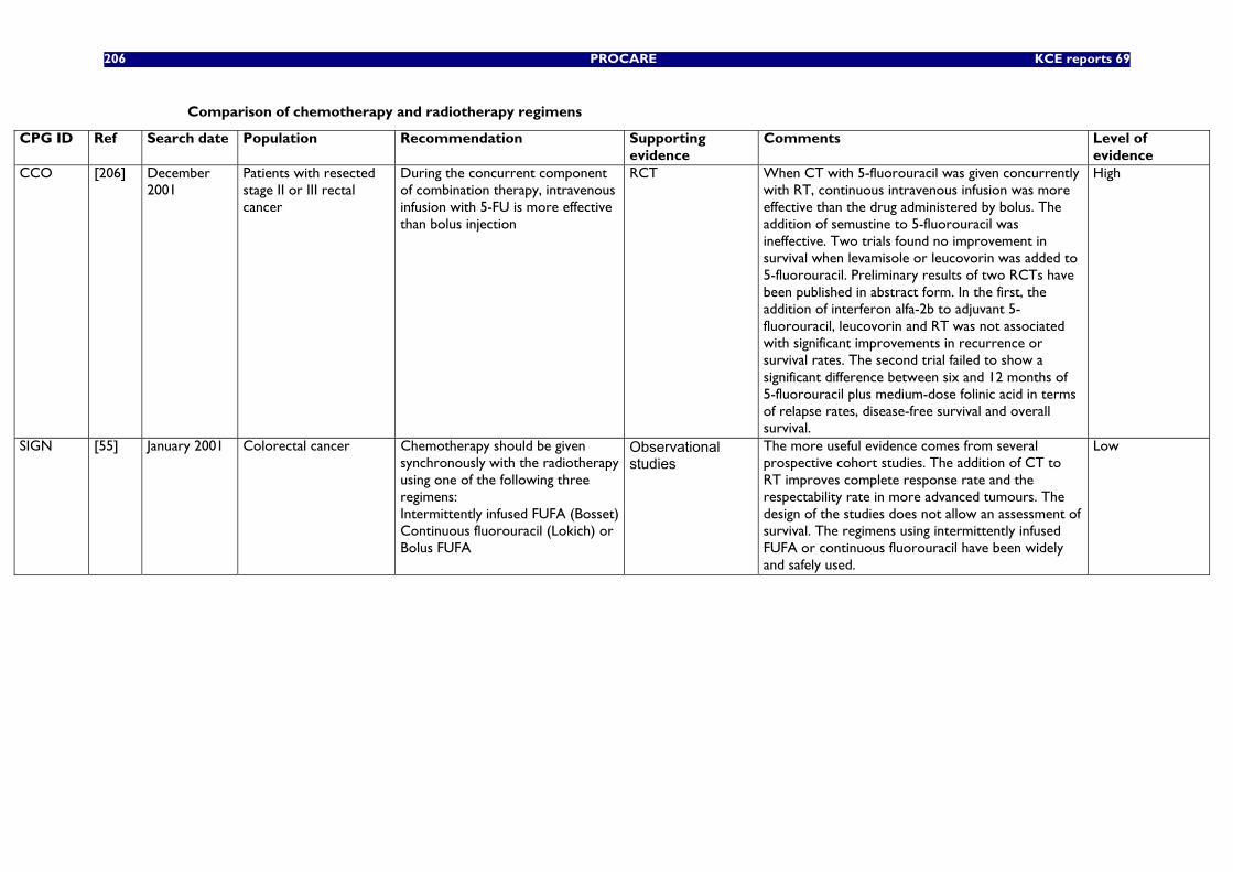
206 PROCARE KCE reports 69
Comparison of chemotherapy and radiotherapy regimens
CPG ID Ref Search date Population Recommendation Supporting evidence
Comments Level of evidence
CCO [206] December 2001
Patients with resected stage II or III rectal cancer
During the concurrent component of combination therapy, intravenous infusion with 5-FU is more effective than bolus injection
RCT When CT with 5-fluorouracil was given concurrently with RT, continuous intravenous infusion was more effective than the drug administered by bolus. The addition of semustine to 5-fluorouracil was ineffective. Two trials found no improvement in survival when levamisole or leucovorin was added to 5-fluorouracil. Preliminary results of two RCTs have been published in abstract form. In the first, the addition of interferon alfa-2b to adjuvant 5-fluorouracil, leucovorin and RT was not associated with significant improvements in recurrence or survival rates. The second trial failed to show a significant difference between six and 12 months of 5-fluorouracil plus medium-dose folinic acid in terms of relapse rates, disease-free survival and overall survival.
High
SIGN [55] January 2001 Colorectal cancer Chemotherapy should be given synchronously with the radiotherapy using one of the following three regimens: Intermittently infused FUFA (Bosset) Continuous fluorouracil (Lokich) or Bolus FUFA
Observational studies
The more useful evidence comes from several prospective cohort studies. The addition of CT to RT improves complete response rate and the respectability rate in more advanced tumours. The design of the studies does not allow an assessment of survival. The regimens using intermittently infused FUFA or continuous fluorouracil have been widely and safely used.
Low
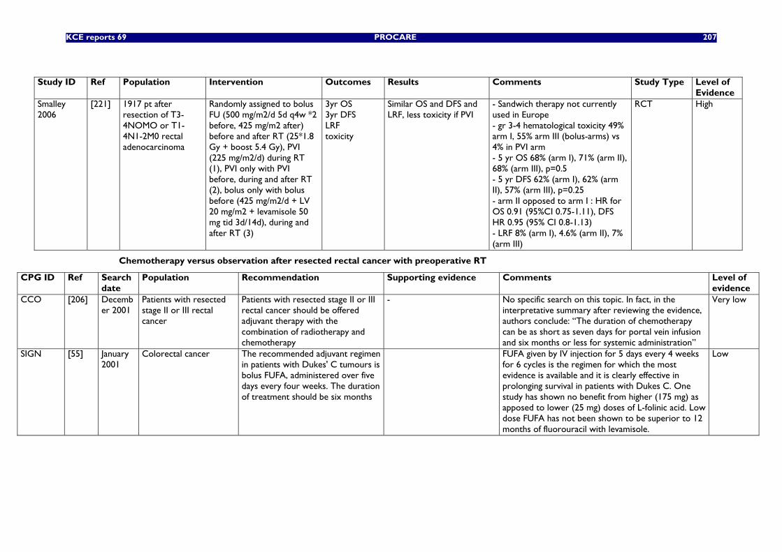
KCE reports 69 PROCARE 207
Study ID Ref Population Intervention Outcomes Results Comments Study Type Level of
Evidence Smalley 2006
[221] 1917 pt after resection of T3-4NOMO or T1-4N1-2M0 rectal adenocarcinoma
Randomly assigned to bolus FU (500 mg/m2/d 5d q4w *2 before, 425 mg/m2 after) before and after RT (25*1.8 Gy + boost 5.4 Gy), PVI (225 mg/m2/d) during RT (1), PVI only with PVI before, during and after RT (2), bolus only with bolus before (425 mg/m2/d + LV 20 mg/m2 + levamisole 50 mg tid 3d/14d), during and after RT (3)
3yr OS 3yr DFS LRF toxicity
Similar OS and DFS and LRF, less toxicity if PVI
- Sandwich therapy not currently used in Europe - gr 3-4 hematological toxicity 49% arm I, 55% arm III (bolus-arms) vs 4% in PVI arm - 5 yr OS 68% (arm I), 71% (arm II), 68% (arm III), p=0.5 - 5 yr DFS 62% (arm I), 62% (arm II), 57% (arm III), p=0.25 - arm II opposed to arm I : HR for OS 0.91 (95%CI 0.75-1.11), DFS HR 0.95 (95% CI 0.8-1.13) - LRF 8% (arm I), 4.6% (arm II), 7% (arm III)
RCT High
Chemotherapy versus observation after resected rectal cancer with preoperative RT
CPG ID Ref Search date
Population Recommendation Supporting evidence Comments Level of evidence
CCO [206] December 2001
Patients with resected stage II or III rectal cancer
Patients with resected stage II or III rectal cancer should be offered adjuvant therapy with the combination of radiotherapy and chemotherapy
- No specific search on this topic. In fact, in the interpretative summary after reviewing the evidence, authors conclude: “The duration of chemotherapy can be as short as seven days for portal vein infusion and six months or less for systemic administration”
Very low
SIGN [55] January 2001
Colorectal cancer The recommended adjuvant regimen in patients with Dukes' C tumours is bolus FUFA, administered over five days every four weeks. The duration of treatment should be six months
FUFA given by IV injection for 5 days every 4 weeks for 6 cycles is the regimen for which the most evidence is available and it is clearly effective in prolonging survival in patients with Dukes C. One study has shown no benefit from higher (175 mg) as apposed to lower (25 mg) doses of L-folinic acid. Low dose FUFA has not been shown to be superior to 12 months of fluorouracil with levamisole.
Low
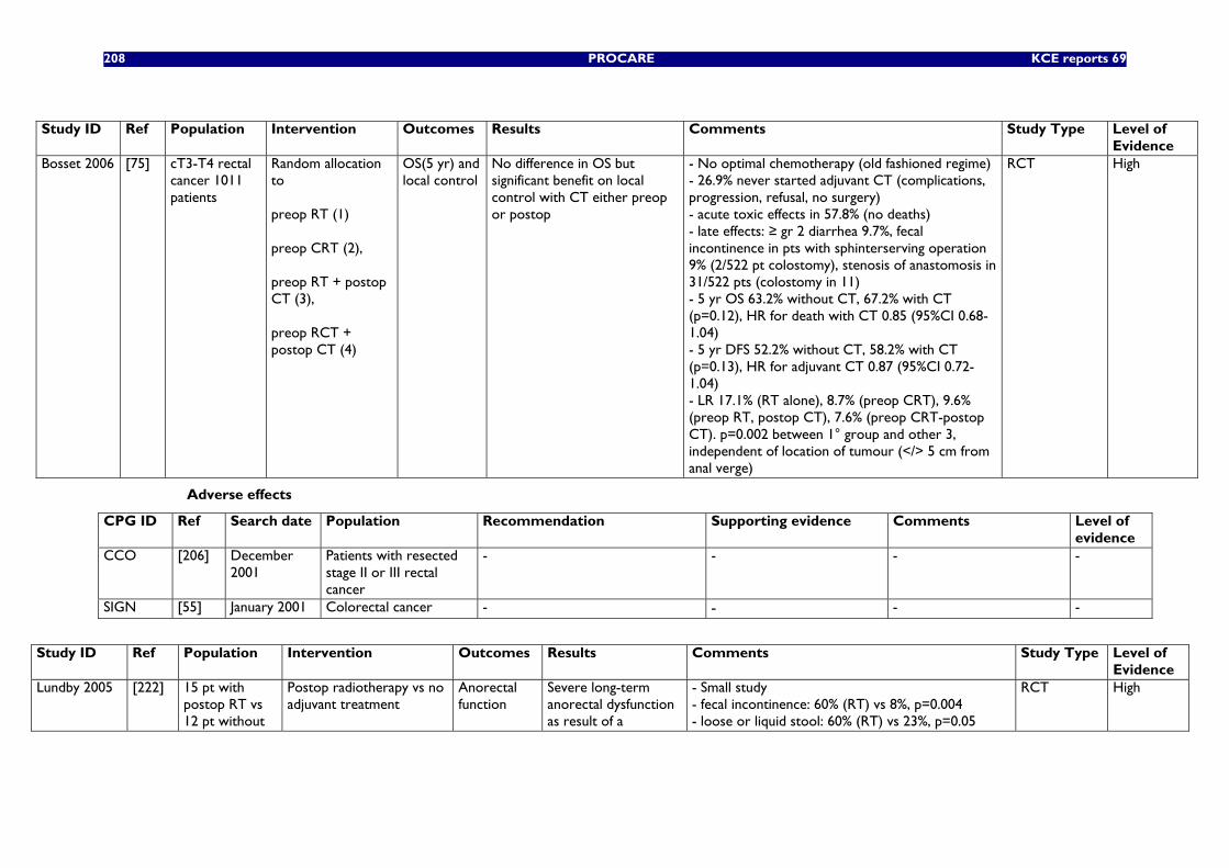
208 PROCARE KCE reports 69
Study ID Ref Population Intervention Outcomes Results Comments Study Type Level of
Evidence Bosset 2006 [75] cT3-T4 rectal
cancer 1011 patients
Random allocation to preop RT (1) preop CRT (2), preop RT + postop CT (3), preop RCT + postop CT (4)
OS(5 yr) and local control
No difference in OS but significant benefit on local control with CT either preop or postop
- No optimal chemotherapy (old fashioned regime) - 26.9% never started adjuvant CT (complications, progression, refusal, no surgery) - acute toxic effects in 57.8% (no deaths) - late effects: ≥ gr 2 diarrhea 9.7%, fecal incontinence in pts with sphinterserving operation 9% (2/522 pt colostomy), stenosis of anastomosis in 31/522 pts (colostomy in 11) - 5 yr OS 63.2% without CT, 67.2% with CT (p=0.12), HR for death with CT 0.85 (95%CI 0.68-1.04) - 5 yr DFS 52.2% without CT, 58.2% with CT (p=0.13), HR for adjuvant CT 0.87 (95%CI 0.72-1.04) - LR 17.1% (RT alone), 8.7% (preop CRT), 9.6% (preop RT, postop CT), 7.6% (preop CRT-postop CT). p=0.002 between 1° group and other 3, independent of location of tumour (</> 5 cm from anal verge)
RCT High
Adverse effects
CPG ID Ref Search date Population Recommendation Supporting evidence Comments Level of evidence
CCO [206] December 2001
Patients with resected stage II or III rectal cancer
- - - -
SIGN [55] January 2001 Colorectal cancer - - - -
Study ID Ref Population Intervention Outcomes Results Comments Study Type Level of
Evidence Lundby 2005 [222] 15 pt with
postop RT vs 12 pt without
Postop radiotherapy vs no adjuvant treatment
Anorectal function
Severe long-term anorectal dysfunction as result of a
- Small study - fecal incontinence: 60% (RT) vs 8%, p=0.004 - loose or liquid stool: 60% (RT) vs 23%, p=0.05
RCT High
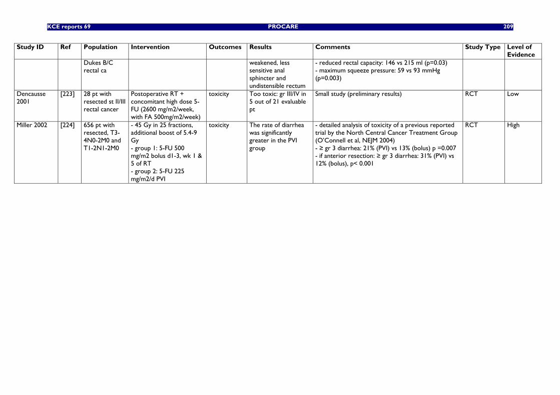
KCE reports 69 PROCARE 209
Study ID Ref Population Intervention Outcomes Results Comments Study Type Level of Evidence
Dukes B/C rectal ca
weakened, less sensitive anal sphincter and undistensible rectum
- reduced rectal capacity: 146 vs 215 ml (p=0.03) - maximum squeeze pressure: 59 vs 93 mmHg (p=0.003)
Dencausse 2001
[223] 28 pt with resected st II/III rectal cancer
Postoperative RT + concomitant high dose 5-FU (2600 mg/m2/week, with FA 500mg/m2/week)
toxicity Too toxic: gr III/IV in 5 out of 21 evaluable pt
Small study (preliminary results) RCT Low
Miller 2002 [224] 656 pt with resected, T3-4N0-2M0 and T1-2N1-2M0
- 45 Gy in 25 fractions, additional boost of 5.4-9 Gy - group 1: 5-FU 500 mg/m2 bolus d1-3, wk 1 & 5 of RT - group 2: 5-FU 225 mg/m2/d PVI
toxicity The rate of diarrhea was significantly greater in the PVI group
- detailed analysis of toxicity of a previous reported trial by the North Central Cancer Treatment Group (O’Connell et al, NEJM 2004) - ≥ gr 3 diarrhea: 21% (PVI) vs 13% (bolus) p =0.007 - if anterior resection: ≥ gr 3 diarrhea: 31% (PVI) vs 12% (bolus), p< 0.001
RCT High
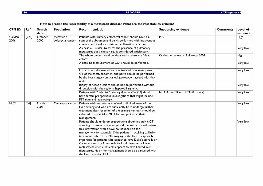
210 PROCARE KCE reports 69
How to precise the resectability of a metastatic disease? What are the resectability criteria?
CPG ID Ref Search date
Population Recommendation Supporting evidence Comments Level of evidence
Patients with primary colorectal cancer should have a CT scan of the abdomen and pelvis performed with intravenous contrast and ideally a maximum collimation of 5 mm.
MA High
A chest CT is ideal to assess the presence of pulmonary metastases but a chest x-ray is considered satisfactory.
Very low
The whole colon should be visualised to ensure a “clean colon”.
Cochrane review on follow-up 2002 High
A baseline measurement of CEA should be performed. Very low
For a patient discovered to have isolated liver metastases, CT of the chest, abdomen, and pelvis should be performed by the liver surgery unit or using protocols agreed with that unit.
Very low
Biopsy of hepatic lesions should not be performed without discussion with the regional hepatobiliary unit.
Very low
Garden 2006
[228] October 2000
Metastatic colorectal cancer
Patients with “high risk” primary disease (T4, C2) should have careful preoperative investigations that might include PET scan and laparoscopy.
No MA nor SR nor RCT (8 papers) Very low
Patients with metastases confined to limited areas of the liver or lung and who are sufficiently fit to undergo further treatment after resection of the primary tumour, should be referred to a specialist MDT for an opinion on their management.
Very low NICE [54] March 2003
Colorectal cancer
Patients should undergo preoperative abdomino-pelvic CT scanning to assess cancer stage and metastatic spread, unless this information would have no influence on the management-for example, if the patient is receiving palliative treatment only. CT or MR imaging of the liver is especially important for patients who appear to have Duke’s stage B or C cancers and are fit enough for local treatment of liver metastases; when a patients appears to have limited liver metastases, his or her management should be discussed with the liver resection MDT.
Very low
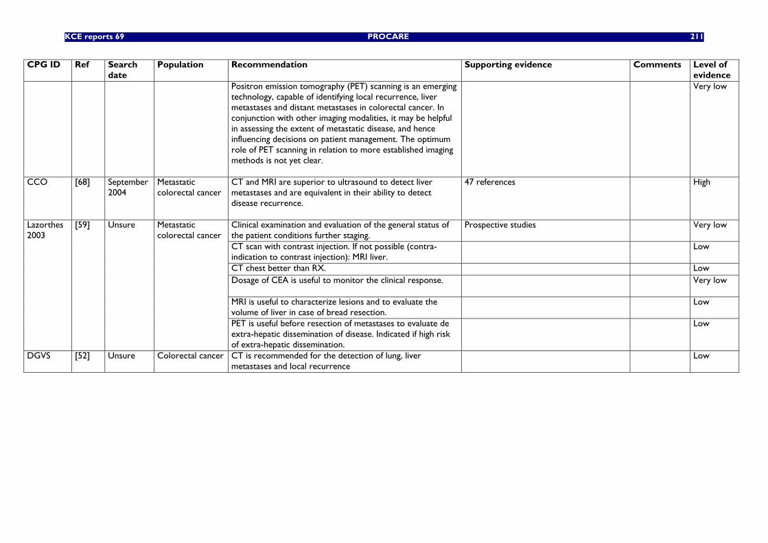
KCE reports 69 PROCARE 211
CPG ID Ref Search date
Population Recommendation Supporting evidence Comments Level of evidence
Positron emission tomography (PET) scanning is an emerging technology, capable of identifying local recurrence, liver metastases and distant metastases in colorectal cancer. In conjunction with other imaging modalities, it may be helpful in assessing the extent of metastatic disease, and hence influencing decisions on patient management. The optimum role of PET scanning in relation to more established imaging methods is not yet clear.
Very low
CCO [68] September 2004
Metastatic colorectal cancer
CT and MRI are superior to ultrasound to detect liver metastases and are equivalent in their ability to detect disease recurrence.
47 references High
Clinical examination and evaluation of the general status of the patient conditions further staging.
Prospective studies Very low
CT scan with contrast injection. If not possible (contra-indication to contrast injection): MRI liver.
Low
CT chest better than RX. Low Dosage of CEA is useful to monitor the clinical response. Very low
MRI is useful to characterize lesions and to evaluate the volume of liver in case of bread resection.
Low
Lazorthes 2003
[59] Unsure Metastatic colorectal cancer
PET is useful before resection of metastases to evaluate de extra-hepatic dissemination of disease. Indicated if high risk of extra-hepatic dissemination.
Low
DGVS [52] Unsure Colorectal cancer CT is recommended for the detection of lung, liver metastases and local recurrence
Low
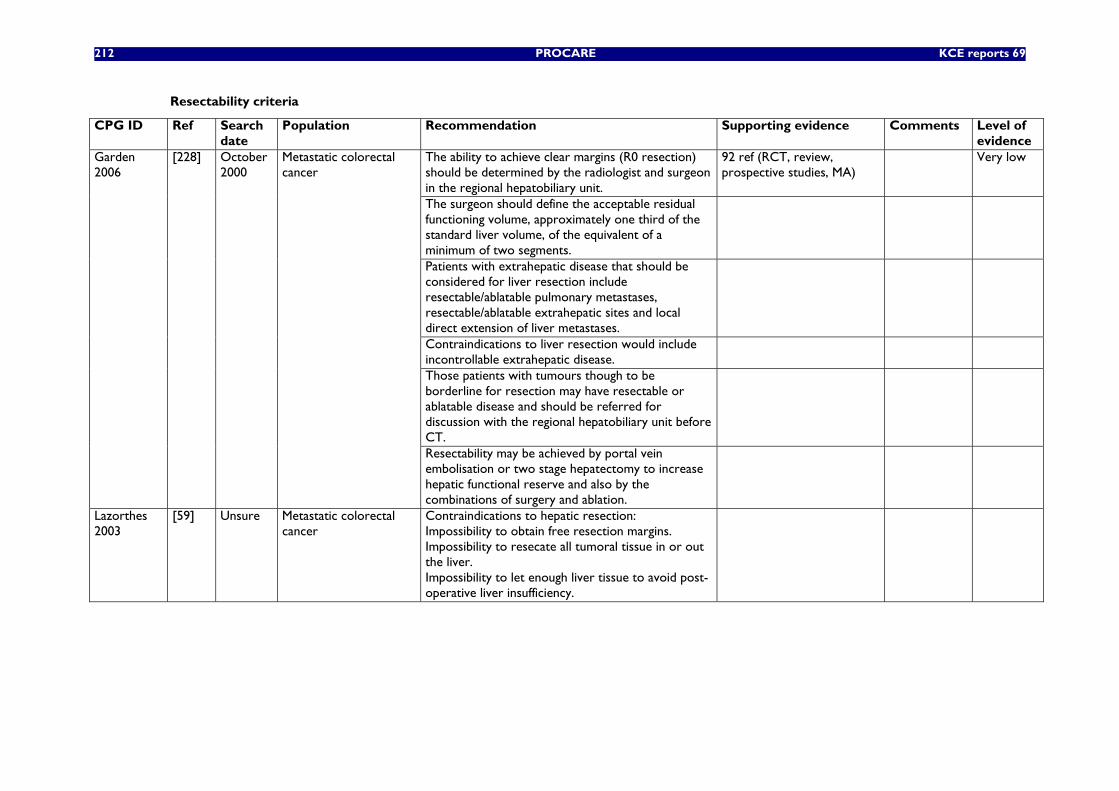
212 PROCARE KCE reports 69
Resectability criteria
CPG ID Ref Search date
Population Recommendation Supporting evidence Comments Level of evidence
The ability to achieve clear margins (R0 resection) should be determined by the radiologist and surgeon in the regional hepatobiliary unit.
92 ref (RCT, review, prospective studies, MA)
Very low
The surgeon should define the acceptable residual functioning volume, approximately one third of the standard liver volume, of the equivalent of a minimum of two segments.
Patients with extrahepatic disease that should be considered for liver resection include resectable/ablatable pulmonary metastases, resectable/ablatable extrahepatic sites and local direct extension of liver metastases.
Contraindications to liver resection would include incontrollable extrahepatic disease.
Those patients with tumours though to be borderline for resection may have resectable or ablatable disease and should be referred for discussion with the regional hepatobiliary unit before CT.
Garden 2006
[228] October 2000
Metastatic colorectal cancer
Resectability may be achieved by portal vein embolisation or two stage hepatectomy to increase hepatic functional reserve and also by the combinations of surgery and ablation.
Lazorthes 2003
[59] Unsure Metastatic colorectal cancer
Contraindications to hepatic resection: Impossibility to obtain free resection margins. Impossibility to resecate all tumoral tissue in or out the liver. Impossibility to let enough liver tissue to avoid post-operative liver insufficiency.
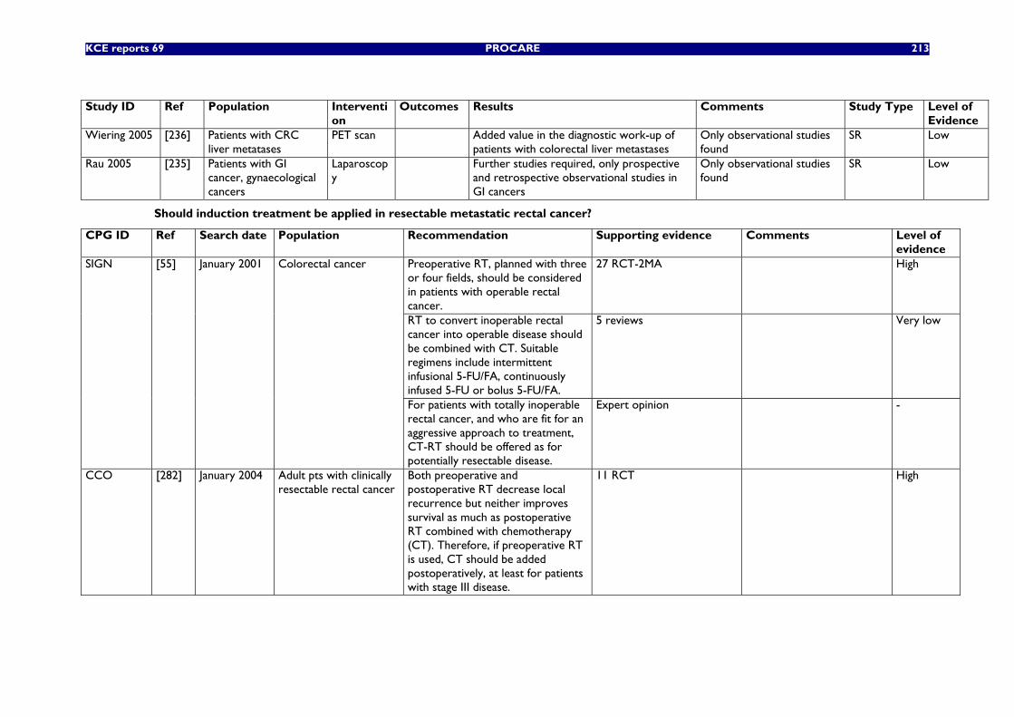
KCE reports 69 PROCARE 213
Study ID Ref Population Interventi
on Outcomes Results Comments Study Type Level of
Evidence Wiering 2005 [236] Patients with CRC
liver metatases PET scan Added value in the diagnostic work-up of
patients with colorectal liver metastases Only observational studies found
SR Low
Rau 2005 [235] Patients with GI cancer, gynaecological cancers
Laparoscopy
Further studies required, only prospective and retrospective observational studies in GI cancers
Only observational studies found
SR Low
Should induction treatment be applied in resectable metastatic rectal cancer?
CPG ID Ref Search date Population Recommendation Supporting evidence Comments Level of evidence
Preoperative RT, planned with three or four fields, should be considered in patients with operable rectal cancer.
27 RCT-2MA High
RT to convert inoperable rectal cancer into operable disease should be combined with CT. Suitable regimens include intermittent infusional 5-FU/FA, continuously infused 5-FU or bolus 5-FU/FA.
5 reviews Very low
SIGN [55] January 2001 Colorectal cancer
For patients with totally inoperable rectal cancer, and who are fit for an aggressive approach to treatment, CT-RT should be offered as for potentially resectable disease.
Expert opinion -
CCO [282] January 2004 Adult pts with clinically resectable rectal cancer
Both preoperative and postoperative RT decrease local recurrence but neither improves survival as much as postoperative RT combined with chemotherapy (CT). Therefore, if preoperative RT is used, CT should be added postoperatively, at least for patients with stage III disease.
11 RCT High
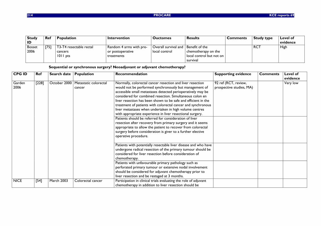
214 PROCARE KCE reports 69
Study ID
Ref Population Intervention Ouctomes Results Comments Study type Level of evidence
Bosset 2006
[75] T3-T4 resectable rectal cancers 1011 pts
Random 4 arms with pre- or postoperative treatments
Overall survival and local control
Benefit of the chemotherapy on the local control but not on survival
RCT High
Sequential or synchronous surgery? Neoadjuvant or adjuvant chemotherapy?
CPG ID Ref Search date Population Recommendation Supporting evidence Comments Level of evidence
Normally, colorectal cancer resection and liver resection would not be performed synchronously but management of accessible small metastases detected perioperatively may be considered for combined resection. Simultaneous colon en liver resection has been shown to be safe and efficient in the treatment of patients with colorectal cancer and synchronous liver metastases when undertaken in high volume centres with appropriate experience in liver resectional surgery.
92 ref (RCT, review, prospective studies, MA)
Very low
Patients should be referred for consideration of liver resection after recovery from primary surgery and it seems appropriate to allow the patient to recover from colorectal surgery before consideration is giver to a further elective operative procedure.
Patients with potentially resectable liver disease and who have undergone radical resection of the primary tumour should be considered for liver resection before consideration of chemotherapy.
Garden 2006
[228] October 2000 Metastatic colorectal cancer
Patients with unfavourable primary pathology such as perforated primary tumour or extensive nodal involvement should be considered for adjuvant chemotherapy prior to liver resection and be restaged at 3 months.
NICE [54] March 2003 Colorectal cancer Participation in clinical trials evaluating the role of adjuvant chemotherapy in addition to liver resection should be
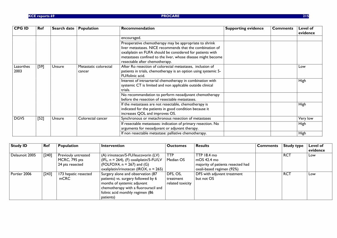
KCE reports 69 PROCARE 215
CPG ID Ref Search date Population Recommendation Supporting evidence Comments Level of evidence
encouraged. Preoperative chemotherapy may be appropriate to shrink liver metastases. NICE recommends that the combination of oxaliplatin en FUFA should be considered for patients with metastases confined to the liver, whose disease might become resectable after chemotherapy.
After Ro resection of colorectal metastases, inclusion of patients in trials, chemotherapy is an option using systemic 5-FU/folinic acid.
Low
Interest of intraarterial chemotherapy in combination with systemic CT is limited and non applicable outside clinical trials.
High
No recommandation to perform neoadjuvant chemotherapy before the resection of resectable metastases.
Lazorthes 2003
[59] Unsure Metastatic colorectal cancer
If the metastases are not resectable, chemotherapy is indicated for the patients in good condition because it increases QOL and improves OS.
High
Synchronous or metachronous resection of metastases Very low If resectable metastases: indication of primary resection. No arguments for neoadjuvant or adjuvant therapy.
High DGVS [52] Unsure Colorectal cancer
If non resectable metastase: palliative chemotherapy. High
Study ID Ref Population Intervention Ouctomes Results Comments Study type Level of
evidence Delaunoit 2005 [240] Previouly untreated
MCRC, 795 pts 24 pts resected
(A) irinotecan/5-FU/leucovorin (LV) (IFL, n = 264), (F) oxaliplatin/5-FU/LV (FOLFOX4, n = 267) and (G) oxaliplatin/irinotecan (IROX, n = 265)
TTP Median OS
TTP 18.4 mo mOS 42.4 mo majority of patients resected had oxali-based regimen (92%)
RCT Low
Portier 2006 [243] 173 hepatic resected mCRC
Surgery alone and observation (87 patients) vs. surgery followed by 6 months of systemic adjuvant chemotherapy with a fluorouracil and folinic acid monthly regimen (86 patients)
DFS, OS, treatment related toxicity
DFS with adjuvant treatment but not OS
RCT Low
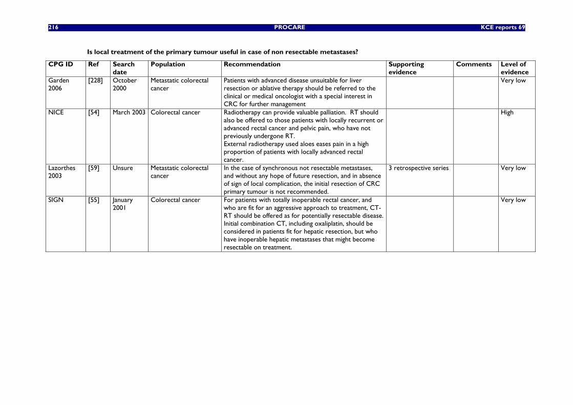
216 PROCARE KCE reports 69
Is local treatment of the primary tumour useful in case of non resectable metastases?
CPG ID Ref Search date
Population Recommendation Supporting evidence
Comments Level of evidence
Garden 2006
[228] October 2000
Metastatic colorectal cancer
Patients with advanced disease unsuitable for liver resection or ablative therapy should be referred to the clinical or medical oncologist with a special interest in CRC for further management
Very low
NICE [54] March 2003 Colorectal cancer Radiotherapy can provide valuable palliation. RT should also be offered to those patients with locally recurrent or advanced rectal cancer and pelvic pain, who have not previously undergone RT. External radiotherapy used aloes eases pain in a high proportion of patients with locally advanced rectal cancer.
High
Lazorthes 2003
[59] Unsure Metastatic colorectal cancer
In the case of synchronous not resectable metastases, and without any hope of future resection, and in absence of sign of local complication, the initial resection of CRC primary tumour is not recommended.
3 retrospective series Very low
SIGN [55] January 2001
Colorectal cancer For patients with totally inoperable rectal cancer, and who are fit for an aggressive approach to treatment, CT-RT should be offered as for potentially resectable disease. Initial combination CT, including oxaliplatin, should be considered in patients fit for hepatic resection, but who have inoperable hepatic metastases that might become resectable on treatment.
Very low
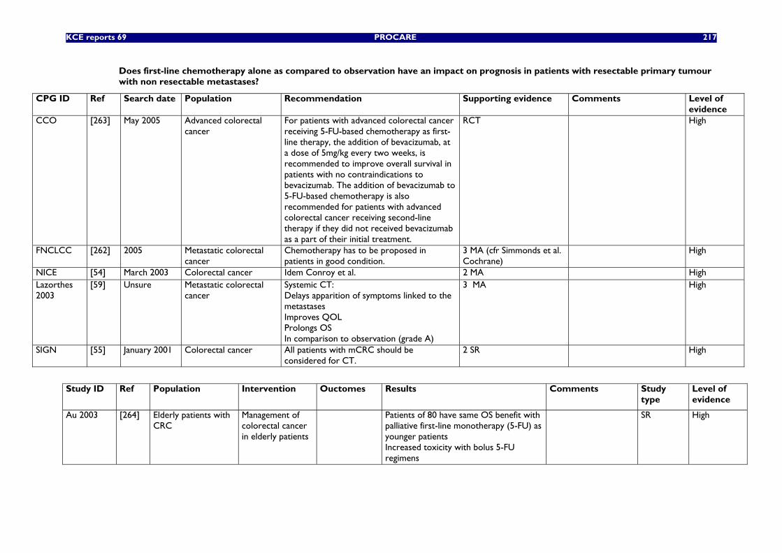
KCE reports 69 PROCARE 217
Does first-line chemotherapy alone as compared to observation have an impact on prognosis in patients with resectable primary tumour with non resectable metastases?
CPG ID Ref Search date Population Recommendation Supporting evidence Comments Level of evidence
CCO [263] May 2005 Advanced colorectal cancer
For patients with advanced colorectal cancer receiving 5-FU-based chemotherapy as first-line therapy, the addition of bevacizumab, at a dose of 5mg/kg every two weeks, is recommended to improve overall survival in patients with no contraindications to bevacizumab. The addition of bevacizumab to 5-FU-based chemotherapy is also recommended for patients with advanced colorectal cancer receiving second-line therapy if they did not received bevacizumab as a part of their initial treatment.
RCT High
FNCLCC [262] 2005 Metastatic colorectal cancer
Chemotherapy has to be proposed in patients in good condition.
3 MA (cfr Simmonds et al. Cochrane)
High
NICE [54] March 2003 Colorectal cancer Idem Conroy et al. 2 MA High Lazorthes 2003
[59] Unsure Metastatic colorectal cancer
Systemic CT: Delays apparition of symptoms linked to the metastases Improves QOL Prolongs OS In comparison to observation (grade A)
3 MA High
SIGN [55] January 2001 Colorectal cancer All patients with mCRC should be considered for CT.
2 SR High
Study ID Ref Population Intervention Ouctomes Results Comments Study
type Level of evidence
Au 2003 [264] Elderly patients with CRC
Management of colorectal cancer in elderly patients
Patients of 80 have same OS benefit with palliative first-line monotherapy (5-FU) as younger patients Increased toxicity with bolus 5-FU regimens
SR High
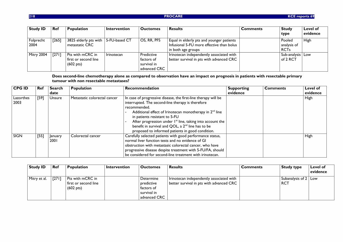
218 PROCARE KCE reports 69
Study ID Ref Population Intervention Ouctomes Results Comments Study type
Level of evidence
Folprecht 2004
[265] 3825 elderly pts with metastatic CRC
5-FU-based CT OS, RR, PFS Equal in elderly pts and younger patients Infusional 5-FU more effective than bolus in both age groups
Pooled analysis of RCTs
High
Mitry 2004 [271] Pts with mCRC in first or second line (602 pts)
Irinotecan Predictive factors of survival in advanced CRC
Irinotecan independently associated with better survival in pts with advanced CRC
Sub-analysis of 2 RCT
Low
Does second-line chemotherapy alone as compared to observation have an impact on prognosis in patients with resectable primary tumour with non resectable metastases?
CPG ID Ref Search date
Population Recommendation Supporting evidence
Comments Level of evidence
Lazorthes 2003
[59] Unsure Metastatic colorectal cancer In case of progressive disease, the first-line therapy will be interrupted. The second-line therapy is therefore recommended. - Additional effect of Irinotecan monotherapy in 2nd line
in patients resistant to 5-FU - After progression under 1st line, taking into account the
benefit in survival and QOL, a 2nd line has to be proposed to informed patients in good condition.
High
SIGN [55] January 2001
Colorectal cancer Carefully selected patients with good performance status, normal liver function tests and no evidence of GI obstruction with metastasic colorectal cancer, who have progressive disease despite treatment with 5-FU/FA, should be considered for second-line treatment with irinotecan.
High
Study ID Ref Population Intervention Ouctomes Results Comments Study type Level of
evidence
Mitry et al. [271] Pts with mCRC in first or second line (602 pts)
Determine predictive factors of survival in advanced CRC
Irinotecan independently associated with better survival in pts with advanced CRC
Subanalysis of 2 RCT
Low
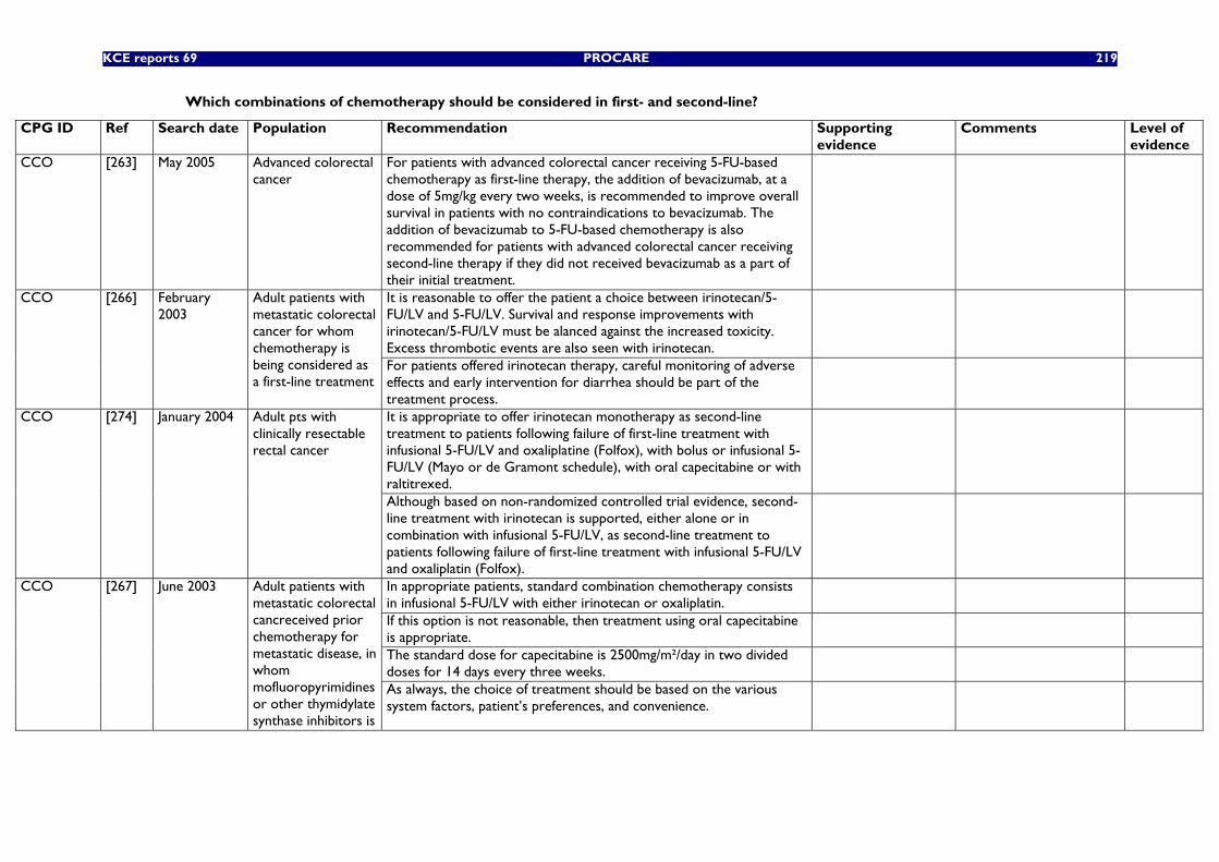
KCE reports 69 PROCARE 219
Which combinations of chemotherapy should be considered in first- and second-line?
CPG ID Ref Search date Population Recommendation Supporting evidence
Comments Level of evidence
CCO [263] May 2005 Advanced colorectal cancer
For patients with advanced colorectal cancer receiving 5-FU-based chemotherapy as first-line therapy, the addition of bevacizumab, at a dose of 5mg/kg every two weeks, is recommended to improve overall survival in patients with no contraindications to bevacizumab. The addition of bevacizumab to 5-FU-based chemotherapy is also recommended for patients with advanced colorectal cancer receiving second-line therapy if they did not received bevacizumab as a part of their initial treatment.
It is reasonable to offer the patient a choice between irinotecan/5-FU/LV and 5-FU/LV. Survival and response improvements with irinotecan/5-FU/LV must be alanced against the increased toxicity. Excess thrombotic events are also seen with irinotecan.
CCO [266] February 2003
Adult patients with metastatic colorectal cancer for whom chemotherapy is being considered as a first-line treatment
For patients offered irinotecan therapy, careful monitoring of adverse effects and early intervention for diarrhea should be part of the treatment process.
It is appropriate to offer irinotecan monotherapy as second-line treatment to patients following failure of first-line treatment with infusional 5-FU/LV and oxaliplatine (Folfox), with bolus or infusional 5-FU/LV (Mayo or de Gramont schedule), with oral capecitabine or with raltitrexed.
CCO [274] January 2004 Adult pts with clinically resectable rectal cancer
Although based on non-randomized controlled trial evidence, second-line treatment with irinotecan is supported, either alone or in combination with infusional 5-FU/LV, as second-line treatment to patients following failure of first-line treatment with infusional 5-FU/LV and oxaliplatin (Folfox).
In appropriate patients, standard combination chemotherapy consists in infusional 5-FU/LV with either irinotecan or oxaliplatin.
If this option is not reasonable, then treatment using oral capecitabine is appropriate.
The standard dose for capecitabine is 2500mg/m²/day in two divided doses for 14 days every three weeks.
CCO [267] June 2003 Adult patients with metastatic colorectal cancreceived prior chemotherapy for metastatic disease, in whom mofluoropyrimidines or other thymidylate synthase inhibitors is
As always, the choice of treatment should be based on the various system factors, patient’s preferences, and convenience.
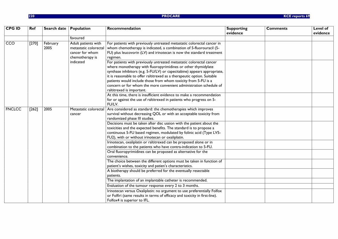
220 PROCARE KCE reports 69
CPG ID Ref Search date Population Recommendation Supporting evidence
Comments Level of evidence
favoured For patients with previously untreated metastatic colorectal cancer in whom chemotherapy is indicated, a combination of 5-fluorouracil (5-FU) plus leucovorin (LV) and irinotecan is now the standard treatment regimen.
For patients with previously untreated metastatic colorectal cancer where monotherapy with fluoropyrimidines or other thymidylate synthase inhibitors (e.g. 5-FU/LV) or capecitabine) appears appropriate, it is reasonable to offer raltitrexed as a therapeutic option. Suitable patients would include those from whom toxicity from 5-FU is a concern or for whom the more convenient administration schedule of raltitrexed is important.
CCO [270] February 2005
Adult patients with metastatic colorectal cancer for whom chemotherapy is indicated
At this time, there is insufficient evidence to make a recommendation for or against the use of raltitrexed in patients who progress on 5-FU/LV.
Are considered as standard: the chemotherapies which improves survival without decreasing QOL or with an acceptable toxicity from randomized phase III studies.
Decisions must be taken after disc ussion with the patient about the toxicities and the expected benefits. The standard is to propose a continuous 5-FU based regimen, modulated by folinic acid (Type LV5-FU2), with or without irinotecan or oxaliplatin.
Irinotecan, oxaliplatin or raltitrexed can be proposed alone or in combination to the patients who have contra-indication to 5-FU.
Oral fluoropyrimidines can be proposed as alternative for the convenience.
The choice between the different options must be taken in function of patient’s wishes, toxicity and patien’s characteristics.
A biotherapy should be preferred for the eventually resectable patients.
The implantation of an implantable catheter is recommended. Evaluation of the tumour response every 2 to 3 months.
FNCLCC [262] 2005 Metastatic colorectal cancer
Irinotecan versus Oxaliplatin: no argument to use preferentially Folfox or Folfiri (same results in terms of efficacy and toxicity in first-line). Folfox4 is superior to IFL.
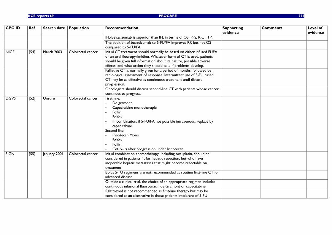
KCE reports 69 PROCARE 221
CPG ID Ref Search date Population Recommendation Supporting evidence
Comments Level of evidence
IFL-Bevacizumab is superior than IFL in terms of OS, PFS, RR, TTP. The addition of bevacizumab to 5-FU/FA improves RR but not OS compared to 5-FU/FA
Initial CT treatment should normally be based on either infused FUFA or an oral fluoropyrimidine. Whatever form of CT is used, patients should be given full information about its nature, possible adverse effects, and what action they should take if problems develop.
Palliative CT is normally given for a period of months, followed be radiological assessment of response. Intermittent use of 5-FU based CT may be as effective as continuous treatment until disease progression.
NICE [54] March 2003 Colorectal cancer
Oncologists should discuss second-line CT with patients whose cancer continues to progress.
DGVS [52] Unsure Colorectal cancer First line: - De gramont - Capecitabine monotherapie - Folfiri - Folfox - In combination: if 5-FU/FA not possible intravenous: replace by
capecitabine Second line: - Irinotecan Mono - Folfox - Folfiri - Cetux-Iri after progression under Irinotecan
Initial combination chemotherapy, including oxaliplatin, should be considered in patients fit for hepatic resection, but who have inoperable hepatic metastases that might become resectable on treatment
Bolus 5-FU regimens are not recommended as routine first-line CT for advanced disease
Outside a clinical trial, the choice of an appropriate regimen includes continuous infusional fluorouracil, de Gramont or capecitabine
SIGN [55] January 2001 Colorectal cancer
Raltitrexed is not recommended as first-line therapy but may be considered as an alternative in those patients intolerant of 5-FU
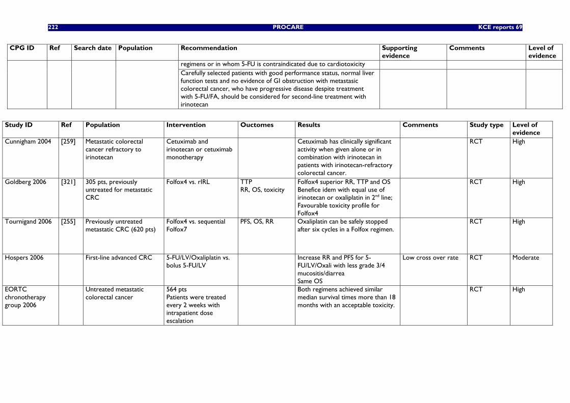
222 PROCARE KCE reports 69
CPG ID Ref Search date Population Recommendation Supporting evidence
Comments Level of evidence
regimens or in whom 5-FU is contraindicated due to cardiotoxicity Carefully selected patients with good performance status, normal liver function tests and no evidence of GI obstruction with metastasic colorectal cancer, who have progressive disease despite treatment with 5-FU/FA, should be considered for second-line treatment with irinotecan
Study ID Ref Population Intervention Ouctomes Results Comments Study type Level of
evidence Cunnigham 2004 [259] Metastatic colorectal
cancer refractory to irinotecan
Cetuximab and irinotecan or cetuximab monotherapy
Cetuximab has clinically significant activity when given alone or in combination with irinotecan in patients with irinotecan-refractory colorectal cancer.
RCT High
Goldberg 2006 [321] 305 pts, previously untreated for metastatic CRC
Folfox4 vs. rIRL TTP RR, OS, toxicity
Folfox4 superior RR, TTP and OS Benefice idem with equal use of irinotecan or oxaliplatin in 2nd line; Favourable toxicity profile for Folfox4
RCT High
Tournigand 2006 [255] Previously untreated metastatic CRC (620 pts)
Folfox4 vs. sequential Folfox7
PFS, OS, RR Oxaliplatin can be safely stopped after six cycles in a Folfox regimen.
RCT High
Hospers 2006 First-line advanced CRC 5-FU/LV/Oxaliplatin vs. bolus 5-FU/LV
Increase RR and PFS for 5-FU/LV/Oxali with less grade 3/4 mucositis/diarrea Same OS
Low cross over rate RCT Moderate
EORTC chronotherapy group 2006
Untreated metastatic colorectal cancer
564 pts Patients were treated every 2 weeks with intrapatient dose escalation
Both regimens achieved similar median survival times more than 18 months with an acceptable toxicity.
RCT High
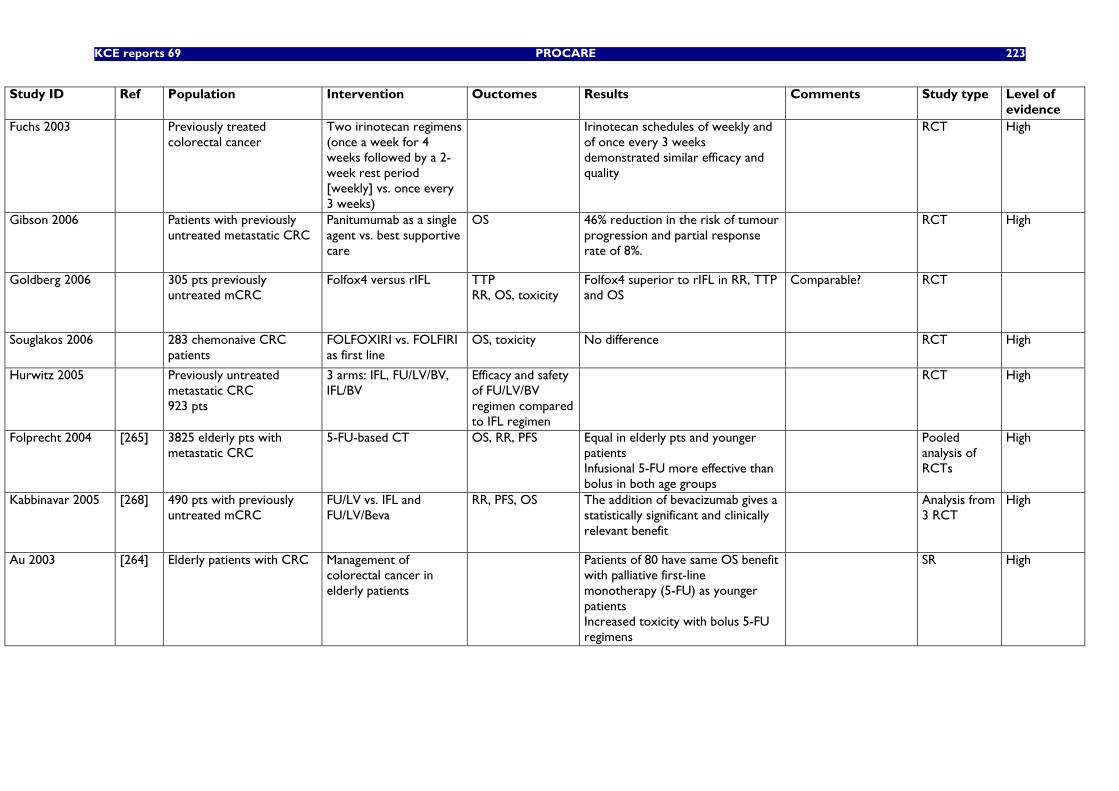
KCE reports 69 PROCARE 223
Study ID Ref Population Intervention Ouctomes Results Comments Study type Level of evidence
Fuchs 2003 Previously treated colorectal cancer
Two irinotecan regimens (once a week for 4 weeks followed by a 2-week rest period [weekly] vs. once every 3 weeks)
Irinotecan schedules of weekly and of once every 3 weeks demonstrated similar efficacy and quality
RCT High
Gibson 2006 Patients with previously untreated metastatic CRC
Panitumumab as a single agent vs. best supportive care
OS 46% reduction in the risk of tumour progression and partial response rate of 8%.
RCT High
Goldberg 2006 305 pts previously untreated mCRC
Folfox4 versus rIFL TTP RR, OS, toxicity
Folfox4 superior to rIFL in RR, TTP and OS
Comparable? RCT
Souglakos 2006 283 chemonaive CRC patients
FOLFOXIRI vs. FOLFIRI as first line
OS, toxicity No difference RCT
High
Hurwitz 2005
Previously untreated metastatic CRC 923 pts
3 arms: IFL, FU/LV/BV, IFL/BV
Efficacy and safety of FU/LV/BV regimen compared to IFL regimen
RCT High
Folprecht 2004 [265] 3825 elderly pts with metastatic CRC
5-FU-based CT OS, RR, PFS Equal in elderly pts and younger patients Infusional 5-FU more effective than bolus in both age groups
Pooled analysis of RCTs
High
Kabbinavar 2005 [268] 490 pts with previously untreated mCRC
FU/LV vs. IFL and FU/LV/Beva
RR, PFS, OS The addition of bevacizumab gives a statistically significant and clinically relevant benefit
Analysis from 3 RCT
High
Au 2003 [264] Elderly patients with CRC Management of colorectal cancer in elderly patients
Patients of 80 have same OS benefit with palliative first-line monotherapy (5-FU) as younger patients Increased toxicity with bolus 5-FU regimens
SR High
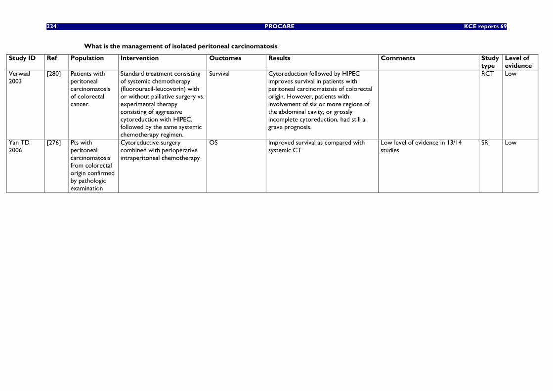
224 PROCARE KCE reports 69
What is the management of isolated peritoneal carcinomatosis
Study ID Ref Population Intervention Ouctomes Results Comments Study type
Level of evidence
Verwaal 2003
[280] Patients with peritoneal carcinomatosis of colorectal cancer.
Standard treatment consisting of systemic chemotherapy (fluorouracil-leucovorin) with or without palliative surgery vs. experimental therapy consisting of aggressive cytoreduction with HIPEC, followed by the same systemic chemotherapy regimen.
Survival Cytoreduction followed by HIPEC improves survival in patients with peritoneal carcinomatosis of colorectal origin. However, patients with involvement of six or more regions of the abdominal cavity, or grossly incomplete cytoreduction, had still a grave prognosis.
RCT Low
Yan TD 2006
[276] Pts with peritoneal carcinomatosis from colorectal origin confirmed by pathologic examination
Cytoreductive surgery combined with perioperative intraperitoneal chemotherapy
OS Improved survival as compared with systemic CT
Low level of evidence in 13/14 studies
SR
Low
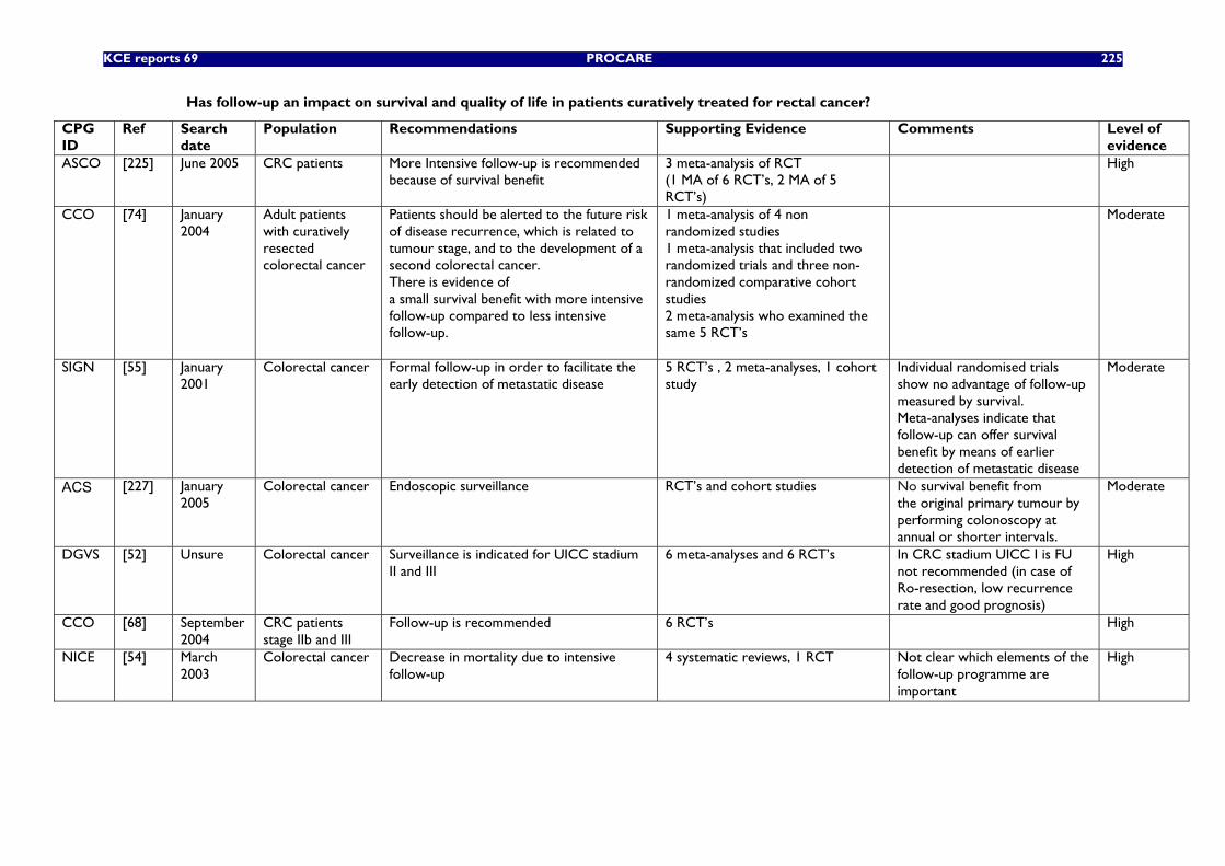
KCE reports 69 PROCARE 225
Has follow-up an impact on survival and quality of life in patients curatively treated for rectal cancer?
CPG ID
Ref Search date
Population Recommendations Supporting Evidence Comments Level of evidence
ASCO [225] June 2005 CRC patients More Intensive follow-up is recommended because of survival benefit
3 meta-analysis of RCT (1 MA of 6 RCT’s, 2 MA of 5 RCT’s)
High
CCO [74] January 2004
Adult patients with curatively resected colorectal cancer
Patients should be alerted to the future risk of disease recurrence, which is related to tumour stage, and to the development of a second colorectal cancer. There is evidence of a small survival benefit with more intensive follow-up compared to less intensive follow-up.
1 meta-analysis of 4 non randomized studies 1 meta-analysis that included two randomized trials and three non-randomized comparative cohort studies 2 meta-analysis who examined the same 5 RCT’s
Moderate
SIGN [55] January 2001
Colorectal cancer Formal follow-up in order to facilitate the early detection of metastatic disease
5 RCT’s , 2 meta-analyses, 1 cohort study
Individual randomised trials show no advantage of follow-up measured by survival. Meta-analyses indicate that follow-up can offer survival benefit by means of earlier detection of metastatic disease
Moderate
ACS
[227] January 2005
Colorectal cancer Endoscopic surveillance RCT’s and cohort studies No survival benefit from the original primary tumour by performing colonoscopy at annual or shorter intervals.
Moderate
DGVS [52] Unsure Colorectal cancer Surveillance is indicated for UICC stadium II and III
6 meta-analyses and 6 RCT’s In CRC stadium UICC I is FU not recommended (in case of Ro-resection, low recurrence rate and good prognosis)
High
CCO [68] September 2004
CRC patients stage IIb and III
Follow-up is recommended 6 RCT’s High
NICE [54] March 2003
Colorectal cancer Decrease in mortality due to intensive follow-up
4 systematic reviews, 1 RCT Not clear which elements of the follow-up programme are important
High
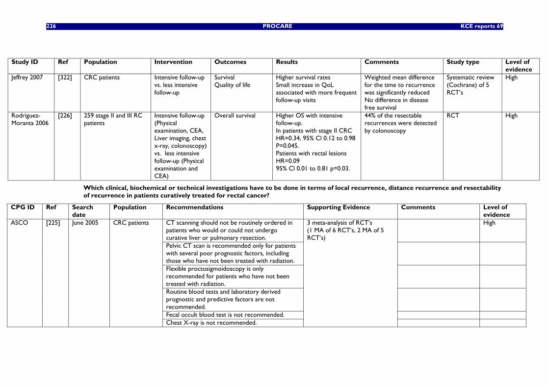
226 PROCARE KCE reports 69
Study ID Ref Population Intervention Outcomes Results Comments Study type Level of
evidence Jeffrey 2007 [322] CRC patients Intensive follow-up
vs. less intensive follow-up
Survival Quality of life
Higher survival rates Small increase in QoL associated with more frequent follow-up visits
Weighted mean difference for the time to recurrence was significantly reduced No difference in disease free survival
Systematic review (Cochrane) of 5 RCT’s
High
Rodriguez-Moranta 2006
[226] 259 stage II and III RC patients
Intensive follow-up (Physical examination, CEA, Liver imaging, chest x-ray, colonoscopy) vs. less intensive follow-up (Physical examination and CEA)
Overall survival Higher OS with intensive follow-up. In patients with stage II CRC HR=0.34, 95% Cl 0.12 to 0.98 P=0.045. Patients with rectal lesions HR=0.09 95% Cl 0.01 to 0.81 p=0.03.
44% of the resectable recurrences were detected by colonoscopy
RCT
High
Which clinical, biochemical or technical investigations have to be done in terms of local recurrence, distance recurrence and resectability of recurrence in patients curatively treated for rectal cancer?
CPG ID Ref Search date
Population Recommendations Supporting Evidence Comments Level of evidence
CT scanning should not be routinely ordered in patients who would or could not undergo curative liver or pulmonary resection.
High
Pelvic CT scan is recommended only for patients with several poor prognostic factors, including those who have not been treated with radiation.
Flexible proctosigmoidoscopy is only recommended for patients who have not been treated with radiation.
Routine blood tests and laboratory derived prognostic and predictive factors are not recommended.
Fecal occult blood test is not recommended.
ASCO [225] June 2005 CRC patients
Chest X-ray is not recommended.
3 meta-analysis of RCT’s (1 MA of 6 RCT’s, 2 MA of 5 RCT’s)
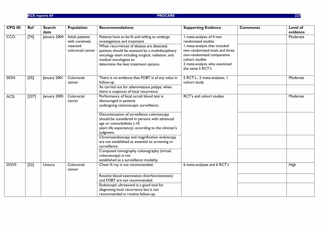
KCE reports 69 PROCARE 227
CPG ID Ref Search date
Population Recommendations Supporting Evidence Comments Level of evidence
Patients have to be fit and willing to undergo investigations and treatment
Moderate CCO [74] January 2004 Adult patients with curatively resected colorectal cancer
When recurrences of disease are detected, patients should be assessed by a multidisciplinary oncology team including surgical, radiation, and medical oncologists to determine the best treatment options.
1 meta-analysis of 4 non randomized studies 1 meta-analysis that included two randomized trials and three non-randomized comparative cohort studies 2 meta-analysis who examined the same 5 RCT’s
There is no evidence that FOBT is of any value in follow-up
Moderate SIGN [55] January 2001 Colorectal cancer
As carried out for adenomatous polyps; when there is suspicion of local recurrence
5 RCT’s , 2 meta-analyses, 1 cohort study
Performance of fecal occult blood test is discouraged in patients undergoing colonoscopic surveillance.
Moderate
Discontinuation of surveillance colonoscopy should be considered in persons with advanced age or comorbidities (-10 years life expectancy), according to the clinician’s judgment.
Chromoendoscopy and magnification endoscopy are not established as essential to screening or surveillance.
ACS
[227] January 2005 Colorectal cancer
Computed tomography colonography (virtual colonoscopy) is not established as a surveillance modality.
RCT’s and cohort studies
Chest X-ray is not recommended.
High
Routine blood examination (liverfunctiontests) and FOBT are not recommended.
DGVS [52] Unsure Colorectal cancer
Endoscopic ultrasound is a good tool for diagnosing local recurrence but is not recommended in routine follow-up.
6 meta-analyses and 6 RCT’s
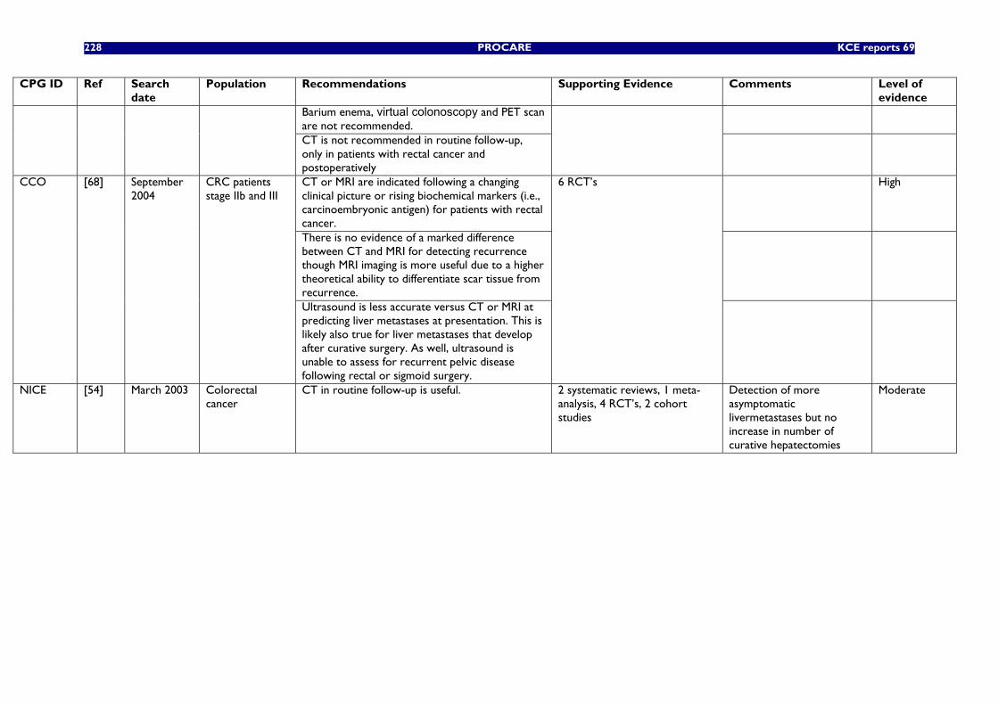
228 PROCARE KCE reports 69
CPG ID Ref Search date
Population Recommendations Supporting Evidence Comments Level of evidence
Barium enema, virtual colonoscopy and PET scan are not recommended.
CT is not recommended in routine follow-up, only in patients with rectal cancer and postoperatively
CT or MRI are indicated following a changing clinical picture or rising biochemical markers (i.e., carcinoembryonic antigen) for patients with rectal cancer.
High
There is no evidence of a marked difference between CT and MRI for detecting recurrence though MRI imaging is more useful due to a higher theoretical ability to differentiate scar tissue from recurrence.
CCO [68] September 2004
CRC patients stage IIb and III
Ultrasound is less accurate versus CT or MRI at predicting liver metastases at presentation. This is likely also true for liver metastases that develop after curative surgery. As well, ultrasound is unable to assess for recurrent pelvic disease following rectal or sigmoid surgery.
6 RCT’s
NICE [54] March 2003 Colorectal cancer
CT in routine follow-up is useful. 2 systematic reviews, 1 meta-analysis, 4 RCT’s, 2 cohort studies
Detection of more asymptomatic livermetastases but no increase in number of curative hepatectomies
Moderate
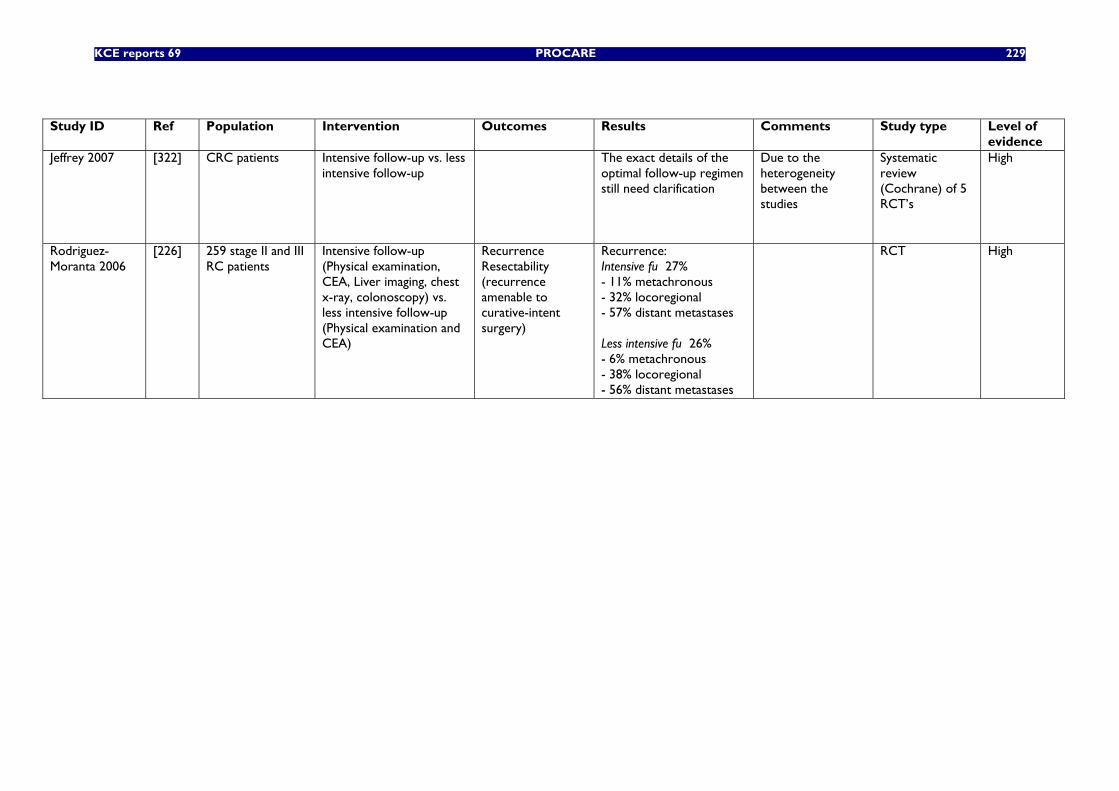
KCE reports 69 PROCARE 229
Study ID Ref Population Intervention Outcomes Results Comments Study type Level of
evidence Jeffrey 2007 [322] CRC patients Intensive follow-up vs. less
intensive follow-up The exact details of the
optimal follow-up regimen still need clarification
Due to the heterogeneity between the studies
Systematic review (Cochrane) of 5 RCT’s
High
Rodriguez-Moranta 2006
[226] 259 stage II and III RC patients
Intensive follow-up (Physical examination, CEA, Liver imaging, chest x-ray, colonoscopy) vs. less intensive follow-up (Physical examination and CEA)
Recurrence Resectability (recurrence amenable to curative-intent surgery)
Recurrence: Intensive fu 27% - 11% metachronous - 32% locoregional - 57% distant metastases Less intensive fu 26% - 6% metachronous - 38% locoregional - 56% distant metastases
RCT High
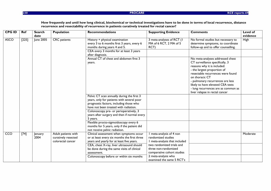
230 PROCARE KCE reports 69
How frequently and until how long clinical, biochemical or technical investigations have to be done in terms of local recurrence, distance recurrence and resectability of recurrence in patients curatively treated for rectal cancer?
CPG ID Ref Search date
Population Recommendations Supporting Evidence Comments Level of evidence
History + physical examination every 3 to 6 months first 3 years, every 6 months during years 4 and 5.
3 meta-analyses of RCT (1 MA of 6 RCT, 2 MA of 5 RCT)
No formal studies but necessary to determine symptoms, to coordinate follow-up and to offer counselling.
High
CEA every 3 months for at least 3 years after diagnosis.
Annual CT of chest and abdomen first 3 years.
No meta-analyses addressed chest CT surveillance specifically, 3 reasons why it is included: - the largest proportion of resectable recurrences were found on thoracic CT - pulmonary recurrences are less likely to have elevated CEA tests - lung recurrences are as common as liver relapse in rectal cancer
Pelvic CT scan annually during the first 3 years, only for patients with several poor prognostic factors, including those who have not been treated with radiation.
Colonoscopy pre- or perioperatively, 3 years after surgery and then if normal every 5 years.
ASCO [225] June 2005 CRC patients
Flexible procto-sigmoidoscopy every 6 months for 5 years, only if the patient did not receive pelvic radiation.
Clinical assessment when symptoms occur or at least every six months the first three years and yearly for at least five years.
Moderate
CEA, chest X-ray, liver ultrasound should be done during the same visits of clinical assessment.
CCO [74] January 2004
Adult patients with curatively resected colorectal cancer
Colonoscopy before or within six months
1 meta-analysis of 4 non randomized studies 1 meta-analysis that included two randomized trials and three non-randomized comparative cohort studies 2 meta-analysis who examined the same 5 RCT’s
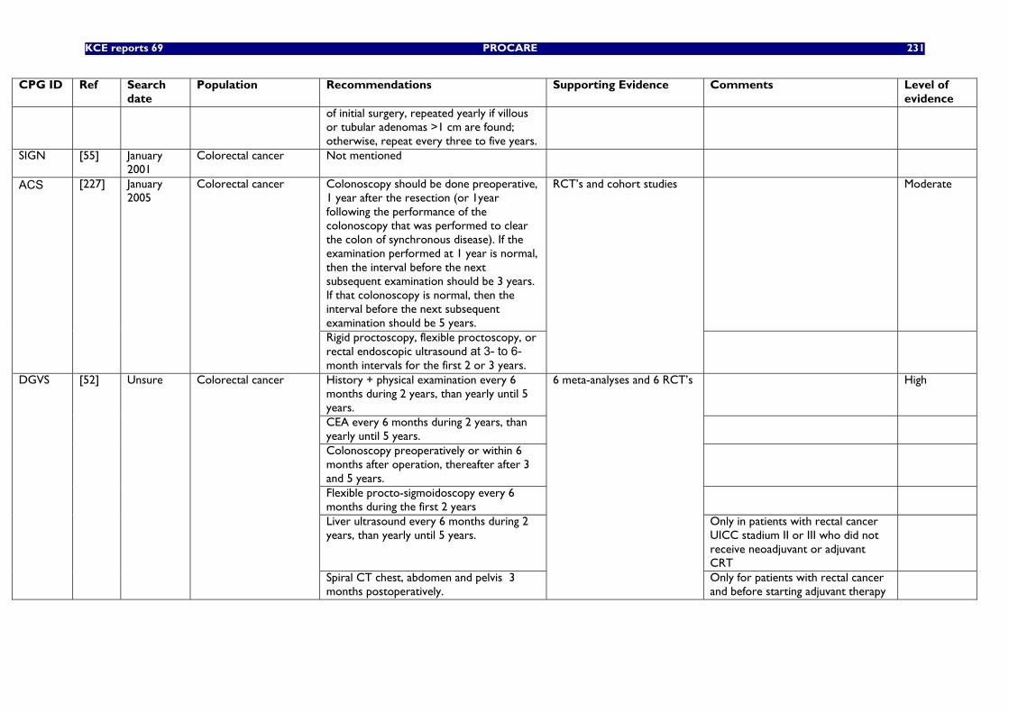
KCE reports 69 PROCARE 231
CPG ID Ref Search date
Population Recommendations Supporting Evidence Comments Level of evidence
of initial surgery, repeated yearly if villous or tubular adenomas >1 cm are found; otherwise, repeat every three to five years.
SIGN [55] January 2001
Colorectal cancer Not mentioned
Colonoscopy should be done preoperative, 1 year after the resection (or 1year following the performance of the colonoscopy that was performed to clear the colon of synchronous disease). If the examination performed at 1 year is normal, then the interval before the next subsequent examination should be 3 years. If that colonoscopy is normal, then the interval before the next subsequent examination should be 5 years.
Moderate ACS
[227] January 2005
Colorectal cancer
Rigid proctoscopy, flexible proctoscopy, or rectal endoscopic ultrasound at 3- to 6-month intervals for the first 2 or 3 years.
RCT’s and cohort studies
History + physical examination every 6 months during 2 years, than yearly until 5 years.
High
CEA every 6 months during 2 years, than yearly until 5 years.
Colonoscopy preoperatively or within 6 months after operation, thereafter after 3 and 5 years.
Flexible procto-sigmoidoscopy every 6 months during the first 2 years
Liver ultrasound every 6 months during 2 years, than yearly until 5 years.
Only in patients with rectal cancer UICC stadium II or III who did not receive neoadjuvant or adjuvant CRT
DGVS [52] Unsure Colorectal cancer
Spiral CT chest, abdomen and pelvis 3 months postoperatively.
6 meta-analyses and 6 RCT’s
Only for patients with rectal cancer and before starting adjuvant therapy
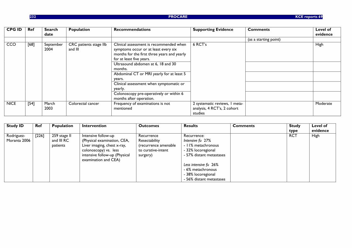
232 PROCARE KCE reports 69
CPG ID Ref Search date
Population Recommendations Supporting Evidence Comments Level of evidence
(as a starting point) Clinical assessment is recommended when symptoms occur or at least every six months for the first three years and yearly for at least five years.
High
Ultrasound abdomen at 6, 18 and 30 months.
Abdominal CT or MRI yearly for at least 5 years.
Clinical assessment when symptomatic or yearly.
CCO [68] September 2004
CRC patients stage IIb and III
Colonoscopy pre-operatively or within 6 months after operation.
6 RCT’s
NICE [54] March 2003
Colorectal cancer Frequency of examinations is not mentioned
2 systematic reviews, 1 meta-analysis, 4 RCT’s, 2 cohort studies
Moderate
Study ID Ref Population Intervention Outcomes Results Comments Study
type Level of evidence
Rodriguez-Moranta 2006
[226] 259 stage II and III RC patients
Intensive follow-up (Physical examination, CEA, Liver imaging, chest x-ray, colonoscopy) vs. less intensive follow-up (Physical examination and CEA)
Recurrence Resectability (recurrence amenable to curative-intent surgery)
Recurrence: Intensive fu 27% - 11% metachronous - 32% locoregional - 57% distant metastases Less intensive fu 26% - 6% metachronous - 38% locoregional - 56% distant metastases
RCT High
KCE reports 69 PROCARE 233
5 REFERENCES 1. Stichting Kankerregister. Kanker in België 2003. Available from:
http://www.kankerregister.org.
2. Capet, F., et al., Colorectale kanker: huidige toestand en bijdrage van informatie voor het opbouwen van een gezondheidsbeleid. 1999, CROSP Wetenschappelijk Instituut Volksgezondheid, Administratie Gezondheidszorg. Ministerie van de Vlaamse Gemeenschap.
3. Hewitt, M. and J.V. Simone, Ensuring Quality Cancer Care. 1999: National Academy Press.
4. O'Malley, A.S., et al., Clinical practice guidelines and performance indicators as related--but often misunderstood--tools. Jt Comm J Qual Saf, 2004. 30(3): p. 163-71.
5. McGory, M.L., P.G. Shekelle, and C.Y. Ko, Development of quality indicators for patients undergoing colorectal cancer surgery. J Natl Cancer Inst, 2006. 98(22): p. 1623-33.
6. Walter, L.C., et al., Pitfalls of converting practice guidelines into quality measures: lessons learned from a VA performance measure. JAMA, 2004. 291(20): p. 2466-70.
7. Beauduin, M., et al., The management of rectal cancer in Belgium: a survey of our practice. Acta Gastroenterol Belg, 2004. 67(1): p. 9-13.
8. Penninckx, F., Surgeon-related aspects of the treatment and outcome after radical resection for rectal cancer. Acta Gastroenterol Belg, 2001. 64(3): p. 258-62.
9. Penninckx, F., et al., Survival of rectal cancer patients in Belgium 1997-98 and the potential benefit of a national project. Acta Chir Belg, 2006. 106(2): p. 149-57.
10. Beets-Tan, R.G., et al., Accuracy of magnetic resonance imaging in prediction of tumour-free resection margin in rectal cancer surgery. Lancet, 2001. 357(9255): p. 497-504.
11. Brown, G., et al., Preoperative assessment of prognostic factors in rectal cancer using high-resolution magnetic resonance imaging. Br J Surg, 2003. 90(3): p. 355-64.
12. Penninckx, F. and E. Danse, On the role of radiologists in the Belgian PROject on CAncer of the REctum, PROCARE. Jbr-Btr, 2006. 89(1): p. 19-22.
13. Hermanek, P. and P.J. Hermanek, Role of the surgeon as a variable in the treatment of rectal cancer. Semin Surg Oncol, 2000. 19(4): p. 329-35.
14. Kapiteijn, E. and C.J. van de Velde, Developments and quality assurance in rectal cancer surgery. Eur J Cancer, 2002. 38(7): p. 919-36.
15. Wexner, S.D. and N.A. Rotholtz, Surgeon influenced variables in resectional rectal cancer surgery. Dis Colon Rectum, 2000. 43(11): p. 1606-27.
16. Sobin, L.H., C. Wittekind, and International Union Against Cancer (UICC), TNM classification on malignant tumours. 5th Ed. 1997, New York: Wiley-Liss Publications.
17. Radice, E. and R.R. Dozois, Locally recurrent rectal cancer. Dig Surg, 2001. 18(5): p. 355-62.
18. Beattie, G.C., et al., Improvement in quality of colorectal cancer pathology reporting with a standardized proforma--a comparative study. Colorectal Dis, 2003. 5(6): p. 558-62.
19. Nagtegaal, I.D. and J.H. van Krieken, The role of pathologists in the quality control of diagnosis and treatment of rectal cancer-an overview. Eur J Cancer, 2002. 38(7): p. 964-72.
20. Edge, S.B., D.L. Cookfair, and N. Watroba, The role of the surgeon in quality cancer care. Curr Probl Surg, 2003. 40(9): p. 511-90.
21. Hodgson, D.C., et al., Relation of hospital volume to colostomy rates and survival for patients with rectal cancer. J Natl Cancer Inst, 2003. 95(10): p. 708-16.
234 PROCARE KCE reports 69
22. Meyerhardt, J.A., et al., Impact of hospital procedure volume on surgical operation and long-term outcomes in high-risk curatively resected rectal cancer: findings from the Intergroup 0114 Study. J Clin Oncol, 2004. 22(1): p. 166-74.
23. Porter, G.A., et al., Surgeon-related factors and outcome in rectal cancer. Ann Surg, 1998. 227(2): p. 157-67.
24. Rabeneck, L., et al., Surgical volume and long-term survival following surgery for colorectal cancer in the Veterans Affairs Health-Care System. Am J Gastroenterol, 2004. 99(4): p. 668-75.
25. Schrag, D., et al., Hospital and surgeon procedure volume as predictors of outcome following rectal cancer resection. Ann Surg, 2002. 236(5): p. 583-92.
26. Schrag, D., et al., Surgeon volume compared to hospital volume as a predictor of outcome following primary colon cancer resection. J Surg Oncol, 2003. 83(2): p. 68-78.
27. Wibe, A., et al., Effect of hospital caseload on long-term outcome after standardization of rectal cancer surgery at a national level. Br J Surg, 2005. 92(2): p. 217-24.
28. Harling, H., et al., Hospital volume and outcome of rectal cancer surgery in Denmark 1994-99. Colorectal Dis, 2005. 7(1): p. 90-5.
29. Smith, J.A., et al., Evidence of the effect of 'specialization' on the management, surgical outcome and survival from colorectal cancer in Wessex. Br J Surg, 2003. 90(5): p. 583-92.
30. Martling, A., et al., Impact of a surgical training programme on rectal cancer outcomes in Stockholm. Br J Surg, 2005. 92(2): p. 225-9.
31. McArdle, C.S. and D.J. Hole, Influence of volume and specialization on survival following surgery for colorectal cancer. Br J Surg, 2004. 91(5): p. 610-7.
32. Holm, T., et al., Influence of hospital- and surgeon-related factors on outcome after treatment of rectal cancer with or without preoperative radiotherapy. Br J Surg, 1997. 84(5): p. 657-63.
33. Dowdall, J.F., D. Maguire, and O.J. McAnena, Experience of surgery for rectal cancer with total mesorectal excision in a general surgical practice. Br J Surg, 2002. 89(8): p. 1014-9.
34. Smedh, K., et al., Reduction of postoperative morbidity and mortality in patients with rectal cancer following the introduction of a colorectal unit. Br J Surg, 2001. 88(2): p. 273-7.
35. Seow-Choen, F., Adjuvant therapy for rectal cancer cannot be based on the results of other surgeons. Br J Surg, 2002. 89(8): p. 946-7.
36. Dominitz, J.A., et al., Race, treatment, and survival among colorectal carcinoma patients in an equal-access medical system. Cancer, 1998. 82(12): p. 2312-20.
37. Dignam, J.J., et al., Outcomes among African-Americans and Caucasians in colon cancer adjuvant therapy trials: findings from the National Surgical Adjuvant Breast and Bowel Project. J Natl Cancer Inst, 1999. 91(22): p. 1933-40.
38. Boyd, C., et al., Associations between community income and cancer survival in Ontario, Canada, and the United States. J Clin Oncol, 1999. 17(7): p. 2244-55.
39. Gorey, K.M., et al., An international comparison of cancer survival: Toronto, Ontario, and Detroit, Michigan, metropolitan areas. Am J Public Health, 1997. 87(7): p. 1156-63.
40. Ramsey, S.D., et al., Quality of life in survivors of colorectal carcinoma. Cancer, 2000. 88(6): p. 1294-303.
41. Porter, G.A. and J.M. Skibber, Outcomes research in surgical oncology. Ann Surg Oncol, 2000. 7(5): p. 367-75.
42. Gagliardi, A.R., et al., Development of quality indicators for colorectal cancer surgery, using a 3-step modified Delphi approach. Can J Surg, 2005. 48(6): p. 441-52.
43. Fervers, B., et al., Adaptation of clinical guidelines: literature review and proposition for a framework and procedure. Int J Qual Health Care, 2006. 18(3): p. 167-76.
KCE reports 69 PROCARE 235
44. Guyatt, G., et al., Grading strength of recommendations and quality of evidence in clinical guidelines: report from an american college of chest physicians task force. Chest, 2006. 129(1): p. 174-81.
45. Kapiteijn, E., et al., Preoperative radiotherapy combined with total mesorectal excision for resectable rectal cancer. New Engl J Med, 2001. 345: p. 638-646.
46. Ross, H.M., N. Mahmoud, and R.D. Fry, The current management of rectal cancer. Curr Probl Surg, 2005. 42(2): p. 72-131.
47. Sebag-Montefiore, D., et al., Routine short course pre-op radiotherapy or selective post-op chemoradiotherapy for resectable rectal cancer? Preliminary results of the MRC CR07 randomised trial. Journal of Clinical Oncology. ASCO Annual Meeting Proceedings Part I., 2006. 24(18S): p. 3511.
48. American Joint Committee on Cancer, Cancer staging handbook. 5th Ed. 1998: Lippincott-Raven.
49. Ueno, H., et al., Risk factors for an adverse outcome in early invasive colorectal carcinoma. Gastroenterology, 2004. 127(2): p. 385-94.
50. Roels, S., et al., Definition and delineation of the clinical target volume for rectal cancer. Int J Radiat Oncol Biol Phys, 2006. 65(4): p. 1129-42.
51. ACPGBI, Guidelines for the management of colorectal cancer. 2001, The Association of Coloproctology of Great Britain and Ireland.
52. Schmiegel, W., et al., [S3-guideline conference "Colorectal Cancer" 2004]. Dtsch Med Wochenschr, 2005. 130 Suppl 1: p. S5-53.
53. Barillari, P., et al., Effect of preoperative colonoscopy on the incidence of synchronous and metachronous neoplasms. Acta Chir Scand, 1990. 156(2): p. 163-6.
54. NICE, Guidance on Cancer Services. Improving outcomes in colorectal cancers. 2004, National Institute for Clinical Excellence.
55. SIGN, Management of colorectal cancer: a national clinical guideline. 2003, Scottish Intercollegiate Guidelines Network (SIGN).
56. Halligan, S., et al., CT colonography in the detection of colorectal polyps and cancer: systematic review, meta-analysis, and proposed minimum data set for study level reporting. Radiology, 2005. 237(3): p. 893-904.
57. Mulhall, B.P., G.R. Veerappan, and J.L. Jackson, Meta-analysis: computed tomographic colonography. Ann Intern Med, 2005. 142(8): p. 635-50.
58. Locker, G.Y., et al., ASCO 2006 update of recommendations for the use of tumor markers in gastrointestinal cancer. J Clin Oncol, 2006. 24(33): p. 5313-27.
59. Lazorthes, F., et al., [Therapeutic management of hepatic metastases from colorectal cancers]. Gastroenterol Clin Biol, 2003. 27 Spec No 2: p. B7.
60. Bipat, S., et al., Colorectal liver metastases: CT, MR imaging, and PET for diagnosis--meta-analysis. Radiology, 2005. 237(1): p. 123-31.
61. Bentrem, D.J., et al., T1 adenocarcinoma of the rectum: transanal excision or radical surgery? Ann Surg, 2005. 242(4): p. 472-7.
62. Mellgren, A., et al., Is local excision adequate therapy for early rectal cancer? Dis Colon Rectum, 2000. 43(8): p. 1064-71.
63. Nascimbeni, R., et al., Long-term survival after local excision for T1 carcinoma of the rectum. Dis Colon Rectum, 2004. 47(11): p. 1773-9.
64. You, Y., et al., Is Local Excision Adequate For T1 Rectal Cancer? A Nationwide Cohort Study From The National Cancer Database (NCDB). J Clin Oncol, 2005. 23(16S): p. 3526.
65. Worrell, S., et al., Endorectal ultrasound detection of focal carcinoma within rectal adenomas. Am J Surg, 2004. 187(5): p. 625-9.
236 PROCARE KCE reports 69
66. Kwok, H., I.P. Bissett, and G.L. Hill, Preoperative staging of rectal cancer. Int J Colorectal Dis, 2000. 15(1): p. 9-20.
67. Will, O., et al., Diagnostic precision of nanoparticle-enhanced MRI for lymph-node metastases: a meta-analysis. Lancet Oncol, 2006. 7(1): p. 52-60.
68. Simunovic, M., et al., Cross-Sectional Imaging in Colorectal Cancer. Recommendations Report. 2006, Cancer Care Ontario.
69. Lahaye, M.J., et al., Imaging for predicting the risk factors--the circumferential resection margin and nodal disease--of local recurrence in rectal cancer: a meta-analysis. Semin Ultrasound CT MR, 2005. 26(4): p. 259-68.
70. Diagnostic accuracy of preoperative magnetic resonance imaging in predicting curative resection of rectal cancer: prospective observational study. Bmj, 2006. 333(7572): p. 779.
71. Fuchsjager, M.H., et al., Comparison of transrectal sonography and double-contrast MR imaging when staging rectal cancer. AJR Am J Roentgenol, 2003. 181(2): p. 421-7.
72. Burton, S., et al., MRI directed multidisciplinary team preoperative treatment strategy: the way to eliminate positive circumferential margins? Br J Cancer, 2006. 94(3): p. 351-7.
73. Knaebel, H.P., et al., Diagnostics of rectal cancer: endorectal ultrasound. Recent Results Cancer Res, 2005. 165: p. 46-57.
74. Figueredo, A., et al., Follow-up of Patients with Curatively Resected Colorectal Cancer. Practice Guideline Report #2-9. 2004, Cancer Care Ontario.
75. Bosset, J.-F., et al., Chemotherapy with preoperative radiotherapy in rectal cancer. N Engl J Med, 2006. 355(11): p. 1114-23.
76. Gerard, J.P., et al., Preoperative radiotherapy with or without concurrent fluorouracil and leucovorin in T3-4 rectal cancers: results of FFCD 9203. J Clin Oncol, 2006. 24(28): p. 4620-5.
77. Marijnen, C.A., et al., Acute side effects and complications after short-term preoperative radiotherapy combined with total mesorectal excision in primary rectal cancer: report of a multicenter randomized trial. J Clin Oncol, 2002. 20(3): p. 817-825.
78. Marijnen, C.A., et al., Radiotherapy does not compensate for positive resection margins in rectal cancer patients: report of a multicenter randomized trial. Int J Radiat Oncol, 2003. 55: p. 1311-1320.
79. Marijnen, C.A., et al., No downstaging after short-term preoperative radiotherapy in rectal cancer patients. J Clin Oncol, 2001. 19: p. 1976-1984.
80. Marijnen, C.A., et al., Impact of short-term preoperative radiotherapy on health-related quality of life and sexual functioning in primary rectal cancer: report of a multicenter randomized trial. J Clin. Oncol, 2005. 23: p. 1847-1858.
81. Quirke, P., et al., Local recurrence after rectal cancer resection is strongly related to the plane of surgical dissection and is further reduced by pre-operative short course radiotherapy. Preliminary results of the Medical Research Council (MRC) CR07 trial. 2006. 24(18S): p. 3512.
82. Sauer, R., et al., Preoperative versus postoperative radiochemotherapy for rectal cancer. N Engl J Med, 2004. 351: p. 1731-1740.
83. Bosset, J.F., et al., Determination of the optimal dose of 5-fluorouracil when combined with low dose D,L-leucovorin and irradiation in rectal cancer: results of three consecutive phase II studies. Eur J Cancer, 1993. 29A(10): p. 1406-1410.
84. Lokich, J.J., et al., A prospective randomized comparison of continuous infusion fluorouracil with a conventional bolus schedule in metastatic colorectal carcinoma: a Mid-Atlantic Oncology Program Study. J Clin Oncol, 1989. 7(4): p. 425-32.
85. Rich, T.A., et al., Preoperative infusional chemoradiation therapy for stage T3 rectal cancer. Int J Radiat Oncol Biol Phys, 1995. 32(4): p. 1025-1029.
KCE reports 69 PROCARE 237
86. O’Connell, M.J., et al., Improving adjuvant therapy for rectal cancer by combining protracted-infusion Fluorouracil with radiation therapy after curative surgery. N Engl J Med, 1994. 331: p. 502–7.
87. Kim, J.S., et al., Comparison of the efficacy of oral capecitabine versus bolus 5-FU in preoperative radiotherapy of locally advanced rectal cancer. J Korean Med Sci, 2006. 21(1): p. 52-7.
88. Kim, N.K., et al., Intravenous 5-fluorouracil versus oral doxifluridine as preoperative concurrent chemoradiation for locally advanced rectal cancer: prospective randomized trials. Jpn J Clin Oncol, 2001. 31(1): p. 25_29.
89. Bujko, K., et al., Long-term results of a randomized trial comparing preoperative short-course radiotherapy with preoperative conventionally fractionated chemoradiation for rectal cancer. British Journal of Surgery, 2006. 93: p. 1215–1223.
90. Francois, Y., et al., Influence of the interval between preoperative radiation therapy and surgery on downstaging and on the rate of sphincter-sparing surgery for rectal cancer: the Lyon R90-01 randomized trial. J Clin Oncol, 1999. 17: p. 2396-2402.
91. Gerard, J.P., et al., Improved sphincter preservation in low rectal cancer with high dose preoperative radiotherapy: the LYON R96-02 randomized trial. J Clin Oncol, 2004. 22: p. 2404-2409.
92. Vuong, T., et al., Conformal preoperative endorectal brachytherapy treatment for locally advanced rectal cancer: early results of a phase I/II study. Dis Colon Rectum, 2002. 45(11): p. 1486-93.
93. Ahmad, N.R., G. Marks, and M. Mohiuddin, High-dose preoperative radiation for cancer of the rectum: Impact of radiation dose on patterns of failure and survival. Int J Radiat Oncol Biol Phys, 1993. 27: p. 773–778.
94. Chan, A.J.P., A.O.W.a.J. Langevin, and et al., Preoperative chemotherapy and pelvic radiation for tethered or fixed rectal cancer: A Phase II dose escalation study. Int J Radiat Oncol Biol Phys, 2000. 48: p. 843–856.
95. Janjan, N.A., C.N.C.a.B.W. Feig, and et al., Prospective trial of preoperative concomitant boost radiotherapy with continuous infusion 5-fluorouracil for locally advanced rectal cancer. Int J Radiat Oncol Biol Phys, 2000. 47: p. 713–718.
96. Mohiuddin, M., W.F.R.a.W.J. John, and et al., Preoperative chemoradiation in fixed distal rectal cancer: Dose time factors for pathological complete response. Int J Radiat Oncol Biol Phys, 2000. 46: p. 883–888.
97. Movsas, B., et al., Phase I dose escalating trial of hyperfractionated pre-operative chemoradiation for locally advanced rectal cancer. Int J Radiat Oncol Biol Phys., 1998. 42(1): p. 43-50.
98. Myerson, R.J., et al., A phase I/II trial of three-dimensionally planned concurrent boost radiotherapy and protracted venous infusion of 5-FU chemotherapy for locally advanced rectal carcinoma. Int J Radiat Oncol Biol Phys, 2001. 50: p. 1299–1308.
99. Freedman, G.M., et al., Phase I trial of preoperative hypofractionated intensity-modulated radiotherapy with incorporated boost and oral capecitabine in locally advanced rectal cancer. Int J Radiat Oncol Biol Phys, 2007. 67(5): p. 1389-93.
100. Kim, J.Y., et al., Intensity-modulated radiotherapy with a belly board for rectal cancer. Int J Colorectal Dis., 2007. 22(4): p. 373-9.
101. Andreola, S., et al., Adenocarcinoma of the lower third of the rectum: metastases in lymph nodes smaller than 5 mm and occult micrometastases; preliminary results on early tumor recurrence. Ann Surg Oncol., 2001. 8(5): p. 413-7.
102. Oberg, A., et al., Are lymph node micrometastases of any clinical significance in Dukes Stages A and B colorectal cancer? Dis Colon Rectum., 1998. 41(10): p. 1244-9.
103. Perez, R.O., et al., Lymph node micrometastasis in stage II distal rectal cancer following neoadjuvant chemoradiation therapy. Int J Colorectal Dis., 2005(5): p. 434-9.
238 PROCARE KCE reports 69
104. Habr-Gama, A., et al., Patterns of failure and survival for nonoperative treatment of stage c0 distal rectal cancer following neoadjuvant chemoradiation therapy. J Gastrointest Surg., 2006. 10(10): p. 1319-28.
105. Bosset, J.F., et al., Preoperative chemoradiotherapy versus preoperative radiotherapy in rectal cancer patients: assessment of acute toxicity and treatment compliance. Report of the 22921 randomised trial conducted by the EORTC Radiotherapy Group. Eur J Cancer, 2004. 40: p. 219–24.
106. Bosset, J.F., et al., Enhanced tumorocidal effect of chemotherapy with preoperative radiotherapy for rectal cancer: preliminary results--EORTC 22921. J Clin Oncol, 2005. 23(24): p. 5620-7.
107. Bujko, K., et al., Does rectal cancer shrinkage induced by preoperative radio(chemo)therapy increase the likelihood of anterior resection? A systematic review of randomised trials. Radiother Oncol. Epub, 2006. 80(1): p. 4-12.
108. Bujko, K., et al., Postoperative complications in patients irradiated pre-operatively for rectal cancer: report of a randomised trial comparing short-term radiotherapy vs chemoradiation. Colorectal Dis, 2005. 7(4): p. 410-416.
109. Bujko, K., et al., Sphincter preservation following preoperative radiotherapy for rectal cancer: report of a randomized trial comparing short-term radiotherapy vs. conventionally fractionated radiochemotherapy. Radiother Oncol, 2004. 72: p. 15-24.
110. Bedrosian, I., et al., Predicting the node-negative mesorectum after preoperative chemoradiation for locally advanced rectal carcinoma. J Gastrointest Surg, 2004. 8(1): p. 56-62.
111. Benzoni, E., et al., The predictive value of clinical evaluation of response to neoadjuvant chemoradiation therapy for rectal cancer. Tumor, 2005. 91(5): p. 401-5.
112. Bernini, A., et al., Preoperative adjuvant radiation with chemotherapy for rectal cancer: its impact on stage of disease and the role of endorectal ultrasound. Ann Surg Oncol, 1996. 3: p. 131–135.
113. Calvo, F.A., et al., 18F-FDG positron emission tomography staging and restaging in rectal cancer treated with preoperative chemoradiation. Int J Radiat Oncol Biol Phys, 2004. 58(2): p. 528-35.
114. Capirci, C., et al., Restaging after neoadjuvant chemoradiotherapy for rectal adenocarcinoma: role of F18-FDG PET. Biomed Pharmacother, 2004. 58(8): p. 451-7.
115. Capirci, C., et al., Long-term prognostic value of 18F-FDG PET in patients with locally advanced rectal cancer previously treated with neoadjuvant radiochemotherapy. Ajr, 2006. 187: p. 20220.
116. Chen, C.C., et al., How Accurate is Magnetic Resonance Imaging in Restaging Rectal Cancer in Patients Receiving Preoperative Combined Chemoradiotherapy? Dis Colon Rectum, 2005. 48: p. 722-728.
117. Fleshman, J.W., et al., Accuracy of transrectal ultrasound in predicting pathologic stage of rectal cancer before and after preoperative radiation therapy. Dis Colon Rectum, 1992. 35(9): p. 823-9.
118. Gavioli, M., et al., Usefulness of endorectal ultrasound after preoperative radiotherapy in rectal. Dis Colon Rectum, 2000. 43(8): p. 1075-83.
119. Guillem, J.G., et al., Clinical examination following preoperative chemoradiation for rectal cancer is not a reliable surrogate end point. J Clin Oncol., 2005. 23(15): p. 3475-9.
120. Hiotis, S.P., et al., Assessing the predictive value of clinical complete response to neoadjuvant therapy for rectal cancer: an analysis of 488 patients. J Am Coll Surg, 2002. 194(2): p. 131-5 discussion 135-6.
KCE reports 69 PROCARE 239
121. Houvenaeghel, G., et al., Staging of rectal cancer: a prospective study of digital examination and endosonography before and after preoperative radiotherapy. Acta Chir Belg, 1993. 93(4): p. 164-8.
122. Kahn, H., et al., Preoperative staging of irradiated rectal cancers using digital rectal examination, computed tomography, endorectal ultrasound, and magnetic resonance imaging does not accurately predict T0,N0 pathology. Dis Colon Rectum, 1997. 40(2): p. 140-4.
123. Kuo, L.J., et al., Interpretation of magnetic resonance imaging for locally advanced rectal carcinoma after preoperative chemoradiation therapy. Dis Colon Rectum, 2005. 48(1): p. 23-28.
124. Liersch, T., et al., Präoperative Diagnostik beim lokal fortgeschrittenen Rektumkarzinom (= T3 oder N+). Chirurg, 2003. 74: p. 224-234.
125. Maor, Y., et al., Endoscopic ultrasound staging of rectal cancer: diagnostic value before and following chemoradiation. J Gastroenterol Hepatol, 2006. 21: p. 454-458.
126. Romagnuolo, J., et al., Predicting residual rectal adenocarcinoma in the surgical specimen after preoperative brachytherapy with endoscopic ultrasound. Can J Gastroenterol, 2004. 18(7): p. 435-40.
127. Vanagunas, A., D.E. Lin, and S.J. Stryker, Accuracy of endoscopic ultrasound for restaging rectal cancer following neoadjuvant chemoradiation therapy. Am J Gastroenterol, 2004. 99(1): p. 109-12.
128. Williamson, P.R., et al., Endorectal ultrasound of T3 and T4 rectal cancers after preoperative chemoradiation. Dis Colon Rectum, 1996. 39: p. 45–49.
129. Frykholm, G.J., L. Pahlman, and B. Glimelius, Combined chemo- and radiotherapy vs. radiotherapy alone in the treatment of primary, nonresectable adenocarcinoma of the rectum. Int J Radiat Oncol Biol Phys., 2001. 50(2): p. 427-34.
130. Bosset, J.F., et al., Preoperative radiochemotherapy in rectal cancer: long-term results of a phase II trial. Int J Radiat Oncol Biol Phys, 2000. 46(2): p. 323-327.
131. Chari, R.S., et al., Preoperative radiation and chemotherapy in the treatment of adenocarcinoma of the rectum. Ann Surg, 1995. 221(6): p. 778-786.
132. Habr-Gama, A., et al., Low rectal cancer: impact of radiation and chemotherapy on surgical treatment. Dis Colon Rectum, 1998. 41(9): p. 1087-1096.
133. Janjan, N.A., et al., Improved overall survival among responders to preoperative chemoradiation for locally advanced rectal cancer. Am J Clin Oncol., 2001. 24(2): p. 107-112.
134. Ngan, S.Y., et al., Early toxicity from preoperative radiotherapy with continuous infusion 5-fluorouracil for resectable adenocarcinoma of the rectum: a Phase II trial for the Trans-Tasman Radiation Oncology Group. Int J Radiat Oncol Biol Phys., 2001. 50(4): p. 883-887.
135. Borschitz, T., W. Kneist, and T. Junginger, [Evaluation of residual urine volume by ultrasound for detection of urinary bladder dysfunction after surgical therapy of rectal cancer]. Chirurg, 2005. 76(7): p. 696-701; discussion 701-2.
136. Kneist, W. and T. Junginger, Residual urine volume after total mesorectal excision: an indicator of pelvic autonomic nerve preservation? Results of a case-control study. Colorectal Dis, 2004. 6(6): p. 432-7.
137. Grumann, M.M., et al., Comparison of quality of life in patients undergoing abdominoperineal extirpation or anterior resection for rectal cancer. Ann Surg, 2001. 233(2): p. 149-56.
138. Gosselink, M.P., et al., Quality of life after total mesorectal excision for rectal cancer. Colorectal Dis, 2006. 8(1): p. 15-22.
240 PROCARE KCE reports 69
139. Jess, P., J. Christiansen, and P. Bech, Quality of life after anterior resection versus abdominoperineal extirpation for rectal cancer. Scand J Gastroenterol, 2002. 37(10): p. 1201-4.
140. Pachler, J. and P. Wille-Jorgensen, Quality of life after rectal resection for cancer, with or without permanent colostomy. Cochrane Database Syst Rev, 2005(2): p. CD004323.
141. Schmidt, C.E., et al., Prospective evaluation of quality of life of patients receiving either abdominoperineal resection or sphincter-preserving procedure for rectal cancer. Ann Surg Oncol, 2005. 12(2): p. 117-23.
142. Bucher, P., et al., Mechanical bowel preparation for elective colorectal surgery: a meta-analysis. Arch Surg, 2004. 139(12): p. 1359-64.
143. Platell, C. and J. Hall, What is the role of mechanical bowel preparation in patients undergoing colorectal surgery? Dis Colon Rectum, 1998. 41(7): p. 875-82.
144. Slim, K., et al., Meta-analysis of randomized clinical trials of colorectal surgery with or without mechanical bowel preparation. Br J Surg, 2004. 91(9): p. 1125-30.
145. Wille-Jorgensen, P., et al., Pre-operative mechanical bowel cleansing or not? an updated meta-analysis. Colorectal Dis, 2005. 7(4): p. 304-10.
146. Borly, L., P. Wille-Jorgensen, and M.S. Rasmussen, Systematic review of thromboprophylaxis in colorectal surgery -- an update. Colorectal Dis, 2005. 7(2): p. 122-7.
147. Chaudhri, S., et al., Preoperative intensive, community-based vs. traditional stoma education: a randomized, controlled trial. Dis Colon Rectum, 2005. 48(3): p. 504-9.
148. Mynster, T., et al., Blood loss and transfusion after total mesorectal excision and conventional rectal cancer surgery. Colorectal Dis, 2004. 6(6): p. 452-7.
149. Bulow, S., et al., Recurrence and survival after mesorectal excision for rectal cancer. Br J Surg, 2003. 90(8): p. 974-80.
150. Kapiteijn, E., et al., Impact of the introduction and training of total mesorectal excision on recurrence and survival in rectal cancer in The Netherlands. Br J Surg, 2002. 89(9): p. 1142-9.
151. Maeda, K., et al., "On table" positioning for optimal access for cancer excision in the lower rectum. World J Surg, 2004. 28(4): p. 416-9.
152. Martling, A., et al., Prognostic significance of both surgical and pathological assessment of curative resection for rectal cancer. Br J Surg, 2004. 91(8): p. 1040-5.
153. Nagtegaal, I.D., et al., Low rectal cancer: a call for a change of approach in abdominoperineal resection. J Clin Oncol, 2005. 23(36): p. 9257-64.
154. Peeters, K.C.M.J., et al., Risk factors for anastomotic failure after total mesorectal excision of rectal cancer. Br J Surg, 2005. 92(2): p. 211-6.
155. Kanemitsu, Y., et al., Survival benefit of high ligation of the inferior mesenteric artery in sigmoid colon or rectal cancer surgery. Br J Surg, 2006. 93(5): p. 609-15.
156. Kim, J.C., et al., The clinicopathological significance of inferior mesenteric lymph node metastasis in colorectal cancer. Eur J Surg Oncol, 2004. 30(3): p. 271-9.
157. Nagawa, H., et al., Randomized, controlled trial of lateral node dissection vs. nerve-preserving resection in patients with rectal cancer after preoperative radiotherapy. Dis Colon Rectum, 2001. 44(9): p. 1274-80.
158. Araujo, S.E.A., et al., Conventional approach x laparoscopic abdominoperineal resection for rectal cancer treatment after neoadjuvant chemoradiation: results of a prospective randomized trial. Rev Hosp Clin Fac Med Sao Paulo, 2003. 58(3): p. 133-40.
KCE reports 69 PROCARE 241
159. Aziz, O., et al., Laparoscopic versus open surgery for rectal cancer: a meta-analysis. Ann Surg Oncol, 2006. 13(3): p. 413-24.
160. Breukink, S., J. Pierie, and T. Wiggers, Laparoscopic versus open total mesorectal excision for rectal cancer. Cochrane Database Syst Rev, 2006(4): p. CD005200.
161. Jayne, D.G., et al., Bladder and sexual function following resection for rectal cancer in a randomized clinical trial of laparoscopic versus open technique. Br J Surg, 2005. 92(9): p. 1124-32.
162. Quah, H.M., et al., Bladder and sexual dysfunction following laparoscopically assisted and conventional open mesorectal resection for cancer. Br J Surg, 2002. 89(12): p. 1551-6.
163. Schwenk, W., et al., Short term benefits for laparoscopic colorectal resection. Cochrane Database Syst Rev, 2005(3): p. CD003145.
164. Zhou, Z.G., et al., Laparoscopic versus open total mesorectal excision with anal sphincter preservation for low rectal cancer. Surg Endosc, 2004. 18(8): p. 1211-5.
165. Chapuis, P.H., et al., Risk factors for tumour present in a circumferential line of resection after excision of rectal cancer. Br J Surg, 2006. 93(7): p. 860-5.
166. Eriksen, M.T., et al., Inadvertent perforation during rectal cancer resection in Norway. Br J Surg, 2004. 91(2): p. 210-6.
167. Wibe, A., et al., Oncological outcomes after total mesorectal excision for cure for cancer of the lower rectum: anterior vs. abdominoperineal resection. Dis Colon Rectum, 2004. 47(1): p. 48-58.
168. Maeda, K., et al., Irrigation volume determines the efficacy of "rectal washout". Dis Colon Rectum, 2004. 47(10): p. 1706-10.
169. Terzi, C., et al., Is rectal washout necessary in anterior resection for rectal cancer? A prospective clinical study. World J Surg, 2006. 30(2): p. 233-41.
170. Laurent, A., et al., Colonic J-pouch-anal anastomosis for rectal cancer: a prospective, randomized study comparing handsewn vs. stapled anastomosis. Dis Colon Rectum, 2005. 48(4): p. 729-34.
171. Park, J.-G., et al., Colonic J-pouch anal anastomosis after ultralow anterior resection with upper sphincter excision for low-lying rectal cancer. World J Gastroenterol, 2005. 11(17): p. 2570-3.
172. Sailer, M., et al., Randomized clinical trial comparing quality of life after straight and pouch coloanal reconstruction. Br J Surg, 2002. 89(9): p. 1108-17.
173. Furst, A., et al., Colonic J-pouch vs. coloplasty following resection of distal rectal cancer: early results of a prospective, randomized, pilot study. Dis Colon Rectum, 2003. 46(9): p. 1161-6.
174. Jiang, J.-K., S.-H. Yang, and J.-K. Lin, Transabdominal anastomosis after low anterior resection: A prospective, randomized, controlled trial comparing long-term results between side-to-end anastomosis and colonic J-pouch. Dis Colon Rectum, 2005. 48(11): p. 2100-8.
175. Pimentel, J.M., et al., Transverse coloplasty pouch and colonic J-pouch for rectal cancer--a comparative study. Colorectal Dis, 2003. 5(5): p. 465-70.
176. Machado, M., et al., Similar outcome after colonic pouch and side-to-end anastomosis in low anterior resection for rectal cancer: a prospective randomized trial. Ann Surg, 2003. 238(2): p. 214-20.
177. Machado, M., et al., Functional and physiologic assessment of the colonic reservoir or side-to-end anastomosis after low anterior resection for rectal cancer: a two-year follow-up. Dis Colon Rectum, 2005. 48(1): p. 29-36.
242 PROCARE KCE reports 69
178. Ulrich, A., et al., Functional results of the colon J-pouch versus transverse coloplasty pouch in Heidelberg. Recent Results Cancer Res, 2005. 165: p. 205-11.
179. Amin, A.I., et al., Comparison of transanal stent with defunctioning stoma in low anterior resection for rectal cancer. Br J Surg, 2003. 90(5): p. 581-2.
180. Matthiessen, P., et al., Population-based study of risk factors for postoperative death after anterior resection of the rectum. Br J Surg, 2006. 93(4): p. 498-503.
181. Poon, J.T.C., W.-L. Law, and K.-W. Chu, Small bowel obstruction following low anterior resection: the impact of diversion ileostomy. Langenbecks Arch Surg, 2004. 389(4): p. 250-5.
182. Brown, S.R., et al., A prospective randomised study of drains in infra-peritoneal rectal anastomoses. Tech Coloproctol, 2001. 5(2): p. 89-92.
183. Matthiessen, P., et al., Defunctioning stoma reduces symptomatic anastomotic leakage after low anterior resection of the rectum for cancer: a randomized multicenter trial. Ann Surg, 2007. 246(2): p. 207-14.
184. Law, W.L., K.W. Chu, and H.K. Choi, Randomized clinical trial comparing loop ileostomy and loop transverse colostomy for faecal diversion following total mesorectal excision. Br J Surg, 2002. 89(6): p. 704-8.
185. Winde, G., et al., Surgical cure for early rectal carcinomas (T1). Transanal endoscopic microsurgery vs. anterior resection. Dis Colon Rectum, 1996. 39(9): p. 969-76.
186. Lezoche, E., et al., Transanal endoscopic versus total mesorectal laparoscopic resections of T2-N0 low rectal cancers after neoadjuvant treatment: a prospective randomized trial with a 3-years minimum follow-up period. Surg Endosc, 2005. 19(6): p. 751-6.
187. Nascimbeni, R., et al., Risk of lymph node metastasis in T1 carcinoma of the colon and rectum. Dis Colon Rectum, 2002. 45(2): p. 200-6.
188. Nagtegaal, I.D., et al., Macroscopic evaluation of rectal cancer resection specimen: clinical significance of the pathologist in quality control. J Clin Oncol, 2002. 20(7): p. 1729-34.
189. Marr, R., et al., The modern abdominoperineal excision: the next challenge after total mesorectal excision. Ann Surg, 2005. 242(1): p. 74-82.
190. Maughan, N.J. and P. Quirke, Modern management of colorectal cancer--a pathologist's view. Scand J Surg, 2003. 92(1): p. 11-9.
191. Quirke, P., Training and quality assurance for rectal cancer: 20 years of data is enough. Lancet Oncol, 2003. 4(11): p. 695-702.
192. Quirke, P. and M.F. Dixon, The prediction of local recurrence in rectal adenocarcinoma by histopathological examination. Int J Colorectal Dis, 1988. 3(2): p. 127-31.
193. Quirke, P., et al., Local recurrence of rectal adenocarcinoma due to inadequate surgical resection. Histopathological study of lateral tumour spread and surgical excision. Lancet, 1986. 2(8514): p. 996-999.
194. Jouret-Mourin, A., Recommendations for pathological examination and reporting for colorectal cancer. Belgian consensus. Acta Gastroenterol Belg, 2004. 67(1): p. 40-5.
195. Compton, C.C., et al., Prognostic factors in colorectal cancer. College of American Pathologists Consensus Statement 1999. Arch Pathol Lab Med, 2000. 124(7): p. 979-94.
196. Becouarn, Y., et al., Cancer of the rectum. Br J Cancer, 2001. 84 Suppl 2: p. 69-73.
197. Nelson, H., et al., Guidelines 2000 for colon and rectal cancer surgery. J Natl Cancer Inst, 2001. 93(8): p. 583-96.
KCE reports 69 PROCARE 243
198. Quirke, P., et al., The future of the TNM staging system in colorectal cancer: time for a debate? Lancet Oncol, 2007. 8(7): p. 651-7.
199. Parfitt, J.R. and D.K. Driman, The total mesorectal excision specimen for rectal cancer: a review of its pathological assessment. Journal of Clinical Pathology, 2007. 60(8): p. 849-55.
200. Wheeler, J.M., et al., Quantification of histologic regression of rectal cancer after irradiation: a proposal for a modified staging system. Dis Colon Rectum, 2002. 45(8): p. 1051-6.
201. Mandard, A.M., et al., Pathologic assessment of tumor regression after preoperative chemoradiotherapy of esophageal carcinoma. Clinicopathologic correlations. Cancer, 1994. 73(11): p. 2680-6.
202. Dworak, O., L. Keilholz, and A. Hoffmann, Pathological features of rectal cancer after preoperative radiochemotherapy. Int J Colorectal Dis, 1997. 12(1): p. 19-23.
203. Compton, C.C., Updated protocol for the examination of specimens from patients with carcinomas of the colon and rectum, excluding carcinoid tumors, lymphomas, sarcomas, and tumors of the vermiform appendix: a basis for checklists. Cancer Committee. Arch Pathol Lab Med, 2000. 124(7): p. 1016-25.
204. Compton, C.C., Key issues in reporting common cancer specimens: problems in pathologic staging of colon cancer. Arch Pathol Lab Med, 2006. 130(3): p. 318-24.
205. Jass, J.R., et al., Recommendations for the reporting of surgically resected specimens of colorectal carcinoma. Hum Pathol, 2007. 38(4): p. 537-545.
206. Germond, C., et al., Postoperative adjuvant radiotherapy and/or chemotherapy for resected stage II or III rectal cancer in Practice Guideline Report. 2001, Cancer Care Ontario.
207. De Placido, S., et al., Modulation of 5-fluorouracil as adjuvant systemic chemotherapy in colorectal cancer: the IGCS-COL multicentre, randomised, phase III study. Br J Cancer, 2005. 93(8): p. 896-904.
208. Tsavaris, N., et al., Leucovorin and fluorouracil vs levamisole and fluorouracil as adjuvant chemotherapy in rectal cancer. Oncol Rep, 2004. 12(4): p. 927-32.
209. Kotake, K., et al., A multicenter randomized study comparing 5-fluorouracil continuous infusion (ci) plus 1-hexylcarbamoyl-5-fluorouracil and 5-FU ci alone in colorectal cancer. Oncol Rep, 2005. 14(1): p. 129-34.
210. Iwagaki, H., et al., Post-operative adjuvant chemotherapy for colorectal cancer with 5-fluorouracil (5-FU) infusion combined with 1-hexylcarbamoyl-5-fluorouracil (HCFU) oral administration after curative resection. Anticancer Res, 2001. 21(6A): p. 4163-8.
211. Watanabe, M., et al., Randomized controlled trial of the efficacy of adjuvant immunochemotherapy and adjuvant chemotherapy for colorectal cancer, using different combinations of the intracutaneous streptococcal preparation OK-432 and the oral pyrimidines 1-hexylcarbamoyl-5-fluorouracil and uracil/tegafur. Int J Clin Oncol, 2004. 9(2): p. 98-106.
212. James, R.D., et al., Randomized clinical trial of adjuvant radiotherapy and 5-fluorouracil infusion in colorectal cancer (AXIS). Br J Surg, 2003. 90(10): p. 1200-12.
213. Adjuvant therapy for patients with colon and rectum cancer. Consens Statement, 1990. 8(4): p. 1-25.
214. Bosset, J.F., et al., Postoperative pelvic radiotherapy with or without elective irradiation of para-aortic nodes and liver in rectal cancer patients. A controlled clinical trial of the EORTC Radiotherapy Group. Radiother Oncol, 2001. 61(1): p. 7-13.
215. Cafiero, F., et al., Randomised clinical trial of adjuvant postoperative RT vs. sequential postoperative RT plus 5-FU and levamisole in patients with stage II-III resectable rectal cancer: a final report. J Surg Oncol, 2003. 83(3): p. 140-6.
244 PROCARE KCE reports 69
216. Akasu, T., et al., Adjuvant chemotherapy with uracil-tegafur for pathological stage III rectal cancer after mesorectal excision with selective lateral pelvic lymphadenectomy: a multicenter randomized controlled trial. Jpn J Clin Oncol, 2006. 36(4): p. 237-44.
217. Taal, B.G., et al., Adjuvant 5FU plus levamisole in colonic or rectal cancer: improved survival in stage II and III. Br J Cancer, 2001. 85(10): p. 1437-43.
218. Glimelius, B., et al., Adjuvant chemotherapy in colorectal cancer: a joint analysis of randomised trials by the Nordic Gastrointestinal Tumour Adjuvant Therapy Group. Acta Oncol, 2005. 44(8): p. 904-12.
219. Kato, T., et al., Efficacy of oral UFT as adjuvant chemotherapy to curative resection of colorectal cancer: multicenter prospective randomized trial. Langenbecks Arch Surg, 2002. 386(8): p. 575-81.
220. Sakamoto, J., et al., Efficacy of oral adjuvant therapy after resection of colorectal cancer: 5-year results from three randomized trials. J Clin Oncol, 2004. 22(3): p. 484-92.
221. Smalley, S.R., et al., Phase III trial of fluorouracil-based chemotherapy regimens plus radiotherapy in postoperative adjuvant rectal cancer: GI INT 0144. J Clin Oncol, 2006. 24(22): p. 3542-7.
222. Lundby, L., et al., Long-term anorectal dysfunction after postoperative radiotherapy for rectal cancer. Dis Colon Rectum, 2005. 48(7): p. 1343-9; discussion 1349-52; author reply 1352.
223. Dencausse, Y., et al., Adjuvant radio-chemotherapy in stage II-III rectal cancer with 24-hour infusion of high-dose 5-fluorouracil and folinic acid: evaluation of feasibility. Onkologie, 2001. 24(5): p. 476-80.
224. Miller, R.C., et al., Acute diarrhea during adjuvant therapy for rectal cancer: a detailed analysis from a randomized intergroup trial. Int J Radiat Oncol Biol Phys, 2002. 54(2): p. 409-13.
225. Desch, C.E., et al., Colorectal cancer surveillance: 2005 update of an American Society of Clinical Oncology practice guideline. J Clin Oncol, 2005. 23(33): p. 8512-9.
226. Rodriguez-Moranta, F., et al., Postoperative surveillance in patients with colorectal cancer who have undergone curative resection: a prospective, multicenter, randomized, controlled trial. J Clin Oncol, 2006. 24(3): p. 386-93.
227. Rex, D.K., et al., Guidelines for colonoscopy surveillance after cancer resection: a consensus update by the American Cancer Society and the US Multi-Society Task Force on Colorectal Cancer. Gastroenterology, 2006. 130(6): p. 1865-71.
228. Garden, O.J., et al., Guidelines for resection of colorectal cancer liver metastases. Gut, 2006. 55 Suppl 3: p. iii1-8.
229. Pawlik, T.M., et al., Effect of surgical margin status on survival and site of recurrence after hepatic resection for colorectal metastases. Ann Surg, 2005. 241(5): p. 715-22, discussion 722-4.
230. Adam, R., et al., Two-stage hepatectomy: A planned strategy to treat irresectable liver tumors. Ann Surg, 2000. 232(6): p. 777-85.
231. Farges, O., et al., Portal vein embolization before right hepatectomy: prospective clinical trial. Ann Surg, 2003. 237(2): p. 208-17.
232. Neeleman, N. and R. Andersson, Repeated liver resection for recurrent liver cancer. Br J Surg, 1996. 83(7): p. 893-901.
233. Oshowo, A., et al., Radiofrequency ablation extends the scope of surgery in colorectal liver metastases. Eur J Surg Oncol, 2003. 29(3): p. 244-7.
234. Nordlinger, B., et al., Does chemotherapy prior to liver resection increase the potential for cure in patients with metastatic colorectal cancer? A report from the European Colorectal Metastases Treatment Group. Eur J Cancer, 2007. 43(14): p. 2037-45.
KCE reports 69 PROCARE 245
235. Rau, B. and M. Hunerbein, Diagnostic laparoscopy: indications and benefits. Langenbecks Arch Surg, 2005. 390(3): p. 187-96.
236. Wiering, B., et al., The impact of fluor-18-deoxyglucose-positron emission tomography in the management of colorectal liver metastases. Cancer, 2005. 104(12): p. 2658-70.
237. Nordlinger, B., et al., Final results of the EORTC Intergroup randomized phase III study 40983 [EPOC] evaluating the benefit of peri-operative FOLFOX4 chemotherapy for patients with potentially resectable colorectal cancer liver metastases. J Clin Oncol (Meeting Abstracts), 2007. 25(18_suppl): p. LBA5.
238. Adam, R., et al., Five-year survival following hepatic resection after neoadjuvant therapy for nonresectable colorectal. Ann Surg Oncol, 2001. 8(4): p. 347-53.
239. Tournigand, C., et al., FOLFIRI followed by FOLFOX6 or the reverse sequence in advanced colorectal cancer: a randomized GERCOR study. J Clin Oncol, 2004. 22(2): p. 229-37.
240. Delaunoit, T., et al., Chemotherapy permits resection of metastatic colorectal cancer: experience from Intergroup N9741. Ann Oncol, 2005. 16(3): p. 425-9.
241. Folprecht, G., et al., Neoadjuvant treatment of unresectable colorectal liver metastases: correlation between tumour response and resection rates. Ann Oncol, 2005. 16(8): p. 1311-9.
242. Van Cutsem, E., et al., Towards a pan-European consensus on the treatment of patients with colorectal liver metastases. Eur J Cancer, 2006. 42(14): p. 2212-21.
243. Portier, G., et al., Multicenter randomized trial of adjuvant fluorouracil and folinic acid compared with surgery alone after resection of colorectal liver metastases: FFCD ACHBTH AURC 9002 trial. J Clin Oncol, 2006. 24(31): p. 4976-82.
244. Mitry, E., et al., [Importance of a multidisciplinary approach to metastatic cancer of the rectum]. Bull Cancer, 1998. 85(8): p. 716-20.
245. Jonker, D.J., J.A. Maroun, and W. Kocha, Survival benefit of chemotherapy in metastatic colorectal cancer: a meta-analysis of randomized controlled trials. Br J Cancer, 2000. 82(11): p. 1789-94.
246. Simmonds, P.C., Palliative chemotherapy for advanced colorectal cancer: systematic review and meta-analysis. Colorectal Cancer Collaborative Group. Bmj, 2000. 321(7260): p. 531-5.
247. Douillard, J.Y., et al., Irinotecan combined with fluorouracil compared with fluorouracil alone as first-line treatment for metastatic colorectal cancer: a multicentre randomised trial. Lancet, 2000. 355(9209): p. 1041-7.
248. Saltz, L.B., et al., Irinotecan plus fluorouracil and leucovorin for metastatic colorectal cancer. Irinotecan Study Group. N Engl J Med, 2000. 343(13): p. 905-14.
249. de Gramont, A., et al., Leucovorin and fluorouracil with or without oxaliplatin as first-line treatment in advanced colorectal cancer. J Clin Oncol, 2000. 18(16): p. 2938-47.
250. Giacchetti, S., et al., Phase III multicenter randomized trial of oxaliplatin added to chronomodulated fluorouracil-leucovorin as first-line treatment of metastatic colorectal cancer. J Clin Oncol, 2000. 18(1): p. 136-47.
251. Hoff, P.M., et al., Comparison of oral capecitabine versus intravenous fluorouracil plus leucovorin as first-line treatment in 605 patients with metastatic colorectal cancer: results of a randomized phase III study. J Clin Oncol, 2001. 19(8): p. 2282-92.
252. Van Cutsem, E., et al., Oral capecitabine compared with intravenous fluorouracil plus leucovorin in patients with metastatic colorectal cancer: results of a large phase III study. J Clin Oncol, 2001. 19(21): p. 4097-106.
253. Hurwitz, H., et al., Bevacizumab plus irinotecan, fluorouracil, and leucovorin for metastatic colorectal cancer. N Engl J Med, 2004. 350(23): p. 2335-42.
246 PROCARE KCE reports 69
254. Cunningham, D., Mature results from three large controlled studies with raltitrexed ('Tomudex'). Br J Cancer, 1998. 77 Suppl 2: p. 15-21.
255. Tournigand, C., et al., OPTIMOX1: a randomized study of FOLFOX4 or FOLFOX7 with oxaliplatin in a stop-and-Go fashion in advanced colorectal cancer--a GERCOR study. J Clin Oncol, 2006. 24(3): p. 394-400.
256. Cunningham, D., et al., Randomised trial of irinotecan plus supportive care versus supportive care alone after fluorouracil failure for patients with metastatic colorectal cancer. Lancet, 1998. 352(9138): p. 1413-8.
257. Rougier, P., et al., Randomised trial of irinotecan versus fluorouracil by continuous infusion after fluorouracil failure in patients with metastatic colorectal cancer. Lancet, 1998. 352(9138): p. 1407-12.
258. Giantonio, B.J., et al., Bevacizumab in combination with oxaliplatin, fluorouracil, and leucovorin (FOLFOX4) for previously treated metastatic colorectal cancer: results from the Eastern Cooperative Oncology Group Study E3200. J Clin Oncol, 2007. 25(12): p. 1539-44.
259. Cunningham, D., et al., Cetuximab monotherapy and cetuximab plus irinotecan in irinotecan-refractory metastatic colorectal cancer. N Engl J Med, 2004. 351(4): p. 337-45.
260. Wiseman, L.R., et al., Oxaliplatin: a review of its use in the management of metastatic colorectal cancer. Drugs Aging, 1999. 14(6): p. 459-75.
261. Rothenberg, M.L., et al., Superiority of oxaliplatin and fluorouracil-leucovorin compared with either therapy alone in patients with progressive colorectal cancer after irinotecan and fluorouracil-leucovorin: interim results of a phase III trial. J Clin Oncol, 2003. 21(11): p. 2059-69.
262. Conroy, T., et al., [Recommendations for clinical practice: management with first-line palliative chemotherapy for patients with metastatic colorectal cancer]. Bull Cancer, 2006. 93(2): p. 197-200.
263. Welch, S., et al., The Role of Bevacizumab (Avastin™) Combined With Chemotherapy in the Treatment of Patients With Advanced Colorectal Cancer: A Clinical Practice Guideline. 2005, Cancer Care Ontario.
264. Au, H.J., K.E. Mulder, and A.L. Fields, Systematic review of management of colorectal cancer in elderly patients. Clin Colorectal Cancer, 2003. 3(3): p. 165-71.
265. Folprecht, G., et al., Efficacy of 5-fluorouracil-based chemotherapy in elderly patients with metastatic colorectal cancer: a pooled analysis of clinical trials. Ann Oncol, 2004. 15(9): p. 1330-8.
266. Gastrointestinal Cancer Disease Site Group, Use of Irinotecan (Camptosar®, CPT-11) Combined with 5-Fluorouracil and Leucovorin (5FU/LV) as First-Line Therapy for Metastatic Colorectal Cancer. Practice Guideline Report #2-16b. 2003, Cancer Care Ontario.
267. Kocha, W., et al., Oral Capecitabine (Xeloda ) in the First-line Treatment of Metastatic Colorectal Cancer: A Clinical Practice Guideline. 2005, Cancer Care Ontario.
268. Kabbinavar, F.F., et al., Combined analysis of efficacy: the addition of bevacizumab to fluorouracil/leucovorin improves survival for patients with metastatic colorectal cancer. J Clin Oncol, 2005. 23(16): p. 3706-12.
269. Zondor, S.D. and P.J. Medina, Bevacizumab: an angiogenesis inhibitor with efficacy in colorectal and other malignancies. Ann Pharmacother, 2004. 38(7-8): p. 1258-64.
270. Germond, C., et al., Use of Raltitrexed (Tomudex) in the Management of Metastatic Colorectal Cancer. Practice Guideline Report #2-17. 2005, Cancer Care Ontario.
KCE reports 69 PROCARE 247
271. Mitry, E., et al., Predictive factors of survival in patients with advanced colorectal cancer: an individual data analysis of 602 patients included in irinotecan phase III trials. Ann Oncol, 2004. 15(7): p. 1013-7.
272. Chau, I., et al., Elderly patients with fluoropyrimidine and thymidylate synthase inhibitor-resistant advanced colorectal cancer derive similar benefit without excessive toxicity when treated with irinotecan monotherapy. Br J Cancer, 2004. 91(8): p. 1453-8.
273. Frieze, D.A. and J.S. McCune, Current status of cetuximab for the treatment of patients with solid tumors. Ann Pharmacother, 2006. 40(2): p. 241-50.
274. Figueredo, A., et al., Use of Irinotecan in the Second-Line Treatment of Metastatic Colorectal Carcinoma. Practice Guideline Report #2-16. 2004, Cancer Care Ontario.
275. Khot, U.P., et al., Systematic review of the efficacy and safety of colorectal stents. Br J Surg, 2002. 89(9): p. 1096-102.
276. Yan, T.D., et al., Systematic review on the efficacy of cytoreductive surgery combined with perioperative intraperitoneal chemotherapy for peritoneal carcinomatosis from colorectal carcinoma. J Clin Oncol, 2006. 24(24): p. 4011-9.
277. Elias, D., et al., Treatment of peritoneal carcinomatosis from colorectal cancer: impact of complete cytoreductive surgery and difficulties in conducting randomized trials. Ann Surg Oncol, 2004. 11(5): p. 518-21.
278. Kavanagh, M. and J.F. Ouellet, [Clinical practice guideline on peritoneal carcinomatosis treatment using surgical cytoreduction and hyperthermic intraoperative intraperitoneal chemotherapy]. Bull Cancer, 2006. 93(9): p. 867-74.
279. Verwaal, V.J., et al., Recurrences after peritoneal carcinomatosis of colorectal origin treated by cytoreduction and hyperthermic intraperitoneal chemotherapy: location, treatment, and outcome. Ann Surg Oncol, 2004. 11(4): p. 375-9.
280. Verwaal, V.J., et al., Randomized trial of cytoreduction and hyperthermic intraperitoneal chemotherapy versus systemic chemotherapy and palliative surgery in patients with peritoneal carcinomatosis of colorectal cancer. J Clin Oncol, 2003. 21(20): p. 3737-43.
281. Verwaal, V.J., et al., Predicting the survival of patients with peritoneal carcinomatosis of colorectal origin treated by aggressive cytoreduction and hyperthermic intraperitoneal chemotherapy. Br J Surg, 2004. 91(6): p. 739-46.
282. Figueredo, A., et al., The use of preoperative radiotherapy in the management of patients with clinically resectable rectal cancer: a practice guideline. BMC Med, 2003. 1(1): p. 1.
283. Anthony, T., et al., Practice parameters for the surveillance and follow-up of patients with colon and rectal cancer. Dis Colon Rectum, 2004. 47(6): p. 807-17.
284. Bast, R.C., Jr., et al., 2000 update of recommendations for the use of tumor markers in breast and colorectal cancer: clinical practice guidelines of the American Society of Clinical Oncology. J Clin Oncol, 2001. 19(6): p. 1865-78.
285. National Cancer, I.
286. National Comprehensive Cancer Network, Rectal Cancer. 2006.
287. [Therapeutic choices for rectal cancer]. Gastroenterol Clin Biol, 2006. 30(1): p. 59-69.
288. Tjandra, J.J., et al., Practice parameters for the management of rectal cancer (revised). Dis Colon Rectum, 2005. 48(3): p. 411-23.
289. Davila, R.E., et al., ASGE guideline: the role of endoscopy in the diagnosis, staging, and management of colorectal cancer. Gastrointest Endosc, 2005. 61(1): p. 1-7.
290. Scholefield, J.H. and R.J. Steele, Guidelines for follow up after resection of colorectal cancer. Gut, 2002. 51 Suppl 5: p. V3-5.
248 PROCARE KCE reports 69
291. [Recommendations on the diagnosis and multimodal primary therapy of rectal carcinomas 2004]. Wien Klin Wochenschr, 2005. 117(4): p. 154-71.
292. Tveit, K.M. and V.V. Kataja, ESMO Minimum Clinical Recommendations for diagnosis, treatment and follow-up of rectal cancer. Ann Oncol, 2005. 16 Suppl 1: p. i20-1.
293. Van Cutsem, E.J., J. Oliveira, and V.V. Kataja, ESMO Minimum Clinical Recommendations for diagnosis, treatment and follow-up of advanced colorectal cancer. Ann Oncol, 2005. 16 Suppl 1: p. i18-9.
294. Purkayastha, S., et al., Diagnostic precision of magnetic resonance imaging for preoperative prediction of the circumferential margin involvement in patients with rectal cancer. Colorectal Dis, 2007. 9(5): p. 402-11.
295. van Erkel, A.R., et al., Hepatic metastases in patients with colorectal cancer: relationship between size of metastases, standard of reference, and detection rates. Radiology, 2002. 224(2): p. 404-9.
296. Dietrich, C.F., et al., Assessment of metastatic liver disease in patients with primary extrahepatic tumors by contrast-enhanced sonography versus CT and MRI. World J Gastroenterol, 2006. 12(11): p. 1699-705.
297. Kulig, J., et al., The role and value of endorectal ultrasonography in diagnosing T1 rectal tumors. Ultrasound Med Biol, 2006. 32(4): p. 469-72.
298. Bipat, S., et al., Rectal cancer: local staging and assessment of lymph node involvement with endoluminal US, CT, and MR imaging--a meta-analysis. Radiology, 2004. 232(3): p. 773-83.
299. Marusch, F., et al., Routine use of transrectal ultrasound in rectal carcinoma: results of a prospective multicenter study. Endoscopy, 2002. 34(5): p. 385-90.
300. Poon, F.W., et al., Accuracy of thin section magnetic resonance using phased-array pelvic coil in predicting the T-staging of rectal cancer. Eur J Radiol, 2005. 53(2): p. 256-62.
301. Tatli, S., et al., Local staging of rectal cancer using combined pelvic phased-array and endorectal coil MRI. J Magn Reson Imaging, 2006. 23(4): p. 534-40.
302. Kulinna, C., et al., Local staging of rectal cancer: assessment with double-contrast multislice computed tomography and transrectal ultrasound. J Comput Assist Tomogr, 2004. 28(1): p. 123-30.
303. Mathur, P., et al., Comparison of CT and MRI in the pre-operative staging of rectal adenocarcinoma and prediction of circumferential resection margin involvement by MRI. Colorectal Dis, 2003. 5(5): p. 396-401.
304. Branagan, G., et al., Can magnetic resonance imaging predict circumferential margins and TNM stage in rectal cancer? Dis Colon Rectum, 2004. 47(8): p. 1317-22.
305. Bianchi, P.P., et al., Endoscopic ultrasonography and magnetic resonance in preoperative staging of rectal cancer: comparison with histologic findings. J Gastrointest Surg, 2005. 9(9): p. 1222-7; discussion 1227-8.
306. Hsieh, P.S., et al., Comparing results of preoperative staging of rectal tumor using endorectal ultrasonography and histopathology. Chang Gung Med J, 2003. 26(7): p. 474-8.
307. Strassburg, J., Magnetic resonance imaging in rectal cancer: the MERCURY experience. Tech Coloproctol, 2004. 8 Suppl 1: p. s16-8.
308. Martling, A., et al., The Stockholm II trial on preoperative radiotherapy in rectal carcinoma: long-term follow-up of a population-based study. Cancer, 2001. 92: p. 896-902.
309. Holm, T., et al., Tumour location and the effects of preoperative radiotherapy in the treatment of rectal cancer. Br J Surg, 2001. 88(6): p. 839-843.
KCE reports 69 PROCARE 249
310. Folkesson, J., et al., Swedish Rectal Cancer Trial: long lasting benefits from radiotherapy on survival and local recurrence rate. J Clin Oncol, 2005. 23(24): p. 5644-5650.
311. Birgisson, H., et al., Adverse effects of preoperative radiation therapy for rectal cancer: long-term follow-up of the Swedish Rectal Cancer Trial. J Clin Oncol, 2005. 23(34): p. 8697-8705.
312. Petersen, S., et al., Brief preoperative radiotherapy in surgical therapy of rectal carcinoma. Long-term results of a prospective randomized study. Chirurg, 1998. 69(7): p. 759-765.
313. Kapiteijn, E., et al., Total mesorectal excision (TME) with or without preoperative radiotherapy in the treatment of primary rectal cancer. Prospective randomised trial with standard operative and histopathological techniques. Dutch ColoRectal Cancer Group. Eur J Surg, 1999. 165(5): p. 410-420.
314. Rodel, C., et al., Prognostic significance of tumor regression after preoperative chemoradiotherapy for rectal cancer. J Clin Oncol, 2005. 23(34): p. 8688-8696.
315. Hyams, D.M., E.P. Mamounas, and N. Petrelli, A clinical trial to evaluate the worth of preoperative multimodality therapy in patients with operable carcinoma of the rectum: a progress report of National Surgical Breast and Bowel Project Protocol R-03. Dis Colon Rectum 1997. 40: p. 131–139.
316. Roh, M.S.R., et al., Response to preoperative multimodality therapy predicts survival in patients with carcinoma of the rectum. Journal of Clinical Oncology. ASCO Annual Meeting Proceedings (Post-Meeting Edition). 2004. 22(14S): p. 3505.
317. Bujko, K., et al., Prediction of mesorectal nodal metastases after chemoradiation for rectal cancer: results of a randomised trial: implication for subsequent local excision. Radiother Oncol, 2005. 76(3): p. 234-40.
318. Glehen, O., et al., Long-term results of the Lyons R90-01 randomized trial of preoperative radiotherapy with delayed surgery and its effect on sphincter-saving surgery in rectal cancer. Br J Surg., 2003. 90(8): p. 996-8.
319. Chaudhri, S., et al., Successful voiding after trial without catheter is not synonymous with recovery of bladder function after colorectal surgery. Dis Colon Rectum, 2006. 49(7): p. 1066-70.
320. Nowacki, M.P., et al., Prospective, randomized trial examining the role of gentamycin-containing collagen sponge in the reduction of postoperative morbidity in rectal cancer patients: early results and surprising outcome at 3-year follow-up. Int J Colorectal Dis, 2005. 20(2): p. 114-20.
321. Goldberg, P.A., et al., Long-term results of a randomized trial of short-course low-dose adjuvant pre-operative radiotherapy for rectal cancer: reduction in local treatment failure. Eur J Cancer, 1994. 30A: p. 1602-1606.
322. Jeffery, M., B.E. Hickey, and P.N. Hider, Follow-up strategies for patients treated for non-metastatic colorectal cancer. Cochrane Database Syst Rev, 2007(1): p. CD002200.


This page is left intentionally blank.

Wettelijk depot : D/2007/10.273/54
KCE reports
1. Effectiviteit en kosten-effectiviteit van behandelingen voor rookstop. D/2004/10.273/1. 2. Studie naar de mogelijke kosten van een eventuele wijziging van de rechtsregels inzake
medische aansprakelijkheid (fase 1). D/2004/10.273/2. 3. Antibioticagebruik in ziekenhuizen bij acute pyelonefritis. D/2004/10.273/5. 4. Leukoreductie. Een mogelijke maatregel in het kader van een nationaal beleid voor
bloedtransfusieveiligheid. D/2004/10.273/7. 5. Het preoperatief onderzoek. D/2004/10.273/9. 6. Validatie van het rapport van de Onderzoekscommissie over de onderfinanciering van de
ziekenhuizen. D/2004/10.273/11. 7. Nationale richtlijn prenatale zorg. Een basis voor een klinisch pad voor de opvolging van
zwangerschappen. D/2004/10.273/13. 8. Financieringssystemen van ziekenhuisgeneesmiddelen: een beschrijvende studie van een
aantal Europese landen en Canada. D/2004/10.273/15. 9. Feedback: onderzoek naar de impact en barrières bij implementatie – Onderzoeksrapport:
deel 1. D/2005/10.273/01. 10. De kost van tandprothesen. D/2005/10.273/03. 11. Borstkankerscreening. D/2005/10.273/05. 12. Studie naar een alternatieve financiering van bloed en labiele bloedderivaten in de
ziekenhuizen. D/2005/10.273/07. 13. Endovasculaire behandeling van Carotisstenose. D/2005/10.273/09. 14. Variaties in de ziekenhuispraktijk bij acuut myocardinfarct in België. D/2005/10.273/11. 15. Evolutie van de uitgaven voor gezondheidszorg. D/2005/10.273/13. 16. Studie naar de mogelijke kosten van een eventuele wijziging van de rechtsregels inzake
medische aansprakelijkheid. Fase II : ontwikkeling van een actuarieel model en eerste schattingen. D/2005/10.273/15.
17. Evaluatie van de referentiebedragen. D/2005/10.273/17. 18. Prospectief bepalen van de honoraria van ziekenhuisartsen op basis van klinische paden en
guidelines: makkelijker gezegd dan gedaan.. D/2005/10.273/19. 19. Evaluatie van forfaitaire persoonlijk bijdrage op het gebruik van spoedgevallendienst.
D/2005/10.273/21. 20. HTA Moleculaire Diagnostiek in België. D/2005/10.273/23, D/2005/10.273/25. 21. HTA Stomamateriaal in België. D/2005/10.273/27. 22. HTA Positronen Emissie Tomografie in België. D/2005/10.273/29. 23. HTA De electieve endovasculaire behandeling van het abdominale aorta aneurysma (AAA).
D/2005/10.273/32. 24. Het gebruik van natriuretische peptides in de diagnostische aanpak van patiënten met
vermoeden van hartfalen. D/2005/10.273/34. 25. Capsule endoscopie. D/2006/10.273/01. 26. Medico–legale aspecten van klinische praktijkrichtlijnen. D2006/10.273/05. 27. De kwaliteit en de organisatie van type 2 diabeteszorg. D2006/10.273/07. 28. Voorlopige richtlijnen voor farmaco-economisch onderzoek in België. D2006/10.273/10. 29. Nationale Richtlijnen College voor Oncologie: A. algemeen kader oncologisch
kwaliteitshandboek B. wetenschappelijke basis voor klinische paden voor diagnose en behandeling colorectale kanker en testiskanker. D2006/10.273/12.
30. Inventaris van databanken gezondheidszorg. D2006/10.273/14. 31. Health Technology Assessment prostate-specific-antigen (PSA) voor
prostaatkankerscreening. D2006/10.273/17. 32. Feedback : onderzoek naar de impact en barrières bij implementatie – Onderzoeksrapport :
deel II. D/2006/10.273/19. 33. Effecten en kosten van de vaccinatie van Belgische kinderen met geconjugeerd
pneumokokkenvaccin. D/2006/10.273/21. 34. Trastuzumab bij vroegtijdige stadia van borstkanker. D/2006/10.273/23. 35. Studie naar de mogelijke kosten van een eventuele wijziging van de rechtsregels inzake
medische aansprakelijkheid (fase III)- precisering van de kostenraming. D/2006/10.273/26. 36. Farmacologische en chirurgische behandeling van obesitas. Residentiële zorg voor ernstig
obese kinderen in België. D/2006/10.273/28. 37. HTA Magnetische Resonantie Beeldvorming. D/2006/10.273/32.
38. Baarmoederhalskankerscreening en testen op Human Papillomavirus (HPV). D/2006/10.273/35
39. Rapid assessment van nieuwe wervelzuil technologieën : totale discusprothese en vertebro/ballon kyfoplastie. D/2006/10.273/38.
40. Functioneel bilan van de patiënt als mogelijke basis voor nomenclatuur van kinesitherapie in België? D/2006/10.273/40.
41. Klinische kwaliteitsindicatoren. D/2006/10.273/43. 42. Studie naar praktijkverschillen bij electieve chirurgische ingrepen in België. D/2006/10.273/45. 43. Herziening bestaande praktijkrichtlijnen. D/2006/10.273/48. 44. Een procedure voor de beoordeling van nieuwe medische hulpmiddelen. D/2006/10.273/50. 45. HTA Colorectale Kankerscreening: wetenschappelijke stand van zaken en budgetimpact
voor België. D/2006/10.273/53. 46. Health Technology Assessment. Polysomnografie en thuismonitoring van zuigelingen voor de
preventie van wiegendood. D/2006/10.273/59. 47. Geneesmiddelengebruik in de belgische rusthuizen en rust- en verzorgingstehuizen.
D/2006/10.273/61 48. Chronische lage rugpijn. D/2006/10.273/63. 49. Antivirale middelen bij seizoensgriep en grieppandemie. Literatuurstudie en ontwikkeling van
praktijkrichtlijnen. D/2006/10.273/65. 50. Eigen betalingen in de Belgische gezondheidszorg. De impact van supplementen.
D/2006/10.273/68. 51. Chronische zorgbehoeften bij personen met een niet- aangeboren hersenletsel (NAH)
tussen 18 en 65 jaar. D/2007/10.273/01. 52. Rapid Assessment: Cardiovasculaire Primaire Preventie in de Belgische Huisartspraktijk.
D/2007/10.273/03. 53. Financiering van verpleegkundige zorg in ziekenhuizen. D/2007/10 273/06 54. Kosten-effectiviteitsanalyse van rotavirus vaccinatie van zuigelingen in België 55. Evidence-based inhoud van geschreven informatie vanuit de farmaceutische industrie aan
huisartsen. D/2007/10.273/12. 56. Orthopedisch Materiaal in België: Health Technology Assessment. D/2007/10.273/14. 57. Organisatie en Financiering van Musculoskeletale en Neurologische Revalidatie in België.
D/2007/10.273/18. 58. De Implanteerbare Defibrillator: een Health Technology Assessment. D/2007/10.273/21. 59. Laboratoriumtesten in de huisartsgeneeskunde. D2007/10.273/24. 60. Longfunctie testen bij volwassenen. D/2007/10.273/27. 61. Vacuümgeassisteerde Wondbehandeling: een Rapid Assessment. D/2007/10.273/30 62. Intensiteitsgemoduleerde Radiotherapie (IMRT). D/2007/10.273/32. 63. Wetenschappelijke ondersteuning van het College voor Oncologie: een nationale
praktijkrichtlijn voor de aanpak van borstkanker. D/2007/10.273/35. 64. HPV Vaccinatie ter Preventie van Baarmoederhalskanker in België: Health Technology
Assessment. D/2007/10.273/41. 65. Organisatie en financiering van genetische diagnostiek in België. D/2007/10.273/44. 66. Health Technology Assessment: Drug-Eluting Stents in België. D/2007/10.273/47 67. Hadrontherapie. D/2007/10.273/50. 68. Vergoeding van schade als gevolg van gezondheidszorg – Fase IV : Verdeelsleutel tussen het
Fonds en de verzekeraars. D/2007/10.273/52 69. Kwaliteit van rectale kankerzorg – Fase 1: een praktijkrichtlijn voor rectale kanker
D/2007/10.273/54