Embed Size (px)
Citation preview

Collaborating to improve the management of
acute malnutrition worldwide
Kwashiorkor: still an enigma –
the search must go on
André Briend, MD, PhD
Department of Nutrition, Exercise and Sports,
Faculty of Science,
University of Copenhagen,
DK-1958 Frederiksberg,
Denmark
and
Department for International Health,
University of Tampere School of Medicine,
FIN-33014, Tampere,
Finland
CMAM Forum Technical Brief, December 2014

www.cmamforum.org
1
Acknowledgments We would like to thank the following experts for their very helpful comments and input during the
development of this brief: Per Ashorn, Robert Bandsma, Nicky Dent, Christian Fabiansen, François
Feillet, Merry Fitzpatrick, Terrence Forrester, Tsinuel Girma, Mike Golden, Kelsey Jones, Maren
Heilskov-Rytter, Indi Trehan.
Abbrieviations
ADH Antidiuretic Hormone
CMAM Community-based Management of Acute Malnutrition
CoA Coenzyme A
EFA Essential Fatty Acids
GAG Glycosaminoglycans
G6PD Glucose 6-Phosphate Dehydrogenase
HSPG Heparin Sulphate Proteoglycan
NADPH Nicotinamide Adenine Dinucleotide Phosphate (reduced form)
LPS Lipopolysaccharide
RUTF Ready-to-Use Therapeutic Food
TNF Tumor Necrosing Factor
VLDL Very Low Density Lipoprotein
WHO World Health Organisation

www.cmamforum.org
2
Table of Contents Acknowledgments ..............................................................................................................................1
Abbrieviations ....................................................................................................................................1
Introduction ........................................................................................................................................3
Public health importance .....................................................................................................................3
Diagnosis and management .................................................................................................................4
The kwashiorkor enigma .....................................................................................................................7
Kwashiorkor as a consequence of insufficient protein intake ...............................................................8 Limitations of the protein hypothesis ......................................................................................................... 8
The possible role of insufficient intake of some amino acids ............................................................. 12
A possible role of kidney dysfunction ............................................................................................... 13
Kwashiorkor resulting from a dysadaptation to a low protein high carbohydrate diet ......................... 14
Kwashiorkor and aflatoxins............................................................................................................... 16
The role of oxidative stress – the free radical hypothesis ................................................................... 17
Disruption of sulphated glycosaminoglycans (GAGs)........................................................................ 22
Possible role of the gut microbiota .................................................................................................... 23
Conclusions: kwashiorkor remains an enigma ................................................................................... 25
The way forward – priorities for research .......................................................................................... 25
Epidemiological studies............................................................................................................. 25
Pathophysiological studies ........................................................................................................ 26
Clinical trials ............................................................................................................................. 27
References ........................................................................................................................................ 29
Tables Table 1: Estimation of protein requirements by different committees over the last few decades ...........8
Figures Figure 1: Basic disturbances in homeostasis during the development of kwashiorkor. ........................ 15
Figure 2: Initial interpretation of the causal association between increased oxidative stress
and clinical features of kwashiorkor ...................................................................................... 17
Figure 3: Alternative interpretation of the association between increased oxidative stress and
clinical features of kwashiorkor. ........................................................................................... 20
Figure 4: Percentage of CD4+ cells of lymphocytes in malnourished children with and
without oedema. ................................................................................................................... 21
Figure 5: Number of publications with “kwashiorkor” as keyword since 1945 ................................... 25
Photos Photo 1: Pitting oedema on the feet .....................................................................................................4
Photo 2: Pitting oedema on the legs .....................................................................................................5
Photo 3: Kwashiorkor with oedema of the face (oedema +++) .............................................................5
Photo 4: Skin lesions of kwashiorkor - scaly skin. ...............................................................................6
Photo 5: Skin lesions of kwashiorkor - peeling skin .............................................................................6

www.cmamforum.org
3
Introduction The term kwashiorkor was introduced in the medical literature by Cicely Williams, a Jamaican physician
working in what is now Ghana in an article published in the Lancet in 19351 (republished in 2003
2). It is
derived from a word of the Krobo language from Ghana and refers to a child displaced from the breast by
the birth of a younger sibling.3 The paper was not the first to describe this form of oedematous
malnutrition. Arguably it had been known under different names in different languages back to biblical
times.4 It was not even the first description of the condition in English, as Cecily Williams herself had
already given a full picture of the disease in 19335 (republished in 1983
6).
In its original description, kwashiorkor was presented as a full clinical syndrome, seen mainly in children
under the age of 2 years fed a monotonous diet and associating oedema, skin lesions, hair changes and
affect dominated both by irritability and apathy. Its reported case fatality was 90% and at post mortem,
fatty infiltration of the liver was a constant finding. In the following years, some authors used the term
kwashiorkor to describe conditions which included only some of the clinical signs of the original
description. This resulted in some confusion and following a meeting of experts at the invitation of the
Wellcome Trust in 1970, it was recommended to use the terms kwashiorkor and marasmic kwashiorkor
only for children having nutritional oedema, independently of other associated symptoms.7 To be more
specific, some authors went on to abandon the term “kwashiorkor” and just use the term “oedematous
malnutrition.”
In the original Wellcome classification, marasmus was defined by a weight-for-age less than 60% of the
US reference used at that time. Currently, it is defined by a weight-for-age less than -3 z-score of the
World Health Organisation (WHO) growth standard, or a mid-upper arm circumference less than 115 mm
in 6-60 month old children and absence of oedema.8 This changing definition complicates the
interpretation of studies based on the comparison of the pathophysiology of marasmus and kwashiorkor
as they refer to different comparison groups.
Public health importance Currently, there is no reliable estimate of the number of children suffering from kwashiorkor around the
world. This condition is usually transient, i.e. children usually recover or die within a few weeks of onset,
and kwashiorkor is poorly captured by cross-sectional surveys which are commonly used to assess the
importance of malnutrition.9 As a result, it is not mentioned in the recent Lancet series on nutrition,
although it is the most common form of severe acute malnutrition in many parts of Africa.10,11,12,13
Kwashiorkor incidence can be high in some areas. During an attempt to prevent kwashiorkor by an
antioxidant mix in rural Malawi, 2.6% of children developed oedema during the 20 weeks follow-up.14
In
a study in Malawi following up twins, in about half of the pairs of twins, at least one developed
kwashiorkor.15
Extrapolated at the southern Africa regional level, these studies suggest that hundreds of
thousands of children are affected in this area only, every year. The number of kwashiorkor cases
worldwide would be even larger.
The number of children who die worldwide from kwashiorkor every year is difficult to establish. Several
studies suggest that presence of oedema is an aggravating factor for severe acute malnutrition and is
associated with a high risk of death.16,17,18,19
In contrast, a community study from Malawi suggests a lower
mortality among kwashiorkor children compared with children with non oedematous malnutrition.20

www.cmamforum.org
4
There is indirect evidence that the number of children suffering from kwashiorkor has declined over the
last 30 years or more, presumably in parallel with a reduction of infectious diseases, especially with the
increased coverage of measles immunisation.21,22
Despite this encouraging trend, the problem remains
important in terms of public health and available data suggest it should not be neglected.
Diagnosis and management To determine the presence of oedema, normal thumb pressure should be applied to the dorsum of both
feet for at least 3 seconds. If a shallow print persists, then the child has oedema.23
The severity of oedema
is often graded as mild (+) when it is present in feet only, as moderate (++) when it is present in legs and
feet and lower arms, and severe (+++) when it is visible on the face and / or arms (Photos 1 to 3).
Predominance of oedema in the legs is more frequent than in nephritic and nephrotic syndromes, the most
common causes of non nutritional oedema. There are no skin lesions in the oedema of renal origin and
apathy is more pronounced in kwashiorkor. If in doubt, a simple urine test strip shows the presence of
albumin in the urine of children with a renal disease in contrast to kwashiorkor where it is absent or
present in very small amounts.24
Blood, present in urine during nephritis, is always absent in kwashiorkor.
Photo 1: Pitting oedema on the feet
Credit: Nicky Dent (Nutritionist)

www.cmamforum.org
5
Photo 2: Pitting oedema on the legs
Credit: Nicky Dent (Nutritionist)
Photo 3: Kwashiorkor with oedema of the face (oedema +++)
Credit: Kerstin Hanson (MSF)

www.cmamforum.org
6
Skin lesions are associated with a higher risk of death.25
Skin may be scaly, or peeling (Photos 4 and 5).
Skin lesions are associated with a higher risk of hypothermia and also predisposes to infections.23
The liver is usually enlarged, but there is no associated jaundice. Slight elevation of bilirubin can be
observed and is associated with an increased risk of dying.26
The presence of significant elevation of liver
enzymes is also a sign of profound illness and high risk of death.27
Photo 4: Skin lesions of kwashiorkor - scaly skin
Credit: Nicky Dent (Nutritionist)
Photo 5: Skin lesions of kwashiorkor - peeling skin
Credit: Nicky Dent (Nutritionist)

www.cmamforum.org
7
Compared to children with marasmus, children with kwashiorkor are more apathic. They often suffer
from anorexia, but in the absence of a quantitative measure, it is not clear whether this is more
pronounced than in marasmus.
Children with kwashiorkor seem susceptible to an excessive sodium load leading to heart failure.28
They
seem especially at risk at the time of starting intensive feeding.29
This sodium load can result from
inappropriate use of oral rehydration solutions (including ReSoMal) or from intravenous infusion.23
The management of kwashiorkor improved considerably after decades of research and mortality
decreased dramatically first in a few pilot centres and then more generally after the publication of a
standardised protocol by WHO in 1999.30,31
Still, mortality remains high in referral treatment centres,
especially in the context of high HIV prevalence.32,33
The WHO treatment protocol has been recently updated.34
With the endorsement of the Community-
based management of severe acute malnutrition in 2007 by WHO and UNICEF35
kwashiorkor cases in
the mild and moderate categories, in the absence of complications - the vast majority of cases - are
usually treated entirely in the community using ready-to-use therapeutic food (RUTF) and antibiotics.34
RUTF provides all essential nutrients and is designed to replace deficits, promote rapid catch-up growth
of lean as well as adipose tissue, and repair physiological function. Early detection of oedema by
community health workers facilitates the treatment of less severe forms of kwashiorkor and contributes to
the reduction of its mortality.
When complications are present, children should be treated as inpatients and receive a broad spectrum
antibiotic treatment and are fed F-75, a low protein milk-based diet with added vitamins and minerals,
until they have a major decrease in their oedema, recover their appetite and show improvement in their
affect. They receive an intake of 100 kcal/kg/day, which is designed just to maintain their body weight.
Once associated infections are under control and oedema is resolving, children are fed RUTF in the
community until full recovery.
High doses of vitamin A (60 mg) have been reported to be associated with higher risk of death in
kwashiorkor.36
As F-75 and RUTF are already fortified with sufficient levels of vitamin A, children with
kwashiorkor should not be given pharmacological doses of vitamin A on admission.34,37
The kwashiorkor enigma The first paper from Cicely Williams, originally published in 1933, remained understandably circumspect
regarding the origin of the disease.6 It noted simply that it is frequently associated with a monotonous
maize-based diet and concludes with the following statement:
“Breast milk is probably deficient in some factors, which are at present uncertain. As maize is the only
source of supplementary food, some amino acid or protein deficiency cannot be excluded as a cause. As
regards vitamin deficiency, there is no evidence pointing to lack of vitamin A, C, D or E. That there is a
deficiency of some part of vitamin B complex cannot be excluded, although the disease described here
does not resemble either pellagra or beri-beri.”
In those days, vitamins were in fashion and some authors argued that the newly described syndrome was
just a variation of pellagra. In her Lancet 1935 paper, Cecily Williams insisted that this was a different
condition but again remained circumspect about the aetiology.2
www.cmamforum.org
8
Despite the spectacular improvements in its management, 80 years later, the aetiology of kwashiorkor
remains an enigma. There is no doubt that it usually occurs at the time of weaning, in a context of
poverty, in children having a monotonous diet with low nutrient density. Kwashiorkor responds to dietary
treatment, in which a milk-based diet seems important, suggesting a key role of some form of macro or
micro nutrient deficiency in its aetiology. But beyond this, there is no general agreement on what is its
original cause and there are still many uncertainties regarding its management and particularly its
prevention. This technical brief will review different explanations which have been proposed as a cause
of kwashiorkor, describe their shortcomings and highlight areas which deserve attention.
Kwashiorkor as a consequence of insufficient protein intake Following failed attempts to treat kwashiorkor with niacin, the specific treatment for pellagra, it became
generally accepted that protein deficiency was the cause of kwashiorkor. This hypothesis was supported
by the apparent consumption of a low protein diet and also by the frequent observation of low plasma
albumin concentration in oedematous children. A simple mechanism was postulated: low protein intake
resulted in insufficient albumin synthesis which in turn was the cause of oedema as a result of low plasma
oncoticipressure.
38 The association of fatty liver with oedema could further be explained by a depression
of the synthesis of apo-lipoprotein. As this protein is needed for the release of triglycerides from the liver
into the plasma, an insufficient synthesis was supposed to explain the accumulation of fat.39,40
This
hypothesis however, was later challenged and this simple mechanism seems now unlikely to explain the
clinical picture of kwashiorkor.
Limitations of the protein hypothesis
Lack of supporting epidemiological evidence
First, the protein deficiency hypothesis was put forward at a time when different committees thought that
child protein requirements were quite high in comparison with currently accepted values (Table 1). With
the successive readjustments which took place over the years, it became less clear that children in areas
where kwashiorkor was prevalent had an insufficient protein intake, unless their overall food (and energy)
intake was itself insufficient.41,42
Of note, the protein requirements of children has been recently
challenged based on stable isotope studies and may be higher than currently estimated.43
Table 1: Estimation of protein requirements by different committees over the last few decades
Year Protein (g/day) Source
1948
1957 1964
1965
1968
1969
1973
1974
1985
2007
3.3
2.0 2.5
1.1
1.8
1.3
1.27
1.35
1.57
1.14
NRC (USA)
FAO NRC (USA)
FAO/WHO
NRC (USA)
DHSS (UK)
FAO/WHO
NRC (USA)
FAO/WHO
FAO/WHO
Source: adapted from Waterlow and Payne,42 FAO-WHO-UNU,198 WHO74
With this background of falling estimates of protein requirements, the protein deficiency hypothesis,
which was predominant until the 1980s, was also challenged on different grounds. First, a study from
i Oncotic pressure is the fraction of the osmotic pressure which is due to protein. In contrast to proteins, electrolytes and small
molecules which readily cross capillaries membranes have no effect on oncotic pressure. The effect of oncotic pressure is to
maintain fluids within the capillaries.

www.cmamforum.org
9
India examined the food and nutrient intake of about 1800 children among which 23 developed
kwashiorkor.44
According to the authors:
“…the most careful examination failed to show that the dietary pattern of the children who developed
kwashiorkor or marasmus was qualitatively different from those of other children in the community.”
This simple sentence, published in a narrative book chapter, not in a formal scientific journal, with little
information on the dietary intake measure, and which was not supported by quantitative data nor
statistical analysis, would not meet present criteria to be considered as solid evidence. Yet, it was
sufficient at the time to question the relevance of the protein deficiency hypothesis. A hospital based
study from Nigeria, published in 1976, also mentioned an absence of detectable difference in the diet of
children with marasmus or kwashiorkor without giving a quantitative description of their respective
diets.45
These findings were confirmed years later by more rigorous studies. A first retrospective study
failed to find marked differences of intake of protein rich foods, except for a slightly lower consumption
of eggs in children with kwashiorkor compared to those with marasmus.46
A later retrospective case-
control study compared the food diversity of children from households where kwashiorkor or marasmus
cases came from and found a slightly lower consumption of eggs in households of children with
kwashiorkor. Frequency of consumption of other protein rich foods, such as milk, beans or fish was not
significantly different.46
Longitudinal studies provide more rigorous information but the quantity of data that can be collected is
always limited. A first longitudinal study from Malawi used food frequency questionnaires that were used
afterwards to calculate the nutrient content of the diet of children in the community using standard portion
sizes obtained from a previous survey. This study found that children who later developed kwashiorkor
had on average a higher protein intake than those who did not during follow-up.47
A second study
examined the food frequency of children every three months and compared the food frequency among
children who later developed kwashiorkor and others who did not. This study failed to show a lower
frequency of consumption of high protein foods in children who later developed kwashiorkor.48
Taken all together, studies on the relationship between diet and the occurrence of kwashiorkor are not in
favour of the hypothesis of a lower protein intake in children who develop kwashiorkor. It should be
acknowledged, however, that these studies rely on a very rough estimate of nutrient intake based on food
frequency questionnaires, and consumption of lower amounts of high quality protein rich foods or of
specific amino acids in children who developed kwashiorkor cannot be ruled out.
Link between low protein intake and low plasma albumin concentration
The link between low protein intake and low plasma albumin concentration was first suggested by early
experimental animal studies, going back to the 1940s.38
Albumin concentrations, however, fell only with
diets which are very low in proteins, levels unlikely to be consumed by children in real life.49
A link between protein intake and albumin synthesis was also suggested by a study in malnourished
children estimating de novo albumin synthesis indirectly by the shape of the curves of distribution over
time of albumin labelled with [131
I] in the intra and extra vascular compartments.50
This early study
compared estimated albumin synthesis in children on a low (0.7-1g/kg/day) and a high (2.0-4.8 g/kg/day)
protein diet and found it to be higher in the latter. This finding was later supported by a comparison of
albumin concentrations in Ugandan children receiving a diet providing either 2.3 or 4.4 g/kg/day of
protein suggesting a faster increase of serum albumin to normal concentrations in children in the high
protein diet.51
A later study, however, using a direct method of estimation of albumin synthesis based on
measurement of the incorporation of [2H3] labelled leucine, failed to show a difference in albumin

www.cmamforum.org
10
synthesis at the beginning of treatment in children with oedematous and non oedematous forms of
malnutrition.52
Albumin concentrations are also influenced by concurrent infections,49,53
and more generally by the level
of metabolic stress, and cannot be regarded as a specific consequence of a low protein intake.54
The effect
of protein-losing enteropathy on plasma albumin concentration is uncertain: it has been reported to
influence albumin concentrations in children with marasmus, but not in those with kwashiorkor.55
Link between low plasma albumin concentrations and presence of oedema
Water represents about 60% of the total body weight in adults, more in children, depending on age. About
two thirds of this water is within the cells and represents the intracellular compartment. Water outside the
cells (about 33 % of total body water) represents the extracellular compartment. About 25% of this water
is in blood vessels, in the intravascular compartment. Extracellular water which is not in blood vessels
(about 75% of the extracellular water) represents the interstitial compartment. Pitting oedema, as seen in
kwashiorkor, is the clinical manifestation of increased water in the interstitial compartment.56
Water is continuously flowing at the capillary level from the intravascular to the interstitial sector. Excess
water accumulation is prevented by the lymphatic system which actively returns the water from the
intravascular compartment into the venous system. Normally, the capacity of the lymphatic system to
move the water from the interstitial compartment back into the venous system can increase 10 to 50-fold
and represents an important safety factor to prevent the occurrence of oedema. Oedema occurs only when
there is a massive increase of the filtration of water into the interstitial space.56
The passage of fluid from the intravascular to the interstitial sector is ruled by the Starling’s equation:
Filtration = Kf (Pc – Pif - Πp + Πif)
where Kf is the capillary filtration coefficient which depends on the membrane permeability, Pc and Pif
are the hydrostatic pressures within the capillaries and the interstitial fluid respectively, and Πp and Πif
are the respective oncotic pressure of plasma and interstitial fluid.56
Albumin is a major determinant of the plasma oncotic pressure (Πp) and the low plasma albumin
concentrations frequently observed in kwashiorkor were proposed to explain the oedema.57
The observed
association, however, does not necessarily imply direct causality. One can argue that, to some extent, a
correlation between a reduction of serum albumin and the extent of oedema is almost inevitable as a result
of a dilution effect if the intravascular volume increases and the total albumin pool remains constant.58
This dilution mechanism, however, implies an increased plasma volume, which does not seem to be
present in kwashiorkor.38
Nephrotic syndrome has some similarities with kwashiorkor as it associates low plasma albumin
concentration and oedema, but the causal link between these two events is debated.59
Oedema in the
nephrotic syndrome seems related to change in the endothelial capillary barrier, i.e. an increase in its
hydraulic conductivity and permeability to proteins, rather than to an imbalance of Starling’s forces.
These changes may be indirectly related to hypoalbuminaemia via an increase in intracellular calcium and
possibly to an increased TNFα (Tumor Necrosing Factor) plasma concentration.60
A primary role of low albumin concentrations as a cause of oedema has been questioned in view of the
large overlap of albumin concentrations between children with oedematous and non oedematous
malnutrition which has been noted for some time.61,62
Also, oedema can disappear without any major
change in plasma albumin concentration.58,63
The frequent absence of oedema, and, if present, its minor

www.cmamforum.org
11
importance in patients who have analbuminaemia,64
a rare genetic disease preventing the synthesis of
albumin, also suggests that low albumin concentrations do not necessarily lead to oedema.
These observations however, do not exclude a low albumin from being a possible contributing cause of
oedema. They suggest that other factors are also involved, as described by Starling’s equation, and that
these other factors may play a more important role than albumin. Among the other terms in the equation,
a change in albumin concentration in the interstitial sector has an effect on its oncotic pressure (Πif) but
experimental studies suggest it decreases during malnutrition, opposing the development of oedema. This
decrease in albumin concentration in the interstitial sector possibly results from an increased fluid flow
across the capillary membrane57
and a redistribution of albumin in favour of the intravascular
compartment.50,65
The hydrostatic pressure of the interstitial fluid (Pif) decreases in response to some bacterial endotoxins,
thereby increasing the pressure gradient which drives filtration across the capillary wall,66
with different
toxins having different effects.67
Studies on the relationship between low plasma albumin concentrations and oedema are difficult to
interpret. The effect of the difference:
(Pc – Pif - Πp + Πif)
(also called the net filtration pressure) on interstitial space volume is non linear and increases sharply
about 3 mmHg above the normal range.56
This suggests that hardly detectable changes in albumin and on
oncotic pressure can have a dramatic effect on appearance of oedema. This can mask the effect of
albumin especially if other factors influencing the appearance of oedema, notably sodium and potassium
intake, change at the same time.68
Fatty liver and export proteins
Fat accumulation in the liver is unlikely to be due to a lack of dietary protein leading to an insufficient
production of lipoproteins needed to export fat from the liver. The evidence in favour of this mechanism
is indirect and weak, based mainly on the low concentration of beta-lipoprotein in the plasma of children
with kwashiorkor,39,69
which represents a poor indicator of its synthesis and seems an inconsistent
finding.70
Direct measure of very low density lipoprotein (VLDL) apolipoprotein B100 synthesis by a
stable isotope method disproved this interpretation by showing that children who had the highest
proportion of fat in their liver had the fastest rate of synthesis.70
Response to treatment
Response to treatment has been central in the discussion on the cause of kwashiorkor. Initially, the
hypothesis that protein deficiency was a possible cause was supported by response to treatment of
children receiving milk described in early reports.2 However, as was pointed out at a meeting in Uganda
in the 1950s, curing headache with aspirin does not mean that headache is due to aspirin deficiency.38
Milk-based diets provide many more nutrients than proteins, and have diverse effects on metabolism, and
it is not correct to conclude that the effect of milk is necessarily due to its protein content.
In an early metabolic study on kwashiorkor, oedema was shown to disappear in 5 children while receiving
for 4 to 7 days a nitrogen-free but potassium rich electrolyte solution, pointing to the possible role of
potassium deficiency as contributing to the development of oedema.71
Disappearance of oedema was later
found to be unrelated to the level of protein intake in Jamaica also suggesting this is not the main causal
factor.72
Since 1999, WHO has recommended the low protein milk-based formula F-75 diet for children
with oedema31
and daily experience shows that it is effective in treating kwashiorkor in the initial phase.

www.cmamforum.org
12
These observations of response to low protein diets are hardly compatible with a central role of protein
deficiency as a cause of oedema, and here the logic seems absolute, in contrast to the link between protein
intake associated with milk-based diets and the disappearance of oedema.4 To continue the previous
comparison, one cannot argue that headache is due to aspirin deficiency if it can be cured without aspirin.
Interestingly, in one experimental study of induced kwashiorkor in children, the oedema was cured by the
addition of egg yolk to the diet, but not by the addition of egg white, which is rich in protein,73
suggesting
that the effect of protein rich diets may be due to the other nutrients they provide beyond proteins.
Arguably, the quantity of protein provided by F-75 is slightly above the maintenance level. F-75 contains
9 g/L of protein. When 135 ml/kg/day of F75 is consumed, the protein intake is equivalent to 1.2 g/kg/day
of proteins which is clearly higher than the currently estimated daily requirement for body maintenance,
which is 0.66 g/kg/day in children.74
Also, the lack of effect of protein intake on the disappearance of
oedema was established based on comparisons of diets with different protein contents given over different
time periods, and not by a direct comparative trial. So some doubt theoretically persists about the relation
between protein intake, protein synthesis and disappearance of oedema.38
The protein content in F-75 is
low, however, representing about 5% of its energy content, a proportion which is in the lower range of
observed intake in the poorest countries.42
Thus, the consistent response to treatment by low protein diets
makes the hypothesis of a protein deficiency as the primary cause of kwashiorkor unlikely.
The possible role of insufficient intake of some amino acids In her early papers, Cicely Williams mentioned an insufficient intake of individual amino acids as a
possible cause of kwashiorkor. This hypothesis was tested in the 1960s by a multicentric study examining
the plasma amino acid profile of 64 children suffering from kwashiorkor in 9 different countries.75
The
authors observed that the aminogram of these children was remarkably similar across countries, despite
great variation in the type of protein consumed. In all countries, plasma concentrations of branched chain
amino acids (valine, leucine and isoleucine) were markedly depressed. Among aromatic amino acids,
tyrosine plasma concentration was depressed but phenyl-alanine was maintained. The concentration of 3
non essential amino acids (tyrosine, arginine, citrulline) was decreased whereas that of the others was
increased. These aminograms were quite different from those observed in experimental amino acid
deficiencies and the authors concluded that overall protein deficiency, and not the lack of a specific amino
acid, was the cause of the observed abnormal plasma amino acid profile. The decrease of some amino
acid concentrations was attributed to insufficient protein intake. The decrease of tyrosine plasma
concentrations when its essential precursor phenylalanine was maintained was explained by an
insufficient conversion.75
The increase of some of the non essential amino acids was attributed to altered
enzymatic pathways involved in their metabolism, suggested by the similarities of the excretion in the
urine of products of amino acid metabolism with what is seen in inborn errors of amino acid
metabolism.76
The evidence ruling out the role of individual amino acids was however indirect, and was challenged by
Roediger who observed that many characteristics of kwashiorkor could be explained by an insufficient
intake or metabolism of sulphur amino acids.77
Among his arguments was the marked decrease of the
plasma concentration of methionine, the only essential sulphur amino acid, before starting nutritional
therapy (mean reduction in 5 different studies, ±SE: 60.3% ±11.8). Also, he noted the marked reduced
urine excretion of sulphur in children with kwashiorkor, with hardly any overlap with concentrations
observed in marasmic patients.78
Kwashiorkor is also associated with a decreased plasma concentration of
glutathione,79
which could also result from an insufficient sulphur amino acid intake.
www.cmamforum.org
13
Formation of coenzyme A (CoA) requires methionine derived cysteine. Methionine is important for
maintenance of liver CoA. CoA is central to the control of lipid synthesis and breakdown and methionine
deficiency leads to production of fatty liver in experimental animals.77
Methionine insufficient intake or
availability could play a role in the fatty liver associated with kwashiorkor.
Skin is rich in sulphur, especially in the young,80
and some of the skin lesions could also be explained by
sulphur amino acid deficiency. In addition, kwashiorkor is commonly seen in population consuming
cassava,81
a staple food often contaminated with cyanogens which require sulphur amino acid for
detoxification.
A clinical trial giving cysteine supplement (as N-acetyl-cysteine) showed a faster disappearance of
oedema in children who were supplemented, compared to children who received alanine supporting the
hypothesis of a role of sulphur amino acids in the occurrence of oedema.82
A similar effect was not
observed with methionine supplementation, although cysteine can be synthesised from methionine which
is considered to be the only indispensible sulphur amino acid in healthy children.83
The ability to convert
methionine to cysteine, however, is limited and in a number of cases where cysteine demand is high,
cysteine must be supplied directly through the diet - thus cysteine can be considered as "conditionally
essential."83
All together, available evidence suggests that insufficient intake or availability of sulphur amino acid may
be involved in the development of kwashiorkor. The absence of efficacy of a supplement providing 300
mg of N-Acetyl-cysteine (equivalent to 222 mg of cysteine) to prevent kwashiorkor in Malawi14
- when
the estimated requirement is of 22 mg/kg/day of sulphur amino acid in children aged 1 to 2 years - is
intriguing in this context. Methionine, however, has specific functions and cannot be synthesised from
cysteine. A deficiency of the two sulphur amino acids as a cause of kwashiorkor cannot be ruled out by
this finding.
A possible role of other isolated amino acid deficiency is also suggested by the kwashiorkor-like clinical
presentation of Hartnup disease when associated with malnutrition.84,85
Hartnup disease is caused by a
genetic metabolic disorder affecting the transport of neutral amino acids, and in particular the intestinal
uptake of tryptophan.86
A possible role of kidney dysfunction An involvement of kidney dysfunction has also been postulated to explain the presence of oedema in
some children. With the protein deficiency hypothesis in mind, it has been proposed that this kidney
dysfunction was a consequence of the low plasma albumin concentration which would result in low
plasma volume, low cardiac output, low blood pressure, decreased peritubular hydrostatic pressure and
increased reabsorption of salt and water, possibly associated with increased renin and angiotensin and
aldosetrone concentrations, as a result of a decreased glomerular filtration rate.87
This possible
mechanism has been little explored but the rare observations of variations of urinary aldosterone
excretion and oedema during treatment do not support this mechanism.88
Arguably, a hyperactivity of the
renin-angiotensin system in oedematous malnutrition has been reported,89
but it is difficult to state
whether this is a cause of oedema or the consequence of cardiac dysfunction.90
An excessive production of antidiuretic hormone (ADH) has also been proposed as contributing to the
formation of oedema. The proposed mechanism is that liver damage leads to the release of ferritin into the
plasma and that ferritin has a stimulating effect on ADH secretion by the posterior pituitary.91
This
hypothesis was supported by a study showing by a biological assay an ADH effect of plasma from

www.cmamforum.org
14
children with kwashiorkor that decreased during treatment.92
Using the same assay, an increased ADH
effect was observed in children with kwashiorkor compared to those with marasmus.93
This observation is
not consistent, however, with a decreased ADH activity in malnutrition reported by other authors.87
An
increased ADH activity should be associated with an increase in urine osmolarity in kwashiorkor, which
is inconsistent with the decreased urine osmolarity observed in malnutrition.87
A direct dysfunction at the kidney level has been proposed in view of the increased leakiness of leucocyte
cell membranes with a cellular response of increasing sodium efflux by the sodium pump. If an increased
permeability existed as well at the renal tubule level this could explain an excessive sodium retention.58
A
study in dogs suggested that cell membranes permeability is influenced by some trace elements such as
zinc, copper and cobalt.94
The nature of the membrane lipids may also affect cell membrane permeability.
The sodium pump activity is also regulated by vanadium58
which may play a role as well in the
development of oedema.95
This hypothesis, however, is difficult to explore as vanadium has different
oxidation states of which only the one with the highest oxidation level, vanadate, apparently has an effect
on the sodium pump.95,96
Presence or absence of oedema in malnourished subjects seemed to correlate well with the dietary history
of salt intake.87
Sodium is mainly distributed in the extracellular sector and in case of excessive intake it
may lead to expansion of the interstitial space and cause oedema. Animal studies suggest that this sodium
retention is aggravated in cases of low potassium intake.97
A combination of excessive sodium intake and
a low potassium intake may also explain the development of oedema in malnourished children.
Kwashiorkor resulting from a dysadaptation to a low protein high
carbohydrate diet Gopalan in 1968 was the first to raise the possibility that kwashiorkor was not the consequence of a low
protein diet itself but the result of a failure of the organism to adapt to a low protein diet, which would
explain why some children would develop kwashiorkor, whereas others would not, while consuming
similar low protein diets.44
There was no mechanism leading to this dysadaptation described in this initial
paper.
A detailed mechanism leading to kwashiorkor by a failure to adapt to a low protein diet was proposed a
few years later.98,99
In brief, these authors suggested that this dysadaptation was the result of a high
carbohydrate intake, leading to an increased insulin secretion inhibiting amino acid release from muscle,
which in turn may lead to reduced albumin synthesis and oedema (Figure 1). The high carbohydrate
intake would also inhibit the synthesis of beta lipoprotein which would limit the release of triglycerides
from fat stores leading to hepatic steatosis. These changes in insulin were associated with low cortisol
concentrations which also inhibited the use of muscle amino acids which usually takes place in children
with marasmus. Growth hormone concentration is increased in kwashiorkor, with an inverse correlation
with albumin, possibly as a result of low somatomedin concentration (IGF1 and 2 with the current
nomenclature).100

www.cmamforum.org
15
Figure 1 – Basic disturbances in homeostasis during the development of kwashiorkor
Adapted from Whitehead and Alleyne98
This hypothesis of an involvement of high insulin and low cortisol plasma concentration in the
mechanism leading to kwashiorkor was supported by comparison of apparently healthy children in
Uganda, where kwashiorkor was the commonest form of malnutrition, with children from the Gambia
where marasmus was predominant: after 6 months of age, children in Uganda had high insulin and low
cortisol plasma concentrations compared to children in the Gambia.100
This comparison between Uganda
and the Gambia should be interpreted with caution, as it was cross-sectional, based on community surveys
carried out years apart, and relied on comparison between countries (ecological studies), both aspects of
which represent a very low level of evidence in favour of causality.
This interpretation ascribing many of the clinical features of kwashiorkor to an insufficient mobilisation
of protein stores has received some confirmation from metabolic studies using stable isotopes.101
In brief,
these studies showed a slower amino acid flux, indicating a slower protein breakdown, in children with
oedematous malnutrition compared to the non oedematous form.102,103,104,105
In the same way, a study of
fat metabolism with stable isotopes showed that fat release from adipocytes and fatty acids oxidation is
lower in kwashiorkor compared to marasmus, again suggesting a failure to use fat as an energy source, as
suggested by this dysadaptation hypothesis.106
The role of increased insulin production in the process of inadequate protein and fat mobilisation
associated once kwashiorkor is established is questionable. In Uganda, plasma concentrations of insulin
were lowest in children with the lowest plasma concentration of albumin.107
Children with kwashiorkor
and those with marasmus both have an impaired insulin production and there is no indication that they
differ in this respect once malnutrition is established.108,109
An increased insulin secretion may only play a
role as an underlying factor at an early stage of the disease.
Observations made in malnourished baboons suggest that addition of sugar to a nutrient poor diet lead to
clinical deterioration and precipitates the onset of kwashiorkor.110
This intriguing observation may be
related to an inappropriate insulin secretion leading to hypophosphatemia and hypokalemia as seen in
refeeding syndrome.111
Serum phosphate is more depressed in children with kwashiorkor than in
marasmus.112

www.cmamforum.org
16
Another mechanism than hormonal imbalance may be involved in the insufficient protein and fat
mobilisation observed in kwashiorkor. A study from Jamaica found that children with kwashiorkor had a
higher birth weight than those with marasmus.113
They speculated that children with marasmus adapted to
an inadequate nutrient intake right from foetal life. In support of this, they refer to the higher protein turn-
over in children with kwashiorkor after recovery compared to those with marasmus.101
In absence of a
healthy control, however, this study could be interpreted as evidence that a low birth weight is a risk
factor with marasmus, with less effect on the risk of kwashiorkor. In a prospective study from Malawi,
children who later developed kwashiorkor were found to be more wasted and stunted that those who did
not.47
Experimental studies suggest that a low protein, high energy diet represses the transcription of albumin
mRNA in rat liver and this is the main mechanism behind the low albumin plasma concentration observed
in this model of malnutrition.114,115
Kwashiorkor and aflatoxins Aflatoxins represent a family of toxins produced by Aspergillus flavus, a fungus which grows worldwide
but produces its toxins mainly in tropical climates. In view of the difficulties with explaining the
epidemiology of kwashiorkor by a protein deficiency, it was proposed in the 1980s that aflatoxins may
play a role in its pathogenesis.116
This hypothesis was suggested by a similar geographical distribution of
kwashiorkor and of aflatoxin presence in food, and by the similarities of the metabolic disturbances
induced by aflatoxin in animals and those observed in kwashiorkor. Aflatoxins have an effect on several
organs but especially on the liver where they induce a depression of protein synthesis. Hence, it seemed
plausible that aflatoxin contamination could explain the low plasma albumin concentrations in
kwashiorkor, and then oedema and an altered lipid metabolism. The association with a background of
malnutrition could be explained by a higher toxicity of aflatoxins in young and malnourished children as
suggested by animal studies.117
This hypothesis was supported by studies from Sudan showing that aflatoxin was more commonly found
at higher concentrations in plasma of children with kwashiorkor compared to those with marasmus.118
A
similar observation was found in Kenya.119
This also was supported by pathological studies showing a
higher concentration of aflatoxin in the liver of children with kwashiorkor compared to control
children.120,121
When put on an aflatoxin-free diet, it was found that children with kwashiorkor excreted
aflatoxin for a longer period compared to children with marasmus.122
These observations, however, should be interpreted with caution. First, in all these clinical observations,
there was a considerable overlap between the aflatoxin concentration of children with kwashiorkor and
marasmus. Second, the difference in aflatoxin concentrations in the liver of children with marasmic
kwashiorkor was not shown in a later series which found that children dying from marasmus had higher
hepatic aflatoxin concentrations than those dying from kwashiorkor.4 Also, aflatoxin was found in post
mortem analysis in children who died from causes other than kwashiorkor.123
An alternative interpretation
of these findings is that of a reverse causality as aflatoxins are detoxified in the liver and the impaired
liver function in kwashiorkor could cause aflatoxin accumulation in the tissues. In this interpretation, all
children are exposed to aflatoxin in some countries, but those with kwashiorkor or who are severely ill for
another reason lose the capacity to rapidly detoxify aflatoxin which then accumulates in the liver or other
organs.
A primary role of aflatoxin as a cause of kwashiorkor is not consistent either with observations made
during recent outbreaks of acute aflatoxin poisoning. During these outbreaks there are consistent

www.cmamforum.org
17
observations of leg oedema, but they are associated with abdominal pain, vomiting, fever, jaundice and
ascites124
which are not part of the clinical picture of kwashiorkor. There was no mention in the
observations made during these outbreaks of an increase of the association of oedema as seen in
kwashiorkor, with hepatic steatosis and skin lesions but without fever and without jaundice. Nevertheless,
aflatoxins can stimulate free radicals production and may aggravate kwashiorkor as discussed in the next
section.
The role of oxidative stress – the free radical hypothesis In 1985 and 1987, in two landmark papers, Golden and Ramdath proposed an alternative interpretation of
the pathophysiology of kwashiorkor, denying any role of protein deficiency and presenting oxidative
stress as the initial cause of kwashiorkor.125,126
Free radicals are atoms or molecules which have an
unpaired electron which makes them chemically hyper-reactive. They are produced in small quantities in
healthy subjects in the mitochondria during respiration but their production is greatly increased in
leukocytes in response to infection as they are involved in mechanisms killing potential pathogens. The
body uses multiple mechanisms to deactivate these free radicals which have a strong oxidative action and
may damage other molecules. According to the free radical hypothesis, the production of free radicals is
increased in kwashiorkor as a result of infections or toxic aggression (both grouped under the general
term of noxae) but crucially the defence mechanisms needed to remove these free radicals are inadequate
(Figure 2). Protecting the organism against oxidative stress involves multiple detoxification mechanisms
requiring the presence of many essential nutrients, including sulphur amino acids, several vitamins (E,
riboflavin, nicotinic acid), carotene, selenium, copper, zinc and manganese, many of which are usually
lacking in the monotonous diet typically consumed in regions where kwashiorkor is prevalent.126
This
results in an oxidative stress which would explain the clinical features of kwashiorkor.
Figure 2 . Initial interpretation of the causal association between increased oxidative stress and clinical features of kwashiorkor
Adapted from Golden and Ramdath126
The hypothesis was initially proposed following the observation that plasma concentration of glutathione,
a tripeptide involved in the detoxification of free radicals, was lower in children with kwashiorkor
compared to marasmus with minimal overlap between the two forms of malnutrition.79
This suggested an
increased oxidative stress in kwashiorkor,127
which was confirmed in several other settings.128,129,130
Other
studies found an increase in free iron in the plasma of children having kwashiorkor compared to

www.cmamforum.org
18
marasmus, which is also in favour of a role for the oxidative stress as free iron is a powerful pro-
oxidant.131,132,133
The free radical hypothesis is attractive as it offers a unique explanation to many apparently unrelated
clinical features of kwashiorkor. Oedema could be related to an increased cell membrane permeability
due to lipid oxidation or to alteration of membrane pore permeability which is speculated to be due to
oxidation of a sulphydryl group on band-3 pore protein.134
The consequent increase in intracellular
sodium could explain the increased activity of the sodium pump seen in children with
kwashiorkor.135,136,137
Fatty liver could be related to an impairment of fatty acid oxidation by peroxisomes in the liver.138
Peroxisomes are small subcellular bodies which are present in large quantities in the liver and are
involved in the initial stage of the beta-oxidation of long chain fatty acids, producing hydrogen peroxide,
a strong and potentially damaging oxidant. In the case of inadequate protection against oxidation, this
could lead to destruction of peroxisomes and inadequate fatty acid oxidation, leading to fatty liver.138
This
mechanism is consistent with the observation of lower fatty acid oxidation in children with kwashiorkor
compared to marasmus.106
It could be tested by examining the lipid profile of fatty liver, an excess of long
chain fatty acids being in favour of a dysfunction of peroxisomes. Peroxisomes, however, oxidise only a
small part of lipids in the liver and are mainly involved in the oxidation of very long chain fatty acids
such as hexacosanoic acid (C26:0) or branched chain fatty acids which are mainly found in dairy products
or meat, rarely consumed by children with kwashiorkor.
Skin lesions could be related to an insufficient availability of reduced nicotinamide adenine dinucleotide
phosphate (NADPH) consumed in large quantities to reduce glutathione as part of the defence against free
radicals. The similarity of skin lesions of kwashiorkor to those of pellagra, due to an insufficient intake of
niacin, could be explained by this mechanism as niacin is an essential component of NADPH which is
likely to be reduced as well in pellagra.4 In contrast to pellagra where the total NADP+/NADPH is
decreased, only the reduced form NADPH is decreased in kwashiorkor.127
Limitations of the free radical hypothesis
In its initial version, the free radical hypothesis postulated that kwashiorkor was not related at all with
protein and /or amino acid deficiency. The low concentrations of reduced glutathione observed in
kwashiorkor could be in theory due either to an insufficient intake or availability of amino acids needed
for its synthesis or to an excessive oxidation, but the emphasis was put on the latter mechanism based on
the observation that glutathione concentration in whole blood of malnourished children increased in vitro
when oxidation was prevented.126
The role of an insufficient intake of sulphur amino acids is suggested,
however, by a clinical study showing that supplementing children with kwashiorkor with cysteine
increased their glutathione concentrations.82
Cysteine is one of the three amino acids (glutamic acid,
cysteine and glycine) needed for glutathione synthesis and can be obtained from the diet or from
mobilisation of body proteins, or synthesised from methionine, which is an essential amino acid. This
effect of cysteine suggests either an insufficient dietary intake or an inadequate release of cysteine from
the body protein pool, as part of an overall dysfunction of protein metabolism.139
An abnormal protein
metabolism could explain the low concentration of glutathione in addition to the increased level of
oxidative stress. Another possible link complicating the interpretation of these results is that serum
albumin, the concentration of which is markedly decreased in kwashiorkor, is also a major antioxidant
due to the presence of cysteine residues.140
So it is not clear which is the first causal mechanism in the
association between altered protein metabolism and increased oxidative stress. The difficulty of
determining whether oxidative stress is the real cause of kwashiorkor or one of its many consequences
has been highlighted previously.128

www.cmamforum.org
19
Link between free radicals and oedema
The mechanism which could explain how excessive free radical production can lead to oedema is not
clear. A major difficulty for accepting a causal link is that the association of oxidative stress with oedema
is inconstant. Oedema has been described in premature infants receiving a diet high in polyunsaturated
fatty acids favouring the production of free radicals and which was to be corrected by the addition of the
antioxidant vitamin E to the diet.141
Consumption of edible oil adulterated with argemone oil results in
oxidative stress and epidemics of oedema (epidemic dropsy).142
But exposure to ionizing radiation, as
occurs during a nuclear accident, exposes the organism to an intense attack by free radicals and is not
associated with oedema.143
Exercise at high altitude generates a significant oxidative stress which can
lead to cerebral or pulmonary oedema,144
but not generalised oedema as observed in kwashiorkor.
Preeclampsia is associated with an increased oxidative stress associated with oedema, but antioxidants do
not reduce the risk,145
which suggests a non causal association. AIDS, which is known to be associated
with increased oxidative stress146
is not associated with oedema. Among children treated for severe acute
malnutrition, those infected with HIV are less likely to have kwashiorkor than marasmus compared to
uninfected children.147,148,149,150,160
Although these differences may partly be explained by a bias in
treatment seeking behaviour, they are also at odds with the hypothesis that oxidative stress leads to
oedema.
Lack of association between genetic variants of enzymes involved in the protection against oxidative
stress and kwashiorkor
When deactivating free radicals, reduced glutathione is oxidised and the oxidised form has to be reduced
by NADPH to be used again. The NADPH used in this reaction is mainly produced by the pentose
phosphate pathway which oxidises glucose into ribose 5-phosphate by a series of reactions of which the
first one is catalysed by glucose 6-phosphate dehydrogenase (G6PD). This enzyme has several genetic
variants, some of which are less effective and lead to a reduced capacity to produce NADPH. Clinical
manifestation of G6PD deficiency results mainly from the effect of oxidative stress on red cells. As red
cells have no mitochondria, they rely only on the pentose phosphate pathway to produce NADPH and are
especially vulnerable in case of G6PD deficiency.151
G6PD, however, is present in all cells, and one
would expect kwashiorkor to occur more frequently in patients with less effective G6PD variants. This
was examined in a study in Nigeria which failed to find an association between G6PD activity and
kwashiorkor.152
Arguably, the level of G6PD deficiency may have been insufficient in these patients to
lead to kwashiorkor, especially considering the other existing pathways to produce NADPH in all cells
except red cells.
The same approach of examining the effect of genetic variants of enzymes involved in the protection
against oxidative stress was used in a later study from Jamaica. Genes coding for different enzymes
involved in a wide range of protection against free radicals were compared in children with marasmus and
kwashiorkor.153
Variations of tested genes could not explain the occurrence of kwashiorkor or marasmus
in some children. The authors acknowledged that this does not rule out the free radical hypothesis, as
differences in important untested genes may have remained unnoticed, but again this finding is not in
favour of failure to respond to an oxidative stress as the primary cause of kwashiorkor.
Absence of effect of supplementation with antioxidants on kwashiorkor incidence
A double blind randomized controlled trial failed to show a preventive effect of a supplementation with
an antioxidant cocktail (1.8 mg riboflavin, 23 mg Vitamin E as d-α tocopheryl acetate, 55 mcg selenium
as sodium selenate, and 300 mg N-acetylcysteine) on the incidence of kwashiorkor.14
The N-Acetyl-
cysteine dose was equivalent to 222 mg of cysteine, to be compared with an estimated requirement of 22
mg/kg/day of sulphur amino acid in healthy children aged 1 to 2 years. The incidence of kwashiorkor was
www.cmamforum.org
20
higher in the intervention group (3.3%) than in the placebo group (1.9%) with a relative risk of 1.7 almost
reaching statistical significance (95% CI: 0.98 to 2.42).
Another trial carried out also in Malawi in moderately wasted children receiving three different food
supplements, two being more effective than the last one to prevent wasting, failed to show a differential
effect on the incidence of kwashiorkor, which was 8% for the three groups over the study period despite
the presence of a vitamin and mineral antioxidant mix in all the supplements.154
These results are not in favour of inadequate protection against an oxidative stress as being the primary
cause of kwashiorkor. This negative finding, however, does not formally eliminate the free radial
hypothesis. In all these studies, neither antioxidant status nor oxidative stress were measured so it can be
argued that the antioxidant mix was not optimal, or not sufficient in relation to an important oxidative
stress.155
A large dietary survey found an association between the intake of high carotene foods, likely to have
antioxidant properties, with a reduced incidence of kwashiorkor.48
This association, in favour of a role of
carotene in protecting against kwashiorkor, however, should be confirmed by intervention studies. Also,
carotene-rich foods may contain other nutrients which are needed for kwashiorkor prevention and the
effect of carotene is uncertain.
Alternative interpretation of the high level of oxidative stress observed in kwashiorkor
The association between kwashiorkor and oxidative stress is now well established and appears to be part
of the pathogenesis of kwashiorkor. Its role as a primary cause of kwashiorkor, however, seems uncertain.
A possible interpretation of these findings is that oxidative stress may come at a late stage of the causal
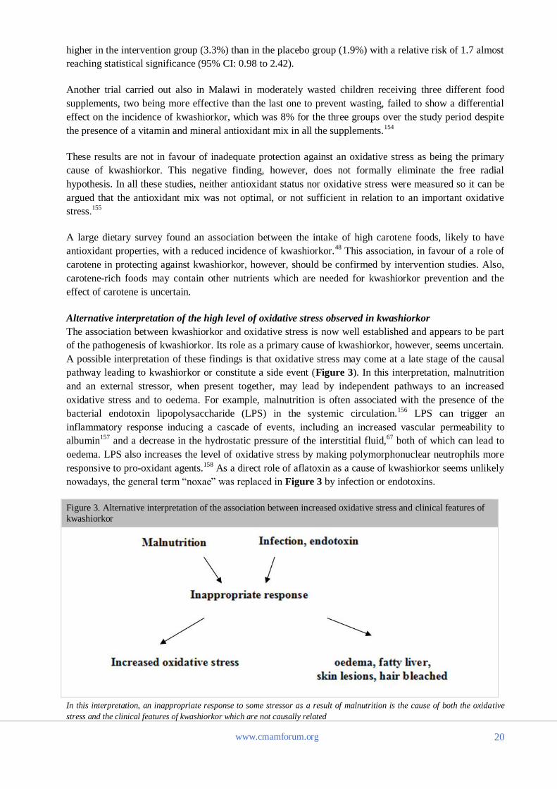
pathway leading to kwashiorkor or constitute a side event (Figure 3). In this interpretation, malnutrition
and an external stressor, when present together, may lead by independent pathways to an increased
oxidative stress and to oedema. For example, malnutrition is often associated with the presence of the
bacterial endotoxin lipopolysaccharide (LPS) in the systemic circulation.156
LPS can trigger an
inflammatory response inducing a cascade of events, including an increased vascular permeability to
albumin157
and a decrease in the hydrostatic pressure of the interstitial fluid,67
both of which can lead to
oedema. LPS also increases the level of oxidative stress by making polymorphonuclear neutrophils more
responsive to pro-oxidant agents.158
As a direct role of aflatoxin as a cause of kwashiorkor seems unlikely
nowadays, the general term “noxae” was replaced in Figure 3 by infection or endotoxins.
Figure 3. Alternative interpretation of the association between increased oxidative stress and clinical features of
kwashiorkor
In this interpretation, an inappropriate response to some stressor as a result of malnutrition is the cause of both the oxidative
stress and the clinical features of kwashiorkor which are not causally related

www.cmamforum.org
21
This interpretation, not involving oxidative stress as the primary cause of kwashiorkor, is compatible with
an inadequate protection against oxidative stress being an aggravating factor of kwashiorkor and is
compatible with a possible role of antioxidants in the treatment of kwashiorkor. In this regard, a first pilot
study showed that supplementing children with kwashiorkor with cysteine (as N-acetyl-cysteine)
increased glutathione synthesis and plasma concentrations and was associated with a more rapid
resolution of oedema compared to controls (9 ±1, vs. 14±2 days).82
Another pilot study showed that
supplementation with gluthathione or the antioxidant alpha-lipoic acid had a favourable effect on
survival.159
Similar studies to test the potential of different antioxidant supplementations on the clinical
outcome of kwashiorkor seem warranted.
Immunity and inflammation
The activation of the immune system seems to operate early in the causal pathway leading to
kwashiorkor. This is suggested by the already mentioned lower prevalence of HIV infection among
children admitted to hospital with kwashiorkor compared to marasmus. This is also suggested by the
finding of a study from Uganda showing that among HIV negative children with severe malnutrition, the
presence of oedema was associated with a higher CD4 count160
(Figure 4).
Figure 4: Percentage of CD4+ cells of lymphocytes in malnourished children with and without oedema
Source: Bachou et al160
These observations are difficult to explain with the hypothesis of an insufficient protein intake, or to a
dysadaptation to a low protein, high carbohydrate diet. It is also not easily explained by the free radical
hypothesis, at least in its initial form assuming a direct link between an increased oxidative stress and
kwashiorkor.
Box and whisker plot showing the median and the interquartile range of the percentages of CD+ cells in
severely malnourished children who were grouped based on their HIV status and type of malnutrition

www.cmamforum.org
22
Among HIV infected children with severe acute malnutrition, treatment with antiretroviral therapy to
restore immunity has been shown to be frequently associated with development of oedema.161
Whether
this is due to the restoration of immunity or to a refeeding syndrome with hypophosphataemia and
hypokalaemia162
is unclear, however.
An inappropriate inflammatory response to an external stimulus is suggested by an elevated concentration
of inflammatory mediators in children with kwashiorkor compared to marasmus. Among these mediators
Interleukin-6 and soluble receptors of tumor necrosing factor alpha sTNFR-p55 and sTNFR-p75 seem
elevated even in the absence of clinical infection.163
The production and excretion of leukotrienes in kwashiorkor also suggests an inappropriate inflammatory
response as a contributing cause.195
Particularly intriguing is the increased urinary excretion of interleukin
E4, as its synthesis requires glutathione which is in short supply as it is needed to respond to the oxidative
stress. Quantities of glutathione needed for interleukin E4 synthesis are small, however, compared to its
concentration in plasma.
The dysregulation of the inflammatory response, and in particular the increased leukotrienes production,
suggests a possible contributing role of essential fatty acids (EFA) in the pathway leading to kwashiorkor,
as these inflammation mediators are produced from fatty acids of the omega-6 family (n-6). Also, the type
of predominant EFA in the diet has a modulating effect on the inflammatory response.164
A role for an
excessive (n-6) EFA in kwashiorkor has been previously suggested, but with the hypothesis that cellular
immune response, especially CD4, is decreased in kwashiorkor,165
which is not consistent with available
evidence suggesting that the CD4 response is actually increased. A possible role of EFA is also suggested
by the observation that children with cystic fibrosis, which is associated with severe fat malabsorption,
may have a kwashiorkor-like clinical aspect.166,167
Of note, the early observation of Cicely Williams
suggested that cod liver oil, which has a high content of eicosapentaenoic acid (n-3) had a favourable
effect on kwashiorkor.2 An early description of kwashiorkor in Vietnam noted that it rarely occurred in
fishing communities.168
And whereas in Malawi as a whole, kwashiorkor is the predominant form of
severe acute malnutrition, it is not mentioned in a longitudinal study examining the growth and nutrition
of children in the community of Lungwena, near the lake Malawi, with fish being part of the common
foods.169
At the individual level, however, the fish intake is not significantly lower in children who later
develop kwashiorkor compared to other children of the same community.47,48
Disruption of sulphated glycosaminoglycans (GAGs) Water in the interstitial space is mainly entrapped in a gel formed by very long molecules of sulphated
glycosaminoglycans (GAGs) which are long chains of polysaccharides with attached sulphate molecules.
Free water represents a small proportion of interstitial tissues (usually less than 1%) and is normally
contained in small non-communicating pockets. The lack of communication between these vesicles
explains why water does not flow down to the lower parts of the body in healthy individuals. During
oedema, there is an excess of free water and these vesicles grow, come into contact with each other and
channels appear between them. Water can then flow from one vesicle to the other and accumulates in the
lower parts of the body.56
An abnormal GAG structure could favour the development of these micropockets of free water and could
be a cause of oedema. In the early paper presenting the free radical hypothesis, disruption of sulphated
GAGs was presented as a possible consequence of oxidative stress and as a mechanism to explain
occurrence of oedema in kwashiorkor.126
The constitutive antioxidant function of superoxide dismutase, a
key enzyme for neutralising free radicals, is dependent on binding to heparan sulphate proteoglycan

www.cmamforum.org
23
(HSPG), another form of sulphated GAG. Also, complex carbohydrates are a target for oxidative
damage.170
In the kidney, epithelial cells which line the outer surface of the glomerulus have long foot-like processes
(podocytes) that encircle the outer surface of the capillaries.56
These podocytes have an ultrastructure rich
in sulphated GAGs which were examined in a post mortem study of the kidneys of 6 children who died
from kwashiorkor. The histological analysis showed an effacement of glomerular foot processes, similar
to that of minimal change nephritic syndrome, suggesting a disruption of the structure of sulphated
GAGs.171
This abnormality can be reproduced experimentally in animals by infusion of polycationic
substances neutralising the negative ionic charge of GAGs. This suggests that children with kwashiorkor
could have sulphated GAGs with a decreased ionic charge, altering the physical properties of the
interstitial space leading to oedema. A more recent study confirmed that there is a decreased production
of sulphated GAGs, in particular of HSPG in kwashiorkor, but not in marasmus, also suggesting a
possible role of GAGs disruption in kwashiorkor.170
This hypothesis is also suggested by a low urinary
sulphate and sulphated GAG excretion in kwashiorkor.172,78
Sulfated GAG disruption could also explain a
greater resistance to different infections, in particular to cholera observed in kwashiorkor: Vibrio cholerae
binds to GAGs present in the gut and their disruption during kwashiorkor may prevent infection.4
Amadi et al. ascribed this disruption of sulphated GAGs to an interaction of malnutrition and enteric
infection with a genetic predisposition associated with a decreased capacity for GAG synthesis. A genetic
variation leading to anomalies of sulphated GAGs could also explain a lower risk of acquiring HIV
prenatally or during lactation, as HIV entry across endothelial barriers is mediated by interaction with
HSPG.170
In this interpretation, this protection against HIV may explain the lower prevalence of infection
in kwashiorkor compared to marasmic children, an observation which is not consistent with the original
free radical hypothesis.
The acceptance of the hypothesis of disrupted sulphated GAGs as a cause of kwashiorkor should be based
on its capacity to resist testing and to explain all manifestations of kwashiorkor. Children with congenital
defects of the metabolism of heparin sulphate, a sulphated GAG, suffer from a non oedematous form of
malnutrition.173,174
A possible test would be to examine the effect of stimulating sulphated GAGs synthesis either by N-
acetyl glucosamine (GlcNAc) or by heparin analogs on the evolution of kwashiorkor. Examining the
effect of the capacity to synthesise HPSG on the risk of kwashiorkor possibly by examining the
polymorphism of genes involved in sulphated GAGs synthesis also seems a promising option.170
Possible role of the gut microbiota In recent years, the gut microbiota
ii has been shown in experimental models to have an effect on the
overall metabolism and in particular to have influence on the energy harvested from the gut.175,176
While
this has important implications for the control of obesity, a possible role of the gut microbiota as a
contributing factor leading to malnutrition has been also suggested by studies showing a delay in its
maturation in children suffering from severe acute malnutrition.177
The role of the microbiota in the development of kwashiorkor has been suggested by a major longitudinal
study done in Malawi on 317 twin pairs during the first 3 years of their life.15
Among these twin pairs,
ii This term is preferable to the old term “gut flora” which implies that all microbes in the gut are bacteria whereas there are also
archae, viruses and eukaryotes, which belong to different kingdoms.
www.cmamforum.org
24
half remained well-nourished, whereas in 43%, one child developed kwashiorkor, and the other one did
not. Interestingly, among these discordant pairs, there was no significant difference in the incidence of
kwashiorkor among the identical and the fraternal pairs of twins of a case, suggesting that genetic factors
do not have a major role in the origin of kwashiorkor. Analysis of the microbiota of children who
developed kwashiorkor showed they had a delayed maturation compared to the healthy twin.
To disentangle the cause from the effect of this association between abnormal microbiota and
kwashiorkor, the microbiota of 3 discordant pairs of twins were inoculated to germ-free mice, some of
which were fed a standard laboratory diet, while others received the typical food consumed by rural
children in Malawi. Germ-free mice who received the Malawi diet and were also inoculated with the
microbiota of kwashiorkor children suffered a major weight loss, suggesting that the association between
a nutritionally poor diet and an abnormal microbiota was leading to malnutrition. The metabolism of
germ-free mice who were inoculated showed many differences of their metabolism compared to controls,
in particular in carbohydrate and sulphur amino acid metabolism, which suggests a possible link with the
“dysadaptation” hypothesis and with the hypothesis of an insufficient sulphur amino acid availability.
The Malawi twin study attracted wide attention from the scientific community as it definitely offers new
perspectives for the understanding of kwashiorkor.178,179,180,181
A plausible scenario would be that the
effect of microbiota on metabolism and translocation of intestinal bacteria or of bacterial endotoxins
could trigger the development of kwashiorkor. The abnormal metabolism of sulphur amino acids of mice
who received the microbiota of kwashiorkor children deserves attention, as it is also considered as a key
event in other explanations of kwashiorkor, such as the free radical or the disrupted sulphated GAGs
hypotheses. An abnormal microbiota associated with gut dysfunction could explain the presence of
endotoxin in the blood of these children with overstimulation of the immune system and of the
inflammatory response, and increased oxidative stress. Endotoxins produced by intestinal bacteria, and in
particular LPS, are suspected to be involved in the pathophysiology of non alcoholic fatty liver and
associated stimulation of pro-inflammation cytokines.182,183
A similar mechanism could be involved as
well in the steatosis of kwashiorkor. Or an effect of microbiota on sulphur amino acid metabolism leading
to an insufficient availability of methionine could also explain the hepatic steatosis.
The results of the twin Malawi study, however, should be regarded as preliminary. The hypothesis that an
abnormal gut microbiota may cause kwashiorkor, suggested by these early studies, needs further testing.
In this first study, microbiota of kwashiorkor children was compared with that of well nourished controls,
not that of children with marasmus. Hence it is not possible to say if the observed change are specific to
kwashiorkor or are related to malnutrition. The mouse model used in this study is interesting, but in the
absence of observed oedema in experimental mice, this should not be regarded as a kwashiorkor model.
Reproducing kwashiorkor in an animal model if this hypothesis is true might be difficult as humans have
a very different gut physiology compared to other mammals and even to other primates.184
In particular,
their gut microbiota is different, even compared to closely related primates.185
Kwashiorkor, however, has
been reproduced to a large extent in baboons110
and this model could be explored to test the microbiota
hypothesis. Oedema disease, observed in piglets at the time of weaning, and in which the role of a
bacterial toxin is well established,186
could also be considered as a possible experimental model which
could give a clue on the pathophysiology of oedema.
Ultimately, trials having a direct effect on the gut microbiota, such as giving an antibiotic or a specific pre
or probiotic, are needed to confirm a possible primary role of an abnormal gut microbiota as a cause of
kwashiorkor.

www.cmamforum.org
25
0
10
20
30
40
50
60
70
80
90
100
110
120
1945 1955 1965 1975 1985 1995 2005
Year
Nu
mb
er
of
pu
bli
cati
on
s
Conclusions: kwashiorkor remains an enigma None of the many hypotheses about the origin of kwashiorkor have succeeded in explaining correctly all
of its clinical and pathophysiological manifestations. The low prevalence of kwashiorkor in HIV infected
children and the possible early activation of the CD4 immune systems are especially intriguing, as they
are not easily explained by any of the hypotheses proposed over the last 50 years. The hypothesis of an
abnormal microbiota is attractive but this hypothesis, however, is in its early stages, and is based on very
limited evidence and testing it in clinical and animal models is needed before going further in its
acceptance.
The way forward – priorities for research Many studies quoted in this review go back to 30 or 40 years ago, sometimes even more. Kwashiorkor
was the topic of high quality research and intense debate in the 1950-1970s. Surprisingly, it later became
an “orphan disease”, with very few groups actively involved in disentangling its mechanism and
decreasing number of publications over the years (Figure 5). In contrast to most other understudied
diseases, kwashiorkor affects hundreds of thousands if not millions of children every year, killing many
of them, and is a serious public health problem in many parts of the world. The lack of a coherent
explanation to its aetiology is a major obstacle to further optimise treatment and to design effective
prevention strategies.
Figure 5: Number of publications with “kwashiorkor” as keyword since 1945
(Source: PubMed)
Several areas deserve special attention. They are suggested below without ranking their level of priority
beyond the scope of this review.
Epidemiological studies
Assessing the extent of the problem
Currently, there is no reliable estimate on the number of children developing kwashiorkor each year. As
mentioned in a previous section, there was no mention of the problem in the recent Lancet series on child
nutrition. Importance of kwashiorkor, an acute condition, is poorly evaluated by prevalence surveys,
which are often used to assess nutritional problems. These prevalence data could be used for mapping,
www.cmamforum.org
26
and identifying areas where it is a problem, but should be complemented by studies of the ratio of
kwashiorkor to marasmic patients admitted for treatment.
Detection of children with oedema is currently done in community-based programmes of management of
malnutrition and this can generate incidence data in different regions of the world if screening is repeated
in the same area sufficiently frequently. These data should be systematically collected and compared at
the international level.
Getting a clue about aetiology
Kwashiorkor is known to occur only in some areas, but currently, there is no systematic attempt to link
these differences with dietary patterns or other environmental variables. Although this kind of evidence is
very weak to show causality, it may give hints about possible mechanisms involved. A special case is
Malawi where large variations of kwashiorkor prevalence have been reported within small geographical
areas without a clear explanation.187
Anecdotal reports suggest the same pattern occurs in other settings
but has not been documented.
These epidemiological investigations should go beyond simple comparisons on protein or antioxidant
intakes. Comparison could be made on the intake of sulphur amino acids, ideally studying separately
cysteine and methionine intake, as these amino acids have different effects. The pattern of essential fatty
acids intake could also be examined. Sodium and potassium intake should be compared in populations
with high and low prevalence of kwashiorkor. Beyond these pure dietary aspects, the different insulin and
cortisol profiles observed in Uganda and in the Gambia is intriguing, and this pattern should be examined
in other settings with different forms of malnutrition. The nature of the soil could provide a clue as well.
Wet soil with a lot of organic matter which represent a reducing environment can make selenium
unavailable, and lead to selenium deficiency.188
As selenium is needed to protect the organism against
free radicals, natural variations of selenium availability could provide a clue to test the role of a
deficiency of this particular trace element as a contributing cause of kwashiorkor.
Microbiome studies (studies of the collective genome of the microbiota) can be carried out in large scale
human studies as they do not require blood nor tissues and could be used to explore potential differences
in areas of high and low kwashiorkor prevalence.
Although the Malawi twin study, which reported a similar incidence of kwashiorkor in identical and
fraternal twins of a confirmed case, is not in favour of a genetic factor,15
case-control studies examining
genetic variants of genes coding for enzymes involved in the defence against oxidative stress or in the
synthesis of sulphated GAGs could also be considered. Properly designed and well powered genome wide
association studies could give very important mechanistic insights.
Pathophysiological studies
Major aspects of the pathophysiology of kwashiorkor are still poorly understood and deserve exploration
with modern techniques. A few areas deserve urgent attention.
Immunology
The possible role of an active CD4 response suggested in one study160
is intriguing and needs
confirmation. If confirmed, this should be further explored and factors leading to this active role of the
immune system should be explored. Our knowledge of the immune system in malnutrition, and in
particular in kwashiorkor, is incomplete and relies on studies done years ago with techniques which are
currently often regarded as unreliable.189
All possible factors including a continuous stimulation of the
immune system by infections or by specific organisms proliferating in the gut possibly releasing LPS and
other bacterial metabolites in the blood should be examined. Factors possibly regulating the immune

www.cmamforum.org
27
response, and in particular essential fatty acids, should be examined. The role of non-specific immunity in
kwashiorkor should also be explored, as its role was largely ignored at the time most studies on the
immune system in malnutrition were carried out.
Pathophysiology of oedema
The pathophysiology of oedema is still poorly understood. The role of different terms of the Starling’s
equation, and in particular the role of albumin and hydrostatic pressure in the interstitial fluid should be
explored. The possibility of an increased hydraulic conductivity and permeability to proteins, as observed
in the nephrotic syndrome, should also be examined. The possible role of endotoxins, and in particular of
LPS in generating oedema has been largely ignored. Antibodies to different LPS could be looked at, and
compared with those favouring protein leakage into the interstitial sector in animal studies. More
generally, animal models, and ideally primate models, could be used to disentangle factors leading to
oedema. The few studies using non-human primates to reproduce kwashiorkor were carried out more than
40 year ago,110
and go back to a time when the understanding of kwashiorkor was fragmentary and relied
mainly on the protein deficiency hypothesis. Studies exploring more complex aetiology and pathogenic
pathways using the exciting advances in technology and exploring more recent hypothesis are urgently
needed.
Mechanisms leading to fatty liver
The origin of this major characteristic of kwashiorkor is still poorly understood. Hepatic steatosis occurs
in marasmus as well as kwashiorkor190
and is associated with a poor prognosis.38
Fat is very slowly
mobilised from the liver as assessed by repeated biopsies191
or by ultrasound assessment.190
This contrasts
with the successful treatment of children with high fat diets, showing that dietary fat is efficiently utilised.
The reasons for these contrasting observations are not clear. The hypothesis of an insufficient export of
protein synthesis seem hardly tenable70
but no adequate alternative explanation has been validated. The
hypothesis of an insufficient oxidation of fatty acids, proposed more than 20 years ago138
has not been
adequately tested so far. This could be done by examining the fatty acid composition of fatty liver. Here
again, animal studies, preferably in non-human primates, checking the nature of accumulated fatty acids,
would help. This question could be also examined in post mortem liver samples taken from children who
died from kwashiorkor.
Effect of the microbiota on overall metabolism
Results of the pilot Malawi study suggesting that microbiota associated with kwashiorkor can have
general metabolic effects on mouse metabolism with some similarities with those observed in
kwashiorkor should be further explored, ideally in non-human primate models.
Clinical trials
In recent years, WHO’s emphasis when generating guidelines has been to use an evidence-based
approach;192
this has led to giving a special weight to data generated by randomised clinical trials.
Although this approach has its limitations, especially in nutrition,193
randomised trials have an important
role to play in improving current protocols and also in understanding causal links. This approach has been
underused in the context of malnutrition. Most studies describe associations which are not sufficient to
have any conclusion on causality. As a result, many critical aspects of clinical management of severe
acute malnutrition are still based on clinical impression and experience of experts which represents a low
level of evidence.194,34
Several questions should be addressed urgently.
Testing the effect of different antioxidants - minimising the level of oxidative stress
The pilot study suggesting that supplementation with reduced glutathione or with alpha-lipoic acid had an
effect on mortality159
has never been confirmed. There is an urgent need to test the potential of different
antioxidants to improve clinical recovery of children with kwashiorkor.
www.cmamforum.org
28
The level of oxidative stress can be influenced by the nature of the fatty acids included in the diet.141
Leukotrienes which are derived from unsaturated fatty acids are elevated in kwashiorkor compared to
children with marasmus.195
Diets with different fatty acid composition should be compared for their
efficacy for the treatment of children with oedema.
Optimising amino acid intake
Clinical data suggesting that cysteine supplementation can accelerate cure from oedema82
should be
confirmed by large clinical trials in different settings. The use of different protein sources with a high
content of sulphur amino acids, in particular cysteine (egg proteins, or specific milk fractions) should also
be tested. A preliminary study suggested that a diet with egg white as a protein source might have a
favourable effect on acute phase protein synthesis.196
Egg white, however, has a low phosphorus content
and its use was associated with lower phosphate plasma concentration and egg yolk might be a better
choice as suggested by early studies.73
In all these clinical trials skin lesions and other organ systems
dependent upon sulphur metabolism, including sulphated GAGs,77
should be included as secondary
outcomes.
Optimising the fat intake
Little attention has been given to optimising the fat intake of children with kwashiorkor. The nature of
lipids in the diet used for treatment is likely to have an effect on the level of oxidative stress and the
production of leukotrienes and other mediators of inflammation. Medium chain fatty acids are likely to be
better absorbed. When the protein intake is kept constant, increasing the fat intake will decrease the
carbohydrate intake, with possibly a favourable effect on plasma phosphorus concentrations.111
Different
options to optimise fat intake should be tested in formal clinical trials.
Treating skin lesions
The origin of skin lesions of kwashiorkor still remains an open question. Some clue about the
mechanisms involved can be obtained by clinical trials testing supplementation with sulphur amino acids
or changing essential fatty intake during treatment. Skin lesions can also respond to topical application of
some nutrients, especially zinc and fatty acids. This can lead to clinical trials, on a small sample of
children, each child being his own control, by leaving some lesions untreated.197
Modifying the gut microbiota
One of the best ways to confirm or disprove the effect of the gut microbiota on the clinical picture of
kwashiorkor would be to test the efficacy of different interventions which could affect it by using
randomised clinical trials. The microbiota can be modified by antibiotics, prebiotics, and probiotics.
Testing any of these options should be considered. A randomised trial in Malawi showed that treating
children with kwashiorkor with antibiotics has a favourable effect on survival,20
consistent with a possible
role of infection in the pathogenesis of this form of malnutrition.
Despite the high quality of the work led by the few groups, many avenues of research have been left
untouched as is shown by this list. This contrasts with the unprecedented development of techniques to
explore metabolism over the last 20 years. One reason for low research output is the scarcity of human
capacity and poor access to the innovative technologies in areas with the disease burden. The situation
may be improving with increasing academic connectivity and resurgence of interest in nutrition related
health internationally. It is hoped in this context that the challenge of identifying the mechanism of
kwashiorkor will attract the attention of donors and encourage scientists with access to modern
exploration techniques to study its mechanisms in depth.
Questions and comments may be submitted through the CMAM Forum:
[email protected] www.cmamforum.org

www.cmamforum.org
29
References 1 Williams C. Kwashiorkor, a nutritional disease associated with a maize diet. Lancet
1935;224:1151–2.
2 Williams CD, Oxon BM, Lond H. Kwashiorkor: a nutritional disease of children associated with a
maize diet. 1935. Bull World Health Organ 2003;81:912–3.
3 Konotey-Ahulu FI. Kwashiorkor. BMJ 1991;302:180–1.
4 Golden MH. Oedematous malnutrition. Br Med Bull 1998;54:433–44.
5 Williams CD. A nutritional disease of childhood associated with a maize diet. Arch Dis Child
1933;8:423–33.
6 Williams CD. Fifty years ago. Archives of Diseases in Childhood 1933. A nutritional disease of childhood associated with a maize diet. Arch Dis Child 1983;58:550–60.
7 Classification of infantile malnutrition. Lancet 1970;2:302–3.
8 World Health Organization. WHO child growth standards and the identification of severe acute malnutrition in infants and children: joint statement by the World Health Organization and the
United Nations Children’s Fund 2009.
9 Williams CD. Protein-calorie malnutrition. Lancet 1972;1:1333.
10 Black RE, Allen LH, Bhutta ZA, Caulfield LE, de Onis M, Ezzati M, et al. Maternal and child undernutrition: global and regional exposures and health consequences. Lancet 2008;371:243–60.
doi:10.1016/S0140-6736(07)61690-0.
11 Ndekha MJ. Kwashiorkor and severe acute malnutrition in childhood. Lancet 2008;371:1748; author reply 1749. doi:10.1016/S0140-6736(08)60752-7.
12 Black RE, Victora CG, Walker SP, Bhutta ZA, Christian P, de Onis M, et al. Maternal and child
undernutrition and overweight in low-income and middle-income countries. Lancet 2013;382:427–51. doi:10.1016/S0140-6736(13)60937-X.
13 Briend A, Collins S, Golden M, Manary M, Myatt M. Maternal and child nutrition. Lancet
2013;382:1549. doi:10.1016/S0140-6736(13)62315-6.
14 Ciliberto H, Ciliberto M, Briend A, Ashorn P, Bier D, Manary M. Antioxidant supplementation for the prevention of kwashiorkor in Malawian children: randomised, double blind, placebo controlled
trial. BMJ 2005;330:1109. doi:10.1136/bmj.38427.404259.8F.
15 Smith MI, Yatsunenko T, Manary MJ, Trehan I, Mkakosya R, Cheng J, et al. Gut microbiomes of Malawian twin pairs discordant for kwashiorkor. Science 2013;339:548–54.
doi:10.1126/science.1229000.
16 Briend A, Wojtyniak B, Rowland MG. Arm circumference and other factors in children at high risk
of death in rural Bangladesh. Lancet 1987;2:725–8.
17 Dramaix M, Hennart P, Brasseur D, Bahwere P, Mudjene O, Tonglet R, et al. Serum albumin
concentration, arm circumference, and oedema and subsequent risk of dying in children in central
Africa. BMJ 1993;307:710–3.
18 Prudhon C, Golden MH, Briend A, Mary JY. A model to standardise mortality of severely
malnourished children using nutritional status on admission to therapeutic feeding centres. Eur J
Clin Nutr 1997;51:771–7.
19 Talbert A, Thuo N, Karisa J, Chesaro C, Ohuma E, Ignas J, et al. Diarrhoea complicating severe
acute malnutrition in Kenyan children: a prospective descriptive study of risk factors and outcome.
PloS One 2012;7:e38321. doi:10.1371/journal.pone.0038321.
20 Trehan I, Goldbach HS, LaGrone LN, Meuli GJ, Wang RJ, Maleta KM, et al. Antibiotics as part of the management of severe acute malnutrition. N Engl J Med 2013;368:425–35.
doi:10.1056/NEJMoa1202851.
www.cmamforum.org
30
21 Bhattacharyya AK, Chattopadhyay PS, Paladhi PK, Ganguli S, Bhattacharyya N. Kwashiorkor and
marasmus: changing hospital incidence of syndromic presentation (1957-88). Indian Pediatr 1990;27:1191–8.
22 Oyelami OA, Ogunlesi TA. Kwashiorkor--is it a dying disease? South Afr Med J Suid-Afr Tydskr
Vir Geneeskd 2007;97:65–8.
23 Golden M, Grellety Y. Protocol - Integrated Management of Acute Malnutrition 2012.
24 Said A, el-Hawary FS, Sakr R, Abdel Khalek MK, Ibrahim AM. Protein-calorie malnutrition in
Egypt. I. Immunoelectrophoretic studies on urinary proteins. Am J Clin Nutr 1973;26:1355–9.
25 Heilskov S, Rytter MJH, Vestergaard C, Briend A, Babirekere E, Deleuran MS. Dermatosis in
children with oedematous malnutrition (Kwashiorkor): a review of the literature. J Eur Acad
Dermatol Venereol JEADV 2014;28:995–1001. doi:10.1111/jdv.12452.
26 Garrow JS, Pike MC. The short-term prognosis of severe primary infantile malnutrition. Br J Nutr
1967;21:155–65.
27 Mclean AE. Hepatic failure in malnutrition. Lancet 1962;2:1292–4.
28 Wharton BA, Howells GR, McCance RA. Cardiac failure in kwashiorkor. Lancet 1967;2:384–7.
29 Patrick J. Death during recovery from severe malnutrition and its possible relationship to sodium
pump activity in the leucocyte. Br Med J 1977;1:1051–4.
30 Schofield C, Ashworth A. Why have mortality rates for severe malnutrition remained so high? Bull
World Health Organ 1996;74:223–9.
31 World Health Organization. Management of severe malnutrition: a manual for physicians and other
senior health workers. Geneva: World Health Organization; 1999.
32 Heikens GT, Bunn J, Amadi B, Manary M, Chhagan M, Berkley JA, et al. Case management of
HIV-infected severely malnourished children: challenges in the area of highest prevalence. Lancet
2008;371:1305–7. doi:10.1016/S0140-6736(08)60565-6.
33 Trehan I, O’Hare BA, Phiri A, Heikens GT. Challenges in the Management of HIV-Infected
Malnourished Children in Sub-Saharan Africa. AIDS Res Treat 2012;2012:790786.
doi:10.1155/2012/790786.
34 World Health Organization. Updates on the management of severe acute malnutrition in infants and
children 2013.
35 World Health Organization. Community-based management of severe acute malnutrition. Geneva
World Health Organ 2007.
36 Donnen P, Sylla A, Dramaix M, Sall G, Kuakuvi N, Hennart P. Effect of daily low dose of vitamin
A compared with single high dose on morbidity and mortality of hospitalized mainly malnourished
children in senegal: a randomized controlled clinical trial. Eur J Clin Nutr 2007;61:1393–9.
doi:10.1038/sj.ejcn.1602671.
37 Iannotti LL, Trehan I, Manary MJ. Review of the safety and efficacy of vitamin A supplementation
in the treatment of children with severe acute malnutrition. Nutr J 2013;12:125. doi:10.1186/1475-
2891-12-125.
38 Waterlow JC. Protein Energy Malnutrition. London: Edward Arnold; 1992.
39 Flores H, Pak N, Maccioni A, Monckeberg F. Lipid transport in kwashiorkor. Br J Nutr
1970;24:1005–11.
40 Truswell AS, Hansen JD, Watson CE, Wannenburg P. Relation of serum lipids and lipoproteins to
fatty liver in kwashiorkor. Am J Clin Nutr 1969;22:568–76.
41 McLaren DS. The great protein fiasco. Lancet 1974;2:93–6.
42 Waterlow JC, Payne PR. The protein gap. Nature 1975;258:113–7.
43 Elango R, Humayun MA, Ball RO, Pencharz PB. Protein requirement of healthy school-age children determined by the indicator amino acid oxidation method. Am J Clin Nutr 2011;94:1545–
52. doi:10.3945/ajcn.111.012815.
44 Gopalan C. Kwashiorkor and marasmus: evolution and distinguishing features. Calor. Defic. Protein
Defic., Boston, Little, Brown: McCance RA, Widdowson EM; 1968, p. 49–58.
45 Laditan AA, Reeds PJ. A study of the age of onset, diet and the importance of infection in the
pattern of severe protein-energy malnutrition in Ibadan, Nigeria. Br J Nutr 1976;36:411–9.

www.cmamforum.org
31
46 Sullivan J, Ndekha M, Maker D, Hotz C, Manary MJ. The quality of the diet in Malawian children
with kwashiorkor and marasmus. Matern Child Nutr 2006;2:114–22. doi:10.1111/j.1740-
8709.2006.00053.x.
47 Lin CA, Boslaugh S, Ciliberto HM, Maleta K, Ashorn P, Briend A, et al. A prospective assessment
of food and nutrient intake in a population of Malawian children at risk for kwashiorkor. J Pediatr
Gastroenterol Nutr 2007;44:487–93. doi:10.1097/MPG.0b013e31802c6e57.
48 Kismul H, Van den Broeck J, Lunde TM. Diet and kwashiorkor: a prospective study from rural DR
Congo. PeerJ 2014;2:e350. doi:10.7717/peerj.350.
49 Golden MH. Transport proteins as indices of protein status. Am J Clin Nutr 1982;35:1159–65.
50 James WP, Hay AM. Albumin metabolism: effect of the nutritional state and the dietary protein
intake. J Clin Invest 1968;47:1958–72. doi:10.1172/JCI105885.
51 Whitehead RG. The protein needs of malnourished children. Proteins Hum. Nutr. Academic Press,
Porter DWG, Rolls BA; 1973, p. 103–17.
52 Morlese JF, Forrester T, Badaloo A, Del Rosario M, Frazer M, Jahoor F. Albumin kinetics in
edematous and nonedematous protein-energy malnourished children. Am J Clin Nutr 1996;64:952-9.
53 Frood JD. Relationship between pattern of infection and development of hypoalbuminaemia and
hypo-beta-lipoproteinaemia in rural Ugandan children. Lancet 1971;2:1047–9.
54 Fuhrman MP. The albumin-nutrition connection: separating myth from fact. Nutr Burbank Los
Angel Cty Calif 2002;18:199–200.
55 Iputo JE. Protein-losing enteropathy in Transkeian children with morbid protein-energy
malnutrition. South Afr Med J Suid-Afr Tydskr Vir Geneeskd 1993;83:588–9.
56 Guyton AC, Hall JE. Textbook of Medical Physiology. 11th ed. Philadelphia: Elsevier; 2006.
57 Coward WA, Fiorotto M. The pathogenesis of oedema in kwashiorkor--the role of plasma proteins.
Proc Nutr Soc 1979;38:51–9.
58 Patrick J. Oedema in protein energy malnutrition: the role of the sodium pump. Proc Nutr Soc
1979;38:61–8.
59 Rondon-Berrios H. New insights into the pathophysiology of oedema in nephrotic syndrome. Nefrol
Publ Of Soc Esp Nefrol 2011;31:148–54. doi:10.3265/Nefrologia.pre2010.Nov.10724.
60 Doucet A, Favre G, Deschênes G. Molecular mechanism of edema formation in nephrotic
syndrome: therapeutic implications. Pediatr Nephrol Berl Ger 2007;22:1983–90.
doi:10.1007/s00467-007-0521-3.
61 Keys A. The edema problem. Biol. Hum. Starvation. University of Minnesota Press, Keys A,
Brozeck J, Henschel A, Mikkelsen O, Taylor HL; 1950, p. 921–65.
62 Montgomery RD. The Relation of Oedema to Serum Protein and Pseudocholinesterase Levels in the
Malnourished Infant. Arch Dis Child 1963;38:343–8.
63 Golden MH, Golden BE, Jackson AA. Albumin and nutritional oedema. Lancet 1980;1:114–6.
64 Minchiotti L, Galliano M, Caridi G, Kragh-Hansen U, Peters T. Congenital analbuminaemia:
molecular defects and biochemical and clinical aspects. Biochim Biophys Acta 2013;1830:5494–
502. doi:10.1016/j.bbagen.2013.04.019.
65 Coward WA, Sawyer MB. Whole-body albumin mass and distribution in rats fed on low-protein
diets. Br J Nutr 1977;37:127–34.
66 Nedrebø T, Berg A, Reed RK. Effect of tumor necrosis factor-alpha, IL-1beta, and IL-6 on
interstitial fluid pressure in rat skin. Am J Physiol 1999;277:H1857–62.
67 Nedrebø T, Reed RK. Different serotypes of endotoxin (lipopolysaccharide) cause different
increases in albumin extravasation in rats. Shock Augusta Ga 2002;18:138–41.
68 Waterlow JC. Kwashiorkor revisited: the pathogenesis of oedema in kwashiorkor and its
significance. Trans R Soc Trop Med Hyg 1984;78:436–41.
69 Coward WA, Whitehead RG. Changes in serum -lipoprotein concentration during the development
of kwashiorkor and in recovery. Br J Nutr 1972;27:383–94.
70 Badaloo A, Reid M, Soares D, Forrester T, Jahoor F. Relation between liver fat content and the rate
of VLDL apolipoprotein B-100 synthesis in children with protein-energy malnutrition. Am J Clin
Nutr 2005;81:1126–32.
www.cmamforum.org
32
71 Hansen JD, Jenkinson V. Electrolyte and nitrogen metabolism in kwashiorkor. South Afr J Lab Clin
Med Suid-Afr Tydskr Vir Lab- En Kliniekw 1956;2:206–31.
72 Golden MH. Protein deficiency, energy deficiency, and the oedema of malnutrition. Lancet
1982;1:1261–5.
73 Petrides EP. Hunger edema in children. J Pediatr 1948;32:333–50.
74 World Health Organization. Protein and amino acid requirements in human nutrition. WHO 2007.
http://www.who.int/nutrition/publications/nutrientrequirements/WHO_TRS_935/en/ (accessed
September 26, 2014).
75 Holt LE, Snyderman SE, Norton PM, Roitman E, Finch J. The plasma aminogram in kwashiorkor.
Lancet 1963;2:1342–8.
76 Dean RF, Whitehead RG. The metabolism of aromatic aminoacids in kwashiorkor. Lancet
1963;1:188–91.
77 Roediger WE. New views on the pathogenesis of kwashiorkor: methionine and other amino acids. J
Pediatr Gastroenterol Nutr 1995;21:130–6.
78 Ittyerah TR. Urinary excretion of sulfate in kwashiorkor. Clin Chim Acta Int J Clin Chem
1969;25:365–9.
79 Jackson AA. Blood glutathione in severe malnutrition in childhood. Trans R Soc Trop Med Hyg
1986;80:911–3.
80 Bonting SL. Cysteine, cystine and methionine in the skin of young and adult rats. Biochim Biophys
Acta 1950;6:183–6.
81 Kamalu BP. Cassava (Manihot esculenta crantz) in the aetiology of kwashiorkor. Nutr Res Rev
1993;6:121–35. doi:10.1079/NRR19930009.
82 Badaloo A, Reid M, Forrester T, Heird WC, Jahoor F. Cysteine supplementation improves the
erythrocyte glutathione synthesis rate in children with severe edematous malnutrition. Am J Clin
Nutr 2002;76:646–52.
83 Green CO, Badaloo AV, Hsu JW, Taylor-Bryan C, Reid M, Forrester T, et al. Effects of
randomized supplementation of methionine or alanine on cysteine and glutathione production
during the early phase of treatment of children with edematous malnutrition. Am J Clin Nutr
2014;99:1052–8. doi:10.3945/ajcn.113.062729.
84 Sander CS, Hertecant J, Abdulrazzaq YM, Berger TG. Severe exfoliative erythema of malnutrition in a child with coexisting coeliac and Hartnup’s disease. Clin Exp Dermatol 2009;34:178–82.
doi:10.1111/j.1365-2230.2008.02773.x.
85 Orbak Z, Ertekin V, Selimoglu A, Yilmaz N, Tan H, Konak M. Hartnup disease masked by
kwashiorkor. J Health Popul Nutr 2010;28:413–5.
86 Bröer S. Amino acid transport across mammalian intestinal and renal epithelia. Physiol Rev
2008;88:249–86. doi:10.1152/physrev.00018.2006.
87 Klahr S, Alleyne GA. Effects of chronic protein-calorie malnutrition on the kidney. Kidney Int
1973;3:129–41.
88 Lurie AO, Jackson WP. Aldosteronuria and the edema of kwashiorkor. Am J Clin Nutr
1962;11:115–26.
89 Godard CM, Muñoz M, Sanchez MA, Vallotton MB, Riondel A. A study of the renin-angiotensin-
aldosterone system in severe infantile malnutrition. Int J Pediatr Nephrol 1986;7:39-44.
90 Van der Westhuysen JM, Kanengoni E, Jones JJ, van Niekerk CH. Plasma renin activity in oedematous and marasmic children with protein energy malnutrition. South Afr Med J Suid-Afr
Tydskr Vir Geneeskd 1975;49:1729–31.
91 Srikantia SG, Gopalan C. Role of ferritin in nutritional edema. J Appl Physiol 1959;14:829–33.
92 Srikantia SG. Ferritin in nutritional oedema. Lancet 1958;1:667–8.
93 Srikantia SG, Mohanram M. Antidiuretic hormone values in plasma and urine of malnourished
children. J Clin Endocrinol Metab 1970;31:312–4. doi:10.1210/jcem-31-3-312.
94 Castranova V, Miles PR. Sodium permeability of dog red blood cell membranes. I. Identification of
regulatory sites. J Gen Physiol 1976;67:563–78.
95 Golden MH, Golden BE. Trace elements. Potential importance in human nutrition with particular
reference to zinc and vanadium. Br Med Bull 1981;37:31–6.

www.cmamforum.org
33
96 Nutritional oedema, albumin, and vanadate. Lancet 1981;1:646–7.
97 McGuire EA, Young VR. Nutritional edema in a rat model of protein deficiency: significance of the
dietary potassium and sodium content. J Nutr 1986;116:1209–24.
98 Whitehead RG, Alleyne GA. Pathophysiological factors of importance in protein-calorie
malnutrition. Br Med Bull 1972;28:72–9.
99 Coward WA, Lunn PG. The biochemistry and physiology of kwashiorkor and marasmus. Br Med
Bull 1981;37:19–24.
100 Whitehead RG, Lunn PG. Endocrines in protein-energy malnutrition. Proc Nutr Soc 1979;38:69–76.
101 Jahoor F, Badaloo A, Reid M, Forrester T. Protein metabolism in severe childhood malnutrition.
Ann Trop Paediatr 2008;28:87–101. doi:10.1179/146532808X302107.
102 Manary MJ, Broadhead RL, Yarasheski KE. Whole-body protein kinetics in marasmus and
kwashiorkor during acute infection. Am J Clin Nutr 1998;67:1205–9.
103 Jahoor F, Badaloo A, Reid M, Forrester T. Protein kinetic differences between children with edematous and nonedematous severe childhood undernutrition in the fed and postabsorptive states.
Am J Clin Nutr 2005;82:792–800.
104 Jahoor F, Badaloo A, Reid M, Forrester T. Sulfur amino acid metabolism in children with severe
childhood undernutrition: cysteine kinetics. Am J Clin Nutr 2006;84:1393–9.
105 Jahoor F, Badaloo A, Reid M, Forrester T. Sulfur amino acid metabolism in children with severe
childhood undernutrition: methionine kinetics. Am J Clin Nutr 2006;84:1400–5.
106 Badaloo AV, Forrester T, Reid M, Jahoor F. Lipid kinetic differences between children with
kwashiorkor and those with marasmus. Am J Clin Nutr 2006;83:1283–8.
107 Lunn PG, Whitehead RG, Hay RW, Baker BA. Progressive changes in serum cortisol, insulin and
growth hormone concentrations and their relationship to the distorted amino acid pattern during the
development of kwashiorkor. Br J Nutr 1973;29:399–422.
108 Robinson H, Picou D. A comparison of fasting plasma insulin and growth hormone concentrations in marasmic, kwashiorkor, marasmic-kwashiorkor and underweight children. Pediatr Res
1977;11:637–40. doi:10.1203/00006450-197705000-00003.
109 Spoelstra MN, Mari A, Mendel M, Senga E, van Rheenen P, van Dijk TH, et al. Kwashiorkor and
marasmus are both associated with impaired glucose clearance related to pancreatic β-cell
dysfunction. Metabolism 2012;61:1224–30. doi:10.1016/j.metabol.2012.01.019.
110 Coward DG, Whitehead RG. Experimental protein-energy malnutrition in baby baboons. Attempts
to reproduce the pathological features of kwashiorkor as seen in Uganda. Br J Nutr 1972;28:223-37.
111 Mehanna HM, Moledina J, Travis J. Refeeding syndrome: what it is, and how to prevent and treat
it. BMJ 2008;336:1495–8. doi:10.1136/bmj.a301.
112 Waterlow JC, Golden MH. Serum inorganic phosphate in protein-energy malnutrition. Eur J Clin
Nutr 1994;48:503–6.
113 Forrester TE, Badaloo AV, Boyne MS, Osmond C, Thompson D, Green C, et al. Prenatal factors
contribute to the emergence of kwashiorkor or marasmus in severe undernutrition: evidence for the
predictive adaptation model. PloS One 2012;7:e35907. doi:10.1371/journal.pone.0035907.
114 Sakuma K, Ohyama T, Sogawa K, Fujii-Kuriyama Y, Matsumura Y. Low protein--high energy diet
induces repressed transcription of albumin mRNA in rat liver. J Nutr 1987;117:1141–8.
115 Perozzi G, Mengheri E, Faraonio R, Gaetani S. Expression of liver-specific genes coding for plasma
proteins in protein deficiency. FEBS Lett 1989;257:215–8.
116 Hendrickse RG. The influence of aflatoxins on child health in the tropics with particular reference
to kwashiorkor. Trans R Soc Trop Med Hyg 1984;78:427–35.
117 Hendrickse RG. Kwashiorkor: the hypothesis that incriminates aflatoxins. Pediatrics 1991;88:376-9.
118 Hendrickse RG, Coulter JB, Lamplugh SM, Macfarlane SB, Williams TE, Omer MI, et al.
Aflatoxins and kwashiorkor: a study in Sudanese children. Br Med J Clin Res Ed 1982;285:843–6.
119 De Vries HR, Lamplugh SM, Hendrickse RG. Aflatoxins and kwashiorkor in Kenya: a hospital
based study in a rural area of Kenya. Ann Trop Paediatr 1987;7:249–51.
120 Coulter JB, Suliman GI, Lamplugh SM, Mukhtar BI, Hendrickse RG. Aflatoxins in liver biopsies
from Sudanese children. Am J Trop Med Hyg 1986;35:360–5.
www.cmamforum.org
34
121 Lamplugh SM, Hendrickse RG. Aflatoxins in the livers of children with kwashiorkor. Ann Trop
Paediatr 1982;2:101–4.
122 De Vries HR, Maxwell SM, Hendrickse RG. Aflatoxin excretion in children with kwashiorkor or
marasmic kwashiorkor--a clinical investigation. Mycopathologia 1990;110:1–9.
123 Oyelami OA, Maxwell SM, Adelusola KA, Aladekoma TA, Oyelese AO. Aflatoxins in autopsy
kidney specimens from children in Nigeria. J Toxicol Environ Health A 1998;55:317–23.
124 Peraica M, Radić B, Lucić A, Pavlović M. Toxic effects of mycotoxins in humans. Bull World
Health Organ 1999;77:754–66.
125 M H N Golden. The consequences of protein deficiency in man and its relationship to the features
of kwashiorkor. Nutr. Adapt. Man. Applied Science Publishers, London: K Blaxter and JC
Waterlow; 1985, p. 169–87.
126 Golden MH, Ramdath D. Free radicals in the pathogenesis of kwashiorkor. Proc Nutr Soc
1987;46:53–68.
127 Golden Michael, Ramdath DD, Golden BE. Free radicals and malnutrition. Trace Elem. Micronutr.
Free Radic. Humana Press, Totowa, NJ: Dreosti IE; 1991, p. 199–222.
128 Sive AA, Subotzky EF, Malan H, Dempster WS, Heese HD. Red blood cell antioxidant enzyme
concentrations in kwashiorkor and marasmus. Ann Trop Paediatr 1993;13:33–8.
129 Manary MJ, Leeuwenburgh C, Heinecke JW. Increased oxidative stress in kwashiorkor. J Pediatr
2000;137:421–4. doi:10.1067/mpd.2000.107512.
130 Fechner A, Böhme C, Gromer S, Funk M, Schirmer R, Becker K. Antioxidant status and nitric
oxide in the malnutrition syndrome kwashiorkor. Pediatr Res 2001;49:237–43.
131 Dempster WS, Sive AA, Rosseau S, Malan H, Heese HV. Misplaced iron in kwashiorkor. Eur J
Clin Nutr 1995;49:208–10.
132 Ashour MN, Salem SI, El-Gadban HM, Elwan NM, Basu TK. Antioxidant status in children with
protein-energy malnutrition (PEM) living in Cairo, Egypt. Eur J Clin Nutr 1999;53:669–73.
133 Ramdath DD, Golden MH. Non-haematological aspects of iron nutrition. Nutr Res Rev 1989;2:29–
49. doi:10.1079/NRR19890006.
134 Forrester T, Golden M, Brand S, Swales J. Reduction in vitro of red cell glutathione reproduces
defects of cellular sodium transport seen in oedematous malnutrition. Eur J Clin Nutr 1990;44:363-9.
135 Patrick J, Golden M. Leukocyte electrolytes and sodium transport in protein energy malnutrition.
Am J Clin Nutr 1977;30:1478–81.
136 Kaplay SS. Erythrocyte membrane Na+ and K+ activated adenosine triphosphatase in protein-
calorie malnutrition. Am J Clin Nutr 1978;31:579–84.
137 Willis JS, Golden MH. Active and passive transport of sodium and potassium ions in erythrocytes
of severely malnourished Jamaican children. Eur J Clin Nutr 1988;42:635–45.
138 Doherty JF, Golden MH, Brooks SE. Peroxisomes and the fatty liver of malnutrition: an hypothesis.
Am J Clin Nutr 1991;54:674–7.
139 Jahoor F. Effects of decreased availability of sulfur amino acids in severe childhood undernutrition.
Nutr Rev 2012;70:176–87. doi:10.1111/j.1753-4887.2011.00462.x.
140 Anraku M, Chuang VTG, Maruyama T, Otagiri M. Redox properties of serum albumin. Biochim
Biophys Acta 2013;1830:5465–72. doi:10.1016/j.bbagen.2013.04.036.
141 Hassan H, Hashim SA, Van Itallie TB, Sebrell WH. Syndrome in premature infants associated with
low plasma vitamin E levels and high polyunsaturated fatty acid diet. Am J Clin Nutr 1966;19:147-57.
142 Babu CK, Ansari KM, Mehrotra S, Khanna R, Khanna SK, Das M. Alterations in redox potential of
glutathione/glutathione disulfide and cysteine/cysteine disulfide couples in plasma of dropsy
patients with argemone oil poisoning. Food Chem Toxicol Int J Publ Br Ind Biol Res Assoc
2008;46:2409–14. doi:10.1016/j.fct.2008.03.031.
143 Christodouleas JP, Forrest RD, Ainsley CG, Tochner Z, Hahn SM, Glatstein E. Short-term and
long-term health risks of nuclear-power-plant accidents. N Engl J Med 2011;364:2334–41.
doi:10.1056/NEJMra1103676.
144 Bakonyi T, Radak Z. High altitude and free radicals. J Sports Sci Med 2004;3:64–9.
145 Rumbold A, Duley L, Crowther CA, Haslam RR. Antioxidants for preventing pre-eclampsia.
Cochrane Database Syst Rev 2008:CD004227. doi:10.1002/14651858.CD004227.pub3.

www.cmamforum.org
35
146 Israël N, Gougerot-Pocidalo MA. Oxidative stress in human immunodeficiency virus infection. Cell
Mol Life Sci CMLS 1997;53:864–70.
147 Kessler L, Daley H, Malenga G, Graham S. The impact of the human immunodeficiency virus type
1 on the management of severe malnutrition in Malawi. Ann Trop Paediatr 2000;20:50–6.
148 Beau JP, Imboua-Coulibaly L. Kwashiorkor and HIV: new questions. J Trop Pediatr 1997;43:50–1.
149 Amadi B, Kelly P, Mwiya M, Mulwazi E, Sianongo S, Changwe F, et al. Intestinal and systemic
infection, HIV, and mortality in Zambian children with persistent diarrhea and malnutrition. J
Pediatr Gastroenterol Nutr 2001;32:550–4.
150 Angami K, Reddy SVR, Singh KI, Singh NB, Singh PI. Prevalence of HIV infection and AIDS
symptomatology in malnourished children--a hospital based study. J Commun Dis 2004;36:45–52.
151 Cappellini MD, Fiorelli G. Glucose-6-phosphate dehydrogenase deficiency. Lancet 2008;371:64–
74. doi:10.1016/S0140-6736(08)60073-2.
152 Becker K, Leichsenring M, Gana L, Bremer HJ, Schirmer RH. Glutathione and association
antioxidant systems in protein energy malnutrition: results of a study in Nigeria. Free Radic Biol
Med 1995;18:257–63.
153 Marshall KG, Swaby K, Hamilton K, Howell S, Landis RC, Hambleton IR, et al. A preliminary examination of the effects of genetic variants of redox enzymes on susceptibility to oedematous
malnutrition and on percentage cytotoxicity in response to oxidative stress in vitro. Ann Trop
Paediatr 2011;31:27–36. doi:10.1179/146532811X12925735813805.
154 Matilsky DK, Maleta K, Castleman T, Manary MJ. Supplementary feeding with fortified spreads
results in higher recovery rates than with a corn/soy blend in moderately wasted children. J Nutr
2009;139:773–8. doi:10.3945/jn.108.104018.
155 Fuchs GJ. Antioxidants for children with kwashiorkor. BMJ 2005;330:1095–6.
doi:10.1136/bmj.330.7500.1095.
156 Campbell DI, Elia M, Lunn PG. Growth faltering in rural Gambian infants is associated with
impaired small intestinal barrier function, leading to endotoxemia and systemic inflammation. J
Nutr 2003;133:1332–8.
157 Fleck A, Raines G, Hawker F, Trotter J, Wallace PI, Ledingham IM, et al. Increased vascular
permeability: a major cause of hypoalbuminaemia in disease and injury. Lancet 1985;1:781–4.
158 DeLeo FR, Renee J, McCormick S, Nakamura M, Apicella M, Weiss JP, et al. Neutrophils exposed
to bacterial lipopolysaccharide upregulate NADPH oxidase assembly. J Clin Invest 1998;101:455–
63. doi:10.1172/JCI949.
159 Becker K, Pons-Kühnemann J, Fechner A, Funk M, Gromer S, Gross H-J, et al. Effects of antioxidants on glutathione levels and clinical recovery from the malnutrition syndrome
kwashiorkor--a pilot study. Redox Rep Commun Free Radic Res 2005;10:215–26.
doi:10.1179/135100005X70161.
160 Bachou H, Tylleskär T, Downing R, Tumwine JK. Severe malnutrition with and without HIV-1
infection in hospitalised children in Kampala, Uganda: differences in clinical features,
haematological findings and CD4+ cell counts. Nutr J 2006;5:27. doi:10.1186/1475-2891-5-27.
161 Prendergast A, Bwakura-Dangarembizi MF, Cook AD, Bakeera-Kitaka S, Natukunda E, Nahirya
Ntege P, et al. Hospitalization for severe malnutrition among HIV-infected children starting
antiretroviral therapy. AIDS Lond Engl 2011;25:951–6. doi:10.1097/QAD.0b013e328345e56b.
162 Nyirenda C, Zulu I, Kabagambe EK, Bagchi S, Potter D, Bosire C, et al. Acute hypophosphataemia and hypokalaemia in a patient starting antiretroviral therapy in Zambia—a new context for
refeeding syndrome? BMJ Case Rep 2009;2009. doi:10.1136/bcr.07.2008.0469.
163 Sauerwein RW, Mulder JA, Mulder L, Lowe B, Peshu N, Demacker PN, et al. Inflammatory
mediators in children with protein-energy malnutrition. Am J Clin Nutr 1997;65:1534–9.
164 Calder PC. Dietary modification of inflammation with lipids. Proc Nutr Soc 2002;61:345–58.
165 Sammon AM. Dietary linoleic acid, immune inhibition and disease. Postgrad Med J 1999;75:129-32.
166 Phillips RJ, Crock CM, Dillon MJ, Clayton PT, Curran A, Harper JI. Cystic fibrosis presenting as
kwashiorkor with florid skin rash. Arch Dis Child 1993;69:446–8.

www.cmamforum.org
36
167 Kose M, Pekcan S, Kiper N, Aslan AT, Cobanoglu N, Yalcin E, et al. Doll-like face: is it an
underestimated clinical presentation of cystic fibrosis? Pediatr Pulmonol 2008;43:634–7. doi:10.1002/ppul.20819.
168 Normet L. La boufissure d’Annam. Bull Soc Path Exot 1926;19:207–13.
169 Maleta K, Virtanen SM, Espo M, Kulmala T, Ashorn P. Childhood malnutrition and its predictors in rural Malawi. Paediatr Perinat Epidemiol 2003;17:384–90.
170 Amadi B, Fagbemi AO, Kelly P, Mwiya M, Torrente F, Salvestrini C, et al. Reduced production of
sulfated glycosaminoglycans occurs in Zambian children with kwashiorkor but not marasmus. Am J Clin Nutr 2009;89:592–600. doi:10.3945/ajcn.2008.27092.
171 Golden MH, Brooks SE, Ramdath DD, Taylor E. Effacement of glomerular foot processes in
kwashiorkor. Lancet 1990;336:1472–4.
172 Mohanram M, Reddy V. Urinary excretion of acid mucopolysaccharides in kwashiorkor and vitamin A-deficient children. Clin Chim Acta Int J Clin Chem 1971;34:93–6.
173 Murch SH, Winyard PJ, Koletzko S, Wehner B, Cheema HA, Risdon RA, et al. Congenital
enterocyte heparan sulphate deficiency with massive albumin loss, secretory diarrhoea, and malnutrition. Lancet 1996;347:1299–301.
174 Westphal V, Murch S, Kim S, Srikrishna G, Winchester B, Day R, et al. Reduced heparan sulfate
accumulation in enterocytes contributes to protein-losing enteropathy in a congenital disorder of
glycosylation. Am J Pathol 2000;157:1917–25. doi:10.1016/S0002-9440(10)64830-4.
175 Bäckhed F, Ding H, Wang T, Hooper LV, Koh GY, Nagy A, et al. The gut microbiota as an
environmental factor that regulates fat storage. Proc Natl Acad Sci U S A 2004;101:15718–23.
doi:10.1073/pnas.0407076101.
176 Turnbaugh PJ, Ley RE, Mahowald MA, Magrini V, Mardis ER, Gordon JI. An obesity-associated
gut microbiome with increased capacity for energy harvest. Nature 2006;444:1027–31.
doi:10.1038/nature05414.
177 Subramanian S, Huq S, Yatsunenko T, Haque R, Mahfuz M, Alam MA, et al. Persistent gut
microbiota immaturity in malnourished Bangladeshi children. Nature 2014.
doi:10.1038/nature13421.
178 Relman DA. Microbiology. Undernutrition--looking within for answers. Science 2013;339:530–2. doi:10.1126/science.1234723.
179 Ley RE. Nutrition: When guests turn hostile. Nature 2013;494:437–8. doi:10.1038/494437a.
180 Garrett WS. Kwashiorkor and the gut microbiota. N Engl J Med 2013;368:1746–7. doi:10.1056/NEJMcibr1301297.
181 Claus SP. Fighting undernutrition: don’t forget the bugs. Cell Host Microbe 2013;13:239–40.
doi:10.1016/j.chom.2013.02.015.
182 Abu-Shanab A, Quigley EMM. The role of the gut microbiota in nonalcoholic fatty liver disease.
Nat Rev Gastroenterol Hepatol 2010;7:691–701. doi:10.1038/nrgastro.2010.172.
183 Miura K, Ohnishi H. Role of gut microbiota and Toll-like receptors in nonalcoholic fatty liver
disease. World J Gastroenterol WJG 2014;20:7381–91. doi:10.3748/wjg.v20.i23.7381.
184 Milton K, Demment MW. Digestion and passage kinetics of chimpanzees fed high and low fiber
diets and comparison with human data. J Nutr 1988;118:1082–8.
185 Moeller AH, Li Y, Ngole EM, Ahuka-Mundeke S, Lonsdorf EV, Pusey AE, et al. Rapid changes in the gut microbiome during human evolution. Proc Natl Acad Sci 2014:201419136.
doi:10.1073/pnas.1419136111.
186 Imberechts H, De Greve H, Lintermans P. The pathogenesis of edema disease in pigs. A review.
Vet Microbiol 1992;31:221–33.
187 Courtright P, Canner J. The distribution of kwashiorkor in the southern region of Malawi. Ann Trop
Paediatr 1995;15:221–6.

www.cmamforum.org
37
188 Golden MH. Proposed recommended nutrient densities for moderately malnourished children. Food
Nutr Bull 2009;30:S267–342.
189 Rytter MJH, Kolte L, Briend A, Friis H, Christensen VB. The immune system in children with
malnutrition--a systematic review. PloS One 2014;9:e105017. doi:10.1371/journal.pone.0105017.
190 Doherty JF, Adam EJ, Griffin GE, Golden MH. Ultrasonographic assessment of the extent of hepatic steatosis in severe malnutrition. Arch Dis Child 1992;67:1348–52.
191 Waterlow JC. Amount and rate of disappearance of liver fat in malnourished infants in Jamaica. Am
J Clin Nutr 1975;28:1330–6.
192 WHO signals strong commitment to evidence. Lancet 2007;369:1666. doi:10.1016/S0140-
6736(07)60756-9.
193 Blumberg J, Heaney RP, Huncharek M, Scholl T, Stampfer M, Vieth R, et al. Evidence-based
criteria in the nutritional context. Nutr Rev 2010;68:478–84. doi:10.1111/j.1753-
4887.2010.00307.x.
194 Picot J, Hartwell D, Harris P, Mendes D, Clegg AJ, Takeda A. The effectiveness of interventions to treat severe acute malnutrition in young children: a systematic review. Health Technol Assess
Winch Engl 2012;16:1–316. doi:10.3310/hta16190.
195 Mayatepek E, Becker K, Gana L, Hoffmann GF, Leichsenring M. Leukotrienes in the
pathophysiology of kwashiorkor. Lancet 1993;342:958–60.
196 Manary MJ, Brewster DR, Broadhead RL, Graham SM, Hart CA, Crowley JR, et al. Whole-body
protein kinetics in children with kwashiorkor and infection: a comparison of egg white and milk as
dietary sources of protein. Am J Clin Nutr 1997;66:643–8.
197 Golden MH, Golden BE, Jackson AA. Skin breakdown in kwashiorkor responds to zinc. Lancet
1980;1:1256.
198 FAO-WHO-UNU Expert Consultation. Energy and Protein Requirements 1985.