Embed Size (px)
Citation preview

L-6 Lymphatic System and Defense Mechanism (Immune Response)
Dr Than Kyaw
12 March 2011

Lymphatic system
Includes:
1. lymphatic vessels
2. Lymphoid tissues
- lymph node
- nodules
- lymph patches of mucosa of intestinal, respiratory,
reproductive systems; also k/s mucosa-associated
lymphoid tissue (MALT)
- e.g. Payer’s patches
- spleen
- thymus
- tonsils

Lymphatic system of the cow


- Distributed throughout the body
- Have blind beginnings (lymph capillaries)
- In interstitial spaces (between the cells and outside of
the blood vessels)
- similar structure to blood capillaries and veins
Characteristics of Lymphatic vessels

Characteristics of Lymphatic vessels
– One way system toward the heart (unidirectional flow)
– No pump
– Lymph moves toward the heart by
• Contraction of surrounding skeletal muscles
• Rhythmic contraction of smooth muscle in the lymphatic vessel walls
• Pressure changes in the thoracic cavity during respiration
-- Largest lymphatic vessels join with the large veins just cranial to the heart
-- no lymph vessels in the brain (CSF)


Lymph
• Lymph is usually a clear, colorless fluid, similar to blood plasma but low in protein
• Its composition varies from place to place;
e.g. - after a meal, lymph draining from the small intestine, milky lymph (chyle), due to lipid content.
• Lymph may contain numerous lymphocytes, macrophages, inorganic salts, glucose, and other nitrogenous substances
• May also contains viruses, bacteria, cellular debris and even traveling cancer cells.
• Neutrophils – not normally present in large numbers except during acute infections
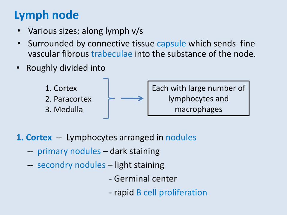
Lymph node • Various sizes; along lymph v/s
• Surrounded by connective tissue capsule which sends fine vascular fibrous trabeculae into the substance of the node.
Each with large number of lymphocytes and
macrophages
• Roughly divided into
1. Cortex 2. Paracortex 3. Medulla
1. Cortex -- Lymphocytes arranged in nodules
-- primary nodules – dark staining
-- secondry nodules – light staining
- Germinal center
- rapid B cell proliferation

2. Paracortex
-- Deep to the cortex
-- Primaily T lymphocytes and dendritic cells (a type of
phagocyte and a type of antigen-presenting cell (APC))
Lymph node
3. Medulla
-- Lymphocytes arranged in medullary cord
-- primarily – accumulation of plasma cells
Dendritic cell: A special type of immune cell that is found in tissues, such as the skin, and boosts immune responses by showing antigens on its surface to other cells of the immune system.
Plasma cell: A type of immune cell that makes large amounts of a specific antibody. Plasma cells develop from B cells that have been activated.
Stopped lecture

Structure of a typical lymph node
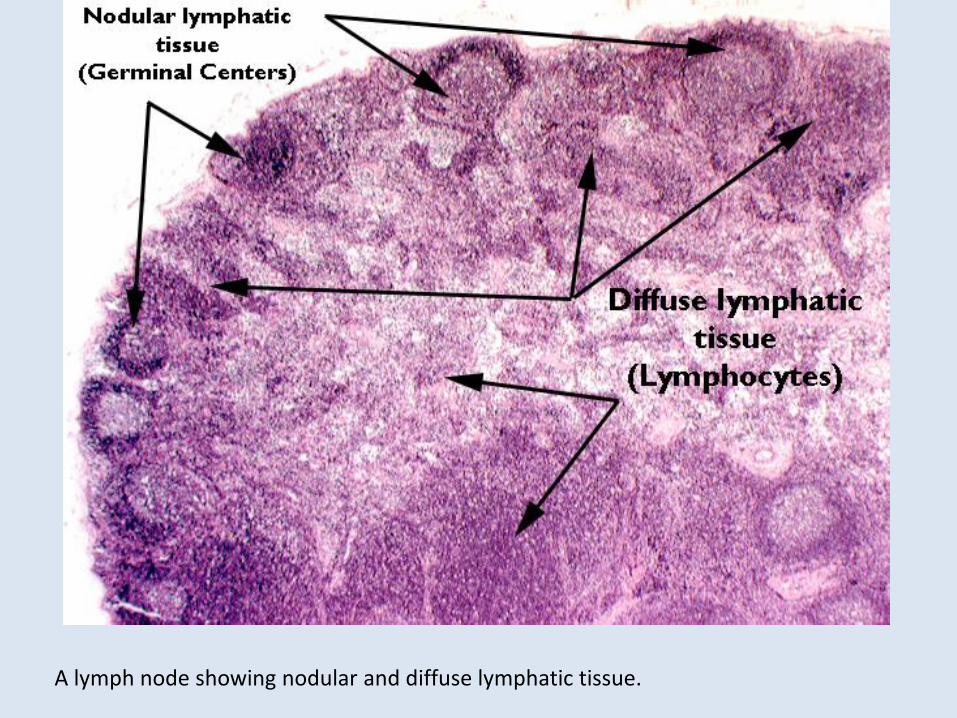
A lymph node showing nodular and diffuse lymphatic tissue.

Lymph node dog
1 = Capsule 8 = Medullary cord GC = Germinal center
2 = Cortical sinus 9 = Medullary sinus
4 = Deep cortex 12 = Subcapsular sinus
7 = Lymph nodule 13 = Trabeculae
GC

A Lymph node of cow
A Lymph node of Pig What are the differences?

Subcapsular sinus
-- space immediately deep to the capsule
-- communicate with other sinuses of cortex and medulla
-- lymph delivered by afferent lymph vessels enters subcapsular sinus and slowly filtered through the cortex and medulla
-- finally emerge at the hilus of the node
Lymph node

Lymphocytes of the lymph node
• T Cells (T lymphocytes) - attack foreign cells or body cells infected by viruses; T cells
mature and divide in the thymus - responsible for cell-mediated immunity (protection directly from
living cells) • B Cells (B lymphocytes) responsible for antibody-mediated immunity (=humoral
immunity); a percentage of circulating B lymphocytes mature into plasma cells; plasma cells produce and secrete antibodies (immunoglobulins) which destroy antigens
• NK Cells (natural killer cells) - attack foreign cells and cells infected
with viruses and cancer cells; also abnormal cells of body
LYMPHOCYTE and MEMORY
• Some B and T cells have what is called “memory”.
• Memory Cells have the ability to divide on short notice to produce more of all of the B and T cells.
• This is the basis of acquired immunity.
• B and T cells amplified in response to antigen are reserved and circulate in lymphatic system..for years or even life.
• If same antigen enters body again immune response will take place rapidly and without full-blown illness.

-- A LN may reflect local health condition
-- E.g. LN of infected area -- enlarged
-- Germinal centers produce additional lymphocytes in response to antigens (bacteria, virus) delivered to the node
-- Frequently associated with pain on palpationn
-- An enlargement of LN indicates infection
-- at least 99% of the pathogens in the lymph are removed
** Neoplastic (cancerous) cells may spread through the lymphatic channels.
Lymph node and infection

Five classes immunoglobulin (Ig)
IgG- active in blood against bacteria and viruses helps activate complement helps phagocytes eliminate antigens most common antibody in the blood can pass v/s and placenta IgM - reacts with certain antigens, usually on first exposure IgA - most common in mucosa
IgD -- rare in blood usually found on B cells (not released) may be involved in B cell activation IgE -- rare in blood involved in allergic reactions sticks to mast cells, which release inflammatory substances

Hemal node
• Small dark red or black nodes in cattle and sheep
• Usually located in the dorsal parts of the abdominal and thoracic cavities
• Resembles LN but re found on the course of blood vessels and contains only blood.

Has multiple interrelated functions: - responsible for the removal of interstitial fluid from tissues - absorbs and transports fatty acids and fats as chyle from the
digestive system - transports WBCs to and from the lymph nodes into the bones - transports antigen-presenting cells (APCs), such as dendritic
cells, to the lymph nodes where an immune response is stimulated.
- body’s most important defence mechanism against invasion by pathogens
- production of immunoglobulin - filters lymph and blood
Functions of lymphatic system

Spleen
- Largest lymphoid organ
Functions
- Contractile – expresses RBCs into the blood vessels
- The only organ to filter the blood
- An active destruction site for RBCs (MPS – mononuclear phagocytic system)
- storage of iron
- initiation of immune responses by B cells and T cells in response to antigens in circulating blood
- acts as a blood reservoir
• Not essential for life
• Splenectomy - bone marrow takes place its function

RED PULP vs. WHITE PULP:
Red pulp
- Area containing a large number of RBCs
- Structurally consists of a network of reticular fibers rich in macrophages
- mainly concerned with disposing of worn-out red blood cells and bloodborn pathogens
White pulp
- Area that resembles lymphoid nodules
- Composed mostly of lymphocytes suspended on reticular fibers
and involved with the immune functions of the spleen
Spleen

- An organ of immature animal
- Undergo involution at puberty
- Lies cranial to the heart
- Accumulation of lymphocytes (k/s thymocytes)
- Embryonic lymphocyte undergo differentiation and leave to populate many other lymphatic tissues of the body
Thymus

TONSILS
-- Unencapsulated aggregate of lymphoid nodules associated with the pharyngeal mucosa -- Lack afferent lymphatic vessels -- rely on the proximity of epithelial surface to make contact with antigens -- have crypts that increase surface area

PEYER’S PATCHES
-- Peyer's patches are clusters of lymphoid nodules deep to the epithelial lining of the small intestine -- Contain lymphocytes and macrophages which remove microorganisms, debris, and antigens from the digestive tract Note: Athough the terms tonsils and Peyer’s patches for phayrnx and small intestine respectively used, identical histological structures are found in the mucous membranes of prepuce and vagina etc.

Chicken
-- no lymph nodes -- Bursa of Fabricius : a sac like dorsal diverticulum of the proctodeum -- unique to birds. -- characterized by tall, thick mucosal folds (plicae)
filled with numerous polyhedral follicles. -- Each follicle, composed of lymphatic tissue, is
divided into a cortex and medulla.

Filtration and edema
• Recall lecture on flow
• The balance between pressure changes between arterioles and veinules (tissue space)


Edema
- Increased venous pressure leads to increased interstitial fluid volume (edema). - 3 counteracting effects (Negative feedback) against edema. 1. An increase in interstitial fluid
hydrostatic pressure reduces the rate of filtration back toward normal.
2. An increase in lymph flow reduces interstitial fluid volume back toward normal.
3. A decrease in interstitial fluid protein concentration reduces the rate of filtration back toward normal.


CIRCULATORY SYSTEMS
Cardiovascular Lymphatic Derived from mesoderm Derived from mesoderm Transport System Transport System Has a pump (heart) No pump Arteries No equivalent Veins for return Lymph vessels for return Veins have valves Lymph vessels have valves Carries RC, WBC, plasma Carries WBC, plasma