Embed Size (px)
Citation preview
Is the Pinworm Causing Acute Suppurative Perforating Appendicitis in Children?A Case SeriesSarah Magdy Abdelmohsen1* and Mohamed Abdelkader Osman2
1Pediatric Surgery, Aswan University Hospital, Egypt2Headmaster of Pediatric Surgery, Assiut University Hospital, Egypt*Corresponding author: Sarah Magdy Abdelmohsen, Pediatric Surgery, Aswan University Hospital, Egypt, Tel: +201012069422/+20224446921; E-mail:[email protected]; [email protected]
Received date: June 06, 2017; Accepted date: June 21, 2017; Published date: June 29, 2017
Copyright: © 2017 Abdelmohsen SM, et al. This is an open-access article distributed under the terms of the Creative Commons Attribution License, which permitsunrestricted use, distribution, and reproduction in any medium, provided the original author and source are credited.
Abstract
Introduction: The pinworm Enterobius vermicularis is the most common parasite reported to be associated withappendicitis. It frequently occurs in children between the age of 5 to 10 years. The aim of our study is to focus on therelationship between the pinworm and acute suppurative perforating appendicitis.
Case series: 4 children from rural areas of Upper Egypt with mean age 11 years presented to emergencydepartment Aswan and Assiut universities hospitals during the period from March 2016 to January 2017 complainedof right lower quadrant abdominal pain. The appendix in all children was perforated at the base or the middle of theappendix with mobile pinworm was noted moving around the perforated edge.
Discussion: Pinworm was associated with different pathologic changes ranging from lymphoid hyperplasia,acute appendicitis, and suppurative appendicitis to a normal appendix. It is wrangling whether pinworm can causeappendicitis or if they are an incidental finding during an appendectomy.
Conclusion: The awareness of pinworm and its preventive measures should be delivered to school children andtheir mothers to overcome the risk of infection and its dangerous drawback. The importance of histopathological andfaecolith examinations to the resected appendix specimens to detect the correct incidence of pinworm causingappendicitis in Egypt.
Keywords: Enterobius vermicularis; Pinworm; Suppurativeperforating appendicitis; Worm appendicitis in pediatrics; A caseseries
IntroductionThe association of Enterobius vermicularis, with acute appendicitis,
varies from 0.2 to 41.8% [1-9]. Infection is seen in all age groups andall socioeconomic classes but it is more common in children. Evermicularis is spread by feco-oral route and is associated with closeliving, poor hygiene, temperate and tropical climates [10,11]. Parasitescan be associated with the development of classic appendicitis. Inseries, there is a range of pathologic findings from nonspecific changesto frankly ruptured appendicitis [9]. The surgeon should understandthat the clinical management of these cases is different from that for anordinary appendicitis, as it requires treatment with antihelminthicdrugs.
Case Series4 children from rural areas of Upper Egypt with mean age 11 years
[10-12], presented to emergency department Aswan and AssiutUniversities Hospitals during the period from March 2016 to January2017, complained of right lower quadrant abdominal pain. The painwas sharp inconsistency, sometimes crampy. All children were livedwith their families and they were students. No significant medical orfamily histories were reported.
On examination, the children appeared pale but nontoxic. Theabdominal examination gives the typical picture of acute appendicitis,the pain localized to the right iliac fossa, tenderness at MC Burney'spoint, +ve Rovsing sing, +ve rebound tenderness. The pain associatedwith nausea, vomiting once or twice, proceeded by diarrhea 5 daysduration in two cases, no constipation. All children had a low-gradefever with temperature ranging (37.7°C-38.4°C), tachycardia pulse raterange (93-105) beats\minute; all children had elevated WBCs countrange (13,000-16,000) mcl, with no significant neutrophilia oreosinophilia. Additional preoperative laboratories finding are withinnormal limits.
Abdominal ultrasound reviled minimal fluid collection at the pelvicin 3 cases with visible pelvic appendix. Plain erect abdominal x raysshowed gaseous distension but no air fluid level or gas under thediaphragm.
A written consent obtained from the parent for appendectomyunder general endotracheal anesthesia. Experienced pediatric surgeonsmade appendectomy through McBurney's incision. The appendix in allchildren was perforated at the base or the middle of the appendix withmobile pinworm was noted moving around the perforated edge. Theappendixes were pelvic in position at 3 cases and retrocecal in onecase. Toilet and lavage made for the pelvic cavity to drainage theminimal amount of suppurative collection. Closure of the abdominalwall with pelvic drain did. The pathology reports revealedappendicular wall infiltrated by acute inflammatory cells and pus cells.
Abdelmohsen et al., J Clin Exp Pathol 2017, 7:3DOI: 10.4172/2161-0681.1000312
Case Report OMICS International
J Clin Exp Pathol, an open access journalISSN:2161-0681
Volume 7 • Issue 3 • 1000312
Jour
nal o
f Clin
ical & Experimental Pathology
ISSN: 2161-0681
Clinical & Experimental Pathology
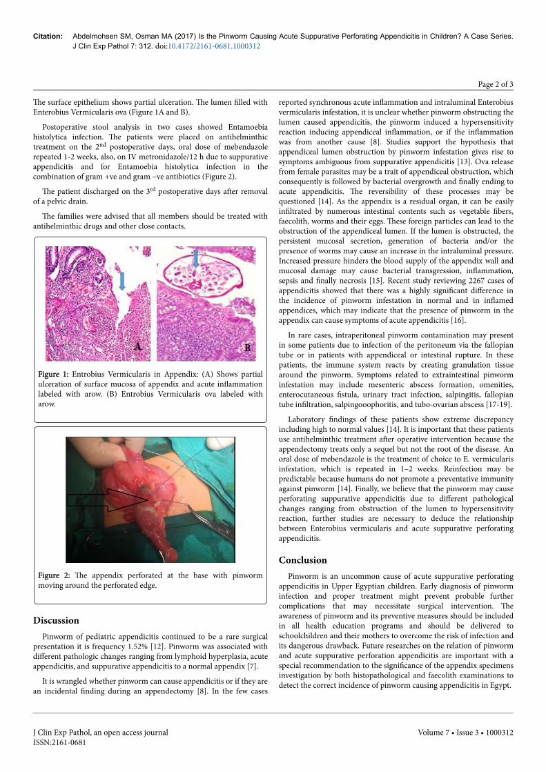
The surface epithelium shows partial ulceration. The lumen filled withEnterobius Vermicularis ova (Figure 1A and B).
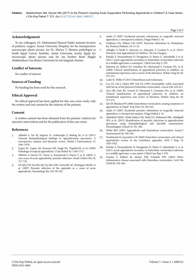
Postoperative stool analysis in two cases showed Entamoebiahistolytica infection. The patients were placed on antihelminthictreatment on the 2nd postoperative days, oral dose of mebendazolerepeated 1-2 weeks, also, on IV metronidazole/12 h due to suppurativeappendicitis and for Entamoebia histolytica infection in thecombination of gram +ve and gram –ve antibiotics (Figure 2).
The patient discharged on the 3rd postoperative days after removalof a pelvic drain.
The families were advised that all members should be treated withantihelminthic drugs and other close contacts.
Figure 1: Entrobius Vermicularis in Appendix: (A) Shows partialulceration of surface mucosa of appendix and acute inflammationlabeled with arow. (B) Entrobius Vermicularis ova labeled witharow.
Figure 2: The appendix perforated at the base with pinwormmoving around the perforated edge.
DiscussionPinworm of pediatric appendicitis continued to be a rare surgical
presentation it is frequency 1.52% [12]. Pinworm was associated withdifferent pathologic changes ranging from lymphoid hyperplasia, acuteappendicitis, and suppurative appendicitis to a normal appendix [7].
It is wrangled whether pinworm can cause appendicitis or if they arean incidental finding during an appendectomy [8]. In the few cases
reported synchronous acute inflammation and intraluminal Enterobiusvermicularis infestation, it is unclear whether pinworm obstructing thelumen caused appendicitis, the pinworm induced a hypersensitivityreaction inducing appendiceal inflammation, or if the inflammationwas from another cause [8]. Studies support the hypothesis thatappendiceal lumen obstruction by pinworm infestation gives rise tosymptoms ambiguous from suppurative appendicitis [13]. Ova releasefrom female parasites may be a trait of appendiceal obstruction, whichconsequently is followed by bacterial overgrowth and finally ending toacute appendicitis. The reversibility of these processes may bequestioned [14]. As the appendix is a residual organ, it can be easilyinfiltrated by numerous intestinal contents such as vegetable fibers,faecolith, worms and their eggs. These foreign particles can lead to theobstruction of the appendiceal lumen. If the lumen is obstructed, thepersistent mucosal secretion, generation of bacteria and/or thepresence of worms may cause an increase in the intraluminal pressure.Increased pressure hinders the blood supply of the appendix wall andmucosal damage may cause bacterial transgression, inflammation,sepsis and finally necrosis [15]. Recent study reviewing 2267 cases ofappendicitis showed that there was a highly significant difference inthe incidence of pinworm infestation in normal and in inflamedappendices, which may indicate that the presence of pinworm in theappendix can cause symptoms of acute appendicitis [16].
In rare cases, intraperitoneal pinworm contamination may presentin some patients due to infection of the peritoneum via the fallopiantube or in patients with appendiceal or intestinal rupture. In thesepatients, the immune system reacts by creating granulation tissuearound the pinworm. Symptoms related to extraintestinal pinworminfestation may include mesenteric abscess formation, omenities,enterocutaneous fistula, urinary tract infection, salpingitis, fallopiantube infiltration, salpingooophoritis, and tubo-ovarian abscess [17-19].
Laboratory findings of these patients show extreme discrepancyincluding high to normal values [14]. It is important that these patientsuse antihelminthic treatment after operative intervention because theappendectomy treats only a sequel but not the root of the disease. Anoral dose of mebendazole is the treatment of choice to E. vermicularisinfestation, which is repeated in 1–2 weeks. Reinfection may bepredictable because humans do not promote a preventative immunityagainst pinworm [14]. Finally, we believe that the pinworm may causeperforating suppurative appendicitis due to different pathologicalchanges ranging from obstruction of the lumen to hypersensitivityreaction, further studies are necessary to deduce the relationshipbetween Enterobius vermicularis and acute suppurative perforatingappendicitis.
ConclusionPinworm is an uncommon cause of acute suppurative perforating
appendicitis in Upper Egyptian children. Early diagnosis of pinworminfection and proper treatment might prevent probable furthercomplications that may necessitate surgical intervention. Theawareness of pinworm and its preventive measures should be includedin all health education programs and should be delivered toschoolchildren and their mothers to overcome the risk of infection andits dangerous drawback. Future researches on the relation of pinwormand acute suppurative perforation appendicitis are important with aspecial recommendation to the significance of the appendix specimensinvestigation by both histopathological and faecolith examinations todetect the correct incidence of pinworm causing appendicitis in Egypt.
Citation: Abdelmohsen SM, Osman MA (2017) Is the Pinworm Causing Acute Suppurative Perforating Appendicitis in Children? A Case Series.J Clin Exp Pathol 7: 312. doi:10.4172/2161-0681.1000312
Page 2 of 3
J Clin Exp Pathol, an open access journalISSN:2161-0681
Volume 7 • Issue 3 • 1000312
AcknowledgementTo my colleagues, Dr. Mohammed Elsayed Nader assistant lecturer
of pediatric surgery Aswan University Hospital, for his intraoperativemacroscopic photo picture, for Dr. Marwa T. Hussien pathologist atSouth Egypt Cancer Institute, Assiut University, Assiut, Egypt formicroscopic photo picture and for my brother Basel MagdyAbdelmohsen Ain Shams University for his languish checker.
Conflict of InterestsNo conflict of interest.
Sources of FundingNo funding has been used for this research.
Ethical ApprovalNo ethical approval has been applied for this case series study, only
the written and oral consent by the relatives of the patients.
ConsentA written consent has been obtained from the patients' relatives for
operative intervention and for the publication of this case series.
References1. Akbulut S, Tas M, Sogutcu N, Arikanoglu Z, Basbug M, et al. (2011)
Unusual histopathological findings in appendectomy specimens: Aretrospective analysis and literature review. World J Gastroenterol 15:1961-1970.
2. Gupta SC, Gupta AK, Keswani NK, Singh PA, TripathiAK, et al. (1989)Pathology of tropical appendicitis. J Clin Pathol 42: 1169-1172.
3. Yildirim S, Nursal TZ, Tarim A, Kayaselcuk F, Noyan T, et al. (2005) Arare cause of acute appendicitis: parasitic infection. Scand J Infect Dis 10:757-759.
4. Da Silva DF, Da Silva RJ, Da Silva MG, Sartorelli AC, Rodrigues MAM, etal. (2007) Parasitic infection of the appendix as a cause of acuteappendicitis. Parasitology Res 102: 99-102.
5. Aydin O (2007) Incidental parasitic infestations in surgically removedappendices: a retrospective analysis. Diagn Pathol 2: 16.
6. Goldman DA, Wilson CM (1997) Pinworm Infestation In: HoekelmanRa, Primary Pediatric 18: 13-24.
7. Akhigbe T, Smith F, Adeyemo A, Adeyanju T, Condon E, et al. (2013)Pinworm And Appendicitis In Children. The Int J Surgery.
8. Panidis S, Paramythiotis D, Panagiotou D, Batsis G, Salonikidis S et al.(2011) Acute appendicitis secondary to Enterobius vermicularis infectionin a middle aged man: a casereport. J Med Case Rep 5: 559.
9. Marjorie JA, Robert LG, Jonathan IG, Hammond S, Caniano DA, et al.(2004) Clinical manifestation of appendiceal pinworm in children: aninstitutional experience and a review of the literature. Pediatr Surg Int 20:372-375.
10. Leder K, Weller P (2011) Enterobiasis and trichuriasis.11. Leo LX, Chi J, Upton MP, Ash LR (1995) Eosinophilic colitis associated
with larvae of the pinworm Enterobius vermicularis. Lancet 346: 410-412.12. Arca MJ, Gate RL, Groner JI, Harmond S, Carniano DA, et al. (2004)
Clinical manifestation of appendiceal pinworm in children: aninstitutional experience and review of literature. Pediatr Surg Int 20:372-375.
13. Sah SP, Bhadani PP (2006) Enterobious vermicularis causing symptoms ofappendicitis in Nepal. Trop Doct 36: 160-162.
14. Aydin O (2007) Incidental parasitic infestations in surgically removedappendices: a retrospective analysis. Diagn Pathol 2: 16.
15. Abdellatif MZM, Abdel-Hafeez EH, Belal US, Mohamed RM, AbdelgelilNH, et al. (2015) Identification of parasitic infections in appendectomyspecimens using histopathological and faecolith examinations.Parasitologists United J 8: 101-106.
16. Wiebe BM (1991) Appendicitis and Enterobious vermicularis. Scand JGastroenterol 26: 336-338.
17. Nordstrand IA, Jayasekera LK (2004) Enterobius vermicularis and clinicalappendicitis: worms in the vermiform appendix. ANZ J Surg 74:1024-1025.
18. Panidis S, Paramythiotis D, Panagiotou D, Batsis G, Salonikidis S, et al.(2011) Acute appendicitis secondary to Enterobius vermicularis infectionin a middle aged man: a case report. J Med Case Rep 5: 559.
19. Tandan T, Pollard AJ, Money DM, Scheifele DW (2002) Pelvicinflammatory disease associated with Enterobius vermicularis. Arch DisChild 86: 439-440.
Citation: Abdelmohsen SM, Osman MA (2017) Is the Pinworm Causing Acute Suppurative Perforating Appendicitis in Children? A Case Series.J Clin Exp Pathol 7: 312. doi:10.4172/2161-0681.1000312
Page 3 of 3
J Clin Exp Pathol, an open access journalISSN:2161-0681
Volume 7 • Issue 3 • 1000312