Embed Size (px)
Citation preview
Volume 6 • Issue 9 10000875J Clin Case Rep, an open access journalISSN: 2165-7920
Open AccessCase Report
Isa and Mohamed, J Clin Case Rep 2016, 6:9DOI: 10.4172/2165-7920.1000875
Journal of Clinical Case ReportsJour
nal o
f Clinical Case Reports
ISSN: 2165-7920
*Corresponding author: Hasan M Isa, Pediatric Department, Arabian GulfUniversity, Manama, Bahrain, Tel: +973-66364449; Fax: +973-17279738;E-mail: [email protected]
Received August 30, 2016; Accepted September 24, 2016; Published September 30, 2016
Citation: Isa HM, Mohamed AM (2016) Abetalipoproteinemia: Three Case Reports, a Novel Microsomal Triglyceride Transfer Protein Gene Mutation and a Literature Review. J Clin Case Rep 6: 875. doi: 10.4172/2165-7920.1000875
Copyright: © 2016 Isa HM, et al. This is an open-access article distributed under the terms of the Creative Commons Attribution License, which permits unrestricted use, distribution, and reproduction in any medium, provided the original author and source are credited.
Abetalipoproteinemia: Three Case Reports, a Novel Microsomal Triglyceride Transfer Protein Gene Mutation and a Literature ReviewHasan M Isa1* and Afaf M Mohamed2
1Pediatric Department, Arabian Gulf University, Manama, Bahrain2Shaikh Jaber Health Centre, Manama, Bahrain
AbstractAbetalipoproteinemia (ABL, OMIM 200100) is a very rare metabolic disease with reported prevalence of less
than one case per 100,000. It is an autosomal recessive disease resulting from mutations in the gene encoding microsomal triglyceride transfer protein (MTP). Affected patients present with a wide range of clinical symptoms during infancy. Typical manifestations are failure to thrive, low level of cholesterol and fat malabsorption. Other features like fatty liver, acanthocytosis and anemia are usually present. Low fat diet and fat-soluble vitamins are the main stay of therapy. This is a retrospective review of three patients admitted to Salmaniya medical complex (SMC), Bahrain, with ABL. We presented the clinical presentations, diagnosis, response to medical therapy and outcome of these three infants along with a literature review about ABL. A novel MTP gene mutation, c.1508_1515delTGGCTACC (p.Leu503Hisfs*7, exon 11, MTTP), was detected. Two patients responded to dietary modifications and one deceased.
Keywords: Abetalipoproteinemia; Children; Novel; Mutation
IntroductionA beta lipoproteinemia (ABL; OMIM#200100) is a rare metabolic
autosomal recessive disorder [1-8]. The prevalence is ranging between less than 1 in 100,000 [2,6] and less than 1 in one million [3]. It is caused by mutations in the microsomal triglyceride transfer protein (MTP) gene [2,9,10]. MTP is the protein responsible for the transfer of triglycerides (TG), cholesteryl ester and phospholipids on to the apolipoprotein B (Apo B) [1]. Apo B promotes the secretion of chylomicrons and very low density lipoproteins (VLDL) from the enterocytes and hepatocytes [1]. Deficiency of the MTP genes results in absence of apo B-containing lipoproteins such as low density lipoproteins (LDL), VLDL and chylomicrons [2-4,9-11].
Patients with ABL usually presented during infancy with history of failure to thrive, severe diarrhea and lipid malabsorption syndrome [2,5,8]. They exhibit symptoms of intestinal lipid malabsorption and low plasma lipid levels [1,5,6]. In later childhood, they present with ataxic neuropathy or retinopathy [2,8]. Loss of fat soluble vitamins due to fat malabsorption might lead to hepatic, neurologic and ophthalmologic symptoms in the form of spinocerebellar degeneration, coagulopathy and pigmented retinopathy [1,3,8,12]. Hepatic steatosis, fibrosis and cirrhosis were also reported in some cases [5].
Early diagnosis and management of ABL patients is important in preventing manifestations of fat soluble vitamin deficiencies [8,9]. This article describes the clinical manifestations, diagnosis and response to therapy of three patients with ABL from Bahrain. A novel mutation of MTP gene has been described.
Case ReportCase 1
This patient is an eight-year-old male who presented to the Salmaniya medical complex (SMC) at the age of five months with a history of failure to thrive and vomiting since the age of one month. He was the first child of a consanguineous (first cousin) marriage. There was a history of one previous abortion. The antepartum course was uneventful. He was a full term baby with intrauterine growth retardation (IUGR) and a birth weight of 2.25 kgs. The patient was exclusively breast-fed initially but at the age of three months he was started on antiregurgitation milk formula as part of management of severe
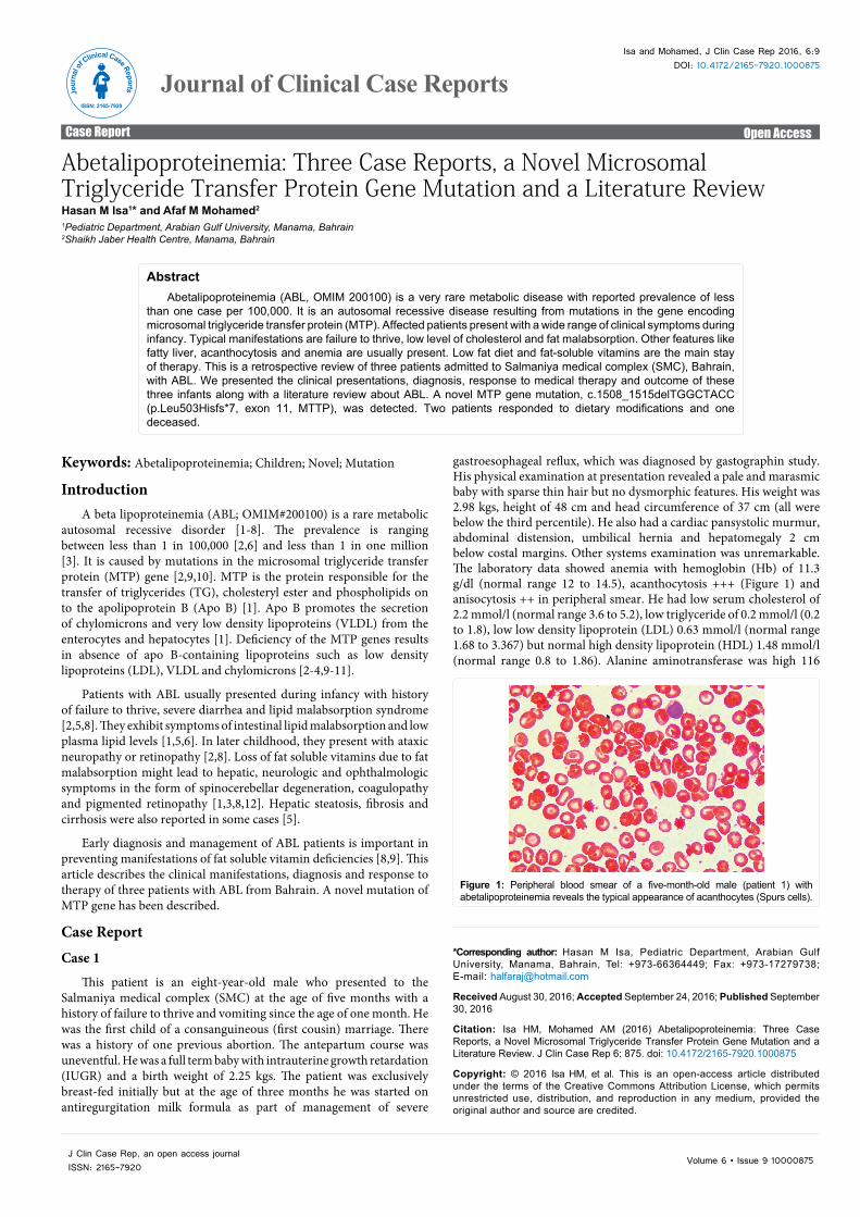
gastroesophageal reflux, which was diagnosed by gastographin study. His physical examination at presentation revealed a pale and marasmic baby with sparse thin hair but no dysmorphic features. His weight was 2.98 kgs, height of 48 cm and head circumference of 37 cm (all were below the third percentile). He also had a cardiac pansystolic murmur, abdominal distension, umbilical hernia and hepatomegaly 2 cm below costal margins. Other systems examination was unremarkable. The laboratory data showed anemia with hemoglobin (Hb) of 11.3 g/dl (normal range 12 to 14.5), acanthocytosis +++ (Figure 1) and anisocytosis ++ in peripheral smear. He had low serum cholesterol of 2.2 mmol/l (normal range 3.6 to 5.2), low triglyceride of 0.2 mmol/l (0.2 to 1.8), low low density lipoprotein (LDL) 0.63 mmol/l (normal range 1.68 to 3.367) but normal high density lipoprotein (HDL) 1.48 mmol/l (normal range 0.8 to 1.86). Alanine aminotransferase was high 116
Figure 1: Peripheral blood smear of a five-month-old male (patient 1) with abetalipoproteinemia reveals the typical appearance of acanthocytes (Spurs cells).
Citation: Isa HM, Mohamed AM (2016) Abetalipoproteinemia: Three Case Reports, a Novel Microsomal Triglyceride Transfer Protein Gene Mutation and a Literature Review. J Clin Case Rep 6: 875. doi: 10.4172/2165-7920.1000875
Page 2 of 5
Volume 6 • Issue 9 1000875J Clin Case Rep, an open access journalISSN: 2165-7920
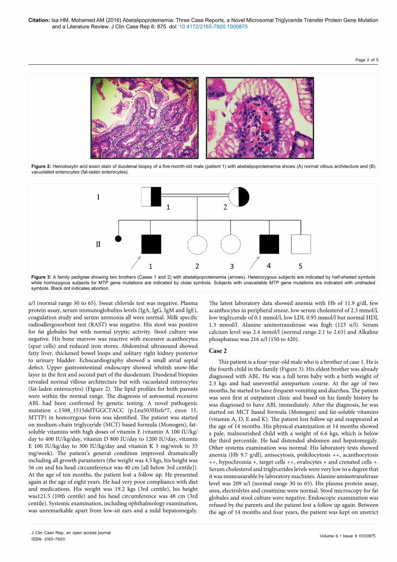
u/l (normal range 30 to 65). Sweat chloride test was negative. Plasma protein assay, serum immunoglobulins levels (IgA, IgG, IgM and IgE), coagulation study and serum ammonia all were normal. Milk specific radioallergosorbent test (RAST) was negative. His stool was positive for fat globules but with normal tryptic activity. Stool culture was negative. His bone marrow was reactive with excessive acanthocytes (spur cells) and reduced iron stores. Abdominal ultrasound showed fatty liver, thickened bowel loops and solitary right kidney posterior to urinary bladder. Echocardiography showed a small atrial septal defect. Upper gastrointestinal endoscopy showed whitish snow-like layer in the first and second part of the duodenum. Duodenal biopsies revealed normal villous architecture but with vacuolated enterocytes (fat-laden enterocytes) (Figure 2). The lipid profiles for both parents were within the normal range. The diagnosis of autosomal recessive ABL had been confirmed by genetic testing. A novel pathogenic mutation c.1508_1515delTGGCTACC (p.Leu503Hisfs*7, exon 11, MTTP) in homozygous form was identified. The patient was started on medium-chain triglyceride (MCT) based formula (Monogen), fat-soluble vitamins with high doses of vitamin E (vitamin A 100 IU/kg/day to 400 IU/kg/day, vitamin D 800 IU/day to 1200 IU/day, vitamin E 100 IU/kg/day to 300 IU/kg/day and vitamin K 5 mg/week to 35 mg/week). The patient’s general condition improved dramatically including all growth parameters (the weight was 4.5 kgs, his height was 56 cm and his head circumference was 40 cm [all below 3rd centile]). At the age of ten months, the patient lost a follow up. He presented again at the age of eight years. He had very poor compliance with diet and medications. His weight was 19.2 kgs (3rd centile), his height was121.5 (10th centile) and his head circumference was 48 cm (3rd centile). Systemic examination, including ophthalmology examination, was unremarkable apart from low-sit ears and a mild hepatomegaly.
The latest laboratory data showed anemia with Hb of 11.9 g/dl, few acanthocytes in peripheral smear, low serum cholesterol of 2.3 mmol/l, low triglyceride of 0.1 mmol/l, low LDL 0.95 mmol/l but normal HDL 1.3 mmol/l. Alanine aminotransferase was high (123 u/l). Serum calcium level was 2.4 mmol/l (normal range 2.1 to 2.63) and Alkaline phosphatase was 216 u/l (150 to 420).
Case 2
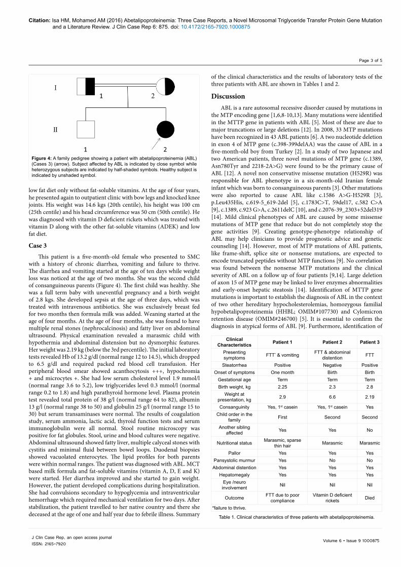
This patient is a four-year-old male who is a brother of case 1. He is the fourth child in the family (Figure 3). His eldest brother was already diagnosed with ABL. He was a full term baby with a birth weight of 2.3 kgs and had uneventful antepartum course. At the age of two months, he started to have frequent vomiting and diarrhea. The patient was seen first at outpatient clinic and based on his family history he was diagnosed to have ABL immediately. After the diagnosis, he was started on MCT based formula (Monogen) and fat-soluble vitamins (vitamin A, D, E and K). The patient lost follow up and reappeared at the age of 14 months. His physical examination at 14 months showed a pale, malnourished child with a weight of 6.6 kgs, which is below the third percentile. He had distended abdomen and hepatomegaly. Other systems examination was normal. His laboratory tests showed anemia (Hb 9.7 g/dl), anisocytosis, poikilocytosis ++, acanthocytosis ++, hypochromia +, target cells ++, ovalocytes + and crenated cells +. Serum cholesterol and triglycerides levels were very low to a degree that it was immeasurable by laboratory machines. Alanine aminotransferase level was 209 u/l (normal range 30 to 65). His plasma protein assay, urea, electrolytes and creatinine were normal. Stool microscopy for fat globules and stool culture were negative. Endoscopic examination was refused by the parents and the patient lost a follow up again. Between the age of 14 months and four years, the patient was kept on unstrict
Figure 2: Hemotoxylin and eosin stain of duodenal biopsy of a five-month-old male (patient 1) with abetalipoproteinemia shows (A) normal villous architecture and (B) vacuolated enterocytes (fat-laden enterocytes).
Figure 3: A family pedigree showing two brothers (Cases 1 and 2) with abetalipoproteinemia (arrows). Heterozygous subjects are indicated by half-shaded symbols while homozygous subjects for MTP gene mutations are indicated by close symbols. Subjects with unavailable MTP gene mutations are indicated with unshaded symbols. Black dot indicates abortion.
Citation: Isa HM, Mohamed AM (2016) Abetalipoproteinemia: Three Case Reports, a Novel Microsomal Triglyceride Transfer Protein Gene Mutation and a Literature Review. J Clin Case Rep 6: 875. doi: 10.4172/2165-7920.1000875
Page 3 of 5
Volume 6 • Issue 9 1000875J Clin Case Rep, an open access journalISSN: 2165-7920
low fat diet only without fat-soluble vitamins. At the age of four years, he presented again to outpatient clinic with bow legs and knocked knee joints. His weight was 14.6 kgs (20th centile), his height was 100 cm (25th centile) and his head circumference was 50 cm (50th centile). He was diagnosed with vitamin D deficient rickets which was treated with vitamin D along with the other fat-soluble vitamins (ADEK) and low fat diet.
Case 3 This patient is a five-month-old female who presented to SMC
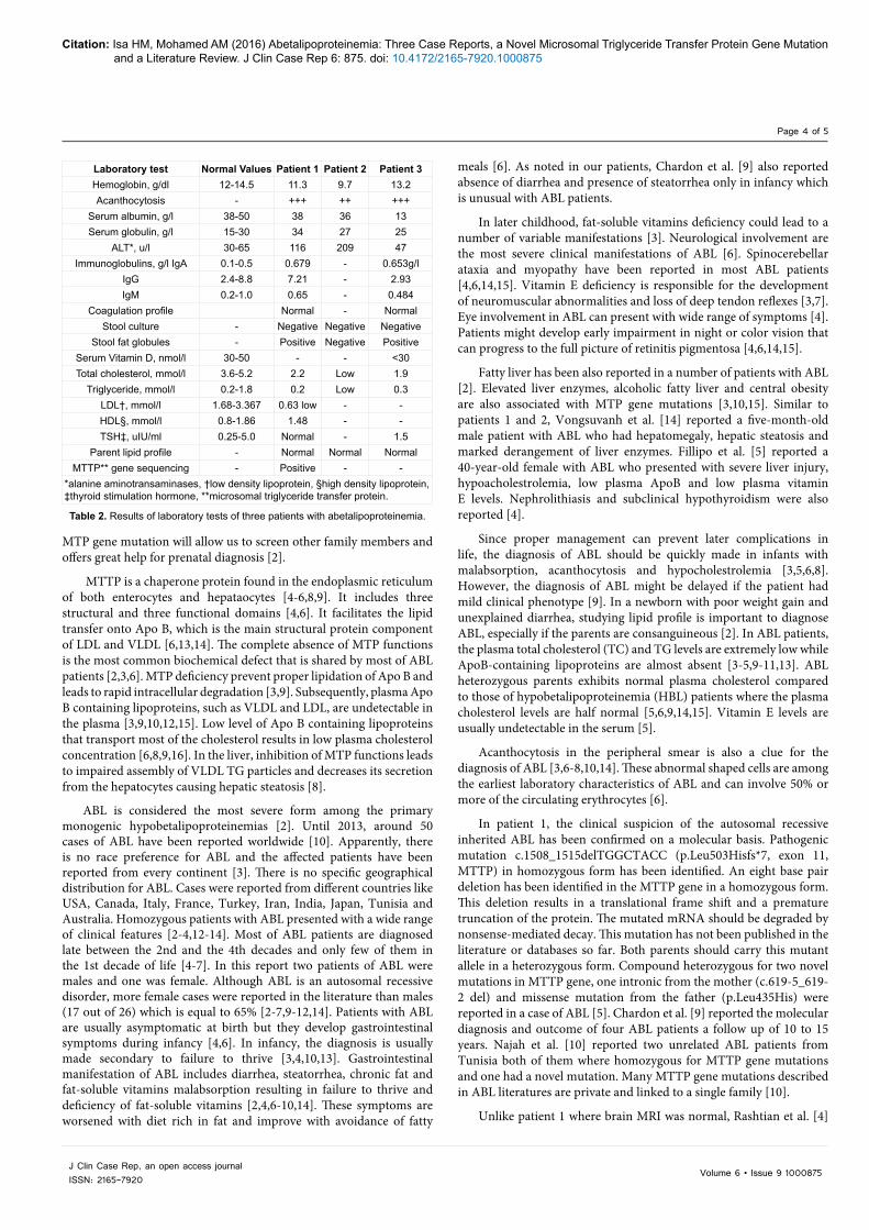
with a history of chronic diarrhea, vomiting and failure to thrive. The diarrhea and vomiting started at the age of ten days while weight loss was noticed at the age of two months. She was the second child of consanguineous parents (Figure 4). The first child was healthy. She was a full term baby with uneventful pregnancy and a birth weight of 2.8 kgs. She developed sepsis at the age of three days, which was treated with intravenous antibiotics. She was exclusively breast fed for two months then formula milk was added. Weaning started at the age of four months. At the age of four months, she was found to have multiple renal stones (nephrocalcinosis) and fatty liver on abdominal ultrasound. Physical examination revealed a marasmic child with hypothermia and abdominal distension but no dysmorphic features. Her weight was 2.19 kg (below the 3rd percentile). The initial laboratory tests revealed Hb of 13.2 g/dl (normal range 12 to 14.5), which dropped to 6.5 g/dl and required packed red blood cell transfusion. Her peripheral blood smear showed acanthocytosis +++, hypochromia + and microcytes +. She had low serum cholesterol level 1.9 mmol/l (normal range 3.6 to 5.2), low triglycerides level 0.3 mmol/l (normal range 0.2 to 1.8) and high parathyroid hormone level. Plasma protein test revealed total protein of 38 g/l (normal range 64 to 82), albumin 13 g/l (normal range 38 to 50) and globulin 25 g/l (normal range 15 to 30) but serum transaminases were normal. The results of coagulation study, serum ammonia, lactic acid, thyroid function tests and serum immunoglobulin were all normal. Stool routine microscopy was positive for fat globules. Stool, urine and blood cultures were negative. Abdominal ultrasound showed fatty liver, multiple calyceal stones with cystitis and minimal fluid between bowel loops. Duodenal biopsies showed vacuolated enterocytes. The lipid profiles for both parents were within normal ranges. The patient was diagnosed with ABL. MCT based milk formula and fat-soluble vitamins (vitamin A, D, E and K) were started. Her diarrhea improved and she started to gain weight. However, the patient developed complications during hospitalization. She had convulsions secondary to hypoglycemia and intraventricular hemorrhage which required mechanical ventilation for two days. After stabilization, the patient travelled to her native country and there she deceased at the age of one and half year due to febrile illness. Summary
of the clinical characteristics and the results of laboratory tests of the three patients with ABL are shown in Tables 1 and 2.
Discussion ABL is a rare autosomal recessive disorder caused by mutations in
the MTP encoding gene [1,6,8-10,13]. Many mutations were identified in the MTTP gene in patients with ABL [5]. Most of these are due to major truncations or large deletions [12]. In 2008, 33 MTP mutations have been recognized in 43 ABL patients [6]. A two nucleotide deletion in exon 4 of MTP gene (c.398-399delAA) was the cause of ABL in a five-month-old boy from Turkey [2]. In a study of two Japanese and two American patients, three novel mutations of MTP gene (c.1389, Asn780Tyr and 2218-2A>G) were found to be the primary cause of ABL [12]. A novel non conservative missense mutation (H529R) was responsible for ABL phenotype in a six-month-old Iranian female infant which was born to consanguineous parents [3]. Other mutations were also reported to cause ABL like c.1586 A>G-H529R [3], p.Leu435His, c.619-5_619-2del [5], c.1783C>T, 59del17, c.582 C>A [9], c.1389, c.923 G>A, c.2611delC [10], and c.2076-39_2303+52del319 [14]. Mild clinical phenotypes of ABL are caused by some missense mutations of MTP gene that reduce but do not completely stop the gene activities [9]. Creating genotype-phenotype relationship of ABL may help clinicians to provide prognostic advice and genetic counseling [14]. However, most of MTP mutations of ABL patients, like frame-shift, splice site or nonsense mutations, are expected to encode truncated peptides without MTP functions [9]. No correlation was found between the nonsense MTP mutations and the clinical severity of ABL on a follow up of four patients [9,14]. Large deletion of axon 15 of MTP gene may be linked to liver enzymes abnormalities and early-onset hepatic steatosis [14]. Identification of MTTP gene mutations is important to establish the diagnosis of ABL in the context of two other hereditary hypocholesterolemias, homozygous familial hypobetalipoproteinemia (HHBL; OMIM#107730) and Cylomicron retention disease (OMIM#246700) [5]. It is essential to confirm the diagnosis in atypical forms of ABL [9]. Furthermore, identification of
Figure 4: A family pedigree showing a patient with abetalipoproteinemia (ABL) (Cases 3) (arrow). Subject affected by ABL is indicated by close symbol while heterozygous subjects are indicated by half-shaded symbols. Healthy subject is indicated by unshaded symbol.
Clinical Characteristics Patient 1 Patient 2 Patient 3
Presenting symptoms FTT* & vomiting FTT & abdominal
distention FTT
Steatorrhea Positive Negative PositiveOnset of symptoms One month Birth Birth
Gestational age Term Term TermBirth weight, kg 2.25 2.3 2.8
Weight at presentation, kg 2.9 6.6 2.19
Consanguinity Yes, 1st casein Yes, 1st casein YesChild order in the
family First Second Second
Another sibling affected Yes Yes No
Nutritional status Marasmic, sparse thin hair Marasmic Marasmic
Pallor Yes Yes YesPansystolic murmur Yes No NoAbdominal distention Yes Yes Yes
Hepatomegaly Yes Yes YesEye /neuro involvement Nil Nil Nil
Outcome FTT due to poor compliance
Vitamin D deficient rickets Died
*failure to thrive.
Table 1. Clinical characteristics of three patients with abetalipoproteinemia.
Citation: Isa HM, Mohamed AM (2016) Abetalipoproteinemia: Three Case Reports, a Novel Microsomal Triglyceride Transfer Protein Gene Mutation and a Literature Review. J Clin Case Rep 6: 875. doi: 10.4172/2165-7920.1000875
Page 4 of 5
Volume 6 • Issue 9 1000875J Clin Case Rep, an open access journalISSN: 2165-7920
MTP gene mutation will allow us to screen other family members and offers great help for prenatal diagnosis [2].
MTTP is a chaperone protein found in the endoplasmic reticulum of both enterocytes and hepataocytes [4-6,8,9]. It includes three structural and three functional domains [4,6]. It facilitates the lipid transfer onto Apo B, which is the main structural protein component of LDL and VLDL [6,13,14]. The complete absence of MTP functions is the most common biochemical defect that is shared by most of ABL patients [2,3,6]. MTP deficiency prevent proper lipidation of Apo B and leads to rapid intracellular degradation [3,9]. Subsequently, plasma Apo B containing lipoproteins, such as VLDL and LDL, are undetectable in the plasma [3,9,10,12,15]. Low level of Apo B containing lipoproteins that transport most of the cholesterol results in low plasma cholesterol concentration [6,8,9,16]. In the liver, inhibition of MTP functions leads to impaired assembly of VLDL TG particles and decreases its secretion from the hepatocytes causing hepatic steatosis [8].
ABL is considered the most severe form among the primary monogenic hypobetalipoproteinemias [2]. Until 2013, around 50 cases of ABL have been reported worldwide [10]. Apparently, there is no race preference for ABL and the affected patients have been reported from every continent [3]. There is no specific geographical distribution for ABL. Cases were reported from different countries like USA, Canada, Italy, France, Turkey, Iran, India, Japan, Tunisia and Australia. Homozygous patients with ABL presented with a wide range of clinical features [2-4,12-14]. Most of ABL patients are diagnosed late between the 2nd and the 4th decades and only few of them in the 1st decade of life [4-7]. In this report two patients of ABL were males and one was female. Although ABL is an autosomal recessive disorder, more female cases were reported in the literature than males (17 out of 26) which is equal to 65% [2-7,9-12,14]. Patients with ABL are usually asymptomatic at birth but they develop gastrointestinal symptoms during infancy [4,6]. In infancy, the diagnosis is usually made secondary to failure to thrive [3,4,10,13]. Gastrointestinal manifestation of ABL includes diarrhea, steatorrhea, chronic fat and fat-soluble vitamins malabsorption resulting in failure to thrive and deficiency of fat-soluble vitamins [2,4,6-10,14]. These symptoms are worsened with diet rich in fat and improve with avoidance of fatty
meals [6]. As noted in our patients, Chardon et al. [9] also reported absence of diarrhea and presence of steatorrhea only in infancy which is unusual with ABL patients.
In later childhood, fat-soluble vitamins deficiency could lead to a number of variable manifestations [3]. Neurological involvement are the most severe clinical manifestations of ABL [6]. Spinocerebellar ataxia and myopathy have been reported in most ABL patients [4,6,14,15]. Vitamin E deficiency is responsible for the development of neuromuscular abnormalities and loss of deep tendon reflexes [3,7]. Eye involvement in ABL can present with wide range of symptoms [4]. Patients might develop early impairment in night or color vision that can progress to the full picture of retinitis pigmentosa [4,6,14,15].
Fatty liver has been also reported in a number of patients with ABL [2]. Elevated liver enzymes, alcoholic fatty liver and central obesity are also associated with MTP gene mutations [3,10,15]. Similar to patients 1 and 2, Vongsuvanh et al. [14] reported a five-month-old male patient with ABL who had hepatomegaly, hepatic steatosis and marked derangement of liver enzymes. Fillipo et al. [5] reported a 40-year-old female with ABL who presented with severe liver injury, hypoacholestrolemia, low plasma ApoB and low plasma vitamin E levels. Nephrolithiasis and subclinical hypothyroidism were also reported [4].
Since proper management can prevent later complications in life, the diagnosis of ABL should be quickly made in infants with malabsorption, acanthocytosis and hypocholestrolemia [3,5,6,8]. However, the diagnosis of ABL might be delayed if the patient had mild clinical phenotype [9]. In a newborn with poor weight gain and unexplained diarrhea, studying lipid profile is important to diagnose ABL, especially if the parents are consanguineous [2]. In ABL patients, the plasma total cholesterol (TC) and TG levels are extremely low while ApoB-containing lipoproteins are almost absent [3-5,9-11,13]. ABL heterozygous parents exhibits normal plasma cholesterol compared to those of hypobetalipoproteinemia (HBL) patients where the plasma cholesterol levels are half normal [5,6,9,14,15]. Vitamin E levels are usually undetectable in the serum [5].
Acanthocytosis in the peripheral smear is also a clue for the diagnosis of ABL [3,6-8,10,14]. These abnormal shaped cells are among the earliest laboratory characteristics of ABL and can involve 50% or more of the circulating erythrocytes [6].
In patient 1, the clinical suspicion of the autosomal recessive inherited ABL has been confirmed on a molecular basis. Pathogenic mutation c.1508_1515delTGGCTACC (p.Leu503Hisfs*7, exon 11, MTTP) in homozygous form has been identified. An eight base pair deletion has been identified in the MTTP gene in a homozygous form. This deletion results in a translational frame shift and a premature truncation of the protein. The mutated mRNA should be degraded by nonsense-mediated decay. This mutation has not been published in the literature or databases so far. Both parents should carry this mutant allele in a heterozygous form. Compound heterozygous for two novel mutations in MTTP gene, one intronic from the mother (c.619-5_619-2 del) and missense mutation from the father (p.Leu435His) were reported in a case of ABL [5]. Chardon et al. [9] reported the molecular diagnosis and outcome of four ABL patients a follow up of 10 to 15 years. Najah et al. [10] reported two unrelated ABL patients from Tunisia both of them where homozygous for MTTP gene mutations and one had a novel mutation. Many MTTP gene mutations described in ABL literatures are private and linked to a single family [10].
Unlike patient 1 where brain MRI was normal, Rashtian et al. [4]
Laboratory test Normal Values Patient 1 Patient 2 Patient 3Hemoglobin, g/dl 12-14.5 11.3 9.7 13.2Acanthocytosis - +++ ++ +++
Serum albumin, g/l 38-50 38 36 13Serum globulin, g/l 15-30 34 27 25
ALT*, u/l 30-65 116 209 47Immunoglobulins, g/l IgA 0.1-0.5 0.679 - 0.653g/l
IgG 2.4-8.8 7.21 - 2.93IgM 0.2-1.0 0.65 - 0.484
Coagulation profile Normal - NormalStool culture - Negative Negative Negative
Stool fat globules - Positive Negative PositiveSerum Vitamin D, nmol/l 30-50 - - <30Total cholesterol, mmol/l 3.6-5.2 2.2 Low 1.9
Triglyceride, mmol/l 0.2-1.8 0.2 Low 0.3 LDL†, mmol/l 1.68-3.367 0.63 low - -HDL§, mmol/l 0.8-1.86 1.48 - -TSH‡, uIU/ml 0.25-5.0 Normal - 1.5
Parent lipid profile - Normal Normal NormalMTTP** gene sequencing - Positive - -
*alanine aminotransaminases, †low density lipoprotein, §high density lipoprotein, ‡thyroid stimulation hormone, **microsomal triglyceride transfer protein.
Table 2. Results of laboratory tests of three patients with abetalipoproteinemia.
Citation: Isa HM, Mohamed AM (2016) Abetalipoproteinemia: Three Case Reports, a Novel Microsomal Triglyceride Transfer Protein Gene Mutation and a Literature Review. J Clin Case Rep 6: 875. doi: 10.4172/2165-7920.1000875
Page 5 of 5
Volume 6 • Issue 9 1000875J Clin Case Rep, an open access journalISSN: 2165-7920
reported the presence of an intraventricular cyst. Patient 1 also had solitary/fused ectopic kidneys, a finding that has not been previously reported in any ABL patient.
Similar to most reported cases [4], our patients had typical features of ABL in the intestinal biopsies. In patients with ABL, intestinal and liver biopsies are characterized by the presence of large amounts of free lipid droplets accumulated in the cytoplasm of enterocytes and hepatocytes at the ultrastructural levels [5,6].
Management of ABL includes dietary fat restrictions, fat-soluble vitamins supplementations, specific formula and polycitrate [4,6,7]. Low fat diet will improve steatorrhea and allow absorption of other vital nutrients needed for normal growth and development [6,15]. Patients with ABL are treated with a low-fat diet (~15 g/d), in order to improve fat malabsorption, and fat-soluble vitamins supplementation (high doses of vitamin E 2400 to 12000 IU) [3,6,8,9].
The use of ultra-long-term high-dose of oral vitamin therapy, including vitamin E, can stop neurological complications in at least some ABL patients [6]. High doses of fat-soluble vitamins are expected to bypass the intestinal chylomicron assembly pathway through the portal circulation [6,15]. Additional supplementation with vitamin A (100 IU/Kg/d to 400 IU/Kg/d) and vitamin D (1000 mg/d) should be considered in all ABL patients [6,7]. Unlike vitamin E supplementation where high doses can only improve the serum level by no more than 30% of the lower limit of normal, high doses of vitamin A can normalize the serum level [6]. ABL patients who have tremors and ataxia may benefit from staged bilateral thalamic deep brain stimulation (DBS) [11]. Serum vitamin E levels can be used to monitor compliance and sufficiency of therapy [6]. Similar to patient 2, Hasosah et al. [17] reported an 18-months-old male with ABL who presented with rickets. Vitamin D deficiency is not constantly described in ABL patients [6]. However, vitamin D supplementation should be always considered along with iron and folate [15]. Fat-soluble vitamins levels should be monitored periodically to avoid toxicity [3,7].
Patients with ABL are prone for essential fatty acids and fat-soluble vitamin deficiencies especially vitamin E [5,6,9,11,13]. This is complicated by ocular and neurological manifestations [7]. Vitamin E deficiency can lead to retinal degeneration and many neurological complications like hyporeflexia, reduced proprioceptive and vibratory sense, and ataxia [7,11,13]. Very low vitamin E levels (<1 µmol/L) not only causes significant neurological damage, but does so even at an early age during infancy [14]. Deafness was also reported in one patient with ABL [7]. Bleeding diathesis secondary to vitamin K deficiency is another complication [6,8]. ABL is not a primary disorder of lipid metabolism alone but also considered as an immune disease [1]. In ABL patients, the primary genetic deficiency of MTP is associated with loss of CD1 function which is a previously unrecognized immune defect [1,3].
Without treatment, symptoms of ABL can be debilitating leading to reduced life expectancy [6]. Factors that can affect the outcome of ABL patients include MTTP genotype, age at diagnosis and age at starting low fat diet and fat-soluble vitamins [6]. The natural history of ABL and the genotype-phenotype correlations are difficult to define due to paucity of reported cases [2]. Early diagnosis can ensure success of nutritional therapeutic strategy based on high vitamin E supplementations and good outcome [9]. Early presentation may be related to more severe phenotype that might be resistant to treatment and have a poor outcome [4]. However, late presentation and longer period of untreated disease is also associated with poor outcomes secondary to fat-soluble vitamins deficiency effects [4].
ConclusionAlthough ABL is a rare disease, it is treatable. Early diagnosis
and proper treatment of patients with ABL are necessary to prevent the devastating long-term serious clinical sequelae. We have described a novel MTP gene mutation, c.1508_1515delTGGCTACC (p.Leu503Hisfs*7, exon 11, MTTP), in homozygous form; in an infant with ABL phenotype.
References
1. Zeissig S, Dougan SK, Barral DC, Junker Y, Chen Z, et al. (2010)Primary deficiency of microsomal triglyceride transfer protein in human abetalipoproteinemia is associated with loss of CD1 function. J Clin Invest 120: 2889-2899.
2. Uslu N, Gurakan F, Yuce A, Demir H, Tarugi P (2010) Abetalipoproteinemia inan infant with severe clinical phenotype and a novel mutation. Turk J Pediatr52: 73-77.
3. Sani MN, Sabbghian M, Mahjoob F, Cefalu AB, Averna MR, et al.(2011). Identification of a novel mutation of MTP gene in a patient with abetalipoproteinemia. Ann Hepatol 10: 221-226.
4. Rashtian P, Sani MN, Jalilian R (2015) A male infant with abetalipoproteinemia: A case report from Iran. Middle East J Dig Dis 7: 181-184.
5. Di Filippo M, Créhalet H, Samson-Bouma ME, Bonnet V, Aggerbeck L, et al.(2012) Molecular and functional analysis of two new MTTP gene mutations inan atypical case of abetalipoproteinemia. J Lipid Res 53: 548-555.
6. Zamel R, Khan R, Pollex RL, Hegele RA (2008) Abetalipoproteinemia: two case reports and literature review. Orphanet J Rare Dis 3: 19.
7. Nagapa M, Bindu PS, Adwani S, Seshagiri SK, Saini J, et al. (2014) Clinical,hematological, and imaging observations in a 25-year-old woman withabetalipoproteinemia. Ann Indian Acad Neurol 17: 113-116.
8. Welty FK (2014) Hypobetalipoproteinemia and abetalipoproteinemia. Curr Opin Lipidol 25: 161-168.
9. Chardon L, Sassolas A, Dingeon B, Michel-Calemard L, Bovier-Lapierre M,et al. (2009) Identification of two novel mutations and long-term follow-up in abetalipoproteinemia: A report of four cases. Eur J Pediar 168: 983-989.
10. Najah M, Youssef SM, Yahia HM, Afef S, Awatef J, et al. (2013) Molecularcharacterization of Tunisian families with abetalipoproteinemia and identification of a novel mutation in MTTP gene. Diagn Pathol 8: 54.
11. Mammis A, Pourfar M, Feigin A, Mogilner AY (2012) Deep brain stimulationfor the treatment of tremor and ataxia associated with abetalipoproteinemia.Tremor Other Hyperkinet Mov.
12. Ohashi K, Ishibashi S, Osuga J, Tozawa R, Harada K, et al. (2000) Novelmutation in the microsomal triglyceride transfer protein gene causingabetalipoproteinemia. J Lipid Res 41: 1199-1204.
13. Khatun I, Walsh MT, Hussain MM (2013) Loss of both phospholipid andtriglyceride transfer activities of microsomal triglyceride transfer protein inabetalipoproteinemia. J Lipid Res 54: 1541-1549.
14. Vongsuvanh R, Hooper AJ, Coakley JC, Macdessi JS, Loughlin EV, et al.(2007) Novel mutations in abetalipoproteinaemia and homozygous familialhypobetalipoproteinaemia. J Inherit metab dis 30: 990.
15. Burnett JR, Bell DA, Hooper AJ, Hegele RA (2012) Clinical utility gene card for: abetalipoproteinemia. Eur J Hum Genet 20.
16. Seckeler MD, Linden J (2008) Maternal abetalipoproteinemia resulting inmultiple fetal anomalies. Neonatol 94: 310-313.
17. Hasosah MY, Shesha SJ, Sukkar GA, Bassuni WY (2010) Rickets anddysmorphic findings in a child with abetalipoproteinemia. Saudi Med J 31: 1169-1171.