Embed Size (px)
Citation preview
L-SPINE
CERVICAL SPINE: C1 exits above C1 vert body
THORACIC & LUMBAR SPINE: T1 nerve exits below T1 at T1-T2 and similarly, L1 nerve exists below L1 at L1-2;
at L3-4 level exiting L3 nerve root within NF and originating L4 nerve root within lateral recess.
At C1-2 --> C2 exits
At C7-T1 --> C8 exits
At T1-T2 -->T1 exits
At T12-L1 -->T12 exits
At L1-2-->L1 exits
At L5-S1-->L5 exits
Sag T1 (LR)
-Alignment (listhesis: ant/post/lat)
-Loss of intervertebral height
-Marrow abnl (compare w/ muscle)
-Disc bulge
-NF narrowing
-Pars defect
-Cong canal stenosis or short pedicle (10mm AP for L-spine; 12mm for C-spine)
Sag T2 STIR
-Conus medullaris (terminates at approp level; above L3 at 3mos)
-Cord signal (confirm on axial T2)
-Disc dessication
-Marrow abnl
-Annular tear/fissure
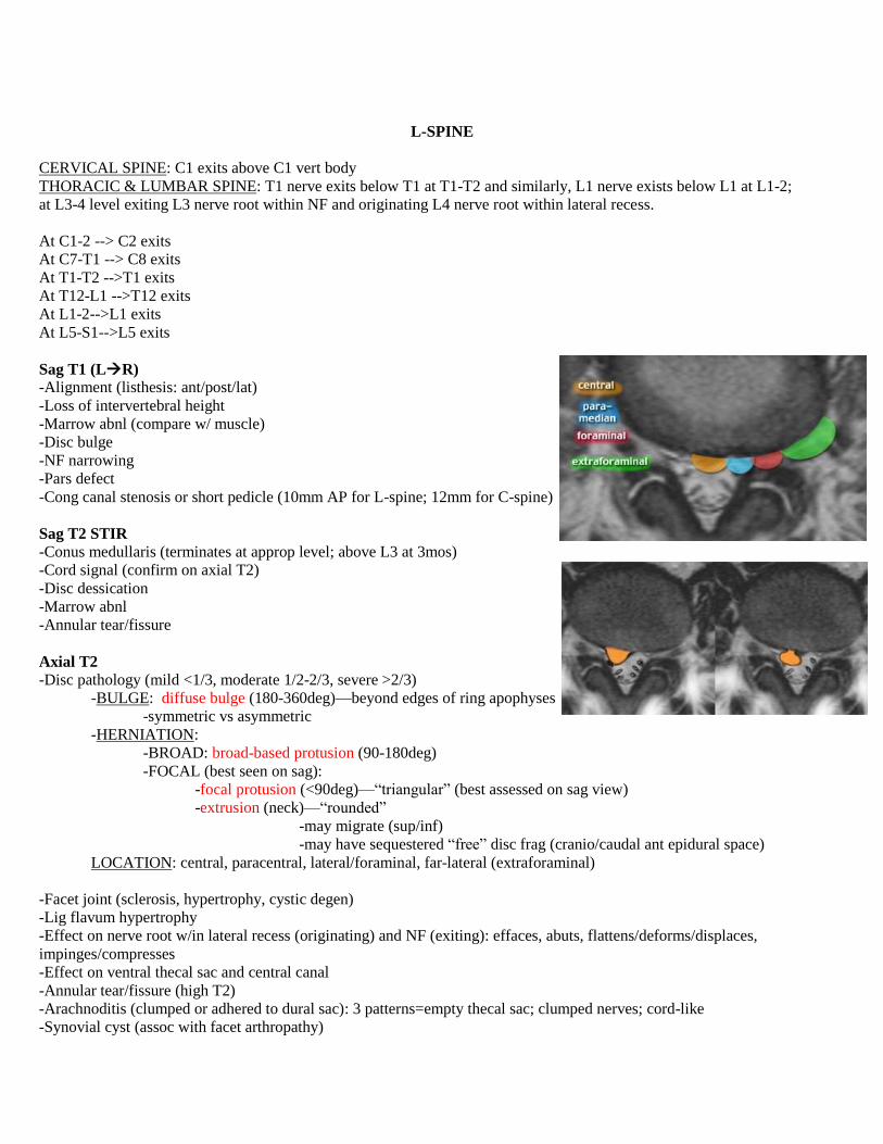
Axial T2
-Disc pathology (mild <1/3, moderate 1/2-2/3, severe >2/3)
-BULGE: diffuse bulge (180-360deg)—beyond edges of ring apophyses
-symmetric vs asymmetric
-HERNIATION:
-BROAD: broad-based protusion (90-180deg)
-FOCAL (best seen on sag):
-focal protusion (<90deg)—“triangular” (best assessed on sag view)
-extrusion (neck)—“rounded”
-may migrate (sup/inf)
-may have sequestered “free” disc frag (cranio/caudal ant epidural space)
LOCATION: central, paracentral, lateral/foraminal, far-lateral (extraforaminal)
-Facet joint (sclerosis, hypertrophy, cystic degen)
-Lig flavum hypertrophy
-Effect on nerve root w/in lateral recess (originating) and NF (exiting): effaces, abuts, flattens/deforms/displaces,
impinges/compresses
-Effect on ventral thecal sac and central canal
-Annular tear/fissure (high T2)
-Arachnoditis (clumped or adhered to dural sac): 3 patterns=empty thecal sac; clumped nerves; cord-like
-Synovial cyst (assoc with facet arthropathy)

-Epidural lipomatosis (narrows/compresses thecal sac)
MISC aorta/iliacs, kidneys, SI joints, pelvis organs
POSTOP FAILED BACK spine: Gad with fat sat (alternative=CT myelography); >6wks-6mos; fibrosis/scar enhances, disk
only has rim-enhancement.
DDX:
-epidural scar/fibrosis (enhances)
-recurrent/residual/new disk prolapse (does not enhance but peripheral enhancement due to surrounding fibrosis)
-inadequate decompression of nerve root
-archnoiditis (clumped centrally vs adherent peripherally to wall w/ empty sac appearance vs cord-like central mass)
-radiculitis (enhancing nerve root)
-hematoma/seroma at laminectomy site (assess for thecal sac compression or extension into site of bone graft at facets which
may impair proper fusion)
-epidural abscess (if immediate post-op)
-pseudomeningocele (due to dural tear)
-hardware failure
-mechanical instability aka subluxation (if not fused)
VERT FX VS TUMOR: fat, air, post-el involvement, multifocal, ST component, post bulge; sup-end plate depressed fx w/
localized edema; chronic compression fx has preserved vert fat but acute is hypointense no T1
TUMOR: Look for focal/circumferential tumor extension with paraspinal or anterior epidural component; degree of spinal
canal compromise (thecal sac deformation vs cord contact vs cord compression vs partial/complete effacement of CSF) and
spinal instability which may need surgical decompression and hardware prior to XRT; look for impending or current fracture
SAMPLE: Alignment is normal without listhesis. No abnormal marrow signal. Conus medullaris terminates at approp level.
Thecal sac and contents are normal. No disc herniation, nerve root impingement/compression, or neuroforaminal/CC stenosis.
No facet arthropathy or LF hypertrophy.
Disc height loss or disc space narrowing. 3mm Grade I anterolisthesis of L3 on L4. Degenerative Modic end-late
changes/signal. Diffuse disc bulge (eccentric to left); Central/left paracentral disc protrusion; Focal disc extrusion measuring
5mm and extending cranially/caudally; which along with LF hypertrophy and facet arthropathy results in mild/mod/sig CC
stenosis and mild to moderate bilateral neuroforaminal stenosis and right lateral recess stenosis.
For purpose of this dictation, the inferior most level of fully formed intervertebral disk will be referred to as L5/S1 or
L6/S1.

Disc Dessic,
Disc space
narrowing,
Listhesis,
Hemangioma
Bulge/herniation
Location/type
Annular
Fissure
Lig
Flavum
Hypertr
Facet
Hypertr/
Arthropathy
Effect
on
ventral
thecal
sac
Effect on
lateral
recess
(orig NR)
Effect on
NF (exit
NR)
Central
Canal
Stenosis
L1-2
L2-3
L3-4
L4-5
L5-S1
-Alignment (listhesis)
-Pars defect
-Marrow signal (modic changes/hemangioma/schmorl)
-Disc space narrowing (disc height loss)
-Disc dessication
-Annular tear/fissure
-Conus medullaris and cauda equina
-Arachanoditis
-Misc: Aorta/iliacs, Kidneys, SI joints, Pelvis organs
Disc dessication.
[severity] disk space narrowing +/- endplate spondylosis
[Central/paracentral/lateral or foraminal/far-lateral or extraforaminal] [diffuse disk bulge] vs [broad-based herniation/
protusion//extrusion (neck) +/-migration/sequestered or free disk fragment located cranio/caudal ant epidural space]
Lig flavum hypertrophy
Facet arthropathy
[abuts/flattens, effaces/displaces ventral thecal sac]
[abuts/effaces lateral recess resulting in originating NR impingement/compression]
[results in neuroforaminal narrowing with exiting NR impingement/compression; but NR exits freely]
[central canal stenosis]
NORMAL VARIANTS:
-epidural lipomatosis (encases thecal sac with loss of CSF signal around cauda equine)
-fatty filum terminale
-terminal ventricle (5th vent; dilated central canal extends below tip of conus into filum)
-congenitally short pedicles (narrowed spinal canal)
-dilated nerve sheath (tarlov cyst)
-paraspinal muscle atrophy
-synovial cyst (facet arthropathy)
-pars defect

-yellow marrow around basivertebral vein (baxton’s plexus)
-heterogenous marrow w/ intermixed red and yellow marrow
-hemangioma (T1 bright; may enhance)
-intraosseous lipoma
C-SPINE
8 cervical nerves, 7 exit above pedicle of respective vertebra, 8th exits below level of it vertebra (i.e. T1); Brachial
plexus C5 – C8/T1. C1 exits below occiput; C4-5 herniated disc will involve C5 nerve root (not so in L-spine)
Axial GRE are good for eval of discogenic ridging (GRE often able to distinguish between disc which is relatively
bright vs osteophyte which is dark). Cong canal stenosis or short pedicle (10mm AP for L-spine; 12mm for C-spine)
Disk-
osteophyte
Complex
Uncovertebral
Hypertrophy;
facet
arthopathy
Ventral
thecal sac
effacement
Cord abuts/
displaces/flattens/
deforms/
compresses
Cord signal
(myelopathy;
myelomalacia)
Central canal
stenosis
NF
narrowing
C2-3
C3-4
C4-5
C5-6
C6-7
C7-T1
-Alignment: spondylolithesis
-Marrow signal:
-Disc height:
-Cord signal alteration:
-Prevertebral ST:
-Paraspinal ST:
-Misc: Posterior fossa (tonsils), Pituitary, Thyroid, Airways, Lung apex
Vertebral alignment is normal w/o lithesis. BM signal is unremarkable.
Atlantoaxial DJD with thickened and post bulging PLL.
[severity] disk space narrowing +/- endplate spondylosis
Uncovertebral hypertrophy; [severity][diffuse/central /paracentral] disk-osteophyte complex or disk protusion
[abuts/effaces] ventral thecal sac
and results in [severity] CC stenosis (w/ residual AP diameter of x mm)
and [left/right/bilat] [severity] NF narrowing.
No cord compression or abnormal cord signal.
Effaces ventral thecal sacAbuts ventral cordDisplaces cordFlattens ventral cordCompresses/Deforms cord
Don’t talk about nerve root impingement.
NORMAL VARIANT:
-atlanto-occ sublux
-unfused ant/post arch of C1
-odontoid cleft
-os odontoideum

-cong fusion of vert body or facet jt
-nerve root sleeve cyst
-CSF pulsation (flow artifact)
-intradural lipoma
-extradural arachnoid cyst (remodels osseous str)
CORD SIGNAL:
-cord edema
-compressive myelopathy (cord compression)
-myelomalacia (chronic compression; usually with atrophy)
-neoplasm (astrocytoma vs ependymoma) vs demyelination vs transverse myelitis
DEGREE OF CORD COMPRESSION:
-mild 8-10mm AP central canal
-mod 6-8mm AP central canal
-severe <6mm AP central canal
Radiculopathy vs Myelopathy?
Radiculopathy: a) Young patients get herniation (acute onset; pain with sitting)
b) Older patients get spondylosis w/ lateral recess stenosis (insidious onset; pain better w/ sitting)
Myelopathy: a) Cervical (hyperreflexia, spasticity)
b) Lumbar (parasthesia, pain, +/-weakness; worse w/ walking mimics vascular claudication)
Neurogenic claudication (due to central canal stenosis): bilat LE pain (prox thighs), pain at rest, worse with
walking/standing, better w/ sitting/bending forward (flexes spine), worse with spine extension
Sciatica: shoots down distal leg, worse with sitting or flexion
Vascular claudication: at rest sx stop immediately, sxs more distal, cold foot w/ decreased pulses

Myotomal pattern of cervical radiculoapthy?
C5: deltoid
C6: biceps (decreased biceps DTR)
C7: triceps (decreased triceps DTR)
C8-T1: interossei, abductor digiti minimi
Myotomal pattern of lumbar radiculopathy?
L2, L3: Iliopsoas
L4: Quadriceps (decreased patellar DTR)
L4: extensor hallucis longus
S1: gastroc-soleus (decreased ankle DTR)
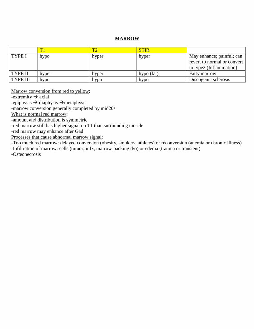
MARROW
T1 T2 STIR
TYPE I hypo hyper hyper May enhance; painful; can
revert to normal or convert
to type2 (Inflammation)
TYPE II hyper hyper hypo (fat) Fatty marrow
TYPE III hypo hypo hypo Discogenic sclerosis
Marrow conversion from red to yellow:
-extremity axial
-epiphysis diaphysis metaphysis
-marrow conversion generally completed by mid20s
What is normal red marrow:
-amount and distribution is symmetric
-red marrow still has higher signal on T1 than surrounding muscle
-red marrow may enhance after Gad
Processes that cause abnormal marrow signal:
-Too much red marrow: delayed conversion (obesity, smokers, athletes) or reconversion (anemia or chronic illness)
-Infiltration of marrow: cells (tumor, infx, marrow-packing d/o) or edema (trauma or transient)
-Osteonecrosis