Embed Size (px)
Citation preview

Lab #15
Muscles

Muscles of the Head and Neck

The Axial Muscles
• Divisions based on location and function:– muscles of head and neck– muscles of vertebral column– oblique and rectus muscles– muscles of pelvic floor

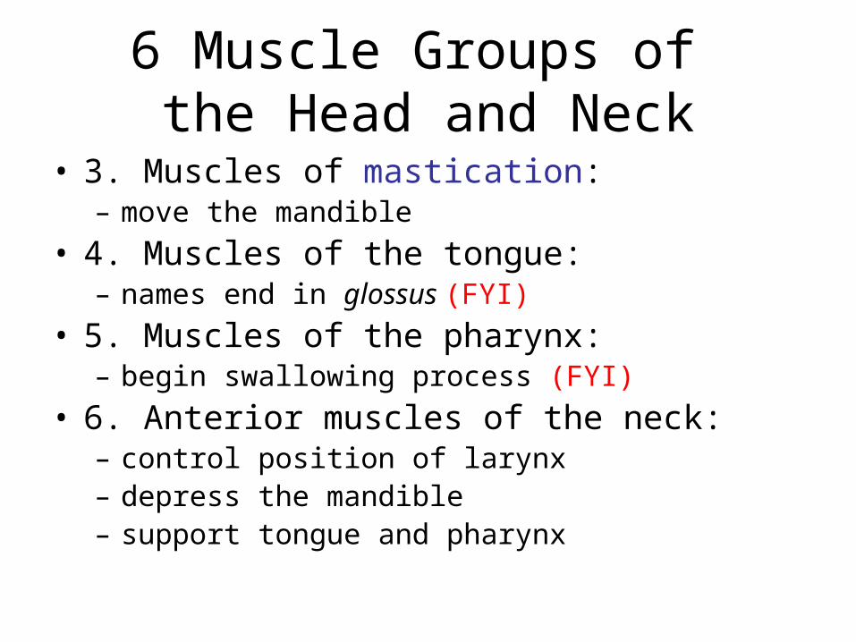
6 Muscle Groups of the Head and Neck
• 1. Muscles of facial expression:– originate on skull
• 2. Extrinsic (outside) eye muscles:
(don’t need to know)
6 Muscle Groups of the Head and Neck
• 3. Muscles of mastication:– move the mandible
• 4. Muscles of the tongue:– names end in glossus (FYI)
• 5. Muscles of the pharynx:– begin swallowing process (FYI)
• 6. Anterior muscles of the neck:– control position of larynx– depress the mandible– support tongue and pharynx

Muscles of Facial Expression
• Orbicularis oris:– constricts the mouth opening
• Buccinator:– moves food around the cheeks
• Corrugator supercilli – wrinkles forehead
• Orbicularis oculi – sphincter of eye

Others
• Zygomaticus – pull skin of mouth up and out when smiling
• Levator labii superiorus – raises upper lip
• Depressor labii inferiorus – “pouting” muscle

Muscles of the Epicranium (Scalp)
• Temporoparietalis
• Occipitofrontalis:– frontal and occipital bellies– separated by epicranial aponeurosis
• Platysma:– covers anterior surface of neck

Muscles of Facial Expression
Figure 11–4a

Muscles of Facial Expression
Figure 11–4b

Summary: Muscles of Facial Expression
Table 11–2 (1 of 2)

Summary: Muscles of Facial Expression
Table 11–2 (2 of 2)
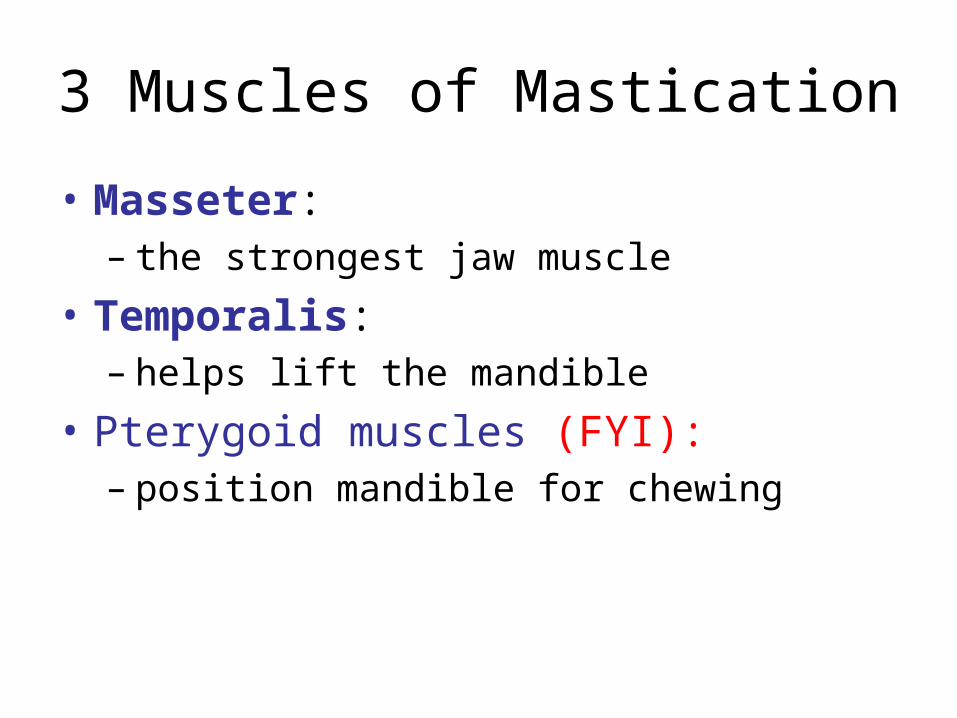
3 Muscles of Mastication
• Masseter:– the strongest jaw muscle
• Temporalis:– helps lift the mandible
• Pterygoid muscles (FYI):– position mandible for chewing

Muscles of Mastication
Figure 11–6

Summary: Muscles of Mastication
Table 11–4

Muscles of the Tongue
Figure 11–7
Don’t need to know any of these

Muscles of the Neck
• Digastric:– from chin to hyoid– also hyoid to mastoid
• Platysma:– covers anterior surface of neck
• Sternocleidomastoid:– from clavicle and sternum to mastoid
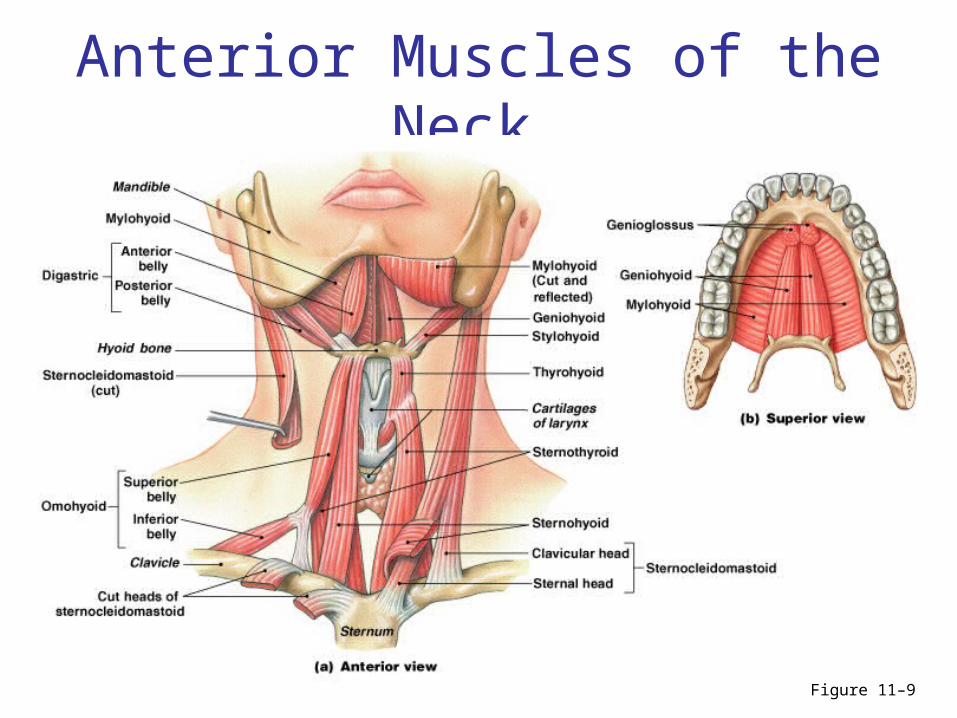
Anterior Muscles of the Neck
Figure 11–9

Summary: Anterior Muscles of the Neck
Table 11–7


Muscles of the chest and abdomen

Lab 19: Muscles of the chest and abdomen
• Remember: if it’s not on the list, you don’t have to locate it or write about it in the lab report

Muscles to know
• pectoralis major• serratus anterior• intercostals (external, internal)• diaphragm• rectus abdominus• obliques (external, internal)• transverse abdominus• trapezius• latissimus dorsi• splenius capitus

Oblique and rectus muscles
• Lie within the body wall
• Generally:– obliques groups are lateral– rectus muscles lie on the anterior midline

Oblique and Rectus Muscles
• Oblique muscles:– compress underlying structures– rotate vertebral column
• Rectus muscles:– flex vertebral column– oppose erector spinae

Cervical Oblique Muscles
• FYI: Cervical region:– scalene muscles– flex the neck

Oblique and Rectus Muscles of the Thoracic region
Figure 11–11a, b

Thoracic Oblique Muscles
• Thoracic region:– intercostal muscles (external and internal
intercostals):• respiratory movements of ribs
– FYI: transversus thoracis:• cross inner surface of ribs

Thoracic Rectus Group
• Diaphragmatic muscle or diaphragm:– divides thoracic and abdominal cavities– performs respiration

Oblique and Rectus Muscles of the Abdominopelvic region
Figure 11–11a, c

Abdominopelvic Oblique Muscles
• Abdominopelvic region (same pattern as thoracic):– external oblique muscles – internal oblique muscles
• Transversus abdominis – deep to internals

Rectus Group -Abdominopelvic
• Rectus abdominis:– between xiphoid process and pubic
symphysis– divided longitudinally by linea alba– divided transversely by tendinous inscriptions

Summary: Oblique and Rectus Muscles
Table 11–9 (1 of 2)

Muscles that Position the Pectoral Girdle
Figure 11–14b

Muscles that Position the Pectoral Girdle (1 of 3)
• Trapezius:– HUGE– superficial– covers back and neck to base of skull– inserts on clavicles and scapular spines

Muscles that Position the Pectoral Girdle (2 of 3)
• Rhomboid and levator scapulae:– deep to trapezius– attach to cervical and thoracic vertebrae– insert on scapular border

Muscles that Position the Pectoral Girdle (3 of 3)
• Serratus anterior:– on the chest– originates along ribs– inserts on anterior scapular margin– “serrated”

Summary: Muscles that Position the Pectoral Girdle
Tables 11–11

Muscles that Move the Arm
Figure 11–15b

Muscles that Move the Arm (1 of 3)
• Deltoid: – the major abductor of arm
• Supraspinatus: – assists deltoid
• Subscapularis and teres major: – produce medial rotation at shoulder

Muscles that Move the Arm (2 of 3)
• Infraspinatus: – produce lateral rotation at shoulder

Muscles that Move the Arm (3 of 3)
• Pectoralis major: – between anterior chest and greater tubercle of
humerus– produces flexion at shoulder joint
• Latissimus dorsi:– between thoracic vertebrae and humerus– produces extension at shoulder joint

Shoulder flexion vs extension
• Pecs and portion of delts
• Lats and portion of delts

The Rotator Cuff
• Muscles involved in shoulder rotation– supraspinatus, subscapularis, infraspinatus,
teres minor,and their tendons

Muscles that Position the Pectoral Girdle
• Rhomboid and levator scapulae:– deep to trapezius– attach to cervical and thoracic vertebrae– insert on scapular border
• Also saw trapezius and serratus anterior

Muscles of the shoulders, arm, and hand

Muscles to know
• supraspinatus• infraspinatus• subscapularis• teres major• rhomboid• levator scapulae• deltoid• biceps brachii• brachialis
• triceps brachii• flexor carpi radialis• flexor carpi ulnaris• flexor digitorum sup.• extensor carpi ulnaris• extensor digitorum• extensor carpi radialis• brachioradialis

Muscles that Move the Arm
Figure 11–15a

Muscles that Move the Arm
Figure 11–15b

Muscles that Move the Arm (1 of 3)
• Deltoid: – the major abductor
• Supraspinatus: – assists deltoid
• Subscapularis and teres major: – produce medial rotation at shoulder

Muscles that Move the Arm (2 of 3)
• Infraspinatus: – produce lateral rotation at shoulder

Muscles that Move the Arm (3 of 3)
• Pectoralis major: – between anterior chest and greater tubercle of
humerus– produces flexion at shoulder joint
• Latissimus dorsi:– between thoracic vertebrae and humerus– produces extension at shoulder joint

The Rotator Cuff
• Muscles involved in shoulder rotation– supraspinatus, subscapularis, infraspinatus,
teres minor,and their tendons

Summary: Muscles that Move the Arm
Table 11–12

Muscles that Position the Pectoral Girdle
• Rhomboid and levator scapulae:– deep to trapezius– attach to cervical and thoracic vertebrae– insert on scapular border

Muscles that Move the Forearm and Hand
• Originate on humerus and insert on forearm
• Exceptions:– the major flexor (biceps brachii)– the major extensor (triceps brachii)

Extensors and Flexors
• Extensors:– mainly on posterior and lateral surfaces of
arm
• Flexors:– mainly on anterior and medial surfaces

Muscles that Move the Forearm and Hand
• Biceps brachii:– flexes elbow– stabilizes shoulder joint– originates on scapula and humerus– inserts on radial tuberosity

Muscles that Move the Forearm and Hand
• Triceps brachii:– extends elbow– originates on scapula (three spots)– inserts on olecranon
• Brachialis and brachioradialis:– assist in flexing elbow (synergists)

Muscles that Move the Forearm and Hand -Extensors
Figure 11–16a

Muscles that Move the Forearm and Hand - Flexors
Figure 11–16b

Muscles that Move the Forearm and Hand
• Flexor carpi ulnaris: – superficial– flexes wrist– adducts wrist

Muscles that Move the Forearm and Hand
• Flexor carpi radialis: – superficial– flexes wrist– abducts wrist

Muscles that Move the Forearm and Hand
• Extensor carpi radialis:– superficial– extends wrist– abducts wrist

Muscles that Move the Forearm and Hand
• Extensor carpi ulnaris:– superficial– extends wrist– adducts wrist

Figure 11–16a

Figure 11–16b

Summary: Muscles that Move the Forearm and Hand
Table 11–13 (1 of 2)

Muscles of the Pelvis, Leg and Foot
Muscles to know
• psoas major• iliacus• gluteus maximus• gluteus medius• sartorius• quadriceps femoris (4)• gracilus• adductor longus• biceps femoris• semitendinosis• semimembranosus
• tibialis anterior• ext hallucis longus• ext digitorum longus• fibularis (peroneus)
longus• gastrocnemius• soleus• flexor hallucis longus• flexor digitorum longus

Muscles of the Pelvis and Lower Limbs
• Pelvic girdle is tightly bound to axial skeleton:– permits little movement– has few muscles

Muscles that Position the Lower Limbs
1. Muscles that move the thigh
2. Muscles that move the leg
3. Muscles that move the foot and toes

Generally…
• Muscles that are lateral are abductors
• Muscles that are medial are adductors
• flexors are on inner surface of joint
• extensors are on outer surface of joint
Note: in legs and feet, look at each joint individually (cf. arms)

Muscles that Move the Thigh
• Gluteal muscles
• Lateral rotators
• Adductors
• Iliopsoas group

Muscles of hip and thigh
• psoas major
• iliacus
• gluteus maximus
• gluteus medius
• adductor longus

Gluteal Muscles (1 of 2)
• Cover lateral surfaces of ilia
• Gluteus maximus:– largest, most posterior gluteal muscle– produces extension and lateral rotation at
hip– Originates on illiac crest, etc., inserts on
illiotibial tract and femur
Gluteal Muscles (2 of 2)
• Gluteus medius and [gluteus minimus]: – originate anterior to gluteus maximus– insert on trochanter of femur

Muscles that Move the Thigh
Figure 11–19a, b

Muscles that Move the Thigh
Figure 11–19c, d

Adductors
• Adductor longus:– hip flexion and adduction
• Gracilis (UPPER LEG):– hip flexion and adduction

Iliopsoas group
• 2 hip flexors insert on the same tendon: – psoas major
• originates on lumbar vetebrae, inserts on femur
– iliacus• originates on illium/fossa

Summary: Muscles that Move the Thigh
Table 11–16 (1 of 2)

Summary: Muscles that Move the Thigh
Table 11–16 (2 of 2)

Muscles that Move the Leg
• Flexors of the knee:– originate on the pelvic girdle– generally: hamstrings
• Extensors of the knee:– originate on the femoral surface– insert on the patella– generally: quads

Flexors of the Knee
• Biceps femoris
• Semimembranosus
• Semitendinosus
• Sartorius:– originates superior to the acetabulum– long, ribbon-like muscle– traverses the quads and wraps around to the
back of the knee
Hamstrings
• Made up of:– biceps femoris (long and short heads)– semimembranosus– semitendinosus
• All are knee flexors

Muscles that Move the Leg
Figure 11–20a

Extensors of the Knee
• 4 muscles of the quadriceps femoris: – 3 vastus muscles:
• vastus lateralis • vastus medialis• vastus intermedius
– rectus femoris muscle

Muscles that Move the Leg
Figure 11–20b, c

Summary: Muscles that Move the Leg
Table 11–17 (1 of 2)

Summary: Muscles that Move the Leg
Table 11–17 (2 of 2)

Muscles that Move the Foot and Toes
• Extrinsic muscles that move the foot and toes include:– muscles that produce extension at the ankle
(Plantar flexion)– muscles that produce flexion at the ankle– muscles that produce extension at the toes– muscles that produce flexion at the toes

Muscles that Produce Extension at the Ankle (Plantar flexion)
Calf muscles
Large posterior calf muscles, both insert on Achilles):
• Gastrocnemius
• Soleus
• Fibularis longus

Muscles that Move the Foot and Toes
Figure 11–21a, b

Muscles that Move the Foot and Toes
Figure 11–21c, d

The Achilles Tendon
• The calcaneal tendon (Achilles tendon):– shared by the gastrocnemius and soleus

Muscles that Produce Flexion at the Ankle
• Tibialis anterior:– opposes the gastrocnemius

Muscles that Move the Foot and Toes
Figure 11–21a, b

Muscles that Move the Foot and Toes
Figure 11–21c, d
Muscles that Produce Extension at the Toes
• Extensor digitorum longus
• Extensor hallucis longus
Toe extensors are on top of foot
Note: there are no muscles in toes themselves (only tendons)

The Intrinsic Muscles of the Foot
Figure 11–22a

Muscles that Produce Flexion at the Toes
• Flexor digitorum longus
• Flexor hallucis longus:– oppose the extensors
Toe flexors are on bottom of foot

The Intrinsic Muscles of the Foot
Figure 11–22b, c

Summary: Muscles that Move the Foot and Toes
Table 11–18

Lab 15
• This is a huge lab (it covers ~3-4 labs worth of material)
• Due on day of practical (11/22)