Embed Size (px)
Citation preview
ХАРЬКОВСКИЙ НАЦИОНАЛЬНЫЙ МЕДИЦИНСКИЙ УНИВЕРСИТЕТ
DIPHTHERIA. TUBERCULOSIS
Methodical instructions for the II and III yearEnglish media students of medical and dentistry
faculties
ДИФТЕРИЯ. ТУБЕРКУЛЕЗМетодические указания для студентов
II и III курсов медицинского и стоматологического факультетов с английским языком преподавания
Утверждено ученым советом ХНМУ
Протокол № 6 от 17 мая 2012 г.
Харьков ХНМУ 2012

Дифтерия. Тубуркулез: Метод. указ. для студентов II и III курсов медицинского и стоматологического факультетов с английским языком преподавания / Составители: А.Я.Цыганенко, Н.И.Коваленко – Харьков: ХНМУ, 2012. – 24 с.
Составители: А.Я.Цыганенко, Н.И.Коваленко
Diphtheria. Tuberculosis: Methodical instructions for the II and III year English media students of medical and dentistry faculties / A.Ya.Tsyganenko, N.I.Kovalenko. – Kharkiv: Kharkiv National Medical University, 2012. – 24 p.
Authors: A.Ya.Tsyganenko, N.I.Kovalenko
2
INTRODUCTION
In parts of the world where diphtheria still occurs, it is primarily a disease of children, and most individuals who survive infancy and childhood have acquired immunity to diphtheria. In earlier times, when nonimmune populations were exposed to the disease, people of all ages were infected and many were killed.
Diphtheria is a serious disease, with fatality rates between 5% and 10%. In children under five years and adults over 40 years, the fatality rate may be as much as 20%.
No bacterial disease of humans has been as successfully studied as diphtheria. The etiology, mode of transmission, pathogenic mechanism and molecular basis of exotoxin structure, function, and action have been clearly established. Consequently, highly effective methods of treatment and prevention of diphtheria have been developed.
The bacterium that caused diphtheria was first described by Klebs in 1883, and was cultivated by Loeffler in 1884, who applied Koch's postulates and properly identified Corynebacterium diphtheriae as the agent of the disease.
In 1884, Loeffler concluded that Corynebacterium diphtheriae produced a soluble toxin, and thereby provided the first description of a bacterial exotoxin.
In 1888, Roux and Yersin demonstrated the presence of the toxin in the cell-free culture fluid of C. diphtheriae which, when injected into suitable lab animals, caused the systemic manifestation of diphtheria.
Two years later, von Behring and Kitasato succeeded in immunizing guinea pigs with a heat-attenuated form of the toxin and demonstrated that the sera of immunized animals contained an antitoxin capable of protecting other susceptible animals against the disease. This modified toxin was suitable for immunizing animals to obtain antitoxin, but it was found to cause severe local reactions in humans and could not be used as a vaccine.
In 1929, Ramon demonstrated the conversion of diphtheria toxin to its nontoxic, but antigenic, equivalent (toxoid) by using formaldehyde. He provided humanity with one of the safest and surest vaccines of all time-the diphtheria toxoid.
In 1951, Freeman made the remarkable discovery that pathogenic (toxigenic) strains of C. diphtheriae are lysogenic, (i.e., are infected by a temperate B phage), while non lysogenized strains are avirulent. Subsequently, it was shown that the gene for toxin production is located on the DNA of the B phage.
In the early 1960s, Pappenheimer and his group at Harvard conducted experiments on the mechanism of a action of the diphtheria toxin.
3
Tuberculosis (TB) is the leading cause of death in the world from a single infectious disease. The disease affects 2.7 billion people per year which is equal to one-third of the entire world population.
Mycobacterium tuberculosis (M.TB.) was the cause of the "White Plague" of the 17th and 18th centuries in Europe. During this period nearly 100 % of the European population was infected with M.TB., and 25 % of all adult deaths were caused by M.TB.
In 1882, the microbiologist Robert Koch discovered the tubercle bacillus, at a time when one of every seven deaths in Europe was caused by ТВ. Tuberculosis spread much more widely in Europe when the industrial revolution began in the late nineteenth century. When streptomycin, the first antibiotic effective against M. tuberculosis, was discovered in the early 1940s, the infection began to come under control. Although other more effective anti-tuberculosis drugs were developed in the following decades, the number of cases of ТВ began to rise again in the mid-1990s. At the same time multiple drug-resistant strains of M. tuberculosis are appearing regularly.
DIPHTHERIAStructure. Corynebacterium diphtheriae is a Gram-positive nonmotile,
nonsporforming, club-shaped bacillus. The club or bar appearance of the cells is due to pockets of inorganic polyphosphate (volutin) which form metachromatic granules in the polar regions that serve as energy. The granules are called Babes Ernst Granules.
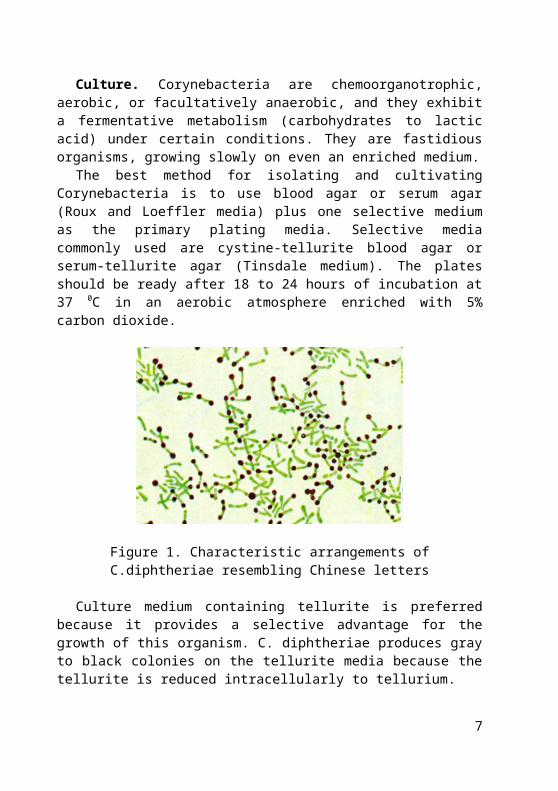
Metachromacity is the phenomenon by which different parts of an organism can get stained in two or more different colours just by applying a single dye. Internal metachromatic granules densely stain dark blue while the rest of the bacillus stains blue, when stained with an aniline dye such as methylene blue (Loeffler stain). Cells appear to be banded or beaded with irregularly staining granules; may show alternate bands of stained and unstained material (giving the appearance of septa). They have the characteristic of forming irregular shaped, club-shaped or V-shaped arrangements in normal growth. They undergo snapping movements just after cell division which brings them into characteristic arrangements resembling Chinese letters (Fig. 1).
Culture. Corynebacteria are chemoorganotrophic, aerobic, or facultatively anaerobic, and they exhibit a fermentative metabolism (carbohydrates to lactic acid) under certain conditions. They are fastidious organisms, growing slowly on even an enriched medium.
The best method for isolating and cultivating Corynebacteria is to use blood agar or serum agar (Roux and Loeffler media) plus one selective medium as the primary plating media. Selective media commonly used are cystine-tellurite blood agar or serum-tellurite agar (Tinsdale medium). The plates should be
4
ready after 18 to 24 hours of incubation at 37 0C in an aerobic atmosphere enriched with 5% carbon dioxide.
Figure 1. Characteristic arrangements of C.diphtheriae resembling Chinese letters
Culture medium containing tellurite is preferred because it provides a selective advantage for the growth of this organism. C. diphtheriae produces gray to black colonies on the tellurite media because the tellurite is reduced intracellularly to tellurium.
Three distinct cultural varieties, mitis, intermedius, and gravis have been recognized:
var. gravis: large, flat, rough, dark-gray colonies; not hemolytic; very few small metachromatic granules; form a pellicle in broth;
var. mitis: smooth, convex, black, shiny, entire colonies; hemolytic; prominent metachromatic granules; diffuse turbidity in broth;
var. intermedius: dwarf, flat, umbilicate colony with a black center and slightly crenated periphery; not hemolytic; fine granular deposit in broth.
Some mitis strains can grow on blood agar with beta-hemolysis.Transmission. Human carriers are the reservoir for C. diphtheriae, and are
usually asymptomatic. Chronic carriers may shed organisms for 6 months or more. Toxigenic strains of C.diphtheriae spread directly from person to person with respiratory secretions and cutaneous lesions. It is known that toxigenic strains may directly colonize the nasopharyngeal cavity.
Diphtheria is highly contagious. It's easily passed from an infected person to others through sneezing, coughing, or even laughing. It also can spread to someone who picks up tissues or drinking glasses that have been used by an infected person. Cutaneous lesions are important in transmission particularly in countries warm climates.
5
People infected with the diphtheria bacteria, even if they don't have any symptoms, can infect others for up to 4 weeks. The incubation period (the time it takes for a person to become infected after being exposed) for diphtheria is 2 to 4 days, although it can range from 1 to 6 days.
Pathogenesis. The pathogenesis of diphtheria is based upon two primary determinants: (1) the ability of a given strain of C.diphtheriae to colonize in the nasopharyngeal cavity and/or on the skin, and (2) its ability to produce diphtheria toxin. Since those determinants involved in colonization of the host are encoded by the bacteria, and the toxin is encoded by the corynebacteriophage, the molecular basis of virulence in C.diphtheriae results from the combined effects of determinants carried on two genomes. Nontoxigenic strains of C.diphtheriae are rarely associated with clinical disease; however, they may become highly virulent following lysogenic conversion to toxigenicity.
As early as 1887, Loeffler described the isolation from healthy individuals of avirulent (nontoxigenic) C.diphtheriae that were indistinguishable from the virulent (toxigenic) strains isolated from patients. It is now recognized that avirulent strains of C.diphtheriae may be converted to the virulent phenotype following infection and lysogenization by one of a number of distinct corynebacteriophages that carry the structural gene for diphtheria toxin, tox. The diphtheria toxin structural gene is not essential for either corynebacteriophage or C.diphtheriae.
Therefore, the toxinogenicity of C.diphtheriae is dependant on phage conversion with a phage that has a functional gene for the toxin protein.
Diphtheria toxin is extraordinarily potent; in sensitive species (e.g., humans, monkeys, rabbits, guinea pigs) as little as 100 to 150 ng/kg of body weight is lethal.
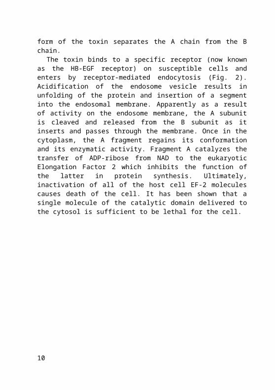
The diphtheria toxin is a two component bacterial exotoxin synthesized as a single polypeptide chain containing an A (catalytically active) domain and a B (binding) domain. Proteolytic nicking of the secreted form of the toxin separates the A chain from the B chain.
The toxin binds to a specific receptor (now known as the HB-EGF receptor) on susceptible cells and enters by receptor-mediated endocytosis (Fig. 2). Acidification of the endosome vesicle results in unfolding of the protein and insertion of a segment into the endosomal membrane. Apparently as a result of activity on the endosome membrane, the A subunit is cleaved and released from the B subunit as it inserts and passes through the membrane. Once in the cytoplasm, the A fragment regains its conformation and its enzymatic activity. Fragment A catalyzes the transfer of ADP-ribose from NAD to the eukaryotic Elongation Factor 2 which inhibits the function of the latter in protein synthesis. Ultimately, inactivation of all of the host cell EF-2 molecules causes
6
death of the cell. It has been shown that a single molecule of the catalytic domain delivered to the cytosol is sufficient to be lethal for the cell.
Figure 2. Uptake and activity of the diphtheria toxin in eukaryotic cells
CDC describes diphtheria as "an upper respiratory tract illness characterized by sore throat, low-grade fever, and an adherent membrane of the tonsil(s), pharynx, and/or nose". Diphtheria is a rapidly developing, acute, febrile
7
infection which involves both local and systemic pathology. A local lesion develops in the upper respiratory tract and involves necrotic injury to epithelial cells. As a result of this injury, blood plasma leaks into the area and a fibrin network forms which is interlaced with rapidly-growing C.diphtheriae cells. This membranous network covers over the site of the local lesion and is referred to as the pseudomembrane.
The diphtheria bacilli do not tend to invade tissues below or away from the surface epithelial cells at the site of the local lesion. At this site they produce the toxin that is absorbed and disseminated through lymph channels and blood to the susceptible tissues of the body. Degenerative changes in these tissues, which include heart, muscle, peripheral nerves, adrenals, kidneys, liver and spleen, result in the systemic pathology of the disease.
Signs and symptoms. C.diphtheriae causes the disease diphtheria which primarily affects the upper respiratory tract. The bacteria can invade any of the mucous membranes and have a 2-5 day incubation period. Different sites of infection cause different clinical forms of diphtheria with varying severity. The most common sites of infection is the pharynx and tonsils, but the bacteria can also invade the nasal tissues, larynx and skin. Anterior nasal diphtheria is usually quite similar to the common cold in symptoms and severity because the location of the infection does not allow for large-scale systemic absorption of the diphtheria toxin.
Pharyngeal, tonsillar and laryngeal diphtheria are more severe because more toxin is absorbed into the bloodstream. Early symptoms of this form of diphtheria include a sore throat and fever, but after a few days a bluish-white adherent membrane, the pseudomembrane, forms over the back of the throat and tonsils. The pseudomembrane is actually a fibrin network infected with multiplying C.diphtheriae cells which grows over a necrotic lesion on the epithelial cells on the back of the throat. The consequences of this membrane growth can be severe if the membrane grows to the extent that it blocks the airway in the throat. This infection quickly becomes acute as the bacterial toxin is absorbed into the bloodstream and lymphatic network.
Symptoms of diphtheria include fever of 38°C (100.4°F) or above, chills, fatigue, bluish skin coloration, sore throat, hoarseness, cough, headache, difficulty swallowing, painful swallowing, difficulty breathing, rapid breathing, foul-smelling bloodstained nasal discharge and lymphadenopathy. Symptoms can also include cardiac arrhythmias, myocarditis, and cranial and peripheral nerve palsies.
Cutaneous diphtheria, or an infection of the skin, is much less common. Symptoms may include a rash or ulcer on the skin. This form of the infection is generally less severe.
8
Diphtheria is unusual as a bacterial infection because the bacteria themselves only invade superficial tissues. However, the toxin released spreads throughout the entire body and attacks cells without specificity. Severe infection with diphtheria toxin can cause death by necrosis of the tissues of essential organs such as the heart, kidneys, liver and lungs by blocking all protein synthesis in infected cells.
The ultimate outcome of the disease depends on how much toxin is absorbed into the bloodstream. In the most severe cases, an infected person will develop myocarditis or neuritis, which leads to heart failure and local paralysis most commonly of the soft palate, respectively. The fatality rate is approximately 5-10% of cases, but is higher for young children or the very old.
Immunity to Diphtheria. Acquired immunity to diphtheria is due primarily to toxin-neutralizing antibody (antitoxin). Passive immunity in utero is acquired transplacentally and can last at most 1 or 2 years after birth. In areas where diphtheria is endemic and mass immunization is not practiced, most young children are highly susceptible to infection. Probably active immunity can be produced by a mild or inapparent infection in infants who retain some maternal immunity, and in adults infected with strains of low virulence (inapparent infections).
Individuals that have fully recovered from diphtheria may continue to harbor the organisms in the throat or nose for weeks or even months. In the past, it was mainly through such healthy carriers that the disease was spread, and toxigenic bacteria were maintained in the population. Before mass immunization of children, carrier rates of C.diphtheriae of 5% or higher were observed.
Diagnosis. Diphtheria can be diagnosed usually by proper clinical examination, throat culture from the infected area and blood tests:
Physical examination may reveal the characteristic gray membrane (pseudomembrane) in the throat, enlarged lymph glands and swelling of the neck or larynx.
MicroscopyPresumptive rapid diagnosis in which methylene blue and Gram stain or
immunofluorescent staining is done on the throat cultures to detect the bacteria. Methylene blue stain (Loeffler stain) shows metachromatic granules. Gram stain of material from the membrane itself can be helpful when trying
to confirm the clinical diagnosis. The Gram stain may show multiple club-shaped forms which look like Chinese characters. Other Corynebacterium species ("diphtheroids") that can normally inhabit the throat may confuse the interpretation of direct stain. However, treatment should be started if clinical diphtheria is suggested, even in the absence of a diagnostic Gram stain.
Culture
9
Definitive diagnosis of diphtheria requires culture of C.diphtheriae from respiratory tract secretions or cutaneous lesions, and a positive toxin assay.
Exudate from the lesion should be inoculated onto Loeffler’s serum or blood agar and selective media: cysteine-tellurite agar; serum tellurite agar.
A selective plate known as tellurite agar allows all Corynebacteria (including C.diphtheriae) to reduce tellurite to metallic tellurium. The tellurite reduction is colormetrically indicated by brown colonies for most Cornyebacteria species or by a black halo around the C.diphtheriae colonies.
Any colonies which appear on the three media should be stained with methylene blue.
Following initial isolation, C.diphtheriae may be identified as mitis, intermedius, or gravis biotype on the basis of carbohydrate fermentation patterns and hemolysis on sheep blood agar plates.
The clinical diagnosis of diphtheria requires bacteriologic laboratory confirmation of toxigenic C.diphtheriae in throat or lesion cultures. Cultures should be obtained from the throat and nose including a portion of the membrane (if possible), and material from beneath the membrane.
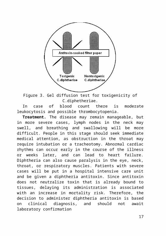
The toxigenicity of C.diphtheriae strains is determined by a variety of in vitro and in vivo tests. The most common in vitro assay for toxigenicity is the Elek double immunodiffusion test (gel precipitation test) (Fig. 3). This test is based on the double diffusion of diphtheria toxin and antitoxin in an agar medium. A sterile, antitoxin-saturated filter paper strip is embedded in the culture medium, and C.diphtheriae isolates are streak-inoculated at a 90° angle to the filter paper. The production of diphtheria toxin can be detected within 18 to 48 hours by the formation of a toxin-antitoxin precipitin band in the agar. Several sensitive in vivo tests for diphtheria toxin have also been described (e.g., guinea pig challenge test, rabbit skin test).
10
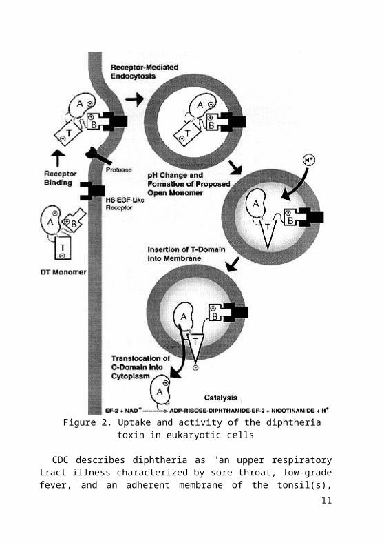
Figure 3. Gel diffusion test for toxigenicity of C.diphetheriae.In case of blood count there is moderate leukocytosis and possible
thrombocytopenia. Treatment. The disease may remain manageable, but in more severe cases,
lymph nodes in the neck may swell, and breathing and swallowing will be more difficult. People in this stage should seek immediate medical attention, as obstruction in the throat may require intubation or a tracheotomy. Abnormal cardiac rhythms can occur early in the course of the illness or weeks later, and can lead to heart failure. Diphtheria can also cause paralysis in the eye, neck, throat, or respiratory muscles. Patients with severe cases will be put in a hospital intensive care unit and be given a diphtheria antitoxin. Since antitoxin does not neutralize toxin that is already bound to tissues, delaying its administration is associated with an increase in mortality risk. Therefore, the decision to administer diphtheria antitoxin is based on clinical diagnosis, and should not await laboratory confirmation
Antibiotics have not been demonstrated to affect healing of local infection in diphtheria patients treated with antitoxin. Antibiotics are used in patients or carriers to eradicate C.diphtheriae and prevent its transmission to others. The CDC recommends either:
Erythromycin (orally or by injection) for 14 days (40 mg/kg per day with a maximum of 2 g/d), or
Procaine penicillin G given intramuscularly for 14 days (300,000 U/d for patients weighing <10 kg and 600,000 U/d for those weighing >10 kg). Patients with allergies to penicillin G or erythromycin can use rifampin or clindamycin.
Control. The control of diphtheria depends upon adequate immunization with diphtheria toxoid: formaldehyde-inactivated diphtheria toxin that remains
11
antigenically intact. The toxoid is prepared by incubating diphtheria toxin with formaldehyde at 37° C under alkaline conditions. Diphtheria toxoid is widely used as a component in the DPT or DTP vaccine which contains diphtheria toxoid, pertussis vaccine, and tetanus toxoid. Children should get 5 doses of DTP, one dose at each of the following ages: 2, 4, 6, and 15-18 months and 4-6 years. DT does not contain pertussis, and is used as a substitute for DTP for children who cannot tolerate pertussis vaccine.
A booster injection should be given about a year later, and it is advisable to administer several booster injections during childhood.
The adult population should be reimmunized with diphtheria toxoid every 10 years. Indeed, booster immunization with diphtheria-tetanus toxoids should be administered to persons traveling to regions with high rates of endemic diphtheria (Central and South America, Africa, Asia, Russia and Eastern Europe). In recent years, the use of highly purified toxoid preparations for immunization has minimized the occasional severe hypersensitivity reaction.
Although antibiotics (e.g., penicillin and erythromycin) are used as part of the treatment of patients who present with diphtheria, prompt passive immunization with diphtherial antitoxin is most effective in reducing the fatality rate. The long half-life of specific antitoxin in the circulation is an important factor in ensuring effective neutralization of diphtheria toxin; however, to be effective, the antitoxin must react with the toxin before it becomes internalized into the cell.
Adverse reactions following vaccination. Local reactions, generally erythema and induration with or without tenderness, are common after the administration of vaccines containing diphtheria toxoid. Local reactions are usually self-limited and require no therapy. A nodule may be palpable at the injection site for several weeks. Abscess at the site of injection has been reported. Fever and other systemic symptoms are not common.
Exaggerated local (Arthus-type) reactions are occasionally reported following receipt of a diphtheria- or tetanus-containing vaccine. These reactions present as extensive painful swelling, often from shoulder to elbow. They generally begin from 2 to 8 hours after injections, and are reported most often in adults, particularly those who have received frequent doses of diphtheria or tetanus toxoid. Persons experiencing these severe reactions usually have very high serum antitoxin levels; they should not be given further routine or emergency booster doses of Td more frequently than every 10 years. Less severe local reactions may occur in persons who have multiple prior boosters.
Rarely, severe systemic reactions such as generalized urticaria, anaphylaxis, or neurologic complications have been reported following administration of diphtheria toxoid.
12
Contraindications and precautions to vaccination. Persons with a history of a severe allergic reaction following a prior dose should not receive additional doses of diphtheria toxoid. Diphtheria toxoid should be deferred for those individuals who have moderate or severe acute illness, but persons with mild illness may be vaccinated. Immunosuppression and pregnancy are not contraindications to diphtheria toxoid.
TUBERCULOSISMycobacterium tuberculosis is the etiologic agent of tuberculosis (TB) in
humans. Humans are the only reservoir for the bacterium. Mycobacterium bovis is the etiologic agent of TB in cows and rarely in
humans. Both cows and humans can serve as reservoirs. Humans can also be infected by the consumption of unpasteurized milk. This route of transmission can lead to the development of extrapulmonary TB, exemplified in history by bone infections that led to hunched backs.
Other human pathogens belonging to the Mycobacterium genus include Mycobacterium avium which causes a TB-like disease especially prevalent in AIDS patients, and Mycobacterium leprae, the causative agent of leprosy.
Structure. Mycobacteria are slender, curved rods in stained clinical specimens. They are non-motile, non-sporforming bacteria.
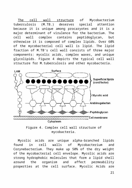
The cell wall structure of Mycobacterium tuberculosis (M.TB.) deserves special attention because it is unique among procaryotes and it is a major determinant of virulence for the bacterium. The cell wall complex contains peptidoglycan, but otherwise it is composed of complex lipids. Over 60% of the mycobacterial cell wall is lipid. The lipid fraction of M.TB's cell wall consists of three major components: mycolic acids, complex waxes, and unique glycolipids. Figure 4 depicts the typical cell wall structure for M.tuberculosis and other mycobacteria.
13
Figure 4. Complex cell wall structure of mycobacteria.
Mycolic acids are unique alpha-branched lipids found in cell walls of Mycobacterium and Corynebacterium. They make up 50% of the dry weight of the mycobacterial cell envelope. Mycolic acids are strong hydrophobic molecules that form a lipid shell around the organism and affect permeability properties at the cell surface. Mycolic Acids are thought to be a significant determinant of virulence in M.TB. Probably, they prevent attack of the mycobacteria by cationic proteins, lysozyme and oxygen radicals in the phagocytic granule. They also protect extracellular mycobacteria from complement deposition in serum.
Other important wall components are trehalose dimycolate (so-called cord factor, as it is thought to induce growth in serpentine cords on artificial medium) and mycobacterial sulfolipids, which may play a role in virulence. Cord factor is toxic to mammalian cells and is also an inhibitor of PMN migration. Cord factor is most abundantly produced in virulent strains of M.TB.
Another unique constituent which may contribute to pathogenesis is lipoarabinomannan (LAM). Purified LAM from virulent and attenuated strains of mycobacteria may differ structurally, and these differences may contribute to their varying abilities to stimulate cytokine production in mononuclear cell cultures.
In summary, the high concentration of lipids in the cell wall of M.TB. has been associated with these properties of the bacterium:
• Impermeability to stains and dyes • Resistance to many antibiotics • Resistance to killing by acidic and alkaline compounds
14
• Resistance to osmotic lysis via complement deposition • Resistance to lethal oxidations and survival inside of macrophagesThis unusual cell wall structure endows mycobacteria with resistance to
dehydration, acids, and alkalis. The resistance to acids and alkalis is useful in the isolation of mycobacteria from contaminated clinical specimens such as sputum. Treatment of sputum with dilute solutions of sulfuric acid or sodium hydroxide will allow mycobacteria to survive and grow on culture medium in the absence of the members of the respiratory flora.
M.TB. is not classified as either Gram-positive or Gram-negative because it does not have the chemical characteristics of either, although the bacteria do contain peptidoglycan (murein) in their cell wall. If a Gram stain is performed on M.TB., it stains very weakly Gram-positive or not at all (referred to as "ghosts").
Mycobacterium species are classified as acid-fast bacteria due to their impermeability by certain dyes and stains. Despite this, once stained, acid-fast bacteria will retain dyes when heated and treated with acidified organic compounds. One acid-fast staining method for M.TB. is the Ziehl-Neelsen stain. When this method is used, the M.TB. smear is fixed, stained with carbol-fuchsin (a pink dye), and decolorized with acid-alcohol. The smear is counterstained with methylene-blue or certain other dyes. Acid-fast bacilli appear pink in a contrasting background.
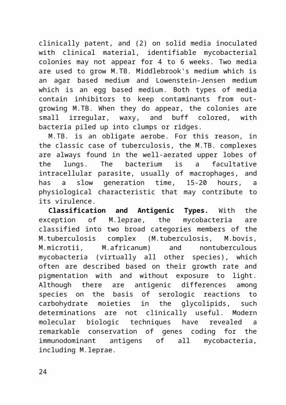
Culture. Although mycobacteria are normally cultured from clinical material by inoculation onto enriched agar media containing bovine serum albumin, they can grow on a chemically defined medium containing asparagine, glycerol, and micronutrients. Even under ideal culture conditions M.TB. and M.bovis grow very slowly, with doubling times on the order of 18 to 24 hours. This extremely slow growth, even in vivo, has two consequences of clinical significance: (1) the infection is an insidious, chronic process, which may take several weeks or months to become clinically patent, and (2) on solid media inoculated with clinical material, identifiable mycobacterial colonies may not appear for 4 to 6 weeks. Two media are used to grow M.TB. Middlebrook's medium which is an agar based medium and Lowenstein-Jensen medium which is an egg based medium. Both types of media contain inhibitors to keep contaminants from out-growing M.TB. When they do appear, the colonies are small irregular, waxy, and buff colored, with bacteria piled up into clumps or ridges.
M.TB. is an obligate aerobe. For this reason, in the classic case of tuberculosis, the M.TB. complexes are always found in the well-aerated upper lobes of the lungs. The bacterium is a facultative intracellular parasite, usually of macrophages, and has a slow generation time, 15-20 hours, a physiological characteristic that may contribute to its virulence.
15

Classification and Antigenic Types. With the exception of M.leprae, the mycobacteria are classified into two broad categories members of the M.tuberculosis complex (M.tuberculosis, M.bovis, M.microtii, M.africanum) and nontuberculous mycobacteria (virtually all other species), which often are described based on their growth rate and pigmentation with and without exposure to light. Although there are antigenic differences among species on the basis of serologic reactions to carbohydrate moieties in the glycolipids, such determinations are not clinically useful. Modern molecular biologic techniques have revealed a remarkable conservation of genes coding for the immunodominant antigens of all mycobacteria, including M.leprae.
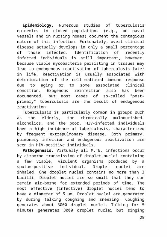
Epidemiology. Numerous studies of tuberculosis epidemics in closed populations (e.g., on naval vessels and in nursing homes) document the contagious nature of this infection. Fortunately, overt clinical disease actually develops in only a small percentage of those infected. Identification of recently infected individuals is still important, however, because viable mycobacteria persisting in tissues may lead to endogenous reactivation of tuberculosis later in life. Reactivation is usually associated with deterioration of the cell-mediated immune response due to aging or to some associated clinical condition. Exogenous reinfection also has been documented, but most cases of so-called "post-primary" tuberculosis are the result of endogenous reactivation.
Tuberculosis is particularly common in groups such as the elderly, the chronically malnourished, alcoholics, and the poor. HIV-infected individuals have a high incidence of tuberculosis, characterized by frequent extrapulmonary disease. Both primary, pulmonary infection and endogenous reactivation are seen in HIV-positive individuals.
Pathogenesis. Virtually all M.TB. infections occur by airborne transmission of droplet nuclei containing a few viable, virulent organisms produced by a sputum-positive individual. Droplet nuclei are inhaled. One droplet nuclei contains no more than 3 bacilli. Droplet nuclei are so small that they can remain air-borne for extended periods of time. The most effective (infective) droplet nuclei tend to have a diameter of 5 um. Droplet nuclei are generated by during talking coughing and sneezing. Coughing generates about 3000 droplet nuclei. Talking for 5 minutes generates 3000 droplet nuclei but singing generates 3000 droplet nuclei in one minute. Sneezing generates the most droplet nuclei by far, which can spread to individuals up to 10 feet away.
The bacilli are deposited in the alveolar spaces of the lungs, where they are engulfed by alveolar macrophages (Fig. 5). A portion of the infectious inoculum resists intracellular destruction and persists, eventually multiplying and killing the macrophage. The ability of virulent mycobacteria to survive within phagocytes justifies their designation as facultative intracellular pathogens. The mechanisms of intracellular survival are not clear and may vary
16
from species to species. There is some evidence that M.TB. can prevent phagosome-lysosome fusion. Other studies have demonstrated that virulent mycobacteria can prevent acidification of the phagolysosome, perhaps by modulating the activity of a membrane proton pump. In addition, some of the components of the mycobacterial cell wall (e.g., cord factor) may be directly cytotoxic to macrophages. Most of the tissue destruction associated with tuberculosis results from cell-mediated hypersensitivity, however, rather than direct microbial aggression.
Figure 5. Engulfment of Mycobacteria by alveolar macrophages.
Although a CMI response is necessary to control an M.TB. infection, it is also responsible for much of the pathology associated with tuberculosis. Activated macrophages may release lytic enzymes and reactive intermediates that facilitate the development of immune pathology. Activated macrophages and T-cells also secrete cytokines that may also play a role in the development of immune pathology, including Interleukin 1 (IL-l), tumor necrosis factor (TNF), and gamma IFN.
It is also at this stage that tubercle formation begins. The center of the tubercle is characterized by "caseation necrosis" meaning semi-solid or "cheesy" consistency. M.TB. cannot multiply within these tubercles because of the low pH and anoxic environment. M.TB. can, however, persist within these tubercles for extended periods.
Although many activated macrophages can be found surrounding the tubercles, many other macrophages present remain unactivated or poorly activated. M.TB. uses these macrophages to replicate and hence the tubercle grows.
The growing tubercle may invade a bronchus. If this happens, M.TB. infection can spread to other parts of the lung. Similarly the tubercle may invade an artery or other blood supply line.
Early in infection, mycobacteria may spread distally either indirectly through the lymphatics to the hilar or mediastinal lymph nodes and thence via
17
the thoracic duct into the blood stream, or directly into the circulation by erosion of the developing tubercle into a pulmonary vessel.
The hematogenous spread of M.TB. may result in extrapulmonary tuberculosis otherwise known as milliary tuberculosis. The name "milliary" is derived from the fact that metastasizing tubercles are about the same size as a millet seed, a grain commonly grown in Africa.
The secondary lesions caused by milliary TB can occur at almost any anatomical location, but usually involve the genitourinary system, bones, joints, lymph nodes, and peritoneum. These lesions are of two types:
1. Exudative lesions result from the accumulation of PMN's around M.TB. Here the bacteria replicate with virtually no resistance. This situation gives rise to the formation of a "soft tubercle".
2. Productive or granulomatous lesions occur when the host becomes hypersensitive to tuberculoproteins. This situation gives rise to the formation of a "hard tubercle".
Extrapulmonary hematogenous dissemination results in the seeding of other organs (e.g., spleen, liver, and kidneys) and, eventually, reinoculation of the lungs. The resulting secondary lung lesions (as opposed to the initial site of implantation) may serve as the origin of reactivation of clinical disease years or decades later owing to the persistence of viable tubercle bacilli.
In parts of the world where bovine tuberculosis has not been eliminated and where dairy products are not properly treated, direct infection of the gastrointestinal tract may occur by ingestion of virulent M.bovis organisms. The gut is also exposed occasionally in pulmonary tuberculosis when large numbers of viable M.TB. cells are coughed up and swallowed.
In either case, the principal site of involvement is the mesenteric lymph nodes, with subsequent dissemination.
An inflammatory focus can mature into a granulomatous lesion characterized by a mononuclear cell infiltrate surrounding a core of degenerating epithelioid and multinucleated giant (Langhans) cells (Fig. 6). It is at this stage that the individual becomes tuberculin-positive. This positive tuberculin reaction is the result of the host developing a vigorous cell mediated immune (CMI) response. A CMI response must be mounted to control an M.TB. infection. An antibody mediated immune (AMI) will not aid in the control of a M.TB. infection because M.TB. is intracellular and if extracellular, it is resistant to complement killing due to the high lipid concentration in its cell wall.
18
Figure 6. Mature a granulomatous lesion characterized by a mononuclear cell infiltrate surrounding a core of epithelioid and multinucleated giant cells
Eventually, the accumulating mycobacteria stimulate an inflammatory focus. At this stage lymphocytes begin to infiltrate. The lymphocytes, specifically T-cells, recognize processed and presented M.TB. This results in T-cell activation and the liberation of cytokines including gamma interferon (IFN). The liberation of IFN causes in the activation of macrophages. These activated macrophages are now capable of destroying M.TB. In the resistant host, the tubercle eventually becomes calcified.
For unknown reasons, the caseous centers of the tubercles liquify. Liquefaction of the caseous material and erosion of the tubercle into an adjacent airway may result in cavitation and the release of massive numbers of bacilli into the sputum (Fig. 7). This liquid is very conducive to M.TB. growth and hence the organism begins to rapidly multiply extracellularly. After time, the large antigen load causes the walls of nearby bronchi to become necrotic and rupture. This results in cavity formation. This also allows M.TB. to spill into other airways and rapidly spread to other parts of the lung.
As stated previously, only a very small percent of M.TB. infections result in disease, and even a smaller percentage of M.TB. infections progress to an advanced stage. Usually the host will begin to control the infection at some point. When the primary lesion heals, it becomes fibrous and calcifies. When this happens the lesion is referred to as the Ghon complex. Depending on the size and severity, the Ghon complex may never subside. Typically the Ghon complex is readily visible upon chest X-ray.
19
Figure 7. Liquefaction of the caseous material and erosion of the tubercle into an adjacent airway results in cavitation and release of bacilli into the sputum.
Small metastatic foci containing low numbers of M.TB. may also calcify. However, in many cases these foci will contain viable organisms. These foci are referred to Simon foci. The Simon foci are also visible upon chest X-ray and are often the sight of disease reactivation.
Host Defenses. Innate susceptibility to pulmonary infection with M.TB. is clearly influenced by genetic and/or ethnic variables that have not been defined. A search for human homologues is currently under way. The mechanism for this genetic effect may reflect the ability of macrophages to process and present mycobacterial antigens to the immune system.
Acquired immunity following mycobacterial infection usually develops within 4 to 6 weeks and is associated temporally with the onset of delayed hypersensitivity to mycobacterial antigens such as PPD. Successful acquired resistance is mediated by T lymphocytes. Antimycobacterial antibodies, although present in many patients, do not play a protective role in tuberculosis. CD4 cells produce factors (e.g., gamma interferon) that activate macrophages and endow them with enhanced mycobacteriostatic or mycobactericidal capabilities. Unlike normal macrophages, these activated cells can limit the replication of intracellular M.TB. and may kill tubercle bacilli. CD8 cells attack infected macrophages expressing mycobacterial antigens and lyse the cells, releasing the mycobacteria from their protective niche and exposing them to activated macrophages.
Since the vast majority (90-95 %) of otherwise healthy individuals who are infected with M.TB. never develop clinically apparent disease, acquired resistance must be quite effective. However, the immune response to mycobacteria is a double-edged sword: the intense cell-mediated hypersensitivity that usually accompanies infection is responsible for much of the pathology associated with clinical tuberculosis. Some clinical studies suggest that inappropriately high levels of circulating cytokines, such as tumor necrosis factor alpha, may be responsible for some of the clinical features of tuberculosis (e.g. fever, weight loss). 20
Diagnosis 1. Presumptive tests for tuberculosis a. Chest X-rays are used to detect confluent granuloma formation in the
lungs, which could be a result of past or present infection with tuberculosis or with some other pulmonary infection that may be mistaken for tuberculosis.
b. Infection in an asymptomatic individual can be diagnosed with the help of the intradermal PPD skin test (the tuberulin or Mantoux test) which detects delayed hypersensitivity to purified protein from the cell wall of M.TB. A positive skin test indicates that the person has developed a cellular immunity to the organism as a result of either a previous or a current infection. It is impossible to distinguish between present and past infection on the basis of a positive tuberculin test. Recent conversion of the reaction from negative to positive warrants clinical attention.
PPD is generated by boiling a culture of M.TB., specifically Old Tuberculin (OT). 5 TU (tuberculin units), which equals 0.000lmg of PPD, in a 0.1 ml volume is intracutaneously injected in the forearm. The transverse diameter of induration is measured 48 to 72 hours later.
The test is considered positive if the diameter of the resulting lesion is 10 mm or greater. The lesion is characterized by erythema (redness) and swelling and induration (raised and hard). 90% of people that have a lesion of 10 mm or greater are currently infected with M.TB. or have been previously exposed to M.TB. 100% of people that have a lesion of 15 mm or greater are currently infected with M.TB. or have been previously exposed to M.TB.
False positive tests usually manifest themselves as lesser reactions. These lesser reactions could indicate prior exposure or infection with other Mycobacteria or vaccination with BCG. However, in places were the vaccine is not used, lesser reactions should be regarded as highly suspicious.
False negatives are more rarely than false positives but are especially common in AIDS patients as they have an impaired CMI response. Other conditions such as malnutrition, steroids, etc., can rarely result in a false negative reaction.
c. Two types of stains are used specifically for detection of mycobacteria: fluorochrome (recommended) and Ziehel-Neelsen stain with carbol fuchsin (acid-fast stain). An acid-fast stain of the sputum may indicate acid-fast bacilli, which is a presumptive test for active tuberculosis. In reporting acid-fast slide results, the slide should be observed for 10-15 minutes before considered negative. Results are reported as positive or negative for acid-fast bacilli. Sometimes the amount of acid-fast bacilli are indicated, with 3-9 per slide reported as rare, 10 or more per slide reported as few, and more than one per oil immersion field reported as numerous. In smears stained with carbol fuchsin,
21

mycobacteria typically appear as red rods (1-10 µm long and 0.2-0.6 µm wide) and often are beaded or banded, but also may appear coccoid or filamentous.
2. Confirmation of Active Tuberculosis Active tuberculosis is confirmed by culturing the organism. Sputum is
usually treated with sodium hydroxide, which is cidal for contaminants but not for M.TB.. The liquified sputum is then neutralized, centrifuged, and the sediment is inoculated onto special enrichment media such as Lowenstein-Jensen agar slants, Middlebrook agar, or 7H 10 Oleic acid agar plates. Felsen Quadrant plates with agar containing different antimicrobial agents are also inoculated to determine drug sensitivity. Plates are incubated at 35° to 37° C in an atmosphere of 5 to 10% CO2. For specimens from cutaneous sites a second set of cultures should be incubated at 30° C. All cultures should be examined weekly for 8 weeks.
The major advantage of culture on solid media is that it allows visualization of colony morphology and pigmentation, which is useful diagnostically for distinguishing colonies of M.TB. from those of some nontuberculous mycobacteria. However, they require 3 or 4 weeks. The more rapid broth systems (e.g. BACTEC) require only 5 to 12 days. The media used in the BACTEC system contains radio-labeled palmitate as the sole carbon source. As M.TB. multiplies, it breaks down the palmitate and liberates radio-labeled CO2.
Nucleic acid amplification methods may prove useful for detection of mycobacteria directly in clinical material within 24 hours or less of specimen receipt.
Treatment. Because administration of a single drug often leads to the development of a bacterial population resistant to that drug, effective regimens for the treatment of TB must contain multiple drugs to which the organisms are susceptible. When two or more drugs are used simultaneously, each helps prevent the emergence of tubercle bacilli resistant to the others. However, when the in vitro susceptibility of a patient's isolate is not known, which is generally the case at the beginning of therapy, selecting two agents to which the patient's isolate is likely to be susceptible can be difficult, and improper selection of drugs may subsequently result in the development of additional drug-resistant organisms.
Hence, tuberculosis is usually treated with four different antimicrobial agents The course of drug therapy usually lasts from 6-9 months. The most commonly used drugs are rifampin (RIF), isoniazid (INH), pyrazinamide (PZA) and ethambutol (EMB) or streptomycin (SM). When adherence with the regimen is assured, this four-drug regimen is highly effective.
Control. A viable, attenuated strain of M.bovis, called bacille Calmette-Guérin (BCG), after the French microbiologists who developed the strain, has been used in more than 120 countries for many years as a vaccine to prevent
22
clinical tuberculosis. Vaccination is the only feasible approach to controlling this disease in much of the developing world. The efficacy of BCG has varied in field trials from 0 to 85 percent, indicating an influence of unknown local environmental or host factors. In the past 5 years, researchers have turned their efforts to the development and animal model testing of new tuberculosis vaccines. Promising results have been obtained in animal experiments with purified subunit vaccines, recombinant BCG strains, and auxotrophic BCG mutants. Excellent animal models for vaccine testing are available using mice, guinea pigs or rabbits infected with M.TB. by the pulmonary route.
REFERENCES1. Bark C.M. Clinical symptoms and microbiological outcomes in
tuberculosis treatment trials / C.M Bark., R. Dietze, A. Okwera, M.I. Quelapio, B.A. Thiel, J.L. Johnson // Tuberculosis. - 2011. – Vol. 91, Issue 6. – P. 601-604.
2. Diphtheria. In: Todar's Online Textbook of Bacteriology. Textbook is available on site: http://textbookofbacteriology.net/diphtheria.html.
3. Holmes, R .K. Diphtheria and other corynebacterial infections. In Kasper; et al. Harrison's Principles of Internal Medicine. - 16th ed. - New York: McGraw-Hill, 2005. – 283 p.
4. Krishnamurthy Natarajan. Innate immune responses to M. tuberculosis infection / Krishnamurthy Natarajan, Manikuntala Kundu, Pawan Sharma, Joyoti Basu // Tuberculosis. - 2011. – Vol. 91, Issue 5. – P. 427-431.
5. Nagasuma Chandra, Dhiraj Kumar, Kanury Rao. Systems biology of tuberculosis // Tuberculosis. - 2011. – Vol. 91, Issue 5. – P. 487-496.
6. Robert L. Hunter. Pathology of post primary tuberculosis of the lung: An illustrated critical review // Tuberculosis. - 2011. - Vol. 91, Issue 6. - P. 497-509.
7. Vaccines and Preventable Diseases: Diphtheria Vaccination. Article is available on site: http://www.cdc.gov/vaccines/vpd-vac/diphtheria/
23
CONTENTSPages
INTRODUCTION……………………………………………………...…..... 3DIPHTHERIA………………………………………………………………. 4
Structure………………………………………………………………….. 4Culture…………………………………………………………………… 4Transmission……………………………………………………………... 5Pathogenesis……………………………………………………………… 6Signs and symptoms……………………………………………………... 8Immunity to diphtheria…………………………………………………... 9Diagnosis………………………………………………………………… 9Treatment………………………………………………………………… 11Control…………………………………………………………………… 11
TUBERCULOSIS…………………………………………………………… 12Structure…………………………………………………………………. 13Culture…………………………………………………………………… 14Classification and antigenic types…………………………………… 15Epidemiology…………………………………………………………. 15Pathogenesis…………………………………………………………... 15Host defenses…………………………………………………………. 19Diagnosis……………………………………………………………… 20Treatment……………………………………………………………… 21Control……………………………………………………………………… 21
REFERENCES…………………………………………………………..….. 22
24
Учебное издание
ДИФТЕРИЯ. ТУБЕРКУЛЕЗМетодические указания для студентов
II и III курсов медицинского и стоматологического факультетов с английским языком преподавания
Авторы: Цыганенко Анатолий Яковлевич,Коваленко Наталья Ильинична
Ответственный за выпуск А.Я.Цыганенко
Компьютерный набор Н.И.Коваленко
План 2012, поз.Подписано к печати . .2012. Формат А5. Бумага печат. Ризография. Усл. печ. л. 1,5. Уч-изд.л. 1,3. Тираж 150 экз. Заказ № .
ХГМУ, 61022, Харьков, пр. Ленина, 4Редакционно-издательский отдел
25